Embed Size (px)
Citation preview
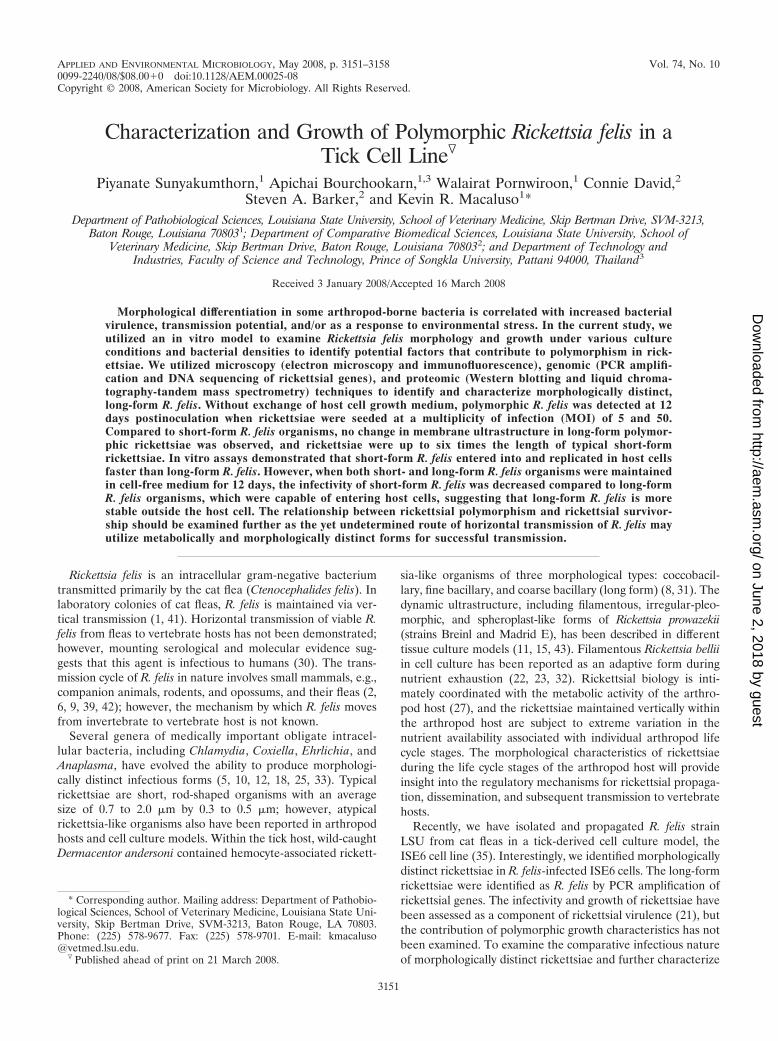
APPLIED AND ENVIRONMENTAL MICROBIOLOGY, May 2008, p. 3151–3158 Vol. 74, No. 100099-2240/08/$08.00�0 doi:10.1128/AEM.00025-08Copyright © 2008, American Society for Microbiology. All Rights Reserved.
Characterization and Growth of Polymorphic Rickettsia felis in aTick Cell Line�
Piyanate Sunyakumthorn,1 Apichai Bourchookarn,1,3 Walairat Pornwiroon,1 Connie David,2Steven A. Barker,2 and Kevin R. Macaluso1*
Department of Pathobiological Sciences, Louisiana State University, School of Veterinary Medicine, Skip Bertman Drive, SVM-3213,Baton Rouge, Louisiana 708031; Department of Comparative Biomedical Sciences, Louisiana State University, School of
Veterinary Medicine, Skip Bertman Drive, Baton Rouge, Louisiana 708032; and Department of Technology andIndustries, Faculty of Science and Technology, Prince of Songkla University, Pattani 94000, Thailand3
Received 3 January 2008/Accepted 16 March 2008
Morphological differentiation in some arthropod-borne bacteria is correlated with increased bacterialvirulence, transmission potential, and/or as a response to environmental stress. In the current study, weutilized an in vitro model to examine Rickettsia felis morphology and growth under various cultureconditions and bacterial densities to identify potential factors that contribute to polymorphism in rick-ettsiae. We utilized microscopy (electron microscopy and immunofluorescence), genomic (PCR amplifi-cation and DNA sequencing of rickettsial genes), and proteomic (Western blotting and liquid chroma-tography-tandem mass spectrometry) techniques to identify and characterize morphologically distinct,long-form R. felis. Without exchange of host cell growth medium, polymorphic R. felis was detected at 12days postinoculation when rickettsiae were seeded at a multiplicity of infection (MOI) of 5 and 50.Compared to short-form R. felis organisms, no change in membrane ultrastructure in long-form polymor-phic rickettsiae was observed, and rickettsiae were up to six times the length of typical short-formrickettsiae. In vitro assays demonstrated that short-form R. felis entered into and replicated in host cellsfaster than long-form R. felis. However, when both short- and long-form R. felis organisms were maintainedin cell-free medium for 12 days, the infectivity of short-form R. felis was decreased compared to long-formR. felis organisms, which were capable of entering host cells, suggesting that long-form R. felis is morestable outside the host cell. The relationship between rickettsial polymorphism and rickettsial survivor-ship should be examined further as the yet undetermined route of horizontal transmission of R. felis mayutilize metabolically and morphologically distinct forms for successful transmission.
Rickettsia felis is an intracellular gram-negative bacteriumtransmitted primarily by the cat flea (Ctenocephalides felis). Inlaboratory colonies of cat fleas, R. felis is maintained via ver-tical transmission (1, 41). Horizontal transmission of viable R.felis from fleas to vertebrate hosts has not been demonstrated;however, mounting serological and molecular evidence sug-gests that this agent is infectious to humans (30). The trans-mission cycle of R. felis in nature involves small mammals, e.g.,companion animals, rodents, and opossums, and their fleas (2,6, 9, 39, 42); however, the mechanism by which R. felis movesfrom invertebrate to vertebrate host is not known.
Several genera of medically important obligate intracel-lular bacteria, including Chlamydia, Coxiella, Ehrlichia, andAnaplasma, have evolved the ability to produce morphologi-cally distinct infectious forms (5, 10, 12, 18, 25, 33). Typicalrickettsiae are short, rod-shaped organisms with an averagesize of 0.7 to 2.0 �m by 0.3 to 0.5 �m; however, atypicalrickettsia-like organisms also have been reported in arthropodhosts and cell culture models. Within the tick host, wild-caughtDermacentor andersoni contained hemocyte-associated rickett-
sia-like organisms of three morphological types: coccobacil-lary, fine bacillary, and coarse bacillary (long form) (8, 31). Thedynamic ultrastructure, including filamentous, irregular-pleo-morphic, and spheroplast-like forms of Rickettsia prowazekii(strains Breinl and Madrid E), has been described in differenttissue culture models (11, 15, 43). Filamentous Rickettsia belliiin cell culture has been reported as an adaptive form duringnutrient exhaustion (22, 23, 32). Rickettsial biology is inti-mately coordinated with the metabolic activity of the arthro-pod host (27), and the rickettsiae maintained vertically withinthe arthropod host are subject to extreme variation in thenutrient availability associated with individual arthropod lifecycle stages. The morphological characteristics of rickettsiaeduring the life cycle stages of the arthropod host will provideinsight into the regulatory mechanisms for rickettsial propaga-tion, dissemination, and subsequent transmission to vertebratehosts.
Recently, we have isolated and propagated R. felis strainLSU from cat fleas in a tick-derived cell culture model, theISE6 cell line (35). Interestingly, we identified morphologicallydistinct rickettsiae in R. felis-infected ISE6 cells. The long-formrickettsiae were identified as R. felis by PCR amplification ofrickettsial genes. The infectivity and growth of rickettsiae havebeen assessed as a component of rickettsial virulence (21), butthe contribution of polymorphic growth characteristics has notbeen examined. To examine the comparative infectious natureof morphologically distinct rickettsiae and further characterize
* Corresponding author. Mailing address: Department of Pathobio-logical Sciences, School of Veterinary Medicine, Louisiana State Uni-versity, Skip Bertman Drive, SVM-3213, Baton Rouge, LA 70803.Phone: (225) 578-9677. Fax: (225) 578-9701. E-mail: [email protected].
� Published ahead of print on 21 March 2008.
3151
on June 2, 2018 by guesthttp://aem
.asm.org/
Dow
nloaded from

the environmental factors contributing to the induction of amorphologically distinct form, we examined rickettsial growthand polymorphism under various environmental conditions,including nutrient availability and rickettsial density, and thencompared virulence attributes of polymorphic R. felis organ-isms.
MATERIALS AND METHODS
Tick cells and rickettsial culture. Ixodes scapularis-derived ISE6 cells, pro-vided by T. Kurtti (University of Minnesota), were maintained in L15Bgrowth medium supplemented with 10% heat-inactivated fetal bovine serum(HyClone) and 10% tryptose phosphate broth (Sigma) (26) at pH 6.8 to 7.0in a humidified 5% CO2 incubator at 32°C. R. felis (LSU), originally isolatedfrom C. felis, was maintained in ISE6 cells as previously described (35). Initialcultures of morphologically distinct R. felis (LSU) organisms were identifiedat passage 8. Nearly homogenous populations of morphologically distinctrickettsiae, consisting of �90% long-form organisms, were maintained inde-pendently; rickettsiae measuring at least three times the length of typicalrickettsiae (0.25 to 0.3 �m in diameter by 0.8 to 1.0 �m) were considered longform. Populations of typical short-form R. felis were fed weekly by replacinghalf of the medium with new medium; long-form R. felis cultures were main-tained by adding 1 ml of fresh medium into 5 ml of culture medium in a25-cm2 tissue culture flask at weekly intervals. Both short-form and long-formR. felis organisms were subcultured to uninfected confluent ISE6 cells every3 to 4 weeks. For all bioassays, rickettsial viability and enumeration wereassessed by staining with a BacLight viability stain kit (Molecular Probes),and rickettsiae were counted in a Petroff-Hausser bacteria counting chamber(21) using a Leica microscope.
PCR amplification and DNA sequencing of rickettsial genes. Genomic DNAof short- and long-form rickettsiae from infected ISE6 cells was extracted usinga DNeasy tissue kit (Qiagen) according to the manufacturer’s protocol. PCRsutilized gene-specific primers for the 17-kDa genus-specific antigen (primersRr17.61p and Rr17.492n [42]) and the R. felis plasmids, pRF and pRF� (primersets pRFa-pRFb and pRFc-pRFd [28]). Reactions were performed using PCRmaster mix (Promega) and thermocycler conditions as previously described (24,35). For each set of reactions, an environmental negative control for genomicDNA extraction (200 �l of phosphate-buffered saline [PBS]) and negative con-trol for PCR (1 �l of water) were included. Amplified products were visualizedon ethidium bromide-stained agarose gels. The PCR products were cloned intoa pCR4-TOPO vector (Invitrogen) and sequenced by the dye terminator methodon a 3130 genetic analyzer (Applied Biosystems) at the Louisiana State Univer-sity School of Veterinary Medicine. Sequencing results were analyzed using theBioEdit sequence alignment editor (Ibis Biosciences); nucleotide similarity com-parisons were made using the GenBank database.
Gel electrophoresis and Western immunoblotting. Semipurified rickettsiaewere recovered from R. felis-infected ISE6 cells via needle lysis of host cellsand low- and high-speed centrifugation (40). Rickettsial pellets and an un-infected ISE6 cell pellet were resuspended in lysis buffer (PBS [pH 7.4], 1%NP-40, and 1� complete protease inhibitor cocktail [Roche]) and disruptedby an ultrasonic bath (Crest) for two 10-min intervals. Cell lysate was col-lected by centrifugation at 16,000 � g for 10 min at 4°C. Protein concentra-tions were determined using a detergent-compatible protein assay (Bio-Rad);30 �g of each protein extract was subjected to sodium dodecyl sulfate-polyacrylamide gel electrophoresis using 4 to 12% Bis-Tris gels (Invitrogen).Separated proteins from companion gels were either stained with PageBlueprotein staining solution (Fermentas) or transferred to a polyvinylidene flu-oride membrane (Bio-Rad). The membrane was blocked with 5% skim milkin Tris-buffered saline–0.1% Tween 20 (TBST) for 1 h at room temperature.The membrane was then incubated with the anti-rickettsial outer membraneprotein B (OmpB) monoclonal antibody (RC-9C2; Fuller Laboratories) for2 h, washed with TBST, and then incubated with horseradish peroxidase-conjugated goat anti-mouse immunoglobulin G (IgG) (Sigma). After thesample was washed with TBST, the signal was detected using a SuperSignalWest Pico chemiluminescent substrate kit (Pierce).
Protein identification. To identify protein in long-form R. felis, the proteinband on the Coomassie-stained sodium dodecyl sulfate gel corresponding to thepositive immunoblot signal was excised, using the ProteomeWorks Spot Cutter(Bio-Rad), and deposited into a 96-well plate before placement in the freezer(�22°C) until digestion. A MassPrep Station (Waters/Micromass) was used asthe digestion robot to carry out an overnight digestion procedure. The gel plugswere automatically destained, reduced, alkylated, and digested with sequencing-
grade modified trypsin. The peptides were automatically extracted from the gelplugs and transferred to another 96-well plate to be used for analysis. Thepeptide samples were then separated by liquid chromatography using an AtlantisdC18 column (75 �m by 100 mm; Waters). Mobile phase A consisted of 95%H2O–5% acetonitrile with 0.1% formic acid, and mobile phase B consisted of 5%acetonitrile–95% H2O with 0.1% formic acid. A Q-Tof (quadrupole time-of-flight) Micro (Waters/Micromass Corp) hybrid mass spectrometer (MS) wasused for analysis. Electrospray analysis was carried out in positive mode. Dataacquisition and analysis were executed using the ProteinLynx Global Server,version 2.0 (Waters/Micromass). A database search was performed using anonline Mascot (Matrix Science) tandem MS (MS/MS) ion search against theNCBInr/Proteobacteria. Search parameters include peptide and MS/MS toler-ances of 1 Da and 0.5 Da, 1 missed cleavage, oxidation of methionines, andcarbamidomethylation of cysteines.
Microscopy. For the immunofluorescence assay (IFA), cytospin prepara-tions of R. felis-infected ISE6 cells were fixed in ice-cold acetone for 10 min;then they were incubated with blocking buffer (3% bovine serum albumin inPBS) for 1 h in a humidified chamber. Rickettsiae were labeled with poly-clonal antibody against short-form R. felis organisms generated in mice.Briefly, a group of four mice were inoculated subcutaneously with a homog-enate of purified, �-propiolactone (Sigma)-inactivated R. felis (25 to 50 �g)mixed 1:1 with Titermax. After two additional inoculations at 2-week inter-vals, anti-R. felis mouse serum was recovered and diluted 1:100 in blockingbuffer for the assay. Fluorescein isothiocyanate-conjugated goat-anti-mouseIgG (KPL) was diluted 1:400 in blocking buffer and served as the secondaryantibody; DAPI (4�, 6-diamidino-2-phenylindole; 1 �g/�l; Sigma) was used asa counter-stain. VectaShield (Vector Laboratories) was applied to the slides,and then slides were visualized using a Zeiss microscope. Slides in which noprimary antibody was added served as a control for the fluorescein isothio-cyanate-conjugated nonspecific binding.
For transmission electron microscopy, R. felis-infected ISE6 cells were pre-pared as described previously (14), with a modified procedure for epon/aralditeembedding (35). For immunoelectron microscopy, R. felis-infected ISE6 cellswere fixed in a mixture of 2.5% formaldehyde and 0.5% glutaraldehyde in 0.1 Mcacodylate buffer (pH 7.3), washed in the same buffer, and then dehydrated inethanol and embedded in LR White (SPI Supplies) as described previously (37).Ultrathin sections were cut by using an ultramicrotome and placed on nickelgrids. Antigen grids were treated in blocking buffer (5% skim milk in PBS–0.1%Tween 20) at room temperature; then they were incubated with rabbit-anti-spotted fever group Rickettsia polyclonal antibody (NIH/RML-I7198; diluted1:20 in blocking buffer) for 1.5 h at room temperature. The grids were washed inblocking buffer and incubated for 1 h at room temperature with goat-anti-rabbitIgG antibody labeled with 10-nm-diameter colloidal gold particles (Sigma) di-luted 1:20 in blocking buffer. Preparations incubated with secondary antibodyalone served as controls for nonspecific binding. Samples (for transmission elec-tron microscopy and immunoelectron microscopy) were visualized on a JEM-1011 transmission electron microscope (JEOL) in the Microscopy Center at theLouisiana State University School of Veterinary Medicine.
Preparation of cell-free rickettsiae for bioassays. Short-form R. felis was semi-purified from R. felis-infected ISE6 cells by needle lysis and centrifugation asdescribed above. To purify long-form R. felis, ISE6 cells were separated fromrickettsiae by centrifugation at 100 � g at 4°C for 3 min; long-form R. felisorganisms in the supernatant were concentrated by centrifugation. Semipurifiedshort-form and long-form rickettsiae were counted, and comparable viability wasconfirmed for both forms prior to use in bioassays.
Polymorphic induction of rickettsiae. To identify the culture parameters thatfacilitate induction of typical short-form R. felis into polymorphic, long-form R.felis, ISE6 cells (5 � 105 cells per well) were seeded into 12-well plates (NUNC)in 2 ml of L15B growth medium. After 2 days of culture at 32°C, ISE6 cells wereexposed, in triplicate, to semipurified short-form R. felis at a multiplicity ofinfection (MOI) of 0.5, 5, or 50, and cells were incubated at 32°C. At 5, 12, and19 days postinfection (dpi), the infected cells were detached by pipetting. Poly-morphic, long-form development was assessed in 100 �l of cell suspensioncollected at 5, 12, and 19 dpi. Cell suspensions were disrupted by forcing thesuspension through a 27-gauge needle 10 times, and the cells were collected bycentrifugation at 16,000 � g at 4°C for 10 min, washed with 500 �l of 0.85%NaCl, and resuspended in 100 �l of 0.85% NaCl. Rickettsiae were assessed forviability and counted as described above.
Rickettsial infectivity and replication in ISE6 cells. To compare infectivity andgrowth of polymorphic R. felis, ISE6 cells (5 � 105 cells per well) were seededinto 12-well plates (NUNC) in 2 ml of L15B growth medium and infected withR. felis at three different MOIs as described for the polymorphic induction assay.For the internalization assessment, 100 �l of cell suspension from 5, 12, and 19
3152 SUNYAKUMTHORN ET AL. APPL. ENVIRON. MICROBIOL.
on June 2, 2018 by guesthttp://aem
.asm.org/
Dow
nloaded from

dpi was stained with Diff-Quik (Dade Behring) according to the manufacturer’sprotocol. For each treatment (different MOIs and periods of infection), 200 ISE6cells were counted by two individuals to estimate the percentage of R. felis-infected ISE6 cells. Only the intact ISE6 cells containing rickettsiae within thecell membrane were considered to be infected cells.
Rickettsiae in cell-free medium. Conditioned L15B growth medium was col-lected from ISE6 cell culture flasks 3 days postseeding and filtered through a2.0-�m-pore-size syringe filter (Whatman) to remove any ISE6 cells. Rickettsiaewere assessed for viability and counted as described above, and serial quantities(2.5 � 105, 2.5 � 106, and 2.5 � 107 rickettsiae) of partially purified short-formand long-form R. felis were incubated in 1 ml of cell-free conditioned medium ina 24-well plate at 32°C. Rickettsial viability and quantity were assessed after 5, 12,and 19 days. To evaluate the infectivity of rickettsiae cultured in cell-free me-dium, uninfected ISE6 cells were exposed to 200 �l of rickettsial suspensionsfrom day 12. After challenge with rickettsiae cultured in cell-free medium, ISE6cells were incubated at 32°C until harvested at 5, 12, and 19 dpi, and theninfectivity was assessed as described above.
Statistical analysis. The SAS statistical package (version 9.1.3) general linearmodel procedure in an analysis of variance was used to examine potentialdifferences in populations of short- and long-form rickettsiae. Data presented arefrom a single bioassay for the rickettsiae in a cell-free medium and two separateinfectivity and growth assays; infectivity was assessed by two different individuals,and percentages represent the determined mean. When overall significance wasidentified, Tukey’s honestly significant difference (HSD) post hoc test was usedto examine pairwise differences of means of main effects. Pairwise t tests of leastsquare means were performed for interaction effects to identify significant dif-ferences in rickettsial infectivity and growth. For all comparisons, a P value of�0.05 was considered significantly different.
RESULTS
PCR amplification and DNA sequencing. The presence ofrickettsiae in culture was confirmed using a PCR assay specificfor portions of the 17-kDa antigen gene and pRF plasmid. Anamplicon of 434 bp for the 17-kDa antigen gene was consis-tently obtained from homogenous populations of long-form R.felis. Likewise, primer sets pRFa-pRFb and pRFc-pRFd gen-erated expected amplicons of 159 bp and 1.3 kb, respectively.The primer set pRFa-pRFd did not amplify DNA from short-or long-form R. felis samples. Representative amplicons for theRr17.61p and Rr17.492n and pRFa-pRFb primer sets werecloned, and two clones were sequenced. Consistent with ourprevious reports (35), the 17-kDa antigen gene sequences ofshort- and long-form R. felis were 100% identical to each otherand to the 17-kDa antigen gene of R. felis strain URRWXCal2(GenBank accession number CP000053). The pRFa-pRFb se-quences of both short and long forms were 100% identical tothe R. felis plasmid, pRF (GenBank accession numberCP000054). Amplification did not occur when the PCR tem-plates used were an environmental control for genomic DNAextraction and water.
Western blot analysis and peptide identification. Westernblot analysis was performed to assess the OmpB protein inlong-form R. felis culture. Anti-OmpB monoclonal antibodystrongly reacted with a 120-kDa protein, which also waspresent in short-form R. felis; no band was identified in theISE6 cell extract not infected with R. felis (Fig. 1). For peptideidentification, the corresponding protein band on a companionCoomassie-stained gel was excised after positions with theimmmunoblot membrane were compared. The protein wasdigested and analyzed by using quadrupole time-of-flight microMS. The data were dominantly matched to the OmpB of R.felis (GenBank accession number AAY61056) with a score of236 using MASCOT MS/MS ion search software. The corre-
sponding peptide and antigenic data confirm the expression ofOmpB by long-form R. felis.
Microscopic analysis. To study morphology, long-form R.felis organisms were stained by Diff-Quik and were character-ized as bacillary bacteria, up to six times the length of short-form R. felis organisms, and present in the cytosol and outsidethe cells, with the majority of bacteria found outside the cells(Fig. 2A). While both short- and long-form R. felis induced acytopathic effect in host cells, compared to short-form R. felis,the cytopathic effect in long-form R. felis-infected ISE6 cellswas greater at 5 dpi. At an MOI of 50, long-form R. felisinfection induced larger vacuoles than short-form R. felis andproduced a sponge-like appearance in the host cell. Additionalconfirmation of the rickettsial origin of the long-form bacteriawas accomplished with IFA utilizing polyclonal anti-short-formR. felis mouse serum. The antibody specifically bound the rick-ettsiae (Fig. 2B), while control slides lacking primary antibodywere negative.
We utilized electron microscopy for further morphologicalcharacterization of long-form R. felis. In many ISE6 cells, themajority of the cytoplasm was occupied by long, filamentousrickettsiae up to 5 �m in length, free in their cytosol. Rickett-siae were usually found within electron-lucent spaces corre-sponding to their slime layers. Round rickettsiae 0.25 to 0.3 �min diameter were most likely cross-sections of long-form R. felisorganisms though some rickettsiae appeared to be of normal
FIG. 1. Western blot analysis of short- and long-form R. felis pro-tein using anti-rickettsial OmpB monoclonal antibody. Proteins wereextracted from semipurified short- and long-form R. felis bacteria.Marker, Kaleidoscope prestained standards (Bio-Rad). Other lanescontain protein extract from uninfected ISE6 cells and short- andlong-form R. felis bacteria.
VOL. 74, 2008 POLYMORPHIC RICKETTSIA FELIS 3153
on June 2, 2018 by guesthttp://aem
.asm.org/
Dow
nloaded from

length of 0.8 to 1.0 �m. Additionally, Wolbachia-like organismswere identified in several cells (Fig. 3A). Long-form R. felis wassurrounded by a typical envelope consisting of the cell wallmembrane and cytoplasmic membrane separated by an unex-panded, narrow periplasmic space (Fig. 3B).
In ultrathin sections of infected cells embedded in LR Whiteand reacted with NIH/RML-I7198 antibody, the label was spe-cifically localized at the surface of rickettsiae and specificallylocalized around the rickettsial membranes (Fig. 3C). Rickett-siae were not stained in control samples when NIH/RML-I7198 primary antibody was omitted and only secondary gold-labeled antibody was used (Fig. 3D).
Induction of rickettsial polymorphism. The polymorphiclong-form R. felis was first observed at 12 dpi at MOIs of 5 and50; however, the number of long-form R. felis organisms wassignificantly increased by 19 dpi (P � 0.05; Tukey’s HSD test).Therefore, the proportion of polymorphic R. felis was depen-dent on the density of rickettsiae and the length of time inculture. Similar results were obtained using diluted (3:4) L15Bgrowth medium with no significant difference between the twomedium concentrations (data not shown).
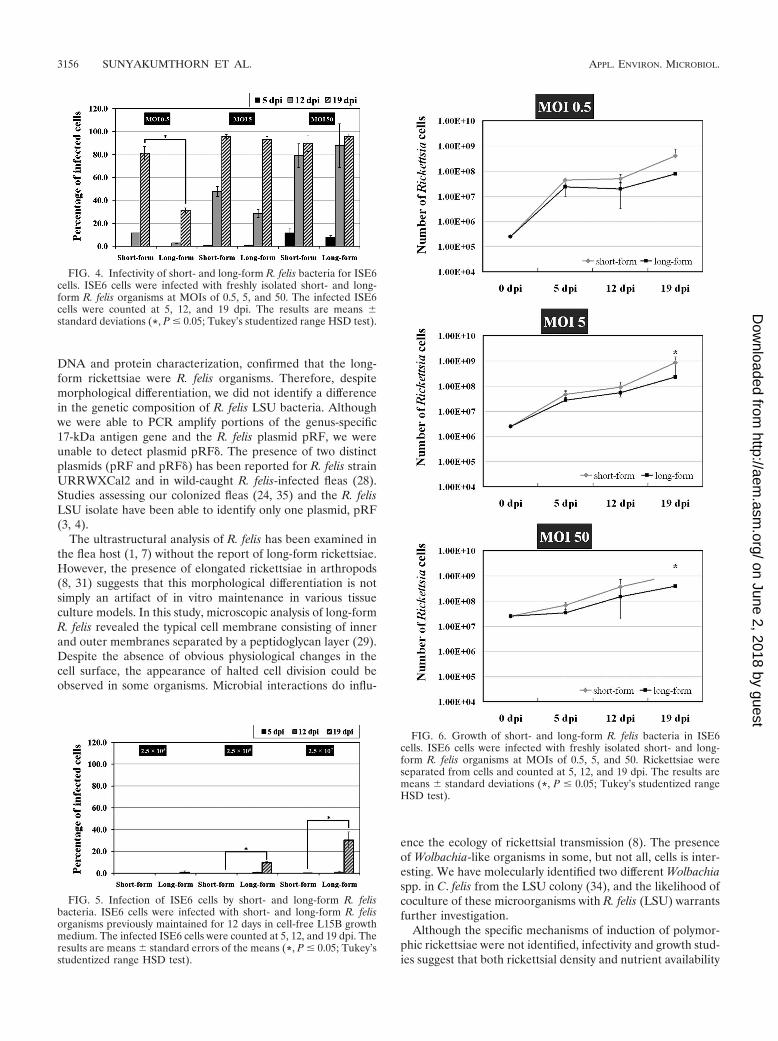
Rickettsial infectivity and replication in ISE6 cells. To as-sess the infectious nature of polymorphic R. felis for the ISE6cell line, short- and long-form R. felis organisms immediatelyharvested from ISE6 cells or maintained in cell-free growthmedium were inoculated at various MOIs onto previously R.felis-uninfected ISE6 cells. Cytospin preparations were stainedwith Diff-Quik, and the percentages of R. felis-infected ISE6cells were counted at 5, 12, and 19 dpi. The ability to detectrickettsiae at the earliest assessment time point (5 dpi) wasdependent on the MOI, with an increase in the percentage ofinfected cells correlating with an increased MOI and durationof infection. By 12 and 19 dpi, rickettsiae were detected inincreased percentages of host cells, reaching nearly 100% in-fection of host cells by day 19 at MOIs of 5 and 50. A lowerMOI correlated with a lower total number of cells infected byday 19; however, only at the lower MOI did we observe asignificant difference between short- and long-form R. felisorganisms, with the short form infecting a significantly higherpercentage of ISE6 cells than the long form at the same MOI(Fig. 4).
To examine environmental stability of polymorphic R.felis, short- and long-form R. felis organisms were recoveredfrom ISE6 cells and maintained in filtered, conditioned cell-free L15B growth medium. While both short- and long-formR. felis organisms retained viability in cell-free L15B growthmedium for up to 5 days, as assessed by BacLight staining,and replicated, as determined by an increase in the totalnumber of rickettsiae, replication did not continue through19 days of cell-free culture, despite the detection of viablerickettsiae (data not shown). To assess the infectious natureof cell-free rickettsiae, short- and long-form R. felis bacteriawere maintained in cell-free L15B growth medium for 12days and then passed onto uninfected ISE6 cells at variousconcentrations; infectivity and replication were assessed at5, 12, and 19 dpi. While still viable, short-form R. felis wasonly infective for ISE6 cells at the highest concentrationassessed, in contrast to the long-form R. felis, which wasinfectious at all concentrations (Fig. 5). At 19 dpi the infec-tivity of long-form R. felis (30.75%) was significantly higherthan that of short-form R. felis (0.25%); this difference ininfectivity was not observed in assays utilizing R. felis freshlyisolated from ISE6 cells. The inability of short-form R. felisto infect new cells when maintained in cell-free L15B growthmedium, in contrast to the long-form R. felis, which re-mained infectious after a prolonged extracellular period,suggests that polymorphism facilitates physiological changesin rickettsiae for survival outside the host cell.
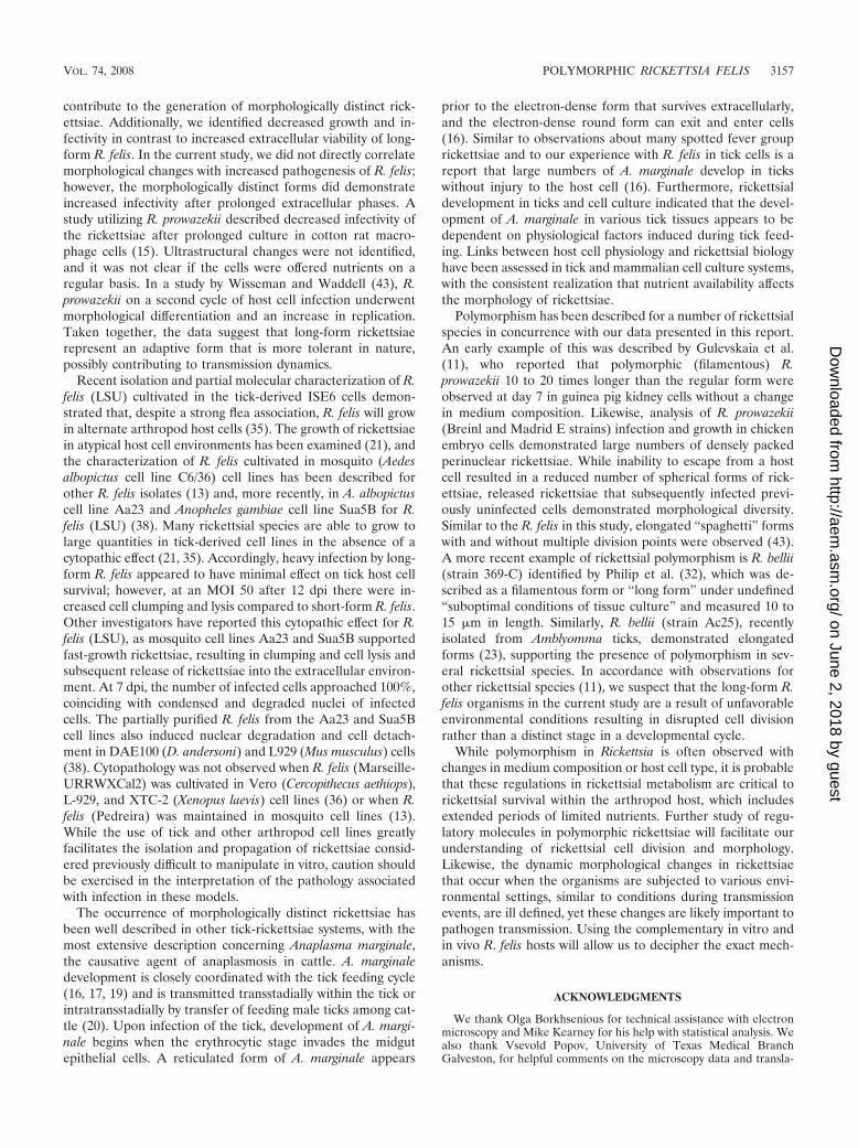
The rate of replication of short- and long-form R. felis or-ganisms was determined by counting total numbers of viablerickettsiae during BacLight viability staining at 5, 12, and 19dpi. Equal numbers of homogenous populations of short- or
FIG. 2. Long-form R. felis cultured in ISE6 cells. (A) Cytospin slideof long-form R. felis-infected ISE6 cells was stained using a Diff-Quikkit. (B) IFA of long-form R. felis-infected ISE6 cells using anti-R. felismouse serum with DAPI counter-staining. A bright-field image wascolor merged with green fluorescence and DAPI images. The rickett-siae were visualized by green fluorescence, and the DNA was stainedwith DAPI. Both images are at a magnification of �63.
3154 SUNYAKUMTHORN ET AL. APPL. ENVIRON. MICROBIOL.
on June 2, 2018 by guesthttp://aem
.asm.org/
Dow
nloaded from

long-form R. felis were seeded onto uninfected ISE6 cells. Ateach time point assessed, the number of short-form rickettsiaewere greater than the number of long-form R. felis organisms,with significant differences in the numbers of short-form R. felisat MOIs of 5 and 50 at 19 dpi (Fig. 6).
DISCUSSION
In the present report, we characterize a homogenous popu-lation of polymorphic rickettsiae maintained in a tick-derivedcell line. Molecular assays, including amplification of rickettsial
FIG. 3. Electron micrographs of polymorphic R. felis in ISE6 cells. (A) Long-form R. felis organisms (arrows) associated with cytoplasmicclearance of ISE6 cells also infected with Wolbachia-like organisms (arrowhead). (B) Portion of long-form R. felis organisms within a cell withtypical rickettsial envelope containing periplasmic space. (C) Immunolabeled long-form R. felis. (D) Secondary antibody alone; negative control.
VOL. 74, 2008 POLYMORPHIC RICKETTSIA FELIS 3155
on June 2, 2018 by guesthttp://aem
.asm.org/
Dow
nloaded from
DNA and protein characterization, confirmed that the long-form rickettsiae were R. felis organisms. Therefore, despitemorphological differentiation, we did not identify a differencein the genetic composition of R. felis LSU bacteria. Althoughwe were able to PCR amplify portions of the genus-specific17-kDa antigen gene and the R. felis plasmid pRF, we wereunable to detect plasmid pRF�. The presence of two distinctplasmids (pRF and pRF�) has been reported for R. felis strainURRWXCal2 and in wild-caught R. felis-infected fleas (28).Studies assessing our colonized fleas (24, 35) and the R. felisLSU isolate have been able to identify only one plasmid, pRF(3, 4).
The ultrastructural analysis of R. felis has been examined inthe flea host (1, 7) without the report of long-form rickettsiae.However, the presence of elongated rickettsiae in arthropods(8, 31) suggests that this morphological differentiation is notsimply an artifact of in vitro maintenance in various tissueculture models. In this study, microscopic analysis of long-formR. felis revealed the typical cell membrane consisting of innerand outer membranes separated by a peptidoglycan layer (29).Despite the absence of obvious physiological changes in thecell surface, the appearance of halted cell division could beobserved in some organisms. Microbial interactions do influ-
ence the ecology of rickettsial transmission (8). The presenceof Wolbachia-like organisms in some, but not all, cells is inter-esting. We have molecularly identified two different Wolbachiaspp. in C. felis from the LSU colony (34), and the likelihood ofcoculture of these microorganisms with R. felis (LSU) warrantsfurther investigation.
Although the specific mechanisms of induction of polymor-phic rickettsiae were not identified, infectivity and growth stud-ies suggest that both rickettsial density and nutrient availability
FIG. 5. Infection of ISE6 cells by short- and long-form R. felisbacteria. ISE6 cells were infected with short- and long-form R. felisorganisms previously maintained for 12 days in cell-free L15B growthmedium. The infected ISE6 cells were counted at 5, 12, and 19 dpi. Theresults are means standard errors of the means (*, P � 0.05; Tukey’sstudentized range HSD test).
FIG. 6. Growth of short- and long-form R. felis bacteria in ISE6cells. ISE6 cells were infected with freshly isolated short- and long-form R. felis organisms at MOIs of 0.5, 5, and 50. Rickettsiae wereseparated from cells and counted at 5, 12, and 19 dpi. The results aremeans standard deviations (*, P � 0.05; Tukey’s studentized rangeHSD test).
FIG. 4. Infectivity of short- and long-form R. felis bacteria for ISE6cells. ISE6 cells were infected with freshly isolated short- and long-form R. felis organisms at MOIs of 0.5, 5, and 50. The infected ISE6cells were counted at 5, 12, and 19 dpi. The results are means standard deviations (*, P � 0.05; Tukey’s studentized range HSD test).
3156 SUNYAKUMTHORN ET AL. APPL. ENVIRON. MICROBIOL.
on June 2, 2018 by guesthttp://aem
.asm.org/
Dow
nloaded from
contribute to the generation of morphologically distinct rick-ettsiae. Additionally, we identified decreased growth and in-fectivity in contrast to increased extracellular viability of long-form R. felis. In the current study, we did not directly correlatemorphological changes with increased pathogenesis of R. felis;however, the morphologically distinct forms did demonstrateincreased infectivity after prolonged extracellular phases. Astudy utilizing R. prowazekii described decreased infectivity ofthe rickettsiae after prolonged culture in cotton rat macro-phage cells (15). Ultrastructural changes were not identified,and it was not clear if the cells were offered nutrients on aregular basis. In a study by Wisseman and Waddell (43), R.prowazekii on a second cycle of host cell infection underwentmorphological differentiation and an increase in replication.Taken together, the data suggest that long-form rickettsiaerepresent an adaptive form that is more tolerant in nature,possibly contributing to transmission dynamics.
Recent isolation and partial molecular characterization of R.felis (LSU) cultivated in the tick-derived ISE6 cells demon-strated that, despite a strong flea association, R. felis will growin alternate arthropod host cells (35). The growth of rickettsiaein atypical host cell environments has been examined (21), andthe characterization of R. felis cultivated in mosquito (Aedesalbopictus cell line C6/36) cell lines has been described forother R. felis isolates (13) and, more recently, in A. albopictuscell line Aa23 and Anopheles gambiae cell line Sua5B for R.felis (LSU) (38). Many rickettsial species are able to grow tolarge quantities in tick-derived cell lines in the absence of acytopathic effect (21, 35). Accordingly, heavy infection by long-form R. felis appeared to have minimal effect on tick host cellsurvival; however, at an MOI 50 after 12 dpi there were in-creased cell clumping and lysis compared to short-form R. felis.Other investigators have reported this cytopathic effect for R.felis (LSU), as mosquito cell lines Aa23 and Sua5B supportedfast-growth rickettsiae, resulting in clumping and cell lysis andsubsequent release of rickettsiae into the extracellular environ-ment. At 7 dpi, the number of infected cells approached 100%,coinciding with condensed and degraded nuclei of infectedcells. The partially purified R. felis from the Aa23 and Sua5Bcell lines also induced nuclear degradation and cell detach-ment in DAE100 (D. andersoni) and L929 (Mus musculus) cells(38). Cytopathology was not observed when R. felis (Marseille-URRWXCal2) was cultivated in Vero (Cercopithecus aethiops),L-929, and XTC-2 (Xenopus laevis) cell lines (36) or when R.felis (Pedreira) was maintained in mosquito cell lines (13).While the use of tick and other arthropod cell lines greatlyfacilitates the isolation and propagation of rickettsiae consid-ered previously difficult to manipulate in vitro, caution shouldbe exercised in the interpretation of the pathology associatedwith infection in these models.
The occurrence of morphologically distinct rickettsiae hasbeen well described in other tick-rickettsiae systems, with themost extensive description concerning Anaplasma marginale,the causative agent of anaplasmosis in cattle. A. marginaledevelopment is closely coordinated with the tick feeding cycle(16, 17, 19) and is transmitted transstadially within the tick orintratransstadially by transfer of feeding male ticks among cat-tle (20). Upon infection of the tick, development of A. margi-nale begins when the erythrocytic stage invades the midgutepithelial cells. A reticulated form of A. marginale appears
prior to the electron-dense form that survives extracellularly,and the electron-dense round form can exit and enter cells(16). Similar to observations about many spotted fever grouprickettsiae and to our experience with R. felis in tick cells is areport that large numbers of A. marginale develop in tickswithout injury to the host cell (16). Furthermore, rickettsialdevelopment in ticks and cell culture indicated that the devel-opment of A. marginale in various tick tissues appears to bedependent on physiological factors induced during tick feed-ing. Links between host cell physiology and rickettsial biologyhave been assessed in tick and mammalian cell culture systems,with the consistent realization that nutrient availability affectsthe morphology of rickettsiae.
Polymorphism has been described for a number of rickettsialspecies in concurrence with our data presented in this report.An early example of this was described by Gulevskaia et al.(11), who reported that polymorphic (filamentous) R.prowazekii 10 to 20 times longer than the regular form wereobserved at day 7 in guinea pig kidney cells without a changein medium composition. Likewise, analysis of R. prowazekii(Breinl and Madrid E strains) infection and growth in chickenembryo cells demonstrated large numbers of densely packedperinuclear rickettsiae. While inability to escape from a hostcell resulted in a reduced number of spherical forms of rick-ettsiae, released rickettsiae that subsequently infected previ-ously uninfected cells demonstrated morphological diversity.Similar to the R. felis in this study, elongated “spaghetti” formswith and without multiple division points were observed (43).A more recent example of rickettsial polymorphism is R. bellii(strain 369-C) identified by Philip et al. (32), which was de-scribed as a filamentous form or “long form” under undefined“suboptimal conditions of tissue culture” and measured 10 to15 �m in length. Similarly, R. bellii (strain Ac25), recentlyisolated from Amblyomma ticks, demonstrated elongatedforms (23), supporting the presence of polymorphism in sev-eral rickettsial species. In accordance with observations forother rickettsial species (11), we suspect that the long-form R.felis organisms in the current study are a result of unfavorableenvironmental conditions resulting in disrupted cell divisionrather than a distinct stage in a developmental cycle.
While polymorphism in Rickettsia is often observed withchanges in medium composition or host cell type, it is probablethat these regulations in rickettsial metabolism are critical torickettsial survival within the arthropod host, which includesextended periods of limited nutrients. Further study of regu-latory molecules in polymorphic rickettsiae will facilitate ourunderstanding of rickettsial cell division and morphology.Likewise, the dynamic morphological changes in rickettsiaethat occur when the organisms are subjected to various envi-ronmental settings, similar to conditions during transmissionevents, are ill defined, yet these changes are likely important topathogen transmission. Using the complementary in vitro andin vivo R. felis hosts will allow us to decipher the exact mech-anisms.
ACKNOWLEDGMENTS
We thank Olga Borkhsenious for technical assistance with electronmicroscopy and Mike Kearney for his help with statistical analysis. Wealso thank Vsevold Popov, University of Texas Medical BranchGalveston, for helpful comments on the microscopy data and transla-
VOL. 74, 2008 POLYMORPHIC RICKETTSIA FELIS 3157
on June 2, 2018 by guesthttp://aem
.asm.org/
Dow
nloaded from

tion of papers. The monoclonal antibody RC-9C2 was provided by LeeFuller, Fuller Laboratories.
This research was supported by the Louisiana Board of Regents(LEQSF), the National Institutes of Health (P20RR0201595), and theNational Institute of Allergy and Infectious Diseases (K22AI60821 andR21AI070705).
REFERENCES
1. Adams, J. R., E. T. Schmidtmann, and A. F. Azad. 1990. Infection of colo-nized cat fleas, Ctenocephalides felis (Bouche), with a rickettsia-like micro-organism. Am. J. Trop. Med. Hyg. 43:400–409.
2. Azad, A. F., S. Radulovic, J. A. Higgins, B. H. Noden, and J. M. Troyer. 1997.Flea-borne rickettsioses: ecologic considerations. Emerg. Infect. Dis. 3:319–327.
3. Baldridge, G. D., N. Y. Burkhardt, R. F. Felsheim, T. J. Kurtti, and U. G.Munderloh. 2008. Plasmids of the pRM/pRF family occur in diverse Rick-ettsia species. Appl. Environ. Microbiol. 74:645–652.
4. Baldridge, G. D., N. Y. Burkhardt, R. F. Felsheim, T. J. Kurtti, and U. G.Munderloh. 2007. Transposon insertion reveals pRM, a plasmid of Rickettsiamonacensis. Appl. Environ. Microbiol. 73:4984–4995.
5. Blouin, E. F., and K. M. Kocan. 1998. Morphology and development ofAnaplasma marginale (Rickettsiales: Anaplasmataceae) in cultured Ixodesscapularis (Acari: Ixodidae) cells. J. Med. Entomol. 35:788–797.
6. Boostrom, A., M. S. Beier, J. A. Macaluso, K. R. Macaluso, D. Sprenger, J.Hayes, S. Radulovic, and A. F. Azad. 2002. Geographic association of Rick-ettsia felis-infected opossums with human murine typhus, Texas. Emerg.Infect. Dis. 8:549–554.
7. Bouyer, D. H., J. Stenos, P. Crocquet-Valdes, C. G. Moron, V. L. Popov, J. E.Zavala-Velazquez, L. D. Foil, D. R. Stothard, A. F. Azad, and D. H. Walker.2001. Rickettsia felis: molecular characterization of a new member of thespotted fever group. Int. J. Syst. Evol. Microbiol. 51:339–347.
8. Burgdorfer, W., S. F. Hayes, and A. J. Marvos. 1981. Nonpathogenic rick-ettsiae in Dermacentor andersoni: a limiting factor for the distribution ofRickettsia rickettsii, p. 585–594. In W. Burgdorfer and R. L. Anacker (ed.),Rickettsiae and rickettsial diseases. Academic Press, New York, NY.
9. Case, J. B., B. Chomel, W. Nicholson, and J. E. Foley. 2006. Serologicalsurvey of vector-borne zoonotic pathogens in pet cats and cats from animalshelters and feral colonies. J. Feline Med. Surg. 8:111–117.
10. Coleman, S. A., E. R. Fischer, D. Howe, D. J. Mead, and R. A. Heinzen. 2004.Temporal analysis of Coxiella burnetii morphological differentiation. J. Bac-teriol. 186:7344–7352.
11. Gulevskaia, S. A., V. L. Popov, and V. F. Ignatovich. 1975. Current data onthe polymorphism of Rickettsia prowazekii and burneti in cultured cells. Zh.Mikrobiol. Epidemiol. Immunobiol. 7:68–72. (In Russian.)
12. Heinzen, R. A., T. Hackstadt, and J. E. Samuel. 1999. Developmental biologyof Coxiella burnettii. Trends Microbiol. 7:149–154.
13. Horta, M. C., M. B. Labruna, E. L. Durigon, and T. T. Schumaker. 2006.Isolation of Rickettsia felis in the mosquito cell line C6/36. Appl. Environ.Microbiol. 72:1705–1707.
14. Ito, S., and Y. Rikihisa. 1981. Techniques for electron microscopy of rick-ettsiae, p. 213–240. In W. Burgdorfer and R. L. Anacker (ed.), Rickettsiaeand rickettsial diseases. Academic Press, Inc., New York, NY.
15. Kekcheeva, N. G., I. N. Kokorin, V. L. Popov, E. A. Chereshkova, N. S.Smirnova, O. A. Vovk, and E. M. Shirokova. 1992. Persistence of Rickettsiaprowazekii in cotton rat macrophage cultures. Acta Virol. 36:103–110.
16. Kocan, K. M. 1986. Development of Anaplasma marginale Theiler in ixodidticks: Coordinated development of a rickettsial organism and its tick host, p.472–505. In J. R. Sauer and J. A. Hair (ed.), Morphology, physiology, andbehavioral biology of ticks. John Wiley & Sons, New York, NY.
17. Kocan, K. M. 1992. Recent advances in the biology of Anaplasma spp. inDermacentor andersoni ticks. Ann. N. Y. Acad. Sci. 653:26–32.
18. Kocan, K. M., S. A. Ewing, D. Holbert, and J. A. Hair. 1982. Morphologiccharacteristics of colonies of Anaplasma marginale Theiler in midgut epithe-lial cells of Dermacentor andersoni Stiles. Am. J. Vet. Res. 43:586–593.
19. Kocan, K. M., W. L. Goff, D. Stiller, P. L. Claypool, W. Edwards, S. A. Ewing,J. A. Hair, and S. J. Barron. 1992. Persistence of Anaplasma marginale(Rickettsiales: Anaplasmataceae) in male Dermacentor andersoni (Acari:Ixodidae) transferred successively from infected to susceptible calves.J. Med. Entomol. 29:657–668.
20. Kocan, K. M., D. Holbert, W. Edwards, S. A. Ewing, S. J. Barron, and J. A.Hair. 1986. Longevity of colonies of Anaplasma marginale in midgut epithe-lial cells of Dermacentor andersoni. Am. J. Vet. Res. 47:1657–1661.
21. Kurtti, T. J., J. A. Simser, G. D. Baldridge, A. T. Palmer, and U. G. Munder-loh. 2005. Factors influencing in vitro infectivity and growth of Rickettsia
peacockii (Rickettsiales: Rickettsiaceae), an endosymbiont of the RockyMountain wood tick, Dermacentor andersoni (Acari, Ixodidae). J. Invertebr.Pathol. 90:177–186.
22. Labruna, M. B., R. C. Pacheco, L. J. Richtzenhain, and M. P. Szabo. 2007.Isolation of Rickettsia rhipicephali and Rickettsia bellii from Haemaphysalisjuxtakochi ticks in the state of Sao Paulo, Brazil. Appl. Environ. Microbiol.73:869–873.
23. Labruna, M. B., T. Whitworth, M. C. Horta, D. H. Bouyer, J. W. McBride,A. Pinter, V. Popov, S. M. Gennari, and D. H. Walker. 2004. Rickettsiaspecies infecting Amblyomma cooperi ticks from an area in the state of SaoPaulo, Brazil, where Brazilian spotted fever is endemic. J. Clin. Microbiol.42:90–98.
24. Macaluso, K. R., W. Pornwiroon, V. Popov, and L. D. Foil. 2008. Identifi-cation of Rickettsia felis in the salivary glands of cat fleas. Vector BorneZoonotic Dis., in press.
25. Munderloh, U. G., S. D. Jauron, V. Fingerle, L. Leitritz, S. F. Hayes, J. M.Hautman, C. M. Nelson, B. W. Huberty, T. J. Kurtti, G. G. Ahlstrand, B.Greig, M. A. Mellencamp, and J. L. Goodman. 1999. Invasion and intracel-lular development of the human granulocytic ehrlichiosis agent in tick cellculture. J. Clin. Microbiol. 37:2518–2524.
26. Munderloh, U. G., and T. J. Kurtti. 1989. Formulation of medium for tickcell culture. Exp. Appl. Acarol. 7:219–229.
27. Munderloh, U. G., and T. J. Kurtti. 1995. Cellular and molecular interrela-tionships between ticks and prokaryotic tick-borne pathogens. Annu. Rev.Entomol. 40:221–243.
28. Ogata, H., P. Renesto, S. Audic, C. Robert, G. Blanc, P. E. Fournier, H.Parinello, J. M. Claverie, and D. Raoult. 2005. The genome sequence ofRickettsia felis identifies the first putative conjugative plasmid in an obligateintracellular parasite. PLoS. Biol. 3:e248.
29. Pang, H., and H. H. Winkler. 1994. Analysis of the peptidoglycan of Rick-ettsia prowazekii. J. Bacteriol. 176:923–926.
30. Parola, P., B. Davoust, and D. Raoult. 2005. Tick- and flea-borne rickettsialemerging zoonoses. Vet. Res. 36:469–492.
31. Philip, R. N., and E. A. Casper. 1981. Serotypes of spotted fever grouprickettsiae isolated from Dermacentor andersoni (Stiles) ticks in westernMontana. Am. J. Trop. Med. Hyg. 30:230–238.
32. Philip, R. N., E. A. Casper, R. L. Anacker, J. Cory, S. F. Hayes, W. Burg-dorfer, and C. E. Yunker. 1983. Rickettsia bellii sp. nov.: a tick-borne rick-ettsia, widely distributed in the United States, that is distinct from thespotted fever and typhus biogroups. Int. J. Syst. Bacteriol. 33:94–106.
33. Popov, V. L., V. C. Han, S. M. Chen, J. S. Dumler, H. M. Feng, T. G.Andreadis, R. B. Tesh, and D. H. Walker. 1998. Ultrastructural differentia-tion of the genogroups in the genus Ehrlichia. J. Med. Microbiol. 47:235–251.
34. Pornwiroon, W., M. T. Kearney, C. Husseneder, L. D. Foil, and K. R.Macaluso. 2007. Comparative microbiota of Rickettsia felis-uninfected and-infected colonized cat fleas, Ctenocephalides felis. ISME J. 1:394–402.
35. Pornwiroon, W., S. S. Pourciau, L. D. Foil, and K. R. Macaluso. 2006.Rickettsia felis from cat fleas: isolation and culture in a tick-derived cell line.Appl. Environ. Microbiol. 72:5589–5595.
36. Raoult, D., S. B. La, M. Enea, P. E. Fournier, V. Roux, F. Fenollar, M. A.Galvao, and X. L. de. 2001. A flea-associated Rickettsia pathogenic forhumans. Emerg. Infect. Dis. 7:73–81.
37. Rikihisa, Y., and S. Ito. 1980. Localization of electron-dense tracers duringentry of Rickettsia tsutsugamushi into polymorphonuclear leukocytes. Infect.Immun. 30:231–243.
38. Sakamoto, J. M., and A. F. Azad. 2007. Propagation of arthropod-borneRickettsia spp. in two mosquito cell lines. Appl. Environ. Microbiol. 73:6637–6643.
39. Schriefer, M. E., J. B. Sacci, Jr., J. P. Taylor, J. A. Higgins, and A. F. Azad.1994. Murine typhus: updated roles of multiple urban components and asecond typhuslike rickettsia. J. Med. Entomol. 31:681–685.
40. Simser, J. A., A. T. Palmer, U. G. Munderloh, and T. J. Kurtti. 2001.Isolation of a spotted fever group Rickettsia, Rickettsia peacockii, in a RockyMountain wood tick, Dermacentor andersoni, cell line. Appl. Environ. Mi-crobiol. 67:546–552.
41. Wedincamp, J., Jr., and L. D. Foil. 2002. Vertical transmission of Rickettsiafelis in the cat flea (Ctenocephalides felis Bouche). J. Vector Ecol. 27:96–101.
42. Williams, S. G., J. B. Sacci, Jr., M. E. Schriefer, E. M. Andersen, K. K.Fujioka, F. J. Sorvillo, A. R. Barr, and A. F. Azad. 1992. Typhus andtyphuslike rickettsiae associated with opossums and their fleas in Los AngelesCounty, California. J. Clin. Microbiol. 30:1758–1762.
43. Wisseman, C. L., Jr., and A. D. Waddell. 1975. In vitro studies on rickettsia-host cell interactions: intracellular growth cycle of virulent and attenuatedRickettsia prowazeki in chicken embryo cells in slide chamber cultures. Infect.Immun. 11:1391–1404.
3158 SUNYAKUMTHORN ET AL. APPL. ENVIRON. MICROBIOL.
on June 2, 2018 by guesthttp://aem
.asm.org/
Dow
nloaded from