Embed Size (px)
Citation preview
Characterization and localization of the Campylobacter jejuni transformation system 1
proteins CtsE, CtsP and CtsX 2
3
Jessica M. Beauchamp¶, Rebecca S. Erfurt¶§, and Victor J. DiRita* 4
1Department of Microbiology and Immunology 5
University of Michigan Medical School 6
7
8
9 10 11 12 13 14 15 *corresponding author 16 Tel: 734-936-3804 17 FAX: 734-764-3562 18 email: [email protected] 19 ¶ equal contribution 20 § present address: Blue Cross Blue Shield of Michigan 21 600 East Lafayette 22 Detroit, MI 48226 23 24
25
26
JB Accepts, published online ahead of print on 1 December 2014J. Bacteriol. doi:10.1128/JB.02434-14Copyright © 2014, American Society for Microbiology. All Rights Reserved.
on March 15, 2018 by guest
http://jb.asm.org/
Dow
nloaded from
27
ABSTRACT 28
The human pathogen Campylobacter jejuni is naturally competent for transformation 29
with its own DNA. Genes required for efficient transformation in C. jejuni include those 30
similar to components of type II secretion systems found in many Gram negative bacteria (1). 31
Two of these, ctsE and ctsP, encode proteins annotated as putative nucleotide binding 32
NTPases or NTP binding proteins. Here we demonstrate that the nucleotide binding motifs 33
of both proteins are essential for their function in transformation of C. jejuni. Localization 34
experiments demonstrate that CtsE is a soluble protein while CtsP is membrane-associated in 35
C. jejuni. A bacterial two hybrid screen identified interaction between CtsP and CtsX, an 36
integral membrane protein also required for transformation. Topological analysis of CtsX 37
using LacZ and PhoA fusions demonstrate it to be a bitopic, integral membrane protein with 38
a cytoplasmic amino terminus and a periplasmic carboxyl terminus. Notwithstanding its 39
interaction with membrane-localized CtsX, CtsP inherently associates with the membrane, 40
requiring neither CtsX or several other Cts proteins for this association. 41
42
on March 15, 2018 by guest
http://jb.asm.org/
Dow
nloaded from
INTRODUCTION 43
The Gram-negative bacterium C. jejuni is a leading cause of bacterial gastroenteritis 44
worldwide (2). C. jejuni often colonizes the avian intestinal tract and, consequently, a common 45
route of infection is through consumption of contaminated poultry (3). 46
A number of Campylobacters are naturally competent for transformation, meaning that 47
they can take up macromolecular DNA from the environment and incorporate it heritably into 48
their genomes (4, 5). The ability to acquire DNA from the environment may contribute to the 49
extensive genetic diversity observed among strains of C. jejuni (6, 7). Horizontal gene transfer 50
in vivo has been demonstrated during experimental infection of chicks, a natural host for this 51
pathogen (8). 52
Multiple genes whose products are involved in natural transformation of C. jejuni have 53
been identified (1, 9-12). Using transposon mutagenesis and a genetic screen for loss of 54
competence, we isolated mutations that mapped to 11 genes encoded in C. jejuni strain 81-176 55
(1). Mutations in these result in reduction in transformability to levels four orders of magnitude 56
below wild type (1). Among these are six genes arranged in a likely operon, some of which 57
encode proteins similar to components of type II secretion and type IV pilus biogenesis systems 58
and are homologous to proteins important for natural transformation in other organisms (13). 59
Two of these, ctsE and ctsP, encode putative NTPases or NTP binding proteins, according to the 60
annotated genome of C. jejuni strain 11168 (1, 14). 61
CtsE is a member of the type II secretion/type IV secretion system superfamily of 62
NTPases collectively referred to as the PulE - VirB11 family (15, 16). Members of this family 63
are involved in diverse processes including secretion, pilus biogenesis, competence for natural 64
transformation, and conjugation (15). PulE – VirB11 family members share several elements, 65
on March 15, 2018 by guest
http://jb.asm.org/
Dow
nloaded from
including the nucleotide-binding motifs – Walker boxes A and B – an Asp box, and a His box 66
(17-19). CtsE also has a tetra-cysteine motif conserved among the GspE and PilB/HofB 67
subfamilies (20, 21). 68
ATPase activity has been demonstrated in vitro for several members of the PulE - VirB11 69
family (22-26). It is hypothesized that this activity powers the transformation process, though 70
this has yet to be conclusively shown. Phylogenetic analyses placed CtsE in the type II secretion 71
subfamily of NTPases; in one such analysis CtsE fell between the ComG1 subfamily of Gram-72
positive NTPases involved in competence and the PilT and PilU subfamily involved in retraction 73
of type IV pili (15, 16). Another analysis placed CtsE in the Gram-positive competence 74
subfamily, closely related to the GspE subfamily of type II secretion machinery (16). Although 75
ATPase activity has not been demonstrated for most of the type II secretion machinery, it has 76
been hypothesized. 77
CtsE is one of two putative identified NTP binding proteins important for natural 78
transformation of C. jejuni. The other is CtsP (1), which has Walker box A and B nucleotide 79
binding motifs, but lacks other characteristics of the PulE – VirB11 superfamily including the 80
His box, the Asp box and the tetra-cysteine motif. By BLAST analysis CtsP exhibits weak 81
homology to several ATPases including PilT homologues, ClpX ATP binding subunits, and 82
members of the AAA family of ATPases. The Walker boxes of CtsP resemble those of the 83
AAA+ superfamily, but CtsP does not appear to have the minimal AAA consensus sequence (18, 84
27). 85
In this study we characterized CtsE and CtsP, the two putative NTPases/NTP binding 86
proteins required for natural transformation of C. jejuni. We also carried out analysis of a third 87
gene product encoded in the cts gene cluster, CtsX, unique to the C. jejuni transformation 88
on March 15, 2018 by guest
http://jb.asm.org/
Dow
nloaded from
system. CtsX lacks significant sequence homology to other proteins, and shares no clearly 89
conserved domains beyond a transmembrane domain. We determined subcellular localization of 90
each of these proteins, and we investigated the roles of the nucleotide binding motifs in CtsE and 91
CtsP. Further analysis investigated protein-protein interactions among constituents of the type 92
II-like Cts system in C. jejuni (1), revealing association between CtsP and CtsX. 93
94
MATERIALS AND METHODS 95
96
Bacterial strains and media 97
Bacterial strains used in this work are listed in Table 1. C. jejuni was routinely cultured 98
on Muller Hinton (MH) agar with 10% sheep’s blood in microaerophilic conditions (5% CO2, 99
10% O2, balanced with N2) in a tri-gas incubator at 37°C. When necessary, media was 100
supplemented with antibiotics in the following concentrations: trimethoprim (10 μg ml-1), 101
kanamycin (as noted, either 30 or 150 µg ml-1), streptomycin (100 μg ml-1), and chloramphenicol 102
(as noted, either 20 or 30 μg ml-1). All C. jejuni strains were stored at -80°C in MH broth with 103
20% glycerol. 104
Escherichia coli strains were routinely cultured at 37°C in Luria-Bertani (LB) broth or 105
agar. When necessary, antibiotics were used at the following concentrations: ampicillin (100 μg 106
ml-1), chloramphenicol (30 μg ml-1), kanamycin (50 μg ml-1), and tetracycline (12.5 μg ml-1). All 107
E. coli strains were stored at -80°C in LB broth with 20% glycerol. 108
Construction of FLAG fusion proteins 109
To express FLAG fusion proteins in C. jejuni we modified pECO102 by mutagenesis 110
with PFU turbo (1). Two primers were made that annealed to pECO102 and would insert the 111
on March 15, 2018 by guest
http://jb.asm.org/
Dow
nloaded from
coding sequence for the FLAG tag and a stop codon downstream of the XhoI site. (All primers 112
used in this study are indicated on Table 2.) pECO102 was amplified by PCR with these primers 113
and the template DNA was digested with DpnI. E. coli DH5α was transformed with the reaction 114
mixture and clones were screened by PCR. Candidate plasmids were sequenced to confirm 115
insertion of the FLAG sequence and one, pRSW211, was used for subsequent cloning. To 116
construct in-frame fusion proteins with a C-terminal FLAG tag, PCR primers were designed to 117
amplify the coding sequence of the protein of interest from the second amino acid through the 118
last amino acid (excluding the stop codon). The 5’ primer had BamHI sites and the 3’ primer 119
had XhoI sites for in-frame insertion into pRSW211. All subsequent clones were verified by 120
sequence determination. 121
Construction of Walker Box point mutants 122
Point mutants were constructed in the Walker boxes of ctsE and ctsP by PFU 123
mutagenesis (Stratagene). Both ctsE and ctsP were cloned into pRSW211 to create C-terminal 124
FLAG fusions, pRSW223 (CtsE-FLAG) and pRSW208 (CtsP-FLAG). The conserved lysine 125
residue (K296) of the Walker box A of CtsE-FLAG was mutated by changing the coding 126
sequence from AAA to CAA resulting in an amino acid change to a glutamine (K296Q); this 127
plasmid was designated pRSW246. The conserved glutamic acid residue (E81A) of the Walker 128
box B of CtsP-FLAG was mutated by changing the coding sequence from GAA to GCA 129
resulting in an amino acid change to an alanine (E81A); this plasmid was designated pRSW228. 130
DH5α. pRK212.1 was transformed with these constructs and conjugations were performed as 131
described previously (28, 29). pRSW208 and pRSW228 were conjugated into DRH212 ΔctsP 132
(RSW115), while pRSW223 and pRSW246 were conjugated into DRH212 ΔctsE (RSW136) (1). 133
on March 15, 2018 by guest
http://jb.asm.org/
Dow
nloaded from
Transconjugants were verified by PCR. Expression of the fusion proteins was detected in whole 134
cell lysates by SDS-PAGE and western blot analysis as discussed below. 135
Construction of His fusion proteins 136
To express a 6x-His tagged version of CtsX, genomic DNA from C. jejuni strain 81-176 137
was used as a template to amplify ctsX from the second codon to the stop codon using a forward 138
primer with a BamHI site and a reverse primer with XhoI site flanking the coding sequence. This 139
fragment was cloned into the BamHI and XhoI sites of pBW206, a derivative of pECO102 that 140
contains a N-terminal 6x-His tag immediately upstream of the BamHI site, creating JMB1. This 141
plasmid was transformed into DH5α pRK212.1 and transferred by conjugation into the ΔctsX 142
strain as described previously (28, 29). Transconjugants were verified by PCR and the stability 143
of the fusion proteins was assessed using SDS-PAGE and Western blot analysis from whole cell 144
lysates. 145
Transformation assays 146
Transformation assays were performed as described previously. (1, 4). Briefly, C. jejuni 147
was grown 16 – 18 hours on MH agar plates supplemented with appropriate antibiotics. Cells 148
were resuspended in MH broth to an optical density at 600 nm of 0.5 and 500 μl aliquots were 149
added to 13 mm test tubes containing one ml solidified agar. After incubation for three hours at 150
37°C in 5% CO2, one microgram of DRH153 (81-176 astA::aphA3) DNA was added and the 151
cultures were incubated for an additional four hours (30). The number of transformants and the 152
total number of bacteria were determined by dilution plating on MH agar with appropriate 153
antibiotics. Transformation efficiency represents the number of transformants per total number 154
of bacteria per microgram of DNA. Transformations were conducted in triplicate and the 155
transformation efficiency represents the average of the three samples from one experiment. 156
on March 15, 2018 by guest
http://jb.asm.org/
Dow
nloaded from
Cell Fractionations 157
Cell fractionation was carried out as previously described for C. jejuni with a few 158
modifications (31). C. jejuni was cultured on MH agar plates under microaerophilic conditions 159
at 37°C for 16 – 18 hrs. Cells were resuspended from plates in MH broth and centrifuged 160
(10,000 x g, 10 min at 4°C) and the resulting pellet was resuspended in 10 mM HEPES pH 7.4. 161
After one freeze-thaw cycle in a dry ice-ethanol bath, cells were sonicated with a micro-tip in a 162
Branson digital sonifier at 30% amplitude six times for 10 seconds. Cell debris was pelleted by 163
centrifugation (10,000 x g, 10 min at 4°C in a Sorvall tabletop centrifuge) and the supernatants 164
were subjected to ultracentrifugation to pellet membranes (100,000 x g, 70 min at 4°C with a 165
SW41 rotor). The supernatant was removed and saved as the soluble fraction; the pellet 166
(membrane fraction) was washed once in 10 mM HEPES and subsequently resuspended in an 167
appropriate volume of 10 mM Tris-HCl pH 8.0. The soluble fraction contains cytoplasmic and 168
periplasmic contents while the membrane fraction contains both inner and outer membranes. 169
The same procedure was followed for fractionation of E. coli. Protein concentrations were 170
determined with the BioRad protein assay or with the ThermoScientific BCA assay per 171
manufacturer’s instructions. Equal concentrations of proteins were then analyzed by SDS-PAGE 172
and Western blot analysis. 173
Membrane Flotation 174
C. jejuni was cultured on MH agar plates under microaerophilic conditions at 37°C for 16 175
– 18 hrs. Cells were resuspended from plates in MH broth and centrifuged (Sorvall tabletop 176
centrifuge, 10,000 x g, 10 min at 4°C) and the resulting pellet was resuspended in P Buffer (100 177
mM sodium phosphate pH 7.6, 50 mM MgCl2, and 10 mM EDTA). Cells were lysed by passage 178
through a French press. Lysates were incubated with DNase for 30 minutes and centrifuged 179
on March 15, 2018 by guest
http://jb.asm.org/
Dow
nloaded from
(5000 rpm for 5 min at 4°C) to remove cell debris. The supernatant was subjected to 180
ultracentrifugation (100,000 x g, 1 hour, at 4°C, SW41 rotor), and a sample of the supernatant, 181
containing the cytosolic and periplasmic proteins, was collected. The pellet was resuspended in P 182
buffer and centrifuged (100,000 x g, 1 hour, at 4°C, SW41 rotor). The supernatant was discarded 183
and the pellet was resuspended in P buffer and mixed with 81% sucrose (dissolved in P buffer) to 184
a final concentration of 71%. The sample was overlaid with 52% and 42% sucrose and subjected 185
to ultracentrifugation (18 hour at 100,000 x g, at 4°C, SW41 rotor). After ultracentrifugation, the 186
presence of the membrane bands was noted. Fractions (1.8 ml) were taken, starting from the top 187
of the sample and labeled 1-6 sequentially. The sucrose was diluted in P buffer and proteins were 188
precipitated using 2% deoxycholate and trichloroacetic acid (TCA) overnight at 4°C. After 189
precipitating the proteins, fractions were subjected to ultracentrifugation (1.5 hour, at 100,000 x 190
g, at 4°C, SW41 rotor). Pellets from each fraction were washed with acetone to remove residual 191
TCA and subsequently with 100% ether to remove lipids. Pellets were vacuum dried and 192
resuspended in water to be tested in the isocitrate dehydrogenase assay or resuspended in 6X 193
sample buffer for SDS-PAGE and immunoblot analysis. 194
SDS-PAGE and Immunoblotting 195
To detect FLAG fusion proteins, samples were diluted 1:1 with 2X SDS sample buffer 196
and boiled for five minutes before loading onto 12% polyacrylamide gels for separation by SDS-197
PAGE. For His-CtsX containing samples, samples were diluted 1:6 with 6X SDS sample buffer 198
and were separated on a 15% polyacrylamide gel. Samples were normalized by protein 199
concentration. Proteins were transferred to nitrocellulose membranes (Hoefer semi-dry, 00 200
mAmps for 2 hours) and, after blocking with 5% milk in TBS, membranes were probed with 201
primary antibody. For FLAG-tagged fusion proteins, an anti-FLAG M2 monoclonal antibody 202
on March 15, 2018 by guest
http://jb.asm.org/
Dow
nloaded from
peroxidase conjugate (Sigma) at 1:1000 was used. This was developed using the Western 203
Lighting Chemiluminescence reagent plus (Perkin-Elmer Life Sciences). For the His-tagged 204
fusion protein, THE™ His-tag antibody (Genscript) at 1:1500 was used. A rat anti-mouse IgG1-205
HRP conjugated antibody (Southern Biotech) was used at 1:3500 for the secondary antibody and 206
blots were developed using SuperSignal West Pico Chemiluminescent Substrate 207
(ThermoScientific). 208
Isocitrate Dehydrogenase assays 209
To demonstrate efficient fractionation, fractions were tested for the presence of isocitrate 210
dehydrogenase, a cytoplasmic protein. Isocitrate dehydrogenase activity was assayed as 211
described previously (31). The reaction mixture contained 100 μl 10 mM MgCl2, 100 μl 50 mM 212
Tris-HCl pH 8.0, 100 μl 10 mM NADP, 550 - 600 μl distilled H2O, and 0 – 50 μl of the cell 213
fraction. The reaction was started by the addition of 100 μl of 50 mM sodium isocitrate and the 214
optical density at 340 nm was read at 15 second intervals for three minutes. The specific activity 215
of isocitrate dehydrogenase was calculated using an absorption coefficient of NADPH of 6.22 216
mM-1 cm-1 at 340 nm. The percent (of total unfractionated sample) of isocitrate dehydrogenase 217
activity present in each fraction was calculated. For all fractionations, a minimum of 90% of the 218
isocitrate dehydrogenase activity was present in the cytoplasmic fraction. 219
Identification of interacting proteins with the bacterial-two hybrid system 220
A bacterial two hybrid system was used to identify potential interactions among Cts 221
proteins. This system is based on functional complementation between two fragments of the 222
catalytic domain of adenylate cyclase from Bordetella pertussis (32). Genes ctsE, ctsP, ctsX, 223
ctsF, ctsD, ctsR, ctsG, ctsW, and dprA were PCR amplified from the coding sequence of the 224
second amino acid through the stop codon and cloned into the PstI and BamHI sites of pT25 to 225
on March 15, 2018 by guest
http://jb.asm.org/
Dow
nloaded from
generate in-frame T25 fusion proteins. The same genes were amplified with primers containing 226
XhoI and ClaI sites from the second amino acid to the last amino acid, excluding the stop codon, 227
and inserted into the XhoI and ClaI sites of pT18 to create in-frame fusion proteins to the 228
adenylate cyclase T18 domain. The insertions in these plasmids were verified by PCR and 229
confirmed by sequencing. 230
E. coli strain Sp850 (cya-) was transformed with all combinations of Cts-pT18 and Cts-231
pT25 plasmids and transformants were screened for protein-protein interactions by growth on LB 232
agar plates at 30°C with 100 μg ml-1 ampicillin, 30 μg ml-1 chloramphenicol, 40 μg ml-1 5-233
bromo-4-chloro-3-indolyl-β-D-galactopyranoside (X-gal), and 0.5 mM isopropyl-β-D-234
thiogalactopyranoside (ITPG). The negative control contained Sp850 with pT18 and pT25 and 235
the positive control was Sp850 with pT18-Zip and pT25-Zip (32). Assays were done in triplicate 236
and the data represent the average of three biological replicates. Standard deviation is indicated. 237
Topology analysis of CtsX 238
LacZ and PhoA Fusions of ctsX were made using pTrcLacZ and pTrcPhoA (33). 239
Translational fusions were generated at amino acid residues 14, 50, and 89 and 195 (the final 240
amino acid in full length CtsX protein). These sites were chosen after analysis of CtsX 241
hydrophobicity by several computer programs (Kyte Doolittle, EMBL) which predicted a 242
transmembrane segment at roughly amino acids 22 – 42. PCR products were cloned into the 243
NcoI and XmaI sites in pTrcLacZ and pTrcPhoA. Plasmids were screened by PCR and verified 244
by sequencing. E. coli strain TG1 was transformed with these plasmids to carry out alkaline 245
phosphatase assays and β-galactosidase assays using standard methods (34). Strains were 246
assayed in triplicate and the data represent the average of three biological replicates. Standard 247
deviations are indicated. 248
on March 15, 2018 by guest
http://jb.asm.org/
Dow
nloaded from
Membrane extractability of CtsP and CtsX fusion proteins 249
For membrane solubility studies, cells were fractionated as described previously. Purified 250
membranes were diluted 1:1 into either a) 10 mM Tris-HCl pH 8.0, b) 1 M NaCl, c) 2 M NaCl, 251
d) 3 M urea, e) 5 M urea, f) 0.5 M Na2CO3 pH 3.0, g) 0.5 M Na2CO3 pH 11.0 or h) 1 M KCl. 252
and rocked on a nutator mixer for 30 minutes at 4°C. Membranes were subsequently collected 253
by ultracentrifugation (1 hour, 100,000 x g, at 4°C, SW41 rotor) and were resuspended in 50 μl 254
of 10 mM Tris, pH 8.0. The supernatant was removed and precipitated by addition of 400 μl of 255
acetone, incubation at -20°C for one hour, and centrifugation at 16,000 x g at 4°C. Precipitated 256
proteins were air dried for 30 minutes and resuspended in 50 μl of 10 mM Tris-HCl pH 8.0. 257
Equal volumes of soluble and membrane fractions were resuspended in SDS sample buffer and 258
loaded onto polyacrylamide gels for separation by SDS-PAGE and Western blot analysis to 259
detect CtsX or CtsP as described above. 260
261
RESULTS 262
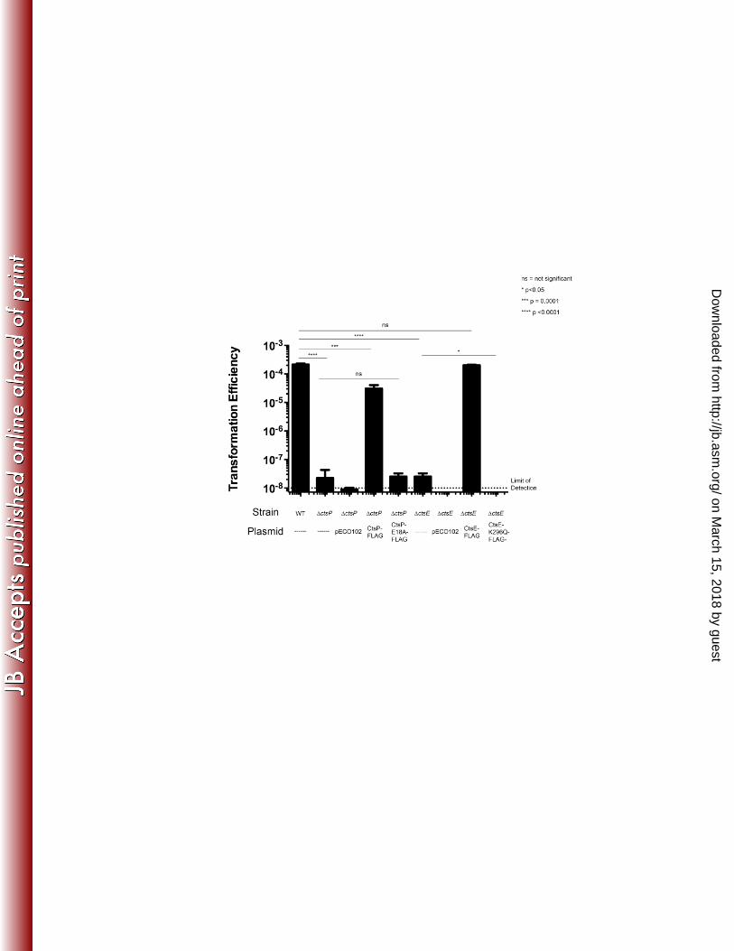
The Walker Boxes of CtsE and CtsP are required for function in vivo 263
The Walker boxes A and B are conserved nucleotide binding motifs important for 264
NTPase function (19), and both motifs are present in CtsP and CtsE. To determine the 265
contribution of CtsP and CtsE for natural transformation of C. jejuni we tested whether Walker 266
box motifs are important in vivo. Carboxyl-terminal FLAG fusions to each protein were 267
expressed C. jejuni ΔctsP and ΔctsE mutants; the fusion proteins complemented the respective 268
mutant alleles to near wild-type levels of transformation, similar to complementation observed 269
with untagged alleles of ctsP or ctsE (Figure 1). (1). 270
on March 15, 2018 by guest
http://jb.asm.org/
Dow
nloaded from
Walker box A (GxxxxGK(S/T), where x is any amino acid) forms a loop structure (the P-271
loop) in which the lysine can directly contact the phosphoryl group of the bound nucleotide (35, 272
36). This Walker box motif is important for the function of many PulE – VirB11 superfamily 273
members and mutation of invariant residues abolishes ATP binding in a number of proteins (17, 274
37-39). The conserved lysine codon (K296) of the Walker box A in ctsE was changed by site 275
directed mutagenesis to encode a glutamine in CtsE-FLAG (CtsE-FLAG K296Q), which was 276
expressed in a ΔctsE strain to determine whether it could restore transformation. While the 277
FLAG-tagged version of CtsE could complement a CtsE deletion mutant, the Walker Box A 278
point mutant in CtsE (CtsE-FLAG K296Q) could not (Figure 1). Immunoblotting confirmed that 279
CtsE-FLAG K296Q was expressed at levels similar to CtsE-FLAG (data not shown). 280
Several alleles of ctsP-FLAG with alterations to the same Walker box A lysine proved 281
unstable (data not shown), preventing functional assessment of Walker box A in CtsP; we thus 282
targeted Walker box B in CtsP for mutagenesis. The conserved sequence of Walker box B is 283
hhhhDExx, where h is a hydrophobic amino acid and x is any amino acid. The coding sequence 284
for the conserved glutamic acid residue (E81) was changed by site-directed mutagenesis to 285
encode an alanine residue in CtsP-FLAG, which was stable (by immunoblotting, data not 286
shown). CtsP-FLAG E81A was unable to complement a ΔctsP mutant (Figure 1). Through 287
these site-directed mutagenesis studies we have determined that intact nucleotide binding motifs 288
are required for the function of both CtsE and CtsP in transformation. 289
290
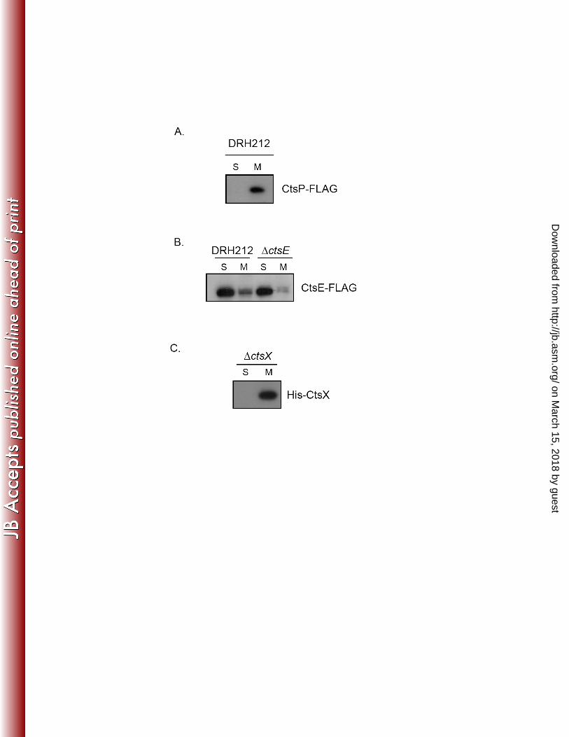
Localization of CtsE and CtsP 291
Members of the PulE – VirB11 superfamily of NTPases are hypothesized to act at the 292
inner membrane of Gram-negative bacteria through association with membrane-localized 293
on March 15, 2018 by guest
http://jb.asm.org/
Dow
nloaded from
proteins within their respective transport systems. To assess localization of CtsP-FLAG and 294
CtsE-FLAG, C. jejuni strains expressing either fusion protein were fractionated into soluble and 295
insoluble fractions as described previously (31). The soluble fraction contains the cytoplasmic 296
and periplasmic contents and the insoluble fraction contains the inner and outer membranes. 297
Activity of isocitrate dehydrogenase, a cytoplasmic enzyme, was monitored to determine the 298
purity of the fractions (31). Immunoblot analysis of these fractions using anti-FLAG monoclonal 299
antibody revealed different subcellular locations for these two putative NTPases. CtsP-FLAG 300
was detected primarily in the membrane fraction, while CtsE-FLAG was located primarily in the 301
soluble fraction (Figures 2A and 2B). To determine whether the cytoplasmic localization of 302
CtsE-FLAG resulted from competition for membrane binding partners with the chromosomally 303
encoded version of CtsE, we assessed localization of CtsE-FLAG in a ΔctsE background. The 304
tagged protein was still observed primarily in the cytoplasmic fraction in the absence of native 305
CtsE (Figure 2B). 306
307
Bacterial two-hybrid screen for identification of Cts interaction partners 308
CtsP localizes to the membrane fraction of C. jejuni, but does not have a predicted 309
hydrophobic region that could serve as a transmembrane domain. We hypothesized that 310
interaction with another protein might be responsible for the membrane localization of CtsP. To 311
explore this, we used a bacterial two-hybrid system based on functional reconstitution of 312
adenylate cyclase activity (32). For two-hybrid analysis, we tested the competence genes ctsE, 313
ctsX, ctsF, ctsD, ctsR, all of which are encoded contiguously with ctsP, and ctsG, ctsW, and 314
dprA, which are unlinked to ctsP. Genes were cloned into both pT18 and pT25, two plasmids 315
that provide different domains of adenylate cyclase. We tested for interactions between all Cts 316
on March 15, 2018 by guest
http://jb.asm.org/
Dow
nloaded from
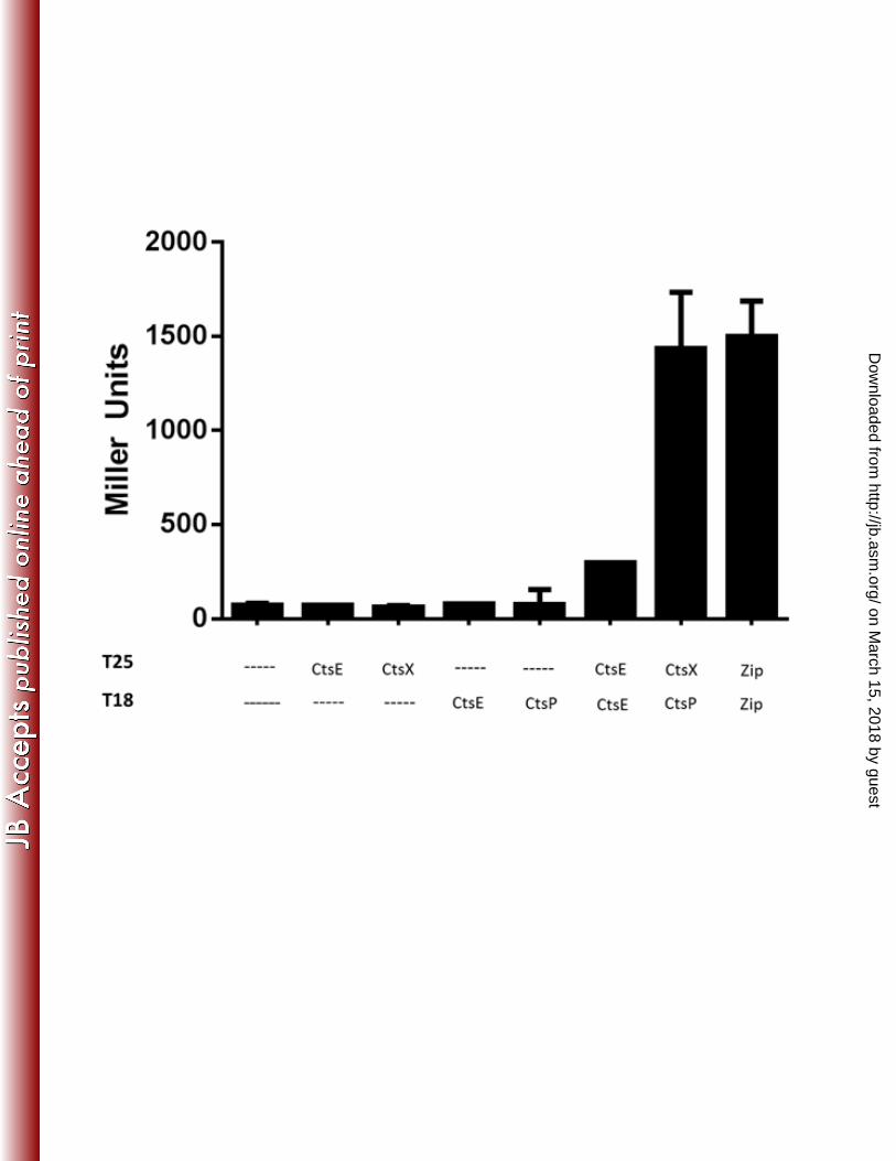
proteins. Two different protein-protein interactions among Cts proteins were identified (Figure 317
3). One was between CtsE-T18 and T25-CtsE, which is consistent with observations that GspE 318
proteins in other type II transport systems form multimers, (26, 40, 41) suggesting that CtsE may 319
form a multi-monomer complex. 320
The other interaction we identified was between CtsP-T18 and T25-CtsX (Figure 3); ctsX 321
is encoded immediately downstream of ctsP (1). Based on Kyte-Doolittle hydropathy analysis, 322
CtsX is predicted to be a membrane protein by virtue of a single putative transmembrane helix 323
from amino acid 21 to 40. To test this experimentally, we expressed a His-CtsX fusion protein in 324
C. jejuni DRH212. Cells were fractionated into soluble and membrane fractions as discussed 325
above for CtsE and CtsP, and fraction purity was assessed by isocitrate dehydrogenase assays 326
(data not shown). His-CtsX was detected by immunoblotting with an anti-His monoclonal 327
antibody. As predicted, the majority of the His-CtsX fusion protein was located in the 328
membrane fraction (Figure 2C). 329
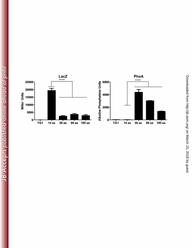
The membrane topology of CtsX was experimentally determined using β-galactosidase 330
and alkaline phosphatase fusions to the CtsX coding sequence. Alkaline phosphatase is active 331
only when transported outside of the cytoplasm (34), whereas β-galactosidase is active in the 332
cytoplasm. Portions of the CtsX coding sequence at different positions on both sides of the 333
predicted transmembrane region of CtsX were fused in frame with lacZ and phoA reporter genes 334
in pTrcLacZ and pTrcPhoA (33) (Figure 4A). 335
The first 14 amino acids of CtsX directed high level β-galactosidase activity but low 336
level alkaline phosphatase activity indicating that the N-terminus of the protein resides in the 337
cytoplasm (Figure 4B and 4C). In contrast, fragments of CtsX including the putative 338
transmembrane domain directed low β-galactosidase levels, but high alkaline phosphatase 339
on March 15, 2018 by guest
http://jb.asm.org/
Dow
nloaded from
activities (Figure 4B and 4C)., consistent with there being a transmembrane domain in the 340
protein as predicted by the Kyte-Doolittle plot. Based on these data, we predict that small 341
portion of the N terminus of CtsX is localized to the cytosol, with the remainder of the protein 342
within the periplasmic space. 343
344
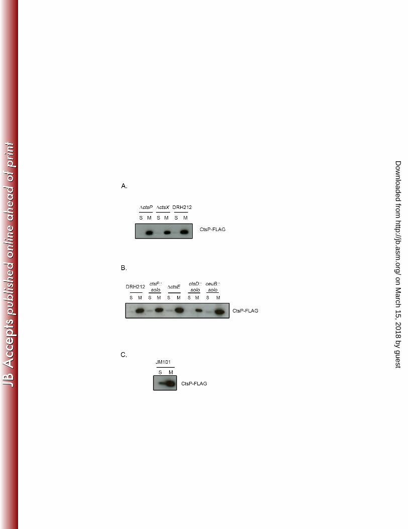
Localization of CtsP in the absence of other Cts proteins 345
Because CtsP lacks an obvious membrane localization signal and interacts with CtsX, 346
which is membrane localized, we hypothesized that membrane localization of CtsP depends on 347
CtsX. However, CtsP-FLAG localized to the membrane fraction in mutant cells lacking ctsX, 348
(Figure 5A), as well as in mutants lacking other transformation genes including ctsF, ctsE, ctsD, 349
ctsR, and ceuB ((1); Figure 5B and data not shown). To test whether membrane localization of 350
CtsP requires any C. jejuni proteins, we expressed CtsP-FLAG in E. coli JM101. Membrane 351
localization was still observed (Figure 5C), suggesting either that membrane localization is an 352
intrinsic feature of CtsP, or that a protein with which it interacts and co-localizes is also present 353
in E. coli. 354
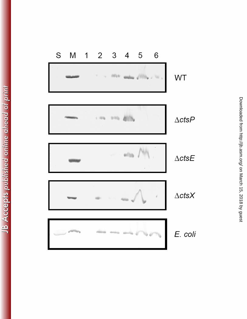
To rule out that the overexpressed protein is simply insoluble and thus pellets with 355
membranes in the fractionation experiments described above, we tested membrane association of 356
CtsP using a membrane flotation assay. Membrane flotation assays utilize a sucrose gradient to 357
separate the membranes from any insoluble proteins that will pellet during ultracentrifugation. 358
Using this assay, we observed that while some CtsP does become insoluble (in fraction 6), the 359
majority is located in membrane containing fractions (fractions 2-5). This is true when CtsP-360
FLAG is expressed in wild type cells, as well as in ΔctsP, ΔctsX, or ΔctsE strains. Even when 361
on March 15, 2018 by guest
http://jb.asm.org/
Dow
nloaded from
CtsP-FLAG is expressed in E. coli, it is primarily localized to the membrane containing fraction 362
and not the insoluble fraction (Figure 6). 363
364
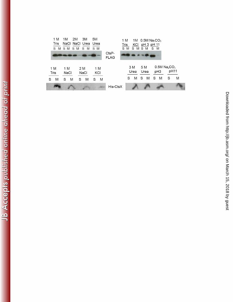
Membrane Extractability of CtsP and CtsX 365
Given its fractionation with the membrane of both C. jejuni and E. coli, we hypothesized 366
that CtsP is a peripheral membrane protein, perhaps associating with the membrane by 367
interacting with the polar head of the phospholipid bilayer. In contrast, CtsX, with its more 368
obvious transmembrane domain and the fusion protein’s ability to localize alkaline phosphatase 369
to the periplasmic space (Figure 4), appears to be an integral membrane protein. To further 370
characterize CtsP and CtsX as integral or peripheral membrane proteins, we analyzed the avidity 371
of their association with C. jejuni membranes using different extraction procedures commonly 372
used to release peripherally associated membrane proteins (42). 373
Membranes of C. jejuni expressing CtsP-FLAG or His-CtsX were treated with high salt 374
(1 & 2 M NaCl, 1 M KCl), urea (3 M, 5 M) or extremes of pH (Na2CO3 pH 3.0, pH 11.0) for 30 375
minutes at 4°C as described in Materials and Methods. Control membranes were treated with 10 376
mM Tris, pH 8.0. CtsP-FLAG was partially extracted from the membranes in high salt (1 & 2 377
M NaCl, 1 M KCl) while CtsX remained predominantly membrane associated (Figure 7). 378
Treatment of the membranes with urea (3 M, 5 M) nearly completely solubilized CtsP-FLAG 379
while His-CtsX was only partially extracted by this treatment (Figure 7). Membrane treatment 380
with 0.5 M Na2CO3 at pH 11 also extracted CtsP-FLAG into the soluble fraction (Figure 7). In 381
contrast, Na2CO3 treatment was not sufficient to affect His-CtsX localization. It remained in the 382
membrane fraction after (Figure 7). These results suggest that while His-CtsX is an integral 383
on March 15, 2018 by guest
http://jb.asm.org/
Dow
nloaded from
membrane protein, the more ready extraction of CtsP-FLAG suggests that it is not integral but 384
peripheral to the membrane, interacting with it perhaps through an as-yet undefined mechanism. 385
386
387
388
DISCUSSION 389
The goal of this study was to characterize two putative NTPases/NTP binding proteins 390
involved in natural transformation of C. jejuni. One of these, CtsE, belongs to a family of well-391
studied NTPases involved in transport of macromolecules in a number of systems including type 392
II secretion, type IV secretion, and type IV pilus biogenesis (15, 16). Members of this family 393
have ATPase activity in vitro (22-26), although the exact ATP-dependent step in transport is 394
unclear. Given the homology of CtsE to other members of this family and the importance of an 395
intact Walker box A for CtsE to function in natural transformation, it is likely that CtsE has 396
ATPase activity critical for its role in transformation. 397
The other putative NTPase/NTP binding protein, CtsP, has little homology to other 398
NTPases and cannot be assigned to a defined family. CtsP Walker box A mutants were all 399
unstable in C. jejuni, but a stable mutant protein lacking Walker box B was unable to correct the 400
transformation defect of a ΔctsP mutant. We suggest that CtsP may bind and hydrolyze 401
nucleotides and that this property is necessary for natural transformation of C. jejuni, although 402
further work is needed to determine whether CtsP has ATPase activity. 403
Transformation in C. jejuni involves a number of proteins with similarity to those 404
required for pilus biogenesis and natural transformation in Neisseria gonorrhoeae and Vibrio 405
cholerae(1, 43-45, 46, 47). In these species, transformation is facilitated by the production of a 406
on March 15, 2018 by guest
http://jb.asm.org/
Dow
nloaded from
pilus that is produced and retracted through the power of two ATPases. Transformation in N. 407
gonorrhoeae requires the activity of two ATPases, PilF and PilT (43, 48) and in V. cholerae it 408
requires PilB and PilT (46). Both N. gonorrhoeae ATPases are members of the PulE – VirB11 409
superfamily (15, 16). PilF is required for elaboration of the pilus while PilT is required for 410
retraction (43, 49). As noted, while CtsE appears to fall into this family, CtsP does not. 411
However, unlike these two systems, C. jejuni has never been shown to produce a pilus (1) and 412
lacks a homolog to the major structural subunits found in these systems, PilE in N. gonorrhoeae 413
and PilA in V. cholerae. Instead, C. jejuni contains a number of genes that may encode pseudo-414
pilins: ctsG, ctsT, and Cjj81176_1096; two of these have been shown to function in 415
transformation (1). Whether a pilus or pseudo-pilus is responsible for DNA binding or uptake 416
across the outer membrane, requiring the actions of CtsE and CtsP, has yet to be determined. In 417
the V. cholerae transformation system, pseudopilins are thought to initiate pilus formation while 418
the major pilin subunit, PilA, is then responsible for the pilus structure (46). Loss of any of these 419
proteins leads to approximately a three-log decrease in transformation efficiency (46). Similarly 420
to the V. cholerae system, the pseudopilins in C. jejuni may be sufficient to induce 421
transformation. A second possibility is that unlike in the V. cholerae system, the pseudopilins 422
might also form enough of a pilus-like structure to facilitate transformation even without the 423
presence of a major pilin subunit. 424
CtsE appears to be predominantly cytosolic in C. jejuni, whereas CtsP is predominantly 425
membrane-associated. A CtsE homologue, ComGA, involved in transformation of Bacillus 426
subtilis localizes to the membrane in that species, where it behaves as a peripheral membrane 427
protein (50). In some other bacteria, CtsE homologues (generally termed GspE proteins) become 428
membrane localized by interacting with an integral membrane protein generally termed GspL 429
on March 15, 2018 by guest
http://jb.asm.org/
Dow
nloaded from
(51, 52) but no GspL homologue was identified in the C. jejuni genome sequence. It is difficult 430
to imagine how CtsE could play a role in the early stages of transformation unless it interacts 431
with proteins at the membrane. In the Vibrio cholerae system, a GFP tagged version of the CtsE 432
homolog, PilB, forms dynamic foci at the membrane that transiently overlap with other 433
components of the Vibrio transformation apparatus (46). Perhaps similarly to the Vibrio system, 434
a transient, or very weak, association of CtsE occurs with the membrane and this could have 435
been disrupted during fractionation; this might account for the small amount of CtsE-FLAG 436
observed in the membrane fraction. 437
Unlike CtsE, CtsP localizes to the membrane in C. jejuni, where it may interact with 438
other components of the transformation machinery. By bacterial two-hybrid analysis we 439
detected interaction between CtsP and CtsX, another protein necessary for efficient C. jejuni 440
transformation. CtsX is encoded immediately downstream of CtsP in a putative operon (1). 441
CtsX is an integral membrane protein with its amino terminus exposed to the cytoplasm, and 442
residues 50 – 195 in the periplasm. Interaction with CtsX is not necessary for CtsP localization 443
to the membrane, nor is interaction with several other components of the transformation 444
machinery. CtsP behaves as a peripheral membrane protein and it may associate directly with 445
the membrane. This possibility is strengthened by the membrane localization of CtsP-FLAG 446
observed during expression in E. coli. If membrane localization required interaction with 447
another protein, it would have to be present in E. coli as well and would likely not be involved in 448
natural transformation, a process that has not been described in E. coli. As noted above, type II 449
secretion system GspE family members, with which CtsE shares homology, associate with the 450
membrane through interaction with another protein, GspL. When these GspE family members 451
are expressed in E. coli cytoplasmic localization is observed (53), which differs from our 452
on March 15, 2018 by guest
http://jb.asm.org/
Dow
nloaded from
observed localization of CtsP to membrane fractions in E. coli. Further study is needed to 453
confirm that CtsP directly associates with the membrane. Furthermore, we were able to replicate 454
the membrane association phenotype using membrane flotation. If this association were an 455
artifact of the initial fractionation procedure, we would expect to see CtsP-FLAG associate 456
predominately with the insoluble aggregate and not float with the membranes. One possibility 457
that has yet to be discounted definitively is that the CtsP-FLAG localization (as well as that of 458
CtsE-FLAG) is due to studying plasmid-encoded proteins, rather than the chromosomally-459
encoded native protein. But the behavior of the plasmid-encoded, FLAG-tagged proteins 460
mitigates this concern, because both CtsE-FLAG and CtsP-FLAG restore transformation 461
efficiency of their respective mutant strains to near wild type levels. 462
CtsX and CtsP represent novel components of the C. jejuni transformation machinery. 463
Homologues of CtsX have not been identified in other transformation systems and BLAST 464
analysis does not provide obvious clues about its function. As it resides largely in the periplasm, 465
perhaps its C-terminus interacts with other components of the transformation machinery in that 466
compartment. CtsX and CtsP are encoded in a putative operon between the type II secretion/type 467
IV pilus biogenesis system homologues ctsD, ctsE, and ctsF. Given the CtsX/CtsP interaction, 468
we hypothesize that these two proteins comprise a component of type II secretion/type IV pilus 469
biogenesis systems specifically involved in C. jejuni competence. Perhaps the interaction is 470
required for assembly or translocation of other transformation components, or it allows assembly 471
of a structure needed specifically for DNA uptake and not pilus biogenesis. 472
473
on March 15, 2018 by guest
http://jb.asm.org/
Dow
nloaded from
Figure Legends 474
Figure 1. Transformation efficiency of C. jejuni CtsE and CtsP mutants. Strains are 475
complemented with pECO102, pECO102 expressing the wild-type coding sequence with a C-476
terminal FLAG tag, or pECO102 expressing the coding sequence with a Walker Box mutation 477
and a C-terminal FLAG tag. The data represent the average of three samples per strain from one 478
experiment. Experiments were repeated at least three times with similar results. Error bars 479
indicate standard deviation. The limit of detection is indicated with a dashed line. Stars indicate p 480
values calculated using one-way ANOVA with Dunnett’s multiple comparison test. 481
482
Figure 2. Localization of CtsE, CtsP, and CtsX fusion proteins. Cellular fractions of DRH212 483
expressing CtsP-FLAG (A), CtsE-FLAG (B), or His-CtsX (C) from pECO102 or ΔctsE 484
expressing CtsE-FLAG (B) were separated by 12% or 15% SDS-PAGE. The presence of the 485
CtsP-FLAG and CtsE-FLAG fusion proteins was detected by immunoblotting using a 486
monoclonal antibody against FLAG. His-CtsX was detected using a monoclonal antibody 487
against His. S, soluble fraction, M, membrane fraction. 488
489
Figure 3. β-galactosidase assays of bacterial two-hybrid system interactions. When fused to 490
proteins that interact, the T25 and T18 fragments of adenylate cyclase are able to associate 491
leading to cAMP synthesis resulting in transcription of lacZ. For a positive control, T25 and T18 492
are fused to a leucine zipper (zip) (32). Activities represent the average of three samples from 493
one experiment which was repeated at least three times with similar results. Error bars indicate 494
standard deviation. 495
496
on March 15, 2018 by guest
http://jb.asm.org/
Dow
nloaded from

497
498
Figure 4. Topology studies of CtsX. Reporter protein activity levels of E. coli TG1 expressing 499
four fragments of ctsX in a translational fusion with lacZ (left) or with phoA (right). Data 500
represent the average of three samples from one experiment that was repeated at least three times 501
with similar results. Error bars indicate standard deviation. Stars represent p-values calculated 502
using Student’s unpaired t test. 503
504
Figure 5. Localization of CtsP in the absence of other Cts proteins. Localization of CtsP-FLAG. 505
(A & B) Cellular fractions of DRH212 or cts mutants or (C) cellular fractions from E. coli 506
JM101 expressing CtsP-FLAG from pECO102 were separated by 12% SDS-PAGE. The 507
presence of the CtsP-FLAG fusion protein was detected by immunoblotting using a monoclonal 508
antibody against the FLAG epitope. S, soluble fraction, M, membrane fraction. 509
510
Figure 6. Membrane flotation of CtsP-FLAG. Soluble and membrane fractions of DRH212, cts 511
mutants (ΔctsP, ΔctsE, and ΔctsX), and E. coli containing the fusion protein CtsP-FLAG 512
expressed on pECO102 were obtained. The membrane fractions were resuspended in a sucrose 513
gradient to separate the membranes from the insoluble material, subjected to ultracentrifugation, 514
and fractions were taken moving sequentially down the gradient (numbered 1-6 where 1 is the 515
top of the gradient and 6 contains the insoluble proteins). These fractions were separated via 12% 516
SDS-PAGE. The presence of the CtsP-FLAG fusion protein was detected by immunoblotting 517
using a monoclonal antibody against the FLAG epitope. S, soluble fraction, M, membrane 518
fraction. 519
on March 15, 2018 by guest
http://jb.asm.org/
Dow
nloaded from
520
Figure 7. Membrane extractability of CtsP-FLAG and His-CtsX. Membranes were incubated 521
with the indicated reagents for 30 min at 4°C and sedimented by ultracentrifugation. Separated 522
fractions were examined by 12% or 15% SDS-PAGE. Fusion proteins were detected by 523
immunoblotting with anti-FLAG M2 antibody for CtsP-FLAG and with an anti-6x His antibody 524
for His-CtsX. S, soluble fraction after washes, M, membrane fraction. 525
526
on March 15, 2018 by guest
http://jb.asm.org/
Dow
nloaded from
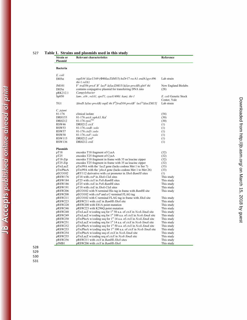
Table 1. Strains and plasmids used in this study 527 Strain or Plasmid
Relevant characteristics Reference
Bacteria E. coli DH5α supE44 ΔlacU169 (Ф80lacZΔM15) hsDr17 recA1 endA1gyrA96
thi-1 relA1 Lab strain
JM101 F’ traD36 proA+B+ lacIq Δ(lacZ)M15/Δ(lax-proAB) glnV thi New England Biolabs DH5α pRK212.1
contains conjugative plasmid for transferring DNA into Campylobacter
(28)
Sp850 lam-, el4-, relA1, spoT1, cyaA1400(::kan), thi-1 E. coli Genetic Stock Center, Yale
TG1 ΔhsdS Δ(lac-proAB) supE thi F0[traD36 proAB+ lacI’qΔlacZM15] Lab strain C. jejuni 81-176 clinical isolate (54) DRH153 81-176 astA::aphA3; Knr (30) DRH212 81-176 rpsLSm (30) RSW46 DRH212 ctsX (1) RSW53 81-176 ceuB::solo (1) RSW57 81-176 ctsD::solo (1) RSW58 81-176 ctsF::solo (1) RSW115 DRH212 ctsP (1) RSW136 DRH212 ctsE (1) Plasmids pT18 encodes T18 fragment of CyaA (32) pT25 encodes T25 fragment of CyaA (32) pT18-Zip encodes T18 fragment in frame with 35 aa leucine zipper (32) pT25-Zip encodes T25 fragment in frame with 35 aa leucine zipper (32) pTrcLacZ pTrc99A with the ‘lacZ gene (lacks codons Met 1 to Ser 7) (33) pTrcPhoA pTrc99A with the ‘phoA gene (lacks codons Met 1 to Met 26) (33) pECO102 pRY112 derivative with cat promoter in XhoI-BamHI sites (1) pRSW176 pT18 with ctsP in XhoI-ClaI sites This study pRSW184 pT25 with ctsX in PstI-BamHI sites This study pRSW186 pT25 with ctsE in PstI-BamHI sites This study pRSW191 pT18 with ctsE in XhoI-ClaI sites This study pRSW206 pRSW208
pECO102 with N terminal His tag in frame with BamHI site pECO102 with ctsP and a C-terminal FLAG tag
This study
pRSW211 pECO102 with C-terminal FLAG tag in frame with XhoI site This study pRSW223 pRSW211 with ctsE in BamHI-XhoI site This study pRSW228 pRSW208 with E81A point mutation This study pRSW246 pRSW223 with K296Q point mutation This study pRSW248 pTrcLacZ w/coding seq for 1st 50 a.a. of ctsX in NcoI-XmaI site This study pRSW249 pTrcLacZ w/coding seq for 1st 100 a.a. of ctsX in NcoI-XmaI site This study pRSW250 pTrcPhoA w/coding seq for 1st 14 a.a. of ctsX in NcoI-XmaI site This study pRSW251 pTrcLacZ w/coding seq for 1st 14 a.a. of ctsX in NcoI-XmaI site This study pRSW252 pTrcPhoA w/coding seq for 1st 50 a.a. of ctsX in NcoI-XmaI site This study pRSW253 pTrcPhoA w/coding seq for 1st 100 a.a. of ctsX in NcoI-XmaI site This study pRSW254 pTrcPhoA w/coding seq of ctsX in NcoI-XmaI site This study pRSW255 pTrcLacZ w/coding seq of ctsX in NcoI-XmaI site This study pRSW256 pRSW211 with ctsX in BamHI-XhoI sites This study pJMB1 pRSW206 with ctsX in BamHI-XhoI This study
528 529 530
531
on March 15, 2018 by guest
http://jb.asm.org/
Dow
nloaded from
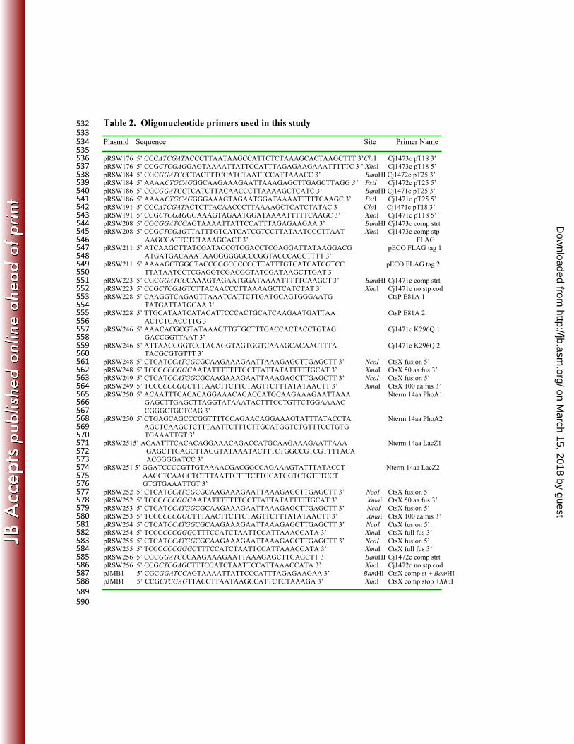
Table 2. Oligonucleotide primers used in this study 532 533 Plasmid Sequence Site Primer Name 534 535 pRSW176 5’ CCCATCGATACCCTTAATAAGCCATTCTCTAAAGCACTAAGCTTT 3’ClaI Cj1473c pT18 3’ 536 pRSW176 5’ CCGCTCGAGGAGTAAAATTATTCCATTTAGAGAAGAAATTTTTC 3 ’ XhoI Cj1473c pT18 5’ 537 pRSW184 5’ CGCGGATCCCTACTTTCCATCTAATTCCATTAAACC 3’ BamHI Cj1472c pT25 3’ 538 pRSW184 5’ AAAACTGCAGGGCAAGAAAGAATTAAAGAGCTTGAGCTTAGG 3’ PstI Cj1472c pT25 5’ 539 pRSW186 5’ CGCGGATCCTCATCTTACAACCCTTAAAAGCTCATC 3’ BamHI Cj1471c pT25 3’ 540 pRSW186 5’ AAAACTGCAGGGGAAAGTAGAATGGATAAAATTTTTCAAGC 3’ PstI Cj1471c pT25 5’ 541 pRSW191 5’ CCCATCGATACTCTTACAACCCTTAAAAGCTCATCTATAC 3 ClaI Cj1471c pT18 3’ 542 pRSW191 5’ CCGCTCGAGGGAAAGTAGAATGGATAAAATTTTTCAAGC 3’ XhoI Cj1471c pT18 5’ 543 pRSW208 5’ CGCGGATCCAGTAAAATTATTCCATTTAGAGAAGAA 3’ BamHI Cj1473c comp strt 544 pRSW208 5’ CCGCTCGAGTTATTTGTCATCATCGTCCTTATAATCCCTTAAT XhoI Cj1473c comp stp 545
AAGCCATTCTCTAAAGCACT 3’ FLAG 546 pRSW211 5’ ATCAAGCTTATCGATACCGTCGACCTCGAGGATTATAAGGACG pECO FLAG tag 1 547
ATGATGACAAATAAGGGGGGCCCGGTACCCAGCTTTT 3’ 548 pRSW211 5’ AAAAGCTGGGTACCGGGCCCCCCTTATTTGTCATCATCGTCC pECO FLAG tag 2 549
TTATAATCCTCGAGGTCGACGGTATCGATAAGCTTGAT 3’ 550 pRSW223 5’ CGCGGATCCCAAAGTAGAATGGATAAAATTTTTCAAGCT 3’ BamHI Cj1471c comp strt 551 pRSW223 5’ CCGCTCGAGTCTTACAACCCTTAAAAGCTCATCTAT 3’ XhoI Cj1471c no stp cod 552 pRSW228 5’ CAAGGTCAGAGTTAAATCATTCTTGATGCAGTGGGAATG CtsP E81A 1 553
TATGATTATGCAA 3’ 554 pRSW228 5’ TTGCATAATCATACATTCCCACTGCATCAAGAATGATTAA CtsP E81A 2 555
ACTCTGACCTTG 3’ 556 pRSW246 5’ AAACACGCGTATAAAGTTGTGCTTTGACCACTACCTGTAG Cj1471c K296Q 1 557
GACCGGTTAAT 3’ 558 pRSW246 5’ ATTAACCGGTCCTACAGGTAGTGGTCAAAGCACAACTTTA Cj1471c K296Q 2 559
TACGCGTGTTT 3’ 560 pRSW248 5’ CTCATCCATGGCGCAAGAAAGAATTAAAGAGCTTGAGCTT 3’ NcoI CtsX fusion 5’ 561 pRSW248 5’ TCCCCCCGGGAATATTTTTTTGCTTATTATATTTTTGCAT 3’ XmaI CtsX 50 aa fus 3’ 562 pRSW249 5’ CTCATCCATGGCGCAAGAAAGAATTAAAGAGCTTGAGCTT 3’ NcoI CtsX fusion 5’ 563 pRSW249 5’ TCCCCCCGGGTTTAACTTCTTCTAGTTCTTTATATAACTT 3’ XmaI CtsX 100 aa fus 3’ 564 pRSW250 5’ ACAATTTCACACAGGAAACAGACCATGCAAGAAAGAATTAAA Nterm 14aa PhoA1 565
GAGCTTGAGCTTAGGTATAAATACTTTCCTGTTCTGGAAAAC 566 CGGGCTGCTCAG 3’ 567
pRSW250 5’ CTGAGCAGCCCGGTTTTCCAGAACAGGAAAGTATTTATACCTA Nterm 14aa PhoA2 568 AGCTCAAGCTCTTTAATTCTTTCTTGCATGGTCTGTTTCCTGTG 569
TGAAATTGT 3’ 570 pRSW2515’ ACAATTTCACACAGGAAACAGACCATGCAAGAAAGAATTAAA Nterm 14aa LacZ1 571
GAGCTTGAGCTTAGGTATAAATACTTTCTGGCCGTCGTTTTACA 572 ACGGGGATCC 3’ 573
pRSW251 5’ GGATCCCCGTTGTAAAACGACGGCCAGAAAGTATTTATACCT Nterm 14aa LacZ2 574 AAGCTCAAGCTCTTTAATTCTTTCTTGCATGGTCTGTTTCCT 575 GTGTGAAATTGT 3’ 576
pRSW252 5’ CTCATCCATGGCGCAAGAAAGAATTAAAGAGCTTGAGCTT 3’ NcoI CtsX fusion 5’ 577 pRSW252 5’ TCCCCCCGGGAATATTTTTTTGCTTATTATATTTTTGCAT 3’ XmaI CtsX 50 aa fus 3’ 578 pRSW253 5’ CTCATCCATGGCGCAAGAAAGAATTAAAGAGCTTGAGCTT 3’ NcoI CtsX fusion 5’ 579 pRSW253 5’ TCCCCCCGGGTTTAACTTCTTCTAGTTCTTTATATAACTT 3’ XmaI CtsX 100 aa fus 3’ 580 pRSW254 5’ CTCATCCATGGCGCAAGAAAGAATTAAAGAGCTTGAGCTT 3’ NcoI CtsX fusion 5’ 581 pRSW254 5’ TCCCCCCGGGCTTTCCATCTAATTCCATTAAACCATA 3’ XmaI CtsX full fus 3’ 582 pRSW255 5’ CTCATCCATGGCGCAAGAAAGAATTAAAGAGCTTGAGCTT 3’ NcoI CtsX fusion 5’ 583 pRSW255 5’ TCCCCCCGGGCTTTCCATCTAATTCCATTAAACCATA 3’ XmaI CtsX full fus 3’ 584 pRSW256 5’ CGCGGATCCCAAGAAAGAATTAAAGAGCTTGAGCTT 3’ BamHI Cj1472c comp strt 585 pRSW256 5’ CCGCTCGAGCTTTCCATCTAATTCCATTAAACCATA 3’ XhoI Cj1472c no stp cod 586 pJMB1 5’ CGCGGATCCAGTAAAATTATTCCCATTTAGAGAAGAA 3’ BamHI CtsX comp st + BamHI 587 pJMB1 5’ CCGCTCGAGTTACCTTAATAAGCCATTCTCTAAAGA 3’ XhoI CtsX comp stop +XhoI 588 589 590
on March 15, 2018 by guest
http://jb.asm.org/
Dow
nloaded from

1. Wiesner RS, Hendrixson DR, DiRita VJ. 2003. Natural transformation of 591 Campylobacter jejuni requires components of a type II secretion system. J Bacteriol 592 185:5408-5418. 593
2. Bereswill S, Kist M. 2003. Recent developments in Campylobacter pathogenesis. 594 Current opinion in infectious diseases 16:487-491. 595
3. Young KT, Davis LM, DiRita VJ. 2007. Campylobacter jejuni: molecular biology 596 and pathogenesis. Nature reviews. Microbiology 5:665-679. 597
4. Wang Y, Taylor DE. 1990. Natural transformation in Campylobacter species. J 598 Bacteriol 172:949-955. 599
5. Wilson DL, Bell JA, Young VB, Wilder SR, Mansfield LS, Linz JE. 2003. Variation 600 of the natural transformation frequency of Campylobacter jejuni in liquid shake 601 culture. Microbiology (Reading, England) 149:3603-3615. 602
6. Dingle KE, Colles FM, Wareing DR, Ure R, Fox AJ, Bolton FE, Bootsma HJ, 603 Willems RJ, Urwin R, Maiden MC. 2001. Multilocus sequence typing system for 604 Campylobacter jejuni. Journal of clinical microbiology 39:14-23. 605
7. Suerbaum S, Lohrengel M, Sonnevend A, Ruberg F, Kist M. 2001. Allelic diversity 606 and recombination in Campylobacter jejuni. Journal of bacteriology 183:2553-2559. 607
8. de Boer P, Wagenaar JA, Achterberg RP, van Putten JP, Schouls LM, Duim B. 608 2002. Generation of Campylobacter jejuni genetic diversity in vivo. Mol Microbiol 609 44:351-359. 610
9. Bacon DJ, Alm RA, Hu L, Hickey TE, Ewing CP, Batchelor RA, Trust TJ, Guerry 611 P. 2002. DNA sequence and mutational analyses of the pVir plasmid of 612 Campylobacter jejuni 81-176. Infection and immunity 70:6242-6250. 613
10. Larsen JC, Szymanski C, Guerry P. 2004. N-linked protein glycosylation is required 614 for full competence in Campylobacter jejuni 81-176. Journal of bacteriology 615 186:6508-6514. 616
11. Takata T, Ando T, Israel DA, Wassenaar TM, Blaser MJ. 2005. Role of dprA in 617 transformation of Campylobacter jejuni. FEMS microbiology letters 252:161-168. 618
12. Jeon B, Zhang Q. 2007. Cj0011c, a periplasmic single- and double-stranded DNA 619 binding protein, contributes to natural transformation in Campylobacter jejuni. J 620 Bacteriol. 621
13. Chen I, Christie PJ, Dubnau D. 2005. The ins and outs of DNA transfer in bacteria. 622 Science (New York, N.Y.) 310:1456-1460. 623
14. Parkhill J, Wren BW, Mungall K, Ketley JM, Churcher C, Basham D, 624 Chillingworth T, Davies RM, Feltwell T, Holroyd S, Jagels K, Karlyshev AV, Moule 625 S, Pallen MJ, Penn CW, Quail MA, Rajandream MA, Rutherford KM, van Vliet 626 AH, Whitehead S, Barrell BG. 2000. The genome sequence of the food-borne 627 pathogen Campylobacter jejuni reveals hypervariable sequences. Nature 403:665-628 668. 629
15. Planet PJ, Kachlany SC, DeSalle R, Figurski DH. 2001. Phylogeny of genes for 630 secretion NTPases: identification of the widespread tadA subfamily and 631 development of a diagnostic key for gene classification. Proceedings of the National 632 Academy of Sciences of the United States of America 98:2503-2508. 633
16. Peabody CR, Chung YJ, Yen MR, Vidal-Ingigliardi D, Pugsley AP, Saier MH, Jr. 634 2003. Type II protein secretion and its relationship to bacterial type IV pili and 635 archaeal flagella. Microbiology (Reading, England) 149:3051-3072. 636
on March 15, 2018 by guest
http://jb.asm.org/
Dow
nloaded from
17. Possot O, Pugsley AP. 1994. Molecular characterization of PulE, a protein required 637 for pullulanase secretion. Molecular microbiology 12:287-299. 638
18. Patel S, Latterich M. 1998. The AAA team: related ATPases with diverse functions. 639 Trends in cell biology 8:65-71. 640
19. Walker JE, Saraste M, Runswick MJ, Gay NJ. 1982. Distantly related sequences in 641 the alpha- and beta-subunits of ATP synthase, myosin, kinases and other ATP-642 requiring enzymes and a common nucleotide binding fold. The EMBO journal 643 1:945-951. 644
20. Possot OM, Letellier L, Pugsley AP. 1997. Energy requirement for pullulanase 645 secretion by the main terminal branch of the general secretory pathway. Molecular 646 microbiology 24:457-464. 647
21. Robien MA, Krumm BE, Sandkvist M, Hol WG. 2003. Crystal structure of the 648 extracellular protein secretion NTPase EpsE of Vibrio cholerae. Journal of 649 molecular biology 333:657-674. 650
22. Rivas S, Bolland S, Cabezon E, Goni FM, de la Cruz F. 1997. TrwD, a protein 651 encoded by the IncW plasmid R388, displays an ATP hydrolase activity essential for 652 bacterial conjugation. The Journal of biological chemistry 272:25583-25590. 653
23. Bhattacharjee MK, Kachlany SC, Fine DH, Figurski DH. 2001. Nonspecific 654 adherence and fibril biogenesis by Actinobacillus actinomycetemcomitans: TadA 655 protein is an ATPase. Journal of bacteriology 183:5927-5936. 656
24. Herdendorf TJ, McCaslin DR, Forest KT. 2002. Aquifex aeolicus PilT, homologue 657 of a surface motility protein, is a thermostable oligomeric NTPase. Journal of 658 bacteriology 184:6465-6471. 659
25. Sexton JA, Pinkner JS, Roth R, Heuser JE, Hultgren SJ, Vogel JP. 2004. The 660 Legionella pneumophila PilT homologue DotB exhibits ATPase activity that is 661 critical for intracellular growth. Journal of bacteriology 186:1658-1666. 662
26. Camberg JL, Sandkvist M. 2005. Molecular analysis of the Vibrio cholerae type II 663 secretion ATPase EpsE. Journal of bacteriology 187:249-256. 664
27. Ogura T, Wilkinson AJ. 2001. AAA+ superfamily ATPases: common structure--665 diverse function. Genes to cells : devoted to molecular & cellular mechanisms 6:575-666 597. 667
28. Figurski DH, Helinski DR. 1979. Replication of an origin-containing derivative of 668 plasmid RK2 dependent on a plasmid function provided in trans. Proceedings of the 669 National Academy of Sciences of the United States of America 76:1648-1652. 670
29. Guerry P, Yao R, Alm RA, Burr DH, Trust TJ. 1994. Systems of experimental 671 genetics for Campylobacter species. Methods in enzymology 235:474-481. 672
30. Hendrixson DR, Akerley BJ, DiRita VJ. 2001. Transposon mutagenesis of 673 Campylobacter jejuni identifies a bipartite energy taxis system required for 674 motility. Mol Microbiol 40:214-224. 675
31. Myers JD, Kelly DJ. 2005. A sulphite respiration system in the chemoheterotrophic 676 human pathogen Campylobacter jejuni. Microbiology (Reading, England) 151:233-677 242. 678
32. Karimova G, Pidoux J, Ullmann A, Ladant D. 1998. A bacterial two-hybrid system 679 based on a reconstituted signal transduction pathway. Proceedings of the National 680 Academy of Sciences of the United States of America 95:5752-5756. 681
on March 15, 2018 by guest
http://jb.asm.org/
Dow
nloaded from
33. Blank TE, Donnenberg MS. 2001. Novel topology of BfpE, a cytoplasmic membrane 682 protein required for type IV fimbrial biogenesis in enteropathogenic Escherichia 683 coli. Journal of bacteriology 183:4435-4450. 684
34. Manoil C. 1991. Analysis of membrane protein topology using alkaline phosphatase 685 and beta-galactosidase gene fusions. Methods in cell biology 34:61-75. 686
35. Fry DC, Kuby SA, Mildvan AS. 1986. ATP-binding site of adenylate kinase: 687 mechanistic implications of its homology with ras-encoded p21, F1-ATPase, and 688 other nucleotide-binding proteins. Proceedings of the National Academy of Sciences 689 of the United States of America 83:907-911. 690
36. Hyde SC, Emsley P, Hartshorn MJ, Mimmack MM, Gileadi U, Pearce SR, 691 Gallagher MP, Gill DR, Hubbard RE, Higgins CF. 1990. Structural model of ATP-692 binding proteins associated with cystic fibrosis, multidrug resistance and bacterial 693 transport. Nature 346:362-365. 694
37. Korangy F, Julin DA. 1992. Alteration by site-directed mutagenesis of the conserved 695 lysine residue in the ATP-binding consensus sequence of the RecD subunit of the 696 Escherichia coli RecBCD enzyme. The Journal of biological chemistry 267:1727-697 1732. 698
38. Turner LR, Lara JC, Nunn DN, Lory S. 1993. Mutations in the consensus ATP-699 binding sites of XcpR and PilB eliminate extracellular protein secretion and pilus 700 biogenesis in Pseudomonas aeruginosa. Journal of bacteriology 175:4962-4969. 701
39. Sandkvist M, Bagdasarian M, Howard SP, DiRita VJ. 1995. Interaction between the 702 autokinase EpsE and EpsL in the cytoplasmic membrane is required for 703 extracellular secretion in Vibrio cholerae. The EMBO journal 14:1664-1673. 704
40. Turner LR, Olson JW, Lory S. 1997. The XcpR protein of Pseudomonas aeruginosa 705 dimerizes via its N-terminus. Molecular microbiology 26:877-887. 706
41. Py B, Loiseau L, Barras F. 1999. Assembly of the type II secretion machinery of 707 Erwinia chrysanthemi: direct interaction and associated conformational change 708 between OutE, the putative ATP-binding component and the membrane protein 709 OutL. Journal of molecular biology 289:659-670. 710
42. Pozidis C, Chalkiadaki A, Gomez-Serrano A, Stahlberg H, Brown I, Tampakaki 711 AP, Lustig A, Sianidis G, Politou AS, Engel A, Panopoulos NJ, Mansfield J, Pugsley 712 AP, Karamanou S, Economou A. 2003. Type III protein translocase: HrcN is a 713 peripheral ATPase that is activated by oligomerization. The Journal of biological 714 chemistry 278:25816-25824. 715
43. Freitag NE, Seifert HS, Koomey M. 1995. Characterization of the pilF-pilD pilus-716 assembly locus of Neisseria gonorrhoeae. Molecular microbiology 16:575-586. 717
44. Tonjum T, Freitag NE, Namork E, Koomey M. 1995. Identification and 718 characterization of pilG, a highly conserved pilus-assembly gene in pathogenic 719 Neisseria. Molecular microbiology 16:451-464. 720
45. Drake SL, Koomey M. 1995. The product of the pilQ gene is essential for the 721 biogenesis of type IV pili in Neisseria gonorrhoeae. Molecular microbiology 18:975-722 986. 723
46. Seitz P, Blokesch M. 2013. DNA-uptake machinery of naturally competent Vibrio 724 cholerae. Proceedings of the National Academy of Sciences of the United States of 725 America 110:17987-17992. 726
on March 15, 2018 by guest
http://jb.asm.org/
Dow
nloaded from
47. Seitz P, Pezeshgi Modarres H, Borgeaud S, Bulushev RD, Steinbock LJ, Radenovic 727 A, Dal Peraro M, Blokesch M. 2014. ComEA is essential for the transfer of external 728 DNA into the periplasm in naturally transformable Vibrio cholerae cells. PLoS 729 genetics 10:e1004066. 730
48. Brossay L, Paradis G, Fox R, Koomey M, Hebert J. 1994. Identification, 731 localization, and distribution of the PilT protein in Neisseria gonorrhoeae. Infection 732 and immunity 62:2302-2308. 733
49. Wolfgang M, Lauer P, Park HS, Brossay L, Hebert J, Koomey M. 1998. PilT 734 mutations lead to simultaneous defects in competence for natural transformation 735 and twitching motility in piliated Neisseria gonorrhoeae. Molecular microbiology 736 29:321-330. 737
50. Chung YS, Dubnau D. 1998. All seven comG open reading frames are required for 738 DNA binding during transformation of competent Bacillus subtilis. Journal of 739 bacteriology 180:41-45. 740
51. Sandkvist M. 2001. Type II secretion and pathogenesis. Infection and immunity 741 69:3523-3535. 742
52. Sandkvist M. 2001. Biology of type II secretion. Molecular microbiology 40:271-283. 743 53. Filloux A. 2004. The underlying mechanisms of type II protein secretion. Biochimica 744
et biophysica acta 1694:163-179. 745 54. Korlath JA, Osterholm MT, Judy LA, Forfang JC, Robinson RA. 1985. A point-746
source outbreak of campylobacteriosis associated with consumption of raw milk. 747 The Journal of infectious diseases 152:592-596. 748
749
Acknowledgements 750
We thank Dr. Jeremy Ellermeir for helpful contributions and discussions. This work was 751
supported in part by NIAID R01AI069383 (to VJD). JMB and RSE were trainees of the 752
University of Michigan Predoctoral Genetics Training Program supported by T32GM07544. 753
on March 15, 2018 by guest
http://jb.asm.org/
Dow
nloaded from


![Campylobacter jejuni routsias [Λειτουργία συμβατότητας]](https://img.pdfslide.net/doc/110x75/61688485d394e9041f70265d/campylobacter-jejuni-routsias-.jpg)