Embed Size (px)
Citation preview

Short sequence-paper
Characterization and regulation of a second gene encoding thioredoxin
from the fission yeast
Yoon-Jong Lee a, Young-Wook Cho a, Daemyung Kim b, Eun-Hee Park c,James A. Fuchs d, Chang-Jin Lim a,*
aDivision of Life Sciences, College of Natural Sciences, Kangwon National University, Chuncheon 200-701, South KoreabDepartment of Genetic Engineering, Chongju University, Chongju 360-764, South Korea
cCollege of Pharmacy, Sookmyung Women’s University, Seoul 140-742, South KoreadDepartment of Biochemistry, Molecular Biology and Biophysics, University of Minnesota, St. Paul, MN 55108, USA
Received 11 September 2001; received in revised form 15 January 2002; accepted 17 January 2002
Abstract
A genomic DNA encoding a second thioredoxin (TRX2) was isolated from the chromosomal DNA of the fission yeast
Schizosaccharomyces pombe. The cloned sequence contains 1823 bp and encodes a protein of 121 amino acids. It has extra N-terminal
17 amino acid residues compared to previously identified thioredoxin (TRX1), which are positively charged and hydrophobic amino acids.
The additional N-terminal region contains a plausible prepeptidase cleavage site, indicating that the TRX2 protein exists in mitochondria. The
cloned TRX2 gene produced functional TRX estimated with insulin reduction assay. The upstream region of the TRX2 gene was fused into the
promoterless h-galactosidase gene of the shuttle vector YEp357R. The 782 bp sequence in the region further upstream of the TRX2 gene was
found to be inhibitory in its expression. Synthesis of h-galactosidase from the fusion plasmid pYFX135-HRL was enhanced by the addition
of aluminum chloride and ferrous chloride, indicating that the TRX2 protein is involved in stress response. D 2002 Elsevier Science B.V. All
rights reserved.
Keywords: Fission yeast; Genomic DNA; Regulation; Stress response; Thioredoxin; Schizosaccharomyces pombe
Thioredoxin (TRX) is a small, ubiquitous and multifunc-
tional protein that has a redox-active disulfide/dithiol within
the conserved active site sequence -Trp-Cys-Gly-Pro-Cys-.
It participates in the reduction of sulfate, methionine sulf-
oxide, and protein disulfide bonds [1]. In Escherichia coli,
TRX functions as an essential subunit of phage T7 DNA
polymerase [2] and is required for the assembly of the fila-
mentous phages M13 and f1 [3]. Furthermore, TRX is an
efficient antioxidant able to reduce hydrogen peroxide [4]
and to scavenge free radicals [5]. Eukaryotic TRX has been
implicated in various physiological functions. It can modu-
late the DNA binding activity of some transcription factors
such as TFIIIC [6], NF-nB [7] and AP-1 [8,9]. TRX was
reported to have preventive capacities towards NO-mediated
cellular injury in monocytic macrophage cells [10], and also
to be a putative oncogene conferring both growth and
survival advantage to tumor cells [11].
There are a few regulatory mechanisms identified in the
expression of TRX genes. Transcription of the Rhodobacter
sphaeroides Y TRX gene is regulated by oxygen tension
[12]. The expression of a human TRX gene is increased by
oxidative agents [13]. This type of regulation supports the
fact that TRX is involved in the regeneration of proteins
inactivated by oxidative stress [14]. Transcript levels of
Dictyostelium TRXs are regulated during the development
cycle, which are low in the growth phase and maximally
high during development [15]. TRX gene expression was
also reported to be transcriptionally up-regulated by retinol
in monkey epithelial cells [16].
More than one TRXs exist in some eukaryotes, such as
yeast [17,18] and rat [19,20]. Mitochondrial TRX has been
identified but its biological functions are not clearly known.
Reported mitochondrial TRXs are found to contain mito-
chondrial translocational signals [18,20]. The first TRX
gene was previously isolated from the fission yeast Schiz-
osaccharomyces pombe [21]. Here, we describe cloning,
characterization, and regulation of genomic DNA encoding
putative mitochondrial TRX from S. pombe.
0167-4781/02/$ - see front matter D 2002 Elsevier Science B.V. All rights reserved.
PII: S0167 -4781 (02 )00246 -4
* Corresponding author. Tel.: +82-33-250-8514; fax: +82-33-242-0459.
E-mail address: [email protected] (C.-J. Lim).
www.bba-direct.com
Biochimica et Biophysica Acta 1575 (2002) 143–147

The chromosomal DNAwas isolated from S. pombe cells
according to the procedure previously described [22]. PCR
was performed as described in the user’s sheet offered by
Roche Molecular Biochemicals. The PCR conditions used
in this study were 94 jC (1 min), 57 jC (1 min) and 72 jC(2 min) for 30 cycles. Thioredoxin-catalyzed reduction of
insulin by dithiothreitol was monitored as a turbidity in-
crease at 650 nm [23]. This method was used to demonstrate
the ability of thioredoxin to function as a protein disulfide
reductase. h-Galactosidase activity in extract was measured
by spectrophotometric method using o-nitrophenyl h-D-galactopyranoside (ONPG) as a substrate [24]. Protein
contents in extracts were measured by the Bradford method
[25] using bovine serum albumin as a standard.
Previously, the first genomic DNA encoding thioredoxin
(later named TRX1) was isolated and characterized from the
fission yeast S. pombe [21]. In addition to the first TRX gene, a
nucleotide sequence homologous to those of TRX genes was
identified on the genomic DNA sequence of S. pombe, which
is known to be similar to higher eukaryotes in aspects of its
physiology and genetics. To understand physiological roles
and regulation of a second thioredoxin gene, the TRX2 gene
was isolated and characterized from S. pombe. Based on the
sequence stored in the GenBank database (AL035085), two
synthetic primers (5V-AACAATT GCTTGCGGATCCC-
Fig. 1. Construction of the three subclones, pRTX602, pUTX118 and
pUTX119, used for sequencing the nucleotide sequence of the insert DNA
contained in the original clone pRTX601.
Fig. 2. The nucleotide sequence and deduced amino acid sequence of S. pombe TRX2 gene. An active site is boxed. A short bar indicates the stop codon.
Y.-J. Lee et al. / Biochimica et Biophysica Acta 1575 (2002) 143–147144
CATCTG-3V and 5V-TTAATCACCTCATAGAATTCCT-
CACGTTGTT AT-3V) were used for PCR amplification by
PyrobestR DNA polymerase. The two primers contain
BamHI and EcoRI restriction sites, respectively, which can
be used for ligation. They were also designed to amplify the
plausible TRX coding region as well as 1090 bp upstream
sequence which should contain a sufficient region for tran-
scriptional regulation. The amplified DNA product was
identified and purified from agarose gel electrophoresis,
and then, completely digested with BamHI and EcoRI. The
BamHI/EcoRI-digested PCR product was ligated into the
BamHI/EcoRI site of the E. coli–yeast shuttle vector
pRS316. The ligation mixture was used to transform the E.
coli strain MV1184 according to the CaCl2 procedure [26].
After isolating plasmid DNA from transformed E. coli cells,
the plasmid DNA was confirmed by restriction mapping as
having the desired size. Plasmid pRTX601 contain a 1823 bp
insert. The PCR product and the insert DNA fragment were
confirmed to have the same size on agarose gel electro-
phoresis (data not shown). To determine the nucleotide
sequence, three subclones were constructed using the unique
HindIII site within the insert of pRTX601. Plasmid pRTX602
contains the BamHI–HindIII fragment, whereas plasmids
pUTX118 and pUTX119 contain the HindIII–EcoRI frag-
ment in vectors pUC118 and pUC119, respectively (Fig. 1).
The original clone and the three subclones were used for
automated sequencing in Bionex Inc., Korea. The nucleotide
sequence of the BamHI–EcoRI insert of the original plasmid
pRTX601was determined by completely overlapping the two
DNA strands. The nucleotide sequence of the S. pombe TRX2
gene was submitted to the GenBank under the accession
number AY034142.
The 1823 bp sequence of the cloned TRX2 gene, shown in
Fig. 2, is identical with the stored GenBank sequence except
for three positions. The coding region contains an intron. The
unique open-reading frame encodes a protein of 121 amino
acids with a calculated mass of 13.7 kDa (Fig. 2). The
determined sequence contains an upstream sequence of
1090 bp and a downstream of 285 bp. Analysis of amino
acid composition indicates that it doesn’t contain histidine
and is rich in alanine, leucine, lysine and serine (Fig. 2). Its
isoelectric point is estimated to be 9.45. By analyzing with
computer programs, it is assumed to have six a-helix regions
and two turns, and to be relatively hydrophobic. The amino
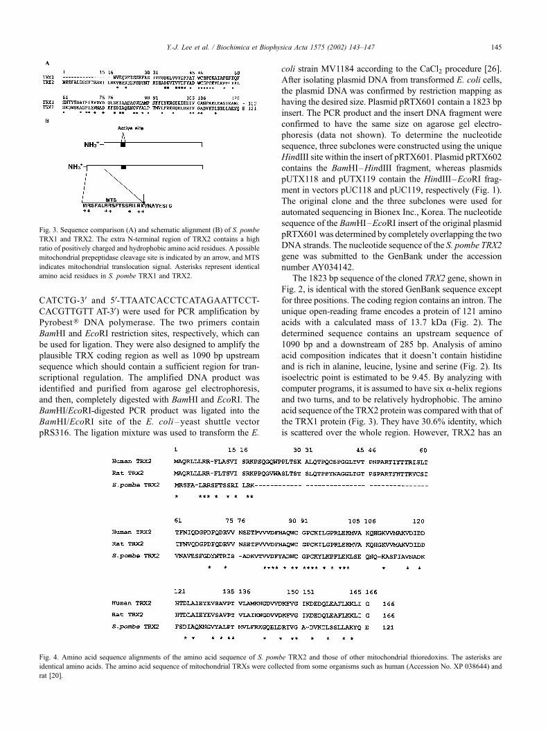
acid sequence of the TRX2 protein was compared with that of
the TRX1 protein (Fig. 3). They have 30.6% identity, which
is scattered over the whole region. However, TRX2 has an
Fig. 3. Sequence comparison (A) and schematic alignment (B) of S. pombe
TRX1 and TRX2. The extra N-terminal region of TRX2 contains a high
ratio of positively charged and hydrophobic amino acid residues. A possible
mitochondrial prepeptidase cleavage site is indicated by an arrow, and MTS
indicates mitochondrial translocation signal. Asterisks represent identical
amino acid residues in S. pombe TRX1 and TRX2.
Fig. 4. Amino acid sequence alignments of the amino acid sequence of S. pombe TRX2 and those of other mitochondrial thioredoxins. The asterisks are
identical amino acids. The amino acid sequence of mitochondrial TRXs were collected from some organisms such as human (Accession No. XP 038644) and
rat [20].
Y.-J. Lee et al. / Biochimica et Biophysica Acta 1575 (2002) 143–147 145

extra N-terminal region, which does not exist in the TRX1
protein (Fig. 3A). The N-terminal region of the TRX2 has a
high content of positively charged and hydrophobic amino
acid residues (Fig. 3B). A possible mitochondrial prepepti-
dase cleavage site is in the N-terminal region (Fig. 3B). These
support that the TRX2 protein exists in the mitochondria. A
sequence alignment of the S. pombe TRX2 with mitochon-
drial TRXs from human and rat is shown in Fig. 4. The amino
acid sequence of the S. pombe TRX2 has 35.5% and 34.7%
homologies with those of human and rat counterparts,
respectively. All three TRXs share very similar mitochondrial
translocational signals (Fig. 4). However, the S. pombe TRX2
appears to be very dissimilar with the corresponding proteins
of the budding yeast S. cerevisiae (data not shown). It might
give one evidence that S. pombe is closer to mammalian cells
than S. cerevisiae.
To examine whether the cloned S. pombe TRX2 gene
produces a functional TRX, plasmid pRTX601 was intro-
duced into the wild-type S. pombe KP1. Then, it was grown
up to mid-exponential phase, and harvested. Cell extract
was used for insulin reduction assay. The S. pombe cells
harboring pRTX601 showed higher specific activity com-
pared with that of vector alone (Fig. 5). The HindIII/EcoRI
insert fragment of pRTX601 was transferred into the shuttle
vector pYES2 to generate the plasmid pYEX-HRL. The S.
pombe cells harboring pYEX-HRL also gave higher specific
activity (data not shown). The assay results with the two
plasmid constructs definitely support that the cloned TRX2
gene is functional in S. pombe cells.
Fig. 5. Expression of the cloned TRX2 gene in S. pombe with insulin
reduction assay. The experiment was carried out as described in the main
text. Absorbance at 650 nm was measured as an indicator of turbidity
produced from the reduction of insulin. Specific activity is represented as
DOD650/min/mg protein.
Fig. 6. Construction of TRX2– lacZ fusions and synthesis in S. pombe of h-galactosidase from the fusion plasmids. The fusion plasmid pYFX135–
HRL lacks 782 bp BamHI/HindIII fragment of the insert DNA contained in
the fusion plasmid pYFX135. Plasmid YEp357R carries 2 A origin and
promoterless h-galactosidase gene. The specific activity is expressed as
DOD420/min/Ag protein.
Fig. 7. Effect of aluminum chloride (A) and ferrous chloride (B) on the
synthesis of h-galactosidase from the fusion plasmid pYFX135–HRL in
S. pombe cells. The yeast cells harboring the fusion plasmid were grown
in minimal medium, and split at the early exponential phase. Aluminum
chloride or ferrous chloride was added to individual culture flask and con-
tinued to be shaken. The h-galactosidase activity was determined at 25 jCby the spectrophotometric assay using ONPG as a substrate. The specific
activity is expressed in DOD420/min/Ag protein.
Y.-J. Lee et al. / Biochimica et Biophysica Acta 1575 (2002) 143–147146

Considering the multiple functions of TRX in living cells,
the regulation of TRX gene may play an important role in its
functioning. To independently monitor the expression of the
S. pombe TRX2 genomic DNA, the upstream region of
pRTX601 was fused into the promoterless h-galactosidasegene of the fusion vector YEp357R. Before the ligation, the
appropriate upstream region was amplified by PCR. After
transformation into E. coli strain MV1184, the desired sub-
clone was confirmed by blue colony formation and restric-
tion mapping. The resultant fusion plasmids are pYFX135
and pYFX135-HRL. These plasmids do not contain original
N-terminal region of the TRX2 protein with the exception of
methionine. Plasmid pYFX135 contains 1090 bp BamHI/
EcoRI upstream region, whereas plasmid pYFX135-HRL
contains 308 bp HindIII/EcoRI upstream region. The S.
pombe cells harboring the two fusion plasmids were found
to contain h-galactosidase activities (Fig. 6). The S. pombe
cells containing plasmid pYFX135-HRL contains higher h-galactosidase activities. This strongly suggests that 782 bp
BamHI/HindIII upstream region is inhibitory in the expres-
sion of the TRX2 gene. The 782 bp upstream region is found
to be very rich in A/T pairs (72.0%).
According to the growth curve of the S. pombe cells
harboring plasmid pYFX135-HRL, the synthesis of h-gal-actosidase was determined. h-Galactosidase activity was
higher in the exponential phase than in the stationary phase
(data not shown). This indicates that the TRX2 gene is
expressed in a higher degree at the exponential phase. The
effect of the addition of aluminum chloride (5 mM) was
examined using exponential S. pombe cells (Fig. 7A). Its
addition significantly increased the synthesis of h-galactosi-dase. Addition of ferrous chloride to the culture also induced
h-galactosidase from the fusion plasmid (Fig. 7B). These
suggest that the TRX2 gene is involved in the response to
oxidative stress. The precise patterns of regulation of the
TRX2 gene should be approached further, which would give
important information on its physiological roles in mitochon-
dria.
Acknowledgements
This work was supported by a grant (No. R01 2000 00133)
from the Basic Research Program of the Korea Science of
Engineering Foundation.
References
[1] E.S. Arner, A. Holmgren, Eur. J. Biochem. 267 (2000) 6102–6109.
[2] D.F. Mark, C.C. Richardson, Proc. Natl. Acad. Sci. U. S. A. 73 (1976)
780–784.
[3] C.-J. Lim, B. Haller, J.A. Fuchs, J. Bacteriol. 161 (1985) 799–802.
[4] M. Russel, P. Model, Proc. Natl. Acad. Sci. U. S. A. 82 (1985) 29–33.
[5] K.U. Schallreuter, J.M. Wood, Biochem. Biophys. Res. Commun. 136
(1986) 630–637.
[6] J.A. Cromlish, R.G. Roeder, J. Biol. Chem. 264 (1989) 18100–
18109.
[7] J.R. Mattews, N. Wakasugi, J.L. Virelizier, J. Yodoi, R.T. Hay, Nu-
cleic Acids Res. 20 (1992) 3821–3830.
[8] C. Abate, L. Patel, F. Rauscher, T. Curran, Science 249 (1990) 1157–
1161.
[9] K. Hirota, M. Matsui, S. Iwata, A. Nishiyama, K. Mori, J. Yodoi,
Proc. Natl. Acad. Sci. U. S. A. 94 (1997) 3633–3638.
[10] P.J. Ferret, E. Soum, O. Negre, E.E. Wollman, D. Fradelizi, Biochem.
J. 346 (2000) 759–765.
[11] T.M. Grogan, C. Fenoglio-Prieser, R. Zeheb, W. Bellamy, Y. Frutiger,
E. Vela, G. Stemmerman, J. MacDonald, L. Richter, A. Gallegos, G.
Powis, Hum. Pathol. 31 (2000) 475–481.
[12] C. Pasternak, K. Assemat, A.M. Breton, J.D. Clement-Metral, G.
Klug, Mol. Gen. Genet. 250 (1996) 189–196.
[13] Y. Taniguchi, Y. Taniguchi-Ueda, K. Mori, J. Yodoi, Nucleic Acids
Res. 24 (1996) 2746–2752.
[14] M.R. Fernando, H. Nanri, S. Yoshitage, K. Nagata-Kuno, S. Minaka-
mi, Eur. J. Biochem. 209 (1992) 917–922.
[15] B. Wetterrauer, J.P. Jacquot, M. Veron, J. Biol. Chem. 267 (1992)
9895–9904.
[16] G. An, R. Wu, Biochem. Biophys. Res. Commun. 183 (1992) 170–
175.
[17] Z.R. Gan, J. Biol. Chem. 266 (1991) 1692–1696.
[18] J.R. Pedrajas, E. Kosmidou, A. Miranda-Vizuete, J.-A. Gustafsson,
A.P.H. Wright, G. Spyrou, J. Biol. Chem. 274 (1999) 6366–6373.
[19] K.F. Tonissen, A.J. Robins, J.R. Wells, Nucleic Acids Res. 17 (1989)
3973.
[20] G. Spyrou, E. Enmark, A. Miranda-Vizuete, J.-A. Gustafsson, J. Biol.
Chem. 272 (1997) 2936–2941.
[21] Y.-W. Cho, Y.H. Shin, Y.-T. Kim, H.-G. Kim, Y.-J. Lee, E.-H. Park,
J.A. Fuchs, C.-J. Lim, Biochim. Biophys. Acta 1518 (2001) 194–199.
[22] C.S. Hoffman, F. Winston, Gene 57 (1987) 267–272.
[23] A. Holmgren, J. Biol. Chem. 264 (1979) 13963–13966.
[24] L. Guarente, Methods Enzymol. 101 (1983) 181–191.
[25] M.M. Bradford, Anal. Biochem. 72 (1976) 248–254.
[26] J. Sambrook, E.F. Fritsch, T. Maniatis, Molecular Cloning: A Labo-
ratory Manual, 2nd edn., Cold Spring Harbor Laboratory Press, Cold
Spring Harbor, NY (1989).
Y.-J. Lee et al. / Biochimica et Biophysica Acta 1575 (2002) 143–147 147