Embed Size (px)
Citation preview

1981 J STAPH PROTEIN A BINDING HUMAN y3 MYELOMA PROTEIN 923
43. Natvig. J. 8.. and H. G. Kunkel. 1974. A hybrid lgG4-lgG2 immunoglobulin. Scand. J. Immunol. 1 :418.
44. Kunkel. H. G., J. B. Natvig. and F. G. Joslin. 1969. The "Lepore" type of J. Immunol. 1 12:1277.
45. Werner, B. C., and A. G. Steinberg. 1974. Structural studies of a human hybrid y-globulin. Proc. Natl. Acad. Sci. 62:144.
46. Michaelsen. T. E., E. C. Franklin, and B. Frangione. 1977. Primary structure hybrid immunoglobulin. Immunogenetics 3:254.
of the "hinge" region of human lgG3: probable quadruplication of a 15 amino acid residue-basic unit, J. Biol. Chem. 252:883.
47. Ponstingl. H.. and N. Hilschmann. 1976. Zur strukturregel der antikorper. Die primarstruktur eiens monoklonalen IgGl-immunoglobulins (Myelompro- tein Nie). 111. Hoppe-Seylers Z. Physiol. Chem. 357:1571.
48. Frangione. 6.. E. Rosenwasser, F. Prelli. and E. C. Franklin. 1980. The
chain disease protein Wis. Biochemistry 19:4304. primary structure of a human y3 immunoglobulin deletion mutant-y3 heavy
49. Prahl. J. 1967. The C terminal sequence of the heavy chains of human immunoglobulin G myeloma proteins of different isotypes and allotypes.
0022-1 767/61/1273-0923802.00/0 THE JOURNAL OF IMMUNOLOGY
Copyright 0 1981 by The American Association of Immunologists
50. Forsgren, A,. and J. Sjoquist. 1966. Protein A from Staphylococcus aureus. Biochem. J. 1051 01 9.
51. Deisenhofer. J.. T. A. Jones, and A. Haber. 1978. Crystallization, crystal Pseudo-immune reaction with human y-globulin. J. Immunol. 97322.
structure analysis, and atomic model of the complex formed by a human Fc fragment and fragment B of protein A from Staphylococcus aureus. Hoppe-
52. Miyata, T.. T. Yasunaga. Y. Yamawaki-Kataoka. M. Obata. and T. Honjo. Seylers 2. Physiol. Chem. 359:975.
y2b chain genes and the hypothesis of intervening sequence-mediated 1980. Nucleotide sequence divergence of mouse immunoglobulin yl and
53. Ito. S.. T. Miyazaki. and H. Matsumoto. 1980. Interaction between normal domain transfer. Proc. Natl. Acad. Sci. 77:2143.
human lgG3 carrying Gm (b', b3), (g), (s. t) and Protein A-Sepharose CL-46. Proc. Japan Academy 56:226.
54. van Loghem. E., and B. G. Grobbelaar. 1971. A new genetic marker of human lgG3 immunoglobulins. Vox Sang. 21 :405.
55. Steinberg. A. G. 1977. A human antibody to Gm (26): an antigen usually present on the y-3 chain of IgG when Gm (1 5) is absent. Vox Sang. 33:266.
Vol. 127. No. 3. September 1981 Pnnted In U S A
CHARACTERIZATION OF AN ANTI-H-2 MONOCLONAL ANTIBODY AND ITS USE IN LARGE-SCALE ANTIGEN PURIFICATION'
KATHRYN C. STALLCUP,* TIMOTHY A. SPRINGER, AND MATTHEW F. MESCHER
From the Department of Pathology, Harvard Medical School. Boston MA 02 1 15
A rat anti-mouse monoclonal antibody (MAb), M1/42, has been found to react with H-2 antigens from cells of the a, b, d, j, k, s, and u haplotypes (all haplotypes tested). This antibody, when bound to cells and reacted with FITC-conjugated anti-rat lg, could be used to quan- titate H-2 expression on several cell types. The antibody was also useful in comparing the H-2 products precipi- tated from a variety of haplotypes. M1/42-coupled Seph- arose-4B beads were used to purify H-2d antigens by affinity chromatography. Pure H-2 molecules eluted from the column in 0.5% DOC, 0.65 M NaCI, 20 mM Tris, pH 8.0, yielding 110 to 180 pg H-2d/10'0 P815 tumor cells. This antibody, when used in series with H-2Kk-specific MAb 114.1, allowed purification of Dk and Dd from RDM- 4 and YAC cells, respectively. H-2d purified by column chromatography on M1/42 was found to be serologically and biologically active, as determined by MAb rebinding, inhibition of cell lysis by alloantisera plus complement, and ability to stimulate alloreactive CTL. This antibody and the described protocols should be useful in the preparation of relatively large quantities of a number of H-2 antigens.
Cell surface antigens encoded by the major histocompatibility complex (MHO3 play important roles in many immune system
Accepted for publication May 14, 1981. Received for publication February 1 1, 1981.
The costs of publication of this article were defrayed in part by the payment of page charges. This article must therefore be hereby marked advertisement in accordance with 18 U.S.C. Section 1734 solely to indicate this fact.
' This work was supported by National Institutes of Health Grant CA-14732. * Recipient of an American Cancer Society Postdoctoral Fellowship, PF-1765.
Abbreviations used in this paper: MAb. monoclonal antibody; CTL. cytotoxic T lymphocyte(s); Triton/Tris buffer. 0.5% Triton X100. 20 mM Tris. DH 8.0: PMSF, phenylmethylsulfonylfluoride; DOC, deoxycholate; NP40, Nonibit P-401 DOC/0.65 M NaCl buffer, 0.5% DOC, 20 mM Tris. pH 8. 0.65 M NaCI; PBES. molecular mass.
phenomena. The glycoproteins coded for by the murine H-2K. H- 2D, and H-2L genes are involved in allograft rejection and are recognized by the cytotoxic T lymphocytes (CTL) that lyse alloge- neic, virus-infected, chemically modified, or transformed cells (1, 2). The polymorphism of the H-2 antigens and their importance in various immune responses have led to a great deal of interest in the structure and function of these molecules.
Murine H-2K, -D, and -L antigens, and the analogous human HLA-A, -8, and -C antigens, are 2-chain structures formed by noncovalent interactions between a heavy chain (m.w. approxi- mately 44-50,000: the MHC gene product), and ,&-microglobulin (rn.w. approximately 13,000) (3, 4). The heavy chain spans the membrane bilayer and is glycosylated (3-5). The primary structure of the polymorphic heavy chains has been studied using a number of methods. In the case of HLA-A, -B, and -C. conventional methods of protein biochemistry (4, 6) have been used to study the relatively large quantities of antigen that can be purified from Epstein-Barr virus-transformed human cell lines (7).
Large quantities of purified H-2 antigens have not previously been available, and most studies of their structure have been done using small amounts of radioactively labeled protein (8, 9). Al- though much structural information has been gained by this ap- proach, further structural, and particularly functional, studies would be facilitated by the availability of large amounts of pure H- 2. There have been several reports of purification of murine MHC antigens by conventional methods, but the protocols are lengthy and give relatively low yields (1 0-1 2). Herrmann and Mescher (1 3) have described a rapid, efficient procedure for affinity column purification of the H-2Kh antigen using a monoclonal antibody (MAb) specific for H-2Kh (1 4). A similar procedure for purification of HLA antigens using MAb has been described by Parham (1 5).
This report describes a new monoclonal antibody, M1/42, which binds to the H-2 antigens of a variety of haplotypes. No allogeneic specificity has been detected in the strains tested. The MAb binds
phosphate-buffered Earle's balanced salts solution; LPO. lactoperoxidase: FACS. fluorescence-activated cell sorter; FITC. fluorescein isothiocyanate; MHC, major histocompatibility complex; OVA, ovalbumin; Ribo A. ribonuclease A; M,. relative

924 K. C. STALLCUP, T. A. SPRINGER, AND M. F. MESCHER [VOL. 127
tightly enough to allow its use for affinity column purification of H- 2, yet the antigens can be eluted under mild conditions. Using this antibody, we have been able to rapidly isolate relatively large quantities of serologically and biologically active H-2K and D antigens from several haplotypes. These results, together with those previously reported (1 4-1 6), demonstrate that MAb against MHC antigens can be useful as affinity purification reagents.
MATERIALS AND METHODS
Laboratory (Bar Harbor, ME) with the exceptions noted below. YAC lym- Mice and tumor cell lines. All mice were purchased from The Jackson
phoma (H-2"). EL-4 lymphoma (H-Zb) , RDM-4 (H-2k) lymphoma, and P815
6 x A/J)f, (66 X A), AKR. and (BALB/c x DBA/Z)F, (CD2F1)(Cumberland mastocytoma (H-2") cells were maintained as ascites in C57BL/6. (C57BL/
View Farms, Clinton, TN) mice, respectively. I/st (H-2') mice and 1.29 lymphoma cells were generous gifts from Dr. Nobuhiko Tada, Memorial Sloan-Kettering Cancer Center, New York, NY. MI /42 monoclonal antibody. The derivation of M1/42 MAb has previ-
ously been described (1 7). It was 1 of 12 rat anti-mouse spleen cell MAb obtained after immunization of a DA rat to 61 0 spleen cells and fusion with the mouse myeloma line NSI. The hybridoma line has been stabilized by 2 reclonings (M1/42.3.9.8). and secretes an IgG2a HLK antibody (containing specific H and L and myeloma K chains) into tissue culture medium to a concentration of about 100pg/ml. This hybridoma cell line is available from the Sak Institute Cell Distribution Center, North Torrey Pines Road, La Jolla. CA.
M1/42 antibody was purified from culture supernatant by ammonium sulfate precipitation and Sephadex G-200 (Pharmacia. Piscataway. NJ) chromatography. The purified antibody was coupled to cyanogen bromide- activated Sepharose 48 (Pharmacia) at 2.5 to 3 mg/ml of wet gel.
Surface labeling with '251. Lactoperoxidase-catalyzed iodination was used to label cell surface proteins. Iodination was done at 25°C using 0.2 mCi of carrier-free lZ5l (New England Nuclear, Boston. MA) and 10 pg of lactoper- oxidase (Calbiochem, San Diego. CA) per 5 x l o 6 cells. The labeling procedure was as described (18) except that the final 2 post-iodination washes were done in phosphate-buffered Earle's balanced salts solution (PBES. GIBCO. Grand Island, NY) or medium RPMl 1640 (GIBCO) medium containing 10% fetal calf serum. Incorporation of label was 1 to 4 cpm/cell.
Immunoprecipitation of H-2 antigens. Iodinated cells were lysed in 0.5% Triton X-1 00, 20 mM Tris. pH 8.0 (Triton/Tris buffer) containing 1 to 2 mM phenylmethylsulfonylfluoride (PMSF) at 5 x lo7 cells/ml. Nuclei were removed by centrifugaton, and M1/42-Sepharose beads were added to the supernatant. Incubation was for 1 hr at 4°C with shaking. The beads were then washed 4 times with 0.5 ml Triton/Tris buffer. Precipitation with 11- 4.1 was as described above, except that after 1 hr the soluble antigen- antibody complexes were precipitated by the addition of protein-A-Sepha- rose beads (Pharmacia) followed by incubation (with shaking) for another hour. Material bound to the antibodies was eluted with the sample buffer described by Laemmli (1 9).
Detergent solubilization of antigen. When whole cell lysates were used,
mM PMSF (all reagents from Sigma Chemical Co.. St. Louis, MO). After 10 cells were lysed at 2 X l o7 cells/ml in Triton/Tris buffer containing 1 to 2
min on ice, nuclei were pelleted at 3600 x G for 10 min and the resulting supernatant was cleared of detergent insoluble material by centrifugation for 45 min at 100,000 x G. The resulting high speed supernatant was loaded onto affinity columns.
When necessary, cell fractionation was done using the protocol de- scribed by Lemonnier et al. (20), and is shown schematically in Figure 3. H-2 antigens could be recovered from both the low-speed pellet and high- speed pellet (see Fig. 3) by solubilization with nonionic detergent. The low- speed pellet, consisting of nuclei and membrane fragments, was solubilized in Triton/Tris buffer containing 1 to 2 mM PMSF for 10 min on ice. Nuclei and detergent insoluble material were pelleted as above, and the resulting high speed supernatant was loaded on to affinity columns. The crude membrane pellet (see Fig. 3) was solubilized in 1 .O% Triton X-100, 20 mM Tris. pH 8.0, 1 to 2 mM PMSF at a detergent:protein ratio of 5 1 . Detergent- insoluble material was pelleted as above, and the supernatant was loaded onto affinity columns.
Affinity chromatography. Detergent lysate or detergent solubilized pro- teins (containing a small amount of '251-labeled surface protein) were passed through a column of M1/42-Sepharose (0.5 ml to 0.9 ml). In most cases, the starting material was first passed through a pre-column of Sepharose 46 (1 ml). The M1/42 column was then extensively washed with Triton/ Tris buffer, usually 25 to 30 column volumes, until '251 radioactivity reached
cholate (DOC) in 20 mM Tris. pH 8. with 0.65 M NaCl (DOC/0.65 M NaCl background levels. Elution of bound antigen was done using 0.5% deoxy-
buffer) unless otherwise indicated. Fractions of about 0.9 ml were collected, and the lZ5l radioactivity was measured. Peak fractions were pooled, sam-
(21), and the remaining ti-2 antigens were frozen until further use. Samples ples were taken for protein determination by the method of Lowry et a/.
containing 10 to 20 pg of protein were precipitated with 6 vol of cold acetone for analysis by sodium dodecyl sulfate-polyacrylamide gel electro- phoresis (SDS-PAGE).
Separation of H-2 heavy chain and Pz-microglobulin. '251-labeled H-2Kh was purified by affinity chromatography on MAb 1 1-4.1 (1 3). The sample (containing 0.3 to 1 .O X 10' cpm of lZ5l) was precipitated with 6 vol of cold acetone, resuspended in approximately 2 ml 6 M guanidine, and incubated for 20 to 30 min at room temperature. Heavy chain and P?-rnicroglobulin were then separated by chromatography on a 1 x 120 cm Biogel A 0.5M (BioRad. Richmond, CA) column in 6 M guanidine, 20 mM Tris, pH 8. Fractions containing peaks of radioactivity were pooled, and samples con- taining either Pz-microglobulin. heavy chain, or a mixture of heavy chain and fln-microglobulin 12:l heavy:light) were dialyzed extensively against 20 mM Tris, pH 8.0, to remove guanidine. After dialysis, Nonidet P-40 (NP40; Particle Data Labs, Evansville, IL) was added to a final concentration of 0.5% and the samples were loaded directly onto M1/42 columns to deter- mine binding.
CTL stimulation using H-2-containing liposomes. Liposomes containing
pg H-2" in DOC/0.65 M NaCl buffer was mixed with 4.6 nmol lipid (chlo- purified H-2Kh or H-2" proteins were prepared as described (22). Briefly, 1
roform:methanol extracted from P815 cells) and 2.8 X 10' cell equivalents of detergent insoluble matrix (from P815 cells) (23). Detergent was removed by dialysis against Tris-buttered saline, pH 8. Responder spleen cells were from CDPF, mice previously primed with YAC cells or C57BL/6 mice primed with P815 cells. CTL response was measured 4 to 5 days later using a standard 5'Cr release assay with P815 (H-2d) or RDM-4 (H-2h) tumor cell targets.
Cytotoxicity assay. M I /42-column purified H-2" antigens were dialyzed against PBS to remove detergent. Allogeneic activity was assayed by adding 2-fold dilutions of pure antigens to a standard cytotoxicity-inhibition assay using 5'Cr-labeled spleen cell targets and conventional alloantisera.
SDS-PAGE. Electrophoresis, in the presence of SDS andp-mercaptoeth- anol. was done by the method of Laemmli (1 9). The running gel consisted of a gradient of 5 to 15% polyacrylamide (BioRad). Gels were stained with Coomassie Blue, and autoradiography of dried gels was done using Kodak XR-5 x-ray film.
RESULTS
Cell binding of M7 /42. The rat MAb M i /42 was raised by immunizing rats with spleen cells from 61 0 (H-2b) mice. Screening of M1/42 reactivity by cell-binding assay (Table I) indicated that the antibody had no identifiable strain or haplotype specificity. In
TABLE I Specificity screening of MAb M1/42
Haplotype Straln/Cell Type Binding' MHC Pre-
cip.'
a
b
d
h2
i5
i
k
S
U
BIO.A Y AC
C57/LJ C57BL/10 C57BL/6 B10 EL-4
LG/J BlO.D2 CDPF, P815
BIO.A (2R)
B1 0.A (5R)
I/St 1.29
B1O.Br AKR RDM-4
BIO.S
PL/ J
s.c.c lymphoma
S.C.
S.C. S.C. S.C. lymphoma
S.C. S.C. S.C. mastocytoma
S.C.
S.C.
S.C.
lymphoma
S.C. S.C. lymphoma
S.C.
S.C.
ND + ND
+ ND
+ (FACS) ND +
+ ND + (FACS) + + ND + (FACS) ND ND +
+
+
+ (FAGS) + + ND
+ ND
ND + ND + ND + ND -k + (FACS) + ND + + ND
Binding was measured by using M1/42 MAb followed by either or FITC- labeled rabbit F(ab% anti-rat IgG (27). Radioactive binding or fluorescent inten- sity of x 2 or more over background was taken as positive.
m.w. of 44,000 to 50,000 and 13.000 from 'Z51-labeled cell lysates. A positive indicates that M1/42-Sepharose Precipitated polypeptides with
s.c., spleen cells.
19811 PURIFICATION OF H-2 ANTIGENS 925
addition, M1/42 did not distinguish normal spleen cells from transformed cells.
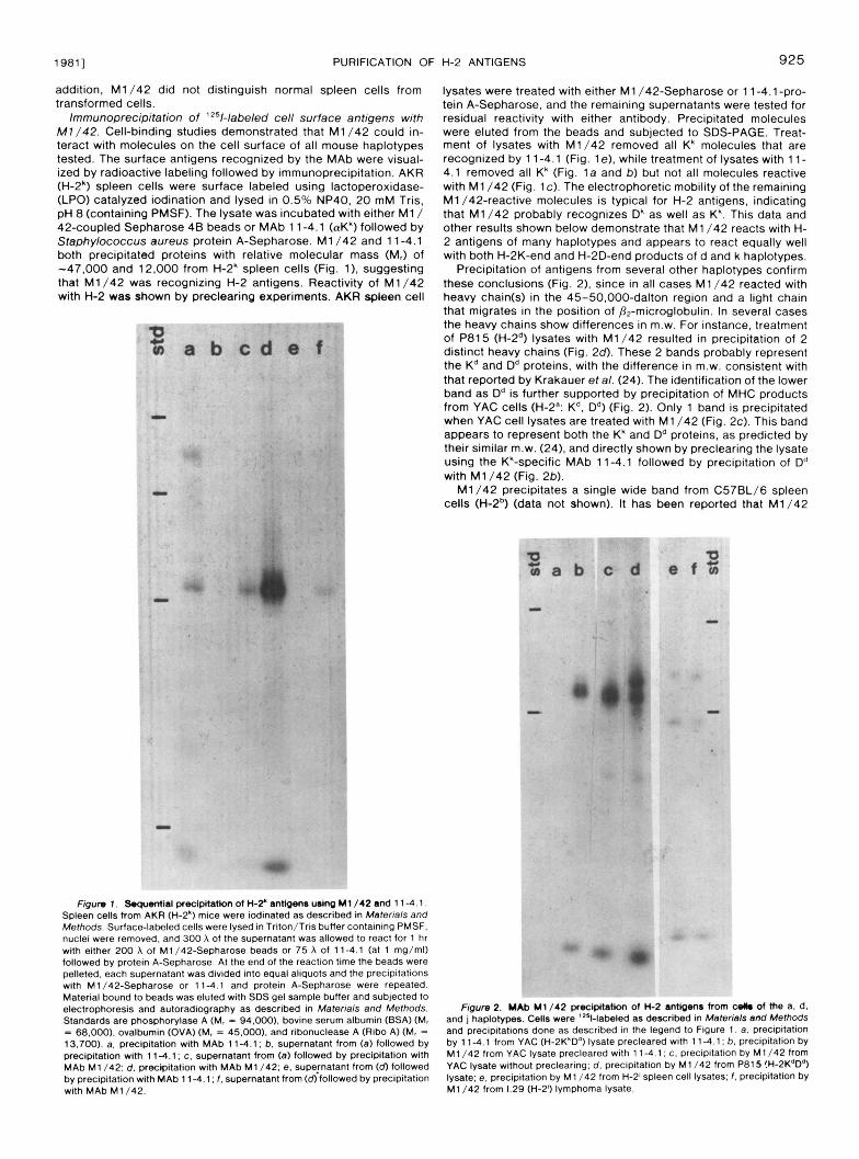
Immunoprecipitation of 'z51-labeled cell surface antigens with M7 /42. Cell-binding studies demonstrated that M1/42 could in- teract with molecules on the cell surface of all mouse haplotypes tested. The surface antigens recognized by the MAb were visual- ized by radioactive labeling followed by immunoprecipitation. AKR (H-2') spleen cells were surface labeled using lactoperoxidase- (LPO) catalyzed iodination and lysed in 0.5% NP40, 20 mM Tris, pH 8 (containing PMSF). The lysate was incubated with either M1/ 42-coupled Sepharose 48 beads or MAb 1 1-4.1 (aK') followed by Staphylococcus aureus protein A-Sepharose. M1/42 and 11 -4.1 both precipitated proteins with relative molecular mass (M,) of -47.000 and 12.000 from H-2' spleen cells (Fig. 11, suggesting that M1/42 was recognizing H-2 antigens. Reactivity of M1/42 with H-2 was shown by preclearing experiments. AKR spleen cell
~ . ". - " .. " -.
lysates were treated with either M1 /42-Sepharose or 11-4.1-pro- tein A-Sepharose, and the remaining supernatants were tested for residual reactivity with either antibody. Precipitated molecules were eluted from the beads and subjected to SDS-PAGE. Treat- ment of lysates with M1/42 removed all K' molecules that are recognized by 11-4.1 (Fig. le), while treatment of lysates with 11- 4.1 removed all Kk (Fig. l a and b) but not all molecules reactive with M1/42 (Fig. IC). The electrophoretic mobility of the remaining M1/42-reactive molecules is typical for H-2 antigens, indicating that M1/42 probably recognizes Dk as well as K'. This data and other results shown below demonstrate that M1/42 reacts with H- 2 antigens of many haplotypes and appears to react equally well with both H-2K-end and H-2D-end products of d and k haplotypes.
Precipitation of antigens from several other haplotypes confirm these conclusions (Fig. 2), since in all cases M1/42 reacted with heavy chain(s) in the 45-50,000-dalton region and a light chain that migrates in the position of Pz-microglobulin. In several cases the heavy chains show differences in m.w. For instance, treatment of P815 (H-2") lysates with M1/42 resulted in precipitation of 2 distinct heavy chains (Fig. 2 d . These 2 bands probably represent the K" and Dd proteins, with the difference in m.w. consistent with that reported by Krakauer et al. (24). The identification of the lower band as Dd is further supported by precipitation of MHC products from YAC cells (H-2': K". D") (Fig. 2). Only 1 band is precipitated when YAC cell lysates are treated with M1/42 (Fig. 2c). This band appears to represent both the Kk and D" proteins, as predicted by their similar m.w. (24), and directly shown by preclearing the lysate using the Kk-specific MAb 11-4.1 followed by precipitation of D" with M1/42 (Fig. 2b).
M1/42 precipitates a single wide band from C57BL/6 spleen cells (H-2b) (data not shown). It has been reported that M1/42
Spleen cells from AKR (H-2*) mice were iodinated as described in Materials and FQum 1 . ~ l a l precipitatkm of H-2' antbenr uJng M1/42 and 1 1-4.1.
Methods. Surface-labeled cells were lysed in Triton/Tris buffer containing PMSF.
with either 200 A of M1/42-Sepharose beads or 75 A of 114.1 (at 1 mg/ml) nuclei were removed, and 300 A of the supernatant was allowed to react for 1 hr
followed by protein A-Sepharose. At the end of the reaction time the beads were pelleted, each supernatant was divided into equal aliquots and the precipitations with M1/42-Sepharose or 114 .1 and protein A-Sepharose were repeated. Material bound to beads was eluted with SDS gel sample buffer and subjected to electrophoresis and autoradiography as described in Materials and Methods. Standards are phosphorylase A (M, = 94.000). bovine serum albumin (BSA) (M, = 68.000). ovalbumin (OVA) (M, = 45.000). and ribonuclease A (Ribo A) (M, = 13.700). a. precipitation with MAb 1 1-4.1 ; b. supernatant from (a) followed by precipitation with 114.1; c. supernatant from (a) followed by precipitation with MAb M1 /42: d. precipitation with MAb M1/42; e, supematant from (d) followed bv DreciDitation with MAb 1 1-4.1 ; f . supernatant from (d) followed by precipitation
and j haplotypes. Cells were 'Z51-labeled as described in Materials and Methods Figure 2. YAb M1/42 precipitation of K 2 anttgenr from wllm of the a, d.
and precipitations done as described In the legend to Figure 1. a, precipitation by 114.1 from YAC (H-2K*Dd) lysate precleared with 11-4.1; b. precipitation by M1/42 from YAC lysate precleared with 114.1 ; c. precipitation by M1/42 from YAC lysate without preclearing; d. precipitation by M1/42 from P815 !H-2KdD4) lysate; e, precipitation by M1 /42 from H-2' spleen cell lysates; f . precipitation by
with MAb M1/42. M1/42 from 1.29 (H-2') lymphoma lysate.

926 K. C. STALLCUP, T. A. SPRINGER, AND M. F. MESCHER [VOL. 127
recognizes only the H-2K end of the b haplotype (1 7). A particularly interesting pattern was found after precipitation of spleen or lym- phoma cells of the H-2' (KIDb) haplotype (Fig. 2e and f). Both spleen and lymphoma cells yielded 2 heavy-chain bands, 1 fairly large (M, = -48,000) and 1 quite small (M, = -38,000). It is not clear whether the smaller band represents an intact H-2 molecule or a breakdown product, although H-2 protein breakdown is usually not observed under the conditions of these immunoprecipitations.
Quantitation of M1/42 binding to various cell types. The speci- ficity of M1/42 for H-2 antigens and the apparent lack of alloanti- genic specificity suggested that M1/42 might provide a means of purifying a variety of H-2 antigens. We first examined several possible sources with respect to the quantity of H-2 expressed on the cell surface, since purification can be greatly facilitated by the use of cells that produce large amounts of the antigen. Herrmann and Mescher (13) had noted that H-2Kk is expressed in large amounts on RDM-4 cells, comparable to the expression of HLA antigens on Epstein-Barr virus-transformed human cells (7).
Cells were stained with M1/42 MAb and mouse IgG-absorbed fluorescein isothiocyanate- (FITC) coupled rabbit F(ab'), anti-rat IgG under saturating or near-saturating conditions and analyzed
TABLE II Quantitation of H-2 Expression Using M 1 / 4 2 and FACS
Cell H-2 (Spleen Cell Relative Units)"
Scatter Intensity (Mean Channel No.)
Spleen (1 ) 52 Con A blastsb 2.3 1 34 LPS blastsb 3.1 RIE/TLEX.l 0 106
1 55
YAC 97
3.2 P81 5 3.5
1 06 140
P388D, 2.7 127 EL-4 3.5 108
RDM-4 9.8-12.7"
Cells were labeled with saturating M1/42 MAb or irrelevant MAb. then FITC- anti-rat IgG and analyzed on the FACS as described (27). The mean fluorescence intensity of each cell type was calculated by integration, corrected for back- ground labeling, and expressed relative to the intensities of C57BL/6J splenic nucleated cells.
Scatter gated to include only blasts. Two-fold more FITC anti-rat IgG gave 1.3-fold brighter labeling.
on the fluorescence-activated cell sorter (FACS) (Table 11). Flu- orescent intensities thus obtained were directly proportional to the number of antibodies bound per cell. T cell and B cell blasts and YAC, P815, EL-4, and P388D1 cell lines expressed 2.3- to 3.5- fold more H-2 than normal spleen cells. Among the lines studied here, RDM-4 expressed unusually high amounts of H-2, approxi- mately 10-fold more than spleen cells, confirming previous results (13). Light-scattering measurements on the FACS and measure- ment of cell size under the microscope suggested that RDM-4 was smaller or similar in size to the other tumor cells, indicating that the increased expression of H-2 on this line is not simply due to greater surface area. The RIE/TL8X.l mutant line, Previously character- ized as negative for Tla and H-2 antigens, due to a complementable mutation suggested to involve carbohydrate or P2-microglobulin (251, expresses no material cross-reactive with M1/42 (Table 11).
These results indicated that tumor cell lines would be a good source of H-2 for purification and suggest that M1/42 could be useful in more extensive studies of H-2 expression on various cell lines.
Purification of H-2 antigens using M1/42. Previous work in this laboratory (1 3) has shown that an MAb coupled to Sepharose 4 8 could be used for affinity purification of H-2 proteins. Since M1/ 42 bound H-2 antigens tightly enough to give good immunoprecip- itates, it appeared that it might be an effective affinity reagent. Small-scale experiments using whole cell lysates from 5 to 25 x 10' cells demonstrated that H-2b.d,andk products could bind and be eluted from M1/42 columns (data not shown). Substantial amounts of H-2 could be eluted using 0.590 DOC in 20 mM Tris, whereas 0.5% DOC in Tris containing 0.65 M NaCl appeared to elute all material bound to the column. Using this protocol, H-2b could be purified from EL-4 tumor cells with a yield of -250 pg H-2 per 1 X 10'' cells. Attempts to purify large amounts of H-2 (e.g., from 10" cells) using whole cell detergent lysate as a source of starting material were not always successful, particularly in the case of P815 (H-2d) cells. The lysates tended to become quite viscous over the course of column loading (which, depending upon the lysate volume, could often be 36 to 50 hr at 4"C), frequently plugging the columns and generally reducing the flow rate to impractical levels. This problem was solved by separating the bulk of the cellular membranes from the cytoplasm with the scheme
c e l l s (1 x loe c e l l s / m t i n PBES) I
4 0 0 p s i x 5 min
cent r i fune 15 min @ 3600a
/ -1 (nuc le i + membrane fragments)
pe l le t supernatant
I I
s o l u b i l i z e !n 0 . 5 % T r i t o n X100 I
cent r i fuge 15 min @ 3600g I
(nuc le i ) pe l le t supernatant
I
cent r i fuge 30 min @ 22,OOOg 1
pe l le t supernatant
(membrane fragments)
I I
s o l u b i l i z e i n 0.5% T r i t o n
cent r i fuge 4 5 min @ lO0,OOOg
p e l l e t A supernatant supernatant pe l le t
monoclonal antibody ' a f f i n i t y chromatography Figure 3. Subcellular fractionation scheme.

19811 PURIFICATION OF H-2 ANTIGENS 927
outlined in Figure 3. Nitrogen cavitation was used to lyse the cells, and membrane fragments were isolated from nuclei and cytoplasm by differential centrifugation. Previous studies (20) had shown that significant amounts of plasma membrane (assayed by 5'-nucleotid- ase activity) pellet with the nuclei after the 1 st low-speed centrifu- gation. We were able to significantly increase recovery of H-2 (often by 100%) by solubilizing the low-speed pellet as well as the 22,000 x G membrane pellet. The low-speed pellet and membrane pellet were solubilized, loaded on MAb-Sepharose columns, and eluted as described in Materials and Methods.
The elution profile and protein composition of a typical H-2d preparation are shown in Figure 4. Elution was monitored using trace amounts of whole cell lysate from cells surface labeled with lZ5l. This method predictably yields between 1 10 and 180 pg H-2/ 10" P815 cells. The Coomassie Blue-stained gel shown in Figure 4 demonstrates that heavy chains of both Kd and Dd and p2- microglobulin are bound to the M1/42 column and that the eluate is free of any major contaminants. Autoradiography of the gel revealed no radioactive contaminants (data not shown). The ratio of heavy chains to Pz-microglobulin is approximately 2 : l based on ' 2 5 1 radioactivity. Individual M1/42 columns have been used 5 to 10 times with no apparent loss in capacity.
During early, relatively small-scale preparations, the efficacy of several elution buffers was tested. Figure 5 shows the profile of a P815 whole cell lysate eluted with buffers of different detergent or salt concentrations. Gels of proteins found in the eluted samples indicate that both Kd and Dd are present in each peak (data not shown). The 1st peak showed an enrichment of H-2Kd; however, attempts to achieve clean separation of Kd and Dd using DOC or salt gradient elutions were unsuccessful. The heterogeneous elu- tion is particularly curious in light of the monoclonality of M1/42 and may reflect slight heterogeneities in parts of the H-2 proteins proximal to the antigenic determinant. Alternatively, heterogeneous elution may reflect heterogeneity of the antibody produced by the M1/42 clone, since the rat lg heavy chain could combine with either a specific rat lg light chain or the myeloma K-chain. Standard large-scale preparations are eluted with DOC/0.65 M NaCl buffer, a buffer that elutes all of the bound antigen.
Using M1/42 as 1 of a series of columns, it becomes possible to isolate individual K or D antigens. Figures 6 and 7 show the
purification of H-2Dd from YAC cells after using MAb 11 -4.1 to isolate Kh. The flow-through from the aKk column, when completely cleared of Kk, was allowed to bind to M1/42. and Dd was subse- quently eluted. We have used a similar protocol to isolate Dh from RDM-4 cells (H-2')). In the case of RDM-4 cells, the material that elutes from M1/42 represents approximately 10 to 14% of the material identified as Kk. This is in agreement with the fact that RDM-4 cells over-express H-2Kk. The possibility exists that M1/ 42 does not bind H-2Dk and that the material bound is partially denatured H-2Kh that did not bind to the 11-4.1 column. This is made unlikely by the results of the preclearing experiment shown in Figure 1 and the results described below.
M1/42 does not bind separated heavy and pz-rnicroglobulin chains. The fact that M1/42 bound to a large number of H-2 molecules suggested that the MAb could be directed toward either a determinant on µglobulin or a region common to all heavy chains, a region that might retain antigenicity even after partial denaturation. To test this possibility, we isolated '251-labeled H-2Kk by affinity chromatography on MAb 11 -4.1. The heavy and light chains were dissociated in 6 M guanidine HCI and separated by chromatography on Biogel A 0.5M. The separated chains were dialyzed into 0.5% Triton X-100, 20 mM Tris. or alternatively, separated heavy chain and P2-microglobulin were mixed (2: 1 heavy chain:P2-microglobulin) and dialyzed together. Samples were ap- plied to M1/42, and the counts bound vs counts in the flow- through were determined. Table 111 shows that 64% of the pure, native Kh bound to M1/42. whereas a mixture of Kh and p2- microglobulin did not bind at all. Separated Pz-microglobulin and heavy chains each bound poorly, if at all. Although these data do not rule out the possibility that M1/42 recognizes µglobulin or partially denatured heavy chain, it seems less likely. These data also argue against the likelihood of M1/42 binding denatured H- 2Kh instead of H-2Dk in MAb 1 1-4.1-absorbed RDM-4 lysates.
Serologic and biologic activity of H-2 antigens purified on M 1 / 42 columns. Affinity chromatography using MAb 11-4.1 columns results in excellent recovery of H-2Kk serologic and biologic activity (13, 22). Similarly mild elution conditions for M1/42 suggested that H-2 antigens purified with this MAb should also retain activity. This was confirmed using both serologic and biologic criteria.
Seroloqic activity was tested by rebinding purified H-2b and H-
0.5 % DOC, 0.65 M NaCI,
Figure 4. Elution profile and SDS-PAGE of H-2' antigens purified by afflnity chroma- tography on M 1 / 4 2 . The high speed pellet (including '251-labeled surface proteins as tracer) (see Fig. 3) from P815 cells was solubilized with Triton X-100 and loaded onto M1 /42 as described in the text. The column was washed extensively with Triton/ Tris buffer and fractions (numbers 1-13) were collected at the end of the wash. DOC/ 0.65 M NaCl elution buffer was then added and elution was monitored by following the
and 1 0 to 20 pg of protein was precipitated I2'l tracer. Fractions 15-18 were pooled
with 6 vol cold acetone and analyzed by SDS gel electrophoresis (inset). The gel was stained with Coomassie Blue and the markers in the outside lanes are BSA. OVA, and Ribo A. Similar results were obtained when H-2 was purified from the low speed pellet (nuclei and membrane fragments, see Fig. 3 ) from these P815 cells.

928 K. C. STALLCUP. T. A. SPRINGER, AND M. F. MESCHER [VOL. 127
Figure 5 . H-2'antigens elute heterogeneously under some conditions. Whole P815 cells were lysed with Triton X-100 and loaded onto an M1 /42 column as described in the text. After extensive washing, a stepwise elution was done using the Indicated buffers. Three to 4 peaks of radioactivity were resolved. Fractions across the elution profile were combined into 5 pools. as shown
A
L 5 IO I5
I $.
ELUTION C (D*)
p
B D s
, 0 5 IO
FRACTfW NUMBER
Figure 6. Purification of H-2Kh and H-2Da. Seven and one-half x 10' YAC cells were iodinated. lysed in Triton/Tris buffer, loaded through a 0.75 ml Sepharose 4 8 precolumn and onto a 0.5 ml MAb 11-4.1 column. The flow- through was collected and saved. The 114 .1 column was washed with Triton/ Tris buffer, then eluted with 0.5% DOC/TBS and collected as 60 drop fractions (elution a). The 114.1 column was re-equilibrated with Triton/Tris and the flow- through from the 1st column application was loaded in series through the 114.1 column and a 0.8 ml column of M1/42-Sepharose. Residual K' was eluted from
of Triton/Tris. then eluted with 0.5% DOC/0.65 M NaCl buffer into 60 drop 114 .1 as above (elution E). The M1/42 column was washed with 15 to 20 vol
fractions (elution C).
Figure 7. SDWAGE analysis of purified K* and od. Radloactive material from elution A and C in Figure 6 was precipitated with cold acetone and subjected to SDS-PAGE. The gel was dried and autoradiographed. Standards are BSA. OVA, and Ribo A. a, H-2Kk from elution A; b, H-2D" from elution C.
TABLE Ill Binding of native or denatured H-2 molecules to MAb 1/42'
Sample Cpm Bound (% of Applled)
f12 Microglobulin 10 Heavy cham. H-2K' 7 Heavy chain (K*) + f12 microglobulin <1 H-2Kh 64
'251-labeled H-2Kh was purified and treated with 6 M guanidine as described
then dialyzed out of guanidine either separately or mixed together at a 2:l (heavy in the text. Heavy chain and f12 microglobulin were separated by gel filtration.
chain:f12-microglobulin) ratio. After dialysis, the samples were made 0.5% with Triton and directly applied to a 0.5 ml M1/42 column. The number Of Counts bound and eluted by DOC/0.65 M NaCl buffer are expressed as percent of applied counts.
2' (both K- and D-ends) or purified H-2Dd (obtained from YAC cells; see Figs. 6 and 7) to M1/42 columns. At least 40 to 60% of these purified molecules rebound to the MAb. Purified H-2" pro- teins were also used to inhibit alloantisera-mediated complement cytotoxicity on appropriate cells (data not shown). By this method of assay, we found that at least 40% of the Kd and Dd serologic activity was recovered after purification.

19811 PURIFICATION OF H-2 ANTIGENS 929
The purified antigens also retained biologic activity. Purified H- 2" antigens, prepared by the protocol shown in Figure 3, were incorporated into lipid vesicles and used to stimulate a secondary allogeneic CTL reponse. Stimulation with liposomes containing 1 to 2 pg of H-2" proteins resulted in a good response. We also tested the biologic activity of H-2d antigens that eluted heteroge- neously from M1/42 (see above, Fig. 4). A single preparation of P815 cells was loaded through M1/42 and sequentially eluted with 0.5% DOC (peak a), 0.25% DOC, 0.1 5 M NaCl (peak b), and DOC/0.65 M NaCl buffer (peak c). Material from each of the 3 peaks was incorporated into liposomes and used to stimulate an allogeneic CTL response. As shown in Figure 8, bottom panel, material from each of the peaks gave a distinct dose-response curve when assayed by CTL stimulation, with the 1 st peak (eluted with DOC alone) yielding the least active material. CTL stimulation by the 2nd and 3rd peaks (b and c, Fig. 8 inset) compared favorably with stimulation by liposomes containing purified H-2Kk (Fig. 8, top panel), where at least 75% of the original biologic activity is retained after purification (1 3, 21 ), The reason(s) for the different biologic activities of the 3 H-2" fractions are not known; however, it does not appear to be due to separation of the K- and D-end molecules (see above).
DISCUSSION
M1/42, a rat anti-mouse MAb, binds to H-2 antigens of a variety of serologic specificities. We have used this antibody to quantitate and precipitate the H-2 antigens from the a, b, d, and k haplotypes. Most interestingly, M1/42, when coupled to Sepharose 4 0 beads, can be used as a highly effective affinity reagent. Affinity columns of M1/42 have been used to purify relatively large quantities of biologically active H-2 antigens from the b, d, and k haplotypes. Mild elution conditions (0.5% DOC and 0.65 M NaCI, pH 8) result in minimal damage to either antibody or antigen. Thus excellent yields of active antigen can be obtained and the MAb columns can be used repeatedly. M1/42 can also be used in series with a MAb 11-4.1 (aKk) affinity column to isolate pure H-2Dd from H-2" cells.
M1/42 appears to recognize a species-specific, rather than allo-specific, antigenic determinant. Failure to bind Pn-micro-
,001 b
H-2d j
40
20 FRACTiON
taining liposomes. H-2Kh purified by affinity chromatography on MAb 114 .1 was Figure 8. Stimulation of a secondary allogeneic CTL response by H-P-con-
incorporated into liposomes as described (22) and various doses were used to stimulate primed CDPF, spleen cells as described in Materials and Methods. H- 2'from P815 whole cell lysates were purified on an MAb M1/42 column. Antigen was eluted in a stepwise fashion with 0.5% DOC (peak a). 0.25% DOC/O.15 M NaCl ( p e a k b). and 0.5% DOC/0.65 M NaCl (peak c) in 20 mM Tris. pH 8.
(22) and used to stimulate primed C57BL/6 Spleen cells as described in Materials Material from each of the 3 peaks was incorporated into liposomes as described
and Methods. CTL activity was assayed by using a standard "Cr release assay with RDM-4 (H-2") or P815 (H-2d) cell targets.
globulin or isolated heavy chain provides preliminary evidence that M I /42 may recognize a determinant common to native, intact H- 2 antigens. Our data indicate that M1/42 recognizes at least 1 and perhaps all 3 H-2 antigens from a number of haplotypes (Table I, Figs. 1 and 2). Specifically, binding to Dd and Kd has been shown by means of SDS-PAGE migration (Fig. 1) and, in the case of H- 2Kk, by the successful binding of pure H-2Kk to M1/42 affinity columns (Table Ill). We also have presumptive evidence that M1/ 42 recognizes H-2Dk, since the MAb can be used to purify a protein from RDM-4 cells that is not bound after several passages over an 11-4.1 column. It is possible, although unlikely, that M1/ 42 may recognize a small proportion of H-2Kk that is partially denatured and therefore not retained by 11-4.1. However, our findings argue against this: separated H-2Kk heavy chain or sep- arated and reassociated H-2k heavy chain and pp-microglobulin do not bind to M1/42 (Table 111). arguing that M1/42 is specific for determinants on native H-2 molecules. Furthermore, M1/42 pre- cipitates a major band from AKR (H-2k) spleen cell lysates that have been cleared of Kk (Fig. 1 ). It seems unlikely that such a large quantity of denatured H-2Kk would be present in spleen cell lysates. It may be possible that M1 /42 recognizes /32-microglobulin, which would account for its wide specificity range. However, we have not found significant binding of separated /3z-microglobulin or p2- microglobulin that has been denatured and partially reassociated with heavy chain (Table 111).
Although not yet determined, it is possible that the product of the H-2L locus is also bound by M I /42. This is deemed likely because M1/42 has no apparent specificity for different H-2 antigens and because H-2L is structurally very similar to H-2K and -D (26). TI and Qa antigens also have some structural similarities to H-2, and it will be interesting to determine whether M I /42 binds these antigens. It should be noted that the tumor cell lines used as H-2 sources in the studies reported here do not bear TI or Qa.
M1/42 will be a useful reagent in a variety of immunologic studies. For instance, we have used M1/42 to quantitate H-2 on several cell types (Table II), and the results confirm the finding (1 3) that the tumor line RDM-4 expresses high levels of H-2k. M1/42 can thus serve as a screening agent, both within and between haplotypes, for discerning fluctuations in H-2 expression. Such screening could be a useful adjunct to purification protocols, as evidenced by the abundant yield of H-2Kk from RDM-4 cells (1 3). As shown in the gels in Figures 1 and 2, M1/42 allows easy comparison of H-2 antigens of different haplotypes. especially in cases where effective precipitating alloantisera are not available. An interesting example is that of the H-2' haplotype, where an unusual -38,000-dalton polypeptide was precipitated along with the expected 47,000 heavy chain (Fig. 2). M1/42 could be used to isolate larger quantities of the small polypeptide, allowing for further characterization of the molecule.
The wide specificity of M1/42 also makes it an excellent reagent for the purification of H-2 molecules, thus combining the ease, rapidity, and specificity of affinity chromatography with the wide applicability of conventional biochemical purifications. The conven- tional purifications of H-2 antigens that have been reported are lengthy, involving several column purifications (some requiring harsh elution conditions) and concentration steps, and result in rather poor yields of purified antigen (1 0-1 2). Using the procedure described in this report, several hundred micrograms of pure H-2 can be prepared in a 3- to 5-step procedure that involves very little loss of material and in which the rate-limiting operation is column- loading time. With respect to column loading time, we have found a good deal of variation between the viscosities of different whole cell lysates. Thus, H-2b can be purified from whole cell lysates of EL-4 cells, whereas P815 cells, due to the viscous nature of the lysates. are usually fractionated before being solubilized and loaded onto the MAb column. Material bound to M1/42 can be eluted under very mild conditions, with 0.5% DOC and 0.65 M NaCI, pH 8.0, sufficient to remove the bound antigen (Fig. 4). Thus, the recovered antigens are exposed to a minimum of harsh treat- ment, can be readily dialyzed out of the elutant and into other buffers, and retain serologic and biologic activity. We estimate that D" purified from M1/42 retains a minimum of 40% of its serologic activity, compared with the 1 to 10% recovery of H-2 typically found after laborious conventional purification methods (1 0-1 2).

930 K. C. STALLCUP, T. A. SPRINGER, AND M. F. MESCHER [VOL. 127
Similarly mild elution of H-2 by DOC has been described for the 1 1-4.1 MAb affinity column (1 3). However, DOC did not elute HLA antigens from 6 different MAb affinity columns (Herrmann, Mescher, and Parham, unpublished), nor did it elute la molecules bound to a MAb (1 6; Turkewitz and Mescher, manuscript in prep- aration), indicating that elution by DOC is not a generalized prop- erty of MHC antigens.
H-Zd antigens obtained from P815 lysates have been found to elute in a heterogeneous manner (Fig. 5). This phenomenon is interesting but not easily explained. We have attempted to use DOC or salt gradient elutions to separate Dd and Kd molecules, and although there is some suggestion of selectivity in the elution, we were unable to obtain effective separation of the molecules in this way. The heterogeneous elution does appear to reflect differences in the molecules, as suggested by the biologic activities of the different peaks (Fig. 8), but it is not clear what this means, since the protein patterns from all peaks are similar.
One of the most interesting applications of this purification technology has been touched upon in this report: the biologic activity of the purified molecules. Using this pure material, it should be possible to investigate the functions of H-2 antigens in the many immunologic processes of which they are a part. This purification protocol makes available several H-2 antigens in quantities and conformations that are appropriate for functional, as well as bio- chemical, characterization.
Acknowledgments. We thank Dr. Martin Dorf for performing the cytotoxicity assays, Dr. Steven Burakoff for performing CTL as- says, and Dr. Steven Herrmann for helpful discussions. We also thank Harriet Yake and Teresa Greenberg for preparing the man- uscript.
1.
2.
3.
4.
5
6.
7.
REFERENCES
Cerottini, J-C.. and R. T. Brunner. 1974. Cell-mediated cytotoxicity, allograft rejection, and tumor immunity. Adv. Immunol. 18:67. Doherty. P. C., R. V. Blanden. and R. M. Zinkernagel. 1976. Specificity of virus-immune effector T cells for H-2K or H-2D compatible interactions:
Vitetta, E. S.. and J. D. Capra. 1978. The protein products of the murine implications for H-2 antigen diversity. Transplant. Rev. 2939.
17th chromosome: genetics and structure. Adv. Immunol. 26:147. Strominger. J. L., V. H. Englehard. A. Fuks. E. C. Guild, S. Hyofil. J. S. Kaufman. A. J. Korman, T. G. Kostyk. M. S. Krangel, D. Lancet, J. A. Lopez de Castro. D. L. Mann. H. T. Orr. P. R. Parham, K. C. Parker, H. L. Ploegh. J. S. Pober. R. J. Robb, D. A. Shackelford. 1981. Biochemical analysis of products of the MHC. In The Role of the Major Histocompatibility Complex in Immunobiology. Edited by M. E. Dorf. Garland Press, New York. Pp. 1 15- 172. Parham, P.. E. N. Alpert, H. T. Orr. and J. L. Strominger. 1977. Carbohydrate moiety of HLA antigens. Antigenic properties and amino acid sequences around the site of glycosylation. J. Biol. Chem. 252:7555. Orr. H. T.. J. R. Lopez de Castro, P. Parham, H. L. Ploegh, and J. L.
histocompatibility antigens, HLA-A2. and HLA-B7: location of putative al- Strominger. 1979. Comparison of amino acid sequences of two human
McCune, J. M., R . E. Humphreys. R. R . Yocum, and J. L. Strominger. 1975. loantigenic sites. Proc. Natl. Acad. Sci. 76:4395.
Enhanced representation of HLA-A antigens on human lymphocytes after
8.
9.
10.
11.
12.
13.
14.
15.
16.
17.
18.
19.
20.
21.
22.
23
24.
25
26
27
mitogenesis induced by phytohemagglutinin of Epstein-Barr virus. Proc.
Maloy. W. L., G. Hammerling, S. G. Nathenson. and J. E. Coligan. 1980. Natl. Acad. Sci. 72:3206.
Comparison of alloantisera and hybridoma antibody for puriftcation of the H- 2Dd murine histocompatibility antigen and preliminary molecular character-
Nathenson. s. G., E. M. Ewenstein. H. Uehara. J. M. Martinko. J. E. Coligan, ization of this antigen. J. Immunol. Methods 37:287.
and T. J. Kindt. Structure of H-2 major histocompatibility complex products: recent studies on the H-2Kb glycoprotein and on the H-2Kb MHC mutants. In Current Trends in Histocompatibility. Edited by S. Ferrone and R. Reisfeld. Plenum Press, New York. In press. Freed, J. H., D. W. Sears, J. Lynne Brown, and S. G. Nathenson. 1979. Biochemical purification of detergent-solubilized H-2 alloantigens. Mol. Im-
Rogers, M. J.. E. A. Robinson, and E. Appella. 1979. The purification of munol. 16:9.
murine histocompatibility antigens (H-29 from RBL-5 tumor cells using detergents. J. Biol. Chem. 254:11126. Henriksen, O., E. A. Robinson, and E. Appella. 1979. Purification and chemical characterization of papain-solubilized histocompatibility-2 antigens
Herrmann, S. H.. and M. F. Mescher. 1979. Purification of the H-2Kk from mouse liver. J. Biol. Chem. 254:7651.
molecule of the murine major histocompatibility complex. J. Biol. Chem. 2542371 3. Oi. V. T.. P. P. Jones, J. W. Goding. L. A. Herzenberg. and L. A. Herzenberg.
la antigens. Curr. Top. Microbiol. Irnmunol. 81 :115. 1978. Properties of monoclonal antibodies to mouse lg allotypes, H-2, and
Parham, P. 1979. Purification of immunologically active HLA-A and -E antigens by a series of monoclonal antibody columns. J. Biol. Chem. 254: 8709. McMaster. R. W.. and A. F. Williams. 1979. Identification of la glycoproteins in rat thymus and purification from rat spleen. Eur. J. Immunol. 9:426. Springer, T. A. 1980. Cell surface differentiation in the mouse. Characteri- zation of 'jumping' and 'lineage' antigens using xenogeneic rat monoclonal antibodies. In Monoclonal Antibodies. Edited by R. H. Kennett. T. J. Mc- Kearn. and K. E. Bechtol. Plenum Press, New York. Pp. 185-21 7. Mescher. M. F.. and R. R. Pollock. 1979. Murine cell surface immunoglob- ulin: two forms of &heavy chain. J. Immunol. 123:1155. Laemmli. U. K. 1970. Cleavage of structural proteins during the assembly of the head of bacteriophage T4. Nature (London) 227:680. Lemonnier, F.. M. Mescher, L. Sherman, and S. Burakoff. 1978. The induction of cytolytic T lymphocytes with purified plasma membranes. J.
Lowry, 0. H.. N. J. Rosebrough. A. L. Farr. and R. J. Randall. 1951. Protein Immunol. 120:1114.
Herrmann, S. H.. and M. F. Mescher. 1981. Secondary cytolytic T lympho- measurement with the Folin phenol reagent. J. Biol. Chem. 193:265.
cyte stimulation by purified H-2Kk in liposomes. Proc. Natl. Acad. Sci. 78: 2488. Mescher. M. F.. M. J. L. Jose, and S. P. Balk. 1981. Actin-containing matrix associated with plasma membrane of murine tumour and lymphoid cells. Nature 289:139. Krakauer, T.. T. H. Hansen. R. D. Camerini-Otero, and D. H. Sachs. 1980. Analysis of the heterogeneity of the mouse H-2K, D. and L gene products. J. Immunol. 124:2149. Hyman. R., and I. Trowbridge. 1977. Analysis of lymphocyte surface antigen expression by the use of variant cell lines. Cold Spring Harbor Symp. Quant.
Coligan. J. E., T. J. Kindt, R. Nairn. S. G. Natheson, D. H. Sachs. and T. H. Biol. 41 :407.
Hansen. 1980. Primary structural studies of an H-2L molecule confirm that it is a unique gene product with homology to H-2K and H-2D antigens. Proc.
Springer, T. A.. G. Galfre, D. S. Secher. and C. Milstein. 1979. Mac-1: A Natl. Acad. Sci. 77:1134.
J. Immunol. 9:301. macrophage differentiation antigen identified by monoclonal antibody. Eur.