Embed Size (px)
Citation preview
H-SC Journal of Sciences (2017) Vol. VI Reinartz and Wolyniak
http://sciencejournal.hsc.edu/
Characterization of arbuscular mycorrhizal fungi and assessing micropropagation potential in Allium tricoccum Dakota M. Reinartz ’18 and Michael J. Wolyniak Department of Biology, Hampden-Sydney College, Hampden-Sydney, VA 23943
INTRODUCTION Allium Tricoccum, commonly known as ramps or wild onion, is one of many plants that holds food value in the Appalachian forest region. Wild onion belongs to the same family of plant as garlic and green onion. Wild onion is a vegetable that can be eaten and has a flavor similar to that of garlic. Wild onion grows on the forest floor in the Appalachia during spring. Even though wild onion is a food source to the people of this region, it is not a cultivated crop. Wild onion is harvested unrestrictedly in the wild during its growing season. Demand for this crop is also increasing, thus increasing the demand to cultivate this crop on private lands. Before the cultivation of wild onion, we need to understand the strategies in which wild onion utilizes for survival. We also need to learn about its optimal growth conditions. The main survival strategy that this project focuses on is the identification and characterization of arbuscular mycorrhiza that is associated with wild onion. Arbuscular mycorrhiza is a type of mycorrhiza, or a symbiotic association, that is comprised of fungus and roots. This fungus gets into the outermost cells of the plants roots (Carter 2013). Previous studies have determined that these relationships exist, but are not characterized. Characterizing this relationship will help us grow wild onion in a controlled way. The other part of the project is to assess the micropropagation potential of wild onion. Micropropagation is the propagation of plants by growing plantlets in nutrient plant media and then planting them in soil. This method of growth has a high production rate and requires only small portions of a parent plant in nutrient rich media. However, these small portions need to include stem cells from the parent plant. These stem cells will eventually divide and differentiate, resulting in the development of a plantlet. Also contained in the media are growth hormones. In this project, auxins were added to the media to stimulate growth of the portions of plant. Auxins are hormones that cause the elongation of cells in shoots, as well as being involved in the regulation of plant growth. Green onion was also used in this project to act as a substitute for wild onion because all the wild onion died. Green onion was used because it is very similar in structure and in the same family as wild onion.
Principal Approaches
Media Preparation for Tissue Culturing. Wild onion plants were received from the
University of Virginia and were potted and stored in a shaded portion of the greenhouse. The preparation of the tissue culturing media was the first step. Murashige and Skoog Basal medium, or MS media, was used as the base nutrient part of the media and is a common medium used in tissue culturing. 1.075g of MS media, 2.5g of Agar, and 5g of sucrose were added and mixed with 250 mL of diH2O in two 500mL flasks. Also added to this mixture were 6-Benzylaminopurine (BAP) and indole-3-acetic acid (IAA), two separate growth hormones. 1mg of BAP and 0.5mg of IAA were added to each flask and mixed with a stir bar. This media was made by altering a recipe from the scientific product provider Sigma- Aldrich (Media). After mixing the mixture for 5 minutes, both flasks were autoclaved to sterilize the mixture. Upon termination of autoclaving, the flasks were moved to a sterile laminar flow hood to ensure the media remained sterile. Using a sterile pipet and an electric pipet, 10 ml of the media were pipetted into sterile 20 mL conical tubes. The tubes were capped loosely and left to dry in the flow hood overnight.
Tissue Culturing.
Gerry Peats discuses in his paper the use of stem disc meristem micropropagation of garlic, and these methods were altered in order to fit the tissue culturing of wild onion in this project. (Peat 2012). Wild onion and green onion plants were obtained from the greenhouse. The wild onion plants were then cut horizontally using a scalpel making pieces that were no greater than an inch. Because of the vastly different parts of the wild onion, pieces cut from different parts of the plant were different sizes and shapes, thus reacting differently. These pieces were then surface sterilized using a modified protocol from the University of New Mexico. Each piece was placed in a petri dish and washed with diH20. The water was drained and the plant pieces were washed with 95% ethanol for 1 minute. The ethanol was drained off and 1% bleach was added to the petri dish to wash the pieces. The bleach was then drained off and diH20 was added and drained off three times to thoroughly wash the bleached pieces. Once the surface sterilization was complete, sterile tweezers were used
H-SC Journal of Sciences (2017) Vol. VI Reinartz and Wolyniak
http://sciencejournal.hsc.edu/
to insert each piece into its own conical tube filled with nutrient media. After all the pieces were in the tubes, the tubes were placed in a growth chamber set for a 12 hour light cycle set at 20 degrees Celsius. The tubes were checked each day for growth and contamination, and were swapped out for fresh tubes every 3 weeks. DNA Extraction and PCR.
The second part of this project involved the extraction of DNA from the roots of wild onion plants to see if arbuscular mycorrhizal fungus were present in the samples. A Qiagen DNeasy Plant minikit protocol was used for the extraction of DNA from wild onion. Small cuttings were taken from the roots of wild onion plants using a scalpel, with DNA extraction being performed as instructed by the manufacturer (Qiagen). After DNA extraction was completed, the resulting liquid was run through a spectrophotometer. This was done to ensure that DNA was successfully extracted. After running the sample through the spectrophotometer, the DNA was stored in a -20 degrees Celsius freezer until it was needed for PCR. All PCR reactions were prepared in the same manner. 10uL of PCR mastermix, 3uL of the upper primer, 3uL of the lower primer, 1uL of the DNA sample, and 3uL of diH2O were added together in a PCR tube. The two different sets of primers that were used included the general fungal primers, LR1 and FLR2, and the Glomeromycota specific primers, FLR3 and FLR4 (Carter 2013). The thermocycler conditions used were as follows: 2 minute initial denaturation at 94°C, followed by 28 cycles of 1 minute denaturation at 94°C, 1 minute annealing at 58°C, and 1 minute extension at 72°C, and lastly a final extension at 72°C for 10 minutes (Carter 2013). Each sample was put through PCR two times. The first trail was completed with the general fungal
primers, and the second trial with the Glomeromycota specific primers. After both PCR cycles, the PCR products were separated on 2% agarose through gel electrophoresis. This protocol was completed on each wild onion plant, in order to see which plant had arbuscular mycorrhizal fungi, allowing that DNA sample to be prepared and sequenced. Each plant contained multiple samples to be run through PCR. DNA Sequencing.
Plants with bands that appeared after PCR, used the Glomeromycota specific primers. These plant samples went on to be prepared for sequencing. The product from the last PCR went through a purification protocol that was provided by the Qiagen Qiaquick PCR purification kit. After the purification of the PCR product, each sample was run through a spectrophotometer in order to make sure enough DNA made it through the purification process. Once the concentration of DNA was received from the spectrophotometer, the DNA was diluted to a concentration of 2 ng/uL. PCR 8-tube strips that hold 0.2 mL were then acquired, and 10 uL of the diluted sample DNA was added to the tubes along with 3uL of Glomeromycota specific primers FLR3 and FLR4, resulting in a final volume of 13 uL. These samples were packed with ice and shipped to Virginia Tech’s Biocomplexity Institute to be sequenced.
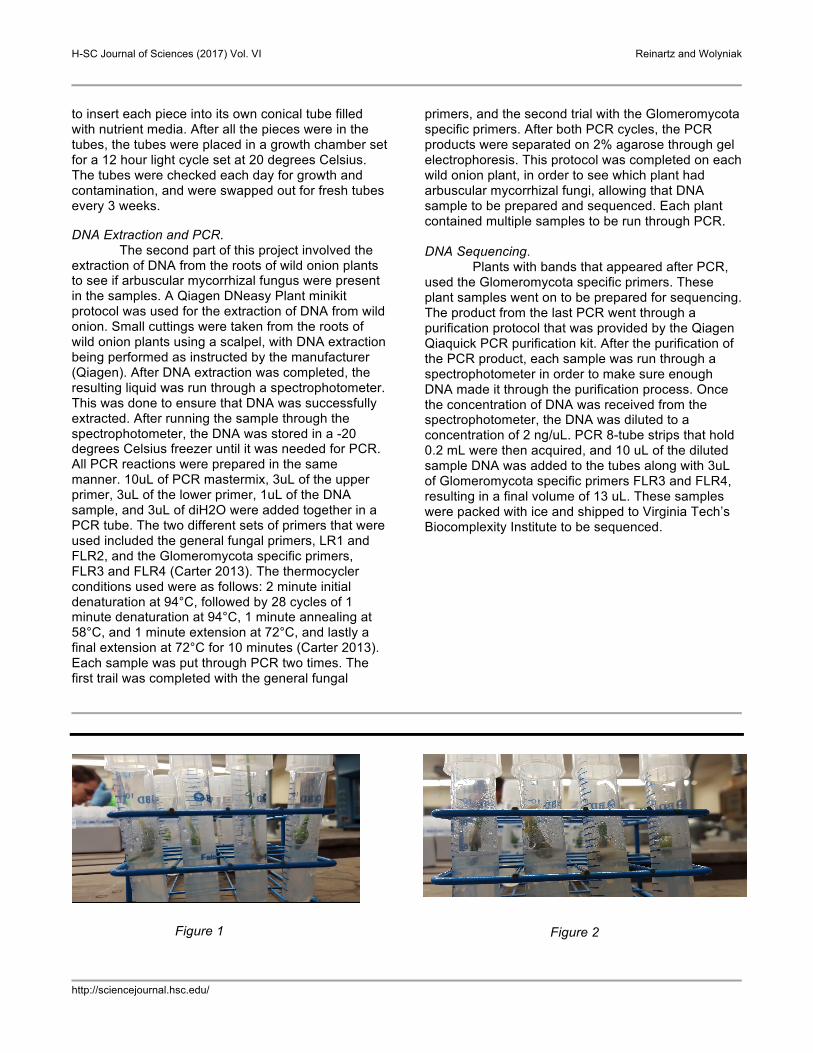
Figure 1
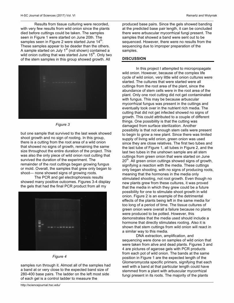
Figure 2
H-SC Journal of Sciences (2017) Vol. VI Reinartz and Wolyniak
http://sciencejournal.hsc.edu/
Results from tissue culturing were recorded, with very few results from wild onion since the plants died before cuttings could be taken. The samples seen in Figure 1 were started on June 20th. The samples seen in Figure 2 were started June 14th. These samples appear to be deader than the others. A sample started on July 1st (not shown) contained a wild onion cutting that was started June 15th. Only two of the stem samples in this group showed growth. All
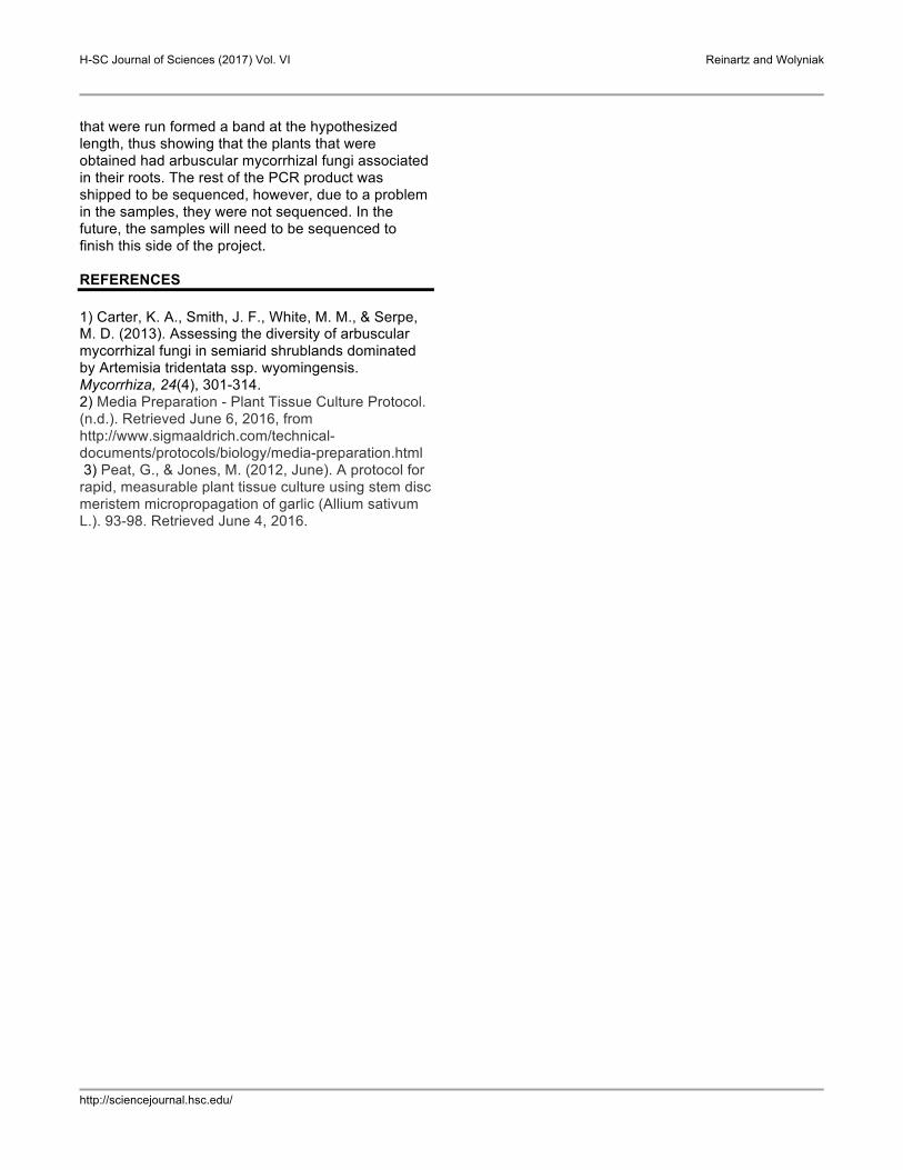
Figure 3
but one sample that survived to the last week showed shoot growth and no sign of rooting. In this group, there is a cutting from the root area of a wild onion that showed no signs of growth, remaining the same size throughout the entire duration of the project. This was also the only piece of wild onion root cutting that survived the duration of the experiment. The remainder of the root cuttings began growing fungus or mold. Overall, the samples that grew only began to shoot— none showed signs of growing roots. The PCR and gel electrophoresis results showed many positive outcomes. Figures 3-4 display the gels that had the final PCR product from all my
Figure 4 samples run through it. Almost all of the samples had a band at or very close to the expected band size of 280-400 base pairs. The ladder on the left most side of each gel is a control ladder to measure the
produced base pairs. Since the gels showed banding at the predicted base pair length, it can be concluded there were arbuscular mycorrhizal fungi present. The samples that showed a band were sent out to be sequenced. However, there were no results from the sequencing due to improper preparation of the samples. DISCUSSION
In this project I attempted to micropropagate
wild onion. However, because of the complex life cycle of wild onion, very little wild onion cultures were started. The cultures that were started were all cuttings from the root area of the plant, since the abundance of stem cells were in the root area of the plant. Only one root cutting did not get contaminated with fungus. This may be because arbuscular mycorrhizal fungus was present in the cuttings and eventually took over in the nutrient rich media. The cutting that did not get infected showed no signs of growth. This could attributed to a couple of different things. One possibility is that the cutting was damaged from surface sterilization. Another possibility is that not enough stem cells were present to begin to grow a new plant. Since there was limited supply of living wild onion, green onion was used since they are close relatives. The first two tubes and the last tube of Figure 1, all tubes in Figure 2, and the last two tubes in the unshown sample are all stem cuttings from green onion that were started on June 20th. All green onion cuttings showed signs of growth, signifying a reaction with the media. These cuttings only began shooting, with no signs of producing roots, meaning that the hormones in the media only stimulated shooting, not root growth. Even though no new plants grew from these cultures, it was proved that the media in which they grew could be a future possibility for one to stimulate shoot growth in wild onion. Figure 2 is an example of the detrimental effects of the plants being left in the same media for too long of a period of time. The tissue cultures of green onion were overall a failure because no plants were produced to be potted. However, this demonstrates that the media used should include a hormone that directly stimulates rooting. Also it is shown that stem cuttings from wild onion will react in a similar way to this media. DNA extraction, amplification, and sequencing were done on samples of wild onion that were taken from alive and dead plants. Figures 3 and 4 are pictures of agarose gels with PCR products from each pot of wild onion. The bands at the same position in Figure 1 are the expected length of the Glomeromycota specific primers, signifying that each well with a band at that particular length could have stemmed from a plant with arbuscular mycorrhizal fungi present in its roots. The majority of the plants
H-SC Journal of Sciences (2017) Vol. VI Reinartz and Wolyniak
http://sciencejournal.hsc.edu/
that were run formed a band at the hypothesized length, thus showing that the plants that were obtained had arbuscular mycorrhizal fungi associated in their roots. The rest of the PCR product was shipped to be sequenced, however, due to a problem in the samples, they were not sequenced. In the future, the samples will need to be sequenced to finish this side of the project. REFERENCES 1) Carter, K. A., Smith, J. F., White, M. M., & Serpe, M. D. (2013). Assessing the diversity of arbuscular mycorrhizal fungi in semiarid shrublands dominated by Artemisia tridentata ssp. wyomingensis. Mycorrhiza, 24(4), 301-314. 2) Media Preparation - Plant Tissue Culture Protocol. (n.d.). Retrieved June 6, 2016, from http://www.sigmaaldrich.com/technical-documents/protocols/biology/media-preparation.html 3) Peat, G., & Jones, M. (2012, June). A protocol for rapid, measurable plant tissue culture using stem disc meristem micropropagation of garlic (Allium sativum L.). 93-98. Retrieved June 4, 2016.