Embed Size (px)
Citation preview
Characterization of Bacteriophages Cp1 and Cp2, the Strain-TypingAgents for Xanthomonas axonopodis pv. citri
Abdelmonim Ali Ahmad, Megumi Ogawa, Takeru Kawasaki, Makoto Fujie, Takashi Yamada
‹Department of Molecular Biotechnology, Graduate School of Advanced Sciences of Matter, Hiroshima University, Higashi-Hiroshima, Japan
The strains of Xanthomonas axonopodis pv. citri, the causative agent of citrus canker, are historically classified based on bacte-riophage (phage) sensitivity. Nearly all X. axonopodis pv. citri strains isolated from different regions in Japan are lysed by eitherphage Cp1 or Cp2; Cp1-sensitive (Cp1s) strains have been observed to be resistant to Cp2 (Cp2r) and vice versa. In this study,genomic and molecular characterization was performed for the typing agents Cp1 and Cp2. Morphologically, Cp1 belongs to theSiphoviridae. Genomic analysis revealed that its genome comprises 43,870-bp double-stranded DNA (dsDNA), with 10-bp 3=-extruding cohesive ends, and contains 48 open reading frames. The genomic organization was similar to that of Xanthomonasphage phiL7, but it lacked a group I intron in the DNA polymerase gene. Cp2 resembles morphologically Escherichia coli T7-likephages of Podoviridae. The 42,963-bp linear dsDNA genome of Cp2 contained terminal repeats. The Cp2 genomic sequence has40 open reading frames, many of which did not show detectable homologs in the current databases. By proteomic analysis, agene cluster encoding structural proteins corresponding to the class III module of T7-like phages was identified on the Cp2 ge-nome. Therefore, Cp1 and Cp2 were found to belong to completely different virus groups. In addition, we found that Cp1 andCp2 use different molecules on the host cell surface as phage receptors and that host selection of X. axonopodis pv. citri strainsby Cp1 and Cp2 is not determined at the initial stage by binding to receptors.
Citrus canker, caused by Xanthomonas axonopodis pv. citri(syn., Xanthomonas campestris pv. citri or Xanthomonas citri),
is a widespread disease in citrus-producing areas of the tropicaland subtropical world (1, 2). Different types of citrus canker, cor-responding to different pathotypes of X. axonopodis pv. citri, havebeen reported (3). The Asiatic type, caused by X. axonopodis pv.citri pathotype A, is the most widespread and the most economi-cally important citrus canker. The host range of pathotype Astrains is wider than that of the other pathotypes, including mostcitrus varieties (3). X. axonopodis pv. citri pathotype A strains areseparated into two groups based on their sensitivity to phages Cp1and Cp2 (4, 5). Phage typing with Cp1 and Cp2 was first demon-strated by Wakimoto (4). Wakimoto found that nearly all strainsisolated from different regions in Japan were lysed by either Cp1or Cp2 and that Cp1-sensitive (Cp1s) strains were resistant to Cp2(Cp2r) and vice versa. Cp1 and Cp2 phages were morphologicallydifferent: Cp1 showed a head-tail structure, whereas Cp2 con-sisted of a polyhedral head without tail (4). In a larger survey,Obata found that Cp1r/Cp2s strains predominated in major cit-rus-producing regions in Japan, with the exception of HiroshimaPrefecture, where Cp1s/Cp2r strains predominated (5). Two dif-ferent strain types can occur in a mixture on the same single leaf ofa tree, but one single lesion usually consists of a single strain.
Notably, the sensitivity of X. axonopodis pv. citri strains to Cp1and Cp2 is associated with differences in their physiological fea-tures and canker aggressiveness. X. axonopodis pv. citri strains thatare Cp1s/Cp2r can assimilate mannitol, while Cp1r/Cp2s strainscannot (6). All strains that are Cp1r/Cp2s are canker aggressive tothe citrus variety “Otachibana,” whereas all the strains with Cp1s/Cp2r are weakly aggressive (7). The Cp1r/Cp2s strains generate a1.8-kbp-specific fragment by repetitive sequence-based PCR (rep-PCR) (8) using enterobacterial repetitive intergenic consensus(ERIC) primers. The 1.8-kbp band corresponds to a region en-compassing XAC1661 (Isxac3 transposase) and XAC1662 (repA)within an insertion element in the genomic sequence of X. ax-
onopodis pv. citri strain 306 (9). Most strains with Cp1s/Cp2r con-tain hssB3.0, a member of the avrBs3 and pthA (avirulence andpathogenicity) gene family (10, 11), which is responsible for thesuppression of virulence on a Citrus grandis cultivar; however,Cp1r/Cp2s strains lack this gene (12). These results suggest somerelatedness between Cp1/Cp2 sensitivity and the virulence andpathogenic features of X. axonopodis pv. citri strains.
In contrast to the large contribution toward characterization ofhost strains, very little information is available about the nature oridentity of phages Cp1 and Cp2. Concerning Cp1 and Cp2, thefollowing issues are of particular interest: (i) the virological iden-tification and phylogenetic relationships of these phages, (ii) theorigin of the above-mentioned 1.8-kbp sequence on the host ge-nome and its possible association with a phage sequence, (iii)hssB3.0 and its possible association with a phage sequence, and(iv) the molecular mechanism of host selection by these phages. Asa first step toward exploring these issues, the present study per-formed genomic and molecular characterization of Cp1 and Cp2.
MATERIALS AND METHODSBacterial strains and phages. Ministry of Agriculture, Forestry and Fish-eries (MAFF) strains of X. axonopodis pv. citri were obtained from theNational Institute of Agrobiological Sciences, Japan. Strain KC33 (7) wasobtained from the National Institute of Fruit Tree Science, National Ag-riculture and Food Research Organization (NAFRO), Japan. Their originsand sensitivity to Cp1 and Cp2 are listed in Table 1. Bacteriophages Cp1
Received 12 July 2013 Accepted 7 October 2013
Published ahead of print 11 October 2013
Address correspondence to Takashi Yamada, [email protected].
Supplemental material for this article may be found at http://dx.doi.org/10.1128/AEM.02310-13.
Copyright © 2014, American Society for Microbiology. All Rights Reserved.
doi:10.1128/AEM.02310-13
January 2014 Volume 80 Number 1 Applied and Environmental Microbiology p. 77– 85 aem.asm.org 77
on February 3, 2019 by guest
http://aem.asm
.org/D
ownloaded from
and Cp2 (4, 5) were obtained from the Yokohama Plant Protection Sta-tion, Japan. Strains MAFF 301080 and MAFF 673010 were used as hostsfor routine propagation of Cp1 and Cp2, respectively. Bacterial cells werecultured in nutrient broth (NB) medium (BBL, Becton, Dickinson andCo., Cockeysville, MD, USA) at 28°C with shaking at 200 to 300 rpm. Anovernight culture of bacterial cells grown in NB was diluted 100-fold with100 ml fresh NB in a 500-ml flask. To collect sufficient phage particles, 1liter of bacterial culture (10 � 100-ml cultures) was grown. When thecultures reached 0.2 units of optical density at 600 nm (OD600), the phageswere added at a multiplicity of infection (MOI) of 0.1. After furthergrowth for 9 to 18 h, the cells were removed by centrifugation with anR12A2 rotor in a Hitachi heavy ion medical accelerator (HIMAC) CR21Ecentrifuge (Hitachi Koki Co. Ltd., Tokyo, Japan), at 8,000 � g for 15 minat 4°C. The supernatant was passed through a 0.45-�m membrane filter,and phage particles were precipitated by centrifugation with a P28S rotorin a Hitachi XII100� centrifuge at 40,000 � g for 1 h at 4°C and dissolvedin SM buffer (50 mM Tris-HCl at pH 7.5, 100 mM NaCl, 10 mM MgSO4,and 0.01% gelatin). Purified phages were stored at 4°C until use. Bacte-riophage particles purified by CsCl gradient ultracentrifugation (with aP28S rotor in a Hitachi XII100� ultracentrifuge) (13) were stained withNa-phosphotungstate before observation in a Hitachi H600A electronmicroscope, according to the method of Dykstra (14). � phage particleswere used as an internal standard marker for size determination. For hostrange determination, standard plaque-forming assays (15) or lysis zoneformation spot tests (16) were performed.
Single-step growth experiment. Single-step growth experiments wereperformed as previously described (17, 18), with some modifications.Strains MAFF 301080 and MAFF 673010 were used as hosts for Cp1 andCp2, respectively. Cells (0.1 U of OD600) were harvested by centrifugationand resuspended in fresh NB (ca. 1 � 108 CFU/ml) to a final culturevolume of 10 ml. Phage was added at an MOI of 1.0 and allowed to adsorbfor 10 min at 28°C. After centrifugation and resuspending in the initialvolume of NB with decimal dilution to a final volume of 10 ml, the cellswere incubated at 28°C. Samples were taken at intervals (every 10 min upto 3.5 h for Cp1 and every 30 min up to 5 h for Cp2), and the titers weredetermined by the double-layered agar plate method.
Phage adsorption test. Phage adsorption was assayed as follows: whenfresh bacterial cultures (10 ml) reached 0.1 unit of OD600, the phage (10�l) was added at an MOI of 0.1. After incubation for 10 min at 28°C, the
cells were removed by centrifugation with an R12A2 rotor in a HitachiHIMAC CR21E centrifuge at 8,000 � g for 10 min at 4°C. The supernatantwas subjected to a plaque assay, where strains MAFF 301080 and MAFF673010 were used as the hosts for Cp1 and Cp2, respectively. Escherichiacoli JM109 was used as a negative control in phage adsorption.
DNA manipulation and sequencing. Standard molecular biologicaltechniques for DNA isolation, digestion with restriction enzymes andother nucleases, and construction of recombinant DNAs were followed,according to Sambrook and Russell (13). Phage DNA was isolated fromthe purified phage particles by phenol extraction. For genome size deter-mination, the purified phage particles were embedded in 0.7% low-melt-ing-point agarose (InCert agarose; FMC Corp., Philadelphia, PA, USA)and, after treatment with proteinase K (1 mg/ml; Merck Ltd., Tokyo,Japan) and 1% Sarkosyl, subjected to pulsed-field gel electrophoresis(PFGE) in a Chef Mapper electrophoresis apparatus (Bio-Rad Lab., Her-cules, CA, USA) according to the method of Higashiyama and Yamada(19). Shotgun sequencing was performed at Hokkaido System ScienceCo., Ltd. (Sapporo, Japan), using the Roche GS Junior Sequence system.The draft assembly of the obtained sequences was assembled using GS Denovo Assembler v2.6. The analyzed sequences corresponded to 94 and 40times the final genome sizes of Cp1 (43,860 bp) and Cp2 (42,963 bp),respectively. Potential open reading frames (ORFs) larger than 150 bp (50codons) were identified using Glimmer (20) and GeneMark. Homologysearches were performed using BLAST/RPS-BLAST (21) against the Uni-Prot sequence database (22) and the NCBI/CDD database (23), using an Evalue lower than e�4 as a cutoff for notable similarity. Multiple-sequencealignments were generated using the DNASIS program (version 3.6; Hi-tachi Software Engineering, Co., Ltd., Tokyo, Japan). For phylogeneticanalysis of RNA polymerase (RNAP) proteins, the unrooted dendrogramwas constructed with the Treeview tool using the maximum likelihoodmethod based on a complete protein sequence alignment of RNAP pro-teins from other phages using ClustalX. The Cp1 cohesive ends (cos) se-quence was determined as follows. A 6.8-kbp PstI fragment of Cp1 DNAwas dissociated into two fragments (4.3 and 2.5 kbp) after heating at 70°Cfor 15 min. The dissociated bands were recovered from the agarose gel andtreated with T4 DNA polymerase to form blunt ends. The nucleotidesequences of these bands were determined. By comparing the nucleotidesequences with each other and with the Cp1 genomic sequence, the cossequence was determined according to the method of Fujiwara et al. (24).
Southern and dot blot hybridization. Genomic DNA from bacterialcells was prepared by the minipreparation method according to Ausubelet al. (25). After digestion with various restriction enzymes (EcoRI,EcoRV, HindIII, and HincII), DNA fragments were separated by agarosegel electrophoresis, blotted onto a nylon membrane (Biodyne; PallGelman Laboratory, Closter, NJ, USA), hybridized with probes (the entireCp1 DNA by combining all the HincII fragments and the entire Cp2 DNAwith all the HincII fragments), labeled with fluorescein (Gene ImagesRandom Prime labeling kit; Amersham Biosciences, Uppsala, Sweden),and detected with a Gene Images CDP-Star detection module (AmershamBiosciences). Hybridization was performed in buffer containing 5� SSC(1� SSC is 0.15 M NaCl plus 0.015 M sodium citrate), 0.1% SDS, 5%liquid block, and 5% dextran sulfate for 16 h at 65°C. The filter was washedat 60°C in 1� SSC and 0.1% SDS for 15 min and then in 0.5% SSC and0.1% SDS for 15 min with agitation, according to the manufacturer’sprotocol. The hybridization signals were detected by exposing the filteronto an X-ray film (RX-U; Fuji Film, Tokyo, Japan).
SDS-PAGE and LC-MS/MS analysis. Purified phage particles weresubjected to SDS-polyacrylamide gel electrophoresis (SDS-PAGE) (10 to12% [wt/vol] polyacrylamide) according to Laemmli (26). Protein bandswere visualized by staining the gel with Coomassie brilliant blue, excisedfrom the gel, digested with trypsin, and subjected to liquid chromatogra-phy-tandem mass spectrometry (LC-MS/MS) (LTQ Orbitrap XL;Thermo Fisher Scientific, Osaka, Japan) analysis at the Natural ScienceCenter for Basic Research and Development, Hiroshima University.
TABLE 1 Bacterial strains and bacteriophages used in this studya
StrainHost(Citrus species) Phage typeb Source
X. axonopodis pv. citriMAFF 301077 C. limon Cp1s/Cp2r NIASc
MAFF 301080 C. sinensis Cp1s/Cp2r NIAS301080 R1 Cp1r/Cp2r This studyMAFF 311130 C. iyo Cp1r/Cp2r NIASMAFF 302102 Citrus sp. Cp1r/Cp2s NIASMAFF 673001 C. natsudaidai Cp1r/Cp2s NIASMAFF 673010 Citrus sp. Cp1r/Cp2s NIAS673010 R2 Cp1r/Cp2r This studyMAFF 673011 C. limon Cp1s/Cp2r NIASMAFF 673013 Citrus sp. CP1
s/CP2r NIAS
MAFF 673018 Citrus sp. Cp1r/Cp2s NIASMAFF 673021 C. limon Cp1r/Cp2s NIASKC33 C. iyo Cp1s/Cp2s Shiotani et al. (12)
PhagesCp1 Wakimoto (4)Cp2 Wakimoto (4)
a All strains originated in Japan.b Sensitivity to phages CP1 and CP2; s, sensitive; r, resistant.c NIAS, National Institute of Agrobiological Sciences, Japan.
Ahmad et al.
78 aem.asm.org Applied and Environmental Microbiology
on February 3, 2019 by guest
http://aem.asm
.org/D
ownloaded from
Staining of bacterial cells by SYBR gold-labeled phages. Phage label-ing and observation of phage-treated bacterial cells were performed ac-cording to Mosier-Boss et al. (27). To 100 ml of the phage lysate, 10 �l of104� SYBR gold (Molecular Probes, Inc., Eugene, OR, USA) in dimethylsulfoxide (DMSO) was added. After 10 min, the labeled phage particleswere precipitated by centrifugation with a P28S rotor in a Hitachi XII100�centrifuge at 40,000 � g for 1 h at 4°C. Three washes using 1� PBS weredone to ensure the removal of excess SYBR gold. A 1-ml sample of anovernight culture of X. axonopodis pv. citri was mixed with 4 ml NB andallowed to grow until an OD600 of 0.5 was reached. To a 10-�l sample ofthe bacterial culture was added 10 �l SYBR gold-labeled phage, and themixture was incubated for 10 to 60 min. As a control, a culture of E. coliJM109 was treated in the same way. After fixation of the mixture with 30�l 4% paraformaldehyde in PBS, 5 ml double-distilled water (ddH2O)was added, and the mixture was filtered through a membrane filter(0.2-�m pore size, Steradisc; Krabo, Osaka, Japan). The bacterial cellswere observed under a fluorescence microscope system with filter sets(Olympus BH2 fluorescence microscope; Olympus, Tokyo, Japan). Mi-croscopic images were recorded with a charge-coupled-device (CCD)camera (Kyence VB-6010; Osaka, Japan).
Nucleotide sequence accession numbers. The sequence data for theCp1 and Cp2 genomes have been deposited in the DDBJ database underaccession no. AB720063 and AB720064, respectively.
RESULTS AND DISCUSSIONCp1 and Cp2 belong to different virus families. Morphology in-dicates that phages Cp1 and Cp2 belong to different virus families:Cp1 as a member of Siphoviridae and Cp2 as a member of Podo-viridae. The purified particles of Cp1 and Cp2 were negativelystained and examined by transmission electron microscopy. Cp1particles had an icosahedral capsid of 60 � 5 nm in diameter witha long noncontractile tail of 135 � 10 nm long by 12 � 2 nm wide(see Fig. S1 in the supplemental material). This morphology wasalmost the same (though in smaller dimensions) as that reportedpreliminarily for CP1 particles replicated in strain N6101 as a host(28), indicating that, morphologically, Cp1 belongs to the familySiphoviridae. In contrast, Cp2 particles showed an icosahedralcapsid of 60 � 5 nm in diameter with a short tail of 15 � 5 nm long(see Fig. S1 in the supplemental material), indicating that Cp2 hasa structure typical of members of the family Podoviridae. In apreliminary work, this phage was reported to have larger polyhe-dral particles without a tail (28). These results raised the question
of whether Cp1 and Cp2 are related to each other in infection andreplication in host strains.
Comparison of infection cycles of Cp1 and Cp2. Both Cp1and Cp2 form clear plaques with various X. axonopodis pv. citristrains, including those shown in Table 1, as hosts (4, 5). Theinfection cycle of each phage was characterized by a single-stepgrowth experiment with appropriate hosts. The growth curves areshown in Fig. 1. In the case of Cp1 replication, the latent periodwas �60 min, followed by a rise period of 20 to 30 min, giving anentire cycle of 80 to 90 min. The average burst size was 20 PFU perinfected cell. For Cp2 replication, the latent period was �90 min,with a 60-min rise period, taking 150 to 180 min for a singlegrowth cycle. The burst size was approximately 100 PFU/cell.These results showed that Cp1 infected and replicated more rap-idly than Cp2, but the burst size was much smaller than that ofCp2. These results contrasted with the observation that more than600 progeny phages were formed in an infected bacterial cell forboth Cp1 and Cp2, as detected by electron microscopy (28). Theburst size of a phage may depend on the host strain, culture me-dium, culture conditions, cell age, and MOI (29). The net ratio ofinfective to noninfective phage particles in the progeny popula-tion and the frequent readsorption of phage particles onto celldebris may partly explain this discrepancy.
Genomic analyses of Cp1: gene organization and homologyto other phages. The Cp1 genome was a linear double-stranded(ds) DNA of approximately 44 kbp, as determined by PFGE (datanot shown). The nucleotide sequence of the Cp1 genome wasdetermined using shotgun sequencing of DNA purified fromphage particles. Sequences were assembled into a circular contig of43,870 bp, suggesting the presence of terminal repeats. The exacttermini of the Cp1 genome were determined to have a 10-nucle-otide (nt) 3=-protruding cos site (5=-CCAGTTGTCT, correspond-ing to positions 43,861 to 43,870) at either end.
The Cp1 genome had a GC content of 53.3%; this value wassignificantly lower than that of the host genome (e.g., 64.7% forstrain 306, accession no. NC_003919). When the databases weresearched using BLAST and BLASTX programs for sequences ho-mologous to the Cp1 DNA sequence, extensive homologies weredetected in the genome sequence of X. campestris phage phiL7(accession no. EU717894), X. oryzae phage OP1 (accession no.
FIG 1 Single-step growth curves for Cp1 (A) and Cp2 (B). The PFU per infected cell in cultures of MAFF 301080 for Cp1 and MAFF 673010 for Cp2 at differenttimes postinfection are shown. Samples were taken at intervals (every 10 min up to 3.5 h for Cp1 and every 30 min up to 5 h for Cp2). Error bars indicate standarddeviations (n 3).
Bacteriophages of Citrus Canker Bacteria
January 2014 Volume 80 Number 1 aem.asm.org 79
on February 3, 2019 by guest
http://aem.asm
.org/D
ownloaded from

AP008979), and Xanthomonas phage Xp10 (accession no.AY299121). All these phages are siphoviruses infecting species ofXanthomonas, which is consistent with the Cp1 morphologicalfeatures. An extended colinearity of the nucleotide sequence ho-mology, with several short interspersed divergent islands, was de-tected throughout almost the entire genomic region between Cp1and phiL7, which gave the highest similarity score (see Fig. S2 inthe supplemental material).
Forty-eight potential ORFs comprising 50 or more codons,starting with ATG or GTG as the initiation codon, and with aShine-Dalgarno ribosome-binding sequence preceding the initia-tion codon, were detected in the Cp1 genome. The genome wasdivided into left and right arms by the ORF29 and ORF30 inter-genic region, with genes on the two arms transcribed convergently(Fig. 2). The 48 deduced proteins included (i) 27 proteins that haddatabase homologs of known function, including phage structuralproteins, DNA packaging proteins, and proteins involved in DNAreplication, transcription, and lysis; (ii) 19 hypothetical proteinsin the databases, including many phiL7 proteins; and (iii) 2 pro-teins with no similarities in the databases (see Table S1 in thesupplemental material). The gene organization of Cp1 was com-pared with that of phiL7 (Fig. 2). This comparison showed thatp21, p25, p26, p30-p34, p36-p38, p42, p45, p49-p52, p55, and p56 ofphiL7, most of which were without similarity in the databases,were missing from Cp1, and instead orf3, orf5, orf30-orf32, orf34,orf44, and orf45 were inserted or replaced in the Cp1 genome.Some of these genes, such as p45, p49, and p56 of phiL7 and orf3,orf5, and orf30 of Cp1, encoded HNH endonuclease, GIY-YIGendonuclease, or group I intron endonuclease, suggesting theirinvolvement in gene rearrangements, especially horizontal genemovements. However, the group I intron inserted in the DNApolymerase gene (p44-p46) in phiL7 (30) was missing from thecorresponding region of the Cp1 gene (orf40).
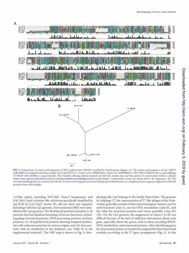
Interestingly, Cp1 encodes a viral RNA polymerase (RNAP;ORF33) (a single-subunit RNAP similar to the T7-type enzymes).Sequence analysis showed that Cp1 ORF33 was 71.4% identical tophiL7 p35 (ACE75775.1) and 31.5% identical to T7 RNAP(NP_041960) (Fig. 3A). Cp1 ORF33 also showed 40 to 50% aminoacid sequence identity with RNAPs of other Xanthomonas phages,
such as Xp10 (AAP58699.1), OP1 (BAE72738.1), and Xop411(ABK00180.1) (Fig. 3A). All important amino acid residues iden-tified in T7 RNAP for structure and function (31, 32) were con-served among these RNAPs. Their phylogenetic relationship isshown in Fig. 3B. The siphovirus Xp10 was shown to rely on bothhost and phage RNAPs, and the shift from host to phage RNAP isregulated by phage protein p7 (33). Xp10 p7 (73 amino acids [aa])is encoded by gene p45L, located after the replication module ofthe Xp10 genome. A similar regulation may work in Cp1 becauseORF44, encoding 66 aa with a sequence similarity to inhibitors oftranscriptional initiators and terminators (33% identical to Xp10p45L; AAP58713.1), was located at the corresponding position onCp1 DNA (Fig. 2). Like Xp10, the protein encoded by ORF44 mayfunction to inhibit host RNAP and act as an antiterminator, al-lowing RNAP to pass through the intrinsic terminator for expres-sion of the late genes (33).
Furthermore, we detected a homolog of the OP1 tail fiber pro-tein (Tfb, OP1-ORF25), which was thought to be involved in hostrange determination, mediated by variation in the combination ofrepetitive motifs at the N terminus (34).
Genomic analyses of Cp2: gene organization and homologyto other phages. The Cp2 genome was also a linear dsDNA ofapproximately 43.0 kbp, based on PFGE analysis (data notshown). The Cp2 DNA isolated from phage particles was sub-jected to shotgun sequencing. Sequences were assembled into acircular contig of 42,963 bp, also suggesting the presence of termi-nal repeats. The precise sequence of the repeat was not deter-mined. The Cp2 genome had a GC content of 66.7%, compara-ble with that of the host genome (ca. 64.7%). To find homologoussequences, nucleotide sequences from Cp2 were used to searchsequence databases. Short-ranged homologies were found in thesequences of Azospirillum brasilense Sp245 plasmid (accession no.HE577328; E value, 2e�11 [84 bit]) and Burkholderia pseudomal-lei bacteriophage phi1026b (accession no. AY453853; E value,5e�09 [76 bit]). Interestingly, a sequence in the genome of X.axonopodis pv. citri strain 306 also showed some homology to theCp2 genome (accession no. AE008923; E value, 8e�08 [72 bit]).The homologous sequence corresponded to a single gene (orf32 ofCp2, encoding a single-strand [ss] binding protein). However, the
FIG 2 Genomic organization of phages Cp1 (A) and phiL7 (B). Colored arrows indicate the directions and categories of the genes. Broken arrows and blackknobs indicate the �70-type promoters and predicted terminators for transcription, respectively. “No similarity” was determined using an E value lower than e�4as a cutoff for notable similarity.
Ahmad et al.
80 aem.asm.org Applied and Environmental Microbiology
on February 3, 2019 by guest
http://aem.asm
.org/D
ownloaded from
1.8-kbp region, including XAC1661 (Isxac3 transposase) andXAC1662 (repA) of strain 306, which was specifically amplified byrep-PCR for Cp1r/Cp2s strains (9), did not show any sequencehomology with the Cp2 genome. Forty potential ORFs were iden-tified in the Cp2 genome. The 40 deduced proteins included (i) 20proteins that had database homologs of known functions, includ-ing phage structural proteins, DNA processing proteins, and lysisproteins; (ii) 16 hypothetical proteins showing marginal similari-ties with unknown proteins of various origins; and (iii) four pro-teins with no similarities in the databases (see Table S2 in thesupplemental material). The ORF map is shown in Fig. 4. Mor-
phologically, Cp2 belongs to the family Podoviridae. The genomeof coliphage T7, the representative of T7-like phages of the Podo-viridae, generally consists of three functional gene clusters: one forearly functions (class I), one for DNA metabolism (class II), andthe other for structural proteins and virion assembly (class III)(35). For the Cp2 genome, the assignment of classes I to III wasdifficult because of the lack of sufficient information about eachgene, especially about key genes, such as those encoding RNAP,DNA metabolism, and structural proteins. After identifying genesfor structural proteins, we tentatively assigned the three functionalmodules according to the T7 gene arrangement (Fig. 4). In this
FIG 3 Comparison of amino acid sequences of RNA polymerases (RNAP) encoded by Xanthomonas phages. (A) The amino acid sequence of Cp1 ORF33(AB720063) was aligned with those of phiL7 p35 (ACE75775.1)., Xop411 p32 (ABK00180.1), Xp10 32L (AAP58699.1), OP1 ORF33 (BAE72738.1), and coliphageT7 RNAP (NP_041960.1) using ClustalX. The ClustalX coloring scheme depends on both the residue type and the pattern of conservation within a column(http://www.cgl.ucsf.edu/chimera/docs/ContributedSoftware/multalignviewer/cxcolor.html). Conservation scores are drawn below the alignment. (B) Theunrooted dendrogram was constructed with the Treeview tool using the maximum likelihood method based on a complete protein sequence alignment of RNAPproteins from other phages.
Bacteriophages of Citrus Canker Bacteria
January 2014 Volume 80 Number 1 aem.asm.org 81
on February 3, 2019 by guest
http://aem.asm
.org/D
ownloaded from
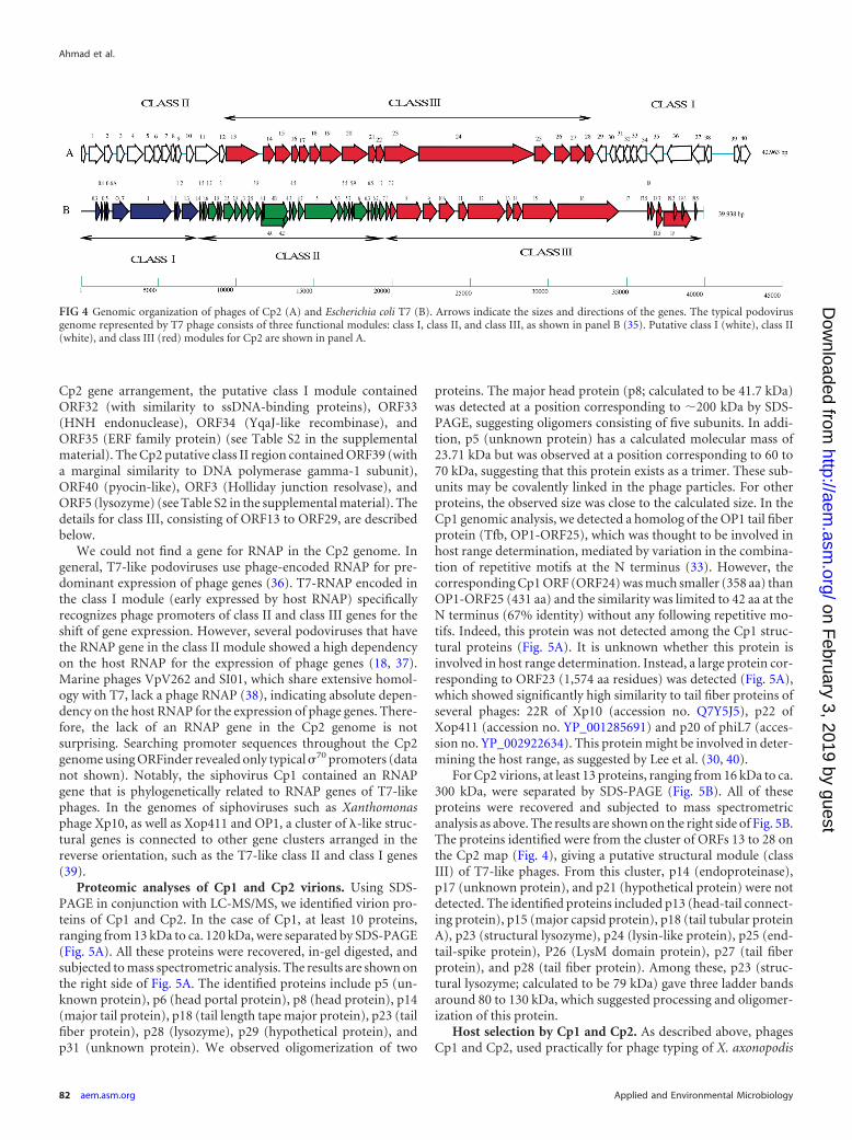
Cp2 gene arrangement, the putative class I module containedORF32 (with similarity to ssDNA-binding proteins), ORF33(HNH endonuclease), ORF34 (YqaJ-like recombinase), andORF35 (ERF family protein) (see Table S2 in the supplementalmaterial). The Cp2 putative class II region contained ORF39 (witha marginal similarity to DNA polymerase gamma-1 subunit),ORF40 (pyocin-like), ORF3 (Holliday junction resolvase), andORF5 (lysozyme) (see Table S2 in the supplemental material). Thedetails for class III, consisting of ORF13 to ORF29, are describedbelow.
We could not find a gene for RNAP in the Cp2 genome. Ingeneral, T7-like podoviruses use phage-encoded RNAP for pre-dominant expression of phage genes (36). T7-RNAP encoded inthe class I module (early expressed by host RNAP) specificallyrecognizes phage promoters of class II and class III genes for theshift of gene expression. However, several podoviruses that havethe RNAP gene in the class II module showed a high dependencyon the host RNAP for the expression of phage genes (18, 37).Marine phages VpV262 and SI01, which share extensive homol-ogy with T7, lack a phage RNAP (38), indicating absolute depen-dency on the host RNAP for the expression of phage genes. There-fore, the lack of an RNAP gene in the Cp2 genome is notsurprising. Searching promoter sequences throughout the Cp2genome using ORFinder revealed only typical �70 promoters (datanot shown). Notably, the siphovirus Cp1 contained an RNAPgene that is phylogenetically related to RNAP genes of T7-likephages. In the genomes of siphoviruses such as Xanthomonasphage Xp10, as well as Xop411 and OP1, a cluster of �-like struc-tural genes is connected to other gene clusters arranged in thereverse orientation, such as the T7-like class II and class I genes(39).
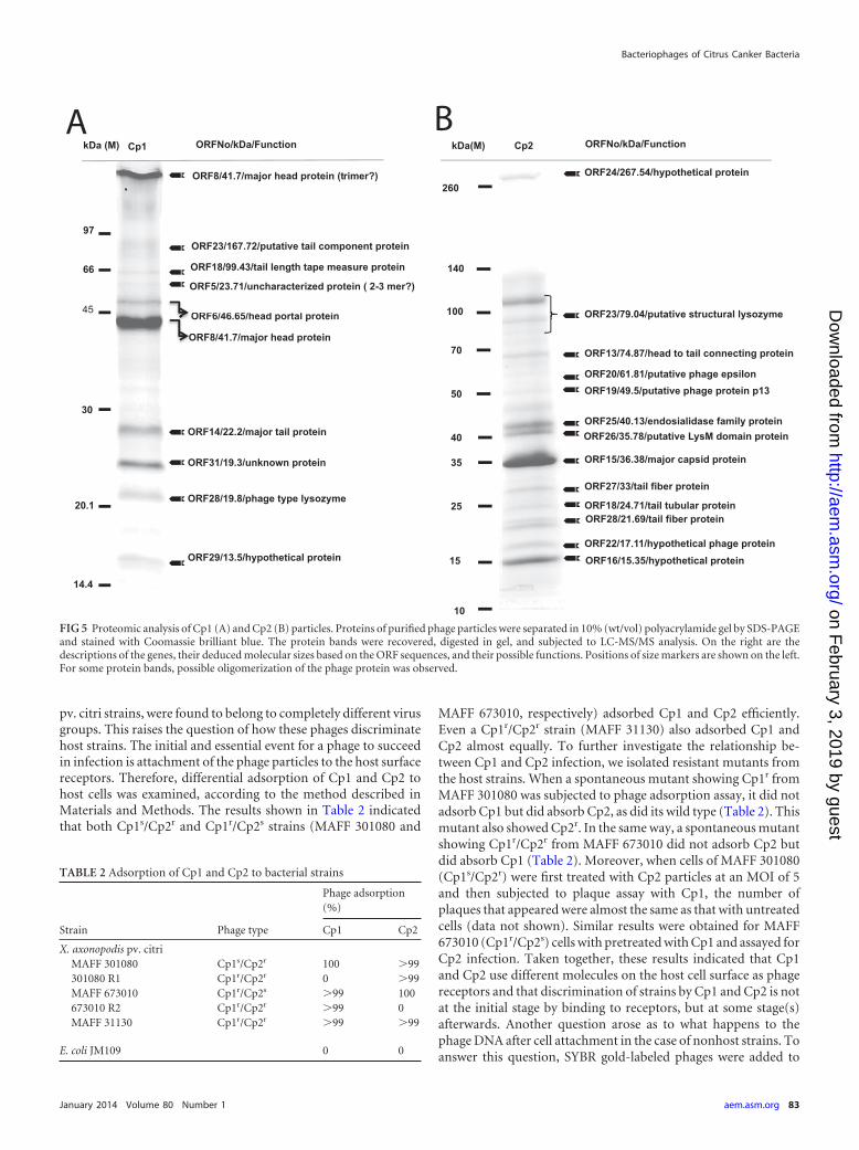
Proteomic analyses of Cp1 and Cp2 virions. Using SDS-PAGE in conjunction with LC-MS/MS, we identified virion pro-teins of Cp1 and Cp2. In the case of Cp1, at least 10 proteins,ranging from 13 kDa to ca. 120 kDa, were separated by SDS-PAGE(Fig. 5A). All these proteins were recovered, in-gel digested, andsubjected to mass spectrometric analysis. The results are shown onthe right side of Fig. 5A. The identified proteins include p5 (un-known protein), p6 (head portal protein), p8 (head protein), p14(major tail protein), p18 (tail length tape major protein), p23 (tailfiber protein), p28 (lysozyme), p29 (hypothetical protein), andp31 (unknown protein). We observed oligomerization of two
proteins. The major head protein (p8; calculated to be 41.7 kDa)was detected at a position corresponding to �200 kDa by SDS-PAGE, suggesting oligomers consisting of five subunits. In addi-tion, p5 (unknown protein) has a calculated molecular mass of23.71 kDa but was observed at a position corresponding to 60 to70 kDa, suggesting that this protein exists as a trimer. These sub-units may be covalently linked in the phage particles. For otherproteins, the observed size was close to the calculated size. In theCp1 genomic analysis, we detected a homolog of the OP1 tail fiberprotein (Tfb, OP1-ORF25), which was thought to be involved inhost range determination, mediated by variation in the combina-tion of repetitive motifs at the N terminus (33). However, thecorresponding Cp1 ORF (ORF24) was much smaller (358 aa) thanOP1-ORF25 (431 aa) and the similarity was limited to 42 aa at theN terminus (67% identity) without any following repetitive mo-tifs. Indeed, this protein was not detected among the Cp1 struc-tural proteins (Fig. 5A). It is unknown whether this protein isinvolved in host range determination. Instead, a large protein cor-responding to ORF23 (1,574 aa residues) was detected (Fig. 5A),which showed significantly high similarity to tail fiber proteins ofseveral phages: 22R of Xp10 (accession no. Q7Y5J5), p22 ofXop411 (accession no. YP_001285691) and p20 of phiL7 (acces-sion no. YP_002922634). This protein might be involved in deter-mining the host range, as suggested by Lee et al. (30, 40).
For Cp2 virions, at least 13 proteins, ranging from 16 kDa to ca.300 kDa, were separated by SDS-PAGE (Fig. 5B). All of theseproteins were recovered and subjected to mass spectrometricanalysis as above. The results are shown on the right side of Fig. 5B.The proteins identified were from the cluster of ORFs 13 to 28 onthe Cp2 map (Fig. 4), giving a putative structural module (classIII) of T7-like phages. From this cluster, p14 (endoproteinase),p17 (unknown protein), and p21 (hypothetical protein) were notdetected. The identified proteins included p13 (head-tail connect-ing protein), p15 (major capsid protein), p18 (tail tubular proteinA), p23 (structural lysozyme), p24 (lysin-like protein), p25 (end-tail-spike protein), P26 (LysM domain protein), p27 (tail fiberprotein), and p28 (tail fiber protein). Among these, p23 (struc-tural lysozyme; calculated to be 79 kDa) gave three ladder bandsaround 80 to 130 kDa, which suggested processing and oligomer-ization of this protein.
Host selection by Cp1 and Cp2. As described above, phagesCp1 and Cp2, used practically for phage typing of X. axonopodis
FIG 4 Genomic organization of phages of Cp2 (A) and Escherichia coli T7 (B). Arrows indicate the sizes and directions of the genes. The typical podovirusgenome represented by T7 phage consists of three functional modules: class I, class II, and class III, as shown in panel B (35). Putative class I (white), class II(white), and class III (red) modules for Cp2 are shown in panel A.
Ahmad et al.
82 aem.asm.org Applied and Environmental Microbiology
on February 3, 2019 by guest
http://aem.asm
.org/D
ownloaded from
pv. citri strains, were found to belong to completely different virusgroups. This raises the question of how these phages discriminatehost strains. The initial and essential event for a phage to succeedin infection is attachment of the phage particles to the host surfacereceptors. Therefore, differential adsorption of Cp1 and Cp2 tohost cells was examined, according to the method described inMaterials and Methods. The results shown in Table 2 indicatedthat both Cp1s/Cp2r and Cp1r/Cp2s strains (MAFF 301080 and
MAFF 673010, respectively) adsorbed Cp1 and Cp2 efficiently.Even a Cp1r/Cp2r strain (MAFF 31130) also adsorbed Cp1 andCp2 almost equally. To further investigate the relationship be-tween Cp1 and Cp2 infection, we isolated resistant mutants fromthe host strains. When a spontaneous mutant showing Cp1r fromMAFF 301080 was subjected to phage adsorption assay, it did notadsorb Cp1 but did absorb Cp2, as did its wild type (Table 2). Thismutant also showed Cp2r. In the same way, a spontaneous mutantshowing Cp1r/Cp2r from MAFF 673010 did not adsorb Cp2 butdid absorb Cp1 (Table 2). Moreover, when cells of MAFF 301080(Cp1s/Cp2r) were first treated with Cp2 particles at an MOI of 5and then subjected to plaque assay with Cp1, the number ofplaques that appeared were almost the same as that with untreatedcells (data not shown). Similar results were obtained for MAFF673010 (Cp1r/Cp2s) cells with pretreated with Cp1 and assayed forCp2 infection. Taken together, these results indicated that Cp1and Cp2 use different molecules on the host cell surface as phagereceptors and that discrimination of strains by Cp1 and Cp2 is notat the initial stage by binding to receptors, but at some stage(s)afterwards. Another question arose as to what happens to thephage DNA after cell attachment in the case of nonhost strains. Toanswer this question, SYBR gold-labeled phages were added to
ORF18/99.43/tail length tape measure protein
97
66
14.4
45
30
20.1
ORF6/46.65/head portal protein
ORF8/41.7/major head protein
ORF5/23.71/uncharacterized protein ( 2-3 mer?)
ORF14/22.2/major tail protein
ORF23/167.72/putative tail component protein
! ORF8/41.7/major head protein (trimer?)
!
!!
!
ORF31/19.3/unknown protein
ORF28/19.8/phage type lysozyme
ORF29/13.5/hypothetical protein
!
!
!
kDa (M) Cp1 ORFNo/kDa/Function
OR
OR
OR
!
ORF27/33/tail fiber protein
ORF22/17.11/hypothetical phage protein
260
50
40
35
25
15
100
70
140
!
!
!
!
!
!!
!
!
!
!
!
ORF24/267.54/hypothetical protein
ORF23/79.04/putative structural lysozyme
ORF13/74.87/head to tail connecting protein
ORF20/61.81/putative phage epsilonORF19/49.5/putative phage protein p13
ORF25/40.13/endosialidase family protein
ORF15/36.38/major capsid protein
ORF18/24.71/tail tubular protein
ORF16/15.35/hypothetical protein
ORF28/21.69/tail fiber protein
kDa(M) Cp2 ORFNo/kDa/Function
!
ORF26/35.78/putative LysM domain protein
A B
FIG 5 Proteomic analysis of Cp1 (A) and Cp2 (B) particles. Proteins of purified phage particles were separated in 10% (wt/vol) polyacrylamide gel by SDS-PAGEand stained with Coomassie brilliant blue. The protein bands were recovered, digested in gel, and subjected to LC-MS/MS analysis. On the right are thedescriptions of the genes, their deduced molecular sizes based on the ORF sequences, and their possible functions. Positions of size markers are shown on the left.For some protein bands, possible oligomerization of the phage protein was observed.
TABLE 2 Adsorption of Cp1 and Cp2 to bacterial strains
Strain Phage type
Phage adsorption(%)
Cp1 Cp2
X. axonopodis pv. citriMAFF 301080 Cp1s/Cp2r 100 �99301080 R1 Cp1r/Cp2r 0 �99MAFF 673010 Cp1r/Cp2s �99 100673010 R2 Cp1r/Cp2r �99 0MAFF 31130 Cp1r/Cp2r �99 �99
E. coli JM109 0 0
Bacteriophages of Citrus Canker Bacteria
January 2014 Volume 80 Number 1 aem.asm.org 83
on February 3, 2019 by guest
http://aem.asm
.org/D
ownloaded from
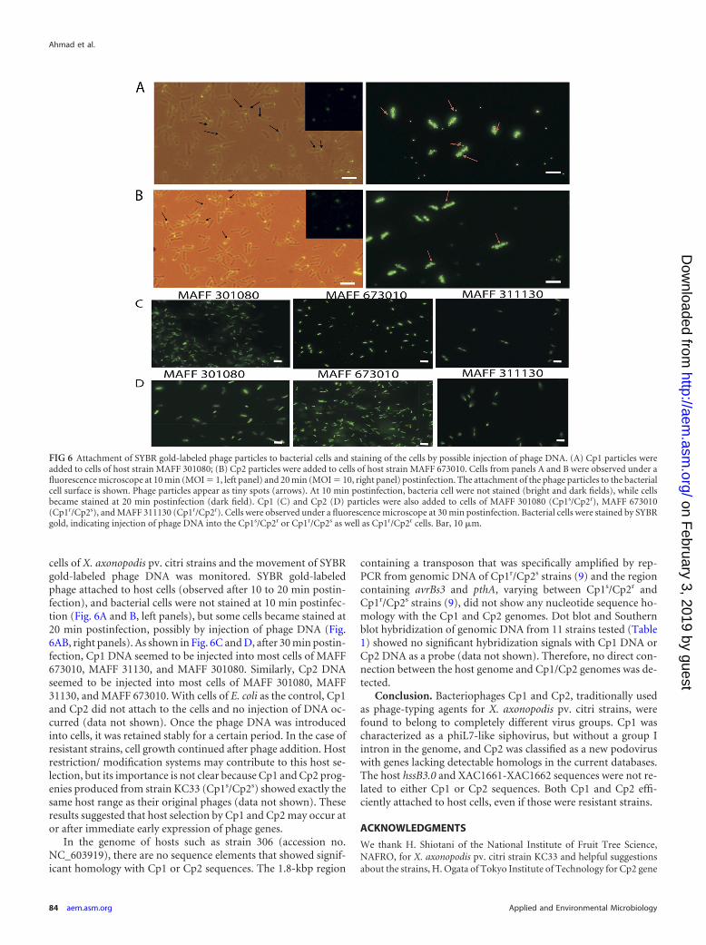
cells of X. axonopodis pv. citri strains and the movement of SYBRgold-labeled phage DNA was monitored. SYBR gold-labeledphage attached to host cells (observed after 10 to 20 min postin-fection), and bacterial cells were not stained at 10 min postinfec-tion (Fig. 6A and B, left panels), but some cells became stained at20 min postinfection, possibly by injection of phage DNA (Fig.6AB, right panels). As shown in Fig. 6C and D, after 30 min postin-fection, Cp1 DNA seemed to be injected into most cells of MAFF673010, MAFF 31130, and MAFF 301080. Similarly, Cp2 DNAseemed to be injected into most cells of MAFF 301080, MAFF31130, and MAFF 673010. With cells of E. coli as the control, Cp1and Cp2 did not attach to the cells and no injection of DNA oc-curred (data not shown). Once the phage DNA was introducedinto cells, it was retained stably for a certain period. In the case ofresistant strains, cell growth continued after phage addition. Hostrestriction/ modification systems may contribute to this host se-lection, but its importance is not clear because Cp1 and Cp2 prog-enies produced from strain KC33 (Cp1s/Cp2s) showed exactly thesame host range as their original phages (data not shown). Theseresults suggested that host selection by Cp1 and Cp2 may occur ator after immediate early expression of phage genes.
In the genome of hosts such as strain 306 (accession no.NC_603919), there are no sequence elements that showed signif-icant homology with Cp1 or Cp2 sequences. The 1.8-kbp region
containing a transposon that was specifically amplified by rep-PCR from genomic DNA of Cp1r/Cp2s strains (9) and the regioncontaining avrBs3 and pthA, varying between Cp1s/Cp2r andCp1r/Cp2s strains (9), did not show any nucleotide sequence ho-mology with the Cp1 and Cp2 genomes. Dot blot and Southernblot hybridization of genomic DNA from 11 strains tested (Table1) showed no significant hybridization signals with Cp1 DNA orCp2 DNA as a probe (data not shown). Therefore, no direct con-nection between the host genome and Cp1/Cp2 genomes was de-tected.
Conclusion. Bacteriophages Cp1 and Cp2, traditionally usedas phage-typing agents for X. axonopodis pv. citri strains, werefound to belong to completely different virus groups. Cp1 wascharacterized as a phiL7-like siphovirus, but without a group Iintron in the genome, and Cp2 was classified as a new podoviruswith genes lacking detectable homologs in the current databases.The host hssB3.0 and XAC1661-XAC1662 sequences were not re-lated to either Cp1 or Cp2 sequences. Both Cp1 and Cp2 effi-ciently attached to host cells, even if those were resistant strains.
ACKNOWLEDGMENTS
We thank H. Shiotani of the National Institute of Fruit Tree Science,NAFRO, for X. axonopodis pv. citri strain KC33 and helpful suggestionsabout the strains, H. Ogata of Tokyo Institute of Technology for Cp2 gene
FIG 6 Attachment of SYBR gold-labeled phage particles to bacterial cells and staining of the cells by possible injection of phage DNA. (A) Cp1 particles wereadded to cells of host strain MAFF 301080; (B) Cp2 particles were added to cells of host strain MAFF 673010. Cells from panels A and B were observed under afluorescence microscope at 10 min (MOI 1, left panel) and 20 min (MOI 10, right panel) postinfection. The attachment of the phage particles to the bacterialcell surface is shown. Phage particles appear as tiny spots (arrows). At 10 min postinfection, bacteria cell were not stained (bright and dark fields), while cellsbecame stained at 20 min postinfection (dark field). Cp1 (C) and Cp2 (D) particles were also added to cells of MAFF 301080 (Cp1s/Cp2r), MAFF 673010(Cp1r/Cp2s), and MAFF 311130 (Cp1r/Cp2r). Cells were observed under a fluorescence microscope at 30 min postinfection. Bacterial cells were stained by SYBRgold, indicating injection of phage DNA into the Cp1s/Cp2r or Cp1r/Cp2s as well as Cp1r/Cp2r cells. Bar, 10 �m.
Ahmad et al.
84 aem.asm.org Applied and Environmental Microbiology
on February 3, 2019 by guest
http://aem.asm
.org/D
ownloaded from
annotation, and M. Nakano of ADSM, Hiroshima University, for hertechnical guidance on LC-MS/MS analysis.
REFERENCES1. Civerelo EL. 1984. Bacterial canker disease of citrus. J. Rio. Grande Val.
Hort. Soc. 37:127–145.2. Gottwald TR, Graham JH, Schubert TS. 2002. Citrus canker: the pathogen
and its impact. Plant health progress. http://www.plantmanagementnetwork.org/pub/php/review/citruscanker/.
3. Stall RE, Civerolo EL. 1991. Research relating to the recent outbreak ofcitrus canker in Florida. Annu. Rev. Phytopathol. 29:399 – 420.
4. Wakimoto S. 1967. Some characteristics of citrus canker bacteria, Xan-thomonas citri (Hasse) Dowson, and the related phages isolated fromJapan. Ann. Phytopathol. Soc. Jpn. 33:301–310.
5. Obata T. 1974. Distribution of Xanthomonas citri strain in relation to thesensitivity to phage CP1 and CP2. Ann. Phytopathol. Soc. Jpn. 40:6 –13.
6. Goto M, Takahashi T, Messina MA. 1980. A comparative study of thestrains of Xanthomonas campestris pv. citri isolated from citrus canker inJapan and cancrosis B in Argentina. Ann. Phytopathol. Soc. Jpn. 46:329 –338.
7. Shiotani H, Tsuyumu S, Ozaki K. 2000. Pathogenic interactions betweenXanthomonas axonopodis pv. citri and cultivars of Pummelo (Citrus grandis).Phytopathology 90:1383–1389. http://dx.doi.org/10.1094/PHYTO.2000.90.12.1383.
8. Louws FJ, Fulbright DW, Stephens CT, de Bruijn FJ. 1994. Specificgenomic fingerprints of phytopathogenic Xanthomonas and Pseudomonaspathovars and strains generated with repetitive sequences and PCR. Appl.Environ. Microbiol. 60:2286 –2295.
9. Shiotani H. 2007. Dissertation. The United Graduate School of Agricul-tural Science, Gifu University, Gifu, Japan.
10. Swarup S, Yang Y, Kingsley MT, Gabriel DW. 1992. An Xanthomonascitri pathogenicity gene, phcA, pleiotropically encodes gratuitous aviru-lence on nonhosts. Mol. Plant Microbe Interact. 5:204 –213.
11. Szurek B, Rossier O, Hause G, Bonas U. 2002. Type III dependent translo-cation of the Xanthomonas AvrBs3 protein into the plant cell. Mol. Microbiol.46:13–23. http://dx.doi.org/10.1046/j.1365-2958.2002.03139.x.
12. Shiotani H, Fujikawa T, Ishihara H, Tsuyumu S, Ozaki K. 2007. A pthAhomolog from Xanthomonas axonopodis pv. citri responsible for host-specific suppression of virulence. J. Bacteriol. 189:3271–3279. http://dx.doi.org/10.1128/JB.01790-06.
13. Sambrook J, Russell DW. 2001. Molecular cloning: a laboratory manual,3rd ed. Cold Spring Harbor Laboratory Press, Cold Spring Harbor, NY.
14. Dykstra MJ. 1993. A manual of applied technique for biological electronmicroscopy. Plenum Press, New York, NY.
15. Yamada T, Kawasaki T, Nagata S, Fujiwara A, Usami S, Fujie M. 2007.New bacteriophages that infect the phytopathogen Ralstonia so-lanacearum. Microbiology 153:2630 –2639. http://dx.doi.org/10.1099/mic.0.2006/001453-0.
16. Hung CH, Yang CF, Yang CY, Tseng YH. 2003. Involvement of tonB-exbBD1D2 operon in infection of Xanthomonas campestris phage phiL7.Biochem. Biophys. Res. Commun. 302:878 – 884. http://dx.doi.org/10.1016/S0006-291X(03)00255-9.
17. Carlson K. 1994. Single-step growth, p 434 – 437. In Karam J, Drake JW,Kreuzer KN, Mosig G, Hall DH, Eiserlig FA, Black LW, Spicer EK, KutterE, Carlson K, Miller ES (ed), Molecular biology of bacteriophage T4. ASMPress, Washington, DC.
18. Kawasaki T, Shimizu M, Satsuma H, Fujiwara A, Fujie M, Usami S,Yamada T. 2009. Genomic characterization of Ralstonia solanacearumphage RSB1, a T7-like wide-host-range phage. J. Bacteriol. 191:422– 427.http://dx.doi.org/10.1128/JB.01263-08.
19. Higashiyama T, Yamada T. 1991. Electrophoretic karyotyping and chro-mosomal gene mapping of Chlorella. Nucleic Acids Res. 19:6191– 6195.
20. Delcher AL, Harmon D, Kasif S, White O, Salzberg SL. 1999. Improvedmicrobial gene identification with GLIMMER. Nucleic Acids Res. 27:4636 – 4641.
21. Altschul SF, Madden TL, Schaffer AA, Zhang Z, Miller W, Lipman DJ.1997. Gapped BLAST and PSI-BLAST: a new generation of protein data-base search programs. Nucleic Acids Res. 25:3389 –3402.
22. UniProt Consortium. 2007. The Universal Protein Resource (UniProt). Nu-cleic Acids Res. 35:D193–D197. http://dx.doi.org/10.1093/nar/gkl929.
23. Wheeler DL, Barrett T, Benson DA, Bryant SH, Canese K, Chetvrnin V,Church DM, DiCuccio M, Edgar E, Federhen S, Geer LY, Kapustin Y,Khovayko O, Landsman D, Lipman DJ, Madden TL, Maglott DR,Ostell J, Miller V, Pruitt KD, Schuler GD, Sequeira E, Sherry ST,Sirotkin K, Souvorov A, Starchenko G, Tatusov RL, Tatusova TA,Wagner L, Yaschenko E. 2007. Database resources of the National Centerfor Biotechnology Information. Nucleic Acids Res. 35:D5–D12. http://dx.doi.org/10.1093/nar/gkl1031.
24. Fujiwara A, Fujisawa M, Hamasaki R, Kawasaki T, Fujie M, Yamada T.2011. Biocontrol of Ralstonia solanacearum by treatment with lytic bacte-riophages. Appl. Environ. Microbiol. 77:4155– 4162. http://dx.doi.org/10.1128/AEM.02847-10.
25. Ausubel F, Brent R, Kingston RE, Moore DD, Seidman JG, Smith JA,Struhl K. 1995. Short protocols in molecular biology, 3rd ed. John Wileyand Sons, Inc., Hoboken, NJ.
26. Laemmli UK. 1970. Cleavage of structural proteins during the assembly ofthe head of bacteriophage T4. Nature 227:680 – 685.
27. Mosier-Boss PA, Lieberman SH, Andrews JM, Rohwer FL, Wegley LE,Breitbart M. 2003. Use of fluorescently labeled phage in the detection andidentification of bacterial species. Appl. Spectrosc. 57:1138 –1144. http://dx.doi.org/10.1366/00037020360696008.
28. Arai K, Shimo H, Doi Y, Yira K. 1974. Electron microscopy of Xan-thomonas citri phages CP1 and CP2 infection. Ann. Phytopathol. Soc. Jpn.40:98 –102.
29. Hadas H, Einav M, Fishov I, Zaritsky A. 1997. Bacteriophage T4 devel-opment depends on the physiology of its host Escherichia coli. Microbiol-ogy 143:179 –185.
30. Lee CN, Lin JW, Weng SF, Tseng YH. 2009. Genomic characterizationof the intron-containing T7-like phage phiL7 of Xanthomonas campestris.Appl. Environ. Microbiol. 75:7828 –7837.
31. Cheetham GM, Jeruzalmi D, Steiz TA. 1999. Structural basis for initia-tion of transcription from an RNA polymerase-promoter complex. Na-ture 399:80 – 83.
32. Osumi-Davis PA, de Aguilera MC, Woody RW, Woody AY. 1992.Asp537, Asp812 are essential and Lys631, His811 are catalytically sig-nificant in bacteriophage T7 RNA polymerase activity. J. Mol. Biol.226:37– 45.
33. Semenova E, Djordjevie M, Shraiman B, Severinov K. 2005. The tale oftwo RNA polymerases: transcription profiling and gene expression strat-egy of bacteriophage Xp10. Mol. Microbiol. 55:764 –777. http://dx.doi.org/10.1111/j.1365-2958.2004.04442.x.
34. Inoue Y, Matsuura T, Ohara T, Azekami K. 2006. Bacteriophage OP1,lytic for Xanthomonas oryzae pv. oryzae, changes its host range by dupli-cation and deletion of the small domain in the deduced tail fiber gene. J.Gen. Plant Pathol. 72:111–118. http://dx.doi.org/10.1007/s10327-005-0252-x.
35. Dunn JJ, Studier FW. 1983. Complete nucleotide sequence of bacterio-phage T7 DNA and the locations of T7 genetic elements. J. Mol. Biol.166:477–535.
36. Molineux IJ. 1999. T7-like phages (Podoviridae), p 1722–1729. In GranoffA, Webster R (ed), Encyclopedia of virology. Academic Press, London,United Kingdom.
37. Ceyssens PJ, Lavigne R, Mattheus W, Chibeu A, Hertveldt K, Mast J,Robben J, Volckaert G. 2006. Genomic analysis of Pseudomonas aerugi-nosa phages LKD16 and LKA1: establishment of the KMV subgroupwithin the T7 subgroup. J. Bacteriol. 188:6924 – 6931. http://dx.doi.org/10.1128/JB.00831-06.
38. Hardies SC, Comeau AM, Serwer P, Suttle CA. 2003. The completesequence of marine bacteriophage VpV262 infecting Vibrio parahaemo-lyticus indicates that an ancestral component of a T7 viral supergroup iswidespread in the marine environment. Virology 310:359 –371. http://dx.doi.org/10.1016/S0042-6822(03)00172-7.
39. Yuzenkova J, Nechaev S, Berlin J, Rogulja D, Kuznedelov K, Inman R,Mushegian A, Severinov K. 2003. Genome of Xanthomonas oryzae bac-teriophage XP10: an odd T-odd phage. J. Mol. Biol. 330:735–748. http://dx.doi.org/10.1016/S0022-2836(03)00634-X.
40. Lee CN, Hu RM, Chow TY, Lin JW, Chen HY, Tseng YH, Weng SF.2007. Comparison of genomes of three Xanthomonas oryzae bacterio-phages. BMC Genomics 8:442– 453. http://dx.doi.org/10.1186/1471-2164-8-442.
Bacteriophages of Citrus Canker Bacteria
January 2014 Volume 80 Number 1 aem.asm.org 85
on February 3, 2019 by guest
http://aem.asm
.org/D
ownloaded from


![arXiv:math/0504269v3 [math.QA] 27 Mar 2006 · Drinfeld [Dr1] and Jimbo [Jim] associated, independently, to any symmetrizable Kac-Moody alge- ... CP1, CP2, CP3, EM, FR, FM, H5, Kas2,](https://img.pdfslide.net/doc/110x75/5c01857b09d3f20a538cc84f/arxivmath0504269v3-mathqa-27-mar-2006-drinfeld-dr1-and-jimbo-jim-associated.jpg)