Embed Size (px)
Citation preview
B I O C H E M I S T R Y
Federation of European Biochemical Societies, Vienna, Pergammon, p 9.
Littauer, U. Z., Revel, M., and Stern, R. (1966), Coldspring Harbor Symp. Quant. Biol. (in press).
Marmur, J. (1961), J . Mol.BioI. 3,208. Monier, R., Stephenson, M. L., and Zamecnik, P. C.
(1960), Biochim. Biophys. Acta 43, 1. Price, T. D., Hinds, H. A,, and Brown, R. S. (1963),
J.Biol.Chem.238,311. Revel, M., and Littauer, U. Z. (1966), J. Mol. Biol. 15,
389.
Sluyser, M., and Bosch, L. (1962), Biochim. Biophys.
Smith, J. D., and D u m , D. B. (1959), Biochem. J. 72,
Srinivasan, P. R., and Borek, E. (1963), Proc. Natl.
Srinivasan, P. R., and Borek, E. (1964), Science 145,
Svensson, I., Boman, H. G., Eriksson, K. G., and
Zubay, G. (1962), J. Mol. Biol. 4,347.
Acta 55,479.
294.
Acad. Sci. (1. S. 49, 529.
548.
Kjellin, K. (1963), J. Mol. Biol. 7,254.
Characterization of Isoenzymes of Adenosine Triphosphate : D-Hexose 6-Phosphotransferase from Rat Liver”
C. Gonzilez, T. Ureta,? J. Babul, E. Rabajille, and H. Niemeyer
ABSTRACT: Four isoenzymes of adenosine triphos- phate (ATP) :D-hexose 6-phosphotransferase have been separated from rat liver by DEAE-cellulose. Three of these isoenzymes ( A X ) are similar to animal hexokinases inasmuch as they exhibit a low K, for glucose (10-j-10-4 M) and the rate of phosphorylation of fructose is slightly higher than that of glucose. Isoen- zyme D has been further purified by fractionation with ammonium sulfate and by chromatography on hy- droxylapatite. This isoenzyme would correspond to glucokinase since it presents a high K, for glucose (1.8 x M), and a low activity with fructose as a substrate; it also catalyzes the phosphorylation of
A great deal of interest has arisen in the last few years on the regulation of glucose phosphorylating activity in rat liver. Changes in the tissue levels of ATP1 :hexose phosphotransferase have been observed under three main circumstances: (1) variations in the supply of glucose in the diet, including total fasting (Vaughan et al., 1960; DiPietro and Weinhouse, 1960; Niemeyer et al., 1962, 1963; Perez et al.. 1964); (2) availability of insulin (DiPietro and Weinhouse, 1960;
mannose and 2-deoxyglucose. The four isoenzymes use only ATP as phosphate donor, with K, values of about 5 X M. The independence of the affinity of gluco- kinase for glucose or ATP on the concentration of the other substrate is in agreement with the presence of two separate binding sites on the enzyme. The four isoenzymes are competitively inhibited by N-acetyl- glucosamine (Ki about 5 X M). High concentrations of glucose inhibit isoenzymes C and D. Mannose also inhibits isoenzyme D. Glucose 6-phosphate inhibits glucokinase (Ki = 1.5 X 10⁻² M); the inhibition is com- petitive with respect to ATP, while it is not clearly defined with respect to glucose.
Salas et al., 1963; Niemeyer et al., 1967); and (3) initial stages of development after birth (Walker, 1963). Of special significance were the reports by Walker (1963) and Viiiuela et al. (1963), describing two protein fractions with glucose phosphorylating activity in liver. Only one of these proteins, characterized by its high K , for glucose and called glucokinase, was amen- able to changes under the various above-mentioned conditions. We separated four fractions or isoenzymes*
460
* From the Instituto de Quimica Fisiolbgica y Patologica, Universidad de Chile, Santiago, Chile. Received September 26, 1966. This work was supported by U. S . Public Health Service Grant A M 07363-03 and by a joint program of the Faculty of Medicine, University of Chile, and the Rockefeller Foundation (Grant 64-53). Parts of this work have been presented at the Annual Meeting of the “Sociedad de Biologia de Santiago” (Gonzllez et al., 1965).
t On leave of absence at the Rockefeller University.
1 Abbreviations used: ATP, adenosine triphosphate; NADPH%, reduced nicotinamide-adenine dinucleotide phos- phate; NADP+, oxidized nicotinamide-adenine dinucleotide phosphate; NAD+, oxidized nicotinamide-adenine dinucleotide; NADH,, reduced nicotinamide-adenine dinucleotide; UTP, uridine triphosphate; Glc-6-P, glucose 6-phosphate.
2 The term isoenzyme is used here in the broad sense accepted by the Standing Committee on Enzymes of the International Union of Biochemistry (Webb, 1964).
C. G O N Z A L E Z , T. U K E T A , J. B A B U L , E. K A B A J I L L E , A N D H. N I E M E Y E R
V O L . 6, N O . 2, F E B R U A R Y 1 9 6 7
of ATP :hexose phosphotransferase by chromatography in DEAE-cellulose (Gonzhlez et al., 1964) and again only one, isoenzyme D, corresponding to glucokinase, varied markedly under the conditions tested.
In the present work the detailed procedures to isolate the four isoenzymes, as well as a study of their kinetic properties, are presented. Special emphasis has been given to the purification and characterization of iso- enzyme D or glucokinase because of its potential physiological significance
Experimental Section
lsolation of the Isoenzymes. Adult albino rats were used. They were well fed with a balanced stock diet (Niemeyer et al., 1962). After killing the animals by decapitation without anesthesia the livers were excised and chilled in cracked ice, blotted, and weighed. Homogenates (50%, w/v) were prepared from the pooled livers of 4-16 rats in 10 mM Tris-1 mM EDTA-6- 10 mM 2-mercaptoethanol, pH 7.0 (medium A), and centrifuged at 105,OOOg for 60 min in a Spinco prepara- tive ultracentrifuge. All procedures were carried out at 0-4". The clear supernatant fluids were treated batch- wise or in a column with CM-Sephadex (or CM-cellu- lose) equilibrated with medium A. The material not absorbed by the exchanger was concentrated at low temperature and reduced pressure and chromato- graphed on DEAE-cellulose ion exchanger. The diam- eter and height of the columns varied according to the number of animals used in each preparation. Linear gradient elution was carried out with KC1 from 0 to 0.5 M in medium A. Fractions were collected and the content of the tubes was examined for glucose phosphorylating activity and for protein.
Further Purification of Glucokinase. The details of the procedure have been reported (Babul and Niemeyer, 1966). In certain experiments the DEAE-cellulose treatment was shortened by washing the column with 0.18 M KC1 prepared in medium A, which eluted iso- enzymes A, B, and partially C. A linear gradient of concentration from 0.18 to 0.5 M KCI in medium A was then applied. The fractions which were free of isoenzyme C and had the highest specific activities were pooled. Glucose was added to a concentration of 50 mM and then solid ammonium sulfate was added to bring the concentration to 55% saturation. The pH was main- tained at 7 with NH40H. The precipitate obtained after centrifugation was discarded and glucokinase was precipitated from the supernatant fluid by adding more solid ammonium sulfate to bring the concentration to 80 saturation. The solution was again centrifuged. The precipitated enzyme could be stored as such at 0" for several months without appreciable loss in activity. Several preparations in this stage could be pooled in order to continue the purification procedure. The pooled precipitates were dissolved in a small amount of 5 mM potassium phosphate buffer, pH 6.8, prepared in medium B (50 mM glucose, 6 mM 2-mercaptoethanol, and 1 mM EDTA), and chromatographed on a column of hydroxylapatite (20 ml of Bio-Gel HT, 0.28 g
dry/ml of settled bed, mixed with 10 g of cellulose powder) equilibrated with the same buffer. The enzyme was eluted with a linear concentration gradient of potassium phosphate, between 5 and 150 mM at a pH increasing from 6.8 to 7.6, prepared in medium B. The flow rate was adjusted to about 3 ml/min and 5- to 6-ml fractions were collected. The eluates with the high- est specific activities were pooled and as soon as possible solid ammonium sulfate was added to bring the con- centration to 80 % saturation to precipitate the enzyme. The sediment collected by centrifugation was suspended in a small volume of 80% saturation ammonium sulfate and stored at 0". Aliquots of this suspension were dis- solved in 0.1 M KCI prepared in medium A for studies of enzyme properties. Table I presents a typical prepara- tion of glucokinase.
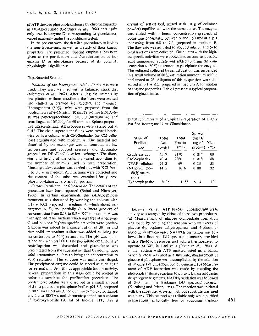
TABLE I : Summary of a Typical Preparation of Highly Purified Isoenzyme D or Glucokinase.
Sp Act. Stage of Total Total (units/ Purifica- Act. Protein mg of Yield
tion (units) (mg) protein) (%) Crude extract 45.7 3 170 0.014 100 CM-Sephadex 40.4 2260 0.018 88 DEAE-cellulose 24.2 69 0.35 53 (PH&SOc (55- 14.5 16.6 0.88 32
80% satura- tion)
Hydroxylapatite 8 .85 1 . 5 7 5.64 19
Enzyme Assay. ATP :hexose phosphotransferase activity was assayed by either of these two procedures. (a) Measurement of glucose 6-phosphate formation was made by coupling the reaction with an excess of glucose 6-phosphate dehydrogenase and 6-phospho- gluconic dehydrogenase. NADPH2 formation was fol- lowed in a Beckman DU spectrophotometer, provided with a Photovolt recorder and with a thermospacer to operate at 30°, in 1-ml cells (PCrez et al., 1964). A similar system with ATP omitted acted as a blank. When fructose was used as a substrate, measurement of glucose 6-phosphate was accomplished by the addition of an excess of phosphoglucose isomerase. (b) Measure- ment of ADP formation was made by coupling the phosphotransferase reaction to pyruvic kinase and lactic dehydrogenase systems. NADH2 oxidation was followed at 340 mp in a Beckman DU spectrophotometer (Kornberg and Pricer, 1951). The reaction was initiated with the substrate. A system without substrate was used as a blank. This method was reliable only when purified preparations, practically free of adenosine triphos- 46 1
A D E N O S I N E T R I P H 0 S P H A T E : D - H E X O S E 6-PHOSPHOTRANSFERASE I S O E N Z Y M E S
B I O C H E M I S T R Y
D
Volume of ef f luent (ml)
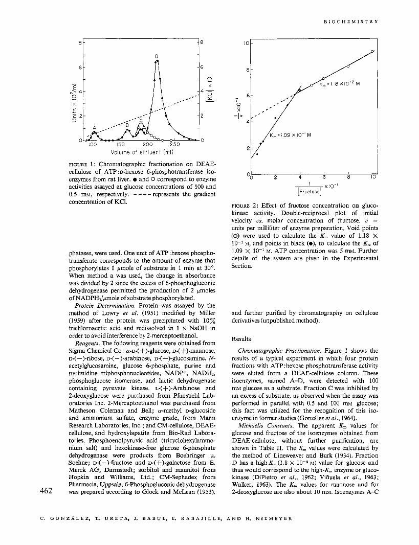
FIGURE 1 : Chromatographic fractionation on DEAE- cellulose of ATP :D-hexose 6-phosphotransferase iso- enzymes from rat liver. and 0 correspond to enzyme activities assayed at glucose concentrations of 100 and 0.5 m, respectively. - --- represents the gradient concentration of KC1.
phatases, were used. One unit of ATP:hexose phospho- transferase corresponds to the amount of enzyme that phosphorylates 1 pnole of substrate in 1 min at 30". When method a was used, the change in absorbance was divided by 2 since the excess of 6-phosphogluconic dehydrogenase permitted the production of 2 pmoles of NADPH2/pmole of substrate phosphorylated.
Protein Determination. Protein was assayed by the method of Lowry et a/. (1951) modified by Miller (1959) after the protein was precipitated with 10% trichloroacetic acid and redissolved in 1 N NaOH in order to avoid interference by 2-mercaptoethanol.
Reagents. The following reagents were obtained from Sigma Chemical Co : a - ~ - ( +)-glucose, D-( +)-mannose, D-( -)-ribose, D-( -)-arabinose, D-( +)-glucosamine, N- acetylglucosamine, glucose 6-phosphate, purine and pyrimidine triphosphonucleotides, NADP+, NADH?, phosphoglucose isomerase, and lactic dehydrogenase containing pyruvate kinase. L-(+)-Arabinose and 2-deoxyglucose were purchased from Pfanstiehl Lab- oratories Inc. 2-Mercaptoethanol was purchased from Matheson Coleman and Bell; a-methyl D-glucoside and ammonium sulfate, enzyme grade, from Mann Research Laboratories, Inc. ; and CM-cellulose, DEAE- cellulose, and hydroxylapatite from Bio-Rad Labora- tories. Phosphoenolpyruvic acid (tricyclohexylammo- nium salt) and hexokinase-free glucose 6-phosphate dehydrogenase were products from Boehringer u. Soehne; D-( -)-fructose and D-(+)-galactose from E. Merck AG, Darmstadt; sorbitol and mannitol from Hopkin and Williams, Ltd. ; CM-Sephadex from Pharmacia, Uppsala. 6-Phosphogluconic dehydrogenase was prepared according to Glock and McLean 11953), 462
I I I I I 2 4 6 a I C
' x10-1 [Fructose]
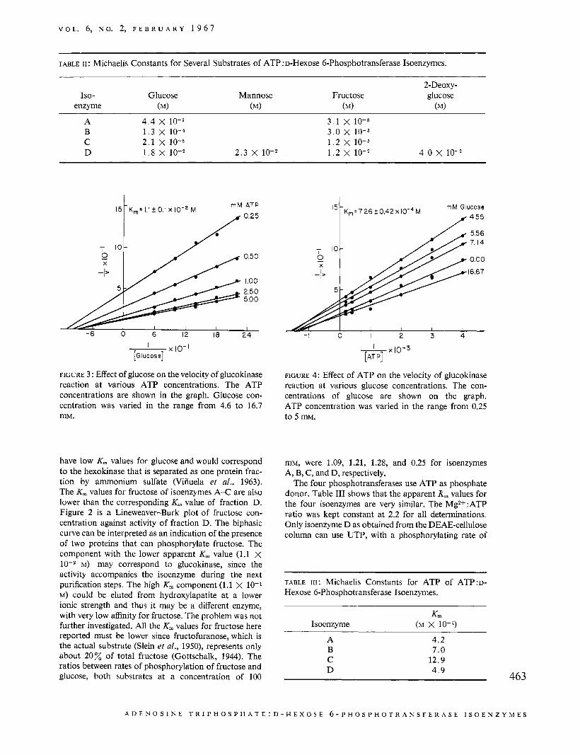
FIGURE 2: Effect of fructose concentration on gluco- kinase activity. Double-reciprocal plot of initial velocity L.S. molar concentration of fructose. u = units per milliliter of enzyme preparation. Void points (0) were used to calculate the K, value of 1.18 X lop2 M, and points in black (o), to calculate the K, of 1.09 X 10⁻¹ M. ATP concentration was 5 m. Further details of the system are given in the Experimental Section.
and further purified by chromatography on cellulose derivatives (unpublished method).
Results
Chrornatographic Fractionation. Figure 1 shows the results of a typical experiment in which four protein fractions with ATP : hexose phosphotransferase activity were eluted from a DEAE-cellulose column. These isoenzymes, named A-D, were detected with 100 mM glucose as a substrate. Fraction C was inhibited by an excess of substrate, as observed when the assay was performed in parallel with 0.5 and 100 mM glucose; this fact was utilized for the recognition of this iso- enzyme in former studies (Gonzhlez et al., 1964).
Michaelis Constants. The apparent K , values for glucose and fructose of the isoenzymes obtained from DEAE-cellulose, without further purification, are shown in Table 11. The K , values were calculated by the method of Lineweaver and Burk (1934). Fraction D has a high K, (1.8 X lo-* M) value for glucose and thus would correspond to the high-K,,, enzyme or gluco- kinase (DiPietro et al., 1962; Viiiuela et al., 1963; Walker, 1963). The K, values for mannose and for 2-deoxyglucose are also about 10 mM. Isoenzymes A-C
C. G O N Z h L E Z , T. U R E T A , J. B A B U L , E. R A B A J I L L E , A N D H. N I E M E Y E R
V O L . 6, N O . 2, F E B R U A R Y 1 9 6 7
~~~ ~
TABLE 11 : Michaelis Constants for Several Substrates of ATP :D-Hexose 6-Phosphotransferase Isoenzymes.
Iso- Glucose enzyme (MI
Mannose Fructose (MI (MI
2-Deoxy- glucose
(MI
A 4.4 x 10⁻⁵ 3 .1 x 10-3 B 1 . 3 x 10⁻⁴ 3 .0 x 10-3 C 2 .1 x 10-5 1 .2 x 10-3 D 1 . 8 x 10⁻² 2 .3 x lo-* 1.2 x 10⁻² 4 . 0 x lo-*
0.50
I .oo 2.50 5.00
1 - - 6 0 6 12 18 24
’ x t o - ’ [GI u c o s e]
FIGURE 3 : Effect of glucose on the velocity of glucokinase reaction at various ATP concentrations. The ATP concentrations are shown in the graph. Glucose con- centration was varied in the range from 4.6 to 16.7 mM.
have low K, values for glucose and would correspond to the hexokinase that is separated as one protein frac- tion by ammonium sulfate (Viiiuela et al., 1963). The K, values for fructose of isoenzymes A-C are also lower than the corresponding K, value of fraction D. Figure 2 is a Lineweaver-Burk plot of fructose con- centration against activity of fraction D. The biphasic curve can be interpreted as an indication of the presence of two proteins that can phosphorylate fructose. The component with the lower apparent K, value (1.1 x
M) may correspond to glucokinase, since the activity accompanies the isoenzyme during the next purification steps. The high K, component (1.1 X 10⁻¹ M) could be eluted from hydroxylapatite at a lower ionic strength and thus it may be a different enzyme, with very low affinity for fructose. The problem was not further investigated. All the K, values for fructose here reported must be lower since fructofuranose, which is the actual substrate (Slein et al., 1950), represents only about 2 0 Z of total fructose (Gottschalk, 1944). The ratios between rates of phosphorylation of fructose and glucose, both substrates at a concentration of 100
7 0 10;
X
-I 0 I 2 3 4
FIGURE 4: Effect of ATP on the velocity of glucokinase reaction at various glucose concentrations. The con- centrations of glucose are shown on the graph. ATP concentration was varied in the range from 0.25 to 5 mM.
mM, were 1.09, 1.21, 1.28, and 0.25 for isoenzymes A, B, C, and D, respectively.
The four phosphotransferases use ATP as phosphate donor. Table I11 shows that the apparent Km values for the four isoenzymes are very similar. The Mg2+*ATP ratio was kept constant at 2.2 for all determinations. Only isoenzyme D as obtained from the DEAE-cellulose column can use UTP, with a phosphorylating rate of
TABLE I I I : Michaelis Constants for ATP of ATP:D- Hexose 6-Phosphotransferase Isoenzymes.
K, Isoenzyme (M x 10-4)
A B C
4.2 7.0
12.9 D 4 .9
A D E N O S I N E T R I P H O S P H A T E : D - H E X O S E 6 - P H O S P H O T R A N S F E R A S E I S O E N Z Y M E S
B I O C H E M I S T R Y
I I I
6 4 2 - l o g s
I 1 I I
015 o o j i l ( I / S ) X 10-2
/ I
OO 2
[Mannose] x IO
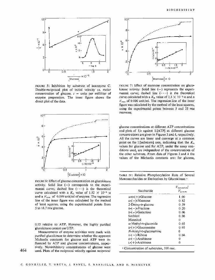
FIGURE 7 : Effect of mannose concentration on gluco- kinase activity. Solid line (-) represents the experi- mental curve; dashed line (---) is the theoretical curve calculated with a K, value of 2.3 X lov2 M and a V,,,, of 0.106 unit/ml. The regressionline of the inner figure was calculated by the method of the least squares, using the experimental points between 5 and 25 rm mannose.
' O W 1
glucose concentrations at different ATP concentrations and plots of I/u against l/[ATP] at different glucose concentrations are given in Figures 3 and 4, respectively. All the curves are linear and converge at a common point on the I/[substrate] axis, indicating that the K,,, values for glucose and for ATP, under the assay con- ditions used, are independent of the concentrations of the other substrate. From data of Figures 3 and 4 the values of the Michaelis constants are: for glucose,
[Glucose] x IO
FIGURE 6: Effect of glucose concentration on glucokinase activity. Solid line (-) corresponds to the experi- mental curve; dashed line (---) is the theoretical curve calculated with a K, value of 1.52 X M and a V,,, of 0.099 unit/ml of enzyme. The regression line of the inner figure was calculated by the method of least squares, using the experimental points from 5 to 16.7 mM glucose.
0.15 relative to ATP. However, the highly purified glucokinase cannot use UTP.
Measurements of enzyme activities were made with purified glucokinase to determine whether the apparent Michaelis constants for glucose and ATP were in- fluenced by ATP and glucose concentrations, respec- tively. Noninhibitory concentrations of glucose were used. Plots of the reciprocal velocity against reciprocal 464
TABLE IV: Relative Phosphorylation Rate of Several Monosaccharides or Derivatives by Glucokinase:
Vsacoharidc/
Saccharide V,l"C",,
a-D-( +)-Glucose D-( +)-Mannose 2-Deoxy-~-glucose D-( -)-Fructose D-(+)-Galactose Sorbitol Mannitol a-Methyl-D-glucoside D-( +)-Glucosamine N- Acety l-D-glucosamine D-( -)-Ribose D-( -)-Arabinose L-(+)-Arabinose
1.00 0.82 0.39 0.20 0.06 0.06 0 0.03 0.03 0 0.04 0 0
a Concentration of substrates, 100 mM.
C. G O N Z A L E Z , T. U R E T A , J. B A B U L , E. K A B A J I L L E , A N D H. N I E M E Y E R
V O L . 6, N O . 2, F E B R U A R Y 1 9 6 7
K, = 8.4 x 1 0 ' ~ M mM G l c - 6 - P
K i = I 5X10⁻² M
6 -
- I 25 0
16.7
100
0
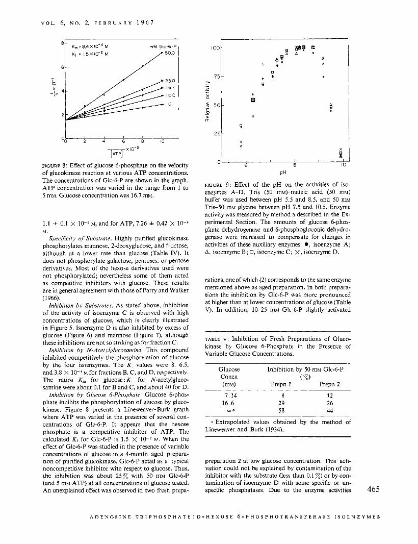
FIGURE 8: Effect of glucose 6-phosphate on the velocity of glucokinase reaction a t various ATP concentrations. The concentrations of Glc-6-P are shown in the graph. ATP concentration was varied in the range from 1 to 5 mM. Glucose concentration was 16.7 mM.
1.1 i 0.1 X M, and for ATP, 7.26 =k 0.42 X M.
SpeciJicity q! Substrate. Highly purified glucokinase phosphorylates mannose, 2-deoxyglucose, and fructose, although at a lower rate than glucose (Table IV). It does not phosphorylate galactose, pentoses, or pentose derivatives. Most of the hexose derivatives used were not phosphorylated ; nevertheless some of them acted as competitive inhibitors with glucose. These results are in general agreement with those of Parry and Walker (1966).
lnhibition by Substrates. As stated above, inhibition of the activity of isoenzyme C is observed with high concentrations of glucose, which is clearly illustrated in Figure 5. Isoenzyme D is also inhibited by excess of glucose (Figure 6) and mannose (Figure 7), although these inhibitions are not so striking as for fraction C .
Inhibition by N-Acetylglucosamine. This compound inhibited competitively the phosphorylation of glucose by the four isoenzymes. The Ki values were 8, 6.5, and 3.8 X M for fractions B, C, and D, respectively. The ratios K, for glucose: Ki for N-acetylgluco- samine were about 0.1 for B and C, and about 40 for D.
inhibition by Glitcose 6-Phosphate. Glucose 6-phos- phate inhibits the phosphorylation of glucose by gluco- kinase. Figure 8 presents a Lineweaver-Burk graph where ATP was varied in the presence of several con- centrations of Glc-6-P. It appears that the hexose phosphate is a competitive inhibitor of ATP. The calculated Ki for Glc-6-P is 1.5 X w. When the effect of Glc-6-P was studied in the presence of variable concentrations of glucose in a 4-month aged prepara- tion of purified glucokinase, Glc-6-P acted a5 a typical noncompetitive inhibitor with respect to glucose. Thus, the inhibition was about 2 5 x with 50 mM Glc-6-P (and 5 mM ATP) at all concentrations of glucose tested. An unexplained effect was observed in two fresh prepa-
' O 0 I 75
P 251 a .
X
A
?
X
A I I I I I 6 8 10 01
PH
FIGURE 9: Effect of the pH on the activities of iso- enzymes A-D. Tris (50 m)-maleic acid (50 mM) buffer was used between pH 5.5 and 8.5, and 50 m~ Tris-50 mM glycine between pH 7.5 and 10.5. Enzyme activity was measured by method a described in the Ex- perimental Section. The amounts of glucose 6-phos- phate dehydrogenase and 6-phosphogluconic dehydro- genase were increased to compensate for changes in activities of these auxiliary enzymes. 0, isoenzyme A ; A, isoenzyme B; 0, isoenzyme C ; X, isoenzyme D.
rations, one of which (2) corresponds to the same enzyme mentioned above as aged preparation. In both prepara- tions the inhibition by Glc-6-P was more pronounced at higher than at lower concentrations of glucose (Table V). In addition, 10-25 mM Glc-6-P slightly activated
TABLE v: Inhibition of Fresh Preparations of Gluco- kinase by Glucose 6-Phosphate in the Presence of Variable Glucose Concentrations.
Glucose Inhibition by 50 mM Glc-6-P
Prepn 1 Prepn 2 Concn ( %)
7.14 8 12 16.6 29 26
m a 58 44
a Extrapolated values obtained by the method of Lineweaver and Burk (1934).
preparation 2 at low glucose concentration. This acti- vation could not be explained by contamination of the inhibitor with the substrate (less than 0.1 2) or by con- tamination of isoenzyme D with some specific or un- specific phosphatases. Due to the enzyme activities 465
A D E N O S I N E T R I P H O S P H A T E : D - H E X O S E ~ - P H O S P H O T R A N S F E R A S E I S O E N Z Y M E S
B I O C H E M I S T R Y
5.0 1
I I I I I 5 IO 15 20
Time (hours)
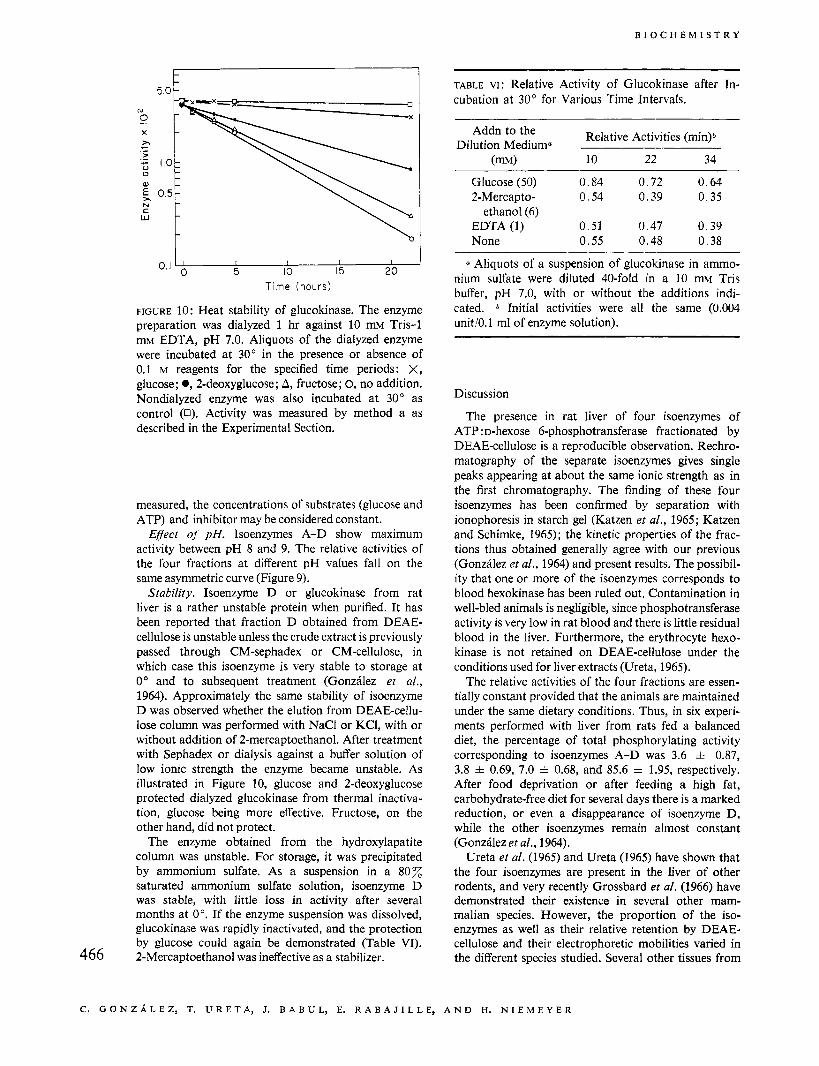
FIGURE 10: Heat stability of glucokinase. The enzyme preparation was dialyzed 1 hr against 10 mM Tris-1 mM EDTA, pH 7.0. Aliquots of the dialyzed enzyme were incubated at 30" in the presence or absence of 0.1 M reagents for the specified time periods: X, glucose; 0,2-deoxyglucose; A, fructose; 0, no addition. Nondialyzed enzyme was also incubated at 30" as control (0). Activity was measured by method a as described in the Experimental Section.
measured, the concentrations of substrates (glucose and ATP) and inhibitor may be considered constant.
Effect oJ' pH. Isoenzymes A-D show maximum activity between pH 8 and 9. The relative activities of the four fractions at different pH values fall on the same asymmetric curve (Figure 9).
Stability. Isoenzyme D or glucokinase from rat liver is a rather unstable protein when purified. It has been reported that fraction D obtained from DEAE- cellulose is unstable unless the crude extract is previously passed through CM-sephadex or CM-cellulose, in which case this isoenzyme is very stable to storage at 0" and to subsequent treatment (Gonzhlez et al., 1964). Approximately the same stability of isoenzyme D was observed whether the elution from DEAE-cellu- lose column was performed with NaCl or KCI, with or without addition of 2-mercaptoethanol. After treatment with Sephadex or dialysis against a buffer solution of low ionic strength the enzyme became unstable. As illustrated in Figure 10, glucose and 2-deoxyglucose protected dialyzed glucokinase from thermal inactiva- tion, glucose being more effective. Fructose, on the other hand, did not protect.
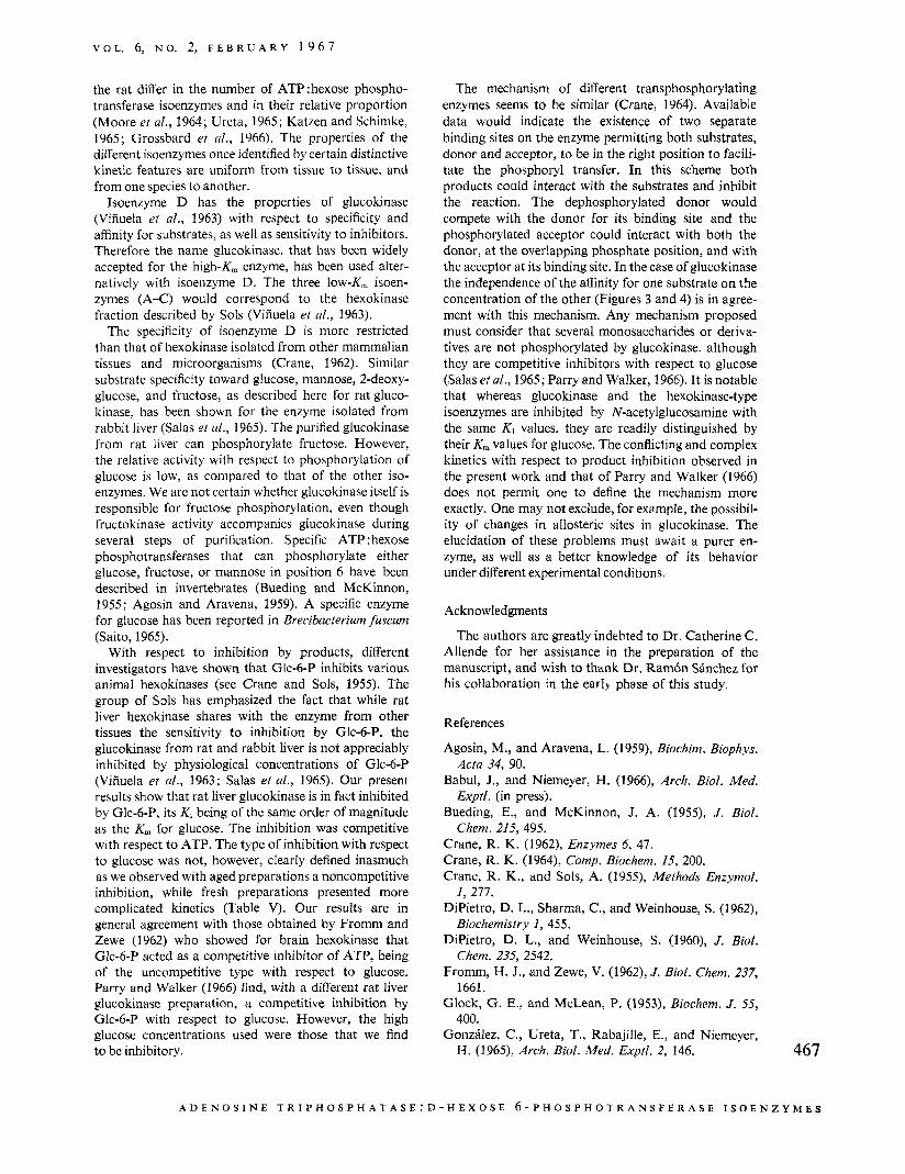
The enzyme obtained from the hydroxylapatite column was unstable. For storage, it was precipitated by ammonium sulfate. As a suspension in a 80% saturated ammonium sulfate solution, isoenzyme D was stable, with little loss in activity after several months at 0". If the enzyme suspension was dissolved, glucokinase was rapidly inactivated, and the protection by glucose could again be demonstrated (Table VI). 2-Mercaptoethanol was ineffective as a stabilizer. 466
~~~ ~~ ~~
TABLE VI: Relative Activity of Glucokinase after In- cubation at 30" for Various Time Intervals.
Relative Activities (min)* Addn to the Dilution Medium.
Glucose (50) 0.84 0.72 0.64 2-Mercapto- 0 .54 0.39 0.35
EDTA (1) 0.51 0.47 0.39 None 0.55 0.48 0.38
ethanol (6)
Q Aliquots of a suspension of glucokinase in ammo- nium sulfate were diluted 40-fold in a 10 mM Tris buffer, pH 7.0, with or without the additions indi- cated. * Initial activities were all the same (0.004 unit/O.l ml of enzyme solution).
Discussion
The presence in rat liver of four isoenzymes of ATP:D-hexose 6-phosphotransferase fractionated by DEAE-cellulose is a reproducible observation. Rechro- matography of the separate isoenzymes gives single peaks appearing at about the same ionic strength as in the first chromatography. The finding of these four isoenzymes has been confirmed by separation with ionophoresis in starch gel (Katzen et al., 1965; Katzen and Schimke, 1965); the kinetic properties of the frac- tions thus obtained generally agree with our previous (Gonzhlez et al., 1964) and present results. The possibil- ity that one or more of the isoenzymes corresponds to blood hexokinase has been ruled out. Contamination in well-bled animals is negligible, since phosphotransferase activity is very low in rat blood and there is little residual blood in the liver. Furthermore, the erythrocyte hexo- kinase is not retained on DEAE-cellulose under the conditions used for liver extracts (Ureta, 1965).
The relative activities of the four fractions are essen- tially constant provided that the animals are maintained under the same dietary conditions. Thus, in six experi- ments performed with liver from rats fed a balanced diet, the percentage of total phosphorylating activity corresponding to isoenzymes A-D was 3.6 f 0.87, 3.8 * 0.69, 7.0 z+= 0.68, and 85.6 f 1.95, respectively. After food deprivation or after feeding a high fat, carbohydrate-free diet for several days there is a marked reduction, or even a disappearance of isoenzyme D, while the other isoenzymes remain almost constant (Gonz6lez et al., 1964).
Ureta et al. (1965) and Ureta (1965) have shown that the four isoenzymes are present in the liver of other rodents, and very recently Grossbard et a/. (1966) have demonstrated their existence in several other mam- malian species. However, the proportion of the iso- enzymes as well as their relative retention by DEAE- cellulose and their electrophoretic mobilities vaned in the different species studied. Several other tissues from
C. G O N Z A L E Z , T. U R E T A , J. B A B U L , E. R A B A J I L L E , A N D H. N I E M E Y E R
V O L . 6, N O . 2, F E B R U A R Y 1 9 6 7
the rat differ in the number of ATP:hexose phospho- transferase isoenzymes and in their relative proportion (Moore et al., 1964; Ureta, 1965; Katzen and Schimke, 1965; Grossbard et ul., 1966). The properties of the different isoenzymes once identified by certain distinctive kinetic features are uniform from tissue to tissue, and from one species to another.
Isoenzyme D has the properties of glucokinase (Viiiuela et al., 1963) with respect to specificity and affinity for substrates, as well as sensitivity to inhibitors. Therefore the name glucokinase, that has been widely accepted for the high-K, enzyme, has been used alter- natively with isoenzyme D. The three low-K,, isoen- zymes (A-C) would correspond to the hexokinase fraction described by Sols (Viiiuela et a/., 1963).
The specificity of isoenzyme D is more restricted than that of hexokinase isolated from other mammalian tissues and microorganisms (Crane, 1962). Similar substrate specificity toward glucose, mannose, 2-deoxy- glucose. and fructose, as described here for rat gluco- kinase, has been shown for the enzyme isolated from rabbit liver (Salas et al., 1965). The purified glucokinase from rat liver can phosphorylate fructose. However, the relative activity with respect to phosphorylation of glucose is low, as compared to that of the other iso- enzymes. We are not certain whether glucokinase itself is responsible for fructose phosphorylation, even though fructokinase activity accompanies glucokinase during several steps of purification. Specific ATP :hexose phosphotransferases that can phosphorylate either glucose, fructose, or mannose in position 6 have been described in invertebrates (Bueding and McKinnon, 1955; Agosin and Aravena, 1959). A specific enzyme for glucose has been reported in Brecibacterium fuscum (Saito, 1965).
With respect to inhibition by products, different investigators have shown that Glc-6-P inhibits various animal hexokinases (see Crane and Sols, 1955). The group of Sols has emphasized the fact that while rat liver hexokinase shares with the enzyme from other tissues the sensitivity to inhibition by Glc-6-P, the glucokinase from rat and rabbit liver is not appreciably inhibited by physiological concentrations of Glc-6-P (Viiiuela et al., 1963; Salas et al., 1965). Our present results show that rat liver glucokinase is in fact inhibited by Glc-6-P, its K, being of the same order of magnitude as the K , for glucose. The inhibition was competitive with respect to ATP. The type of inhibition with respect to glucose was not, however, clearly defined inasmuch as we observed with aged preparations a noncompetitive inhibition, while fresh preparations presented more complicated kinetics (Table V). Our results are in general agreement with those obtained by Fromm and Zewe (1962) who showed for brain hexokinase that Glc-6-P acted as a competitive inhibitor of ATP, being of the uncompetitive type with respect to glucose. Parry and Walker (1966) find, with a different rat liver glucokinase preparation, a competitive inhibition by Glc-6-P with respect to glucose. However, the high glucose concentrations used were those that we find to be inhibitory.
The mechanism of different transphosphorylating enzymes seems to be similar (Crane, 1964). Available data would indicate the existence of two separate binding sites on the enzyme permitting both substrates, donor and acceptor, to be in the right position to facili- tate the phosphoryl transfer. In this scheme both products could interact with the substrates and inhibit the reaction. The dephosphorylated donor would compete with the donor for its binding site and the phosphorylated acceptor could interact with both the donor, at the overlapping phosphate position, and with the acceptor at its binding site. In the case of glucokinase the independence of the affinity for one substrate on the concentration of the other (Figures 3 and 4) is in agree- ment with this mechanism. Any mechanism proposed must consider that several monosaccharides or deriva- tives are not phosphorylated by glucokinase. although they are competitive inhibitors with respect to glucose (Salas et al., 1965; Parry and Walker, 1966). It is notable that whereas glucokinase and the hexokinase-type isoenzymes are inhibited by N-acetylglucosamine with the same Ki values. they are readily distinguished by their K, values for glucose. The conflicting and complex kinetics with respect to product inhibition observed in the present work and that of Parry and Walker (1966) does not permit one to define the mechanism more exactly. One may not exclude, for example, the possibil- ity of changes in allosteric sites in glucokinase. The elucidation of these problems must await a purer en- zyme, as well as a better knowledge of its behavior under different experimental conditions.
Acknowledgments
The authors are greatly indebted to Dr. Catherine C. Allende for her assistance in the preparation of the manuscript, and wish to thdnk Dr. Ramdn SBnchez for his collaboration in the early phase of this study.
References
Agosin, M., and Aravena, L. (1959), Biochim. Biophys.
Babul, J., and Niemeyer, H. (1966), Arch. Biol. Med.
Bueding, E., and McKinnon, J. A. (1955), J. Biol.
Crane, R. K. (1962), Enzymes 6, 47. Crane, R. K. (1964), Comp. Biochem. 15, 200. Crane, R. K., and Sols, A. (1955), Methods Enzymol.
DiPietro, D. L., Sharma, C., and Weinhouse, S. (1962),
DiPietro, D. L., and Weinhouse, S. (1960), J. B id .
Fromm, H. J., and Zewe, V. (1962), J. Biol. Chem. 237,
Glock, G. E., and McLean, P. (1953), Biochem. J . 55,
Gonzhlez, C., Ureta, T., Rabajille, E., and Niemeyer,
Acta 34, 90.
Exptl. (in press).
Chem. 215,495.
1, 277.
Biochemistry 1, 455.
Chem. 235, 2542.
1661.
400.
H. (1965), Arch. Biol. Med. Exptl. 2, 146. 467
A D E N O S I N E T R I P H 0 S P H A T A S E : D - H E X O S E 6-PHOSPHOTRANSFERASE I S O E N Z Y M E S
B I O C H E M I S T R Y
Gonzdlez, C., Ureta, T., Sdnchez, R., and Niemeyer, H. (1964), Biochem. Biophys. Res. Commun. 16, 347.
Gottschalk, A. (1944), Australian J. Exptl. Biol. Med. Sci. 22, 291.
Grossbard, L., Weksler, M . , and Schimke, R. T. (1966), Biochem. Biophys. Res. Commun. 24, 32.
Katzen, H. M., and Schimke, R. T. (1965), Proc. Nut/. Acad. Sci. U. S. 54, 1218.
Katzen, H. M., Soderman, D. D., and Nitowsky, H. M. (1965), Biochem. Biophys. Res. Commun. 19, 317.
Kornberg, A,, and Pricer, W. E. (1951), J . Biol. Chem. 193, 481.
Lineweaver, H., and Burk, D. (1934), J . Am. Chem. Soc. 56, 658
Lowry, 0. H., Rosebrough, N. J., Farr, A. L., and Randall, R. J. (1951), J . Biol. Chem. 193, 265.
Miller, G. L. (1959), Anal. Chem. 31, 964. Moore, R. O., Chandler, A. M., and Tettenhorst, N.
(1964), Biochem. Biophys. Res. Commun. 17, 527. Niemeyer, H., Clark-Turri, L., Garcis, E., and Vergara,
F. E. (1962), Arch. Biochem. Biophys. 98, 77. Niemeyer, H., Clark-Turri, L., and Rabajille, E. (1963),
Nature 198, 1096.
Purification and Properties of Yeast Invertase"
Norbert P. Neumann and J. Oliver Lampen
Niemeyer, H., Pirez, N., and Codoceo, R. (1 967), J .
Parry, M. J., and Walker, D. G. (1966), Biochem. J . 99.
PCrez, N., Clark-Turri, J., Rabajille, E., and Niemeyer,
Saito, N. (1965), J . Biochem. (Tokyo) 57, 363. Salas, J. , Viiiuela, E., Salas, M., and Sols, A. (1963,
Salas, M., Viiiuela, E., and Sols, A. (1963). J . Biol.
Slein, M. W., Cori, G. T., and Cori, C. F. (1950), J .
Ureta, T. (1965), Ph.D. Thesis, UniverCty of Chile,
Ureta, T., Gonzdlez, C., Liilo, S., and Niemeyer, H.
Vaughan, D. A., Hannon, J. P., and Vaughan, L. N.
Viiiuela, E., Salas, M., and Sols, A. (1963), J. Biol.
Walker, D. G. (1963), Biochim. Biophys. Acta 77, 209. Webb, E. C. (1964), Nature 203, 821.
Biol. Chem. (in press).
266.
H. (1964), J. Biol. Chem. 239, 2420.
J . Biol. Chem. 240, 1014.
Chem. 238, 3535.
Biol. Chem. 186, 763.
Santiago, Chile.
(1965), Arch. Biol. Med. Exptl. 2, 170.
(1960), Am. J. Physiol. 199, 1041.
Chem. 238, PCll75.
ABSTRACT : Although the enzyme, yeast invertase, has been studied extensively in the past, it has never been chemically characterized and there has been a great deal of uncertainty concerning a number of its physical properties. The present studies describe a rapid method for the isolation of highly purified enzyme in good yield. The enzyme is homogeneous by polyacrylamide gel electrophoresis. Sedimentation velocity studies reveal a major component with an szo,w of 10.4 S. Heavier components which are also present in much
R ecently, Sutton and Lampen (1962) and Islam and Lampen (1962) have described the secretion of invertase by yeast protoplasts. To understand this process, it is essential to characterize the invertase of the intact yeast cell (which is primarily localized in the cell wall) to permit eventual comparison of this
~~
* From the Institute of Microbiology, Rutgers, The State University, New Brunswick, New Jersey. Received November 14, 1966. This investigation was supported by a U. S. Public Health Service grant (AI-04572). Some of the findings were presented at the 50th Annual Meeting of the Federation of American Societies for Experimental Biology, Atlantic City, N. J., April 1966. 468
smaller amounts are enzymatically active and indicate the presence of an association-dissociation equilib- rium. The molecular weight of the enzyme is about 270,000, as determined by sedimentation equilibrium measurements. The chemical properties of the enzyme have been examined and the amino acid composition determined. Invertase is shown to be a glycoprotein which contains about 50 carbohydrate (predomi- nantly mannan with a small percentage of glucosa- mine).
material with the secreted enzyme and with the inver- tase present inside the cell membrane.
Yeast invertase was first isolated by Berthelot (1 860) by alcohol precipitation. Since that time, this enzyme has been studied by a number of workers. Much of the early work has been reviewed by Neuberg and Roberts (1946). Myrback (1960) has summarized the more recent results. A great deal is known about the enzyme from a kinetic point of view particularly from the inhibition studies by Myrback and his co- workers. Nevertheless, relatively little is known about the detailed chemistry of the molecule. Considerable controversy exists concerning physical properties as
N O R B E R T P. N E U M A N N A N D J. O L I V E R L A M P E N