Embed Size (px)
Citation preview

JOURNAL OF CLINICAL MICROBIOLOGY, Jan. 2003, p. 34–43 Vol. 41, No. 10095-1137/03/$08.00�0 DOI: 10.1128/JCM.41.1.34–43.2003Copyright © 2003, American Society for Microbiology. All Rights Reserved.
Characterization of Members of the Legionellaceae Familyby Automated Ribotyping
Christophe Cordevant,1 Jane S. Tang,2 David Cleland,2 and Marc Lange1*R&D Division, Institut Pasteur de Lille, Lille, France,1 and Bacteriology Program,
American Type Culture Collection, Manassas, Virginia 20110-22092
Received 26 June 2002/Returned for modification 25 July 2002/Accepted 17 September 2002
In order to implement a new and reliable method for characterizing different species of Legionella, a geneticfingerprinting study with an automated ribotyping system (RiboPrinter) was completed with members of thisgenus which were deposited at the American Type Culture Collection. The RiboPrinter examined the differentpatterns of EcoRI digestion fragments from the rRNA operons of 110 strains, representing 48 of the 49described Legionella species as well as 70 serogroups of those species. Distinctive and consistent patterns wereobtained for the type strains of the 48 species investigated. Legionella pneumophila subsp. fraseri and L.pneumophila subsp. pascullei each generated a specific pattern, whereas L. pneumophila subsp. pneumophilaproduced six different fingerprint patterns. No correlation seemed to exist between the ribotypes obtained andthe 15 serotypes of L. pneumophila. For the other species, those with two known serogroups presented twodistinctive patterns with the RiboPrinter with the exception of L. hackeliae and L. quinlivanii, which yielded onlyone pattern. We also encountered ribotypes for strains which were not identified to the species level. Theribotypes generated for these strains with the RiboPrinter did not match those generated for known typestrains, suggesting the putative description of new serogroups or species. Although the automated system didnot have sufficient discriminatory ability to serve as an epidemiological tool in a clinical setting, it appearedto be a powerful tool for general genomic analysis of the Legionella isolates (e.g., determination of new species)and assessment of the interrelationship among Legionella strains through the RiboPrinter database connection.
Since the first isolation of Legionella pneumophila, the caus-ative agent of Legionnaires’ disease, nearly 30 years ago (37),members of this genus have been isolated from a wide range ofenvironments and geographical locations (17, 20). To date,members of the family Legionellaceae comprise 49 describedspecies (Deutsche Sammlung von Mikroorganismen undZellkulturen website [ftp://ftp.dsmz.de/pub/DSMZ/bactnom/bactname.pdf]), with 15 serogroups (Sgs) described for L.pneumophila; 2 Sgs each described for L. bozemanii, L. long-beachae, L. feeleii, L. hackeliae, L. sainthelensi, L. spiritensis, L.erythra, and L. quinlivanii; and a single Sg each described forthe remaining species (3). Over the years, several species ofLegionella that were initially isolated from environmentalsources but that were not implicated as etiological agents havelater been shown to be human pathogens (9, 22, 25, 35). Ap-proximately 70 to 90% of Legionella infections are caused by L.pneumophila Sgs 1 and 6, and others species are responsible forbetween 5 and 30% of the cases of infection (18, 42). Nineteenspecies have been recognized to be pathogenic for humans(14), causing pneumonia (Legionnaires’ disease) (11, 19), amild febrile disease (Pontiac fever) (31, 55), and most recently,soft tissue abscesses (25). Pneumonia caused by Legionella isbecoming a public health problem, since this organism has thepotential to cause large outbreaks (1, 40) and to infect youngimmunocompetent adults (51) or newborns after water birth(21). Legionnaires’ disease, if left untreated, leads to an aver-age mortality rate of 15% (16).
Various methods for typing of members of the family Legio-nellaceae have been developed in the past. These include an-tibiotic susceptibility testing (54), plasmid analysis (47), fattyacid profiling (29), multilocus enzyme electrophoresis (45),pulsed-field gel electrophoresis (39), and various DNA finger-printing protocols by PCR (14, 34, 41, 53). Most of them havefocused specifically on the subtyping of L. pneumophila Sgs 1and 6 because these Sgs are responsible for the majority oflegionellosis cases.
At present, the majority of the Legionella isolates are de-tected and typed by serological designation (3, 30, 36, 49). Thishas been satisfactory for the most commonly occurring speciesand serovars. However, antisera are not available commerciallyfor many of the less well known species. Immunological cross-reactions among some species have also been reported to betroublesome (3, 6, 33, 49). With the increasing number ofdescribed Legionella species, methods based on serology willbecome more difficult and cumbersome to use in environmen-tal and clinical studies.
Ribotyping, a molecular method based on the analysis of therestriction fragment length polymorphisms (RFLPs) of rRNAgenes (23), was also used to characterize Legionella strains (2,52). The feasibility of ribotyping for the differentiation of Le-gionella species was initially tested by Grimont et al. (24) with28 members of the Legionellaceae family.
To further investigate this approach, we tested a larger num-ber of Legionella species using an automated ribotyping sys-tem. A total of 110 strains representing 48 species and 3 sub-species, as well as a newly reported Sg for L. londiniensis (Sg 2;F. Lo Presti [Centre National de Reference des Legionelles,Lyon, France], personal communication to the American TypeCulture Collection [ATCC]) were included in the study. L.
* Corresponding author. Mailing address: R&D Division, InstitutPasteur de Lille, 1 Rue du Professeur Calmette, BP 245, 59019 LilleCedex, France. Phone: 33 3 20 87 72 08. Fax: 33 3 20 87 72 06. E-mail:[email protected].
34
on February 12, 2018 by guest
http://jcm.asm
.org/D
ownloaded from
lytica (28) is a symbiont of amoeba and requires cocultivationwith the host, so it was not feasible to include a culture of thisisolate for testing. The results of that study are presented inthis paper.
MATERIALS AND METHODS
Cultures, media, and growth conditions. A total of 110 Legionella strainscomprising 48 species, including 3 subspecies and 70 Sgs deposited at ATCC,were used for this study (Table 1). Each culture was grown in an atmosphere of5% CO2 at 37°C for 24 to 48 h on ATCC 1099 Charcoal Yeast Extract bufferedmedium (the medium formulation is described at the website http://www.atcc.org/SearchCatalogs/MediaFormulations.cfm).
Sample preparation and processing. All Legionella strains were characterizedby use of the RiboPrinter system (Qualicon Inc., Wilmington, Del.) as describedpreviously (13) with respect to the procedures and conditions recommended bythe manufacturer (12, 46). EcoRI was used as the restriction enzyme. At the endof the process, a densitometric scan depicting the distributions and molecularweights of the restriction fragments was obtained for each sample analyzed. Thisoutput was saved in the RiboPrinter’s computer. Ribotype groups (ribogroups)were defined by the RiboPrinter’s proprietary algorithm, which compared thepattern of each isolate to those of others in the database and assigned groups bythe differences in band number, position, and signal intensity. A given ribogroupwas defined as a group of ribotypes with similarity values �0.93. Strains used forrepeatability testing were analyzed by using the RiboPrinter’s proprietary algo-rithm, which was a modified version of the coefficient of simple correlation (38).Similarity values obtained are reported in Table 2.
Ribotype analysis. For each batch of eight samples, ribotypes were normalizedto the positions of the molecular weight standards with Qualicon software.Computerized ribotypes were exported for analysis in .txt files and imported intoBioNumerics software (version 2.5; Applied Maths, Sint-Martens-Latem, Bel-gium) by using the Qualicon macro. Clustering analysis was performed by theunweighted pair group method with arithmetic averages (UPGMA) methodbased on the Dice (15) coefficient for band matching, with a position tolerancesetting of 1.0% (default values are 1% of position tolerance and 0.5% of opti-mization). Bands for analysis with the Dice coefficient were assigned manually,according to densitometric curves and the accompanying hard-copy photograph.
RESULTS
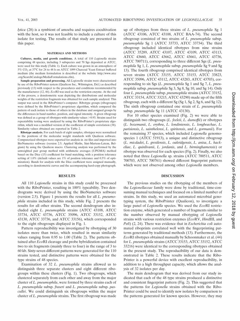
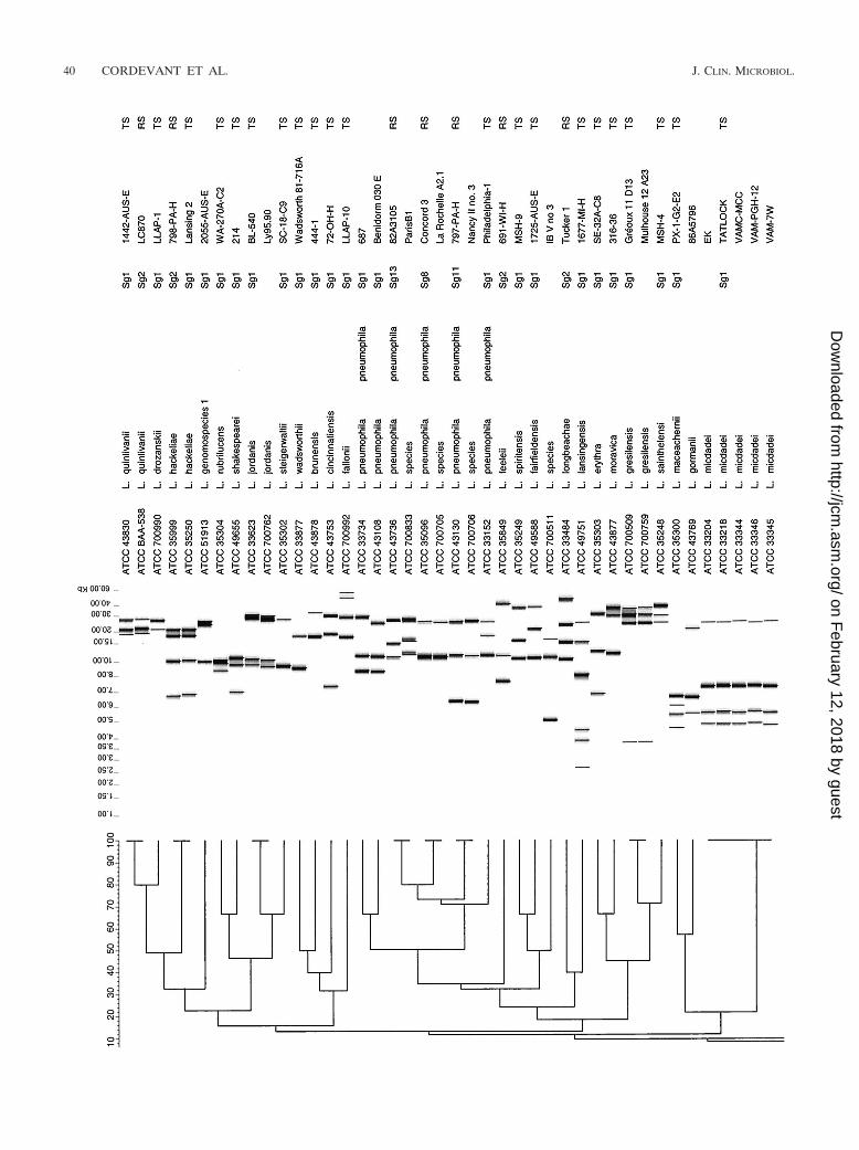
All 110 Legionella strains in this study could be processedwith the RiboPrinter, resulting in 100% typeability. Two den-drograms were derived by using the BioNumerics software(version 2.5). Figure 1 presents the results for all L. pneumo-phila strains included in this study, while Fig. 2 presents theresults for all other strains. The second dendrogram also in-cluded eight L. pneumophila strains (ATCC 43108, ATCC33734, ATCC 43736, ATCC 35096, ATCC 33152, ATCC43130, ATCC 33736, and ATCC 33156), which correspondedto the eight ribogroups displayed in Fig. 1.
Pattern reproducibility was investigated by ribotyping of 30isolates more than twice, which resulted in mean similarityvalues ranging from 0.95 to 1.00 (Table 2). The patterns ob-tained after EcoRI cleavage and probe hybridization containedtwo to six fragments (mainly three to four) in the range of 3 to60 kb. Sixty-seven different patterns were generated for the 110strains tested, and distinctive patterns were obtained for thetype strains of 48 species.
Examination of 32 L. pneumophila strains allowed us todistinguish three separate clusters and eight different ribo-groups within these clusters (Fig. 1). Two ribogroups, whichclustered separately from each other and away from the majorcluster of L. pneumophila, were formed by three strains each ofL. pneumophila subsp. fraseri and L. pneumophila subsp. pas-cullei. We could distinguish six ribogroups within the majorcluster of L. pneumophila strains. The first ribogroup was made
up of ribotypes from three strains of L. pneumophila Sg 1(ATCC 43106, ATCC 43108, ATCC BAA-74). The secondribogroup consisted of two strains of L. pneumophila subsp.pneumophila Sg 1 (ATCC 33733, ATCC 33734). The thirdribogroup included identical ribotypes from nine strains(ATCC 35289, ATCC 43107, ATCC 43109, ATCC 43113,ATCC 43660, ATCC 43662, ATCC 43661, ATCC 43736,ATCC 700711), corresponding to three different Sgs (L. pneu-mophila Sg 1, L. pneumophila subsp. pneumophila Sg 9 and Sg13). The fourth ribogroup comprised identical ribotypes fromseven strains (ATCC 33155, ATCC 33153, ATCC 33823,ATCC 35096, ATCC 43112, ATCC 43283, ATCC 43703), cor-responding to six Sgs (L. pneumophila Sg 1 and Sg 7, L. pneu-mophila subsp. pneumophila Sg 3, Sg 8, Sg 10, and Sg 14). Onlyfour L. pneumophila subsp. pneumophila strains (ATCC 33152,ATCC 33154, ATCC 33215, ATCC 43290) made up of the fifthribogroup, each with a different Sg (Sg 1, Sg 2, Sg 6, and Sg 12).The sixth ribogroup contained one strain of L. pneumophilasubsp. pneumophila Sg 11 (ATCC 43130).
For 10 other species examined (Fig. 2) we were able todistinguish two ribogroups (L. feeleii, L. dumoffii) or ribotypes(L. bozemanii, L. erythra, L. londiniensis, L. longbeachae, L.parisiensis, L. sainthelensi, L. spiritensis, and L. gormanii). Forthe remaining 37 species, which included Legionella genomo-species 1 (ATCC 51913), a unique and distinctive ribogroup(L. micdadei, L. gresilensis, L. oakridgensis, L. anisa, L. hack-eliae, L. quinlivanii, L. jordanis, and L. birminghamensis) orribotype was observed for each species (Fig. 2). Finally, we alsonoted that three Legionella sp. strains (ATCC 700511, ATCC700703, ATCC 700761) showed different fingerprint patternswhich were not observed among other Legionella members.
DISCUSSION
The previous studies on the ribotyping of the members ofthe Legionellaceae family were done by traditional, time-con-suming manual techniques and focused on a limited number ofstrains. In this study, we used an automated microbial geno-typing system, the RiboPrinter (Qualicon), to investigate alarge panel of Legionella species. We used the EcoRI restric-tion enzyme, which generated a number of fragments similar tothe number observed by manual ribotyping of Legionellastrains with various restriction enzymes (EcoRV, HindIII, andPstI) (2, 24). There was evidence that in Escherichia coli auto-mated riboprints correlated well with the fingerprinting pat-terns generated by traditional methods (13). Furthermore, theEcoRI ribotypes obtained manually by Schoonmaker et al. (44)for L. pneumophila strains (ATCC 33153, ATCC 33152, ATCC33216) were identical to the corresponding ribotypes obtainedin the present study. The reproducibility of our data is dem-onstrated in Table 2. These results indicate that the Ribo-Printer is a powerful device with excellent reproducibility, inaddition to a high throughput capacity, which allows the anal-ysis of 32 isolates per day.
The main dendrogram that was derived from our study in-dicated that each of the 48 type strains produced a distinctiveand consistent fingerprint pattern (Fig. 2). This suggested thatthe patterns for Legionella strains obtained with the Ribo-Printer could be used to identify new isolates by comparison tothe patterns generated for known species. However, they may
VOL. 41, 2003 AUTOMATED RIBOTYPING INVESTIGATION OF LEGIONELLACEAE 35
on February 12, 2018 by guest
http://jcm.asm
.org/D
ownloaded from

TABLE 1. Legionella strains included in the studya
Species Subspecies Sg Other Source Original designation ATCC no.
L. adelaidensis 1 TS Cooling tower water, Adelaide, Australia 1762-AUS-E 49625L. anisa 1 TS Hot water, sink, Los Angeles, Calif. WA-316-C3 35292L. anisa Sink faucet, Chicago, Il. CH-47-C3 35291L. anisa Sink faucet, Chicago, Il. CH-47-C1 35290L. beliardensis 1 TS Water from a calorifier in reanimation unit,
Montbeliard, FranceMontbeliard A1 700512
L. birminghamensis 1 TS Lung biopsy, cardiac transplant recipient, Alabama 1407-AL-H 43702L. birminghamensis Water near Clermont-Ferrand, France CF VII no. 3A 700709L. bozemanii 1 TS Lung tissue, pneumonia, Key West, Fla. WIGA 33217L. bozemanii 2 RS Human lung aspirate, Toronto, Ontario, Canada Toronto 3 35545L. brunensis 1 TS Cooling tower water, Brno, Czechoslovakia 444-1 43878L. cherrii 1 TS Thermally altered water, Michigan ORW 35252L. cincinnatiensis 1 TS Lung tissue, pneumonia, Cincinnati, Ohio 72-OH-H 43753L. drozanskii 1 TS Tank of well water, Leeds, United Kingdom LLAP-1 700990L. dumoffii 1 TS Water in cooling tower, New York, N.Y. NY 23 33279L. dumoffii Human lung, Los Angeles, Calif. Wadsworth 81-782A 35850L. dumoffii Thermal spa water A3a3F 700714L. erythra 1 TS Water in cooling tower, Seattle, Wash. SE-32A-C8 35303L. erythra 2 RS Strain isolated in Paris, France LC217 BAA-536L. fairfieldensis 1 TS Cooling tower water, Fairfield, Victoria, Australia 1725-AUS-E 49588L. fallonii 1 TS Ship air-conditioning system, United Kingdom LLAP-10 700992L. feeleii 1 TS Grinding machine coolant fluid, Windsor, Ontario,
CanadaWO-44C-C3 35072
L. feeleii 2 RS Human lung tissue, Wisconsin, Wis. 691-WI-H 35849L. feeleii 1 Bronchoalveolar lavage, pneumonia, Savoy, France Ly126.92b 700513L. feeleii 1 Bronchoalveolar lavage, pneumonia and HIV,
Lyon, FranceLy166.96 700514
L. geestiana 1 TS Hot-water tap, Geest Office building, London,United Kingdom
1308 49504
L. genomospecies 1 Cooling-water tower, Adelaide, Australia 2055-AUS-E 51913L. gormanii 1 TS Soil from a creek bank, Atlanta, Ga. LS-13 33297L. gormanii Bronchial brush, Pneumonia, Calif. 86A5796 43769L. gratiana 1 TS Thermal spa water, Savoy region, France Lyon-8420412 49413L. gresilensis 1 TS Water from shower in a thermal spa. Greoux,
FranceGreoux 11 D13 700509
L. gresilensis Water sample from a potash mine near Mulhouse,France
Mulhouse 12 A23 700759
L. hackeliae 1 TS Human bronchial biopsy specimen, Ann. Arbor.,Mich.
Lansing 2 35250
L. hackeliae 2 RS Human lung aspirate, Pittsburgh, Pa. 798-PA-H 35999L. israelensis 1 TS Water, Israel Bercovier 4 43119L. jamestowniensis 1 TS Wet soil, Janestown, N.Y. JA-26-G16-E2 35298L. jordanis 1 TS Jordan River, Bloomington, Ind. BL-540 33623L. jordanis Patient with pneumonia, France Ly95.90 700762L. lansingensis 1 TS Bronchial aspirate pneumonia and leukemia,
Lansing, Mich.1677-MI-H 49751
L. londiniensis 1 TS Office building cooling tower, London, UnitedKingdom
1477 49505
L. londiniensis 2 RS Water, Mulhouse, France Mulhouse B26 700510L. longbeachae 1 TS Human lung, pneumonia, Long Beach, Calif. Long Beach 4 33462L. longbeachae 2 RS Human lung, Tucker, Ga. Tucker 1 33484L. maceachernii 1 TS Water in home evaporator cooler, Phoeniz, Ariz. PX-1-G2-E2 35300L. micdadei 1 TS Human blood, pneumonia, Fort Bragg, Calif. TATLOCK 33218L. micdadei Lung tissue, pneumonia, Pittsburgh, Pa. EK 33204L. micdadei Transtracheal aspirate, Pittsburgh, Pa. VAMC-MCC 33344L. micdadei Showerhead, Pittsburgh, Pa. VAM-7W 33345L. micdadei Ultrasonic nebulizer, Pittsburgh, Pa. VAM-PGH-12 33346L. moravica 1 TS Cooling-tower water, Jihlava, Czechoslovakia 316-36 43877L. nautarum 1 TS Domestic hot-water tap, Greenwich, London,
United Kingdom1224 49506
L. oakridgensis 1 TS Industrial cooling-tower water, Pennsylvania Oak Ridge 10 33761L. oakridgensis Bronchoalveolar lavage, pneumonia, Nantes,
FranceNantes-930101868 700515
L. oakridgensis Bronchoalveolar lavage, pneumonia, Nantes,France
Nantes-930101937 700516
L. parisiensis 1 TS Water in cooling tower, Paris, France PF-209C-C2 35299L. parisiensis Tracheal aspirate, liver transplant, France FLP2 700174L. pneumophila fraseri 4 TS Human lung, pneumonia, Los Angeles, Calif. Los Angeles-1 33156
Continued on following page
36 CORDEVANT ET AL. J. CLIN. MICROBIOL.
on February 12, 2018 by guest
http://jcm.asm
.org/D
ownloaded from

not be suitable for phylogenetic purposes, as our dendrogramdid not agree with the phylogenetic tree that was generated by16S rRNA analysis (28). By examination of Fig. 1, two separateclusters for L. pneumophila subsp. fraseri and L. pneumophila
subsp. pascullei were clearly observed, while the patterns for allL. pneumophila subsp. pneumophila strains clustered in a com-mon group. This is in agreement with results based on DNAhybridization (10) as well as those of a previous study showing
TABLE 1—Continued
Species Subspecies Sg Other Source Original designation ATCC no.
L. pneumophila fraseri 5 RS Cooling tower, Dallas, Tex. Dallas 1E 33216L. pneumophila fraseri 15 RS Human lung, fatal pneumonia, Royal Oak, Mich. Lansing 3 35251L. pneumophila pascullei TS Water from showerhead, Pittsburgh, Pa. U8W 33737L. pneumophila pascullei 13 RS Water from showerhead, Pittsburgh, Pa. U7W 33736L. pneumophila pascullei Tap water, Pittsburgh, Pa. MICU B 33735L. pneumophila pneumophila 1 TS Human lung, pneumonia, Philadelphia, Pa. Philadelphia-1 33152L. pneumophila pneumophila 1 Tap water, Pittsburgh, Pa. 684 33733L. pneumophila pneumophila 1 Tap water, Pittsburgh, Pa. 687 33734L. pneumophila pneumophila 2 RS Human lung, Togus, Maine Togus-1 33154L. pneumophila pneumophila 3 RS Creek water, Bloomington, Ind. Bloomington-2 33155L. pneumophila pneumophila 6 RS Human lung biopsy specimen, Chicago, Ill. Chicago 2 33215L. pneumophila pneumophila 8 RS Human lung, Concord, Calif. Concord 3 35096L. pneumophila pneumophila 9 RS Tap water, Leden, Holland IN-23-G1-C2 35289L. pneumophila pneumophila 11 RS Human endotracheal tube, Pittsburgh, Pa. 797-PA-H 43130L. pneumophila pneumophila 10 RS Respiratory tract secretions, Holland Leiden 1 43283L. pneumophila pneumophila 12 RS Human lung, pneumonia, Denver, Col. 570-CO-H 43290L. pneumophila pneumophila 14 RS Bronchial aspirate, pneumonia, Minn. 1169-MN-H 43703L. pneumophila pneumophila 13 RS Lung aspirate, pneumonia, Calif. B2A3105 43736L. pneumophila 1 Human lung, Knoxville, Tenn. Knoxville-1 33153L. pneumophila 7 RS Showerhead, Illinois Chicago 8 33823L. pneumophila 1 CDC-Quebec Allentown 1 43106L. pneumophila 1 CDC-Quebec Heysham 1 43107L. pneumophila 1 CDC-Quebec Benidorm 030 E 43108L. pneumophila 1 CDC-Quebec OLDA 43109L. pneumophila 1 CDC-Quebec France 5811 43112L. pneumophila 1 CDC-Quebec Camperdown 1 43113L. pneumophila Clinical sample, State Health Department
CaliforniaRIO 43660
L. pneumophila Hospital faucet, Pittsburgh Veterans AffairsMedical Center, Pittsburgh, Pa.
11EJ 43661
L. pneumophila Hospital faucet, Calif. FAUC 19 43662L. pneumophila 1 Bronchoalveolar lavage fluid, pneumonia, France CA1 700711L. pneumophila 1 Transtracheal aspirate, 1978 F1724 BAA-74L. quateirensis 1 TS Shower in hotel bathroom, Quarteira, Portugal 1335 49507L. quinlivanii 1 TS Water in bus air conditioner, Australia 1442-AUS-E 43830L. quinlivanii 2 RS Cooling-tower pond, London, United Kingdom LC870 BAA-538L. rowbothamii 1 TS Water and sludge from an industrial liquifier
tower, United KingdomLLAP-6 700991
L. rubrilucens 1 TS Tap water, Los Angeles, Calif. WA-270A-C2 35304L. sainthelensi 1 TS Spring water, Mt. St. Helens, Wash. MSH-4 35248L. sainthelensi 2 RS Human bronchial washings, pneumonia, Calif. 1489-CA-H 49322L. santicrucis 1 TS Tap water, St. Croix, U.S. Virgin Islands SC-63-C7 35301L. shakespearei 1 TS Cooling-tower water, Stratford upon Avon, United
Kingdom214 49655
L. species Water from a well, Montpellier, France IB V no 3 700511L. species Water, Bourbonne-les-Bains, France Nancy II no. 1 700703L. species Water, La Rochelle, France La Rochelle A2.1 700705L. species Water, Bourbonne-les-Bains, France Nancy II no. 3 700706L. species Environmental isolate, Venissieux, France IBV no. 2 700761L. species Clinical isolate, France ParisB1 700833L. spiritensis Sg1 TS Spirit Lake, Mt. St. Helens, Wash. MSH-9 35249L. spiritensis Sg2 RS Cooling tower, United Kingdom ML 76 BAA-537L. steigerwalii TS Tap water, St. Croix, U.S. Virgin Islands SC-18-C9 35302L. taurinensis Sg1 TS Water from a hospital oxygen bubble humidifier,
Turin, ItalyTurin no 1 700508
L. tucsonensis TS Pleural fluid, renal transplant, Tucson, Ariz. 1087-AZ-H 49180L. wadsworthii Sg1 TS Human sputum, pneumonia, Los Angeles, Calif. Wadsworth 81-716A 33877L. waltersii Sg1 TS Drinking water distribution system, Adelaide,
Australia2074-AUS-E 51914
L. worsleiensis Sg1 TS Industrial cooling tower, Worsley, UnitedKingdom
1347 49508
a TS, type strain; RS, reference strain; CDC, Centers for Disease Control and Prevention HIV, human immunodeficiency virus.
VOL. 41, 2003 AUTOMATED RIBOTYPING INVESTIGATION OF LEGIONELLACEAE 37
on February 12, 2018 by guest
http://jcm.asm
.org/D
ownloaded from

that L. pneumophila subsp. fraseri could be separated fromother subspecies of L. pneumophila by four restriction enzymes(3). We also noticed that there did not seem to be any corre-lation between the 15 serotypes of L. pneumophila and theirpatterns obtained with the RiboPrinter. This is in accordancewith previous reports indicating that the separation of L. pneu-mophila into different Sgs has no apparent relation to theunderlying genetic structure of the microorganism (27, 45).Furthermore, strains of Sg 1 were present in five of the sixribogroups of L. pneumophila. These results are in agreementwith previous findings which indicated that L. pneumophila Sg1 is a fairly heterogeneous group (3). In addition, we noticedthree strains (ATCC 700706, ATCC 700705, ATCC 700833)deposited without species names clearly clustered with threedifferent L. pneumophila ribogroups (Fig. 2), which stronglysuggests that these strains belong to this species. These resultsshould be further confirmed by other molecular tests such as16S rRNA gene sequencing or DNA-DNA hybridization studies.
A few Legionella species (L. micdadei, L. gresilensis, L.oakridgensis, L. anisa, L. jordanis, L. birminghamensis) from
various origins seemed to display genetic homogeneity withinthe species, exhibiting one ribogroup per species, as seen pre-viously with the subspecies of L. pneumophila. L. hackeliae(ATCC 35250, ATCC 33216) and L. quinlivanii (ATCC 43830,ATCC BAA-538), with two serotypes each, also displayed thisfeature (one ribogroup per species), which is in agreement withearlier studies based on manual ribotyping analysis (8, 24).
On the other hand, L. bozemanii, L. erythra, L. londiniensis,L. longbeachae, L. parisiensis, L. sainthelensi, L. spiritensis, L.gormanii, L. dumoffii, and L. feeleii displayed remarkable ge-netic diversity. Homologies of less than 16% were consistentlyobserved between ribogroups (L. feeleii, L. dumoffii) or ri-botypes (L. bozemanii, L. erythra, L. londiniensis, L. long-beachae, L. parisiensis, L. sainthelensi, L. spiritensis, L. gorma-nii) within a given species. Interestingly, for seven of thesespecies (L. bozemanii, L. erythra, L. londiniensis, L. long-beachae, L. sainthelensi, L. spiritensis, and L. feeleii), two sero-groups have previously been described or reported (5, 7, 26, 43,48, 50). For these species, each Sg seemed to be associatedwith a given ribotype or ribogroup (L. feeleii Sg 1). This ob-servation is in agreement with previous findings based on man-ual ribotyping (24) and randomly amplified polymorphic DNAanalysis (34). L. dumoffii, L. parisiensis, and L. gormanii eachhas a single Sg; however, two different fingerprint patternswere observed for each species. Regarding L. parisiensis ATCC700174 (35), L. gormanii ATCC 43769 (22), and L. dumoffiiATCC 700714 (M. Molmeret [Centre National de Referencedes Legionelles, Lyon, France], personal communication toATCC), our results suggest that these strains may correspondto new putative serotypes, having less than 16% of homologywithin their own ribogroup. This, however, needs to be con-firmed by further serological studies.
For all the other species which were represented in the studyby only one strain, each type strain produced a distinctive andconsistent identifying pattern.
Automated ribotyping may represent an alternative tool fordetermination of putative new species before labor-intensivetechniques are needed. For example, ATCC 700509 andATCC 700761 were deposited at ATCC as Legionella spp. andclearly showed two unequivocal and distinctive patterns in ourstudy. Recently, these two strains were described as two novelspecies with the names L. gresilensis sp. nov. (type strain,ATCC 700509) and L. beliardensis sp. nov. (type strain, ATCC700761) (32). In the same way, the RiboPrinter displayed threedistinctive patterns for three Legionella strains (ATCC 700511,ATCC 700703, ATCC 700761) in our dendrogram (Fig. 2), andthus, these strains may possibly represent new species of Le-gionella or new Sgs of known species. Interestingly, ATCC700511 has been reported to produce a specific randomly am-plified polymorphic DNA pattern (34), and our results couldbe considered new data to support this strain as a new species.Nevertheless, further investigation by 16S rRNA gene se-quencing and DNA homology studies are necessary before aconclusion should be made.
In the same way, ATCC 51913, reported as Legionella geno-mospecies 1, displayed a distinctive pattern with the Ribo-Printer. This strain was related to L. quinlivanii Sg 2 serolog-ically and to L. quinlivanii Sg 1 and Sg 2 genetically (4).However, the pattern obtained with the RiboPrinter clearlydiffered from those associated with L. quinlivanii Sg 1 and Sg
TABLE 2. Strains used for repeatability testing
Strain ATCC no.No. of
repeatedtests
Similarity valuesa
L. dumoffii 33279 2 1.0, 0.99L. longbeachae 43462 3 1.0, 0.99, 0.97L. lansingensis 49751 3 1.0, 0.98, 0.97L. birminghamensis 43702 4 1.0, 0.98, 0.98, 0.98L. feeleii 35849 2 1.0, 0.95L. anisa 35292 2 1.0, 0.95L. pneumophila subsp.
fraseri33156 2 1.0, 0.96
L. pnemophila subsp.pascullei
33737 5 1.0, 0.99, 0.98, 0.96,0.98
L. cincinnatiensis 43753 2 1.0, 0.98L. fallonii 700992 2 1.0, 0.95L. gratiana 49413 2 1.0, 0.98L. parisiensis 700174 2 1.0, 0.96L. dumoffii 700714 2 1.0, 0.95L. pneumophila subsp.
pneumophila33152 3 1.0, 0.96, 0.95
L. pneumophila subsp.pneumophila
43283 2 1.0, 0.99
L. pneumophila subsp.pneumophila
43130 3 1.0, 0.99, 0.99
L. pneumophila BAA-74 3 1.0, 0.99, 0.98L. pneumophila subsp.
pneumophila35289 6 1.0, 0.95, 0.97, 0.98,
0.97, 0.97L. cherrii 33252 2 1.0, 0.98L. moravica 43877 4 1.0, 0.99, 0.97, 0.97L. jordanis 33623 2 1.0, 0.97L. quinlivanii 43830 3 1.0, 1.0L. drosanskii 700990 3 1.0, 0.99L. hackeliae 35250 2 1.0, 0.99L. micdadei 33218 2 1.0, 0.98L. maceachernii 35300 6 1.0, 0.97, 0.98, 0.98,
0.97, 0.98L. bozemanii 33217 7 1.0, 0.98, 0.98, 0.98,
0.99, 0.99, 0.96L. londinensis 49505 4 1.0, 0.98, 0.97, 0.96L. nautarum 49506 2 1.0, 0.98L. steigerwaltii 35302 2 1.0, 0.99
a Similarity values were generated by comparing all runs to the first one withthe RiboPrinters proprietary algorithm (see Materials and Methods).
38 CORDEVANT ET AL. J. CLIN. MICROBIOL.
on February 12, 2018 by guest
http://jcm.asm
.org/D
ownloaded from

FIG
.1.
Com
parativeanalysis
oftheE
coRI
ribotypesobtained
with
theR
iboPrinterfor
thecollection
ofL
.pneumophila
strains.Clustering
was
performed
bythe
UPG
MA
method,and
similarity
analysisw
asbased
onthe
useofthe
Dice
coefficient(seeM
aterialsand
Methods).In
thedendrogram
scale,correlationlevels
were
convertedto
percenthomology
levels.TS,type
strain;RS,reference
strain.
VOL. 41, 2003 AUTOMATED RIBOTYPING INVESTIGATION OF LEGIONELLACEAE 39
on February 12, 2018 by guest
http://jcm.asm
.org/D
ownloaded from
40 CORDEVANT ET AL. J. CLIN. MICROBIOL.
on February 12, 2018 by guest
http://jcm.asm
.org/D
ownloaded from

FIG
.2.
Com
para
tive
anal
ysis
ofth
eE
coR
Iri
boty
pes
obta
ined
with
the
Rib
oPri
nter
for
the
AT
CC
colle
ctio
nof
Leg
ione
llast
rain
s.C
lust
erin
gw
aspe
rfor
med
byth
eU
PGM
Am
etho
d,an
dsi
mila
rity
anal
ysis
was
base
don
the
use
ofth
eD
ice
coef
ficie
nt(s
eeM
ater
ials
and
Met
hods
).In
the
dend
rogr
amsc
ale,
corr
elat
ion
leve
lsw
ere
conv
erte
dto
perc
ent
hom
olog
yle
vels
.T
S,ty
pest
rain
;RS,
refe
renc
est
rain
.
VOL. 41, 2003 AUTOMATED RIBOTYPING INVESTIGATION OF LEGIONELLACEAE 41
on February 12, 2018 by guest
http://jcm.asm
.org/D
ownloaded from
2. Thus, this strain could possibly be considered a new Sg of L.quinlivanii or a novel species.
The automated ribotyping system with EcoRI restrictiondigestion has been shown to be a powerful tool for generalgenomic analysis of Legionella isolates (e.g., determination ofnew species or serotypes within a given species). However, thismethod lacked the discriminatory power required for routineanalysis of nosocomial etiological agents, as epidemiologicallyunrelated strains within a given species may present identicalribotypes. This could be illustrated by examination of the ma-jor cluster of L. pneumophila strains in which six ribogroupshave been identified. Four ribogroups (ribogroups 1, 3, 4, and5) contained epidemiologically unrelated strains, as shown bytheir different serotypes, as well as strains with identical orunknown serotypes which were most unlikely related due totheir very different geographical origins or sources (Table 1).For example, the nine isolates of ribogroup 3 came from Hol-land, the Centers for Disease Control and Prevention-Quebec,California, and Pennsylvania. Strains within ribogroups 4 and 5were isolated from different parts of the United States as wellas other countries.
When our study was initiated, EcoRI was the only enzymeavailable for use with the instrument. In the past few yearsadditional restriction enzymes have been added, which mayimprove the discriminatory ability of the system. Enzymes suchas ClaI, NciI, PstI, and HindIII have already been tested for usein the ribotyping of the Legionellaceae family manually (2, 24,44), but a combination of enzymes was needed for good dif-ferentiation among the species (2).
Conclusion. Automated ribotyping can serve as a rapid andreproducible method for characterization of the members ofthe Legionellaceae family. Increased awareness of the diseasescaused by Legionella has resulted in closer monitoring andinvestigation of potential sources of infection. This will nodoubt increase the number of Legionella species being isolatedand examined from environmental and clinical studies. Givenits worldwide distribution and interconnection, the Ribo-Printer system will enable immediate comparisons of ribotypesthrough connection of databases of ribotypes and assessmentof interrelationships within the Legionellaceae family. Despitethe limitations with the use of the RiboPrinter as a tool forepidemiological analysis of nosocomial members of the Legio-nellaceae family, this automated system holds promise as a veryuseful addition to the ever expanding molecular typing reper-toire.
ACKNOWLEDGMENTS
We are grateful to Maryse De-Re and Armelle Marecat, InstitutPasteur de Lille, for skillful technical assistance. We also thankMonique Reyrolle for providing strain LC217 and Pierre Farge (Cen-tre National de Reference des Legionelles, Lyon, France) for helpfulinformation.
REFERENCES
1. Aavitsland, P. 30 August 2001, posting date. Outbreak of legionellosis inStavanger, Norway. Eurosurv. Wkly. 5(35). [Online.] http://www.eurosurveillance.org/.
2. Bangsborg, J., P. Gerner-Smidt, H. Colding, N. Fiehn, B. Bruun, and N.Hoiby. 1995. Restriction fragment length polymorphism of rRNA genes formolecular typing of members of the family Legionellaceae. J. Clin. Microbiol.33:402–406.
3. Benson, R. F., and B. S. Fields. 1998. Classification of the genus Legionella.Semin. Respir. Infect. 13:90–99.
4. Benson, R. F., W. L. Thacker, M. I. Daneshvar, and D. J. Brenner. 1996.Legionella waltersii sp. nov. and an unnamed Legionella genomospecies iso-lated from water in Australia. Int. J. Syst. Bacteriol. 46:631–634.
5. Benson, R. F., W. L. Thacker, F. C. Fang, B. Kanter, W. R. Mayberry, andD. J. Brenner. 1990. Legionella sainthelensi serogroup 2 isolated from pa-tients with pneumonia. Res. Microbiol. 141:453–463.
6. Benson, R. F., W. L. Thacker, B. B. Plikaytis, and H. W. Wilkinson. 1987.Cross-reactions in Legionella antisera with Bordetella pertussis strains. J. Clin.Microbiol. 25:594–596.
7. Bibb, W. F., R. J. Sorg, B. M. Thomason, M. D. Hicklin, A. G. Steigerwalt,D. J. Brenner, and M. R. Wulf. 1981. Recognition of a second serogroup ofLegionella longbeachae. J. Clin. Microbiol. 14:674–677.
8. Birtles, R. J., N. Doshi, N. A. Saunders, and T. G. Harrison. 1991. Secondserogroup of Legionella quinlivanii isolated from two unrelated sources in theUnited Kingdom. J. Appl. Bacteriol. 71:402–406.
9. Bornstein, N., A. Mercatello, D. Marmet, M. Surgot, Y. Deveaux, and J.Fleurette. 1989. Pleural infection caused by Legionella anisa. J. Clin. Micro-biol. 27:2100–2101.
10. Brenner, D. J., A. G. Steigerwalt, P. Epple, W. F. Bibb, R. M. McKinney,R. W. Starnes, J. M. Colville, R. K. Selander, P. H. Edelstein, and C. W.Moss. 1988. Legionella pneumophila serogroup Lansing 3 isolated from apatient with fatal pneumonia, and descriptions of L. pneumophila subsp.pneumophila subsp. nov., L. pneumophila subsp. fraseri subsp. nov., and L.pneumophila subsp. pascullei subsp. nov. J. Clin. Microbiol. 26:1695–1703.
11. Brenner, D. J., A. G. Steigerwalt, and J. E. McDade. 1979. Classification ofthe Legionnaires’ disease bacterium: Legionella pneumophila, genus novum,species nova, of the family Legionellaceae, familia nova. Ann. Intern. Med.90:656–658.
12. Bruce, J. L. 1996. Automated system rapidly identifies and characterizesmicro organisms in food. Food Technol. 50:77–81.
13. Clermont, O., C. Cordevant, S. Bonacorsi, A. Marecat, M. Lange, and E.Bingen. 2001. Automated ribotyping provides rapid phylogenetic subgroupaffiliation of clinical extraintestinal pathogenic Escherichia coli strains.J. Clin. Microbiol. 39:4549–4553.
14. De Gheldre, Y., N. Maes, F. Lo Presti, J. Etienne, and M. Struelens. 2001.Rapid identification of clinically relevant Legionella spp. by analysis of trans-fer DNA intergenic spacer length polymorphism. J. Clin. Microbiol. 39:162–169.
15. Dice, L. R. 1945. Measures of the amount of ecological associations betweenspecies. J. Ecol. 26:297–302.
16. Edelstein, P. H. 1995. Antimicrobial chemotherapy for Legionnaires’ dis-ease: a review. Clin. Infect. Dis. 21:265–276.
17. Edelstein, P. H., and N. P. Cianciotto. 1999–2002. Legionella species andLegionnaires’ disease. In The prokaryotes. An evolving electronic databasefor the microbiological community, 3rd ed. [Online.] Springer-Verlag, Ber-lin, Germany. http://link.springer.de/link/service/books/10125/bibs/1003001/10030331.htm. Accessed 16 June 2002.
18. Edelstein, P. H., and R. D. Meyer. 1994. Legionella pneumonia, p. 455–484.In J. E. Pennington (ed.), Respiratory infections: diagnosis and management.Raven Press, Ltd., New York, N.Y.
19. File, T. M., Jr., J. S. Tan, and J. F. Plouffe. 1998. The role of atypicalpathogens: Mycoplasma pneumoniae, Chlamydia pneumoniae, and Legionellapneumophila in respiratory infection. Infect. Dis. Clin. N. Am. 12:569–592.
20. Fliermans, C. B., W. B. Cherry, L. H. Orrison, S. J. Smith, D. L. Tison, andD. H. Pope. 1981. Ecological distribution of Legionella pneumophila. Appl.Environ. Microbiol. 41:9–16.
21. Franzin, L., C. Scolfaro, D. Cabodi, M. Valera, and P. A. Tovo. 2001.Legionella pneumophila pneumonia in a newborn after water birth: a newmode of transmission. Clin. Infect. Dis. 33:103–104.
22. Griffith, M. E., D. S. Lindquist, R. F. Benson, W. L. Thacker, D. J. Brenner,and H. W. Wilkinson. 1988. First isolation of Legionella gormanii fromhuman disease. J. Clin. Microbiol. 26:380–381.
23. Grimont, F., and P. A. Grimont. 1986. Ribosomal ribonucleic acid generestriction patterns as potential taxonomic tools. Ann. Inst. Pasteur Micro-biol. 137B:165–175.
24. Grimont, F., M. Lefevre, E. Ageron, and P. A. Grimont. 1989. rRNA generestriction patterns of Legionella species: a molecular identification system.Res. Microbiol. 140:615–626.
25. Gubler, J. G., M. Schorr, V. Gaia, R. Zbinden, and M. Altwegg. 2001.Recurrent soft tissue abscesses caused by Legionella cincinnatiensis. J. Clin.Microbiol. 39:4568–4570.
26. Harrison, T. G., N. A. Saunders, N. Doshi, R. Wait, and A. G. Taylor. 1988.Serological diversity within the species Legionella spiritensis. J. Appl. Bacte-riol. 65:425–431.
27. Harrison, T. G., N. A. Saunders, A. Haththotuwa, N. Doshi, and A. G.Taylor. 1992. Further evidence that genotypically closely related strains ofLegionella pneumophila can express different serogroup specific antigens.J. Med. Microbiol. 37:155–161.
28. Hookey, J. V., N. A. Saunders, N. K. Fry, R. J. Birtles, and T. G. Harrison.1996. Phylogeny of Legionellaceae based on small-subunit ribosomal DNAsequences and proposal of Legionella lytica comb. nov. for Legionella-likeamoebal pathogens. Int. J. Syst. Evol. Microbiol. 46:526–531.
42 CORDEVANT ET AL. J. CLIN. MICROBIOL.
on February 12, 2018 by guest
http://jcm.asm
.org/D
ownloaded from

29. Jantzen, E., A. Sonesson, T. Tangen, and J. Eng. 1993. Hydroxy-fatty acidprofiles of Legionella species: diagnostic usefulness assessed by principalcomponent analysis. J. Clin. Microbiol. 31:1413–1419.
30. Joly, J. R., R. M. McKinney, J. O. Tobin, W. F. Bibb, I. D. Watkins, and D.Ramsay. 1986. Development of a standardized subgrouping scheme for Le-gionella pneumophila serogroup 1 using monoclonal antibodies. J. Clin. Mi-crobiol. 23:768–771.
31. Kaufmann, A. F., J. E. McDade, C. M. Patton, J. V. Bennett, P. Skaliy, J. C.Feeley, D. C. Anderson, M. E. Potter, V. F. Newhouse, M. B. Gregg, and P. S.Brachman. 1981. Pontiac fever: isolation of the etiologic agent (Legionellapneumophila) and demonstration of its mode of transmission. Am. J. Epi-demiol. 114:337–347.
32. Lo Presti, F., S. Riffard, H. Meugnier, M. Reyrolle, Y. Lasne, P. A. Grimont,F. Grimont, R. F. Benson, D. J. Brenner, A. G. Steigerwalt, J. Etienne, andJ. Freney. 2001. Legionella gresilensis sp. nov. and Legionella beliardensis sp.nov., isolated from water in France. Int. J. Syst. Evol. Microbiol. 51:1949–1957.
33. Lo Presti, F., S. Riffard, H. Meugnier, M. Reyrolle, Y. Lasne, P. A. Grimont,F. Grimont, F. Vandenesch, J. Etienne, J. Fleurette, and J. Freney. 1999.Legionella taurinensis sp. nov., a new species antigenically similar to Legio-nella spiritensis. Int. J. Syst. Bacteriol. 49:397–403.
34. Lo Presti, F., S. Riffard, F. Vandenesch, and J. Etienne. 1998. Identificationof Legionella species by random amplified polymorphic DNA profiles.J. Clin. Microbiol. 36:3193–3197.
35. Lo Presti, F., S. Riffard, F. Vandenesch, M. Reyrolle, E. Ronco, P. Ichai, andJ. Etienne. 1997. The first clinical isolate of Legionella parisiensis, from a livertransplant patient with pneumonia. J. Clin. Microbiol. 35:1706–1709.
36. Luck, P. C., L. Bender, M. Ott, J. H. Helbig, and J. Hacker. 1991. Analysisof Legionella pneumophila serogroup 6 strains isolated from a hospital warmwater supply over a three-year period by using genomic long-range mappingtechniques and monoclonal antibodies. Appl. Environ. Microbiol. 57:3226–3231.
37. McDade, J., C. Shepard, D. Fraser, T. Tsai, M. Redus, and W. Dowdle. 1977.Legionnaires’ disease: isolation of a bacterium and demonstration of its rolein other respiratory disease. N. Engl. J. Med. 297:1197–1203.
38. Neter, J., W. Wasseman, and M. H. Kutner. 1990. The coefficient of simplecorrelation, p. 101–102. In Applied linear stastistical models, 3rd ed. Irwin,Boston, Mass.
39. Ott, M., L. Bender, R. Marre, and J. Hacker. 1991. Pulsed-field electro-phoresis of genomic restriction fragments for the detection of nosocomialLegionella pneumophila in hospital water supplies. J. Clin. Microbiol. 29:813–815.
40. Portero, R. C., and C. A. Joseph. 12 July 2001, posting date. Communityoutbreak of Legionnaires’ disease in Murcia, Spain. Eurosurv. Wkly. 5(28).[Online.] http://www.eurosuveillance.org/.
41. Riffard, S., F. L. Presti, F. Vandenesch, F. Forey, M. Reyrolle, and J. Etienne.1998. Comparative analysis of infrequent-restriction-site PCR and pulsed-
field gel electrophoresis for epidemiological typing of Legionella pneumo-phila serogroup 1 strains. J. Clin. Microbiol. 36:161–167.
42. Rodgers, F. G., and A. W. Pasculle. 1991. Legionella, p. 442–453. In A.Balows, W. J. Hausler, Jr., K. L. Herman, H. D. Isenberg, and H. J. Shadomy(ed.), Manual of clinical microbiology, 5th ed. American Society for Micro-biology, Washington, D.C.
43. Saunders, N. A., N. Doshi, and T. G. Harrison. 1992. A second serogroup ofLegionella erythra serologically indistinguishable from Legionella rubrilucens.J. Appl. Bacteriol. 72:262–265.
44. Schoonmaker, D., T. Heimberger, and G. Birkhead. 1992. Comparison ofribotyping and restriction enzyme analysis using pulsed-field gel electro-phoresis for distinguishing Legionella pneumophila isolates obtained during anosocomial outbreak. J. Clin. Microbiol. 30:1491–1498.
45. Selander, R. K., R. M. McKinney, T. S. Whittam, W. F. Bibb, D. J. Brenner,F. S. Nolte, and P. E. Pattison. 1985. Genetic structure of populations ofLegionella pneumophila. J. Bacteriol. 163:1021–1037.
46. Sethi, M. R. 1997. Fully automated microbial characterization and identifi-cation for industrial microbiologists. Am. Lab. 5:31–35.
47. Stout, J. E., J. Joly, M. Para, J. Plouffe, C. Ciesielski, M. J. Blaser, and V. L.Yu. 1988. Comparison of molecular methods for subtyping patients andepidemiologically linked environmental isolates of Legionella pneumophila.J. Infect. Dis. 157:486–495.
48. Tang, P. W., S. Toma, C. W. Moss, A. G. Steigerwalt, T. G. Cooligan, andD. J. Brenner. 1984. Legionella bozemanii serogroup 2: a new etiologicalagent. J. Clin. Microbiol. 19:30–33.
49. Thacker, W. L., B. B. Plikaytis, and H. W. Wilkinson. 1985. Identification of22 Legionella species and 33 serogroups with the slide agglutination test.J. Clin. Microbiol. 21:779–782.
50. Thacker, W. L., H. W. Wilkinson, B. B. Plikaytis, A. G. Steigerwalt, W. R.Mayberry, C. W. Moss, and D. J. Brenner. 1985. Second serogroup ofLegionella feeleii strains isolated from humans. J. Clin. Microbiol. 22:1–4.
51. Tsai, H. C., S. S. Lee, W. R. Lin, C. K. Huang, Y. S. Chen, S. R. Wann, H. H.Lin, M. Y. Yen, and Y. C. Liu. 2001. Legionnaires’ disease in an immuno-competent young adult. Gaoxiong Yi Xue Ke Xue Za Zhi 17:331–335.
52. van Belkum, A. 2002. Molecular tools for epidemiological investigations intoLegionella pneumophila infections, p. 227–236. In R. Marre, Y. Abu Kwaik,C. Bartlett, N. P. Cianciotto, B. S. Fields, M. Frosch, J. Hacker, and P. C.Luck (ed.), Legionella. American Society for Microbiology, Washington,D.C.
53. van Belkum, A., M. Struelens, and W. Quint. 1993. Typing of Legionellapneumophila strains by polymerase chain reaction-mediated DNA finger-printing. J. Clin. Microbiol. 31:2198–2200.
54. Vickers, R. M., J. E. Stout, L. S. Tompkins, N. J. Troup, and V. L. Yu. 1992.Cefamandole-susceptible strains of Legionella pneumophila serogroup 1: im-plications for diagnosis and utility as an epidemiological marker. J. Clin.Microbiol. 30:537–539.
55. Winn, W. C., Jr. 1988. Legionnaires’ disease: historical perspective. Clin.Microbiol. Rev. 1:60–81.
VOL. 41, 2003 AUTOMATED RIBOTYPING INVESTIGATION OF LEGIONELLACEAE 43
on February 12, 2018 by guest
http://jcm.asm
.org/D
ownloaded from