Embed Size (px)
Citation preview
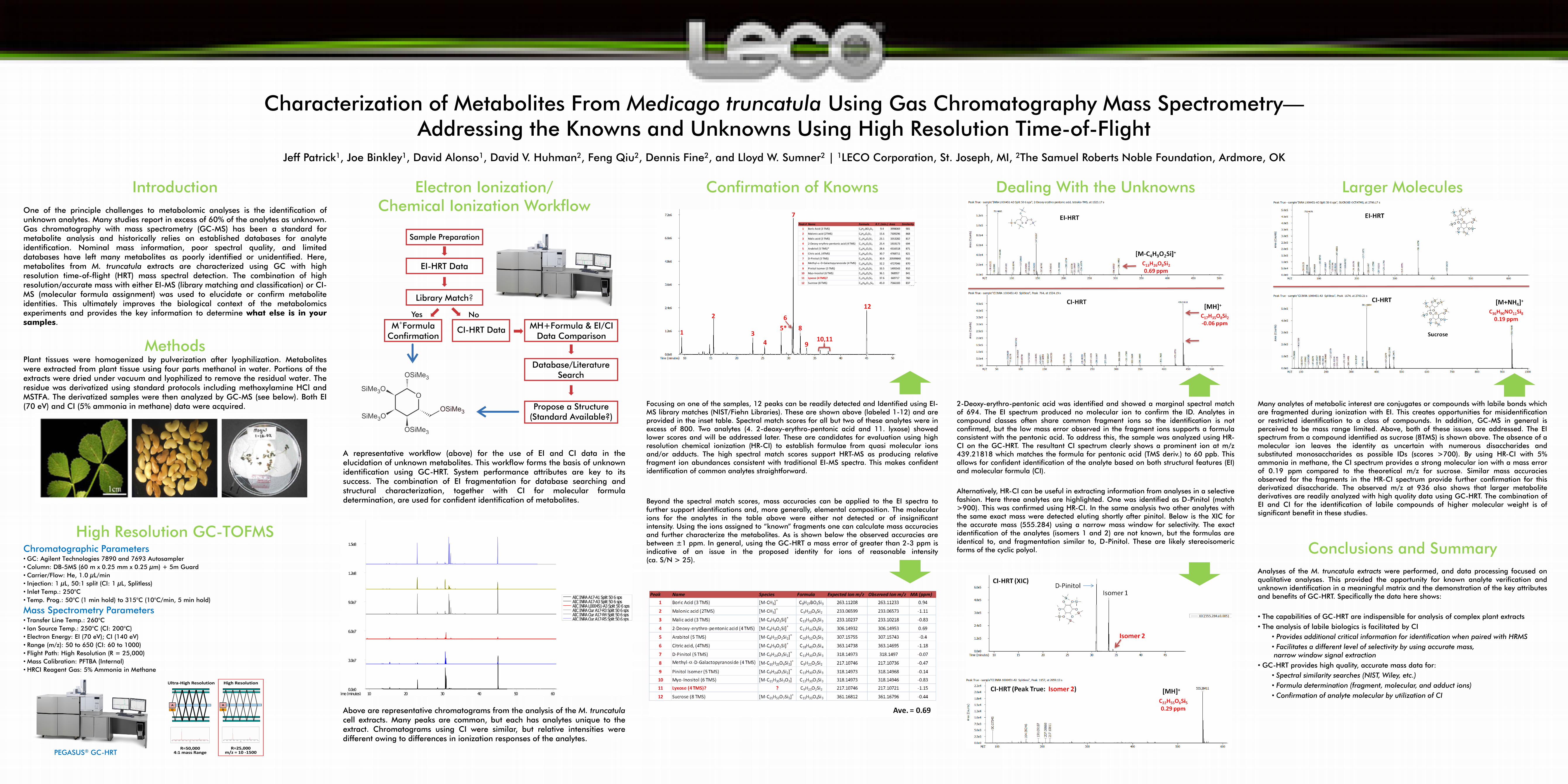
Characterization of Metabolites From Medicago truncatula Using Gas Chromatography Mass Spectrometry—Addressing the Knowns and Unknowns Using High Resolution Time-of-Flight
Introduction One of the principle challenges to metabolomic analyses is the identification of unknown analytes. Many studies report in excess of 60% of the analytes as unknown. Gas chromatography with mass spectrometry (GC-MS) has been a standard for metabolite analysis and historically relies on established databases for analyte identification. Nominal mass information, poor spectral quality, and limited databases have left many metabolites as poorly identified or unidentified. Here, metabolites from M. truncatula extracts are characterized using GC with high resolution time-of-flight (HRT) mass spectral detection. The combination of high resolution/accurate mass with either EI-MS (library matching and classification) or CI-MS (molecular formula assignment) was used to elucidate or confirm metabolite identities. This ultimately improves the biological context of the metabolomics experiments and provides the key information to determine what else is in your samples.
Methods Plant tissues were homogenized by pulverization after lyophilization. Metabolites were extracted from plant tissue using four parts methanol in water. Portions of the extracts were dried under vacuum and lyophilized to remove the residual water. The residue was derivatized using standard protocols including methoxylamine HCl and MSTFA. The derivatized samples were then analyzed by GC-MS (see below). Both EI (70 eV) and CI (5% ammonia in methane) data were acquired.
High Resolution GC-TOFMS Chromatographic Parameters • GC: Agilent Technologies 7890 and 7693 Autosampler • Column: DB-5MS (60 m x 0.25 mm x 0.25 µm) + 5m Guard • Carrier/Flow: He, 1.0 µL/min • Injection: 1 µL, 50:1 split (CI: 1 µL, Splitless) • Inlet Temp.: 250°C • Temp. Prog.: 50°C (1 min hold) to 315°C (10°C/min, 5 min hold)
Mass Spectrometry Parameters • Transfer Line Temp.: 260°C • Ion Source Temp.: 250°C (CI: 200°C) • Electron Energy: EI (70 eV); CI (140 eV) • Range (m/z): 50 to 650 (CI: 60 to 1000) • Flight Path: High Resolution (R = 25,000) • Mass Calibration: PFTBA (Internal) • HRCI Reagent Gas: 5% Ammonia in Methane
Jeff Patrick1, Joe Binkley1, David Alonso1, David V. Huhman2, Feng Qiu2, Dennis Fine2, and Lloyd W. Sumner2 | 1LECO Corporation, St. Joseph, MI, 2The Samuel Roberts Noble Foundation, Ardmore, OK
Electron Ionization/ Chemical Ionization Workflow
A representative workflow (above) for the use of EI and CI data in the elucidation of unknown metabolites. This workflow forms the basis of unknown identification using GC-HRT. System performance attributes are key to its success. The combination of EI fragmentation for database searching and structural characterization, together with CI for molecular formula determination, are used for confident identification of metabolites.
Typical GC/EI Chromatograms
Above are representative chromatograms from the analysis of the M. truncatula cell extracts. Many peaks are common, but each has analytes unique to the extract. Chromatograms using CI were similar, but relative intensities were different owing to differences in ionization responses of the analytes.
Confirmation of Knowns
Focusing on one of the samples, 12 peaks can be readily detected and Identified using EI-MS library matches (NIST/Fiehn Libraries). These are shown above (labeled 1-12) and are provided in the inset table. Spectral match scores for all but two of these analytes were in excess of 800. Two analytes (4. 2-deoxy-erythro-pentonic acid and 11. lyxose) showed lower scores and will be addressed later. These are candidates for evaluation using high resolution chemical ionization (HR-CI) to establish formulae from quasi molecular ions and/or adducts. The high spectral match scores support HRT-MS as producing relative fragment ion abundances consistent with traditional EI-MS spectra. This makes confident identification of common analytes straightforward. Beyond the spectral match scores, mass accuracies can be applied to the EI spectra to further support identifications and, more generally, elemental composition. The molecular ions for the analytes in the table above were either not detected or of insignificant intensity. Using the ions assigned to “known” fragments one can calculate mass accuracies and further characterize the metabolites. As is shown below the observed accuracies are between ±1 ppm. In general, using the GC-HRT a mass error of greater than 2-3 ppm is indicative of an issue in the proposed identity for ions of reasonable intensity (ca. S/N > 25).
Dealing With the Unknowns
TEXT
2-Deoxy-erythro-pentonic acid was identified and showed a marginal spectral match of 694. The EI spectrum produced no molecular ion to confirm the ID. Analytes in compound classes often share common fragment ions so the identification is not confirmed, but the low mass error observed in the fragment ions supports a formula consistent with the pentonic acid. To address this, the sample was analyzed using HR-CI on the GC-HRT. The resultant CI spectrum clearly shows a prominent ion at m/z 439.21818 which matches the formula for pentonic acid (TMS deriv.) to 60 ppb. This allows for confident identification of the analyte based on both structural features (EI) and molecular formula (CI). Alternatively, HR-CI can be useful in extracting information from analyses in a selective fashion. Here three analytes are highlighted. One was identified as D-Pinitol (match >900). This was confirmed using HR-CI. In the same analysis two other analytes with the same exact mass were detected eluting shortly after pinitol. Below is the XIC for the accurate mass (555.284) using a narrow mass window for selectivity. The exact identification of the analytes (isomers 1 and 2) are not known, but the formulas are identical to, and fragmentation similar to, D-Pinitol. These are likely stereoisomeric forms of the cyclic polyol.
Larger Molecules Many analytes of metabolic interest are conjugates or compounds with labile bonds which are fragmented during ionization with EI. This creates opportunities for misidentification or restricted identification to a class of compounds. In addition, GC-MS in general is perceived to be mass range limited. Above, both of these issues are addressed. The EI spectrum from a compound identified as sucrose (8TMS) is shown above. The absence of a molecular ion leaves the identity as uncertain with numerous disaccharides and substituted monosaccharides as possible IDs (scores >700). By using HR-CI with 5% ammonia in methane, the CI spectrum provides a strong molecular ion with a mass error of 0.19 ppm compared to the theoretical m/z for sucrose. Similar mass accuracies observed for the fragments in the HR-CI spectrum provide further confirmation for this derivatized disaccharide. The observed m/z at 936 also shows that larger metabolite derivatives are readily analyzed with high quality data using GC-HRT. The combination of EI and CI for the identification of labile compounds of higher molecular weight is of significant benefit in these studies.
Conclusions and Summary
Analyses of the M. truncatula extracts were performed, and data processing focused on qualitative analyses. This provided the opportunity for known analyte verification and unknown identification in a meaningful matrix and the demonstration of the key attributes and benefits of GC-HRT. Specifically the data here shows: • The capabilities of GC-HRT are indispensible for analysis of complex plant extracts • The analysis of labile biologics is facilitated by CI
• Provides additional critical information for identification when paired with HRMS • Facilitates a different level of selectivity by using accurate mass, narrow window signal extraction
• GC-HRT provides high quality, accurate mass data for: • Spectral similarity searches (NIST, Wiley, etc.) • Formula determination (fragment, molecular, and adduct ions) • Confirmation of analyte molecular by utilization of CI
10 20 30 40 50 600.0e0
3.0e7
6.0e7
9.0e7
1.2e8
1.5e8
Time (minutes)
AIC INRA A17-A1 Split 50 6 spsAIC INRA A17-A3 Split 50 6 spsAIC INRA L000451-A3 Split 50 6 spsAIC INRA Our A17-R3 Split 50 6 spsAIC INRA Our A17-R4 Split 50 6 spsAIC INRA Our A17-R5 Split 50 6 sps
Peak # Name Formula R.T. (min.) Area Similarity1 Boric Acid (3 TMS) C9H27BO3Si3 9.4 3998069 901
2 Malonic acid (2TMS) C9H20O4Si2 15.6 7309296 868
3 Malic acid (3 TMS) C13H30O5Si3 23.1 3353282 817
4 2-Deoxy-erythro-pentonic acid (4 TMS) C17H42O5Si4 25.4 1919173 694
5 Arabitol (5 TMS)* C20H52O5Si5 28.6 4316518 871
6 Citric acid, (4TMS) C18H40O7Si4 30.7 4768711 821
7 D-Pinitol (5 TMS) C22H54O6Si5 30.9 20599840 910
8 Methyl-α-D-Galactopyranoside (4 TMS) C19H46O6Si4 32.2 4727046 870
9 Pinitol Isomer (5 TMS) C22H54O6Si5 33.5 1495543 810
10 Myo-Inositol (6 TMS) C24H60O6Si6 36.1 968957 841
11 Lyxose (4 TMS)? C17H42O5Si4 37.6 669717 760
12 Sucrose (8 TMS) C36H86O11Si8 45.0 7566183 837
PEGASUS® GC-HRT


![Evolutionary flexibility in flooding response circuitry in ... · Medicago truncatula (Mt), and two Solanum spe-cies, domesticated tomato [ Solanum lycopersicum (Sl) cultivar M82]](https://img.pdfslide.net/doc/110x75/60497b0f60455a7a3343524e/evolutionary-flexibility-in-flooding-response-circuitry-in-medicago-truncatula.jpg)