Embed Size (px)
Citation preview

Instructions for use
Title Characterization of nonpathogenic Cadophora gregata, a potential biological control agent, concomitantly isolated fromsoil infested with Cadophora gregata f. sp. adzukicola, the cause of adzuki bean brown stem rot
Author(s) Tanaka, Soichi; Murayama, Keiko; Kondo, Norio; Akino, Seishi
Citation Biological Control, 53(1): 112-121
Issue Date 2010-04
Doc URL http://hdl.handle.net/2115/42760
Type article (author version)
File Information BC53-1_112-121.pdf
Hokkaido University Collection of Scholarly and Academic Papers : HUSCAP
1
Characterization of nonpathogenic Cadophora gregata, a potential biological control
agent, concomitantly isolated from soil infested with Cadophora gregata f. sp.
adzukicola, the cause of adzuki bean brown stem rot
Soichi Tanaka, Keiko Murayama, Norio Kondo and Seishi Akino
Research Faculty of Agriculture, Hokkaido University, Sapporo 060-8589, Japan
Corresponding author: N. Kondo;
E-mail address: [email protected]
Phone/Fax: +81-11-706-4829
2
Abstract
We collected 555 isolates of Cadophora gregata from adzuki bean field soils in Hokkaido,
Japan, from 1997 to 2000. To identify the brown stem rot (BSR) pathogen C. gregata f. sp.
adzukicola, we screened these isolates for pathogenicity to adzuki beans. Of the isolates,
all of which originated in Tokachi District, Hokkaido, Japan, 23 were avirulent to adzuki
bean, soybean, or mung bean. However, polymerase chain reaction (PCR) with specific
primers for C. gregata f. sp. adzukicola (BSRA1 and BSRA2) detected the specific
identifying DNA fragment in these isolates, and cluster analysis with inter-simple
sequence repeat markers showed that the isolates were phylogenetically closer to strains
that are virulent to adzuki bean. Thus, we concluded that the isolates were nonpathogenic
C. gregata. A few selected isolates of the nonpathogenic C. gregata were effective at
3
reducing BSR in vivo and show potential for development as biological control agents.
Keywords: adzuki bean, biological control, brown stem rot, Cadophora gregata,
nonpathogenic
4
1. Introduction
Brown stem rot (BSR), an economically important disease of adzuki bean
[Vigna angularis (Willd.) Ohwi et Ohashi], is caused by Cadophora gregata Harrington
& McNew (Harrington and McNew, 2003). Isolates of C. gregata from adzuki bean and
soybean are host-specific and are recognized as two different formae speciales, C.
gregata f. sp. adzukicola (CGA) and C. gregata f. sp. sojae (CGS), respectively
(Kobayashi et al., 1983, 1991). The pathogens can be differentiated from each other and
other fungi based on isozyme banding patterns (Yamamoto et al., 1990), DNA sequences
of internal transcribed spacer (ITS) regions of rDNA (Chen et al., 1990, 1999; Harrington
et al., 2000), DNA sequences of intergenic spacer (IGS) regions of rDNA (Chen et al.,
2000), microsatellite markers (Chen et al., 2002), and amplified fragment length
5
polymorphisms (AFLPs) or inter-simple sequence repeats (ISSRs; Meng and Chen,
2001). Once soilborne CGA is inside adzuki bean plants by invading root tissues of
young plants, it spreads into the vascular and pith tissue via mycelia and conidia
production (Narita et al., 1971). This fungus infects vascular tissue and causes pith and
vascular tissue discoloration of the stem and petiole, in conjunction with foliar chlorosis
or necrosis. Consequently, the pathogen can cause susceptible plants to wilt and reduce
adzuki bean yields. Although phytotoxic gregatins produced by CGA previously were
thought to be associated with these symptoms (Kobayashi and Ui, 1977), Tanaka et al.
(2007) found that the toxins were unlikely to be essential for pathogenicity.
Extensive studies have been performed on phenotypical variations (races) of
CGA isolates (Kondo et al., 1998, 2002), adzuki bean resistance to BSR (Adachi et al.,
1988; Chiba, 1982, 1985; Chiba et al., 1987; Fujita et al., 1995, 2002, 2007), and the close
6
ecological association of C. gregata with nematodes (Djiwanti, 1999; Sugawara et al.,
1997a; Yamada et al., 2005a, 2005b). To date, disease-resistant cultivars and crop rotation
are the most practical means of controlling BSR. Indeed, Fujita et al. (2007) and Kondo et
al. (2009) found one cultivated adzuki bean variety and one wild adzuki bean accession
that tolerated all of the CGA races, making them useful for breeding BSR-resistant adzuki
bean cultivars. Yamada et al. (2005b) showed that using wild oats [Avena strigosa
Schreb.] as green manure decreased the degree of BSR damage by suppressing
nematodes. However, as shown by the quick appearance of a strain virulent to recently
developed resistant breeding lines (Kondo et al., 2005), BSR outbreaks in new races may
be inevitable, due to the co-evolutionary relationship between crop varieties and their
pathogens.
The distribution of CGA races has been examined using isolates collected from
7
adzuki bean field soils in Hokkaido (Kondo et al., 2002). Pathogenicity tests revealed that
most isolates were virulent to the susceptible cultivar Erimo-shozu, although several
avirulent isolates were discovered. Therefore, we examined the pathogenicity of
additional isolates to investigate the distribution of nonpathogenic C. gregata (NPC).
Some nonpathogenic strains of plant pathogenic species may have the potential to protect
the plant against infections caused by the virulent strains (Sneh, 1998). Moreover,
understanding NPC may enable us to optimize ecological conditions to enhance its
suppressive ability in crop rotations. Our objectives were to identify NPC strains, their
characteristics, and their potential as biological control agents of adzuki bean BSR.
2. Materials and methods
8
2.1. Sources and cultivation of isolates
As described in a previous study (Kondo et al., 2002), C. gregata was isolated
from 44 adzuki bean field soil samples collected from five districts in Hokkaido from
1997 to 2000 using a modified selective medium soil dilution method (Table 1).
Single-spored isolates were stored in green-pea agar (GPA; 200 g frozen green peas
boiled 15 min, filtered through four layers of cheesecloth, and solidified using 20 g
agar/liter) at 4˚C. Sporulation ability and cultural morphology were determined using
V8-juice agar (200 ml V8 juice and 2 g CaCO3 centrifuged at 5,000 rpm for 15 min, with
the supernatant diluted to 1 L with distilled water and solidified using 20 g agar/liter) and
potato dextrose agar (PDA; Difco, Lawrence, Kansas, USA), respectively. For long-term
storage, agar disks containing spores of each isolate grown on GPA were placed in
9
individual cryovials containing 20% sterile glycerol and maintained at -80˚C. In Table 2
four nonpathogenic isolates that were originally obtained as CGA and CGS from diseased
adzuki bean and soybean in Japan, respectively, were included and also used in this study:
a virulence-deficient mutant A’31-2 (Kobayashi et al., 1981); isolates A60K68 and
A60To, which were originally identified as virulent (Yamamoto, 1994); and isolate
A57T22, which was uncertain for virulence.
2.2. Cultural conditions and screening of nonpathogenic strains
The inoculum was grown in V8-juice broth at 25˚C on a reciprocal shaker at
120-oscillations/min. After 3 weeks of incubation, mycelia and spores were collected by
filtration through Whatman No. 1 filter paper and washed by suspending them in distilled
10
water, followed by centrifugation. The fungal pellets were homogenized in distilled water
with a homogenizer (10,000 rpm for 3 min; AN-5, Shin-nihonseiki, Tokyo, Japan) and
the concentration of mycelial fragments and spores was determined using a
hemocytometer; mycelial fragments of all sizes were included in counts. Blended
cultures were then diluted with distilled water to a concentration of 107 propagules per
milliliter. The adzuki bean varieties used to determine fungal races were cvs.
Erimo-shozu (susceptible to all races), Kita-no-otome (resistant to races 1 and 3, but
susceptible to race 2) and Acc259 (resistant to races 1 and 2, but susceptible to race 3).
Seedlings were grown in plastic containers (15 × 20 × 5 cm) with sterilized vermiculite
for about 10 to 14 days in greenhouse, then the roots were washed gently with running tap
water. The roots of 10 seedlings of each cultivar were dipped into each inoculum
suspension (50 ml) for 12 hr, then transplanted into a soil (Pot –ace, Katakura Chikkarin
11
K.K. Tokyo, Japan)/ vermiculite mixture (1:1, v/v) in 12-cm diameter plastic pots. The
response to the pathogen was evaluated after eight weeks growth in greenhouse.
Nighttime low and daytime high temperatures during these tests were 15/32°C. Plants
received supplemental lighting from metal halide sodium lamps (400W) to maintain a
14-h photoperiod. The inoculation experiments were completely randomized, with two
replications (pots) per isolate per cultivar and five plants per pot. Pathogenicity tests were
repeated twice with these cultivars, and nonpathogenic isolates were determined.
Soybean [Glycine max (L.) Merrill. cv. Sapporo-midori] and mung bean [Vigna radiata
(L.) R. Wilczek, susceptible to both CGS and CGA] were also used to determine formae
speciales of C. gregata. The roots of 10 seedlings of each crop grown as described above
were dipped into a suspension (50 ml) of each inoculum (Table 3) for 12 h. The seedlings
were then transplanted into the sterilized mixed soil in 18-cm-diameter plastic pots.
12
Isolates T96-1 (race 1 CGA), T96-5 (race 2 CGA), and S58KS (CGS) also were used as
controls. For BSR assessment, the number of diseased plants in the greenhouse with
foliar symptoms (stunted and necrotic) or vascular discoloration was counted eight weeks
after inoculation.
2.3. Production of gregatins
Isolates were grown in adzuki bean stem medium (5 g dry stem pieces boiled
15 min, filtered through four layers of cheesecloth, with the filtrate diluted to 1 L using
distilled water) containing 5% glucose (Kobayashi and Ui, 1975) on a reciprocal shaker at
120-oscillations/min for 4 weeks at 25˚C. The culture filtrate (200 ml) was adjusted to pH
7.0, extracted once with an equal volume of ethyl acetate, and washed with distilled water.
13
The ethyl acetate solution was evaporated, yielding an oily residue. This extract was
dissolved in acetone (200 μl), and 3 μl was spotted onto a thin-layer chromatography
(TLC) silica gel plate (Silica gel 60 F254; Merck, Darmstadt, Germany). The solvent
system was chloroform: methanol (98:2, v/v). Gregatins were visualized by UV
irradiation (254 nm). For positive and negative controls, extracts from a culture of
wild-type strain T96-5 and non-inoculated medium were used, respectively.
2.4. DNA extraction from mycelia
For DNA extraction, agar plugs were removed from the growing margin of
1-week-old cultures and transferred to V8-juice broth. After a 14-day incubation period at
25˚C, mycelial mats were harvested by filtering the broth through Whatman No.1 filter
14
paper in a Buchner funnel then cleaned by rinsing each mat several times with distilled
water. After removing the excess water, all mycelial mats were frozen at -80˚C until they
were ground to a fine powder in liquid nitrogen using a sterilized mortar and pestle. The
DNA extraction was conducted using the DNeasy Plant Mini Kit (QIAGEN, Hilden,
Germany) following the manufacturer’s instructions. The DNA concentration was
determined by UV/Vis spectrophotometer (DU640; Beckman Coulter Inc., Fullerton, CA,
USA) and the purity of the DNA samples was examined by electrophoresis in 0.8%
agarose (Wako Pure Chemical Industries, Osaka, Japan) gel (TBE buffer).
2.5. Design and specificity of primers specific for C. gregata
Two polymerase chain reaction (PCR) primers, BSRA1
15
(5’-GCTTGCTCCGTGGTGGGCTA-3’) and BSRA2
(5’-GATTTGGGGGTTGCTGGAAG-3’), were designed for CGA based on the ITS
sequence (GenBank accession no. U66731; Chen et al., 1996; Sakuma et al., 1999). The
PCR primers, BSR1 (5’-GCTTGCTCCGTGGCGGGCTG-3’) and BSR2
(5’-AATTTGGGTGTTGCTGGCATG-3’) (Chen et al., 1996), also were used to confirm
CGS. To check DNA quality, PCR was also performed on the DNAs, using the primers
ITS1 (5’-TCCGTAGGTGAACCTGCGG-3’) and ITS4
(5’-TCCTCCGCTTATTGATATGC-3’), which amplify ITS rDNA through ITS 1, the
5.8S gene, and ITS 2 (White et al., 1990). PCR amplifications were generally performed
in 25 µl reactions, with 0.2 mM dNTP mixture, 10 × PCR buffer (10 mM Tris-HCl, pH
8.3, 50 mM KCl, and 1.5 mM MgCl2), 0.75 units Taq polymerase (Takara Inc., Shiga,
Japan), 0.16 µM of each specific primer, and 4 pg template DNA, under the following
16
temperature regimes: initial denaturation at 94˚C for 2 min, followed by 30 denaturation
cycles at 94˚C for 1 min; annealing at 53˚C (for ITS rDNA), and 60˚C or 62˚C for 1 min;
and extension at 72˚C for 1 min, with a final extension at 72˚C for 5 min using a DNA
thermal cycler (TaKaRa Inc., Shiga, Japan). A negative control without the DNA template
was included in each PCR set to monitor possible contamination. To visualize the DNA
fragments, 3 µl PCR products was loaded in a gel containing 1.5% agarose, along with
φHaeⅢ. After running 30 min at 100 V, gels were stained with ethidium bromide and
photographed under ultraviolet light.
2.6. Detection of C. gregata in adzuki bean plants
The colonization of plant roots by C. gregata was determined when the plants
17
were assessed eight weeks after inoculation. Root samples (≥ 10 pieces) from each
leguminous crop, which comprised four plants hosting each isolate, were washed gently
with running tap water for 3 h. DNA was extracted from the samples using the DNeasy
Plant Mini Kit. PCR, using the primers described above, was then performed to detect C.
gregata inoculates.
2.7. Genetic analysis of C. gregata using ISSR
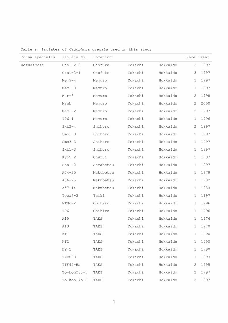
Isolates used in this study are listed in Table 2. DNA extractions were
performed as described above. Microsatellite primers used in this study and the annealing
temperature of each primer are shown in Table 4. The PCR reaction mixture (25 μl)
consisted of 25 ng template DNA, 0.625 units Taq polymerase, 10 × PCR buffer (10 mM
18
Tris-HCl, pH 8.3, 50 mM KCl, and 1.5 mM MgCl2), 0.2 mM dNTP mixture, and 0.5μ M
primer. The PCR thermal cycler conditions were 94˚C for 1 min, followed by 35 cycles at
94˚C for 1 min, each annealing temperature for 1 min (Table 3), and 72˚C for 2 min. A
final elongation was performed at 72˚C for 10 min to ensure a double-stranded
amplification product. The temperature was then reduced and maintained at 4˚C. All
amplifications were repeated at least twice. Negative controls were run with all
amplifications to confirm that the reactions were free of contamination. PCR products
were electrophoresed separately in 1.5% agarose gel (TBE buffer) at 100 V for 40–60 min,
along with λHindⅢ. The gels were stained with ethidium bromide and visualized under
UV light. A digital image of the gel was analyzed using Kodak Digital Science, Image
Analysis Software (Eastman Kodak Company, Rochester, NY, USA).
The PCR reaction fragments were treated as putative distinct loci, and presence
19
or absence was scored as 1 or 0, respectively. Only intense fragments of the isolates were
scored, if reproducible in independent reactions. A binary data matrix was constructed
from the scores of all of the isolates. Distance and similarity matrix computations for
binary data were performed using the Windist program in PHYLIP
(http://evolution.genetics.washington.edu/phylip.html). Cluster analysis, using the
unweighted pair-group method with arithmetic averages (UPGMA), was performed
using the NEIGHBOR program to produce a dendrogram. For each branch of the
phenogram, a supported bootstrap value was generated with 1,000 bootstrapped samples,
using the Winboot program (Yap et al., 1996).
2.8. Screening trials for biocontrol effectiveness
20
Seedlings of cv. Erimo-shozu were co-inoculated with each nonpathogenic
isolate and T96-1 (CGA); suspensions (107 mycelial fragments and spores/ ml) were
mixed at 9:1 (v/v) to yield 50 ml inoculum. Adzuki bean seedlings, spores, and mycelial
fragments were prepared as described above. For each nonpathogenic isolate, 16
seedlings were immersed in the suspension for 24 h and then planted, four per pot, in
12-cm-diameter vinyl pots filled with sterilized soil mixture, consisting of 1:1 (v/v)
vermiculite/Pot-ace. This procedure was conducted twice for all combinations. Nighttime
low and daytime high temperatures during these tests were 15°/ 32°C. Plants received
supplemental lighting from metal halide sodium lamps (400W) to maintain a 14-h
photoperiod. In all tests, controls were inoculated with only the BSR pathogen (106
mycelial fragments and spores/ ml). The suppression of BSR was assessed based on the
occurrence of vascular browning 45 days after inoculation. Average disease severity for
21
each isolate was calculated from the proportion of nodes with browning to the total
number of nodes in each replicate (pot), using the following formula: [Σ(number of nodes
discolored/ number of total nodes) × 100/total number of plants].
2.9. Statistical analysis
All data expressed as percentages were arcsine-transformed (sine-1√x) prior to
analysis. The means were compared using Dunnett’s test, with reference to a positive
control.
3. Results
22
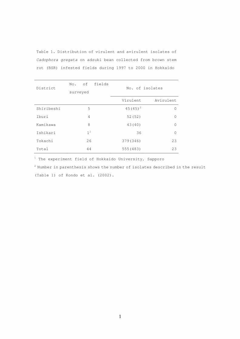
3.1. Identification of NPC
Of the 555 isolates, which included 483 virulent isolates (Kondo et al., 2002),
23 avirulent isolates were obtained only from Tokachi district (Table 1). Although 13
nonpathogenic isolates did not produce conidia on V8 agar (Table 3), nine others (isolate
Hir6-4 was not tested) were morphologically identical to C. gregata: hyaline and septate
hyphae on GPA; hyaline, ovoid or ellipsoidal conidia forming a false head on phialides
with short collarettes; and slow growth on PDA, with colonies reddish-brown or salmon
pink to red. However, five of 43 isolates that were isolated from 1976 to 1996 and
maintained in our laboratory were avirulent; with one exception (mutant isolate A’31-2),
they originated from Tockachi district or the experimental field of Hokkaido University.
When subjected to PCR, using the primers BSRA1 and BSRA2 designed
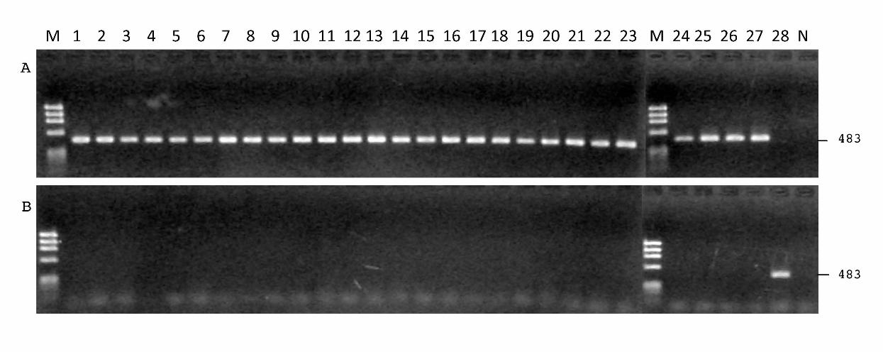
23
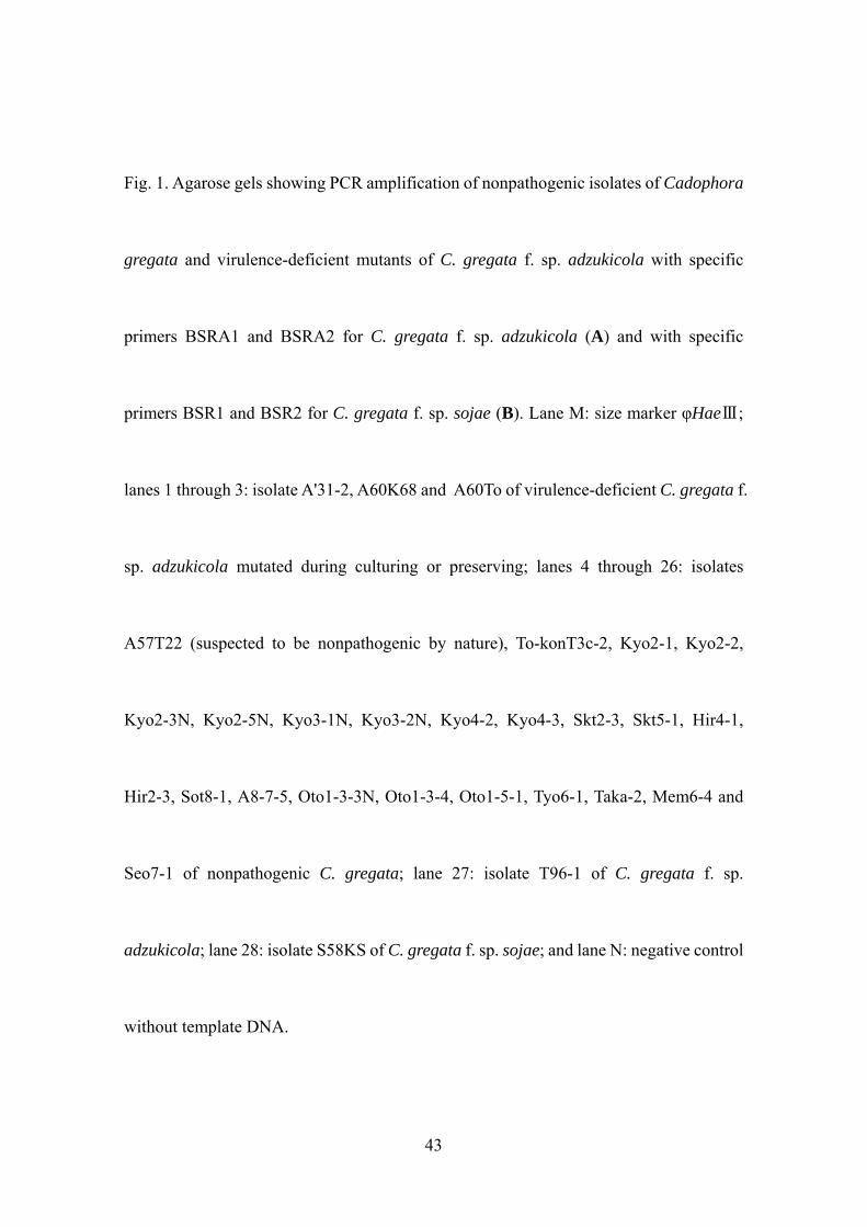
specifically for CGA, all of the NPC isolates tested at an annealing temperature of 62˚C
(Fig. 1A) yielded the expected 483-bp fragment, but the primers BSR1 and BSR2, which
were specific for CGS, did not produce the fragment (Fig. 1B). Occasionally, at 60˚C
annealing temperature, PCR fragments were slightly amplified, even when primers BSR1
and BSR2 were used with CGA isolates (data not shown). This indicates that NPC
isolates are much closer to CGA than to CGS. The PCR products of all isolates resulting
from ITS1 and ITS4, which amplify ITS rDNA, were about the same size, except that of
isolate Taka-2, which was smaller (data not shown).
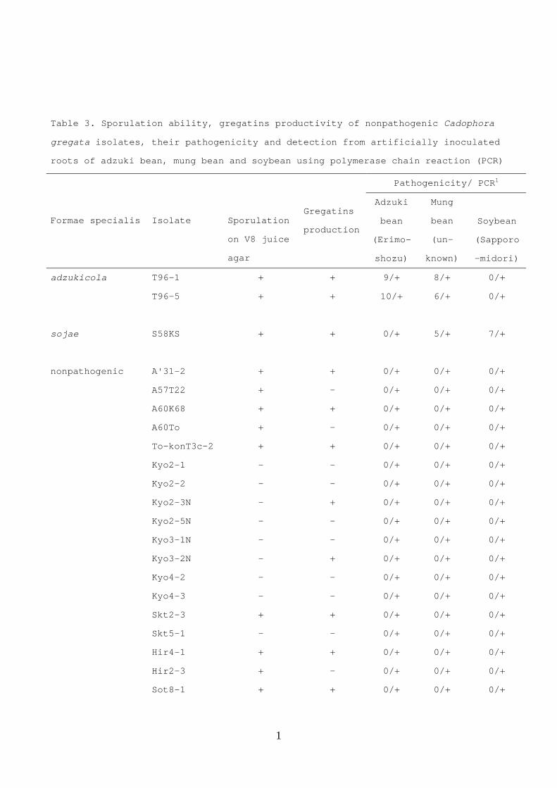
3.2. Host specificity and gregatins productivity
All nonpathogenic isolates, including four virulence-deficient isolates, were
24
not pathogenic to either soybean or mung bean, indicating that they were not CGS (Table
3). Although cononization of treated isolates inside roots is not determined clearly by
PCR because a trace of each fungus on the root surface might be included in the sample,
infected isolates, except isolate Taka-2, were identified and seemed to be colonized in the
inoculated roots due to gentle surface sterilization. The production of gregatins varied,
depending on the isolate; 10 isolates produced gregatins as profusely as did wild-type
CGA isolates, but TLC detected no gregatin in the 16 other isolates. Thus, gregatin
production does not necessarily determine CGA pathogenicity.
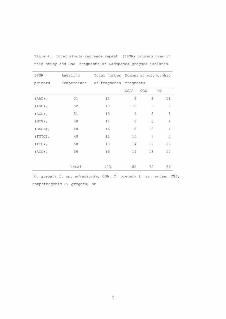
3.3. Cluster analysis with ISSR
Each of the eight primers tested amplified 10 to 18 fragments (Table 4), ranging
25
in size from about 400 to 2500 bps (data not shown). The frequency of polymorphic
fragments was greater among CGA isolates (79.6%) than among isolates of CGS (68.0%)
and NPC (64.1%), indicating higher genetic variation in the CGA population.
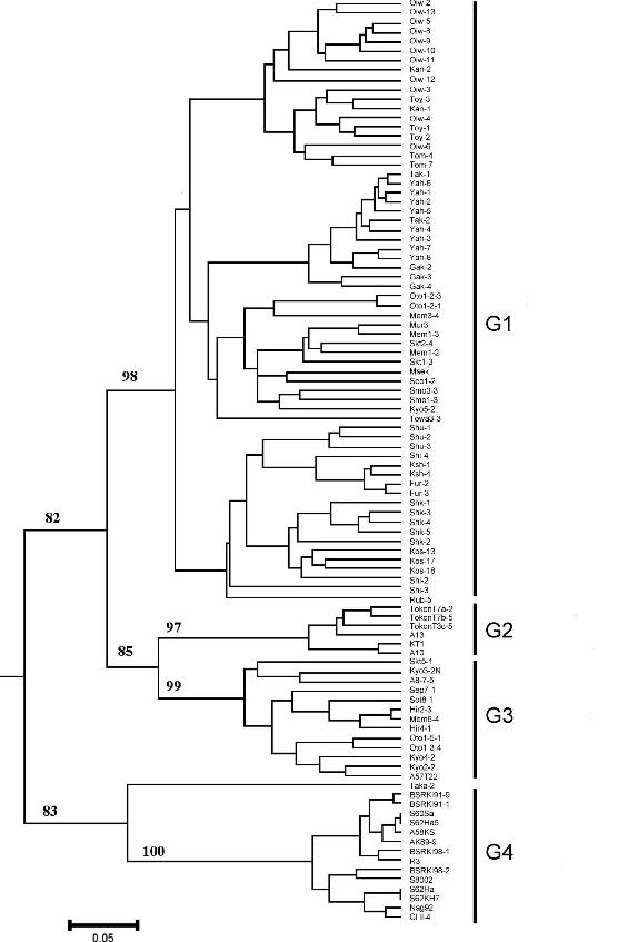
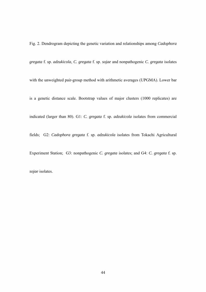
Cluster analysis based on 103 ISSR markers separated isolates into four groups
(G1–G4): CGA isolates from commercial fields (G1); those from Tokachi Agricultural
Experiment Station (G2); NPC isolates, except isolate Taka-2 (G3); and CGS isolates
(G4) supported by high bootstrap values (82–100%; Fig. 2). The UPGMA tree created
with ISSR revealed that G1 was resolved from the clade consisting of G2 and G3, and G1
and G4 were clearly distinguished.
For all of the groups, the ISSR profiles were closely associated with
geographical origins, and all of the CGA races were scattered throughout the tree. Isolates
from the same field tended to cluster in subgroups within CGA. Only isolate Taka-2 was
26
distinct from other NPC or CGA isolates.
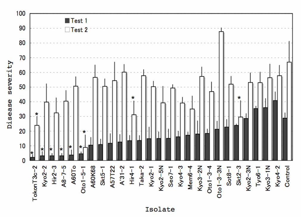
3.4. Effect of NPC on BSR incidence
Effective isolates were found within 26 NPC isolates, including four
virulence-deficient mutants. Co-inoculation with NPC isolates Oto1-5-1 or To-konT3c-2
and CGA isolate T96-1 significantly (P < 0.05) reduced BSR incidence in both trials,
compared with inoculation solely with the CGA isolate (Fig. 3). Isolate Oto1-5-1 was
particularly effective at reducing the disease, with reductions of 84% and 87% in the first
and second trials, respectively.
4. Discussion
27
The 23 isolates obtained from adzuki bean field soils in Tokachi district were
morphologically similar to CGA but were determined to be avirulent to adzuki bean (cv.
Erimo-shozu). In addition, PCR, using the CGA-specific primers BSRA1 and BSRA2,
detected a specific DNA fragment in these isolates (Fig. 1A). Thus, we concluded that
nonpathogenic or saprophytic C. gregata exists concomitantly in soil infested with the
adzuki bean BSR pathogen. Molecular markers for easy discrimination of CGA and CGS
have been developed: a variable DNA region in the ITS (Chen et al., 1996) or a variable
DNA region in the IGS of nuclear rDNA (Chen et al., 2000). However, genotype C,
designated from a variable DNA region in the IGS, does not always coincide with CGA
isolates (Ito et al., 2008). Accordingly, we used the marker from the variable DNA region
in the ITS to identify CGA. Currently, we are genotyping CGA and CGS in Japan using
28
the specific primers BSRIGS1 and BSRIGS2 (Chen et al., 2000).
Although it is not clear why NPC isolate populations are restricted only to
Tokachi district, the UPGMA tree with ISSR revealed that NPC isolates—with the
exception of isolate Taka-2, which is distinct from other NPC or CGA isolates—are
phylogenetically closer to CGA isolates from Tokachi Agricultural Experiment Station
(TAES) in Memuro than to isolates from other adzuki bean fields (Fig. 2). Thus, when the
infested fields of TAES were created for breeding about 40 years ago, diseased residues
of adzuki bean collected from a common location could have been incorporated.
Although the CGA-specific fragment was also detected in isolate Taka-2, the size of the
amplified fragment of the ITS region was smaller than that in other tested NPC isolates,
as well as that of CGA. Of course, this isolate is not pathogenic to soybean (it is not a
CGS pathogen). Thus, it is unique, and its close relationship to CGS in the UPGMA tree
29
could occur by chance. Isolate A57T22, which was also collected from Shimizu in
Tockachi district in 1983 and preserved in our laboratory, is avirulent and included in
NPC group G3 (Fig. 2), indicating that the isolate is probably not a virulence-deficient
mutant but is nonpathogenic by nature. The tree also showed that adzuki bean isolates
(G1–G3) and soybean isolates (G4) of C. gregata are clearly distinguished. Generally,
our results were consistent with previous findings (Meng et al., 2001; Yamamoto, 1994;
Yamamoto et al., 1990), in that CGA and CGS are genetically distinct from each other.
For all of the groups, ISSR profiles are closely associated with geographical origins,
although isolates from the same district are generally not tightly clustered in the AFLP
analysis (Kondo et al., 2002).
We determined a varied range of NPC isolates after screening for the reduction
of BSR symptoms on adzuki bean stems in vivo. One potential isolate, Oto1-5-1, shows
30
potential to control the disease (Fig. 3), due to its disease suppression ability and lack of
gregatin productivity (Table 3). Previously, the pre-inoculation of CGS isolates was
reported to induce resistance to BSR in adzuki bean (Sugawara et al., 1997b). Although
split-root experiments in the same report demonstrated that some CGS isolates induced
systemic resistance to adzuki bean BSR, no systemic resistance with isolate Oto1-5-1 was
determined in preliminary tests using a split-root method (unpublished data). Hence, the
reduction of BSR symptoms in this study could be attributed to parasitic competition for
infection sites on the root.
In naturally occurring suppressive soils, antagonistic Fusarium spp. population
levels typically must be 10–100 times greater than that of the pathogen to be effective
(Alabouvette, 1986; Alabouvette et al., 1993; Paulitz et al., 1987). Although no
suppressive soil has been found in adzuki bean BSR, indigenous NPC isolates may
31
induce disease suppression if appropriate crops or varieties of adzuki bean are cultivated,
and application methods of effective NPC isolates, such as Oto1-5-1, are improved.
Further research is required to develop this antagonist as a biocontrol agent, as well as to
investigate the conditions under which it is effective and ways to improve its consistency
and level of effectiveness.
Acknowledgement
This work was supported by the Japan Beans and Peas Foundation.
References
32
Adachi, T., Narikawa, T., Chiba, I., Murata, K., Hara, M. and Shimada, H., 1988. A new
adzuki bean variety “Hatsune-shozu”. Bulletin of Hokkaido Prefecture
Agricultural Experiment Station 57, pp. 13-24.
Alabouvette, C., 1986. Fusarium-wilt suppressive soils from the Chàteaurenard region:
Review of a 10-year study. Agronomie 6, pp. 273-284.
Alabouvette, C., Lemanceau, P. and Steinberg, C., 1993. Recent advances in the
biological control of Fusarium wilts. Pesticide Science 37, pp. 365-373.
Chen, W., Gray, L. E. and Grau, C. R., 1996. Molecular differentiation of fungi associated
with brown stem rot and detection of Phialophora gregata in resistant and
susceptible soybean cultivars. Phytopathology 86, pp. 1140-1148.
Chen, W., Gray, L. E., Kurle, J. E. and Grau, C. R., 1999. Specific detection of
Phialophora gregata and Plectosporium tabacinum in infected soybean plants.
33
Molecular Ecology 8, pp. 871-877.
Chen, W., Grau, C. R., Adee, E. A. and Meng, X., 2000. A molecular marker identifying
susbspecific populations of the soybean brown stem rot pathogen, Phialophora
gregata. Phytopathology 90, pp. 875-883.
Chen, W., Shi, X. and Chen, Y-G., 2002. Microsatellite markers and clonal genetic
structure of the fungal pathogen Phialophora gregata. Mycological Research
106, pp. 194-202.
Chiba, I., 1982. Breeding for resistance to brown stem rot (BSR) in adzuki beans. I. On
the varietal differences in resistance to BSR. Bulletin of Hokkaido Prefecture
Agricultural Experiment Station 48, pp. 56-63.
Chiba, I., 1985. Breeding for resistance to brown stem rot (BSR) in adzuki beans. II.
Formation of varietal difference to BSR. Bulletin of Hokkaido Prefecture
34
Agricultural Experiment Station 52, pp. 79-84.
Chiba, I., Narikawa, T., Murata, K. and Adachi, T., 1987. Breeding for resistance to
brown stem rot (BSR) in adzuki beans. III. The inheritance of resistance to BSR
and effect of its introduction. Bulletin of Hokkaido Prefecture Agricultural
Experiment Station 56, pp. 1-7.
Djiwanti, S. R., Sugawara, K., Kondo, N., Kobayashi, K. and Ogoshi, A., 1999. Effect of
soybean cyst nematode infestation on the incidence of brown stem rot in adzuki
bean cultivars. Soil Microorganisms 53, pp. 19-25.
Fujita, S., Shimada, H., Murata, K., Shirai, S., Hara, M., Adachi, T. and Chiba, I., 1995. A
new BSR-resistant variety “Kita-no-otome” adzuki bean. Bulletin of Hokkaido
Prefecture Agricultural Experiment Station 68, pp. 17-31.
Fujita, S., Murata, K., Shimada, H., Aoyama, S., Chiba, I., Matsukawa I., Shirai, S.,
35
Miura, T., Ochi, H. and Kondo, N., 2002. A new adzuki bean variety “Syumari”
with soil-borne disease resistance and excellent processing quality. Bull.
Bulletin of Hokkaido Prefecture Agricultural Experiment Station 82, pp. 31-40.
Fujita, S., Kondo N., Shimada, H. and Naito, S., 2007. Re-evaluation and selection of
adzuki beans to breed cultivars resistant to new race of Phialophora gregata f.
sp. adzukicola, the causal agent of adzuki bean brown stem rot (BSR). Breeding
Research 9, pp. 87-95.
Harrington, T. C., Stiemel, J., Workneh, F. and Yang, X. B., 2000. Molecular
identification of fungi associated with vascular discoloration of soybean in the
north central United States. Plant Disease 84, pp. 83-89.
Harrington, T. C. and McNew, D.L., 2003. Phylogenetic analysis places the
Phialophora-like anamorph genus Cadophora in the Helotiales. Mycotaxon 87,
36
pp. 141-151.
Ito, T. and Kondo, N., 2008. Identification of Phialophora gregata genotypes with
molecular marker (Abstr.) Japanese Journal of Phytopathology 74, p. 79.
Kobayashi, K. and Ui, T., 1975. Isolation of phytotoxic substances produced by
Cephalosporium gregatum Allington & Chamberlain. Tetrahedron letters 47,
pp. 4119-4122.
Kobayashi, K. and Ui, T., 1977. Wilt-inducing antibiotic compounds produced by
Cephalosporium gregatum. Physiological Plant Pathology 11, pp. 55-60.
Kobayashi, K., Kondo, N. and Ui, T., 1981. Appearance of a mutant from
Cephalosporium gregatum type A isolate in culture. Annual Phytopathological
Society of Japan 47, pp. 577-580.
Kobayashi, K., Kondo, N. and Ui, T., 1983. Difference in pathogenicity of Phialophora
37
gregata isolates from adzuki bean in Japan and from soybean in the United
States. Plant Disease 67, pp. 387-388.
Kobayashi, K., Yamamoto, H., Negishi, H. and Ogoshi, A., 1991. Formae speciales
differentiation of Phialophora gregata isolates from adzuki bean and soybean
in Japan. Annual Phytopathological Society of Japan 57, pp. 225-231.
Kondo, N., Fujita, S., Murata, K. and Ogoshi, A., 1998. Detection of two races of
Phialophora gregata f. sp. adzukicola, the causal agent of adzuki bean brown
stem rot. Plant Disease 82, pp. 928-930.
Kondo, N., Kobayashi, Y., Sakuma, F., Fujita, S. and Murata, K., 2002. Regional
distribution of two races of Phialophora gregata f. sp. adzukicola, the causal
agent of adzuki bean brown stem rot, and their genetic diversity in Hokkaido,
the northernmost island of Japan. Journal of General Plant Pathology 68, pp.
38
284-291.
Kondo, N., Nakazawa, K., Fujita, S., Shimada, H. and Naito, S., 2005. New virulent race
of Phialophora gregata f. sp. adzukicola associated with continuous cultivation
of adzuki bean cultivar Acc259. Journal of General Plant Pathology 71, pp.
360-363.
Kondo, N., Shimada, H. and Fujita, S., 2009. Screening of cultivated and wild adzuki
bean for resistance to race 3 of Cadophora gregata f. sp. adzukicola, cause of
brown stem rot. Journal of General Plant Pathology 74, pp. 181-187.
Meng, X. and Chen, W., 2001. Applications of AFLP and ISSR techniques in detecting
genetic diversity in the soybean brown stem rot pathogen Phialophora gregata.
Mycological Research 105, pp. 936-940.
Narita, T., Akai, J. and Tsuboki, K., 1971. Adzuki brown stem rot and its pathogenic
39
fungus. Plant Protection 25, pp. 353-358.
Paulitz, T. C., Park, C. S. and Baker, R., 1987. Biological control of Fusarium wilt of
cucumber with nonpathogenic isolates of Fusarium oxysporum. Canadian
Journal of Microbiology 33, pp. 349-353.
Sakuma, F., Haga, H., Kondo, N. and Kobayashi, K., 1999. Detection of Phialophora
gregata f. sp. adzukicola in soil by using PCR-mediated assay (Abstr.). Annual
Phytopahological Society of Japan 65, p. 699.
Sneh, B., 1998. Use of non-pathogenic of hypovirulent fungal strains to protect plants
against closely related fungal pathogens. Biotechnology Advances 16, pp. 1-32.
Sugawara, K., Kobayashi, K. and Ogoshi, A., 1997a. Influence of the soybean cyst
nematode (Heterodelra glycines) on the incidence of brown stem rot of soybean
and adzuki bean. Soil Biology and Biochemistry 29, pp. 1491-1498.
40
Sugawara, K., Furuhashi, S., Haga, H., Kobayashi, K. and Ogoshi, A., 1997b. Induction
of resistance for brown stem rot of adzuki bean by Phialophora gregata f. sp.
adzukicola. In: Ogoshi, A., Kobayashi, K., Homma, Y., Kodama, F., Kondo, N.,
Akino, S. (Eds.), Proceedings of the Fourth International Workshop on Plant
Growth-Promoting Rhizobacteria. The 4th PGPR International Workshop
Organizing Committee, Sapporo, pp. 289-291.
Tanaka, S., Kondo, N. and Naito, S., 2007. Isolation of pathogenicity and
gregatins-deficient mutants of Phialophora gregata f. sp. adzukicola through
Agrobacterium tumefaciens-mediated transformation. Journal of General Plant
Pathology 73, pp. 242-249.
White, T. J., Bruns, T., Lee, S. and Taylor, J., 1990. Amplification and direct sequencing
of fungal ribosomal RNA genes for phylogenetics. In: Innis, M. A., Gelfand, D.
41
H., Sninsky, J. J., White, T. J. (Eds.), PCR Protocols: A Guide to Methods and
Applications. Academic Press, San Diego, CA. pp. 315-322.
Yamada, E. , Sakuma, F., Hashizume, K., Takahashi, M., Fukuhara, Y., Kobayashi, K.
and Kondo, N., 2005a. Effect of Pratylencus penetrans on the infection of
brown stem rot in adzuki bean. Japan Journal Nematology 35, pp. 71-77.
Yamada, E. , Sakuma, F., Hashizume, K., Takahashi, M., Fukuhara, Y., Kobayashi, K.
and Kondo, N., 2005b. Seasonal occurrence of Pratylencus penetrans,
Heterodera glyscines and Phialophora gregata f. sp. adzukicola as affected by
the cultivation of green manure crops. Japan Journal Nematology 35, pp.79-86.
Yamamoto, H., 1994. Studies on taxonomy of the causal fungus of brown stem rot of
adzuki bean and soybean, Phialophora gregata (Allington et Chamberlain)
Gams. Memories of The Faculty of Agriculture Hokkaido University 19, pp.
42
57-98.
Yamamoto, H., Kobayashi, K. and Ogoshi, A., 1990. Isozyme polymorphism in
Phialophora gregata isolates from adzuki bean and soybean in Japan. Annual
Phytopathological Society of Japan 56, pp. 584-590.
Yap, I. and Nelson, R. J., 1996. Winboot: A program for performing bootstrap analysis of
binary data to determine the confidence limits of UPGMA-based dendrograms.
IRRI Research Paper Series No. 14. International Rice Research Institute,
Manila, Phillipines.
43
Fig. 1. Agarose gels showing PCR amplification of nonpathogenic isolates of Cadophora
gregata and virulence-deficient mutants of C. gregata f. sp. adzukicola with specific
primers BSRA1 and BSRA2 for C. gregata f. sp. adzukicola (A) and with specific
primers BSR1 and BSR2 for C. gregata f. sp. sojae (B). Lane M: size marker φHaeⅢ;
lanes 1 through 3: isolate A'31-2, A60K68 and A60To of virulence-deficient C. gregata f.
sp. adzukicola mutated during culturing or preserving; lanes 4 through 26: isolates
A57T22 (suspected to be nonpathogenic by nature), To-konT3c-2, Kyo2-1, Kyo2-2,
Kyo2-3N, Kyo2-5N, Kyo3-1N, Kyo3-2N, Kyo4-2, Kyo4-3, Skt2-3, Skt5-1, Hir4-1,
Hir2-3, Sot8-1, A8-7-5, Oto1-3-3N, Oto1-3-4, Oto1-5-1, Tyo6-1, Taka-2, Mem6-4 and
Seo7-1 of nonpathogenic C. gregata; lane 27: isolate T96-1 of C. gregata f. sp.
adzukicola; lane 28: isolate S58KS of C. gregata f. sp. sojae; and lane N: negative control
without template DNA.
44
Fig. 2. Dendrogram depicting the genetic variation and relationships among Cadophora
gregata f. sp. adzukicola, C. gregata f. sp. sojae and nonpathogenic C. gregata isolates
with the unweighted pair-group method with arithmetic averages (UPGMA). Lower bar
is a genetic distance scale. Bootstrap values of major clusters (1000 replicates) are
indicated (larger than 80). G1: C. gregata f. sp. adzukicola isolates from commercial
fields; G2: Cadophora gregata f. sp. adzukicola isolates from Tokachi Agricultural
Experiment Station; G3: nonpathogenic C. gregata isolates; and G4: C. gregata f. sp.
sojae isolates.
45
Fig. 3. Screening of nonpathogenic Cadophora gregata isolates for biocontrol of brown
stem rot of adzuki bean (C. gregata f. sp. adzukicola). Disease severity is the average
percent of nodes with browning to the total number of the nodes in each replicate.
Vertical bars represent the standard errors of means (n = 4). (*) Significantly different
from each control [inoculation of C. gregata f. sp. adzukicola alone (isolate T96-1)]
according to Dunnett test (P ≤ 0.05).
1
Table 1. Distribution of virulent and avirulent isolates of
Cadophora gregata on adzuki bean collected from brown stem
rot (BSR) infested fields during 1997 to 2000 in Hokkaido
District No. of fields
surveyed No. of isolates
Virulent Avirulent
Shiribeshi 5 45(45)2 0
Iburi 4 52(52) 0
Kamikawa 8 43(40) 0
Ishikari 11 36 0
Tokachi 26 379(346) 23
Total 44 555(483) 23
1 The experiment field of Hokkaido University, Sapporo
2 Number in parenthesis shows the number of isolates described in the result
(Table 1) of Kondo et al. (2002).
1
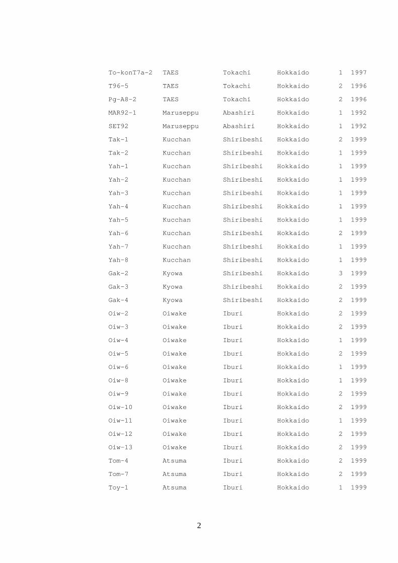
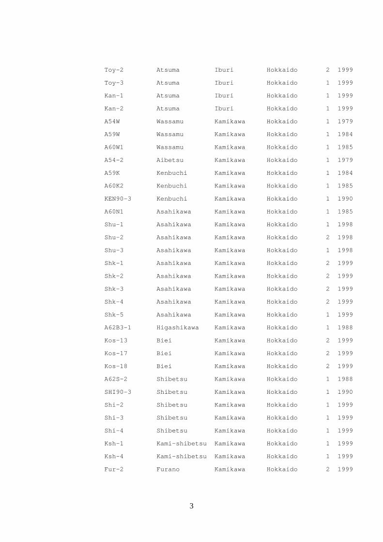
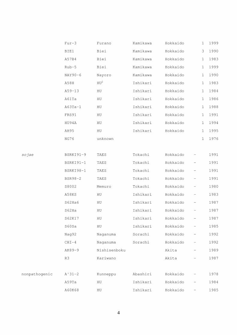
Table 2. Isolates of Cadophora gregata used in this study
Forma specialis Isolate No. Location Race Year
adzukicola Oto1-2-3 Otofuke Tokachi Hokkaido 2 1997
Oto1-2-1 Otofuke Tokachi Hokkaido 3 1997
Mem3-4 Memuro Tokachi Hokkaido 1 1997
Mem1-3 Memuro Tokachi Hokkaido 1 1997
Mur-3 Memuro Tokachi Hokkaido 2 1998
Msek Memuro Tokachi Hokkaido 2 2000
Mem1-2 Memuro Tokachi Hokkaido 2 1997
T96-1 Memuro Tokachi Hokkaido 1 1996
Skt2-4 Shihoro Tokachi Hokkaido 2 1997
Smo1-3 Shihoro Tokachi Hokkaido 2 1997
Smo3-3 Shihoro Tokachi Hokkaido 1 1997
Skt1-3 Shihoro Tokachi Hokkaido 1 1997
Kyo5-2 Churui Tokachi Hokkaido 2 1997
Seo1-2 Sarabetsu Tokachi Hokkaido 1 1997
A54-25 Makubetsu Tokachi Hokkaido 1 1979
A56-25 Makubetsu Tokachi Hokkaido 1 1982
A57T14 Makubetsu Tokachi Hokkaido 1 1983
Towa3-3 Taiki Tokachi Hokkaido 1 1997
NT96-V Obihiro Tokachi Hokkaido 1 1996
T96 Obihiro Tokachi Hokkaido 1 1996
A10 TAES1 Tokachi Hokkaido 1 1976
A13 TAES Tokachi Hokkaido 1 1970
KT1 TAES Tokachi Hokkaido 1 1990
KT2 TAES Tokachi Hokkaido 1 1990
HY-2 TAES Tokachi Hokkaido 1 1990
TAES93 TAES Tokachi Hokkaido 1 1993
TTF95-Ha TAES Tokachi Hokkaido 2 1995
To-konT3c-5 TAES Tokachi Hokkaido 2 1997
To-konT7b-2 TAES Tokachi Hokkaido 2 1997
2
To-konT7a-2 TAES Tokachi Hokkaido 1 1997
T96-5 TAES Tokachi Hokkaido 2 1996
Pg-A8-2 TAES Tokachi Hokkaido 2 1996
MAR92-1 Maruseppu Abashiri Hokkaido 1 1992
SET92 Maruseppu Abashiri Hokkaido 1 1992
Tak-1 Kucchan Shiribeshi Hokkaido 2 1999
Tak-2 Kucchan Shiribeshi Hokkaido 1 1999
Yah-1 Kucchan Shiribeshi Hokkaido 1 1999
Yah-2 Kucchan Shiribeshi Hokkaido 1 1999
Yah-3 Kucchan Shiribeshi Hokkaido 1 1999
Yah-4 Kucchan Shiribeshi Hokkaido 1 1999
Yah-5 Kucchan Shiribeshi Hokkaido 1 1999
Yah-6 Kucchan Shiribeshi Hokkaido 2 1999
Yah-7 Kucchan Shiribeshi Hokkaido 1 1999
Yah-8 Kucchan Shiribeshi Hokkaido 1 1999
Gak-2 Kyowa Shiribeshi Hokkaido 3 1999
Gak-3 Kyowa Shiribeshi Hokkaido 2 1999
Gak-4 Kyowa Shiribeshi Hokkaido 2 1999
Oiw-2 Oiwake Iburi Hokkaido 2 1999
Oiw-3 Oiwake Iburi Hokkaido 2 1999
Oiw-4 Oiwake Iburi Hokkaido 1 1999
Oiw-5 Oiwake Iburi Hokkaido 2 1999
Oiw-6 Oiwake Iburi Hokkaido 1 1999
Oiw-8 Oiwake Iburi Hokkaido 1 1999
Oiw-9 Oiwake Iburi Hokkaido 2 1999
Oiw-10 Oiwake Iburi Hokkaido 2 1999
Oiw-11 Oiwake Iburi Hokkaido 1 1999
Oiw-12 Oiwake Iburi Hokkaido 2 1999
Oiw-13 Oiwake Iburi Hokkaido 2 1999
Tom-4 Atsuma Iburi Hokkaido 2 1999
Tom-7 Atsuma Iburi Hokkaido 2 1999
Toy-1 Atsuma Iburi Hokkaido 1 1999
3
Toy-2 Atsuma Iburi Hokkaido 2 1999
Toy-3 Atsuma Iburi Hokkaido 1 1999
Kan-1 Atsuma Iburi Hokkaido 1 1999
Kan-2 Atsuma Iburi Hokkaido 1 1999
A54W Wassamu Kamikawa Hokkaido 1 1979
A59W Wassamu Kamikawa Hokkaido 1 1984
A60W1 Wassamu Kamikawa Hokkaido 1 1985
A54-2 Aibetsu Kamikawa Hokkaido 1 1979
A59K Kenbuchi Kamikawa Hokkaido 1 1984
A60K2 Kenbuchi Kamikawa Hokkaido 1 1985
KEN90-3 Kenbuchi Kamikawa Hokkaido 1 1990
A60N1 Asahikawa Kamikawa Hokkaido 1 1985
Shu-1 Asahikawa Kamikawa Hokkaido 1 1998
Shu-2 Asahikawa Kamikawa Hokkaido 2 1998
Shu-3 Asahikawa Kamikawa Hokkaido 1 1998
Shk-1 Asahikawa Kamikawa Hokkaido 2 1999
Shk-2 Asahikawa Kamikawa Hokkaido 2 1999
Shk-3 Asahikawa Kamikawa Hokkaido 2 1999
Shk-4 Asahikawa Kamikawa Hokkaido 2 1999
Shk-5 Asahikawa Kamikawa Hokkaido 1 1999
A62B3-1 Higashikawa Kamikawa Hokkaido 1 1988
Kos-13 Biei Kamikawa Hokkaido 2 1999
Kos-17 Biei Kamikawa Hokkaido 2 1999
Kos-18 Biei Kamikawa Hokkaido 2 1999
A62S-2 Shibetsu Kamikawa Hokkaido 1 1988
SHI90-3 Shibetsu Kamikawa Hokkaido 1 1990
Shi-2 Shibetsu Kamikawa Hokkaido 1 1999
Shi-3 Shibetsu Kamikawa Hokkaido 1 1999
Shi-4 Shibetsu Kamikawa Hokkaido 1 1999
Ksh-1 Kami-shibetsu Kamikawa Hokkaido 1 1999
Ksh-4 Kami-shibetsu Kamikawa Hokkaido 1 1999
Fur-2 Furano Kamikawa Hokkaido 2 1999
4
Fur-3 Furano Kamikawa Hokkaido 1 1999
BIE1 Biei Kamikawa Hokkaido 3 1990
A57B4 Biei Kamikawa Hokkaido 1 1983
Rub-5 Biei Kamikawa Hokkaido 1 1999
NAY90-6 Nayoro Kamikawa Hokkaido 1 1990
A58H HU2 Ishikari Hokkaido 1 1983
A59-13 HU Ishikari Hokkaido 1 1984
A61Ta HU Ishikari Hokkaido 1 1986
A63Ta-1 HU Ishikari Hokkaido 1 1988
FRS91 HU Ishikari Hokkaido 1 1991
HU94A HU Ishikari Hokkaido 1 1994
AH95 HU Ishikari Hokkaido 1 1995
NG76 unknown 1 1976
sojae BSRKI91-9 TAES Tokachi Hokkaido - 1991
BSRKI91-1 TAES Tokachi Hokkaido - 1991
BSRKI98-1 TAES Tokachi Hokkaido - 1991
BSR98-2 TAES Tokachi Hokkaido - 1991
S8002 Memuro Tokachi Hokkaido - 1980
A58KS HU Ishikari Hokkaido - 1983
S62Ha6 HU Ishikari Hokkaido - 1987
S62Ha HU Ishikari Hokkaido - 1987
S62K17 HU Ishikari Hokkaido - 1987
S60Sa HU Ishikari Hokkaido - 1985
Nag92 Naganuma Sorachi Hokkaido - 1992
CHI-4 Naganuma Sorachi Hokkaido - 1992
AK89-9 Nishisenboku Akita - 1989
R3 Kariwano Akita - 1987
nonpathogenic A'31-2 Kunneppu Abashiri Hokkaido - 1978
A59Ta HU Ishikari Hokkaido - 1984
A60K68 HU Ishikari Hokkaido - 1985
5
A60To HU Ishikari Hokkaido - 1985
A57T22 Shimizu Tokachi Hokkaido - 1983
To-konT3c-2 TAES Tokachi Hokkaido - 1997
A8-7-5 TAES Tokachi Hokkaido - 1997
Kyo2-1 Churui Tokachi Hokkaido - 1997
Kyo2-2 Churui Tokachi Hokkaido - 1997
Kyo2-3N Churui Tokachi Hokkaido - 1997
Kyo2-5N Churui Tokachi Hokkaido - 1997
Kyo3-1N Churui Tokachi Hokkaido - 1997
Kyo3-2N Churui Tokachi Hokkaido - 1997
Kyo4-2 Churui Tokachi Hokkaido - 1997
Kyo4-3 Churui Tokachi Hokkaido - 1997
Skt2-3 Shihoro Tokachi Hokkaido - 1997
Skt5-1 Shihoro Tokachi Hokkaido - 1997
Sot8-1 Shihoro Tokachi Hokkaido - 1997
Hir2-3 Hiroo Tokachi Hokkaido - 1997
Hir4-1 Hiroo Tokachi Hokkaido - 1997
Hir6-4 Hiroo Tokachi Hokkaido - 1997
Oto1-3-3N Otofuke Tokachi Hokkaido - 1997
Oto1-3-4 Otofuke Tokachi Hokkaido - 1997
Oto1-5-1 Otofuke Tokachi Hokkaido - 1997
Tyo6-1 Taiki Tokachi Hokkaido - 1997
Taka-2 Memuro Tokachi Hokkaido - 1997
Mem6-4 Memuro Tokachi Hokkaido - 1997
Seo7-1 Sarabetsu Tokachi Hokkaido - 1997
1 Tokachi Agriculture Experiment Station, Memuro, Tokachi
2 Experiment field in Hokkaido University, Sapporo, Ishikari
1
Table 3. Sporulation ability, gregatins productivity of nonpathogenic Cadophora
gregata isolates, their pathogenicity and detection from artificially inoculated
roots of adzuki bean, mung bean and soybean using polymerase chain reaction (PCR)
Pathogenicity/ PCR1
Formae specialis Isolate Sporulation
on V8 juice
agar
Gregatins
production
Adzuki
bean
(Erimo-
shozu)
Mung
bean
(un-
known)
Soybean
(Sapporo
-midori)
adzukicola T96-1 + + 9/+ 8/+ 0/+
T96-5 + + 10/+ 6/+ 0/+
sojae S58KS + + 0/+ 5/+ 7/+
nonpathogenic A'31-2 + + 0/+ 0/+ 0/+
A57T22 + - 0/+ 0/+ 0/+
A60K68 + + 0/+ 0/+ 0/+
A60To + - 0/+ 0/+ 0/+
To-konT3c-2 + + 0/+ 0/+ 0/+
Kyo2-1 - - 0/+ 0/+ 0/+
Kyo2-2 - - 0/+ 0/+ 0/+
Kyo2-3N - + 0/+ 0/+ 0/+
Kyo2-5N - - 0/+ 0/+ 0/+
Kyo3-1N - - 0/+ 0/+ 0/+
Kyo3-2N - + 0/+ 0/+ 0/+
Kyo4-2 - - 0/+ 0/+ 0/+
Kyo4-3 - - 0/+ 0/+ 0/+
Skt2-3 + + 0/+ 0/+ 0/+
Skt5-1 - - 0/+ 0/+ 0/+
Hir4-1 + + 0/+ 0/+ 0/+
Hir2-3 + - 0/+ 0/+ 0/+
Sot8-1 + + 0/+ 0/+ 0/+
2
A8-7-5 - - 0/+ 0/+ 0/+
Oto1-3-3N - - 0/+ 0/+ 0/+
Oto1-3-4 + - 0/+ 0/+ 0/+
Oto1-5-1 + - 0/+ 0/+ 0/+
Tyo6-1 + + 0/+ 0/+ 0/+
Taka-2 - - 0/- 0/- 0/-
Mem6-4 - + 0/+ 0/+ 0/+
Seo7-1 + - 0/+ 0/+ 0/+
1 Number of plants diseased out of a total 10 tested/ Presence (+) or absence (-) of
a specific DNA fragment (483-bp) detected from inoculated roots using PCR with each
specific primer set
1
Table 4. Inter simple sequence repeat (ISSR) primers used in
this study and DNA fragments of Cadophora gregata isolates
ISSR
primers
Anealing
Temperature
Total number
of fragments
Number of polymorphic
fragments
CGA1 CGS NP
(AAG)7 51 11 8 9 11
(AGC)7 52 10 10 6 8
(ACC)7 51 10 9 5 8
(GTG)7 50 11 9 6 4
(GAGA)4 48 16 8 12 4
(TGTC)4 48 11 10 7 5
(TCC)5 50 18 14 12 16
(ACG)5 50 16 14 13 10
Total 103 82 70 66
1C. gregata f. sp. adzukicola, CGA; C. gregata f. sp. sojae, CGS;
nonpathogenic C. gregata, NP