Embed Size (px)
Citation preview
Contents lists available at ScienceDirect
Brain Stimulation
journal homepage: www.brainst imjrnl .com
Brain Stimulation 7 (2014) 66e73
Characterizing the Mechanisms of Central and Peripheral Formsof Neurostimulation in Chronic Dysphagic Stroke Patientsq
Emilia Michou a, Satish Mistry a, Samantha Jefferson a, Pippa Tyrrell b, Shaheen Hamdy a,*
aGastrointestinal Centre, Institute of Inflammation and Repair, Faculty of Medical and Human Sciences, University of Manchester (Part of the Manchester Academic Health SciencesCentre [MAHSC]), Salford Royal Hospital, Clinical Sciences Building, Salford M6 8HD, UKb Stroke Medicine, Institute of Cardiovascular Sciences, University of Manchester (Part of the Manchester Academic Health Sciences Centre [MAHSC]), Salford Royal HospitalFoundation Trust, Salford M6 8HD, UK
a r t i c l e i n f o
Article history:Received 9 July 2013Received in revised form12 September 2013Accepted 17 September 2013
Keywords:Chronic dysphagiaStrokeNeurostimulationPlasticity
Abbreviations: cPA, cumulative penetration aspipotentials; MT, motor threshold; NIHSS, National InstPAS, paired associative stimulation; PEG, percutaneoPES, pharyngeal electrical stimulation; rTMS, repestimulation; MI, motor cortex.
q This is an open-access article distributed undCommons Attribution-NonCommercial-No Derivatipermits non-commercial use, distribution, and repprovided the original author and source are credited.
Funding: Supported by the Wellcome Trust (WT0of the Greek State Foundation Scholarship and FSResearch Network. SJ was funded through a grant fro
1935-861X/$ e see front matter � 2014 The Authors.http://dx.doi.org/10.1016/j.brs.2013.09.005
a b s t r a c t
Background: Swallowing problems following stroke may result in increased risk of aspiration pneumonia,malnutrition, and dehydration.Objective/hypothesis: Our hypothesis was that three neurostimulation techniques would produce bene-ficial effects on chronic dysphagia following stroke through a common brain mechanism that wouldpredict behavioral response.Methods: In 18 dysphagic stroke patients (mean age: 66 � 3 years, 3 female, time-post-stroke: 63 � 15weeks [�SD]), pharyngeal electromyographic responses were recorded after single-pulse transcranialmagnetic stimulation (TMS) over the pharyngeal motor cortex, to measure corticobulbar excitabilitybefore, immediately, and 30 min, after real and sham applications of neurostimulation. Patients wererandomized to a single session of either: pharyngeal electrical stimulation (PES), paired associativestimulation (PAS) or repetitive TMS (rTMS). Penetration-aspiration scores and bolus transfer timingswere assessed before and after both real and sham interventions using videofluoroscopy.Results: Corticobulbar excitability of pharyngeal motor cortex was beneficially modulated by PES, PAS andto a lesser extent by rTMS, with functionally relevant changes in the unaffected hemisphere. Followingcombining the results of real neurostimulation, an overall increase in corticobulbar excitability in theunaffected hemisphere (P ¼ .005, F1,17 ¼ 10.6, ANOVA) with an associated 15% reduction in aspiration(P ¼ .005, z ¼ �2.79) was observed compared to sham.Conclusions: In this mechanistic study, an increase in corticobulbar excitability the unaffected projectionwas correlated with the improvement in swallowing safety (P ¼ .001, rho ¼ �.732), but modality-specificdifferences were observed. Paradigms providing peripheral input favored change in neurophysiologicaland behavioral outcome measures in chronic dysphagia patients. Further larger cohort studies of neu-rostimulation in chronic dysphagic stroke are imperative.
� 2014 The Authors. Published by Elsevier Inc. All rights reserved.
Introduction
The presence of oropharyngeal dysphagia is recognized asa major risk factor for pneumonia following stroke and is associated
ration; MEP, motor evokeditute of Health Stroke Scale;us endoscopic gastrostomy;titive transcranial magnetic
er the terms of the Creativeve Works License, whichroduction in any medium,
81741MA). EM was recipientF NIHR North West Strokem Action Medical.
Published by Elsevier Inc. All righ
with increased mortality at 30 days and 1 year post-ictus [1].Dysphagia prolongs hospital length-of-stay [2], and slows recovery;in part secondary to the complications of malnutrition and dehy-dration. Although many patients recover from dysphagia following
Contributors and authorships: EM (blinded to interventions), SJ (blinded tointerventions), SM assisted in the studies. EM and SJ analyzed data and wrote thepaper. EM, SM, SJ and PT helped in interpretation of data. SH was the ChiefInvestigator, wrote the grants to support the studies, helped write the paper andinterpret the analysis.
Disclosures: Nothing to disclose.The studywas sponsored by theUniversity ofManchester, UK,which did not have
a role in the study design or in the collection, analysis, or interpretation of data.* Corresponding author. Tel.: þ44 161 206 4363; fax: þ44 161 206 4364.
E-mail address: [email protected] (S. Hamdy).
ts reserved.
E. Michou et al. / Brain Stimulation 7 (2014) 66e73 67
stroke [3,4], a proportion remain chronically dysphagic, dependenton modified diet and/or enteral feeding. In addition to impairedquality-of-life, patients with long-term dysphagia remain at risk ofmalnutrition, dehydration and aspiration pneumonia. Moreover,while speech and language therapists have employed a variety oflabor intensive compensatory maneuvers to treat chronic dys-phagia, there is very little evidence of their effectiveness [5].
Recent evidence has given insight into the cortical and sub-cortical brain areas, activated during the highly coordinatedsensorimotor activity of swallowing [6]. In dysphagia literature,transcranial magnetic stimulation (TMS) studies have shown thatthe pharyngeal motor cortex (MI) reorganizes following acuteunilateral stroke, and that an increase in cortical excitability in theunaffected hemisphere is associated with the recovery of swallow-ing function [7]. Consequently, the development of novel rehabili-tative approaches to drive beneficial changes in cortical andsubcortical activity, hence promoting improved swallowing func-tion, remains a major imperative [8].
Recently, peripheral stimulation of the pharynx (pharyngealelectrical stimulation (PES) [9e11], and cortical stimulation byrepetitive TMS (rTMS) [12e17], or transcranial direct current stim-ulation (tDCS) [18e20], or the combination of both peripheral andcentral stimulation (paired associative stimulation, PAS) [21,22],have been used in acute and chronic dysphagic stroke researchstudies in an attempt to both modulate cortical activity and induceor promote functional improvements in swallowing. Several studyprotocols have been proposed from different laboratories withvarying parameters, intensity frequency and targeted specificmuscle groups (i.e. mylohyoid [15], upper esophageal area [13,17]).
The aim of our study was to compare the effects of a singleapplication of one of three neurostimulation techniques (PES, PAS,rTMS) on swallowing safety and neurophysiological mechanisms inchronic dysphagic stroke. The study was designed to answer thefollowing questions:
i. What are the neurophysiological and behavioral effects ofeach neurostimulation treatment?
ii. Are the mechanisms of the different neurostimulationmodalities comparable?
iii. Are the changes in activation of corticobulbar pathwayspredictive of the changes in swallowing behavior?
Our hypothesis was that all three interventions would producebeneficial effects on swallowing through a common brain mecha-nism that would predict behavioral response.
Materials and methods
Recruitment
Patients were recruited via referrals from clinics across theNorth West of England or self-referral. Ethical approval was gainedfrom Salford and Trafford Local Research Ethics Committee. Writteninformed consent was obtained from all participants before theexperiments. The study adhered to the Good Clinical PracticeGuidelines from the International Conference on Harmonizationand Code of Ethics of the World Medical Association and wasregistered at UK Clinical Research Network Study Portfolio (UKCRN)(identification number 2499) and at International StandardRandomized Controlled Trial Number Register ISRCTN (83103698).
Participants
Eligible patients were recruited fromdirect and indirect referralsover 4 years (September 2006eAugust 2010). They had a clinical
diagnosis of stroke with dysphagia confirmed by a speech andlanguage therapist, which persisted for more than 6 weeks post-ictus. Patients with a history of dementia, cognitive impairment orneurological deficits prior to stroke, pacemaker/cardiac defibrillatorin situ, severe concomitant medical conditions (i.e. progressiveneurological disorders, chronic respiratory conditions or heartfailure), structural oropharyngeal pathology, history of epilepsy,previous head and neck surgery, pregnancy, any intracranial metalor combination of medications acting on CNS were excluded.
Experimental procedures
TMSFocal TMS was performed using a figure-of-8 shaped magnetic
coil (outer diameter, 70 mm) connected to a Magstim BiStim2
magnetic stimulator (Magstim Co,Wales, UK), withmaximal outputof 2.2 T (see the Supplementary material for full details).
Pharyngeal EMG measurementsPharyngeal electromyographic responses after single TMS pul-
ses, termed as pharyngeal motor evoked potentials (PMEPs), wererecorded via a 3.2 mm diameter intraluminal catheter (Gaeltec Ltd,Isle of Skye), with a built-in pair of bipolar platinum ring electrodes.
Videofluoroscopy (VFS)The research videofluoroscopic assessment [11,12,21] was
carried out at the Radiology Department, Salford Royal NHS Trust,UK. The examination was conducted with 6 swallows of 5 mlboluses of liquid barium (60% w/v, EZ-HD�, E-Z-EM Limited, UK)and the images were acquired in lateral view (Siemens Fluo-rospot�H SIRESKOP SX Unit, Germany).
Experimental interventions
PESAn intraluminal catheter was used for PES, connected to
a constant current generator (DS7, Welwyn-Garden City, UK) anda trigger generator (Neurolog system, Digitimer) allowing titrationof stimulation intensity against individuals’ perception and toler-ance thresholds. PES intensity was then set at 75% of the differencebetween perception and tolerance threshold and delivered ata frequency of 5 Hz for 10 min [9e11].
Repetitive TMSTrains of stimuli were delivered to pharyngeal motor cortex (MI)
with the TMS coil (Magstim,Wales, UK). The optimal parameters forrepetitive TMS were frequency of 5 Hz, intensity 90% of restingthenar Motor Threshold (MT) in train of 250 pulses, in 5 blocks of50 with 10 s between-blocks pause [12].
PASPaired associative stimulation was delivered by pairing
a pharyngeal electrical stimulus (0.2 ms pulse) with a single TMSpulse over the pharyngeal MI at MT intensity plus 20% of thestimulator output. The 2 pulseswere delivered repeatedly every 20 sthrough Signal software (Cambridge Electronic Design, Cambridge,UK), with an inter-stimulus interval of 100 ms for 10 min [21,22].
Sham stimulationFor sham PES, the intraluminal catheter was in situ, but no
stimulationwas delivered, as used in previous studies [9e11]. ShamrTMS stimulation was given using a 90� coil tilt, which producedthe same noise as active stimulation but no cortical stimulation[23]. This approach was applied reliably in the past, where it wasshown that cortical excitability in the pharyngeal motor systemwasnot affected [12,24]. For sham PAS, the coil was held tangentially to
E. Michou et al. / Brain Stimulation 7 (2014) 66e7368
the skull at a 90� angle to the sagittal plane, and no PES wasdelivered through the pharyngeal catheter in situ [21,22].
Experimental protocol
Patients were asked to attend the laboratory on two separateoccasions. On both occasions, VFS was performed initially to obtainbaseline measurements. Following VFS, the catheter was insertedeither transnasally (14e17 cm from the nasal flare to pair elec-trodes) or transorally (13e16 cm) according to patient’s preference.This allowed the placement of the electrodes in themid-pharyngealarea, a position verified by a VFS-still images following insertion(see Supplementary material).
Following identifying the cranial vertex on the scalp [25], thebrain sites evoking the largest pharyngeal responses in eachhemisphere were identified with mapping procedures using singleTMS pulses over MI. Baseline TMS measurements were obtained atMT þ 20% from both hemispheric sites.
All participants then received both real and sham applications ofone of the three neurostimulation treatments in random order ondifferent days. Randomization was also performed for the neuro-stimulation paradigms (PES, PAS or rTMS). Cortical excitability (TMS)measurements were repeated immediately and 30 min post-intervention. A follow-up VFS was performed after the 30-minutesTMSmeasurements on both visits (see also Supplementarymaterial).
Randomization and blindness
A randomization software (minim.exe, Department of Bioengi-neering, Salford Royal NHS Trust) was used for the process of mini-mization to evenlydistributepatients of different age (above orbelow80) and stroke severity (scoring<12 or�12 on the National Instituteof Health stroke Severity Scale (NIHSS) [26,27]). One independentresearcher delivered the real or sham stimulation, while thoseresearchers analyzing the data remained blinded to the procedure.
Data analysis
Neurophysiological measurementsThe peak-to-peak PMEPs amplitude was used as a measure of
cortical excitability. Changes in excitability over-time werecompared, using generalized linear model repeated measuresANOVA (RmANOVA, SPSSv.19.0), following verification of theassumption of sphericity was not violated (Mauchly’s test). Non-
Table 1Table 1 shows the demographics of the patients consented to participate in the trial. PatienQ did not complete one of the two study days.
Groups ID Age Gender Diet We
PES B 51 M PEG 57D 66 M PEG 159K 77 M PEG/modified 28L 72 M PEG 160O 65 F PEG 104P 31 M PEG/Modified 26
rTMS C 66 M PEG 39G 69 M Modified 12H 55 M PEG 10I 64 M Modified 38R 77 M PEG 13T 73 M PEG 70
PAS A 83 M Modified 9E 67 M Modified 72J 76 F PEG 77M 69 M Modified 8N 69 M PEG/Modified 15S 43 F Modified 47
Mean � SD 66 � 3 63
NIHSS ¼ National Institute of Health Stroke Severity, R ¼ right, L ¼ left, * ¼ no data, U ¼
parametric paired-wise comparisons were performed with Fried-man test [28]. P value of <.05 indicated statistical significance (seeSupplementary material).
VFS analysisFrame-by-frame analysis of the videos took place off-line (see
Supplementary material). The safety of all swallows was assessedand scored using the 8-point penetration-aspiration (PA) scale,describing the severity of airway compromise [29]. Abnormallaryngeal protection was verified, if a swallow was scored �3 onone or more occasions.
All pairwise comparisons, non-parametric correlations (Spear-man’s) and Wilcoxon’s tests results were corrected with Holms’step-down technique. All data are presented as group mean � SEM,unless stated otherwise.
Results
Recruitment
From the initial 83 patients enrolled in the study, 4 declined toparticipate, and only 20 met the inclusion criteria for studyparticipation. One patient had a safe swallow upon initial (baseline)assessment and was therefore excluded, while one additionalparticipant was unable to complete both study days and their datawere not used in the analysis. Therefore, 18 patients were left withcomplete data (see below) (see also Supplementary material).
Patients demographics
Clinical symptoms and demographic data are shown in Table 1.Themean age of the participants was 66� 3 (�SD) years and 3werefemale. Mean stroke severity as measured with NIHSS score was7.6 � 1, while 8 patients had a history of multiple strokes. Sevenpatients suffered right-sided symptoms, while three exhibitedbilateral symptoms. Their symptoms side was recorded as “unde-termined.” Fifteen subjects had PEG in situ, four of whom alsoreceived modified diet.
Patient safety
No adverse events were reported, although two patients re-ported coughing in response to the catheter placement alone,which subsided within 2 min.
t F presented safe swallowing on baseline and was therefore excluded, while patient
eks post stroke Previous stroke NIHSS Side of symptoms
0 * R1 * U1 3 R1 6 L1 6 U0 11 L0 5 R0 4 L0 5 L3 CVAs, 1 TIA 8 L1 9 R0 3 L0 4 L0 12 L1 6 R0 6 L1, 1 TIA 8 R0 14 U
� 15 7.6 � 1
undetermined, TIA ¼ transient ischemic attack, CVA ¼ cerebrovascular accident.
E. Michou et al. / Brain Stimulation 7 (2014) 66e73 69
Neurophysiological changes
Mean pharyngeal MT from the affected and unaffected hemi-spheres were 75 � 4% and 73 � 4% respectively. For the subjectswith ‘undetermined’ symptom side, the hemispheric site requiringhigher intensity to elicit PMEPs was termed as ‘affected.’ Nosignificant differences were observed for baseline cortical excit-ability in all patients across the two study days (real and sham)(Wilcoxon’s test, unaffected hemisphere: z ¼ �.86, P ¼ .386,affected hemisphere: z ¼ �1.47, P ¼ .139), allowing for furtherstatistical analysis.
Modality specific neurophysiological effects following 3 differentneurostimulation treatments
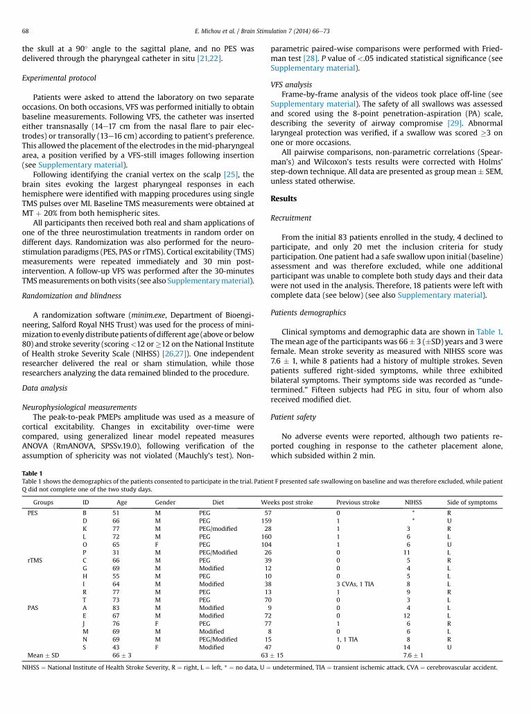
The percentage change in cortical excitability of dysphagicstroke patients recruited in each treatment arm is presented inFig. 1A. Normality assumptions were violated within each groupand non-parametric tests were therefore applied. Three separatenon-parametric Friedman tests were performed within each arm.The distributions within each arm were found to differ, indicatingthat at least one set of responses from the unaffected or affectedhemisphere was driving the effect (PES: P ¼ .009, Chi-square: 22.2;PAS: P ¼ .04, Chi-square: 18.8, rTMS: P ¼ .01, Chi-square:15.07).
Each treatment’s effects were therefore examined individuallywithpairwisecomparisons (Wilcoxon’s)between real and shamarms.
With PES, we observed significant excitability increase imme-diately post-treatment in the unaffected hemisphere (real vs. sham
Figure 1. This figure shows the effects of real and sham neurostimulation techniques on cort(full line) and (B) swallowing safety as measured with cumulative penetration/aspiration scdata: full line shows the significant differences in the unaffected, while dashed line shows
P ¼ .043, z ¼ �2.02, d ¼ 3.51, r ¼ 0.86) and in the affected hemi-sphere 30 min post-intervention (real vs. sham P ¼ .04, z ¼ �2.03,d ¼ 3.36, r ¼ 0.85), indicating bilateral changes in corticalexcitability.
With rTMS, cortical excitability in the unaffected hemisphereappeared to visibly increase following real neurostimulation.However, compared to sham, this change was not statisticallysignificant (immediately [P ¼ .08, z ¼ �1.75], 30 min followingtreatment [P ¼ .08, z ¼ �1.75]). No change in the affected hemi-sphere was observed.
With PAS, cortical excitability increased 30 min post-intervention in the unaffected (P ¼ .043, z ¼ �2.02, d ¼ 2.28,r ¼ .75) compared to sham. A significant increase was also observedin the affected hemisphere immediately following contra-lateralPAS (P ¼ .027, z ¼ �2.07, d ¼ 1.12, r ¼ .49).
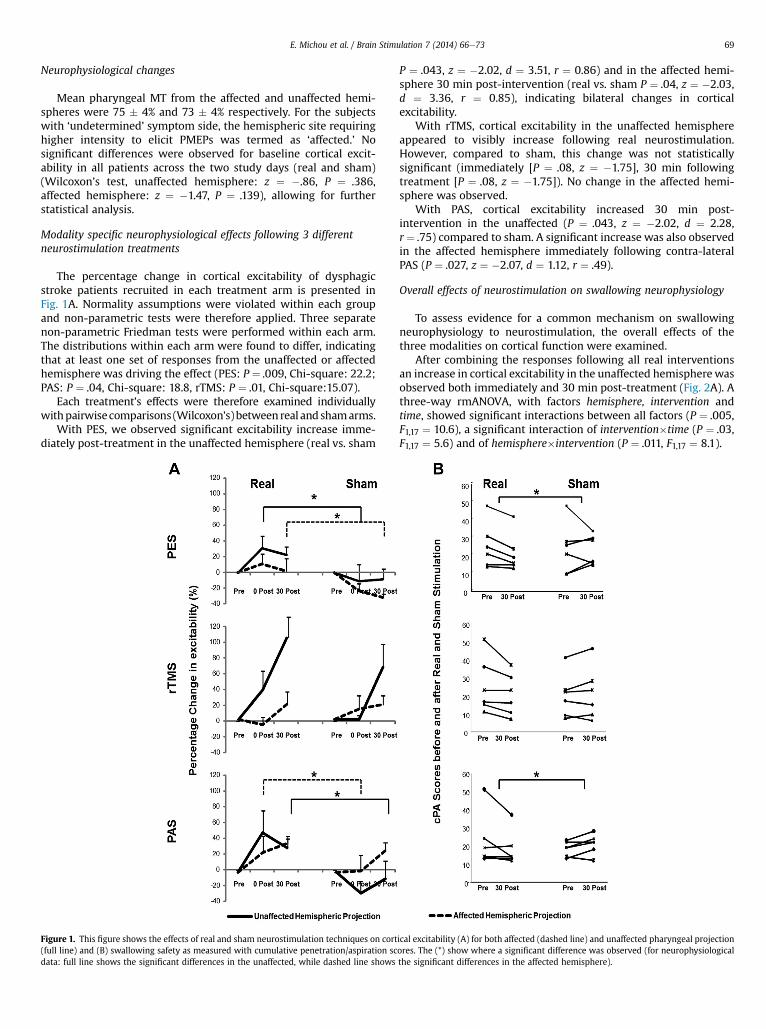
Overall effects of neurostimulation on swallowing neurophysiology
To assess evidence for a common mechanism on swallowingneurophysiology to neurostimulation, the overall effects of thethree modalities on cortical function were examined.
After combining the responses following all real interventionsan increase in cortical excitability in the unaffected hemispherewasobserved both immediately and 30 min post-treatment (Fig. 2A). Athree-way rmANOVA, with factors hemisphere, intervention andtime, showed significant interactions between all factors (P ¼ .005,F1,17 ¼ 10.6), a significant interaction of intervention�time (P ¼ .03,F1,17 ¼ 5.6) and of hemisphere�intervention (P ¼ .011, F1,17 ¼ 8.1).
ical excitability (A) for both affected (dashed line) and unaffected pharyngeal projectionores. The (*) show where a significant difference was observed (for neurophysiologicalthe significant differences in the affected hemisphere).
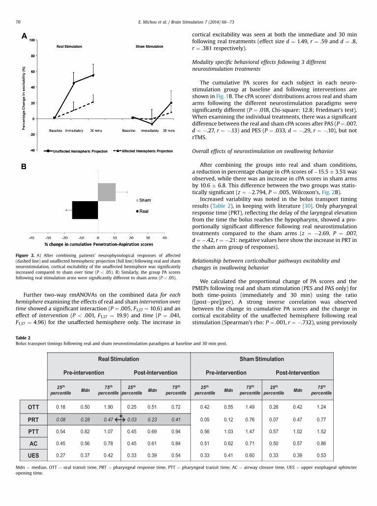
Figure 2. A) After combining patients’ neurophysiological responses of affected(dashed line) and unaffected hemispheric projection (full line) following real and shamneurostimulation, cortical excitability of the unaffected hemisphere was significantlyincreased compared to sham over time (P < .05). B) Similarly, the group PA scoresfollowing real stimulation arms were significantly different to sham arms (P < .05).
E. Michou et al. / Brain Stimulation 7 (2014) 66e7370
Further two-way rmANOVAs on the combined data for eachhemisphere examining the effects of real and sham intervention overtime showed a significant interaction (P ¼ .005, F1,17 ¼ 10.6) and aneffect of intervention (P < .001, F1,17 ¼ 19.9) and time (P ¼ .041,F1,17 ¼ 4.96) for the unaffected hemisphere only. The increase in
Table 2Bolus transport timings following real and sham neurostimulation paradigms at baseline
Mdn ¼ median, OTT ¼ oral transit time, PRT ¼ pharyngeal response time, PTT ¼ pharopening time.
cortical excitability was seen at both the immediate and 30 minfollowing real treatments (effect size d ¼ 1.49, r ¼ .59 and d ¼ .8,r ¼ .381 respectively).
Modality specific behavioral effects following 3 differentneurostimulation treatments
The cumulative PA scores for each subject in each neuro-stimulation group at baseline and following interventions areshown in Fig. 1B. The cPA scores’ distributions across real and shamarms following the different neurostimulation paradigms weresignificantly different (P ¼ .018, Chi-square: 12.8; Friedman’s test).When examining the individual treatments, there was a significantdifference between the real and sham cPA scores after PAS (P¼ .007,d ¼ �.27, r ¼ �.13) and PES (P ¼ .033, d ¼ �.29, r ¼ �.10), but notrTMS.
Overall effects of neurostimulation on swallowing behavior
After combining the groups into real and sham conditions,a reduction in percentage change in cPA scores of �15.5 � 3.5% wasobserved, while there was an increase in cPA scores in sham armsby 10.6 � 6.8. This difference between the two groups was statis-tically significant (z ¼ �2.794, P ¼ .005, Wilcoxon’s, Fig. 2B).
Increased variability was noted in the bolus transport timingresults (Table 2), in keeping with literature [30]. Only pharyngealresponse time (PRT), reflecting the delay of the laryngeal elevationfrom the time the bolus reaches the hypopharynx, showed a pro-portionally significant difference following real neurostimulationtreatments compared to the sham arms (z ¼ �2.69, P ¼ .007,d¼�.42, r¼�.21: negative values here show the increase in PRT inthe sham arm group of responses).
Relationship between corticobulbar pathways excitability andchanges in swallowing behavior
We calculated the proportional change of PA scores and thePMEPs following real and sham stimulation (PES and PAS only) forboth time-points (immediately and 30 min) using the ratio([postepre]/pre). A strong inverse correlation was observedbetween the change in cumulative PA scores and the change incortical excitability of the unaffected hemisphere following realstimulation (Spearman’s rho: P ¼ .001, r ¼ �.732), using previously
and 30 min post.
yngeal transit time, AC ¼ airway closure time, UES ¼ upper esophageal sphincter
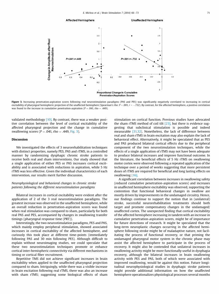
Figure 3. Increasing penetration-aspiration scores following real neurostimulation paradigms (PAS and PES) was significantly negatively correlated to increasing in corticalexcitability of pharyngeal hemispheric projection of the unaffected hemisphere (Spearman’s rho: P ¼ .001, r ¼ �.732). By contrast, for the affected hemisphere, a positive correlationwas found to the increase in cumulative penetration-aspiration (P ¼ .041, rho ¼ .449).
E. Michou et al. / Brain Stimulation 7 (2014) 66e73 71
validated methodology [10]. By contrast, there was a weaker posi-tive correlation between the level of cortical excitability of theaffected pharyngeal projection and the change in cumulativeswallowing scores (P ¼ .041, rho ¼ .449, Fig. 3).
Discussion
We investigated the effects of 3 neurorehabilitation techniqueswith distinct properties, namely PES, PAS and rTMS, in a controlledmanner by randomizing dysphagic chronic stroke patients toreceive both real and sham interventions. Our study showed thata single application of either PES or PAS increases cortical excit-ability and is associated with reductions in aspiration, while 5 HzrTMS was less effective. Given the individual characteristics of eachintervention, our results merit further discussion.
Neurophysiological and behavioral changes in chronic strokepatients following the different neurostimulation paradigms
Bilateral increases in cortical excitability were evident after theapplication of 2 of the 3 real neurostimulation paradigms. Thegreatest increasewas observed in the unaffected hemisphere, whilean overall reduction in penetration-aspiration scores was foundwhen real stimulation was compared to sham, particularly for bothreal PAS and PES, accompanied by changes in swallowing transfertimings (pharyngeal response time (PRT)).
Interestingly, the two neurostimulation paradigms, PES and PAS,which mainly employ peripheral stimulation, showed associatedincreases in cortical excitability of the affected hemisphere, andcuriously this took place at different time-points (immediatelyfollowing PAS and 30 min following PES). Although difficult toexplain without neuroimaging studies, we could speculate thatthese two neurostimulation techniques promote or enhancecortical inter-hemispheric connectivity via different mechanisms intiming or cortical fiber recruitment.
Repetitive TMS did not achieve significant increases in brainexcitability when applied to the unaffected pharyngeal projectioncompared to sham. Intriguingly, while there was a visible increasein brain excitation following real rTMS, there was also an increasewith sham rTMS; suggesting some biological effects of sham
stimulation on cortical function. Previous studies have advocatedthe sham rTMS method of coil tilt [23]. but there is evidence sug-gesting that subclinical stimulation is possible and indeedmeasurable [31,32]. Nonetheless, the lack of difference betweenreal and sham rTMS in brain excitation may also explain the lack ofbehavioral effect. Alternatively, it might be speculated that as PESand PAS produced bilateral cortical effects due to the peripheralcomponent of the two neurostimulation techniques, while theeffects of a single application of rTMS may not have been adequateto produce bilateral increases and improve functional outcome. Inthe literature, the beneficial effects of 5 Hz rTMS on swallowingmotor cortex were observed following a repeated application of thetechnique over a period of weeks suggesting that more persistentdoses of rTMS are required for beneficial and long lasting effects onswallowing [16].
A significant correlation between increases in swallowing safety(reduced cumulative penetration-aspiration scores) and increasesin unaffected hemisphere excitability was observed, supporting thecontention that functional behavioral changes in swallow aremostly driven by improvements in the undamaged circuitry. Hence,our findings continue to support the notion that in (unilateral)stroke, successful neurorehabilitation treatments should bothtarget and promote compensatory changes in the undamaged/unaffected cortex. The unexpected finding that cortical excitabilityof the affected hemisphere increasing in tandemwith an increase incumulative penetration-aspiration scores, might be of importancefor future directions of research. It might be speculated that thelong-term neuroplastic changes occurring in the affected hemi-sphere following stroke might be of maladaptive nature, not facil-itating the process of functional recovery, or that priming theundamaged pharyngeal motor system in the chronic phase mayassist the affected hemisphere to participate in the process ofrecovery. It might also be contended that unilateral increases inswallowing activity might be more functionally useful in dysphagiarecovery, although the bilateral increases in brain swallowingactivity with PES and PAS, both of which were associated withimproved swallowing, would be against this notion. Future longi-tudinal neurophysiological studies in dysphagic stroke patientsmight provide additional information on how the unaffectedhemisphere operationalizes physiological processes several months
E. Michou et al. / Brain Stimulation 7 (2014) 66e7372
post-ictus or to what extent the activity of the corticobulbarprojections from the affected and adjunct to the lesion areas mightbe employed for the restitution of adaptive function, in conjunctionwith the unaffected hemispheric projection.
Since there is limited data on the exact physiologic mechanismsthat drive recovery in stroke, it is not surprising that studiesfocusing on neurostimulation technologies have targeted eitheraffected or unaffected hemisphere without clear rationale. Ourcontention that excitatory stimulation applied to unaffected cortexis likely to produce greatest benefit was also the rationale by Parket al. [16] who recently targeted that hemisphere with 5 Hz rTMSemploying a therapeutic regimen of 2 weeks.
By studying chronic stroke patients, the confounding effect ofspontaneous recovery of function post-ictus was also reduced. Ourresults show that neurostimulation affects neuronal activity evenafter a single application in chronic stroke patients, who usuallypresent a plateau in their ability to change behaviorally. In theliterature, the time-window for functional restoration followingstroke is not clear. However, several factors may play a role in anyrestorative process in the chronic stage, such as the timing andexposure to restorative treatmentsduring theacute stageof recovery[33]. In animals, even weeks and months after a plateau followingthe acute phase, there is potential for change [34]. However, plasticreorganizational changes in chronic dysphagic stroke patients mayinvolve different mechanisms, implicating the need for longitudinalstudies to understand the recovery mechanisms.
Notably, wewere only able to study a carefully selected but well-characterized sample of chronic dysphagic stroke patients, so it isnot possible to generalize our results to a wider more heterogenouspopulation of chronic dysphagic patients. Althoughweused specificinclusion and exclusion criteria, there was still some heterogeneityin the dysphagia severity amongst the patients in each group, whichmay have contributed to the less favorable results following rTMS.Moreover, a cross-over design was employed, with patients servingas their own control. The wash-out period of treatments used incross-over designs is of importance, however, results from studies inhealth [10e12,21] showed that a single application does notpromote effects more than 2 h post-intervention. High variability inbiomechanics and transport bolus timingswas observed,whichmaybe a result of the small sample sizes per treatment arm. Inter-subjectdifferences in swallowing patterns aremoreovert in stroke,with themajority of chronic dysphagic patients requiring artificial feeding toretain nutritional levels. Compensatory strategies during swallow-ing are usually employed, a factor, which by itself may produceadditional changes in neurophysiology.
To conclude our preliminary results suggest that even a singleapplication of some types of neurostimulation (PES and PAS) whenapplied to dysphagic chronic stroke patients can lead to changes incorticobulbar excitability and associated behavioral changes, suchchanges in penetration-aspiration scores. Further studies arerequired to elucidate the optimum stimulation paradigms withadditional classifiers such as the severity of dysphagia, as well asdifferent location and sizes of lesions, which might reduce theincreased heterogeneity in the results. Finally, this study demon-strates the feasibility and acceptability of the interventions inpatients with chronic dysphagia following stroke and providesinformation on likely recruitment rates and effect sizes for thedesign of a future clinical trial.
Acknowledgments
The authors would like to thank all participants and Lisa Renault,Jackie Johnson, and Daniela Burgess (senior radiographers) whohelped conduct the videofluoroscopy examinations. Also we would
like to thank the Local Stroke Research Network (North West) forthe assistance with patient recruitment.
Supplementary data
Supplementary data related to this article can be found at http://dx.doi.org/10.1016/j.brs.2013.09.005.
References
[1] Finlayson O, Kapral M, Hall R, Asllani E, Selchen D, Saposnik G, et al. Riskfactors, inpatient care, and outcomes of pneumonia after ischemic stroke.Neurology 2011;77:1338e45.
[2] Altman KW, Yu GP, Schaefer SD. Consequence of dysphagia in the hospitalizedpatient: impact on prognosis and hospital resources. Arch Otolaryngol HeadNeck Surg 2010;136(8):784e9.
[3] Smithard DG, O’Neill PA, Parks C, Morris J. Complications and outcome afteracute stroke. Does dysphagia matter? Stroke 1996;27(7):1200e4.
[4] Mann G, Hankey GJ, Cameron D. Swallowing function after stroke: prognosisand prognostic factors at 6 months. Stroke 1999;30(4):744e8.
[5] Speyer R, Baijens L, Heijnen M, Zwijnenberg I. Effects of therapy in oropha-ryngeal dysphagia by speech and language therapists: a systematic review.Dysphagia 2010;25(1):40e65.
[6] Martin RE. Neuroplasticity and swallowing. Dysphagia 2009 Jun;24(2):218e29.[7] Hamdy S, Aziz Q, Rothwell JC, Power M, Singh KD, Nicholson DA, et al.
Recovery of swallowing after dysphagic stroke relates to functional reorga-nization in the intact motor cortex. Gastroenterology 1998;115(5):1104e12.
[8] Michou E, Hamdy S. Cortical input in control of swallowing. Curr Opin Oto-laryngol Head Neck Surg 2009;17(3):166e71.
[9] Hamdy S, Rothwell JC, Aziz Q, Singh KD, Thompson DG. Long-term reorgani-zation of human motor cortex driven by short-term sensory stimulation. NatNeurosci 1998;1:64e8.
[10] Fraser C, Power M, Hamdy S, Rothwell J, Hobday D, Hollander I, et al. Drivingplasticity in human adult motor cortex is associated with improved motorfunction after brain injury. Neuron 2002;34(5):831e40.
[11] Jayasekeran V, Singh S, Tyrrell P, Michou E, Jefferson S, Mistry S, et al.Adjunctive functional pharyngeal electrical stimulation reverses swallowingdisability after brain lesions. Gastroenterology 2010;138(5):1737e46.
[12] Jefferson S, Mistry S, Michou E, Singh S, Rothwell JC, Hamdy S. Reversal ofa virtual lesion in human pharyngeal motor cortex by high frequency con-tralesional brain stimulation. Gastroenterology 2009;137(3):841e9.
[13] Khedr EM, Abo-Elfetoh N, Rothwell JC. Treatment of post-stroke dysphagiawith repetitive transcranial magnetic stimulation. Acta Neurol Scand2009;119(3):155e61.
[14] Kim L, Chun MH, Kim BR, Lee SJ. Effect of repetitive transcranial magneticstimulation on patients with brain injury and Dysphagia. Ann Rehabil Med2011;35(6):765e71.
[15] Verin E, Leroi AM. Poststroke dysphagia rehabilitation by repetitive transcranialmagnetic stimulation:anoncontrolledpilotstudy.Dysphagia2009;24(2):204e10.
[16] Park JW,OhJC,Lee JW,YeoJS,RyuKH.Theeffectof5Hzhigh-frequency rTMSovercontralesional pharyngealmotor cortex inpost-stroke oropharyngeal dysphagia:a randomizedcontrolledstudy.NeurogastroenterolMotil 2013;25(4):324ee250.
[17] Khedr EM, Abo-Elfetoh N. Therapeutic role of rTMS on recovery of dysphagiain patients with lateral medullary syndrome and brainstem infarction.J Neurol Neurosurg Psychiatry 2010;81(5):495e9.
[18] Jefferson S, Mistry S, Singh S, Rothwell J, Hamdy S. Characterizing the appli-cation of transcranial direct current stimulation in human pharyngeal motorcortex. Am J Physiol Gastrointest Liver Physiol 2009;297(6):G1035e40.
[19] Yang EJ, Baek SR, Shin J, Lim JY, Jang HJ, Kim YK, et al. Effects of transcranialdirect current stimulation (tDCS) on post-stroke dysphagia. Restor NeurolNeurosci 2012;30(4):303e11.
[20] Kumar S, Wagner CW, Frayne C, Zhu L, Selim M, Feng W, et al. Noninvasivebrain stimulation may improve stroke-related dysphagia: a pilot study. Stroke2011;42(4):1035e40.
[21] Michou E, Mistry S, Jefferson S, Singh S, Rothwell J, Hamdy S. Targetingunlesioned pharyngeal motor cortex improves swallowing in healthy indi-viduals and after dysphagic stroke. Gastroenterology 2012;142(1):29e38.
[22] Singh S, Mistry S, Jefferson S, Davies K, Rothwell J, Williams S, et al. A magneticresonance spectroscopy study of brain glutamate in a model of plasticity inhuman pharyngeal motor cortex. Gastroenterology 2009;136(2):417e24.
[23] Loo CK, Taylor JL, Gandevia SC, McDarmont BN, Mitchell PB, Sachdev PS.Transcranial magnetic stimulation (TMS) in controlled treatment studies: aresome “sham” forms active? Biol Psychiatry 2000;47(4):325e31.
[24] Gow D, Rothwell J, Hobson A, Thompson D, Hamdy S. Induction of long-termplasticity in human swallowing motor cortex following repetitive corticalstimulation. Clin Neurophysiol 2004;115(5):1044e51.
[25] Klem GH, Lüders HO, Jasper HH, Elger C. The ten-twenty electrode system ofthe International Federation. The International Federation of Clinical Neuro-physiology. Electroencephalogr Clin Neurophysiol Suppl 1999;52:3e6.
[26] Alshekhlee A, Ranawat N, Syed TU, Conway D, Ahmad SA, Zaidat OO. NationalInstitutes of Health Stroke Scale assists in predicting the need for

E. Michou et al. / Brain Stimulation 7 (2014) 66e73 73
percutaneous endoscopic gastrostomy tube placement in acute ischemicstroke. J Stroke Cerebrovasc Dis 2010;19:347e52.
[27] Okubo PC, Fábio SR, Domenis DR, Takayanagui OM. Using the National Insti-tute of Health Stroke Scale to predict dysphagia in acute ischemic stroke.Cerebrovasc Dis 2012;33(6):501e7.
[28] Conover WJ. Practical nonparametric statistics. 3rd ed. New York: John Wiley& Sons; 1999.
[29] Rosenbek JC, Robbins JA, Roecker EB, Coyle JL, Wood JL. A penetration-aspiration scale. Dysphagia 1996;11:93e8.
[30] Molfenter SM, Steele CM. Temporal variability in the deglutition literature.Dysphagia 2012;27(2):162e77.
[31] Wassermann EM, Lisanby SH. Therapeutic application of repetitive trans-cranial magnetic stimulation: a review. Clin Neurophysiol 2001;112:1367e77.
[32] Strafella AP, Ko JH, Monchi O. Therapeutic application of transcranial magneticstimulation in Parkinson’s disease: the contribution of expectation. Neuro-image 2006;31(4):1666e72.
[33] Murphy TH, Corbett D. Plasticity during stroke recovery: from synapse tobehaviour. Nat Rev Neurosci 2009;10:861e72.
[34] Zhao LR, Berra HH, Duan WM, Singhal S, Mehta J, Apkarian AV, et al. Beneficialeffects of hematopoietic growth factor therapy in chronic ischemic stroke inrats. Stroke 2007;38(10):2804e11.