Embed Size (px)
Citation preview

Protein Science (1992), I, 892-901. Cambridge University Press. Printed in the USA. Copyright 0 1992 The Protein Society
Charge balance in the a-hydroxyacid dehydrogenase vacuole: An acid test
ANTONIO CORTES,’ DAVID C. EMERY,’ DAVID J. HALSALL,2 RICHARD M. JACKSON,2 ANTHONY R. CLARKE,2 AND J. JOHN HOLBROOK2 ’ Department of Biochemistry and Physiology, University of Barcelona, Marti y FranquCs 1, 08028 Barcelona, Spain Molecular Recognition Centre and Department of Biochemistry, University of Bristol School of Medical Sciences, University Walk, Bristol BS8 lTD, UK
(RECEIVED December 19, 1991; REVISED MANUSCRIPT RECEIVED February 25, 1992)
Abstract
The proposal that the active site vacuole of NAD+-S-lactate dehydrogenase is unable to accommodate any im- balance in electrostatic charge was tested by genetically manipulating the cDNA coding for human muscle lac- tate dehydrogenase to make a protein with an aspartic acid introduced at position 140 instead of the wild-type asparagine. The Asn 140-Asp mutant enzyme has the same kc,, as the wild type (Asn 140) at low pH (4.9, and at higher pH the K , for pyruvate increases 10-fold for each unit increase in pH up to pH 9. We conclude that the anion of Asp 140 is completely inactive and that it binds pyruvate with a K , that is over 1 ,OOO times that of the K, of the neutral, protonated aspartic-140. Experimental results and molecular modeling studies indicate the pK, of the active site histidine-195 in the enzyme-NADH complex is raised to greater than 10 by the presence of the anion at position 140. Energy minimization and molecular dynamics studies over 36 ps suggest that the an- ion at position 140 promotes the opening of and the entry of mobile solvent beneath the polypeptide loop (98- 1 lo), which normally seals off the internal active site vacuole from external bulk solvent.
Keywords: charge complementarity; human muscle isoenzyme; lactate dehydrogenase; protein engineering; site- directed mutagenesis; vacuole stability
There is considerable evidence that the form of lactate de- hydrogenase (LDH) in which very fast hydride transfer takes place is one in which the substrate and the di- hydronicotinamide ring of the coenzyme are trapped in- side the protein in a vacuole that is only sufficiently large to accept substrates up to C4 and which can only form when the ionizing groups within the vacuole have the same total overall formal charge as is present in the wild- type enzyme complex with NAD+ and lactate (Dunn et al., 1991). Some groups that determine the size of the vacuole have been identified and their putative role exam- ined by making them smaller and obtaining an enzyme that would tolerate larger substrates (Wilks et al., 1990).
Reprint requests to: J . John Holbrook, Molecular Recognition Cen- tre and Department of Biochemistry, University of Bristol School of Medical Sciences, University Walk, Bristol BS8 ITD, UK.
The concept that the functional vacuole has an overall charge balance of zero (summed over the substrate, co- enzyme ring, and histidine-195) arose because (1) only complexes of zero overall charge were bound tightly (Par- ker & Holbrook, 1977; Lodola et al., 1978; Parker et al., 1978) and (2) high concentrations of either the competi- tive inhibitor oxamate or of a slow substrate (nitrophe- nylpyruvate) could drive the uptake of a proton from bulk solvent at pH 8 where histidine-195 in the complex with NADH is normally unprotonated (Holbrook, 1973). The charge balance property was used to design a malate dehydrogenase on the LDH framework, and one in which charge balance was preserved (Wilks et al., 1988) was dra- matically better than one in which formal charge balance was not maintained (Clarke et al., 1987; Wilks, 1990).
There is some uncertainty about which positions must be counted as being in the vacuole and which may be con-
892
Charge balance in lactate dehydrogenase vacuole 893
sidered as being on the periphery and only exert a second order ionic effect. Both distance from the hydride trans- fer pathway and exposure to bulk solvent should be im- portant. Ionizing groups that are presently considered to be in the vacuole are arginine-109 (Clarke et al., 1986), arginine-171 (Hart et al., 1987), histidine-195 (Holbrook & Ingram, 1973), and aspartate-140 (this paper); see Kinemage 2 for their positions. Residues that are on the vacuole periphery, partly solvent-exposed, and whose ionization state can change without total disruption of binding or catalysis are glutamate-107 (Scawen et al., 1989), aspartate-197 (Wilks et al., 1988), and tyrosine-237 (Parker et al., 1982). One residue that is part of the cat- alytic pathway and forms a charge couple with histidine- 195 is aspartic acid-168 (Clarke et al., 1988). Yet a neutral mutant at this position, alanine-168, while having a very large K, is still reasonably active, albeit being now rate- limited by chemical bond breaking rather than vacuole closure. There have always been two possible interpreta- tions of poor enzyme properties when a vacuole anion has been substituted by a nonionizing amino acid: (1) the charge-imbalanced vacuole does indeed have weak enzy- mic properties, and (2) a small proportion of bases (e.g., histidine-68 or lysine-243) close to the vacuole could de- protonate to preserve charge neutrality, albeit then in only a minor population of the enzyme molecules.
Rigorous tests of the charge balance requirement have not been possible with the Bacillus stearothermophilus LDH because this enzyme requires fructose- 1,6-bisphos- phate (FBP) binding at histidine-188 for full activity, and the pH dependence of K, for substrate is complicated by the changed phosphate ester ionization in FBP and histidine-188 ionization in a range above pH 6. For these reasons we have had to develop mutagenesis in a cloned, overexpressed human muscle M4 LDH (Kinemage 1; Barstow et al., 1990), which is not regulated by FBP and which has simple binding versus pH curves (Holbrook & Stinson, 1973) to rigorously test whether charge-imbal- anced LDH active site vacuoles can have even weak en- zyme activity. By inserting an aspartic acid instead of asparagine-140, a residue that normally forms a hydro- gen bond to the substrate carbonyl-oxygen, we show that no charged-imbalanced complexes form and that no en- zyme activity exists at levels above 1/1,OOO of that of the charge-balanced complexes. This is a rigorous test of the requirement for charge balance in the active center vacuole.
Results and discussion
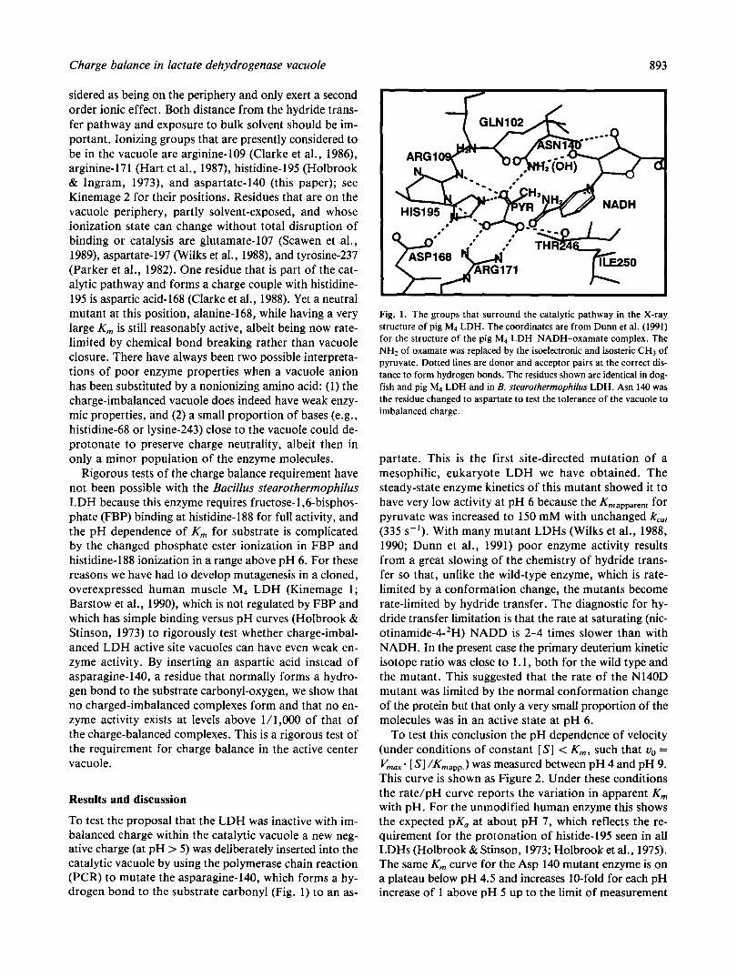
To test the proposal that the LDH was inactive with im- balanced charge within the catalytic vacuole a new neg- ative charge (at pH > 5 ) was deliberately inserted into the catalytic vacuole by using the polymerase chain reaction (PCR) to mutate the asparagine-140, which forms a hy- drogen bond to the substrate carbonyl (Fig. 1) to an as-
I / / GLNlOP I
ARG10 flm Fig. 1. The groups that surround the catalytic pathway in the X-ray structure of pig M4 LDH. The coordinates are from Dunn et al. (1991) for the structure of the pig M4 LDH-NADH-oxamate complex. The NHZ of oxamate was replaced by the isoelectronic and isosteric CH3 of pyruvate. Dotted lines are donor and acceptor pairs at the correct dis- tance to form hydrogen bonds. The residues shown are identical in dog- fish and pig M4 LDH and in B. stearothermophilus LDH. Asn 140 was the residue changed to aspartate to test the tolerance of the vacuole to imbalanced charge.
partate. This is the first site-directed mutation of a mesophilic, eukaryote LDH we have obtained. The steady-state enzyme kinetics of this mutant showed it to have very low activity at pH 6 because the Kmapparent for pyruvate was increased to 150 mM with unchanged kc,, (335 s"). With many mutant LDHs (Wilks et al., 1988, 1990; Dunn et al., 1991) poor enzyme activity results from a great slowing of the chemistry of hydride trans- fer so that, unlike the wild-type enzyme, which is rate- limited by a conformation change, the mutants become rate-limited by hydride transfer. The diagnostic for hy- dride transfer limitation is that the rate at saturating (nic- 0tinamide-4-~H) NADD is 2-4 times slower than with NADH. In the present case the primary deuterium kinetic isotope ratio was close to 1.1, both for the wild type and the mutant. This suggested that the rate of the N140D mutant was limited by the normal conformation change of the protein but that only a very small proportion of the molecules was in an active state at pH 6.
To test this conclusion the pH dependence of velocity (under conditions of constant [SI < K,, such that u,, = V,,. [SI /Kmapp.) was measured between pH 4 and pH 9. This curve is shown as Figure 2. Under these conditions the rate/pH curve reports the variation in apparent K , with pH. For the unmodified human enzyme this shows the expected pK, at about pH 7, which reflects the re- quirement for the protonation of histide-195 seen in all LDHs (Holbrook & Stinson, 1973; Holbrook et al., 1975). The same K, curve for the Asp 140 mutant enzyme is on a plateau below pH 4.5 and increases IO-fold for each pH increase of 1 above pH 5 up to the limit of measurement

894 A. Cortes et al.
100.00000
10.00000
1 .oO000
0.1 0000
0.01000
0.00100
0.00010
0.00001
E HUMAN M4
KM PYRUVATE 2
U I
PIG
+ 0.01000 3
0.00100 8 E,
I c. (D
h c E
o.ooolo i 0
0.00001 g M4 -NADH Q OXAMATE
I I = I
10 9 8 7 6 5 4 PH
Fig. 2. The pH dependence of pyruvate binding to wild-type (M4) human LDH (Asn 140) and its Asp 140 mutant. The buffers were 0.1 M citrate-0.2 M phosphate (pH 4-8), 0.1 M Tris (pH 7.5-9.0). [Pyruvate] was 10 r M (wild type) or 0.35 mM (mu- tant). Initial velocities (left hand axis) are pmol/min/mg determined in triplicate for wild type (-A-) or for the Dl40 mutant (t). No loss of enzyme activity was observed at pH 24.5. At this pH -A340/min due to NADH decomposition was <5% of the enzyme-catalyzed rate, and progress curves were linear. A complete measure of kc,, and K, at pH 4.5 gave kc,, = 300 s", = 10 mM. The (t) for pig M4 NADH-oxamate results (right-hand axis) are replotted from Holbrook and Stinson (1973) and are modeled by an acid K d limit of 28 gM and pK, = 7. The K , of human M4 enzyme for pyruvate is modeled also by pK, = 7. The slope 2 curve models the situation where both Asp 140 and His 195 (pK, = 7) must be protonated in a functional complex. The unprotonated limit curve models the situation where the anion of Asp 140 is active with K, 10,OOO times greater than that of the protonated acid. The model of Figure 3 predicts a linear change in uo (K,) for pyruvate for the Asp 140 mutant from pH 9 to pH 4.
at pH 9. This monotonic decrease shows that pyruvate will not bind in either an active or inactive state to the en- zyme-NADH complex containing ionized aspartate-140. Substrate is only reduced when a group with pK, < 4 is protonated. The K,,, is I 3 mM at pH 4. We first thought the group responsible for the plateau at pH 4 was proton- ated aspartic acid. Indeed, other values measured in Ta- ble l show that interpretation of these results in terms of the protonation state of Asp 140 is reasonable and allows neglect of possible changed binding of NADH.
Table 1 shows that the Kd(NADH) was unaltered by the mutation. Both the Kj for a strict competitive inhibitor (oxamate) and the Kd oxamate from enzyme-NADH were increased to the same extent by the mutation as was the K, for pyruvate. The Kd for this complex was also increased by the mutation in the same way as the K, for pyruvate. At pH 4.5 the kcat and K, for pyruvate could still be measured under conditions where the rate of de- composition of NADH was small compared to the en- zyme rate. This curve showed, as expected from the interpretation of the curve of Figure 2, that kc,, was un- changed at 300 s" but that K , had decreased to about 10 mM. Technically, it was necessary to work at 0.125 p M Asp 140 enzyme subunits, because at lower enzyme con- centrations the progress curves started to slow with time, indicating that irreversible denaturation was occurring in
the assay cuvette. Muscle LDH readily dissociated from a stable tetramer to unstable monomer in acid. Oxalate forms a complex with unprotonated His 195 enzyme- NAD+ and as expected from the required charge balance in the vacuole, its Kj is also much increased by the mu- tation.
The mechanism of Figure 3 was used to model the ef- fects of certain mechanistic assumptions. (1) Assume that it was possible for there to be activity with the anionic form of Asp 140 but with a K, increased to 0.01 M. This assumption predicts the linear decrease in rate with increased pH with a plateau at an unprotonated limit above pH 8. No such limit is visible in the experimental results, and we conclude that such activity or binding must be less than 1/1,000 of that of the unprotonated Asp 140. (2) Assume that the pK, of His 195 remains at 7 (its value in the binary complex [Holbrook & Ingram, 19731) and that in the mutant containing Asp 140 both groups must be protonated to maintain charge balance and activity. This predicts that starting from pH 6 the de- crease in rate would be 100-fold for each increase in pH of 1, a slope of 2 on Figure 2. There is no evidence of such limit appearing up to pH 9. We thus conclude that in the presence of Asp 140 the pK, of His 195 is raised to greater than 9. It is likely that the pK, of His 195 would be increased by the nearby anion at position 140.

Charge balance in lactate dehydrogenase vacuole 895
Table 1. Kinetic and binding properties of wild-type human (M4) LDH and the Asp 140 mutant
Wild type (Asn 140)
350 0.08 0.02 0.014 1.2 0.018 1.15d 1.2f
Mutant (Asp 140)
335 150 43 40
1.1 31 n.d.e
1.15'
300 = 10
a Steady-state values with [pyruvate] 0.04-0.4 mM (wild type) and 4-40 mM (Asp 140) mutant enzyme 2.7 nM subunits, NADH (0.15 mM). Oxamate (0.01-0.04 mM [wild type] and 20-80 mM [for the Asp 140 mutant]) is a competitive inhibitor of pyruvate and Ki is the K. of the LDH-NADH-oxamate complex. Oxalate at the same concentratlons is an uncompetitive inhibitor and the Kt is a complex constant.
'Obtained fluorimetrically with 6 pM NADH. Obtained fluorimetrically with 12 pM subunits and 10 pM NADH. Primary deuterium kinetic isotope effects were from single turn-
over experiments. e n.d., not determined.
The mutant has high Km(pyruvate) and its primary kinetic isotope ra-
*Steady-state values with [pyruvate] 0.1-5 mM, [NADH] = 0.15 tio could only be deduced from steady-state measurements of kc,,.
mM, 0.125 fiM (subunits) Asp 140 mutant enzyme.
Because bringing two acids together normally increases the acidity of the more acidic and decreases that of the weaker acid, this suggests that increasing the pK, of His 195 by over 3 units (say to 10) results in the pK, of Asp 140 decreasing from its expected value of 3.5 to 0.5. This analysis demonstrates that the apparent pK, of 4.5 shown in Figure 2 is not the intrinsic pK, of Asp 140 (a point confirmed by modeling - see below).
Ionized aspartate-53 facilitates the distinction between NAD+ and NADP+ (Feeney et al., 1990). The measure-
pK,=7 Kpyr=O. 1mM ,~yruvate
E-B: d E-BH+ - - E-BH+ - catalysis 'CH2. COOH \CH2. COOH 'CH2. COOH 1 pKa'3.5 \ pKa=0.5
E-B: --' E-BH+ pKa=10
\ai* ." \ ai* .coo-
Fig. 3. Model for the effect of the nearby charge of Asp 140 on the pK, of His 195 in the vacuole of LDHs. As with Figure 2, only a com- plex with protonated His 195 and protonated Asp 140 is active and able to catalyze hydride transfer. The pK, of His 195 with Asp 140 ionized was calculated (Table 2) from modeling to be 210. B: is His 195; -CHz.COOH is Asp 140. All complexes contain NADH.
ments of Figure 2 were made at 0.2 mM NADH, some 20 times the K,,, (NADH). However, because the LDH mechanism involves compulsory order of addition of co- enzyme followed by substrate, any increase in K,,, for NADH above 0.2 mM could lead to an apparent plateau in Figure 2 (Holbrook et al., 1975). The experimental re- sults of Fawcett and Kaplan (1962) indicate that the pK, of the acid controlling the NADH/NADPH discrimina- tion is much less than 5 and thus an unlikely explanation for the pH 4 plateau.
Calculated shift in the pKa of His 195 due to the effect of the Asn 140-Asp mutation
The calculated change in pK, of His 195 for two differ- ent conformations of the aspartate side chain (crystal and model-built) are given in Table 2. The effect of neglect- ing the contribution of asparagine in the calculation of ApK, and the dependence of the calculations on the par- tial charge distribution are also assessed. The model pre- dicts that introduction of an aspartate residue at 140 will cause a significant positive shift in the macroscopic pK, of histidine (6 > ApK, > 1.3). The calculations predict that deprotonation of His 195 is unlikely within the pH range of the enzyme assays (4-9). However, the aspartate side chain conformation creates a very large uncertainty in the His pK,. The neglect of charge distribution on Asn 140 strongly influences the lower limit of ApK,,
Table 2. Calculated shifl in the pK, of His 195 due to the effect of the Asn 140-Asp mutationa
APKa
Model Crystal Conf-I1
I 7.01 2 . 0 1 b I1 6.31 1.25 111 6.71 1.26
a Two extreme conformational states for the aspartate side chain were analyzed. Crystal corresponds to the observed conformation of asparagine-140 in the apoenzyme crystal structure and Conf-I1 to a model-built structure in which the side chain was rotated away from His 195 as far as was sterically reasonable, leaving the side chain tor- sion angle (xl) in a gauche+ conformation. The inclusion of the poten- tial generated by histidine at asparagine-140 4asn was deemed important as the closest appfoach of heavy atoms for the two residues in the crystal structure is 4.2 A. Thus, model I was calculated from the difference in potential generated at all atoms on Asn and Asp for the united atom Biosym charge set (ApK, = E qi[4asni - daspi]/2.303). Model I1 is the same as above; however, it neglects the contribution of aspara- gine (ApK, = C q, [ -$aspi] 12.303). In Model I11 only the side chain carboxyl oxygens are considered charged with a value of -0.5e on either oxygen, whereas the contribution of Asn is neglected (ApK, = E qi [ -4aspi] /2.303). Note: All values of ApK, are positive, leading to an increase in the histidine pK,. Asp 140 is fixed in the conformation of Asn 140 in the crystal structure.
bAsp 140 with a conformation of the minimized model-built structure.

896 A . Cortes et al.
whereas partial charge distribution on Asp 140 has much less effect.
The results were somewhat dependent on grid size, with the results for the two finest grid sizes differing by about 10%. However they were less dependent on rota- tional averaging (Gilson et al., 1987) at the finest grid spacing. The results of four runs on the finest grid spac- ing using rotational averaging varied from the mean value of ApK, by less than 2% when using the coordinates of enzyme crystal structure (which had the largest ApK, de- pendence on grid orientation and during focusing).
If the anion of Asp 140 continually becomes more pro- tonated as pH is decreased to <4 then why does the curve of Figure 2 plateau at pH 4? A likely explanation is that only a minute fraction of the other acids in the LDH vac- uole are protonated above pH 5, and the amount is not sufficient to disrupt charge balance and influence the ap- parently monotonic pH dependence from pH 5 to 9. However, at below pH 5, there will begin to be an appre- ciable fraction of the glutamic and aspartic acids, for ex- ample Glu 107 (Scawen et al., 1989), Asp 197 (Clarke et al., 1987), Glu 194 and Asp 168 (Clarke et al., 1988), which line the vacuole in a protonated state (assuming they have standard pK, values). If these acids are re- quired to be unprotonated to achieve charge balance, then protonating them will result at first in a maximum and then a decrease in activity below pH 4. The optimum and decrease are experimentally inaccessible, as irrevers- ible denaturation of the protein and NADH decomposi- tion become very rapid below pH 4.
Calculated effect of Asn 140-Asp mutation on the free energy of binding pyruvate
The difference in electrostatic free energy of binding py- ruvate in the mutant ternary complexes for both the apo (loop up) and ternary (loop down) enzyme conformations are given in Table 3. The protonated form of aspartate has a negligible effect on A(AGbind). It is predicted to slightly enhance the enzyme's affinity for pyruvate, i.e., to be slightly better than Asn 140. The negatively charged
Table 3 . Calculated effect of Asn 140-Asp mutation on the free energy of binding pyruvate
A ( AGbind)a (kcalhol)
Mutation Apo-crystal Ternary-crystal
Asn 140-Asph n.d.b -1 .53 Asn 140-Asp- 8.96 29.49
aspartate species is predicted to abolish substrate binding in the catalytically competent loop down ternary com- plex. These calculations suggest that only the protonated form of the Asp 140 enzyme is a kinetically significant species. This was also the experimental result.
The active site loop conformation has some effect on the enzyme's affinity for pyruvate, A(AGGbind) being three times more unfavorable in the loop closed confor- mation than in the loop open conformation. The value of the effective dielectric constant (eeff) calculated from a simple Coulomb's law approximation (for the interaction of two point charges of given separation) indicates that eeff is reduced by a factor of three upon closure of the active site loop. Thus loop closure involving removal of bulk solvent from active site residues brings about a dra- matic increase in the importance of electrostatic inter- actions in the active site vacuole. However, to view this region as a low dielectric medium is clearly not helpful, as the internalized active site is a highly polar environ- ment composed of acidic and basic groups involved in ion-pair interactions. Internalization of polar active site residues clearly serves to increase the importance of charge conservation, albeit in a polar environment (Yadav et al., 1991).
The large positive value of eeff will clearly have conse- quences for the conformational viability of the charged mutant ternary complex. It has been noted by Warshel and Russell (1984) that the upper limit of protein stabil- ization for a repulsive charge interaction cannot exceed the folding energy of the protein (which is on the order of 15 kcal/mol [Smith et al., 19911) otherwise the protein will undergo local denaturation. Because the charged form of the mutant loop down ternary complex is desta- bilized by about 30 kcal/mol over wild type, it was pre- dicted there would be local unfolding if the system was treated as a dynamic entity. This conclusion was tested by the use of molecular dynamics, which allows the active site to move toward an energetic equilibrium. A dynam- ics and energy-minimized model of the B. stearothermo- philus LDH vacuole has already been constructed and shown to behave in accord with experiment (Clarke et al., 1991).
Molecular dynamics study
The results in Table 4 show that only the active site loop moves to a significant extent following energy minimiza- tion and molecular dynamics for 35 ps. Both wild-type and the mutant enzymes move, with the structure of post- dynamics main chain backbone atoms having a root mean square (rms) difference from the crystal structure of between 1.1 and 1.5 A for the large loop (residues 101- 121) and 0.52 and 0.5 A for all other residues. The main
aA(AGbind) = E %(@aspi - @as%) for the united atom BiosYm quantitative difference in loop motion comes between charge set. Asph and Asp- are the protonated and anionic form of aspartic acid-140. the tip of the loop (residues 103-109) in the wild-type
n.d., not determined. enzyme (1.85 A) and the two mutant models (2.36 and

Charge balance in lactate dehydrogenase vacuole
Table 4. The rms fit of backbone atoms (N, CA, C, 0) of crystal structure with the initial minimized (min) structure and the minimized structure following 35 ps dynamics (dyn)
Wild type Model Ia Model IIb
Region Min Dyn Min Dyn Min Dyn
AllC 0.56 0.66 0.55 0.72 0.58 0.71 Stabled 0.49 0.55 0.46 0.52 0.53 0.53 Largeloope 0.89 1.15 0.94 1.63 0.82 1.51 Small loop‘ 1.34 1.85 1.34 2.95 1 . 0 6 2.36
a Model I refers to the Asp 140 structure in which the system has
Model I1 refers to the Asp 140 structure in which Asp 197 is con-
All corresponds to all mobile residues in the simulation. Stable corresponds to all mobile residues with the exception of
overall net negative charge (see Materials and methods).
sidered charge neutral and the system has an overall neutral charge.
loop residues 101-121. eLarge loop corresponds to residues 101-121.
Small loop corresponds to residues that constitute the active site loop “tip” (103-109).
2.95 A). The residues Asn 102 and Arg 109 of the loop tip project into the active site vacuole, and hence this re- gion, which is known to have a high degree of conforma- tional flexibility, will be most sensitive to changes in the active site environment. In the crystal structure of B. stearothermophilus the loop is less closely packed down on the surface of the enzyme than in other LDH crystal ternary complexes (Wigley et al., 1992). It was found that on minimization of the crystal structure that the loop and remainder of the protein formed a more close packed in- terface. This occurred in both wild-type and mutant com- plexes. However, following dynamics it became evident that whereas the wild-type enzyme loop tip retained the residue-residue interactions and conformation of the
I
897
minimized structure (rms of 0.91 A), the mutant models drift away from their minimized starting conformations (rms of 1.659 and 2.427 A for model I1 and model I, re- spectively), illustrating that the mutant complexes devi- ate to a much larger degree during the time of the dynamics simulation. Visual inspection of the superirn- posed loop structures (Fig. 4; Kinemage 3) shows that for the two mutant complexes there is a net translation of the loop tip away from the crystal conformation resulting in new contacts between residues in the loop tip and the rest of the protein. These contacts are not seen in the crystal structure or in the wild-type enzyme following dynamics. As might be expected, the conformational changes were greatest in the mutant model, which has a net charge im- balance in the active site. One external water molecule penetrates the active site to form a hydrogen bond to Asp 140, the side chain of which undergoes significant pertur- bation from the crystal conformation of Asn 140 to a po- sition more closely associated with the bulk solvent. Thus again the modeling gives an explanation for the experi- mental result that the enzyme has high K,,, at pH where residue 140 is mainly the charged aspartate.
These calculations were performed in order to access the stability of the active site conformation in the wild- type and mutant Asn 140-Asp ternary complexes. Al- though the simulations were of relatively short duration, it is evident that while the wild-type complex retained the essential conformational and bonding characteristics of the crystal structure, the mutant enzyme complexes showed concerted movements away from the crystal structure leading to new active site loop conformations and in one case to solvent penetration into the active site. Thus molecular dynamics provides further support for the experimental observation that the charged mutant en- zyme ternary complex is energetically unstable and sug- gests that instability is due to the charge imbalance
Fig. 4. The effect of charge imbalance on the struc- ture of the active site loop (between a D and PD). Four loop structures are shown: the wild type after energy minimization of the crystal structure confor- mation (-); the wild type after 36 ps of molecu- lar dynamics ( - - - ); model I after similar dynamics (. . . . .); model I1 after similar dynamics (-----). Models are defined in the legend to Table 4.

898 A. Cortes et al.
forcing open the mobile loop that normally covers the ac- tive site vacuole in ternary complexes of the enzyme.
Materials and methods
Site-directed mutagenesis
The mutant protein with aspartic acid at position 140 was generated by the PCR-based overlap extension method (Higuchi et al., 1988; Ho et al., 1989). The template used was the plasmid pHLDHA22 (Barstow et al., 1990), which consists of an EcoRI-SmaI DNA fragment con- taining the entire human & LDH coding region together with a 60-bp fragment containing the transcriptional and translational signals of the B. stearothermophilus LDH inserted into the expression vector pKK223-3 (Pharmacia LKB Biotechnology, Milton Keynes, UK). The conditions used to generate two overlapping fragments were identi- cal: 100 ng of template DNA; 50 mM KC1; 10 mM Tris, pH 8.3; 1.5 mM MgC12; 0.01% w/v gelatin; 200 pM each dNTP; 1 pM of each primer; 2.5 units TAQ DNA polymerase (Perkin Elmer Cetus) in a final volume of 50 p1. Each reaction was subjected to 10 cycles of PCR with the following parameters (A): 1 min 30 s at 94 "C, 2 min at 57 "C, 2 min at 72 "C in a Perkin Elmer thermal cycler. In one reaction the antisense mutant primer (25- mer) was used in combination with the 5'-flanking primer (20-mer) specific to the tac promoter. In a second reac- tion the "sense" mutant primer was used in combination with the 3'-flanking primer (20-mer) specific to the 5s re- gion of pKK223-3. The sequences of these oligonucleo- tides are listed in Table 5.
Each fragment was then separated from excess primers and template DNA by agarose gel electrophoresis using Ultrapure low melting point agarose (BRL, Gaithers- burg, Maryland). Each purified fragment (50 ng) was added to a single PCR reaction and subjected to five cycles of annealing and extension in the absence of prim- ers (1 min 94 "C, 1 min at 45 "C, heat to 72 "C in 2 min, 1 min 72 "C). Flanking primers were then added and full- length product amplified for 25 cycles with parameters
Table 5. Oligonucleotides used in the PCR-based overlap extension method of mutagenesisa
I leValSer&ProValAsp Human A4 LDH wild-type sequence 5'-TTATTGTTTCAAATCCAGTGGATAT-3' Human A4 LDH sense primer 5'-TTATTGTTTCAGATCCAGTGGATAT-3' Human A4 LDH antisense primer 3'-AATAACAAAGTCTAGGTCACCTATA-5'
I leValSerAspProValAsp
pKK223-3 flanking primer to fac 5'-AGCGGATAACAATTTCACAC-3' pKK223-3 flanking primer to 5s 5'-GGGATCCGTCGACCTGCAGC-3'
a Double-standard DNA was obtained by mixing complementary sense and antisense single-stranded DNA. The sense strand was amplified off the wild-type gene between a mutant sense primer and a primer to the 5s region of pKK223-3. The antisense strand was amplified with a primer flanking the lac promoter region of the vector pKK223-3 (see text for details).
(A) above. The full-length product was purified by eth- anol precipitation and digested with SmaI and EcoRI prior to subcloning into similarly cut pKK223-3 thus forming plasmid pHLDHAacid. The ligation products were microdialyzed and transformed into Escherichia coli TG2 cells made competent by electroporation. LDH overexpression in ampicillin-resistant colonies was iden- tified from an Mr = 33,000 band appearing in sodium dodecyl sulfate (SDS)-polyacrylamide gel electrophore- sis (Pharmacia Phast system). The entire coding region from an overexpressing candidate clone was resequenced using a DuPont Genesis 2000 automated sequencing sys- tem and showed only the required change.
Enzyme purgications
The wild-type human M4 LDH was purified as described by Barstow et al. (1990). The N140D mutant did not bind to oxamate-Sepharose at pH 6 and required a different purification. The cells were grown up in NZCYM broth (1 L). A crude cell extract was obtained by sonication; soluble protein was precipitated with 65% (NH4)$04 and dialyzed against 25 mM triethanolamine hydrochloride (TEA) buffer, pH 7, with charcoal outside the bag as de- scribed by Barstow et al. (1990). The dialysate was ap- plied to a 1.6 x 20-cm column of Q-Sepharose Fast Flow equilibrated with the above buffer. Human LDH was not retained, but the E. coli LDH was. The separation of the E. coli and human LDHs was checked by isoelectric fo- cusing on a pH 3-9 gel with lactate/NAD+ formazan staining for enzyme activity (Fine & Costello, 1963). The LDH in the breakthrough peak was recovered by precip- itation with 65% (NH&S04, desalted on a Sephadex G- 25 column in 20 mM Na phosphate, pH 6.8, and applied to a 1.6 x 20-cm column of S-Sepharose in the same buffer and was eluted in a linear gradient (0-0.5 M) of NaCl at about 0.1 M NaC1. The enzyme was estimated to be >98% pure from the Coomassie blue stain of both SDS-polyacrylamide and pH 3-9 isoelectric focusing gels run in a Pharmacia Phast system. Protein was estimated from A280 ,,,,, = 1.2 for 1 mg/ml protein derived from the Trp and Tyr contents of the protein sequence. The cloned human enzyme is identical to the authentic enzyme iso- lated from human tissue except that the N-terminal ala- nine is not acetylated (Barstow et al., 1990).
Steady-state kinetics
Assays were monitored from -A340nm/min at 25 "C in 67 mM Na phosphate buffer pH 6. Kinetic parameters
mined at saturation with NADH (0.15 mM), and initial rates were fitted using a nonlinear regression (Leatherbar- row, 1990) to the appropriate rate equation (Cleland, 1963). The pH dependence of pyruvate binding to LDH was measured in 0.1 M citric acid and 0.2.M phosphoric acid brought to a pH in the range 4-9 with 10 M NaOH.
kcot, Km(pyruvate) 3 K;(oxamate) 9 and K;(oxalate) were deter-

Charge balance in lactate dehydrogenase vacuole 899
To obtain the pH dependence of K,, the variation of LDH activity with pH was obtained at [pyruvate] < K,,,, where uo = kcat. [pyruvate] /K, (see legend to Fig. 2). To maintain this condition the rates with the wild-type en- zyme were obtained in a rapid mixing spectrometer.
Primary kinetic isotope ratios
Deuterium effects on k,,, were measured during a single turnover in a Hi-Tech stopped flow spectrometer SF-51 by comparing rates of -A340nm when 72 pM of either NADH or NADD and 80 pM enzyme subunits (M, 33,000) in 67 mM Na phosphate buffer, pH 6, was rap- idly mixed with 20 mM pyruvate.
Binding studies
The dissociation constants of enzyme-NADH and of ox- amate from enzyme-NADH-oxamate were measured from the enhanced fluorescence of NADH or its quench by oxamate. Excitation was at 340 nm and emission at 450 nm in an SLM8000 spectrofluorimeter (SLM Instru- ments, Urbana, Illinois). The buffer was 67 mM Na phosphate, pH 6. For the binary complex, concentrated enzyme was continuously added to 6 pM NADH (fluo- rescence enhancement). For the ternary complex, concen- trated oxamate was continuously added to 12 pM LDH and 10 pM NADH. The titration curves were fitted by a nonlinear regression, which corrected for the condition [NADH],,,,, does not equal [NADHIfre,.
Effect of Asn 140-Asp mutation on the pKa of the active-site histidine-I95 in the enzyme-NADH binary complex
To estimate the electrostatic effect of the Asn 140-Asp mutation on the pK, of the active site His 195 we used numerical solution of the Poisson-Boltzmann equation (Klapper et al., 1986; Gilson & Honig, 1988) to calculate the electrostatic potential (4) generated at reside 140 due to the charge on His 195. The atoms of both Asp 140 and Asn 140 were defined explicitly, and the potential at ei- ther residue was therefore calculated in separate runs. ApK, can be calculated from C qi ( 4asni - 4aspi)/2.303.
The high resolution coordinates of the dogfish apo- enzyme (Abad-Zapatero et al., 1987) were used. The co- enzyme was docked into the apostructure to generate the enzyme-NADH binary complex, in accordance with the coenzyme conformation in the dogfish ternary complex. The following side chain torsion angles were changed to relieve bad van der Waals contacts with the coen- zyme; Val 31(x1), Asp 52(xI,x2), Met 5 4 ( x l , x 2 , ~ 3 ) , Thr 95(x1), Thr 245(xl), and Ile 241(xI,x2). In all cases torsion angles were changed to adopt the confor- mation observed in the ternary crystal structure as closely as was possible.
Effect of Asn 140-Asp mutation on the pyruvate binding in the enzyme-NADH-pyruvate ternary complex
Numerical solution of the Poisson-Boltzmann equation was used to calculate the change in electrostatic free en- ergy of binding pyruvate due to the charge on the mutant Asp 140 in its neutral (protonated) and anionic states, in a similar manner to the approach used by Hurley et al. (1990). This approach neglects the contribution of the free energy difference between the wild-type and mutant binary enzyme complexes (the whole thermodynamic cy- cle should be considered in order to calculate A(AGbind). Hence, the value of A(AGbind) given in Table 3 is not strictly the free energy of binding. However, the inclusion of a negatively charged residue (aspartate-140) in the ac- tive site vacuole is analogous to the addition (binding) of the negatively charged substrate (albeit at a different po- sition in the active site) and would be expected to further destabilize the binding of substrate to the mutant enzyme, i.e., the value of A(AGbind) given in Table 3 would be more positive if we considered the whole thermodynamic cycle.
Two conformational states of the enzyme in the en- zyme-NADH-pyruvate ternary complex were assessed. The first corresponds to a loop open ternary complex. This was generated from the binary complex (see above) by docking pyruvate into the active site to reproduce its observed binding mode in the ternary enzyme crystal. Docking required only minimal adjustment of the side chain torsion angles of Arg 171 and His 195 (to adopt their observed conformations of the ternary crystal com- plex). The carboxyl group of the substrate can form a bifurcated ion-pair interaction with Arg 171 while main- taining a hydrogen bond with the side chain hydroxyl group (OG1) of Thr 246. Furthermore, the substrate car- bonyl oxygen can hydrogen bond to the protonated NE2 of His 195. Thus all the direct hydrogen bonds except those of Arg 109 to the substrate seen in the enzyme (loop down) ternary crystal can be satisfied in the loop open model built ternary complex. The second enzyme confor- mation corresponds to the loop down state, for which the high resolution coordinates of the dogfish ternary com- plex were used.
Models and parameters
The linearized Poisson-Boltzmann equation was solved using the program DELPHI (Biosym Technologies) run on a Silicon Graphics Personal IRIS 4D-20 workstation. The enzyme, NADH, and pyruvate were considered as low-dielectric regions surrounded by high dielectric sol- vent. The molecular dielectric ( E , ) was set at 2, and the solvent dielectric (E , ) was that of water (SO) (Sharp & Honig, 1990). However, because we were unable to ex- perimentally quantify ApK, or A ( AGbind) it was unnec- essary to calculate the dependence of the results on the

900 A. Cortes et al.
molecular dielectric (e,). The parameter set of the united atom consistent valence force field (CVFF) (Dauber-Osguthorpe et al., 1988) provided the charges for histidine, asparagine, and both protonated and de- protonated aspartate-140. The united atom force field, which defines polar hydrogens explicitly, was used to de- fine the shape of the low dielectric regions, and hydrogen, nitrogen, and oxygen were given radii of 1.2 A, 1.5 A, and 1.6 A, respectively (Spedding & Gsneidner, 1975). The ionic strength of the phosphate/citrate buffer used for the enzyme assays was used in the calculation (0.15 M) in addition to an ion exclusion layer of 2.0 A (determined from the average ionic radii of phosphate buffer). Cer- tain enzyme assays were carried out in citrate buffer; however, this is expected to have little influence on the re- sults, given the small difference in the radii of a phos- phate and carboxylate.
In the enzyme binary complex, potentials generated at Asn and Asp 140 due to the charge on histidine-195 were calculated using a three-step focusing procedure: with the first calculation using a 2.90-A grid spacing and a Coulombic boundary approximation, followed by two successive focusing calculations using grids of 0.89 A and 0.53 A.
In the enzyme ternary complex, potentials generated at pyruvate due to the charge on Asn and Asp 140 (in its protonated and charged states) were calculated using a two-step focusing procedure using grids with 2.90-A and 0.89-A spacings. The three-step focusing was unnecessary due to a difference of less than 10% in ApK, between the first and second focusing runs in the previous calcu- lations.
Dynamics and energy minimization
Simulations were based upon the high resolution coordi- nates of B. stearothermophilus LDH in a loop down ter- nary complex with NADH and oxamate (Wigley et al., 1992). A fixed boundary method was used with a 15-A re- action zone centered at the carbonyl carbon of oxamate with a 3-A fixed buffer region and water molecules fill- ing nonprotein regions within the 15-A reaction zone. Within this zone the sequences of both the eukaryote and prokaryote LDH are highly conserved, right down to the position of the “frozen” (low B-value) waters, which are thought to order the seven H-bonds from protein to sub- strate (Dunn et al., 1991; Wigley et al., 1992). Initial con- formations for the simulation were generated by changing the NH2 of oxamate to CH3 to generate pyruvate. The mutant complex was generated from this model by changing Asn 140 to Asp leaving the side chain torsion angles unchanged. The protein, coenzyme, and pyruvate were all modeled using the CVFF (Dauber-Osguthorpe et al., 1988) with the exception of charges on the coen- zyme and substrate. Charges on coenzyme, pyruvate, and known fragments from the CVFF library were generated using MNDO semiempirical molecular orbital calcula-
tions (Dewar & Thiel, 1979) in the program MOPAC ver- sion 4.0. A best fit scaling factor, which made the charges of the known fragments match those of the CVFF charge library, was deduced using several charged and un- charged species. This scale factor was then applied to the charges of NADH and pyruvate. A united atom model, which incorporates nonpolar hydrogens into the carbon heavy atoms, was used for amino acid residues, whereas all atom models were used for the coenzyme and sub- strate.
All algorithms were implemented by a vectorized ver- sion of DISCOVER 2.4 (Biosym Technologies) run on an IBM 3090-1508 computer. A nonbonded cutoff of 12 A with a switching function between 10 and 12 A was used. The nonbonded pair list was updated every 30 cycles, and a dielectric constant of 1 was used in all calculations. Only Arg 109, Arg 171, His 195, Asp 168, Asp 197, and the substrate carboxyl group were considered charged. To this was added Asp 140 in the mutant simulation but no counter charge was added because the nearest potentially positively charged residue (Lys 103) was some 14 A dis- tant and in a solvent-exposed position. The incorporation of a solvent counter ion was considered; however, it can- not approach Asp 140 by more than 5-6 A without per- turbation of the active site loop conformation because Asp 140 is in a part of the catalytic vacuole inaccessible to bulk solvent. Hence the simulation represents the vi- ability of the closed active site incorporating a negative charge at position 140. However, another simulation in which Asp 197 was considered as charge neutral was also performed, maintaining overall electroneutrality in the system. Asp 197 is partially solvent exposed but interacts with the active site Arg 109 through a water bridge. It is known that mutation of this residue to an asparagine has minimal effect on the values of kc,, and K, (Wilks et al., 1988). All complexes were minimized with all protein at- oms initially fixed and all waters allowed to move with the oxygen atoms lightly tethered (10 kcal A - 3 ) for 200 cycles of conjugate gradients minimization. The system was further relaxed with 200 cycles of conjugate gradients with heavy atoms of the protein, coenzyme, and substrate constrained (1,000 kcal A-3) to crystallographic posi- tions. This procedure allows the protein to relieve bad contacts of the added hydrogen atoms. Minimization was then continued to convergence (average derivative of <0.002 kcal/mol/A). The resulting structures were then equilibrated at 300 O K for 4 ps and data collected for a further 32 ps using a 1-fs timestep throughout. The final structures from dynamics were then minimized as above to an average derivative of <0.002 kcal/mol/A.
Acknowledgments
This work was supported by the Science and Engineering Re- search Council (UK), SmithKline Beecham Research (UK) Ltd., and The British Council (Acciones Integradas). A.C.’s sabbat- ical year in Bristol was supported by grant BE90-326 from

Charge balance in lactate dehydrogenase vacuole 90 1
Direccion General de Investigacion Cientifica y Tecnica of histidine residue in pig heart lactate dehydrogenase. Biochem. J . 131, Spain. 729-738.
Holbrook, J.J., Liljas, A., Steindel, S.J., & Rossmann, M.G. (1975). Lactate dehydrogenase. The Enzymes Xla, 191-293.
Holbrook, J.J. & Stinson, R.A. (1973). The use of ternary complexes to study ionizations and isomerizations during catalysis by lactate References
Abad-Zapatero, C., Griffith, J.P., Sussman, J.L., & Rossmann, M.G. (1987). Refined crystal structure of dogfish M4 apo-lactate dehydrog- enase. J. Mol. Biol. 198, 445-467.
Barstow, D.A., Black, G.W., Shaman, A.F., Scawen, M.D., Atkinson, T., Li, S.S.-S., Chia, W.N., Clarke, A.R., & Holbrook, J.J. (1990). Expression of the human LDH A and B cDNAs in E. coli. Biochim. Biophys. Acta 1087, 73-79.
Clarke, A.R., Colebrook, S., Cortes, A., Emery, D.C., Halsall, D.J., Hart, K.W., Jackson, R.M., Wilks, H.M., & Holbrook, J.J. (1991). Towards the construction of a universal NAD(P)+-dependent dehy- drogenase: Comparative and evolutionary considerations. Biochem. Soc. Trans. 19, 576-581.
Clarke, A.R., Smith, C.J., Hart, K.W., Birktoft, J.J., Banaszak, L.J., Wilks, H.M., Barstow, D.A., Atkinson, T., Lee, T.V., Chia, W.N., & Holbrook, J.J. (1987). Rational construction of a 2-hydroxyacid dehydrogenase with new substrate specificity. Biochem. Biophys. Res. Commun. 148, 15-23.
Clarke, A.R., Wigley, D.B., Chia, W.N., Barstow, D., Atkinson, T., & Holbrook, J.J. (1986). Site-directed mutagenesis reveals the role of a mobile arginine residue in lactate dehydrogenase catalysis. Na- ture 324, 699-702.
Clarke, A.R., Wilks, H.M., Barstow, D.A., Atkinson, T., Chia, W.N., & Holbrook, J.J. (1988). An investigation of the contribution made by the carboxylate group of an active site histidine-aspartate cou- ple to binding and catalysis in lactate dehydrogenase. Biochemistry
Cleland, W.W. (1963). The kinetics of enzyme-catalyzed reactions with two or more substrates. Biochim. Biophys. Acta 67, 173-187.
Dauber-Osguthorpe, P., Roberts, V.A., Osguthorpe, D.J., Wolff, J., Genest, M.G., & Hagler, A.T. (1988). Structure and energetics of ligand-binding to proteins: E. coli dihydrofolate reductase-trimeth- oprim, a drug receptor system. Proteins Struct. Funct. Genet. 4, 37-47.
Dewar, M.J.S. & Thiel, W.J. (1979). Ground states of molecules. 38. The MNDO method. Approximations and parameters. J. Am. Chem. SOC. 99, 4899-4907.
Dunn, C.R., Wilks, H.M., Halsall, D.J., Atkinson, T., Clarke, A.R., Muirhead, H., & Holbrook, J.J. (1991). Design and synthesis of new enzymes based upon the lactate dehydrogenase framework. Philos. Trans. R. SOC. Lond. B332, 177-185.
Fawcett, C.B. & Kaplan, N.O. (1962). Preparation and properties of some NAD analogues with pentose and purine modifications. J. Biol. Chem. 237, 1709-1715.
Feeney, R., Clarke, A.R., & Holbrook, J.J. (1990). A single amino acid substitution in lactate dehydrogenases improves the catalytic effi- ciency with an alternative coenzyme. Biochem. Biophys. Res. Com- rnun. 166, 667-672.
Fine, I.H. & Costello, L.A. (1963). The use of starch electrophoresis in dehydrogenase studies. Methods Enzymol. 6, 958-972.
Gilson, M. & Honig, B. (1988). Energetics of charge-charge interactions in proteins. Proteins Struct. Funct. Genet. 3, 32-52.
Gilson, M., Sharp, K., & Honig, B.J. (1987). Calculation of electrostatic interactions in biomolecules: Method and error assessment. Com- put. Chem. 9, 327-335.
Hart, K.W., Clarke, A.R. , Wigley, D.B., Waldman. A.D.B., Chia, W.N., Barstow, D.A., Atkinson, T., Jones, D.B., & Holbrook, J.J. (1987). A strong carboxylate-arginine interaction is important in sub- strate orientation and recognition in lactate dehydrogenase. Bimhim. Biophys. Acta 914, 294-298.
Higuchi, R., Krummel, B., & Saiki, R.K. (1988). A general method of in vitro preparation and specific mutagenesis of DNA fragments. Nucleic Acids Res. 16, 7351-7367.
Ho, S.N., Hunt, H.D., Horton, R.M., Pullen, J.K., & Pease, L.R. (1989). Site-directed mutagenesis by overlap extension using the poly- merase chain reaction. Gene 77, 51-59.
Holbrook, J.J. (1973). Direct measurement of proton binding to the ac-
27, 1617-1622.
tive ternary complex of pig heart lactate dehydrogenaseyBiochem. J. 133, 847-849.
Holbrook, J.J. & Ingram, V.A. (1973). Ionic properties of an essential
dehydrogenase. Biochem. J. 131, 739-748. Hurley, J.H., Dean, A.M., Sohl, J.L., Koshland, D.E., & Stroud, R.M.
(1990). Regulation of an enzyme by phosphorylation at the active site. Science 249, 1012-1016.
Klapper, I . , Hagstrom, R., Fine, R., Sharp, K., & Honig, B. (1986). Focussing of electric fields in the active site of Cu-Zn superoxide dis- mutase: Effects of ionic strength and amino acid modification. Pro- teins Struct. Funct. Genet. 1 , 47-59,
Leatherbarrow, R.J. (1990). Grafit version 2.0. Erithracus Software Ltd., Staines, UK.
Lodola, A., Parker, D.M., Jeck, R., &Holbrook, J.J. (1978). Malate dehydrogenase of the cytosol. Ionizations of the enzyme-reduced- coenzyme complex and a comparison with lactate dehydrogenase. Biochem. J. 173, 597-605.
Parker, D.M. & Holbrook, J.J. (1977). An oil-water-histidine mecha- nism for the activation of coenzyme in the 2-hydroxyacid dehydrog- enases. In Pyridine Nucleotide-Dependent Dehydrogenases (Sund, H., Ed.), pp. 485-495. Walter de Gruyter, New York.
Parker, D.M., Jeckel, D., & Holbrook, J.J. (1982). Slow structural changes shown by the 3-nitrotyrosine-237 residue in pig heart [Tyr(3N02)237] lactate dehydrogenase. Biochem. J. 201, 465-471.
Parker, D.M., Lodola, A,, & Holbrook, J.J.(1978). Use of the sulphite adduct of nicotinamide-adenine dinucleotide to study ionizations and the kinetics of lactate dehydrogenase and malate dehydrogenase. Biochem. J. 173, 959-967.
Scawen, M.D., Barstow, D.A., Nichols, D.J., Atkinson, A., Clarke, A.R., Wigley, D.B., Hart, K., Chia, W.N., & Holbrook, J.J. (1989). Lactate dehydrogenase. The effects of amino acid changes on prop- erties. In Gesellschaft fuer Biologisches Forschung GmbH Mono- graph. Advances in Protein Design, Vol. 12 (Bloecker, H., Collins, J., Schmid, R.D., & Schomburg, D., Eds.), pp. 103-115. VCH, Weinheim.
Sharp, K.A. & Honig, B. (1990). Electrostatic interactions in macromol- ecules. Theory and applications. Annu. Rev. Biophys. Biophys. Chem. 19, 301-322.
Smith, C.J., Clarke, A.R., Chia, W.N., Irons, L.I., Atkinson, T., & Holbrook, J.J. (1991). Detection and characterization of interme- diates in the folding of large proteins by the use of genetically in- serted tryptophan probes. Biochemistry 30, 1028-1036.
Spedding, F.H. & Gsneidner, H. (1975). Crystal ionic radii of the ele- ments. In Handbook of Chemistry and Physics, 56th Ed. (Weast, R.C., Ed.) pp. F-198-F-200. CRC Press, Cleveland, Ohio.
Waldman, A.D.B., Hart, K.W., Clarke, A.R., Wigley, D.B., Barstow, D.A., Atkinson, T., Chia, W.N., & Holbrook, J.J. (1988). A genet- ically engineered single-tryptophan reporter identifies the movement of a peptide domain of lactate dehydrogenase as the event which lim- its the maximum enzyme velocity. Biochem. Biophys. Res. Com- rnun. 150,152-759.
Warshel, A. & Russell, S.T. (1984). Calculations of electrostatic inter- actions in biological systems and in solutions. Q. Rev. Biophys. 17, 283-422.
Wigley, D.B., Gamblin, S. J., Turkenburg, S.P., Dodson, E. J. , Pion- tek, K., Muirhead, H., & Holbrook, J.J. (1992). The structure of a ternary complex of an a!losteric lactate dehydrogenase from B. stearothermophitusat 2.5 A resolution. J. Mol. Biol. 223,317-335.
Wilks, H.M. (1990). Design and synthesis of new catalysts on the lac- tate dehydrogenase framework. Ph.D. Thesis, University of Bristol, UK.
Wilks, H.M., Halsall, D.J., Atkinson, T., Chia, W.N., Clarke, A.R., & Holbrook, J.J. (1990). Designs for a broad substrate specificity 2-hydroxyacid dehydrogenase. Biochemistry 29, 8587-8591.
Wilks, H.M., Hart, K.W., Feeney, R., Dunn, C.R., Muirhead, H., Chia, W.N., Barstow, D.A., Atkinson, T., Clarke, A.R., & Hol- brook, J.J. (1988). A specific and highly active malate dehydroge- nase by redesign of a lactate dehydrogenase framework. Science 242, 1541-1544.
Yadav, A., Jackson, R., Holbrook, J.J., & Warshel, A. (1991). Role of solvent reorganization energies in the catalytic activity of enzymes. J. Am. Chem. SOC. 113, 4800-4805.