Embed Size (px)
Citation preview
Chemical and Electrical Synapses
Two Kinds of Synapses
1. Chemical 2. Electrical • Both types of synapses relay information, but do
so by very different mechanisms. • Much more is known about chemical than about
electrical synapses. - Information gleaned from NMJ in frog leg
(sciatic n. – gastrocnemius m.). - However, this is n-m, rather than n-n. - n-m relay is much faster than n-n.

Electrical Synapses • Symmetrical morphology. • Bidirectional transfer of information, but can be
unidirectional. • Pre- and postsynaptic cell membranes are in close
apposition to each other (~ 3.5 vs. ~ 20 nm in other cells), separated only by regions of cytoplasmic continuity, called gap junctions.
- Ions can flow through these gap junctions, providing low-resistance pathway for ion flow between cells without leakage to the extracellular space: signal transmission = electrotonic transmission.
- Instantaneous, fast transfer from 1 cell to the next ( < 0.3 msec), unlike the delay seen with chemical synapses.

Electrical synapses are built for speed
Electrical Synapses (cont’) Putative Functions
• Synchronization of the electrical activity of large populations of neurons;
- e.g., the large populations of neurosecretory neurons that synthesize and release biologically active peptide neurotransmitters and hormones are extensively connected by electrical synapses.
- e.g., Synchronization may be required for neuronal development, including the development of chemical synapses.
- e.g., Synchronization may be important in functions that require instantaneous responses, such as reflexes and pacemakers.

Electrical coupling is a way to synchronize neurons with one another


Gap junctions are formed exclusively from hexameric pores, called connexons (Cx36), which connect cells with each other for robust electrical coupling.

Electrical Synapses: Anatomy A. Have bridged = gap junctions between presynaptic and post-synaptic cells
B. Space between the pre- and post-synaptic cells is ~3.5 nm vs. 20 nm for “normal” cells
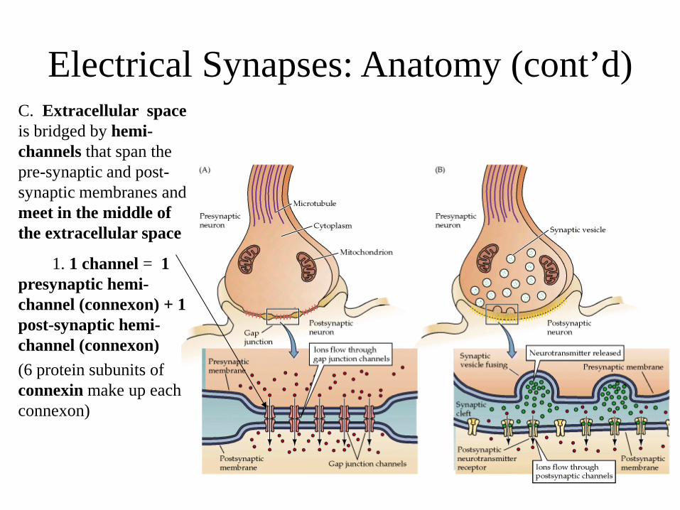
Electrical Synapses: Anatomy (cont’d) C. Extracellular space is bridged by hemi-channels that span the pre-synaptic and post-synaptic membranes and meet in the middle of the extracellular space
1. 1 channel = 1 presynaptic hemi-channel (connexon) + 1 post-synaptic hemi-channel (connexon) (6 protein subunits of connexin make up each connexon)
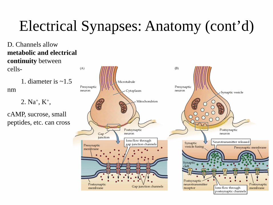
Electrical Synapses: Anatomy (cont’d) D. Channels allow metabolic and electrical continuity between cells-
1. diameter is ~1.5 nm
2. Na+, K+,
cAMP, sucrose, small peptides, etc. can cross

Chemical Synapses
• Asymmetric morphology with distinct features found in the pre- and postsynaptic parts.
• Enlarged extracellular space with no cytoplasmic continuity = Synaptic cleft is ~ 200-300 A wide.
• CHO moities intersperse the synapse. • Most presynaptic endings are axon terminals. • Most postsynaptic elements in the CNS are
dendrites.

Chemical Synapses (cont’d)
• Convergence. • Divergence. • Presynaptic ending: - swelling of the axon terminal. - mitochondria. - a variety of vesicular structures,
clustered at/near the very edge of the axon terminal.

Chemical Synapses (cont’d)
• Postsynaptic element - comprised largely of an electron-dense
structure, called the postsynaptic density (PSD).
Function of PSD? - Anchor receptors for neurotransmitters
in the postsynaptic membrane. - Involved in the conversion of a
chemical signal into an electrical one = transduction.
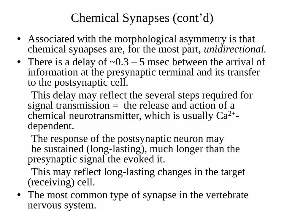
Chemical Synapses (cont’d) • Associated with the morphological asymmetry is that
chemical synapses are, for the most part, unidirectional. • There is a delay of ~0.3 – 5 msec between the arrival of
information at the presynaptic terminal and its transfer to the postsynaptic cell.
This delay may reflect the several steps required for signal transmission = the release and action of a chemical neurotransmitter, which is usually Ca2+-dependent.
The response of the postsynaptic neuron may be sustained (long-lasting), much longer than the presynaptic signal the evoked it.
This may reflect long-lasting changes in the target (receiving) cell.
• The most common type of synapse in the vertebrate nervous system.
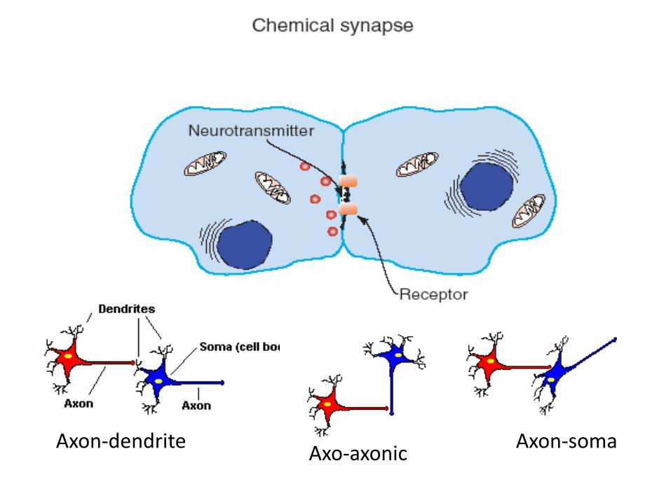
Axon-dendrite Axo-axonic Axon-soma
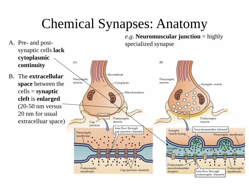
Chemical Synapses: Anatomy A. Pre- and post-
synaptic cells lack cytoplasmic continuity
B. The extracellular space between the cells = synaptic cleft is enlarged (20-50 nm versus 20 nm for usual extracelluar space)
e.g. Neuromuscular junction = highly specialized synapse

Chemical Synapses: Anatomy (cont’d)
Synaptic bouton = terminal knob
mitochondrion
Active zone
e.g. Neuromuscular junction = highly specialized synapse
C. Axon of pre-synaptic cell is highly branched and terminates in terminal knobs = synaptic boutons
D. Both pre- and postsynaptic cells have membrane specializations –
1. presynaptic boutons with – a. Synaptic vesicles = neurotransmitter vesicles b. Lots of mitochondria c. Active zones for docking/release of contents
of vesicles 2. post-synaptic membrane with membrane
spanning neurotransmitter receptor protein that serves as both receptor and ion channel
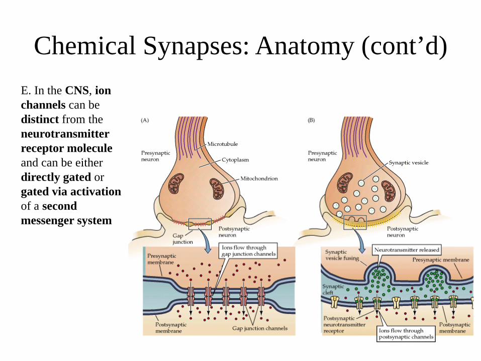
Chemical Synapses: Anatomy (cont’d) E. In the CNS, ion channels can be distinct from the neurotransmitter receptor molecule and can be either directly gated or gated via activation of a second messenger system

Summary Comparison of the 2 Principal kinds of Synapses: Electrical and Chemical
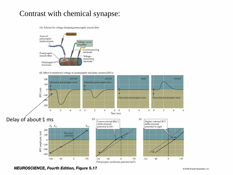
Contrast with chemical synapse:
Delay of about 1 ms

Physiology of Electrical Synapses: A. Experimental set-up:
Gap junction channel
“pre-synaptic” cell “post-synaptic” cell
Current passing electrode
Voltage measuring electrodes

Physiology of Electrical Synapses: (cont’d)
B. Experiment #1: inject threshold current in presynaptic cell
I presynaptic cell
Vm presynaptic cell
Vm postsynaptic cell
0.3 msec or less between pre- and postsynaptic action potentials
Thus, very short synaptic delay

Physiology of Electrical Synapses: (cont’d)
C. Experiment #2: inject subthreshold current in presynaptic cell
I presynaptic cell
Vm presynaptic cell
Vm postsynaptic cell
0.3 msec or less between pre- and postsynaptic membrane depolarizations
Thus, 1) very short synaptic delay and little decrement of original signal, and, 2) does not require a threshold depolarization for signal transmission
Change in Vm slightly less than presynaptic cell
Physiology of Electrical Synapses: (cont’d)
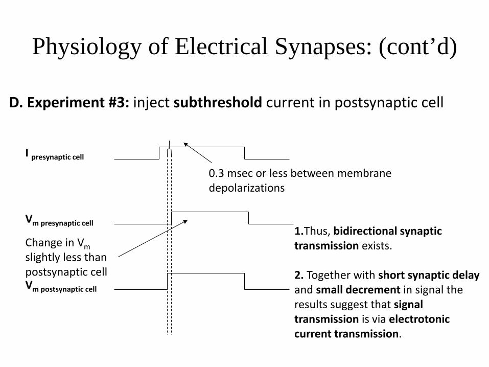
D. Experiment #3: inject subthreshold current in postsynaptic cell
I presynaptic cell
Vm presynaptic cell
Vm postsynaptic cell
0.3 msec or less between membrane depolarizations
1.Thus, bidirectional synaptic transmission exists. 2. Together with short synaptic delay and small decrement in signal the results suggest that signal transmission is via electrotonic current transmission.
Change in Vm slightly less than postsynaptic cell

Physiology of Electrical Synapses: (cont’d)
E. Experiment #4: inject subthreshold current in postsynaptic cell
I presynaptic cell
Vm presynaptic cell
Vm postsynaptic cell
Thus, unidirectional synaptic transmission also exists. Rectifying electrical synapses that conduct current in a single direction. May be due to heterotypic channels formed from different forms of the connexin protein.
No signal transmission
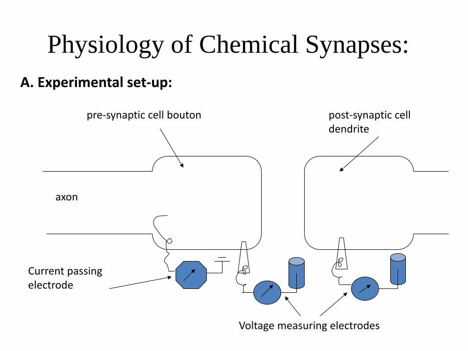
Physiology of Chemical Synapses: A. Experimental set-up:
pre-synaptic cell bouton post-synaptic cell dendrite
Current passing electrode
Voltage measuring electrodes
axon

Physiology of Chemical Synapses: (cont’d)
B. Experiment #1: inject threshold current in presynaptic cell
I presynaptic cell
Vm presynaptic cell
Vm postsynaptic cell
0.3-5 msec delay between pre- and postsynaptic action potentials
Thus, synaptic delay is significantly longer than for an electrical synapse.

Physiology of Chemical Synapses: (cont’d)
C. Experiment #2: inject subthreshold current in presynaptic cell
I presynaptic cell
Vm presynaptic cell
Vm postsynaptic cell
Thus, requires a threshold change in Vm in the presynaptic cell for signal transmission. No response

Physiology of Chemical Synapses: (cont’d)
D. Experiment #3: inject threshold current in postsynaptic cell
I presynaptic cell
Vm presynaptic cell
Vm postsynaptic cell
Thus, signal transmission is unidirectional. Together with other experimental results, this result suggests that signal transmission is not via electrotonic current transmission and that it requires a presynaptic AP.
No signal transmission
AP

Physiology of Chemical Synapses: (cont’d) What Current is Required for Signal Transmission
E. Experiment #4: inject threshold current in presynaptic cell bathed in tetrodotoxin to block Na+ current
I presynaptic cell
Vm presynaptic cell
Vm postsynaptic cell
0.3-5 msec delay between pre- and postsynaptic action potentials
Thus, the Na+ current is not required for chemical synaptic transmission.

Physiology of Chemical Synapses: (cont’d) What Current is Required for Signal Transmission
F. Experiment #5: inject threshold current in presynaptic cell bathed in tetraethylammonium ion to block K+ current
I presynaptic cell
Vm presynaptic cell
Vm postsynaptic cell
0.3-5 msec delay between pre- and postsynaptic action potentials
Thus, the K+ current is not required for chemical synaptic transmission.

Physiology of Chemical Synapses: (cont’d) What Current is Required for Signal Transmission
G. Experiment #6: inject threshold current in presynaptic cell bathed in Ca2+-free medium
I presynaptic cell
Vm presynaptic cell
Vm postsynaptic cell
Thus, the Ca+ current is required for chemical synaptic transmission.
No postsynaptic response
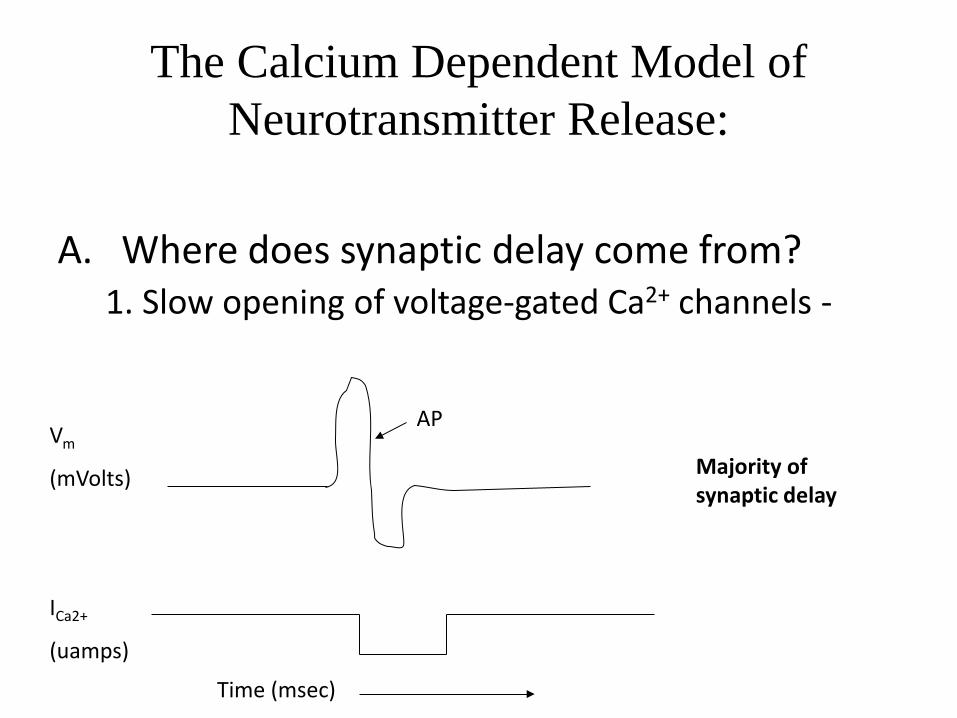
The Calcium Dependent Model of Neurotransmitter Release:
A. Where does synaptic delay come from?
1. Slow opening of voltage-gated Ca2+ channels -
Vm
(mVolts)
ICa2+
(uamps)
AP
Time (msec)
Majority of synaptic delay

The Calcium Dependent Model of Neurotransmitter Release: (cont’d)
Where does synaptic delay come from? (continued) 2. Time for exocytosis of synaptic vesicles 3. Diffusion of neurotransmitter across the synapse 4. Molecular events at the postsynaptic membrane that
lead to AP production following neurotransmitter binding

Calcium influx is necessary for neurotransmitter release
Voltage-gated calcium channels

Calcium influx is sufficient for neurotransmitter release