Embed Size (px)
Citation preview
Louisiana State UniversityLSU Digital Commons
LSU Historical Dissertations and Theses Graduate School
1970
Chemical and Physical Studies of the Structure andComposition of Invertase From NeurosporaCrassa.Zackey Donnell Meachum JrLouisiana State University and Agricultural & Mechanical College
Follow this and additional works at: https://digitalcommons.lsu.edu/gradschool_disstheses
This Dissertation is brought to you for free and open access by the Graduate School at LSU Digital Commons. It has been accepted for inclusion inLSU Historical Dissertations and Theses by an authorized administrator of LSU Digital Commons. For more information, please [email protected].
Recommended CitationMeachum, Zackey Donnell Jr, "Chemical and Physical Studies of the Structure and Composition of Invertase From NeurosporaCrassa." (1970). LSU Historical Dissertations and Theses. 1869.https://digitalcommons.lsu.edu/gradschool_disstheses/1869
71-6589MEACHUM, Jr., Zackey Donnell, 1943-
CHEMICAL AND PHYSICAL STUDIES OF THE STRUCTURE AND COMPOSITION OF INVERTASE FROM NEUROSPORA CRASSA.
The Louisiana State University and Agricultural and Mechanical College,Ph.D., 1970 Biochemistry
University Microfilms, Inc., Ann Arbor, Michigan
CHEMICAL AND PHYSICAL STUDIES OF THE
STRUCTURE AND COMPOSITION OF INVERTASE
FROM NEUROSPORA CRASSA
A Dissertation
Submitted to the Graduate Faculty of the Louisiana State University and
Agricultural and Mechanical College in partial fulfillment of the
requirements for the degree of Doctor of Philosophy
in
The Department of Microbiology
byZ. D. Meachum, Jr.
B.S., Northwestern State College, 1965 S., Northeast Louisiana State College, 1966
August, 1970
To Janet Together we serve God
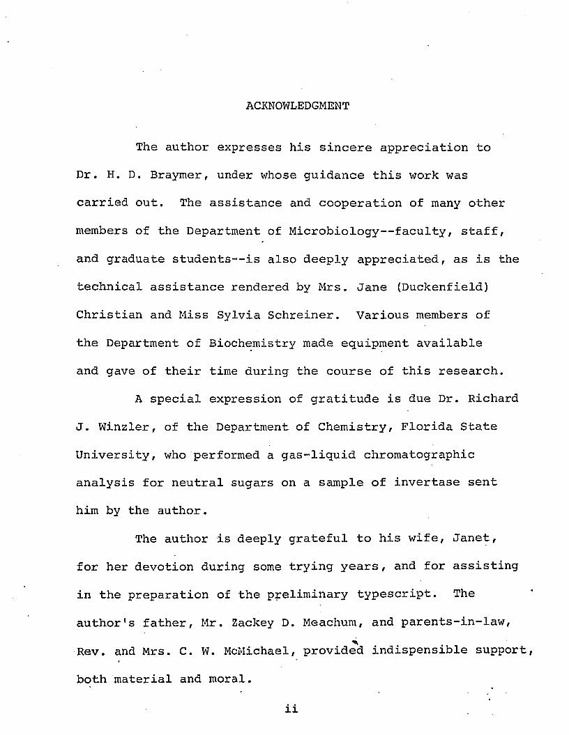
ACKNOWLEDGMENT
The author expresses his sincere appreciation to
Dr. H. D. Braymer, under whose guidance this work was
carried out. The assistance and cooperation of many other
members of the Department of Microbiology— faculty, staff,
and graduate students— is also deeply appreciated, as is the
technical assistance rendered by Mrs. Jane (Duckenfield)
Christian and Miss Sylvia Schreiner. Various members of
the Department of Biochemistry made equipment available
and gave of their time during the course of this research.
A special expression of gratitude is due Dr. Richard
J. Winzler, of the Department of Chemistry, Florida State
University, who performed a gas-liquid chromatographic
analysis for neutral sugars on a sample of invertase sent
him by the author.
The author is deeply grateful to his wife, Janet,
for her devotion during some trying years, and for assisting
in the preparation of the preliminary typescript. The
author's father, Mr. Zackey D. Meachum, and parents-in-law,
Rev. and Mrs. C. W. Mc&lichael, provided indispensible support
both material and moral.
TABLE OF CONTENTS
PageACKNOWLEDGMENT............................................ ii
LIST OF TABLES............................................ V
LIST OF F I G U R E S .......................................... vi
ABSTRACT................. * ................................ vii
Chapter
I. INTRODUCTION..................................... 1
II. LITERATURE REVIEW ........................ 4
INTRODUCTION
YEAST INVERTASE
NEUROSPORA INVERTASE
III. MATERIALS AND METHODS ........... . . . . . . . 28
ENZYME PREPARATION
CHEMICAL METHODS
Amino acid analysis
Carbohydrate Analysis
Preparation of Pronase peptides
PHYSICAL METHODS
CELL WALL METHODS
GEL FILTRATION METHODS
Chapter
IV. RESULTS AND DISCUSSION .......................
PHYSICAL STUDIES
Sedimentation velocity centrifugation
Sedimentation equilibrium centrifugation
CHEMICAL STUDIES
Amino acid composition
Carbohydrate composition
The carbohydrate-protein linkage
BINDING OF INVERTASE TO THE FUNGAL CELL WALL
Enzymatic release of invertase from the cell wall
Binding of invertase by cell wall fractions
ADDITIONAL OBSERVATIONS
Kinetic investigation of invertase active sites (Hill plot)
Refractometric determination of ^280
Behavior of invertase on Sephadex G-100 at low pH
V. SUMMARY.........................................
LITERATURE CITED . . . . . . . . . ..................
VITA ........... .. ...................................
Page
38
73
76
83
iv
LIST OF TABLES
Table Page
1. Amino acid composition of N. crassa invertase. . 60
2. Carbohydrate content of invertase fromvarious purifications........................ 61
3. Composition of Pronase glycopeptides ............ 62
4. Release of invertase from cell wallmaterial by chitinase........................ 63
5. Binding of invertase by cell wall fractions. . . 64
v
LIST OF FIGURES
Figure Page
1. Concentration-dependence of the S20,wof i n v e r t a s e ..................................... 66
2. a) Sedimentation rate study of invertasein 0.05 M acetic-acid (schlieren pattern). . . 68
b) Sedimentation rate study of invertasein 0.05 M acetate buffer, pH 5 . 0 .............. 68
3. Loge net fringe displacement as a functionof comparator x coordinate in a Yphantis run . 70
4. a) Behavior of invertase on Sephadex G-100in 0.05 M acetic acid............................ 72
b) Rerun of fractions indicated by bar inFig. 4a under same conditions................... 72
ABSTRACT
The composition and structure of Neurospora crassa
invertase -fructofuranosidase, E.C.3.2.1.26) have been
investigated by chemical and physical means. The enzyme
was found to be a glycoprotein containing about 11% mannose
and 3% glucosamine (presumed to be in the N-acetyl form) .
Small amounts of galactose and fucose were also present. The
amino acid composition of the enzyme was determined.
Ultracentrifugal studies established an s^ q w of
10.50 for native invertase. The molecular weight varied
slightly with carbohydrate content but was found to be about
210,000 for a preparation containing 11% mannose. Under
dissociating conditions (6 M guanidine hydrochloride plus 0.1
mercaptoethanol) the molecular weight of the enzyme was
51,500, indicating a tetrameric structure for native inver
tase. Both reduction and hydrophobic bond disruption were
necessary for complete dissociation of the molecule. In
dilute acids (e.g., 0.05 M acetic) the enzyme appeared both
to dissociate and to form complexes of high molecular weight.
About half of the invertase activity present in N.
crassa is rather firmly attached to the cell wall. It was
vii
found that a large percentage of this activity could be re
leased by the action of chitinase. A chitin-rich fraction
prepared from the Neurospora cell wall was shown to be
capable of binding invertase in vitro.
The carbohydrate-protein bond in invertase was found
to be alkali-stable. Preliminary analysis of glycopeptides
suggested the presence of the N-glycosidic glucosaminyl-
asparagine bond.
Kinetic studies indicated that invertase exhibits no
cooperative binding of substrate.
CHAPTER I
INTRODUCTION
In recent years the problem of glycoprotein structure
and the problem of secretion of proteins by living, undamaged
cells have received increasing attention. Worthy of special
note is the fact that the intimate interrelationship of
these two problems has been recognized and is now the subject
of intense and increasing interest.
Soodak (1966), in an article concerned with the
relationships among Leloir coenzymes, cell membranes, and
protein synthesis, hypothesizes that glycoproteins are
obligatory intermediates of protein' synthesis. He states
the view that those proteins destined for excretion by the
cell retain their carbohydrate components (and perhaps
receive additional such components) whereas those to be
retained within the cell membrane are enzymatically shorn
of their attached carbohydrate. Eylar (1965) has amassed
a large amount of evidence in support of the thesis that the
presence of carbohydrate covalently attached to a protein
molecule constitutes a "passport" which signals the cell to.
excrete the molecule so identified.
It is interesting to note that the existence and,
in some cases, the purification, of highly specific protein
(or polypeptide) : glycosyl transferases, associated with
cell membranes, has been reported (see, for example,
Hagopian and Eylar, 1969a and b ) . Some of the pertinent
literature has been briefly reviewed by Gottschalk (1969).
It should be noted that in the reviews referred to
above the authors deal almost exclusively with glycoproteins
of higher animals since, as Eylar (1965) says, " . . . infor
mation on glycoproteins from microorganisms and plants is
lacking." Some work with these neglected glycoproteins is
beginning to appear, e.g., Pazur, Kleppe, and Ball (1963),
Lineback (1968) .
We can thus begin to appreciate the significance of
structural and compositional studies on Neurospora crassa
invertase. This enzyme is a glycoprotein, readily isolable
from a representative of a category of organisms whose
glycoproteins have not been extensively studied. It occurs
almost exclusively outside the cell membrane, either in the
external medium (Gratzner and Sheehan, 1969), or in the
intramural space (i.e., the space between the cell wall and
the cell membrane; Metzenberg, 1963a), or in both environ
ments (Trevithick and Metzenberg, 1966a). About half of the
3
invertase associated with N. crassa cells is apparently
tightly bound to the cell walls (Sargent and Woodward, 1969).
Of additional interest is the fact that an
invertaseless mutant of N. crassa, "timex," produces a
protein (CRM) which cross-reacts with antiserum prepared
against wild-type invertase (Sargent and Woodward, 1969).
A precise knowledge of the structural and other properties
of the wild type and mutant gene products would permit a
comparison of the two and would provide knowledge of the
sort which will be required to further elucidate the nature
of gene action.
This dissertation will deal primarily with the
physical and chemical characterization of wild type N. crassa
invertase, with some observations on the nature of the
binding of the enzyme to the fungal cell wall.
It seems desirable to point out that the precise
name of the enzyme being considered is 'yfi-D-fructofuranoside
fructohydrolase" (E.C. 3.2.1.26). The trivial name is
"/3-f ructofuranosidase," and the traditional name is
"invertase." The traditional name will be used throughout
this dissertation. The reaction of primary interest cata
lyzed by the enzyme is the hydrolytic cleavage of sucrose
to yield glucose and fructose.
CHAPTER II
LITERATURE REVIEW
I . INTRODUCTION
The invertase of yeast has been studied extensively
by a number of workers over a period of many years. In
fact, the existence of invertase was first hinted at by the
observation that growing yeast cells would cause inversion
of optical rotation in sucrose solutions. This observation
was made by Persoz in 1833 (Neuberg, 1946).
Most of the studies on yeast invertase bearing
upon the subject treated in this dissertation have been
conducted by J. 0. Lampen and his colleagues. Colvin (1969)
has provided a review of earlier and additional work on the
yeast enzyme. The invertase of N. crassa has received
attention most notably from Robert L. Metzenberg and his
coworkers. This review will be largely concerned v/ith
papers from the laboratories of Lampen and Metzenberg.
II. YEAST INVERTASE
In an attempt to determine the location of invertase
in yeast (Saccharomyces cerevisiae) , Islam and Lampen (1962)
studied invertase secretion and sucrose fermentation by
yeast protoplasts. They found that protoplasts,- produced
by incubating washed, log phase cells with snail (Helix
pomatia) digestive juice, were free of external invertase
and were incapable of utilizing sucrose, although their
ability to utilize glucose was unimpaired'.' If a small
amount of monosaccharide (fructose or glucose) was offered
to protoplasts in the presence of sucrose, utilization of
the sucrose began after a lag period during which synthesis
and secretion of large amounts of invertase and accumulation
of free hexoses occurred. Washing of these "adapted"
protoplasts removed the sucrose-utilizing ability, and the
lag period preceeding sucrose utilization was again evident
when the washed protoplasts were re-incubated with sucrose
and small quantities of glucose.
It was found that non-protoplasted log phase cells
synthesized large amounts of invertase when incubated with
sucrose and glucose. Most of this invertase remained
attached to the cells. However, as increasing amounts of
cell wall were removed (by regulating the time of incubation
with snail digestive juice), an increasing fraction of the
newly synthesized invertase was secreted into the medium.
6
This work established that the integrity of the
yeast cell wall is required for the retention of invertase
by the cell and that the yeast cell membrane is not permeable
to sucrose per se. It is noteworthy that a small fraction
of the total cell invertase activity appeared to be
intracellular.
The purification of yeast invertase was reported in
1967 by Neumann and Lampen. The procedure described was
specifically designed to purify the external form of the
enzyme, i.e., that form secreted by protoplasts and re
tained by the cell walls of intact cells. This procedure
gave a highly active product which was homogeneous by
polyacrylamide gel electrophoresis and was essentially
homogeneous in sedimentation velocity studies in the
analytical ultracentrifuge.
One of the principal differences between this proce
dure and previously described procedures for yeast invertase
purification is that the Neumann-Lampen procedure employed
a French pressure cell to rupture the yeast cells; previous
methods had relied upon autolysis to disrupt the cells and
generally resulted in heterogenous preparations.
The preparation obtained by Neumann and Lampen was
purified 113-fold overall with a recovery of about 22% of
the initial enzymatic activity. The enzyme was characterized
as a glycoprotein consisting of a protein moiety and a
mannan moiety of equal weight, containing about 3%
glucosamine. In sedimentation velocity studies the presence
of a major peak with an s° of 10.4 was revealed. The J 20, wmolecular weight of the mannan-protein, determined by the
high-speed equilibrium method, was 270,000 + 11,000. Thus,
the protein moiety of the molecule had a molecular weight
of 135,000. The amino acid composition of the enzyme was
determined and will be referred to later, as will certain
of its other characteristics. The homogeneity of the
preparation with respect to the carbohydrate content of the
individual molecules was not believed to be absolute; a
slight non-coincidence of the respective peaks was noted
when the enzyme was chromatographed on DEAE-Sephadex and
the fractions were analyzed for protein and carbohydrate
content. The implication is that invertase can probably be
isolated with widely varying amounts of carbohydrate depend
ing upon the purification procedure employed, a given
procedure favoring the isolation of a given class of molecules.
In 1968 Gascon and Lampen reported the purification
of the internal form of yeast invertase. This form occurred
only in small amounts in wild-type cells under cultural
conditions favoring external invertase production, v i z .,
absence of high levels of glucose. In the presence of
high glucose levels the internal form of the enzyme pre
dominated. In the procedure under discussion, the source
of the enzyme was a mutant Saccharomyces strain whose
invertase production was relatively unaffected by high
glucose levels and which produced compartively high levels
of both internal and external invertase, especially in
low-glucose media. However, even in this system, the ratio
of external to internal invertase was 25:1.
Following mechanical disruption of the cells, the
two forms of invertase were separated from one another by
taking advantage of the fact that the external enzyme
remains in solution (presumably because of its large
carbohydrate component) at concentrations of ammonium sulfate
(70% of saturation) which cause precipitation of the internal
form. Further purification steps resulted in a preparation
with a slightly higher specific activity than that of pure
external invertase. The purification factor was 2500-fold,
with 5% recovery of the internal invertase activity initially
present.
The molecular weight of the purified enzyme was
estimated to be 130,000— 140,000 by measuring its elution
volume on a calibrated Sephadex column. This corresponds
roughly to the molecular weight of the protein moiety of the
external invertase. Sedimentation velocity experiments
in the analytical ultracentrifuge revealed a single peak
with an s2n „ of 8.8. zO,WGascon, Neumann, and Lampen (1968) compared the
two forms of yeast invertase and found striking similarities
and marked differences. Carbohydrate analysis revealed
that the internal form contained less than 3% carbohydrate;
it contained no glucosamine. Polyacrylamide gel
electrophoresis at pH 6.8 showed that the mobility of the
internal invertase was four to five times that of the
external; at pH 8.6 the difference in the relative mobilities
was even greater, with the internal form moving with the
tracking dye and the external form migrating slowly as a
diffuse band. Immunodiffusion tests indicated that the two
forms of the enzyme are immunologically related but not
identical.
The kinetic properties of the two enzymes were
investigated by determining Km values for the hydrolysis of
sucrose and raffinose. The two enzymes had identical Km
values for raffinose (150 mM) and closely similar values
for sucrose (26 mM for the external and 25 mM for the
10
internal form). Both enzymes were shown to lack transferase
activity when incubated with sucrose. Both enzymes
exhibited identical pH-activity curves, but the respective
pH stability ranges (at 30 C) were considerably different:
pH 3.0-7.5 for the external and 6.0-9.0 for the internal.
The acid inactivation of the internal invertase (pH 4.0, 4 C)
did not appear to be due to aggregation or dissociation of
the enzyme.
The most significant dissimilarities between the
two invertases appeared when their amino acid compositions
were compared. There were marked differences in the content
of most residues. Cysteine was totally lacking in the
internal enzyme, while five residues occurred in the
external form.
In hopes of shedding some light on the possibility
of a biosynthetic relationship between the two forms of
invertase, three sucrose-negative Saccharomyces strains
were examined. All three lacked both external and internal
invertase.
Thus, while similarities between the two invertases
are abundant, and genetic evidence tends to support the view
that the two are biosynthetically related, the extensive
differences in amino acid composition represent a signal
11
complication in defending this view. A reconciling
interpretation was offered by the authors, however. "The
best available explanation is that both enzymes represent
aggregates of different subunits of which one (or more)
is active and identical in both." The physical differences
(e.g., in solubility and stability) between the two
enzymes were attributed largely to the presence of
carbohydrate in the external species.
In this connection the paper by Arnold (1969) is
of interest. This investigator examined the kinetics of
heat inactivation of yeast invertase and explored the
possibility of a correlation between heat stability and
polysaccharide content. The enzyme used in these studies
corresponded approximately to Lampen's external enzyme, but
was obtained from a cell autolysate. Following purification,
a sample of the enzyme was subjected to gradient elution
(0-0.2M NaCl) chromatography on DEAE- cellulose at pH 6.0
under carefully controlled conditions. The enzyme was
eluted as a single peak, and specific activity, percent
polysaccharide, and first order rate constant for heat
inactivation at 65 C were determined for each of the
fractions in the peak.
12
Studies on the unfractionated invertase had shown
that, while the order of the heat inactivation reaction with
respect to time (n^) was greater than one, the order of the
reaction with respect to concentration (n ), determined
by plotting the log of the initial rate of inactivation
as a function of the log of the enzyme concentration, was
unity. Examination of the individual chromatographic
fractions brought several interesting facts to light. While
the specific activity showed no significant variation from
one fraction to another, there was a continuous increase in
susceptibility to heat inactivation (as indicated by the
magnitude of the first order rate constant) as one pro
ceeded from the leading edge to the trailing edge of the
peak. The carbohydrate content of the enzyme decreased
continuously from 54% at the front of the peak to 39% at the
rear. Furthermore, the individual fractions exhibited an
n^.=l, although, as noted above, there was an increase in
rate constant magnitude from the front to the rear of the
peak. Thus, in unfractionated invertase preparations, the
result that nt>l was a consequence of the relative enrich
ment of the sample in more stable species as inactivation
proceeded. In determining n only the susceptibility of .the
most sensitive species was considered and a value of unity
13
(the true order of reaction for heat denaturation of this
enzyme) was obtained.
This work plainly supports the hypothesis that the
carbohydrate moiety functions to stabilize the enzyme. The
nature of the linkage of this moiety to the protein of the
molecule is thus of great interest. This problem has been
investigated by Neumann and Lampen (1969).
In their study, a sample of external yeast invertase
(containing 52% carbohydrate) was subjected to exhaustive
proteolysis by Pronase. A glycopeptide fraction was
isolated by gel filtration of the digested material on
Sephadex G-25. The glycopeptides were further fractionated
by gel filtration on a column of Sephadex G-50. The eluate
from this column was divided into five fractions. Four of
these fractions (1,11,1V, and V) were obtained in sufficient
quantity for compositional analysis. Aspartic acid was found
to be the most abundant amino acid residue in all four
fractions, followed by threonine, serine, and glutamic acid;
generally smaller amounts of the other amino acids were
present. Glucosamine occurred in all four fractions, and was
equimolar with aspartic acid in three of them. Relatively
large amounts of hexose also occurred in all four fractions,
especially II and IV. Procedures designed to reveal the .
14
presence of O-glycosidic linkages (to serine or threonine
hydroxyl groups) and ester bonds (between the carbohydrate
moiety and the co-carboxyl group of aspartyl or glutamyl
residues) gave negative results. The authors concluded,
therefore, that the major (if not exclusive) type of
protein-carbohydrate bond in the invertase molecule is the
N-glycosidic glucosaminyl-asparaginyl bond, as has been
found in several other proteins. (For a discussion of
the various types of protein-carbohydrate linkages and the
means for their elucidation, see Neuberger, Gottschalk, and
Marshall, 1966.) The authors estimated that each invertase
molecule contains approximately 30 chains of polysaccaride
of varying length. . ,
Lampen (1968) has reviewed the nature and formation
of invertase and other external enzymes of yeast. He
proposes a model for the structure of the yeast cell wall
in which the outer layer of the wall is composed of large
mannan molecules held together by 1,6-phosphodiester bonds; the glycoproteins which occur in the wall are localized just
below the outer limit of this mannan layer (which occurs
externally to a glucan layer) by either additional
phosphodiester bridges between the mannan molecules and
the mannan "tufts" on the protein, or perhaps by "other
associative forces."
15
III. NEUROSPORA INVERTASE
Since, as will be shown, Neurospora invertase is,
like the yeast enzyme, cell wall associated, a brief
discussion of some of the work relating to the chemistry
and structure of the Neurospora cell wall seems to be
in order.
Whereas the yeast cell wall is composed primarily
of an inner glucan layer and an outer mannan layer, the
analogous structure in Neurospora has as its main constituents
chitin and glucan (Mahadevan and Tatum, 1965; Potgeiter
and Alexander, 1965). Additional components are also
present, notably galactosamine and peptides (Mahadevan and
Tatum, 1965, 1967). Mahadevan and Tatum (1967) presented
chemical and ultrastructural evidence that the Neurospora
cell wall is composed of an outer layer of coarse fibrils
(chemically, a glucan-peptide-galactosamine complex, i.e.,
Fraction I of Mahadevan and Tatum, 1965) , and an inner
layer of /Q-l,3-glucan in which a core of thin chitin fibrils
is imbedded. A general review of fungal cell wall chemistry
and its relationship to morphogenesis and taxonomy has been
authored by Bartnicki-Garcia (1968).
16
Metzenberg (1963a) described the purification and
properties of N. crassa invertase. He purified that
fraction of the enzyme which is extractable from mycelial
powder by pH 5.0 buffer; a 105-fold increase in specific
activity was realized in the course of the purification.
The preparation obtained was homogeneous by the criteria of
polyacrylamide gel electrophoresis and sedimentation
velocity studies in the analytical untracentrifuge. By the
latter technique an S2Q w value of 10.3 was calculated for the enzyme using a 0.4% solution and assuming a partial
specific volume of 0.75 for the molecule. The preparation
was examined for the presence of carbohydrate both before
acid hydrolysis (by the anthrone reaction) and following
acid hydrolysis (by the anthrone reaction and by the Somogyi
test for reducing sugars). The results indicated a neutral
sugar content of less than 0.2%. The purified invertase
was found to contain 2.4% hexosamine. The hexosamine was
not identified. The extinction coefficient of the enzyme1 %at 280 nm (Ej^) was found to be 18.6 on a dry weight basis.
The enzymatic activity of. the preparation was found
to be maximal at pH 5.0-5.5. It was fairly stable upon
storage at 0C (at least a week without measurable loss of
activity) and at 38 C (at least 2 hr at pH 5.0 without loss
17
of activity) even in very dilute solutions (0.1 /jug/ml) .It had a half-life of 6-7 minutes at 55C.
The enzyme was subjected to an extensive battery
of inhibition tests. Among the metal ions tested, only
copper and zinc showed notable degrees of inhibition (79 and
33%, respectively). Inhibition by p-hydroxymercuribenzoate
(PHMB) was dependent both upon time and inhibitor concen
tration, and the enzyme.could be protected to some extent
from the effect of this inhibitor by prior incubation with
substrate (sucrose) at 0 C. The Neurospora enzyme appears
to exhibit somewhat greater sensitivity to PHMB than does
the yeast external invertase. In common with invertases
from other sources (Pressey, 1968), the Neurospora enzyme
was rather strongly inhibited by aniline.
The substrate specificity of the enzyme was
investigated using the /6-fructofuranosides sucrose, raffinose,
and -methyl fructoside. It was found that sucrose was the
most readily utilized substrate, exhibiting the smallest
K (6.1 x 10*"3 M) and the highest V (set at 100 for pur- m 3 maxposes of comparison). The respective values of these
_3parameters were 6.5 x 10 M and 22 for raffinose and 3.3 x
10“2 M and 30 for -methyl fructoside. The enzyme did not
attack trehalose (a non-reducing glucoside) or melezitose
(which contains a substituted fructofuranoside ring).
18
Thus it resembles yeast invertase in its substrate
specificity.
The author attempted to relate the presence of
hexosamine in Neurospora invertase to its possible
localization in the fungal cell wall, which-, as we have
seen, is known to contain hexosamine polymers. He suggested
an analogy with yeast invertase, a mannan-protein localized
in a mannan cell wall layer, and, noting the apparent lack
of significant quantities of glucose in the Neurospora
enzyme, suggests that the glucose-containing polysaccaride
of Neurospora cell wall was either loosely bound to the
invertase or not associated with it at all in vivo.
Eberhart (1961) reported that invertase activity
could be recovered in water washings of Neurospora conidia.
Metzenberg (1963b) reported further experiments aimed at
determining the in vivo location of invertase in Neurospora.
He studied the accessibility of conidial invertase to
substrate, to the inhibitor aniline, to hydrogen ions, and
to pepsin. He also studied solubilization of the conidial
enzyme by simple washing and by sonication.
It was found that the pH-activity curve of whole-
conidial invertase was practically identical to that of
highly purified invertase. The I<m for sucrose and the
relative rates of hydrolysis of raffinose and sucrose were
19likewise practically indistinguishable when comparing
whole-conidial and purified enzyme. The conidial enzyme
was fully as sensitive as pure invertase to inhibition by
aniline, and was completely inactivated by brief exposure
to 0.2 N HC1, a treatment which did not impair conidial
viability and which failed to destroy the activity of
alkaline phosphatase, an intracellular enzyme. Simple
washing of the conidia with water could remove about 16%
of the invertase activity present in whole conidia.
Successive extractions of conidia with water showed that
practically all the soluble invertase was removed by the
first washing, thus indicating that, "... . the soluble
fraction represents a discrete proportion of the total
enzyme rather than a random dissociation from a chemically
and topographically homogeneous pool of enzyme."
Metzenberg (1963b) further found that sonication of
acid treated conidia released practically no invertase
activity into the medium, whereas the sonication of untreated
conidia showed a linear solubilization of activity with
time, with the total activity present in the sonicate
(i.e., both soluble and insoluble) remaining constant. Thus
it appears that there is no fraction of invertase present in
the cell which is insensitive to acid treatment by virtue
of its location.
20
The foregoing studies established the ready
accessibility of conidial invertase to small molecules
up to the size of a trisaccharide, indicating the absence of
an effective permeability barrier (by implication, the cell
membrane) between the invertase and the external milieu.
Certain of these studies were repeated using young mycelia
instead of conidia, with identical results.
To ascertain further the localization of the enzyme
in vivo, the sensitivity of invertase activity in whole
conidia to the action of pepsin was determined. It was
found that the conidial enzyme was completely insensitive
to a concentration of the protease 50-fold greater than that
which almost completely destroyed the activity of purified
invertase.
All of the above studies strongly suggested that
invertase is localized in vivo between the fungal cell wall
and the cell membrane, or that it is in some way associated
with some region of the cell wall other than its outer
surface.
Marzluf and Metzenberg (1967) investigated the
possible physiological significance of this apparent
"intramural" location of Neurospora invertase. Preliminary
experiments established that sucrose per se could not be
21
taken up by the cell. Of particular interest was the
possibility of "cytotropic release" of sucrose hydrolysis
products, i.e., release of the glucose and/or fructose
moieties from the surface of the invertase molecule in such
a way that they enter the cell immediately without
equilibrating with the external milieu.
It was found that hexose transport requires energy
and that fructose uptake is strongly inhibited in the presence
of glucose (but not vice versa), although fructose was
readily taken up and metabolized after the disappearance of
glucose from the medium.
Numerous experiments revealed that the presence of
invertase in its in vivo location in conidia conferred no
advantage in the uptake of glucose from sucrose; invertaseless
conidia, supplied exogenously with purified invertase, or
even incubated in the presence of conidia possessing the
enzyme, took up glucose to the same extent and at the same
rate as if they possessed a full complement of intramural
invertase. This observation argues very strongly against
a cytotropic release mechanism.
In 1964, Eilers, Allen, Hill, and Sussman reported
the occurrence of two bands of invertase activity when crude
extracts of several strains of N. crassa were examined by
22
the method of polyacrylamide gel electrophoresis. This
report prompted Metzenberg (1964) (whose previous work had
not included examination of crude extracts) to re-examine
Neurospora invertase in hopes of clarifying the relationship
between the two isozymic bands.
The occurrence of two bands of activity upon gel
electrophoresis of crude extract was confirmed. Typically
65-85% of the activity appeared in the band of lower
mobility. It was found that chromatography of a crude
extract on Sephadex G-200 at pH 7.5 resulted in the
appearance of two distinct peaks of activity; the first
peak (in order of elution) proved to correspond to the band
of lower mobility and the second peak to the band of higher
mobility seen on gel electrophoresis. The material in the
two peaks was subsequently referred to as "heavy invertase"
(first peak) and "light invertase" (second peak).
The heat stability of the two forms of the enzyme
was compared, it was found that the heavy form was roughly
50 times as stable as the light form at 50 C. Heating
caused partial conversion of heavy invertase to the light
form, a process which was greatly accelerated in the presence
of salt (e.g., 1.0 M NaCl). Total conversion of heavy to
light invertase (within the limits of detection) could be
23
effected at 0 C by treating the enzyme with 1.0 M NaCl
in 0.05 M formic or acetic acid, and the light invertase
so produced was observed to reaggregate when precipitated
from neutral solution with ammonium sulfate, dissolved in
pH 5.0 buffer at high protein concentrations (20 mg/ml),
and dialyzed overnight against water. Dissociation resulted
in a 31% loss of activity; reaggregation was accompanied
by neither gain nor loss of activity. Each form of the
enzyme was active per se.
Ultracentrifugal studies showed S2Q w values of 5.2 and 10.3, respectively, for the light and heavy forms of the
enzyme at the same protein concentration. The author points
out that this two-fold difference in sedimentation constant
would correspond to a four-fold difference in molecular
weight if both molecules were assumed to be spherical. The
S20 w va-*-ue was clearly concentration dependent.The occurrence of an "anomalous" form of invertase
with an s20 w = 9.8 was noted in preparations which had been treated so as to give about 50% conversion of heavy
to light enzyme. The interpretation was offered that this
represented a "pre-clastic" form of the molecule, i.e., one
whose conformation is favorable for cleavage into subunits.
It was observed that solutions of light invertase
foamed copiously upon shaking, whereas solutions of heavy
24
invertase showed little tendency to foam. To the author
this suggested the exposure of hydrophobic sidechains as
heavy invertase is split, and implied the importance of
hydrophobic bonding in maintaining the quaternary structure
of heavy invertase. The effect of salt upon the rate of
splitting of heavy invertase suggested that ionic bonds
are likewise of importance.
In an attempt to discover the site of aggregation of
invertase subunits in vivo, Trevithick and Metzenberg (1964)
investigated the nature of the invertase secreted by
Neurospora protoplasts. In experiments similar to those of
Islam and Lampen (1962) with yeast, they found that
protoplasts produced by treating young mycelia with snail
digestive juice secreted considerable amounts of invertase.
This invertase occurred predominantly as the heavy form.
A small amount of invertase was found inside the cells
throughout the incubation period (18 hr); this enzyme was
also of the heavy variety. The presence of actidione
abolished invertase excretion.
These results indicated to the authors that the
aggregation of invertase subunits occurred inside the cell
membrane during or shortly after their synthesis. The
attractive hypothesis that the subunits were excreted as
such into the intramural space, where their high concentration
25
would favor aggregation, seemed unlikely in the light of
these results.
Trevithick and Metzenberg (1966a) investigated
the excretion of invertase by wild type N. crassa and by
several mutant strains (viz., crisp, osmotic, and crisp
osmotic) believed to possess altered cell walls. A slow
but finite rate of transition between heavy and light
invertase had been shown by Metzenberg (1964) to obtain
under physiological conditions. Assuming, then, the
existence of cell wall pores in Neurospora which provide a
means of egress for invertase molecules, a mutant whose
cell wall contained pores smaller than those of the wild
type would be expected to show both a decrease in percent
of total invertase activity excreted into the medium and an
increase in the degree to which this excreted invertase is
fractionated. In other words, one would expect the inver
tase excreted by such a mutant to show a greater enrichment
in the fraction occurring as light invertase, when compared
to the invertase remaining in the intramural space, than
would be seen with the wild type. This effect could result
from increased molecular sieving by the pores of the mutant
cell wall. Strains whose cell wall pores were larger than
those of wild type would be expected to exhibit the opposite
tendencies in percent of invertase excreted and degree of
26
fractionation. Protoplasts, which may be considered to have
cell wall pores of infinite size, had been shown (Trevithick
and Metzenberg, 1964) to excrete practically all of their
invertase, almost exclusively as heavy invertase.
It was found that cultures of wild type and the
various mutants did indeed show an inverse relationship
between percent of total invertase activity occurring in
the medium and the degree to which this excreted invertase
was enriched for the light form. The mutants all showed
increased excretion and decreased fractionation relative
to the wild type. The results indicated increasing pore
size in the order wild type < crisp < crisp osmotic < osmotic.
Cytolysis was excluded as a significant factor in these
results.
In an accompanying paper, Trevithick and Metzenberg
(1966b) presented evidence for the existence of chemical
and physical differences between the cell wall of the wild
type and osmotic. Using a series of polyethylene glycol
and dextran polymers of known molecular weights, the
exclusion thresholds of the isolated cell walls of the two
strains were evaluated by statistical treatment of data
obtained by the "volume of distribution" technique. The
molecular weight limits for permeation of wild type and3osmotic cell walls were, respectively, 2.55 x 10 —
8.84 x 10^ and 1.03 x 10^— 3.37 x 10^. These figures are
believed to have only statistical and not absolute
significance, since at least a small number of very large
pores (larger than the largest indicated in osmotic) would
be required to explain the slow, steady excretion of
invertase which occurs in wild type. Chemically, it was
found that osmotic had a radically altered cell wall
galactosamine/glucosamine ratio (about 30-fold higher than •
wild type) .
Manocha and Colvin (1967) have found evidence for
the existence of pores in Neurospora cell walls which serve
as conduits for macromolecules. Thus the proposal made by
Trevithick and Metzenberg (1966a) concerning the relationship
of cell wall pore size to molecular sieving appears to have
received some experimental support.
CHAPTER III
MATERIALS AND METHODS
I. ENZYME PREPARATION
Neurospora crassa strain SF 26 (Gratzner and
Sheehan, 1969) was the source of the invertase used in all
the studies described herein (except for the performic
oxidation determination of cysteine, in which invertase
from strain L5D was used)’. The growth conditions,
purification procedure, enzyme assay and unitage calculation,
and all ancillary techniques involved in the purification
of the enzyme have been described by Colvin (1969) . The
purification procedure employed was based upon that of
Metzenberg (1963b). The initial extraction was performed
using 0.075 M sodium acetate buffer, pH 4.2, instead of
0.05 M, pH 5.0, as indicated by Colvin (1969).
II. CHEMICAL METHODS
A. Amino acid analysis
Amino acid analyses were performed according to
principles developed by Spackman, Stein, and Moore (1958)•
29
on a Beckman Spinco Model 120C amino acid analyzer. The
two-column, four-hour accelerated analysis procedure was
used.
Glucosamine was quantitated on the short column
of the analyzer (R. J. Winzler, personal communication).
It eluted shortly after the neutral amino acids and .
several minutes before lysine, with good resolution.
Glucosamine (as N-acetylglucosamine; Neuberger, Marshall,
and Gottschalk, 1966) was considered to be a component of
the protein moiety of the enzyme for purposes of molecular
weight and partial specific volume calculation.
In preparing samples for Moore-Stein analysis, the
protein concentration of an invertase solution (dialyzed
against water to remove metal ions) was determined
spectrophotometrically using the extinction coefficient.
Samples (ca. 1 mg) were pipetted into 30 ml hydrolysis tubes
which had been soaked in aqua regia overnight and rinsed
repeatedly with deionized-distilled water. The samples were
lyophilized and 1 mi of 6 N HC1 (Analytical Reagent,
Mallinckrodt) was added to each sample. The hydrolysis
tubes were then evacuated and sealed and placed in an oven
at 110 C. Triplicate samples were removed from the oven
at 24, 48, and 72 hr. The tubes were broken open at the
30
neck.and their contents taken to dryness under a vacuum
in a desiccator over NaOH pellets and concentrated
H2SO4. Each sample was dissolved in 1.0 ml of sample
dilution buffer, pH2.2, and filtered through a Millipore
filter (0.45>c) in a Swinney syringe adapter to remove any
particulate matter before aliquots (0.2 ml) were applied
to the columns of the amino acid analyzer.
Tryptophan was determined by the spectrophotometric
method of Goodwin and Morton (1946); the value for tyrosine
obtained by Moore-Stein analysis was used in the calculations.
Cysteine was determined as cysteic acid following
performic oxidation by the method of Hirs (1967a), and
as S-carboxymethylcysteine following reduction and S-
carboxymethylation by the method of Hirs (1967b).
The amide content of the enzyme was measured by the
method of Leach and Parkhill (1955) as described by Wilcox
(1967). In this method, ammonia released from the protein
by hydrolysis with 2 N HC1 at 100 C is determined at
intervals from 2— 8 hr and the amide content is estimated by extrapolation to the zero time value. In this study
ammonia was measured using the amino acid analyzer by adding
1.0 ml of sample dilution buffer to the 1.0 ml of 0.02 N
H2SO4 in the center well of the diffusion vessel (after'

31
diffusion had been allowed to proceed overnight) and
transferring an aliquot (0.2 ml) to the short column of the
instrument. Corrections were applied for free ammonia in
the protein (Wilcox, 1967) and in the HC1.
The partial specific volume (v) of invertase was
calculated from its composition by the method of Cohn and
Edsall (1943), using figures supplied by them for the v of
amino acid residues and figures supplied by Gibbons (1966)
for the v of hexoses and N-acetylhexosamines.
Free sulfhydryl groups were estimated with 5,5'-
dithiobis-(2-nitrobenzoic acid) by the method of Ellman (1959).
B. Carbohydrate analysis
Neutral sugar analyses were performed by three
methods: (1) the phenol-E^SC^ method of Dubois, et al.
(1956) with glucose or mannose as the standard; (2) the
tryptophan-112SO4 method of Shetlar, Foster, and Everett
(194 8) with mannose as the standard (for purposes of
quantitation by this method, the borosulfuric acid modifica
tion of Badin, Jackson, and Schubert, 1953, was used); and
(3) the gas-liquid chromatographic (GLC) procedure of
Lehnhardt and Winzler (196 8). Invertase was assayed for
sialic acid by the thiobarbituric acid method described by
Spiro (1966).
32
C. Preparation of Pronase peptides
Peptides were prepared by digestion of invertase
(40-50 mg) with one mg (45,000 units) of Pronase (Streptomyces
griseus protease, grade B, Calbiochem) in ten ml of 0.05 M
phosphate buffer, pH 7.4, under toluene. The digestion was
carried out with stirring at. 37 C for 24 hr. The digest was
taken to dryness under a vacuum in a desiccator over NaOH
pellets and concentrated H2SO4, dissolved in sec-butanol- formic acid-water (70 : 9 : 21), spotted on a sheet of
Whatman 3MM paper (9 x 22.5 in) and chromatographed (descen
ding) in the same solvent overnight. The paper was dried
and test strips were cut off and stained for peptides with
ninhydrin (0.3% in acetone) and for glycopeptides by the
Elson-Morgan procedure, modified for dipping (Smith, 1960).
Appropriate bands were cut from the paper and eluted with
0.01 N acetic acid. The eluates were taken to dryness in a
desiccator as previously described and prepared for amino
acid analysis.
III. PHYSICAL METHODS
The extinction coefficient of invertase1%E280nm,lcm^ was determined by the interferometric method of
Babul and Stellwagen (1969) .
Molecular weight determinations were carried out
using the high-speed equilibrium centrifugation method of
Yphantis (1964; Van Holde, 1967). The interference optical
system of the Beckman Spinco Model E analytical ultracentri
fuge was utilized in these studies and interference patterns
were recorded on Kodak spectroscopic plates, type II G. A
3 mm solution column was used and centrifugation was allowed
to proceed for a minimum of 16 hr before photographs were
taken for measurement. (The estimated time for attainment
of equilibrium as calculated by the approximation equation
of Yphantis, 1964, was 10 hr; 16 hr was empirically deter
mined to be a satisfactory run length.)
Weight-average molecular weights were calculated by
plotting the logarithm of the net fringe displacement as a
function of comparator x-axis reading. The slope of the
line so obtained is related to the molecular weight (M) by the
equationM= slope x m . RT_____ ,
r co2 (1 - vf>)in which "m" is the magnification factor from comparator
x units to cm in the cell, "r" is the radius (cm) in
the cell of the point being considered (generally chosen
so as to represent the midpoint of the region in which the
fringes showed a net displacement greater than 100/^) , "R" is
34
the universal gas constant, "T" is the absolute temperature,
" c * > " is the angular velocity of the rotor in radians/sec,
"v" is the partial specific volume of the protein, and
is the solvent density.
Sedimentation velocity studies were performed, using
the schlieren optical system of the Model E and photographs
were taken on Kodak metallographic plates. All photographic
plates were measured with the aid of a Nikon profile pro
jector, Model 6C.Double sector (2h°) cells were used in all runs.
Some, of the rate studies‘were performed with capillary-type
snythetic boundary cells. Sapphire windows were used in
most runs.
All samples to be used in ultracentrifugal studies
were dialyzed exhaustively against the solvent to be used in
the run. Equilibrated solvent was always used in the refer
ence side of the ultracentrifuge cell.
Molecular weight determinations in which the solvent
contained guanidine hydrochloride (Sequanal grade, Pierce
Chemical Co.) were performed' at 25 C. The solvent densities
used were those determined by Kawahara and Tanford (1966).
Other molecular weight determinations and all sedimentation
rate studies were performed at 20 C, or at 18.3 C and the.
results were corrected to 20 C. Solvent parameters (viz.,
35
density and viscosity) in' these runs were computed from
values supplied by Svedberg and Pedersen (1940).
IV. CELL WALL METHODS
Chitin (arthropod shell) was obtained from Sigma
Chemical Co. and chitinase from Worthington Biochemical
Corp. In studying the release of invertase by chitinase,
0.5 g quantities of cell wall material (or whole mycelia)
were weighed out and suspended in 7.5 ml of 0.05 M phosphate
buffer, pH 7.0, in 50 ml flasks. Two mg of chitinase was
added and the flasks were incubated at 37 C for 2h hr with
shaking. Following incubation the insoluble material was
sedimented by centrifugation at 0 C. The supernatant fluid
was decanted and saved, and the sediment was washed once in
0.05 M acetate buffer, pH 5.0, and homogenized in a Sorvall
Omni-mixer for 30-60 sec. Both fractions were appropriately
diluted with 0.05 M acetate buffer, pH 5.0, and assayed for
invertase activity.
Neurospora cell wall fractions were prepared
essentially according to the' procedure of Mahadevan and
Tatum (1965). In studying the ability of the resulting
polysaccharide materials to bind invertase, 0.64 mg (about
9,000 units) of the enzyme was incubated with 10.0 mg portions
of the fraction to be tested in 5.0 ml of buffer in 50 ml
flasks at room temperature for three hr with shaking. At
the end of the incubation period the polysaccharide material
was sedimanted by centrifugation, washed once with the in
cubation buffer, and suspended in 0.05 M acetate buffer, pH
5.0, for invertase assay. The amount of invertase activity
appearing in the final suspensions was expressed as a
percentage of that found in controls which were incubated
in the same manner as the experimental flasks but received
no polysaccharide material. The buffers used in these experi
ments were sodium acetate (pH 5.0), sodium phosphate (pH 7.0),
and Tris-hydrochloride (pH 8.1); all were 0.1 M.
V. GEL FILTRATION METHODS
In the gel filtration studies, a column of Sephadex
G-100 (Pharmacia Fine Chemicals), 2.5 x 53 cm, was used. The
dextran was rapidly swollen according to the manufacturer's
recommendations in 0.05 M acetic acid. Elution was performed
at room temperature, also with 0.05 M acetic acid.
Where it was deemed necessary or desirable in the
following section, the protein used in a particular experiment
was identified as. to the number of the purification in which
it was obtained and the ammonium sulfate fraction in which it
precipitated. For example, #42, 41-48(1) refers to purifica
tion #42, the 41-48% ammonium sulfate fraction. The numeral
in parentheses indicates that the protein was subjected to
chromatography on carboxymethylcellulose (i.e., the terminal
step in the purification procedure, which was sometimes omit
ted) and that it eluted in the first peak.
CHAPTER IV
RESULTS AND DISCUSSION
I. PHYSICAL STUDIES
A. Sedimentation velocity centrifugation
Metzenberg (1964) reported an S2Q w value for N. crassa invertase of 10.3 in 0.005 M sodium acetate buffer,
pH 5.0, containing 0.1 M NaCl. He observed the dependence
of S2Q w on protein concentration but did not determine an value.zu ,w
In the present study the effect of concentration on
s20,w ^as been more thoroughly investigated. Figure 1 shows
this effect graphically for protein concentrations between
one and six mg/ml (0.1-0.6%). All runs were conducted at
56,000 rpm in 0.05 M sodium acetate buffer, pH 5.0 (referred
to henceforth as "pH 5 buffer") containing 0.1 M NaCl (the
solvent used in all runs contained 0.1 M NaCl unless an ex
ception is noted). The curve, obtained by two-factor regressior
analysis ("least squares" fitting), extrapolated to an S2o,w value of 10.50. Based on these results, the concentration
dependence of S2Q w may be expressed by the relationship
s20,w = •*0«076 (c ) + 10.50,
where "c" is the protein concentration in mg/ml.
The behavior of invertase at low pH (0.05 M acetic
acid; pH 3.1) in the ultracentrifuge is not readily inter
pretable. Metzenberg (1964) reported the occurrence of
subunits with s2o^w = 5*2 when the enzyme was dissociated by heat or by high salt concentration at low or high pH;
his preparations were readjusted to pH 5 before examination
in the ultracentrifuge, however. In the current investigation
the effect of low pH on the enzyme was quite complex. In
general, there was a dissociation of the enzyme into two
components which occurred in varying but usually roughly
equal proportions. A typical schlieren pattern is shown in
Figure 2a. The pattern given by invertase in pH 5 buffer
is shown in Figure 2b for comparison. The faster peak seen
at low pH generally had an S2Q w of about 5.9 and the slower one, a value of about 1.0, This pattern was seen in several
separate experiments. On at least two separate occasions,
however, only one peak appeared during the run. The single
peak had an S2o,w ^.0 in one of these runs and 16.1 in the other. In another experiment, this time with 0.05 M formic
acid as the denaturing agent, two peaks were again observed,
with son „ values of 1.0 and 14.8.2U, w
40
A possible implication of these findings will be
discussed below. It should be pointed out that the protein
concentrations were comparable in all of these runs and that
the parent material showed only the 10.5 S peak, or at most
only a small amount «,10%) of lighter material, in pH 5
buffer.
The s of invertase was determined under reducing .20,wconditions (pH 5 buffer with 0.005 M dithiothreitol). The
value obtained was 10.3 S at a protein concentration of
4 mg/ml, which is well within experimental error of the value
expected in the absence of dithiothreitol. This suggests
that the enzyme retains its native configuration even when
its disulfide bonds are cleaved (if cleavage does indeed occur
under these conditions; see below). Contradicting this sug
gestion is the finding that reduced, S-carboxymethylated
invertase exhibited an observed s value of 3.4 S in 0.2 N
NH4HCO3 (no NaCl), pH 8.6 , indicating probable occurrence
of the enzyme in low molecular weight form. Heterogeneity
was observed in this preparation in the form of a faster
sedimenting boundary. It was not technically feasible to
measure the s value of this peak. It may be suggested that
disulfide cleavage is more extensive under the conditions for
reduction and S-carboxymethylation (since guanidine hydrochloric
41
is used in the procedure to denature the protein) than in
the presence of dithiothreitol only.
B. Sedimentation equilibrium centrifugation
An investigation of the molecular weight of invertase
under various conditions was carried out by the Yphantis
(1964) sedimentation equilibrium technique.
The molecular weight of native invertase was estab
lished by performing the centrifugation with the enzyme in
pH 5 buffer. The runs to be discussed here were made with
protein from #42, 41-48(1) and #38, 48-76(1). These prepar
ations contained 10.7% and 14.3% hexose as mannose by the
phenol-H2SC>4 procedure and gave average molecular weights (three determinations each) of 209,900 and 218,100, res
pectively. A plot of log net fringe displacement versus
comparator x-axis reading for a typical run is shown in
Figure 3.
Correction of the observed molecular weights for
hexose content brought the average molecular weights for the
material in the two preparations to 187,500 and 186,900, and
yielded an overall average of 187,200 daltons with a standard
deviation of 12,900, or 6.9%. This, then, is taken to be
the molecular weight of the protein portion of the enzyme.

42
In an attempt to arrive at an estimate of the molecu
lar weight of the invertase subunit, sedimentation equilibrium
studies were performed in which the enzyme was subjected to
a variety of denaturing agents reported to cause dissociation
of multimeric proteins. It was assumed that the presence
of these agents had no effect upon the v of the protein.
In a solvent consisting of 5.889 M guanidine
hydrochloride and 0.1 M 2-mercaptoethanol, the protein gave
no evidence of heterogeneity and the calculated molecular
weight was 51,500. The sample in this case was #42, 41-48(1)/
the molecular weight of which was 209,900, in pH 5 buffer.
Thus, under dissociating conditions, the molecular weight
of the protein was almost exactly one-fourth that of the
parent molecule. In another experiment, protein from the
same preparation was dissolved in 5.889. M guanidine
hydrochloride alone (no NaCl was used in either of these runs)
and its molecular weight was determined. In this case
heterogeneity was observed, at least two components being
present. The molecular weight of the smaller species was
calculated to be 45,400, a value within experimental error of
the subunit weight previously determined. The molecular
weight of the larger species was estimated to be 84,000, a
value at the lower limit of the expected dimer molecular
43
weight. It is conceivable that guanidine hydrochloride
alone is capable of dissociating the molecule to the dimer
stage only, and that the presence of monomer in this run
could be accounted for by adventitious oxidation of cystine
to cysteic acid. In any case, the evidence again favors the
view that both disulfide and hydrophobic bonding must be
disrupted in order to effect complete dissociation of the
molecule.
These observations lend strong support to the pro
posal by Colvin (1969) of a tetrameric structure for invertase.
His molecular weight figures (246,800 for the tetramer and
61,800 for the monomer) have been revised, however.
An attempt was made to determine the molecular weight
of invertase in 0T05 M acetic acid. In order to resolve the
two peaks seen in the sedimentation velocity runs with this
solvent, the material to be used for molecular weight deter
mination was passed through a column of Sephadex G-100, with
0.05 M acetic acid as solvent and eluant. Two major
glycoprotein peaks were resolved by this means (see below)
and material from each peak (specifically, the fraction in
each peak showing the highest OD2qo) was dialyzed against
0.05 M acetic acid containing 0.1 M NaCl and subjected to
equilibrium centrifugation.
44
The results obtained are equivocal since only a
small number of data points were obtained from measurement
of the photographic plates due to an apparent concentration
of the material at the centrifugal extreme of the solution
column; this was especially true of the material from the
second peak. Reduction of the rotor speed did not alleviate
this condition, suggesting that the bulk of the material
had precipitated from solution.
The material from the first peak was heterogeneous.
The molecular weight of the smaller of the two discernible
components was 269,300, or about five times the subunit
molecular weight. The molecular weight of the other com
ponent was approximately seven times that of the subunit.
Anomalously, the material from the second G-100 peak ap
peared to have a higher molecular weight than that from the
first: 490,300. It should be emphasized, however, that
the validity of these measurements is in doubt. Despite, or
perhaps because'of, their lack of clarity, these observations
appear to be qualitatively in harmony with those from the
sedimentation rate studies which suggested that invertase
subunits form aggregates of varying sizes, some quite large,
in solvents of low pH.
45
II. CHEMICAL STUDIES
A. Amino acid composition
Several different preparations of invertase were
subjected to amino acid analysis. The most extensive study
was undertaken with material from #37, 41-48(1) and only
results from this study will be dealt with here. The results
are presented in Table 1.
The general makeup of the enzyme is not at all
unusual for an acid protein and is similar to that of other
fungal carbohydrases (Huotari, et al., 1968; Pazur, Kleppe,
and Ball, 1963). The relatively large amounts of the acidic
and hydroxylated residues is apparent: together they make up
about 36% of the weight of the protein moiety of the enzyme.
The amino acid composition of yeast external invertase is
shown for comparison. Amide determination indicated the
presence of 42 amide groups per mole of enzyme.
The cysteine which is present in the protein appears
to occur mainly as cystine. When the protein was dissolved
in 6 M urea, only about one sulfhydryl group per mole of enzyme was titrable with the Ellman reagent. Therefore,
there should be at least six disulfide bonds per mole (the
number of cysteine residues indicated by the performic
46
oxidation method probably represents a minimum; Hirs,
1967a). It should be pointed out that data obtained from
analysis of a 30 hr acid hydrolysis of S-carboxymethylated
invertase indicated only seven residues of cysteine per mole
of enzyme. Thus, the cysteine content of invertase remains
to be unequivocally determined.
From the number of lysine plus arginine residues in
the protein one would expect about 27-28 peptides to be
produced by tryptic digestion of invertase if the enzyme is
indeed composed of four subunits identical in composition
(and sequence). However, digestion with trypsin which has
been treated to destroy chymotryptic activity (TPCK-trypsin)
produces about 43 peptides (H. D. Braymer, unpublished),
indicating that there may be more than one type of subunit.
The ultracentrifugal data indicate, however, that, regardless
of the composition of the subunits, they must be very similar
in molecular weight.
The partial specific volume of the protein moiety of
the enzyme was calculated from its composition. For this
purpose it was arbitrarily assumed that half of the amide
groups present were in asparagine and half in glutamine. The
value obtained was 0.723 cc/g. Calculations were also made
for invertase containing 10.7% and 14.3% neutral sugar.
47
These calculations yielded values of 0.712 and 0.709 cc/g,
respectively. The relationship between hexose content and
v was found to be a linear one which could be expressed as
v = -9.92 x 10“4 (p) + 0.723,
where "p" is the percent hexose.
B. Carbohydrate composition.
Although Metzenberg (1963a) had not detected neutral
sugars in invertase by means of the anthrone and Somogyi
tests, it was reasoned that the occurrence of the enzyme as
three fractions with respect to its precipitability by
ammonium sulfate was a manifestation of heterogeneity
which could be explained by assuming that the three fractions
represented three classes of molecules which differed from
one another in average neutral sugar content. On the basis
of recommendations made by Ashwell (1966) it was decided to
assay the enzyme for neutral sugars by the phenol-H2S04 method
of Dubois, et al., (1956). The results obtained with protein
from several different purifications are presented in Table 2.
As can be seen, all preparations examined contained signifi
cant quantities of neutral sugar. Overall the results seem
to support the concept that the carbohydrate content of the
molecule positively affects its solubility: the 48-76%.
48
fraction almost invariably had the highest percentage of
hexose. The results with the other two fractions are some
what less clear cut.
It will be noted that the percentage of hexose in
the 48-76% fraction is somewhat variable, especially in the
earlier purifications. The reason (s) for this is not known
but it might be suggested that this variability reflects
inconsistencies in handling the protein during purification.
Perhaps rigid standardization of all details of the procedure,
particularly with respect to the time taken for the comple
tion of each step, would minimize this variability; it is
possible that this would even eliminate one or two of the
three fractions which are obtained.
In the phenol-H2SC>4 test, by. which the results given
in Table 2 were obtained, hexoses and methylated hexoses
produce a chromophore which absorbs in the region of 490 nm.
Glucose was arbitrarily chosen as the standard at the beginning
of these studies. Later, however, evidence accumulated that
the major hexose component of invertase was not glucose. It
was found that the chromophore produced by invertase in the
reaction showed an absorption maximum at about 488 nm,
corresponding closely to the maximum seen with mannose but
slightly different from the one seen with glucose (about
49
487 run). This observation, though far from conclusive in
itself, led to the application of the tryptophan-H^SO^ test,
in which the glucose chromophore absorbs maximally at 440 nm
and that of mannose at 500 nm. It was found that the
reaction product of invertase in this test also exhibited an
absorption maximum at 500 nm. This result definitely excluded
glucose as a major component of invertase but did not exclude
fucose, which also produces an absorption peak at 500 nm.
Final identification of the main carbohydrate
component as mannose was made by the gas-liquid chromatographic
procedure of Lehnhardt and Winzler (1968) (courtesy of Dr.
R. J. Winzler, Florida State University, Tallahassee). In
this procedure the sugars are released from the glycoprotein
by acid hydrolysis and chromatographed as their alditol
acetate derivatives. Mannose was found to account for about
6.5% of the glycoprotein on a dry weight basis. Galactose
(0.5%) and fucose (0.03%) were also present. Along with
N-acetylglucosamine (2.9%), these constitute the carbohydrate
portion of the enzyme molecule. Sialic acid, which is a
common constituent of many glycoproteins, especially many
of those of animal origin (Eylar, 1965), was not detected in
this enzyme by the thiobarbituric acid assay (Spiro, 1966).
50
The protein preparation (#43, 48-76(1)) used in the
GLC analysis contained 11.4% hexose as mannose by the phenol-
I^SO^ assay, whereas the GLC results indicated a total of
only 7.0% neutral sugars. Almost perfect quantitative agree
ment was found between the phenol-H2S04 method and the borosulfuric acid modification of the tryptophan-K^SO^ method.
The reason for the quantitative discrepancy between the
colorimetric methods and the GLC analysis is not clear, but
it has been observed with other glycoproteins (Winzler,
personal communication).
C. The carbohydrate - protein linkage
The nature of the carbohydrate-protein linkage in
invertase has been investigated in a variety of ways but has
not been unambiguously determined. To check for the presence
of O-glycosidic linkages (to threonine and/or serine) a sample
of invertase was treated with 0.5 N NaOH at 4 C for 48 hr,
dialyzed, and prepared for amino acid analysis. Treatment
with alkali under these conditions is known to cause release
of O-glycosidically linked sugar residues by means of a
/5-elimination reaction, with conversion of the serine and/or
threonine residues involved in the linkage to the correspond
ing «<-amino acrylic acid derivative, which is destroyed upon
51
acid hydrolysis (Neuberger, Gottschalk, and Marshall, 1966).
Thus a significant reduction in the serine and/or threonine
content of a glycoprotein foil-owing alkali treatment provides
evidence for the occurrence of 0-glycosidic linkages.A previous experiment had shown that there was no
detectable loss of hexose upon dialysis of a similarly
alkali-treated sample of invertase, indicating either that
no yff-elimination occurred or that such carbohydrate as was
released was in the form of a polymer (s) too large to be
dialyzable, an alternative which seems unlikely.
The results obtained when the alkali-treated enzyme
was analyzed for threonine and serine content were reasonably
consistent with the suggestive evidence from the previous
experiment that O-glycosidic linkages are not the principle
mode of carbohydrate-protein bonding in invertase: the
recoveries of threonine and serine were 94.2% and 95.7%, res
pectively, based upon-four separate determinations and
assuming 100% recovery of leucine. Heller and Lawrence (1970)
reported losses of hydroxyamino acids ranging from 5 to 15%
upon alkali treatment of carbohydrate-free proteins. They
concluded that the loss of a few percent of its hydroxyamino
acid content by a protein relatively rich in threonine and
52
serine residues (as invertase certainly is) cannot be
construed as evidence for O-glycosidic bonding.
A very slight increase in optical density at 241 nm
(due to absorption by the unsaturated residues formed from
serine and/or threonine by the /^-elimination reaction;
Neuberger, Gottschalk, and Marshall, 1966) was noted (about
0.02 OD unit) when invertase was treated with 0.5 N NaOH for
20 min at room temperature. The extremely high background
absorption by the protein at this wavelength (the initial OD
was about 3.4) makes the significance of this observation
difficult to assess. Again, however, the implication is that
if O-glycosidic linkages do occur in invertase their number
must be quite small.
A study of the composition of peptides produced by
Pronase digestion of invertase and partially purified by paper
chromatography showed that one of these peptides contained
glucosamine and aspartic acid in nearly equimolar amounts,
suggesting the presence of the N-glycosidic glucosaminyl
asparagine bond which has been found to represent the car-
bohydrate-protein linkage point in many glycoproteins (Eylar,
1965; Neuberger, Gottschalk, and Marshall, 1966). This type
of linkage has been suggested by Neuman and Lampen (1969) .to
be the principal one in yeast invertase.
53
The composition of two of the glycopeptides which
were isolated is shown in Table 3. It will be noted that
both contain varying amounts of other residues in addition to
aspartic acid and glucosamine. A similar situation was
found by Neuman and Lampen (1969) in their study of yeast
invertase glycopeptides produced by Pronase digestion. In
this connection the suggestion by Eylar (1965) that the
amino acid sequence (in particular, the occurrence of a
threonine residue) in the neighborhood of an asparagine
residue may play a role in marking the asparagine residue
as a site for the enzymatic attachment of an N-acetylglucosamine
residue, is of interest. The glycopeptides from Neurospora,
as well as yeast, invertase contained hydroxyamino acid
residues.
III. BINDING OF INVERTASE TO THE FUNGAL CELL WALL
A. Enzymatic release of invertase from the cell wall.
As has been noted, chitin has been found to be a
significant constituent of N. crassa cell wall, and it has
been suggested (Metzenberg, 1963b) that invertase might be
associated with chitin in vivo. Accordingly the possibility
that chitinase could cause the release of invertase from 'the
N. crassa cell wall was investigated.
54
Preliminary studies indicated that chitinase could
indeed cause release of bound invertase activity from previ
ously extracted mycelial powder (referred to as "cell
debris"). Forty percent more invertase activity was solu
bilized when cell debris was incubated with chitinase than
when it was incubated with no added enzyme. By way of
comparison, lysozme gave 2% more solubilization, cellulase,
13%, and neuraminidase, 5%. Later observations confirmed
this finding and showed that snail digestive juice was at
least as effective as chitinase in solubilizing invertase.
This substance, however, probably contains many enzymatic
activities (Mahadevan and Tatum, 1965) and it was not used
in studies in which specificity of action was desired.
A quantitative study of the.release of invertase from
whole mycelia (lyophilized but not ground or extracted) and
from cell debris is summarized in Table 4. It appears that
the invertase which remains bound to the cell wall following
extraction is much more susceptible to release by chitinase
than that present in undisturbed mycelia. The question of
whether the "unextractable" invertase is covalently linked to
the cell wall chitin via glucosamine or simply mechanically
emmeshed in a network of chitin fibrils (or perhaps associated
with the cell wall in some other way) remains to be answered.
55
B. Binding of invertase by cell wall fractions.
Yabuki (1966) reported a pH-dependent binding of
invertase from Aspergillus amstelodami by a homologous
ascospore wall preparation and by chitin from crab shells.
Similar results were obtained with Neurospora invertase and
mycelial wall preparations (Table 5). In accordance with
the designations of Mahadevan and Tatum (1965) , Fraction III
is the glucan-rich and Fraction IV, the chitin-rich mycelial
wall fraction. A commercial chitin preparation was also used
for comparison. It should be pointed out that the pH at which
the most binding was observed (pH 5.0) is very near the
isoelectric point of the enzyme, i.e., pH 4.76 (H. J. Colvin,
unpublished). This would suggest that the role of electro
static forces in the binding may not be great. However this
cannot be stated categorically, especially since in a prelim
inary experiment using buffers of higher ionic strength
less binding was observed.
Chitin appears to be capable of binding proteins
generally (Yabuki, 1966); it may play an important role in
localizing enzymes like invertase in vivo.
56
IV. ADDITIONAL OBSERVATIONS
A. Kinetic investigation of invertase active sites (Hill plot)
An attempt was made to determine the number of
active sites on the invertase molecule from kinetic data.
Initial reaction velocities, obtained by incubating the enzyme
(about 100 units) with varying concentrations of sucrose
(1 x 10"2 — 5 x 10~4 M)- in a volume of 0.5 ml for 30 sec at37 C and determining the amounts of glucose produced, were
treated according to the Hill equation:
vmax " vlog --------- = -n log S + log K ,vwhere "v" is the initial reaction velocity, "Vmax" is the
maximum reaction velocity, "S" is the substrate concentration,
and "K^ 1 is the Michaelis constant. The absolute value of
the slope of the line obtained when log ^ma_ — is plottedv
as a function of log S is equivalent to the number of inter
acting sites (n) on the molecule. With invertase, the slope
was found to be -1.06, indicating that the active sites on the
molecule are non-interacting (i.e., that the enzyme exhibits
no cooperative binding of substrate). The possibility that
only one site occurs on the molecule is unlikely in view of
the fact that active subunits of the enzyme are known to exist
57
(Metzenberg, 1964). It is possible, of course, that only one
of the four subunits is active.
B. Refractometrie determination of E28O
The extinction coefficient of invertase was deter
mined by the method of Babul and Stellwagen (1969). In this
method, synthetic boundary runs are made on the analytical
ultracentrifuge, with .the interference optics in place, using
a number of invertase solutions of different concentrations.
The protein concentration of each solution, which is a
simple function of the number of interference fringes across
the boundary, is then related to the previously measured
od280 of solution. The extinction coefficient (E£|o,lcm)
calculated by this method was 18.8 + 0.6, in close agreement
with the value of 18.6 reported by Metzenberg (1963b)r
determined on the basis of dry weight. Routinely, the value
of 18.6 was used in calculating invertase concentrations.
C. Behavior of invertase on Sephadex G-100 at low pH
The complexity of the effect of dilute acid on in
vertase was again illustrated by the behavior of the enzyme
on Sephadex G-100 in 0.05 M acetic acid. A typical elution
profile is shown in Figure 4a. The fraction volume was
3.1 ml and the flow rate was 18.6 ml/hr. About 91% of
the 33 mg of invertase loaded onto the column was recovered.
The fractions indicated by the bar in Figure 4a were pooled
and rerun under the same conditions. The fractions were
adjusted to pH 5 with 3 M sodium acetate and their OD28O
was measured. An aliquot of each fraction was taken and its
hexose content was determined by the phenol-H2SC>4 method
(these results are shown as the curve in Figure 4b) .
Calculations indicated that the protein, especially
the material in the second peak, had been enriched in hexose
when compared to the material loaded onto the column. The
protein used in the run depicted was #43, 48-76(1), which
contained 11.4% hexose as mannose. Analysis of the column
fractions indicated that the peak "1" material contained an average of 13% hexose and the peak "2" material an average
of about 17%. The column was washed exhaustively between
runs in order to remove any material which might remain on
the column after fraction collection was terminated. The
possibility of a change in the extinction coefficient of the
protein, leading to underestimation of recovery and thus
spuriously high hexose percentages, is rendered unlikely by
noting that the protein concentrations in the G-100 fractions
determined by the method of Lowry, et al., (1951) were in
59
good agreement, making due allowance for the chromogenicity
of invertase in this test, with those obtained bv use of
the extinction coefficient. No explanation for these
observations can be offered at the present time.
1I
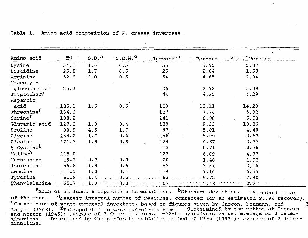
Table 1. Amino acid composition of N. crassa invertase.
Amino acid S.D.b ' S.E.M.C Intearal^ Percent YeastePercentLysine 54.1 1.6 0.5 55 3.95 5.37Histidine 25.8 1.7 0.6 26 2.04 1.53Arginine 52.6 2.0 0.6 54 4.65 2.94N-acetyl-glucosamine^ 25.2 26 2.92 5.39
Tryptophan^ 44 4.35 4.29Asparticacid 185.1 1.6 0.6 189 12.11 14.29
Threonine-^ 134.6 137 7.74 5.92Serine^ 138.2 141 6.80 6.93Glutamic acid 127.6 l.ti 0.4 130 9.33 ■ 10.36Proline 90.9 4.6 1.7 93 . 5.01 4.40Glycine 154.2 1.7 0.6 .158' 5.00 2,83Alanine 121.3 1.9 0.8 124 4.87 3.37h Cystine1 13 0.71 0.36Valine*1 119.0 122 6.69 4.77Methionine 19.3 0.7 0.3 20 1.46 1.92Isoleucine 55. 8 1.9 0.6 57 3.61 3.16Leucine 111.5 1.0 0.4 114 7.16 6.55Tyrosine 61.8 . . 1.4.... ...0.5...... ..... 63 ...... .... 5.72 7.40Phenylalanine ' 65.7 • ■ 1.0 ‘ '. . . .Qv3 . . . . . . ..... 67 •::••• .... 5.48 • •........8.21
aMean of at least 6 separate determinations. ^Standard deviation. cStandard error of the mean. ^Nearest integral number of residues, corrected for an estimated 97.9% recovery. eComposition of yeast external invertase, based on figures given by Gascon, Neumann, and Lampen (1968). ^Extrapolated to zero hydrolysis time. ^Determined by the method of Goodwinand Morton (1946) ; average of 3 determinations. n 72-hr hydrolysis -value; average of 3 determinations. ^Determined by the performic oxidation method of Hirs (1967a); average of 2 determinations .
ou
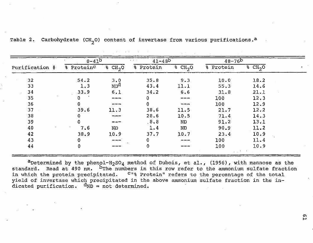
Table 2. Carbohydrate (CH^O) content of invertase from various purifications.3
Purification #0-41b 41-48b 48-76b
•% Proteinc % c h 2q % Protein % c h 2o % Protein % c h 2o
32 54.2 3.0 35. 8 9.3 10.0 18.233 1.3 NDa 43.4 11.1 55.3 14.634 33.9 6.1 34.2 6.6 31.8 21.135 0 — ™ 0 --- 100 12.336 0 --- 0 --- 100 12.937 39.6 11.3 38,6 11.5 21.7 12.238 0 --- 28.6 10.5 71.4 14.339 0 --- .8.8 ND 91.2 13.140 7.6 ND 1.4 ND 90.9 11.242 38.9 10.9 37.7 10.7 23.4 10.943 0 --- ■ 0 --- 100 11.444 0 0 100 10.9
aDetermined by the phenol-H2S04 method of Dubois, et al., (1956), with mannose as the standard. Read at 490 nm. bThe numbers in this row refer to the ammonium sulfate fraction in which the protein precipitated. c "% Protein" refers to the percentage of the total yield of invertase which precipitated in the above ammonium sulfate fraction in the indicated purification. - not determined.
<y> I—*
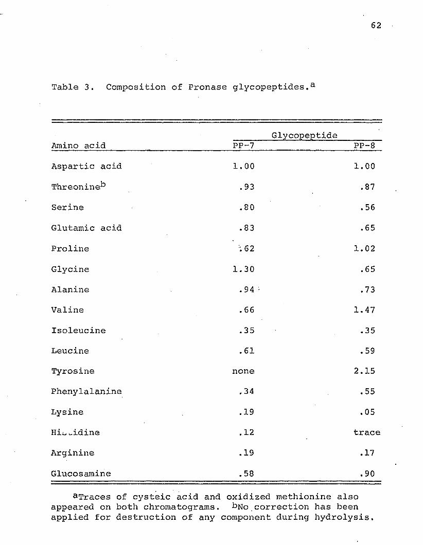
62
Table 3. Composition of Pronase glycopeptides.a
Amino acid PP-7Glycopeptide
PP-8Aspartic acid 1.00 1.00Threonine^ .93 .87
Serine .80 .56
Glutamic acid .83 .65
Proline :62 1.02Glycine 1.30 .65
Alanine .94 • .73
Valine .66 1.47
Isoleucine .35 .35
Leucine .61 .59
Tyrosine none 2.15
Phenylalanine .34 .55
Lysine .19 .05
Hi;_ widine .12 trace
Arginine .19 .17
Glucosamine .58 .90
aTraces of cysteic acid and oxidized methionine also appeared on both chromatograms. bN0 correction has been applied for destruction of any component during hydrolysis.
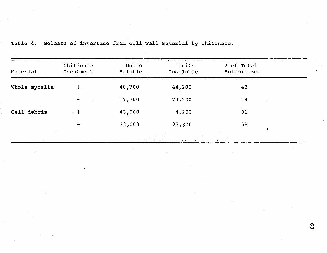
Table 4. Release of invertase from cell wall material by chitinase.
MaterialChitinaseTreatment
Units . Soluble
UnitsInsoluble
% of Total Solubilized
Whole mycelia + 40,700 44,200 48
- 17,700 74,200 19
Cell debris + 43,000 4,200 91
- 32,000 25,800 55 »
cr>u>
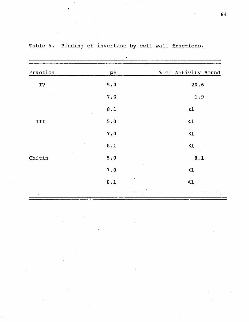
64
Table 5. Binding of invertase by cell wall fractions.
Fraction PH % of Activity Bound
IV 5.0 20.67.0 1.9
8.1 <1III 5.0 <1
7.0 <18.1 <1
Chitin 5.0 8.17.0 <18.1 <1
65
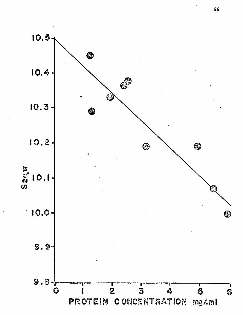
S20,W
66
10.5
10.4-
10.3-
10. 2 -
1 0 . 1 -
10.0
1 2 3 4 3P R O T E I N C O N C ENTR A TIO N m g / m l
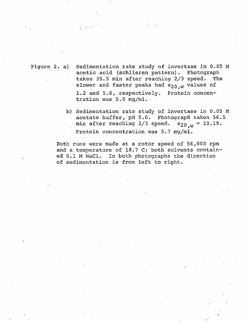
Figure 2. a) Sedimentation rate study of invertase in 0.05 Macetic acid (schlieren pattern). Photograph
. . taken 35.5 min after reaching 2/3 speed. Theslower and faster peaks had S2o,w values of1.2 and 5.6, respectively. Protein concentration was 5.0 mg/ml.
b) Sedimentation rate study of invertase in 0.05 M acetate buffer, pH 5.0. Photograph taken 56.5 min after reaching 2/3 speed. S20 w = 10.19.Protein concentration was 5.7 mg/ml.
Both runs were made at a rotor speed of 56,000 rpm and a temperature of 18.7 C? both solvents contained 0.1 M NaCl. In both photographs the direction of sedimentation is from left to right.
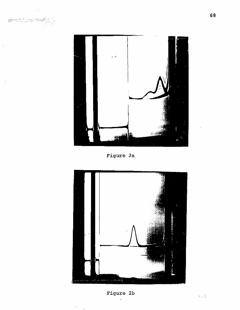
Figure 2b
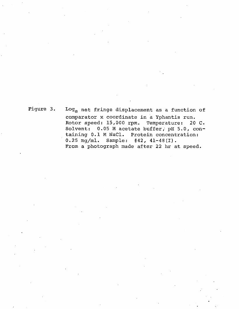
Figure 3. L°ge net fringe displacement as a function ofcomparator x coordinate in a Yphantis run. Rotor speed: 15,000 rpm. Temperature: 20 C.Solvent: 0.05 M acetate buffer, pH 5.0, containing 0.1 M NaCl. Protein concentration: 0.25 mg/ml. Sample: #42, 41-48(1).From a photograph made after 22 hr at speed.
70
6. 5-ifr-zi a
2m o <©. 0 -i £L CO
UJ©z 5.5'I”fcj
©o©
5.0"
4.5 ”f31.2 31.8 .0 32.2
X- COORDINATE' mm
Figure 4 a) Behavior of invertase on Sephadex G-100 in 0.05 M acetic acid.
b) Rerun of fractions indicated by bar in Figure 4a under same conditions. See text for further details.
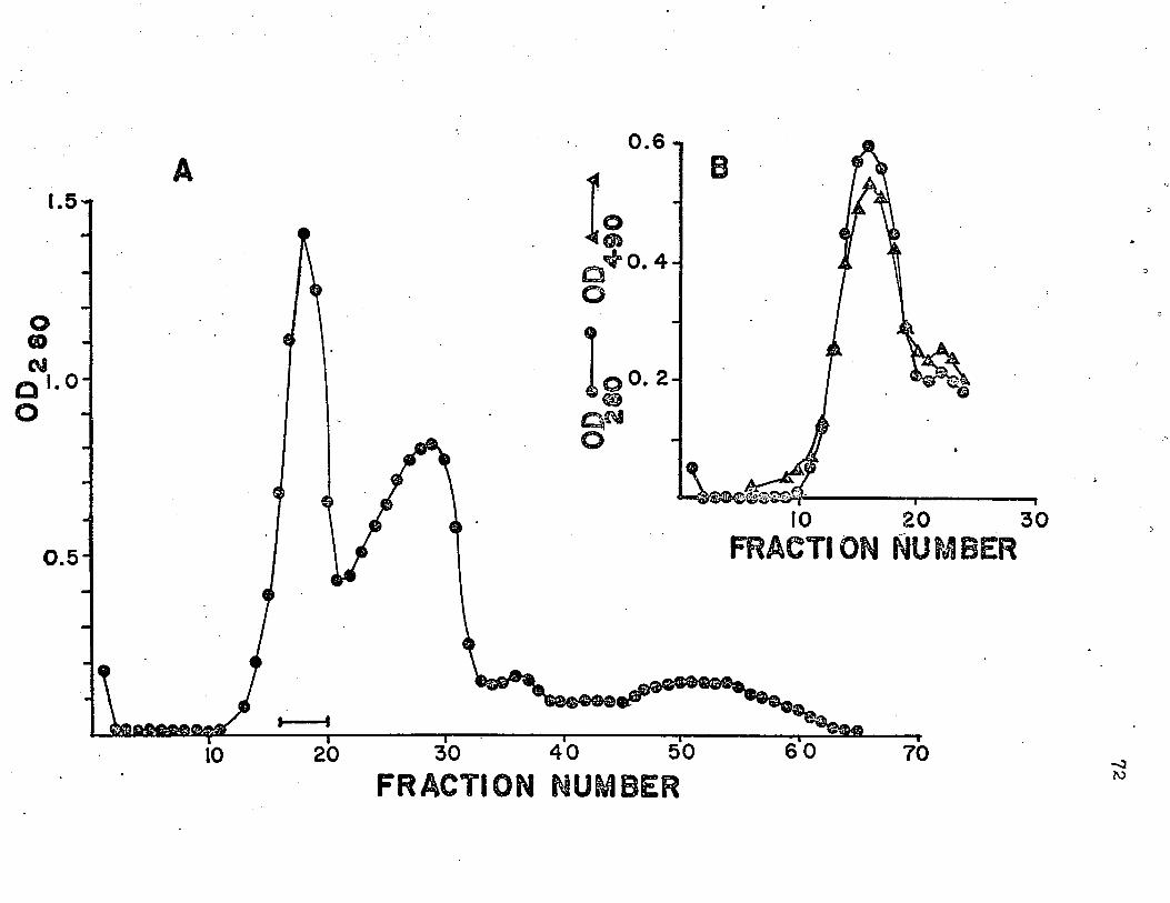
0.6 t
1.5-r
OQCM1.0
0 .5
&0. 4
10 20 30FRACTION NUMBER
FRACTION NUMBER-n3W
CHAPTER V
SUMMARY
In the opinion of the author the most striking
overall impression to emerge from the work described in
this dissertation is that of the similarity between yeast
(external) and Neurospora invertase, especially in a quali
tative sense. Both enzymes are mannan-proteins; both
presumably have the same type of protein-oligosaccharide
linkage; both are cell wall associated; they show impressive
overall similarities in amino acid composition. Many other
similarities, such as response to inhibitors, substrate
specificity, and variability in hexose content, have been
brought to light in this and in previous work.
This work appears definitely to have established the
tetrameric nature of the invertase molecule. Its amino acid
composition (with the probable exception of cysteine content)
has been determined, as has its molecular weight and s20,w*The behavior of the enzyme under dissociating conditions
has been partially explored. Many of its other characteristics
have been investigated. Of special interest are the data
74
obtained on the carbohydrate content of the enzyme. As
pointed out earlier, such data are not plentiful where
microbial glycoproteins are concerned. This enzyme
contains no sugar residues not previously found in glyco
proteins (Neuberger, Marshall, and Gottschalk, 1966).
•Although no direct evidence was obtained concerning
the number of oligosaccharide chains present on the inver
tase molecule, the calculated number of glucosamine residues
(i.e., 26; see Table 1) sets an upper limit upon this number
if glucosamine-to-asparagine bonding does indeed represent the
oligosaccharide attachment point in this glycoprotein. It
would not be surprising to find a smaller number of
oligosaccharide chains, however, since glucosamine has also
been found to occur internally in glycoprotein oligosaccharide
chains (see for example Heller and Lawrence, 1970; Scocca
and Lee, 1969).
Many important aspects of the structure of invertase
do, of course, remain to be clarified. Among these, the
number of types of polypeptide subunits comprising the mole
cule is especially important. The number of kinds of
oligosaccharide chains present and the composition and struc
ture of each, the proof of the nature of the carbohydrate-
protein linkage, and the determination of the amino acid
sequence of highly purified glycopeptides (especially in
the immediate neighborhood of the sugar-peptide bond) are also
problems worthy of immediate attention. In view of the
paucity of data relevant to the question of how oligosaccharide
attachment sites are identified, the last named problem seems
to merit considerable priority.
LITERATURE CITED
LITERATURE CITED
Arnold, W. N. 1969. Heat inactivation kinetics of yeast/?-fructofuranosidase. A polydisperse system. Biochem. Biophys. Acta 178 :347-353.
Ashwell, G. 1966. New colorimetric methods of sugar analysis, p. 8 5-9 5. In E. F. Neufeld and V. Ginsburg (eds.),Methods in enzymology, vol. VIII. Academic Press,New York.
Babul, J . , and E. Stellwagen. 1969. Measurement of protein concentration with interference optics. Anal. Biochem. 28:216-221.
Badin, J . , C. Jackson and M. Schubert. 1953. Improved method for determination of plasma polysaccarides with tryptophan. Proc. Soc. Exp. Biol. Med. 84 :288-291.
Bartnicki-Garcia, S. 1968. Cell wall chemistry, morphogenesis, and taxonomy of fungi. Ann. Rev. Microbiol. 22:87-108.
Cohn, E. J . , and J. T. Edsall. 1943. Proteins, amino acids, and peptides as dipolar ions. p. 370. Reinhold,New York.
Colvin, H. J . , Jr. 1969. Structural studies on invertase from Neurospora crassa. Ph.D. Thesis. Louisiana State University, Baton Rouge, Louisiana.
Dubois, M . , K. Gilles, J. Hamilton, P. Rebers, and F. Smith. 1956. Colorimetric method for determination of sugars and related substances. Anal. Chem. 28:350.
Eberhart, B. 1961. Exogenous enzymes of Neurospora conidia and mycelia. J. Cell. Comp. Physiol. 58:11-16.
Eilers, F. , J. Allen, E. Hill and A. Sussman. 1964. Localization of disaccaridases in extracts of Neurospora after electrophoresis in polyacrylamide gels. J.Histochem. Cytochem. 12:448-450.
Ellman, G. 1959. Tissue sulfhydryl groups. Arch. Biochem. Biophys. 82:70-77.
Eylar, E. 1965. On the biological role of glycoproteins.J. Theoret. Biol. 10:89-113.
Gascon, S., and J. O. Lampen. 1968. Purification of the internal invertase of yeast. J. Biol. Chem. 243:1567- 1572.
Gascon, S., N. Neumann and J. 0. Lampen. 1968. Comparative study of the properties of the purified internal and external invertases from yeast. J. Biol. Chem. 243: 1573-1577.
Gibbons, R. A. 1966. Physico-chemical methods for the determination of the purity, molecular size and shape of glycoproteins, p. 61. Iri A. Gottschalk (ed.), Glycoproteins. Elsevier, Amsterdam.
Goodwin, T . , and R. Morton. 1946. The spectrophotometric determination of tyrosine and tryptophan in proteins. Biochem. J. 40:628.
Gottschalk, A. 1969. Biosynthesis of glycoproteins and its relationship to heterogeneity. Nature 222:452-454.
Gratzner, H . , and D. Sheehan. 1969^ Neurospora mutant exhibiting hyperproduction of amylase and invertase.J. Bacteriol. 97:544-549.
Hagopian, A., and E. Eylar. 1969a. Glycoprotein biosynthesis: the solubilization of glycosyl transferasesfrom membranes of Hela cells and bovine submaxillary glycoproteins. Arch. Biochem. Biophys. 129^:447-455.
Hagopian, A . , and E. Eylar. 1969b. Glycoprotein biosynthesis: the purification and characterization of apolypeptide:N-acetylgalactosaminyl transferase from bovine submaxillary glands. Arch. Biochem. Biophys. 129:515-524.
Heller, J . , and M. Lawrence. 1970. Structure of the glyco- peptide from bovine visual pigment 500. Biochemistry 9:864-869.
79
Hirs, C. 1967a. Determination of cystine as cysteic acid, p. 59-62. Iri C. Hirs (ed.), Methods in enzymology, vol. XI. Academic Press, New York.
Hirs, C. 1967b. Reduction and S-carboxymethylation of proteins, p. 199-203. In C. Hirs (ed.), Methods in enzymology, vol. XI. Academic Press, New York.
Huotari, F . , T. Nelson, F. Smith and S. Kirkwood. 1968. Purification of an exo-^-D-(l— > 3)-glucanase from Basidi- omycete species QM 806. J. Biol. Chem. 243:952-956.
Islam, M . , and J. O. Lampen. 1962. Invertase secretion and sucrose fermentation by Saccharomyces cerevisiae pro- .toplasts. Biochem. Biophys. Acts 58:294-302.
Kawahara, K . , and C. Tanford. 1966. Viscosity and density of aqueous solutions of urea and guanidine hydrochloride.J. Biol. Chem. 241:3228-3232.
Lampen, J. O. 1968. External enzymes of yeast: their natureand formation. Antonie van Leeuwenhoek 3 4 :1-18.
Leach, S., and E. Parkhill. 1955. p. 92. Proc. Intern. Wool Textiles Res. Conf. Australia, vol. C.
Lehnhardt, W . , and R. J. Winzler. 1968. Determination of neutral sugars in glycoproteins by gas-liquid chromatography. J. Chromatog. 34_:471-479.
Lineback, D. 1968. The carbohydrate-polypeptide linkages in a fungal glucoamylase. Carbohyd. Res. 7_: 106-108.
Lowry, 0., N. Rosebrough, A. Farr and R. Randall. 1951. Protein measurement with the Folin phenol reagent. J. Biol. Chem. 193:265-275.
Mahadevan, P., and E. Tatum. 1965. Relationship of the major constituents of the Neurospora crassa cell wall to wild-type and colonial morphology. J. Bacteriol. 9 0 : 1073-1081.
Mahadevan, P., and E. Tatum. 1967. Localization of structural polymers in the cell wall of Neurospora crassa. J.Cell Biol. 35:295-302
80
Manocha, M . , and J. Colvin. 1967. Structure and composition of the cell wall of Neurospora crassa. J. Bacteriol.94^ 202-212.
Marzluf, G . , and R. L. Metzenberg. 1967. Studies on the functional significance of the transmembrane location of invertase in Neurospora crassa. Arch. Biochem. Biophys. 120 :487-496.
Metzenberg, R. L. 1963a. The localization of /)-fructofurano- sidase in Neurospora. Biochem. Biophys. Acta 77:455-465.
Metzenberg, R. L. 1963b. 'The purification and properties of invertase of Neurospora. Arch. Biochem. Biophys. 100: 503-511.
Metzenberg, R. L. 1964. Enzymatically active subunits of Neurospora invertase. Biochem. Biophys. Acta 89:291-302.
Neuberg, C. 194 6. Invertase. Sugar Research Foundation, Inc. Science Report Series No. 4.
Neuberger, A., A. Gottschalk and R. Marshall. 1966. Carbohy- drate-peptide linkages in glycoproteins and methods for their elucidation, p. 273-295. In A. Gottschalk (ed.), Glycoproteins. Elsevier, Amsterdam.
Neuberger, A., R. Marshall and A. Gottschalk. 1966. Some aspects of the chemistry of the component sugars of glycoproteins, p. 158. In A. Gottschalk (ed.), Glycoproteins. Elsevier, Amsterdam.
Neumann, N . , and J. O. Lampen. 1967. Purification and properties of yeast invertase. Biochemistry £:468-475.
Neumann, N . , and J. 0. Lampen. 1969. The glycoprotein structure of yeast invertase. Biochemistry 3552-3556.
Pazur, J . , K. Kleppe and E. Ball. 1963. The glycoprotein nature of some fungal carbohydrases. Arch. Biochem.Biophys. 103:515-516.
Potgieter, H., and M. Alexander. 1965. Polysaccharide components of Neurospora crassa hyphal walls. Can. J. Microbiol. 11:122-125.
81
Pressey, R. 1968. Inhibition of invertases by pyridoxal and its analogues. Biochem. Biophys. Acta 159:414-416.
Sargent, M . , and D. 0. Woodward, 1969. Gene-enzyme relationships in Neurospora invertase. J. Bacteriol. 97 :867-872.
Scocca, J., and Y. C. Lee. 1969. The composition and structure of the carbohydrate of pineapple stem bromelain. J.Biol. Chem. 244:4852-4863.
Shetlar, M . , J. Foster and M. Everett. 1948. Determination of serum polysaccharides by the tryptophane reaction. Proc. Soc. Exp. Biol. Med. 67:125.
Smith, I. 1960. Sugars and related compounds, p. 252. In I. Smith (ed.), Chromatographic and electrophoretic techniques, vol. I. William Heinemann Medical Books,Ltd., London.
Soodak, M. 1966. Leloir coenzymes, cell membranes, and protein synthesis, p. 459-479. Iri N. Kaplan and E. Kennedy (eds.), Current aspects of biochemical energetics. Academic Press, New York.
Spackman, D . , W. Stein and S. Moore. 1958. Automatic recording apparatus for use in the chromatography of amino acids. Anal. Chem. 30:1190-1206.
Spiro, R. G. 1966. Analysis of sugars found in glycoproteins, p. 16-17. In E. Neufeld and V. Ginsburg (eds.), Methods in enzymology, vol. VIII. Academic Press, New York.
Svedberg, T. , and K. O. Pedersen. 1940. The ultracentrifuge. Appendices III and IV. Oxford University Press, London.
Trevithick, J. R . , and R. L. Metzenberg. 1964. The invertase isozyme formed by Neurospora protoplasts. Biochem.Biophys. Res. Comm. 16:319~325.
Trevithick, J. R . , and R. L. Metzenberg..1966a. Molecular siev ing by Neurospora cell walls during secretion of invertase isozymes. J. Bacteriol. 92:1010-1015.
Trevithick, J. R . , and R. L. Metzenberg. 1966b. Genetic alteration of pore size and other properties of Neuro- ’ spora cell wall. J. Bacteriol. 92:1016-1020.
82
Van Holde, K. 1967. Sedimentation equilibrium. Fractions,No. 1. Beckman Instruments, Inc., Pala Alto.
Wilcox, P. 1967. Determination of amide residues by chemical methods, p. 63-65. In C. Hirs (ed.), Methods in enzymology, vol. XI. Academic Press, New York.
Yabuki, M. 1966. Studies on Aspergillus amstelodami. Part VI, The possible role of chitin in binding with ^-fructo- furanosidase within the cell wall of ascospores. Agr.Biol. Chem. 3 0 :A18.
Yphantis, D. 1964. Equilibrium ultracentrifugation of dilute solutions. Biochemistry 3:297-317.
VITA
Zackey Donnell Meachum, Jr. was born on 20 September,
1943, in Shreveport, Louisiana. He attended public grade
and secondary school in Ringgold, Louisiana, and graduated
from Ringgold High School in May, 1961. He received the
degree of B.S. in microbiology at Northwestern State College,
Natchitoches, Louisiana, in May, 1965, and the degree of B.S.
in mathematics at Northeast Louisiana State College, Monroe,
Louisiana, in June, 1966. He entered graduate school at
Louisiana State University, Baton Rouge, Louisiana, in
September, 19 66, and is a candidate for the Ph.D. degree in
the Department of Microbiology in August, 1970.
He is married to the former Janet Faye McMichael,
and has one daughter, Janet Donnelle, one year old.
83
EXAMINATION AND THESIS REPORT
Candidate:
Major Field:
Title of Thesis:
Z. D. Meachum, Jr.
Microbiology
Chemical and physical studies of the structure and composition of invertase from Neurospora crassa.
Approved:
Major Professor and Chairman
Dean of the Graduate School
EXAMINING COMMITTEE:
I UvAVKl'S. vj
Cjz
Date of Examination:
July 15, 1970