Embed Size (px)
Citation preview

CHEMICAL COMBINATION SCREENING

PhD Thesis – M. Farha; McMaster University – Biochemistry and Biomedical Sciences
iii
CHEMICAL-CHEMICAL COMBINATIONS AS TOOLS OF BIOLOGY AND AS ROUTES TO DRUG DISCOVERY
By MAYA A. FARHA, B.Sc.
A Thesis Submitted to the School of Graduate Studies In Partial Fulfillment of the
Requirements for the Degree of Doctor of Philosophy
McMaster University © Maya A. Farha, July 2014

PhD Thesis – M. Farha; McMaster University – Biochemistry and Biomedical Sciences
i
DOCTOR OF PHILOSOPHY (2014) McMaster University (Biochemistry and Biomedical Sciences) Hamilton, Ontario TITLE: Chemical-chemical combinations as tools of biology and as routes to drug discovery AUTHOR: Maya A. Farha, B.Sc. (McMaster University) SUPERVISOR: Dr. Eric D. Brown NUMBER OF PAGES: x, 121

PhD Thesis – M. Farha; McMaster University – Biochemistry and Biomedical Sciences
ii
Abstract
With dramatic rises in bacterial resistance both in the clinic and community, there
is an urgent need for new chemical matter and strategies. Over the years, antibacterial
drug discovery has been centered almost entirely on identifying single agents that target
essential proteins or enzymes. Recent advances in systems biology have suggested that
this paradigm may not be as therapeutically effective as combinations of drugs. Further,
chemical-chemical combinations, rather than single agents, can be used to probe
relationships between target proteins in a biological system, maximizing insight into its
functional organization. In this regard, we first demonstrate that systematic testing of
chemical-chemical combinations can yield information on the functional connections that
exist among molecular targets. Indeed, chemical-chemical interaction profiles with
various drug classes provided powerful means to elucidate the mode of action of two
uncharted compounds active against E. coli. In fact, this approach proved invaluable as a
method for target identification in all phenotype-based screening efforts that followed.
Systematic testing of combinations of chemicals can also lead to the discovery of
synergistic interactions with enhanced antibacterial activity. In this respect, two unique
screening strategies were adopted to identify novel combinations effective against drug-
resistant pathogens. The first uncovered ticlopidine, a novel adjuvant to β-lactam
antibiotics that together, synergize against MRSA through an intricate mechanism
involving the concerted synthesis of wall teichoic acid polymers and cell wall. The
second screening approach identified unique synergistic combinations through a search
for molecules that modulate each of the compensatory pathways that regulate bacterial

PhD Thesis – M. Farha; McMaster University – Biochemistry and Biomedical Sciences
iii
proton motive force. This unconventional screening approach led to the identification of
nine novel synergistic pairs effective against MRSA. Overall, this work illustrates the
powerful capacity of chemical-chemical combinations as tools to probe mechanism of
novel compounds and as routes to new drug discovery. Importantly, such combinatorial
approaches at probing systems and targeting pathogens are consonant with the modern
view of the cell.

PhD Thesis – M. Farha; McMaster University – Biochemistry and Biomedical Sciences
iv
Acknowledgments
First and foremost, I would like to thank my supervisor and mentor, Dr. Eric
Brown. Thank you for the opportunity to complete my graduate work with you and for
your confidence in my abilities as a researcher. Mostly, thank you for your unwavering
support over the years. I am truly thankful for the experience.
Thank you to my very supportive committee members, Dr. Gerry Wright and Dr.
Justin Nodwell. Gerry, I have enjoyed collaborating with you on many projects; I have
learned a great deal from you. Thank you to both for your guidance, suggestions and
support.
A special thank you to Soumaya Zlitni, my fellow chemical geneticist. We have
taken this journey together from beginning to end. I am happy and thankful to have made
a lifelong friend.
To my mother and best friend, Terri. Thank you for your endless love and tireless
support. I am certain that I would not have gotten here without you. A special thank you
to my brother, Philip. You have always been interested in hearing about my work – thank
you for listening and for your support.
To my husband, Shawn. Thank you for your encouragement during graduate
school and mostly, for the laughs in times of stress. Your support has meant the world to
me. I love you.
To my son, Daniel, you are a true blessing. There are no words to describe how
much I love you. I hope this work will one day inspire you to lead a path of innovation
and scholarship.

PhD Thesis – M. Farha; McMaster University – Biochemistry and Biomedical Sciences
v
Finally, I dedicate this work to the memory of my late father Edward Farha, who I
know would be very proud of me. This is for you.

PhD Thesis – M. Farha; McMaster University – Biochemistry and Biomedical Sciences
vi
Table of Contents Abstract .............................................................................................................................. ii Acknowledgments ............................................................................................................. iv Table of Contents .............................................................................................................. vi List of Figures ................................................................................................................. viii List of Tables ..................................................................................................................... ix List of Abbreviations ......................................................................................................... x CHAPTER ONE - Introduction ....................................................................................... 1 Antibiotic Discovery -‐ Successes and Failures ......................................................................... 2 Approaches to drug discovery ....................................................................................................... 3 Target Identification ......................................................................................................................... 5 Chemical Genetics .............................................................................................................................. 6 Challenging Erhlich’s Philosophy ................................................................................................. 7 Combination Therapy ....................................................................................................................... 8 Combination chemical genetics ................................................................................................. 12 Chemical-‐Chemical Combinations as Tools for Biology ..................................................... 12 Chemical-‐Chemical Combinations as Routes to Drug Discovery .................................... 14 Antibiotic adjuvants ....................................................................................................................... 14 Research objectives and organization of thesis ................................................................... 16 References ......................................................................................................................................... 17
CHAPTER TWO - Chemical Probes of Escherichia coli Uncovered through Chemical-Chemical Interaction Profiling with Compounds of Known Biological Activity .............................................................................................................................. 21 Preface ................................................................................................................................................ 22 Summary ............................................................................................................................................ 23 Introduction ...................................................................................................................................... 24 Results ................................................................................................................................................ 28 A screen for growth inhibitory small molecules ............................................................. 28 Combination profiling screen ................................................................................................ 28 Evaluation of uncovered synergistic interactions .......................................................... 32 Clustering of chemical-‐chemical interaction profiles .................................................... 35 MAC-‐0038968 is synergistic with sulfamethoxazole and active against dihydrofolate reductase (DHFR) ........................................................................................... 38 MAC-‐0003199 is a DNA gyrase inhibitor, uncovered through synergy with norfloxacin ................................................................................................................................... 39 Promiscuously synergistic molecules ................................................................................. 44 Discussion ..................................................................................................................................... 45
Significance ....................................................................................................................................... 49 Experimental Procedures ............................................................................................................ 50 Acknowledgements ........................................................................................................................ 53 References ......................................................................................................................................... 53

PhD Thesis – M. Farha; McMaster University – Biochemistry and Biomedical Sciences
vii
CHAPTER THREE - Inhibition of WTA Synthesis Blocks the Cooperative Action of PBPs and Sensitizes MRSA to ß-lactams ...................................................................... 57 Preface ................................................................................................................................................ 58 Abstract .............................................................................................................................................. 59 Introduction ...................................................................................................................................... 60 Results and Discussion .................................................................................................................. 64 Deletion of tarO sensitizes MRSA to β-‐lactams ................................................................ 64 Combination screening identifies ticlopidine .................................................................. 66 Characterization of the mode of action of ticlopidine ................................................... 69 A basis for the synergy among ticlopidine and ß-‐lactams ............................................ 73
Conclusions ....................................................................................................................................... 75 Methods .............................................................................................................................................. 76 Acknowledgments .......................................................................................................................... 78 References ......................................................................................................................................... 78
CHAPTER FOUR – Collapsing the Proton Motive Force to Identify Synergistic Combinations Against Staphylococcus aureus .............................................................. 83 Preface ................................................................................................................................................ 84 Summary ............................................................................................................................................ 85 Introduction ...................................................................................................................................... 86 Results ................................................................................................................................................ 89 Development of a high-‐throughput screen for alterations in PMF. ........................... 89 Follow-‐up to the HTS ................................................................................................................. 92 Uncovering agents that selectively dissipate components of PMF ............................ 93 Characterizing I1, I2 and I3, potential dissipaters of ΔΨ ............................................. 97 Characterizing D1, D2 and D3, potential dissipaters of ΔpH ....................................... 99 Characterizing I4, I5, I6 and I7: other molecules that caused an increase in DiSC3(5) fluorescence ............................................................................................................ 100 Combinations of molecules I1-‐I3 and D1-‐D3 lead to synergistic interactions ... 103 Cytotoxicity studies ................................................................................................................ 104
Discussion ....................................................................................................................................... 106 Significance .................................................................................................................................... 110 Acknowledgements ..................................................................................................................... 113 References ...................................................................................................................................... 113
CHAPTER FIVE – Future Directions and Conclusions ............................................ 116 Summary and Future Directions ............................................................................................ 117 Concluding remarks .................................................................................................................... 120 References ...................................................................................................................................... 121
PhD Thesis – M. Farha; McMaster University – Biochemistry and Biomedical Sciences
viii
List of Figures CHAPTER ONE Figure 1.1 Assessing interactions among chemical-chemical combinations……11 Figure 1.2 Mechanisms of antibiotic adjuvants………………………….......….15 CHAPTER TWO
Figure 2.1. Summary of the approach of profiling chemical-chemical interactions………………………………………………………………………29 Figure 2.2 Chemical-chemical interaction profiling screen with known bioactives………………………………………………………………………..33 Figure 2.3. Hierarchical clustering of chemical-chemical interaction profiles…………………………………………………………………….….…36 Figure 2.4. MAC-0038968 is active against DHFR…………………………….40 Figure 2.5. MAC-0003199 inhibits DNA gyrase…………………………….…43
CHAPTER THREE
Figure 3.1. Sensitivity of CA- and HA-MRSA ΔtarO deletion strains to β-lactam antibiotics……………………………………………………………………….65 Figure 3.2. Ticlopidine potentiates the activity of the β-lactam antibiotic cefuroxime against CA-MRSA USA300, but not CA-MRSA USA300 Δtar…..69 Figure 3.3. Ticlopidine inhibits wall teichoic acid biosynthesis in S. aureus…..72 Figure 3.4. Synthetic lethal interaction when targeting TarO and PBP2….……74
CHAPTER FOUR
Figure 4.1. High-throughput screen to identify dissipaters of ΔΨ and ΔpH……91 Figure 4.2. Hit compounds that dissipate ΔΨ of S. aureus……………………..95 Figure 4.3. Hit molecules that dissipate ΔpH of S. aureus………………….......96 Figure 4.4. Cellular effects of dissipaters of ΔΨ………..………...……….…...99 Figure 4.5. Cellular effects of dissipaters of ΔpH………..………...……..…...100 Figure 4.6. Combinations of dissipaters of ΔΨ and ΔpH exhibit synergistic interactions……………………………………………………………………..104 Figure 4.7. Cytotoxic effects of hit compounds on a HeLa cell line…………..106
PhD Thesis – M. Farha; McMaster University – Biochemistry and Biomedical Sciences
ix
List of Tables CHAPTER TWO
Table 2.1. Examples of molecules with similar structures and biological fingerprints. ………………………………………………………………….…37
CHAPTER THREE
Table 3.1. In vitro interactions between ticlopidine and cefuroxime in various S. aureus species……………………………………………………………..…….68

PhD Thesis – M. Farha; McMaster University – Biochemistry and Biomedical Sciences
x
List of Abbreviations ATP Adenosine triphosphate CLSI Clinical and Laboratory Standards Institute DHFR Dihydrofolate reductase DiSC3(5) 3,3'-Dipropylthiadicarbocyanine Iodide DMSO Dimethylsulfoxide DNA Deoxyribonucleic acid FIC Fractional inhibitory concentration FICI or ΣFIC Fractional inhibitory concentration Index GFP Green fluorescent protein HEPES 4-(2-hydroxyethyl)-1-piperazineethanesulfonic acid IPTG Isopropyl ß-D-1-thiogalactopyranoside LB Luria-Bertani MIC Minimum inhibitory concentration MDR Multi-drug resistant MHB Muller Hinton Broth MOA Mechanism of action MRSA Methicillin-resistant Staphylococcus aureus OD Optical density PAD Previously-approved drug PBP Penicillin-binding protein PG Peptidoglycan PMF Proton motive force RNA Ribonucleic acid TSB Tryptic soy broth VRE Vancomycin-resistant Enterococcus faecium WTA Wall teichoic acid

PhD Thesis – M. Farha; McMaster University – Biochemistry and Biomedical Sciences
1
CHAPTER ONE - Introduction
PhD Thesis – M. Farha; McMaster University – Biochemistry and Biomedical Sciences
2
Antibiotic Discovery - Successes and Failures
The introduction of β-lactam antibiotics in the 1940’s revolutionized medicine and
was undoubtedly a key factor in significantly expanding human lifespan (Bax, Mullan et
al. 2000). Since, several new classes of antibiotics, both natural products and synthetic
molecules, have been introduced. Indeed, innovation would reach its peak in the next 20
years, and antibiotic molecules, whether novel chemical structures or improvements of
existing classes, were successfully uncovered and designed at a rapid rate. Most of the
start of the 20th century was thus marked by declining mortality due to infectious disease
(Armstrong, Conn et al. 1999). This ‘golden era’ of antibiotic discovery, however, did not
last and ended abruptly in the 1960’s. In fact, discovery efforts during the next fifty years
would only lead to two novel antibiotics: linezolid and daptomycin, introduced in 2000
and 2003, respectively (Lewis 2013). What’s worse is this failure in novel discoveries
coincides with the emergence of multi-drug resistant bacteria (Boucher, Talbot et al.
2009).
The large majority of antibiotics target either the cell wall or a macromolecular
biosynthetic process (DNA, RNA or protein) within the bacterial cell (Bax, Mullan et al.
2000). Over time, however, bacteria have developed mechanisms to outsmart antibiotic
action, either by mutagenesis or by acquiring new genes from other bacteria. Resistance
mechanisms have included efflux proteins that rapidly pump antibiotics out of cells,
enzymes that modify or degrade the antibiotic molecule, rendering it inactive, and
alterations to the cellular target in order to prevent drug binding (Wright 2005).

PhD Thesis – M. Farha; McMaster University – Biochemistry and Biomedical Sciences
3
Remarkably, almost as soon as the discovery of penicillin, resistant bacteria were
isolated.
Today, emergence of multidrug-resistant bacterial pathogens has become the main
challenge facing clinicians and researchers in infectious disease medicine. Resistance has
now emerged to all classes of known antibiotics diminishing their usefulness. The crisis is
epitomized by the spread of multidrug-resistant ‘ESKAPE’ pathogens (Enterococcus
faecium, Staphylococcus aureus, Klebsiella pneumoniae, Acinetobacter baumannii,
Pseudomonas aeruginosa and Enterobacter species), highlighted by the Infectious
Diseases Society of America as causing the majority of hospital infections in the United
States (Boucher, Talbot et al. 2009). Rates of infection due to the Gram-positive
pathogens, methicillin-resistant S. aureus (MRSA) and vancomycin-resistant E. faecium
(VRE), are rapidly increasing. In fact, MRSA, which accounts for the majority of
infections, causes higher mortality than HIV/AIDS and tuberculosis combined (Klevens,
Edwards et al. 2006). The threat of MDR Gram-negative pathogens rivals that of MRSA
and VRE. Indeed, in the case of some Gram-negative bacteria, such as A. baumannii,
there are strains that are resistant to all currently available antibiotics (Higgins,
Dammhayn et al. 2010).
As therapeutic options dwindle, it is increasingly becoming clear that clinicians
will soon face a therapeutic dead end in treating certain types of bacterial infections. The
urgent need to develop novel strategies to combat MDR bacteria cannot be overstated.
Approaches to drug discovery
There are two main screening approaches to drug discovery: target-based and

PhD Thesis – M. Farha; McMaster University – Biochemistry and Biomedical Sciences
4
phenotypic screening. The former measures the effects of compounds on a purified
protein of interest in in vitro assays, whereas the latter looks at the effects that compounds
induce in cells (Zheng, Thorne et al. 2013). Advances in chemical synthesis platforms
during the 20th century have allowed progress toward more systematic approaches to drug
discovery. Indeed, synthesis of large libraries of compounds has drastically changed the
throughput of compound screening.
Drug development was once mostly driven by phenotypic screening (Swinney and
Anthony 2011). While such efforts had considerable power in discovering new
molecules that modify a disease phenotype, subsequently determining the relevant
cellular target often proved difficult. Advances in molecular biology and genomics at the
start of the 1980’s caused a shift to target-based screening, with technologies enabling the
characterization and screening of novel targets. Here, the target is defined and the
challenging task of identifying the cellular target(s) becomes obsolete. Over the next two
decades, this screening approach would in fact become the main paradigm for drug
discovery (Zheng, Thorne et al. 2013).
In the last decade, a shift back to phenotype-based screening has taken place with
the realization that target-based screening has in fact resulted in reduced success in
discovering first-in-class medicines (Swinney and Anthony 2011; Zheng, Thorne et al.
2013). While compounds identified via target-based screening may potently inhibit the
target of interest in vitro, they may not cross the bacterial envelope in an in vivo setting or
may be victim to drug efflux (Roemer and Boone 2013). There is also the possibility that
they may end up affecting other undescribed targets in vivo leading to unfavorable side

PhD Thesis – M. Farha; McMaster University – Biochemistry and Biomedical Sciences
5
effects. These factors have thus placed phenotype-based screening in a more favorable
light in more recent years. Target identification of hit compounds, however, remains a
daunting task.
Target Identification The challenge of target identification has long plagued early stage discovery
efforts and has been simply due to a paucity of systematic methods to characterize the
cellular target of small molecules (Burdine and Kodadek 2004). Classically, protein
targets have been identified biochemically using labeled or immobilized molecules. The
advent of genomics brought forth novel and powerful methodologies to investigate the
mode of action of compounds. These have included high-throughput genomic and
proteomic approaches, such as genome-scale clone sets, (e.g. gene deletions,
overexpression constructs and promoter-reporter strains) and microarrays (Barker, Farha
et al. 2010). Further, advances in genomics have made whole-genome sequencing of
drug-resistant mutants an apt method for target identification (Roemer and Boone 2013).
Although these approaches have proven useful in a number of target identification efforts,
they remain far from ideal, as most require chemical modifications of the small molecules
(Lomenick, Olsen et al. 2011), large-scale expression profiles (Fischer, Brunner et al.
2004) or the generation of pathogen-specific clone sets (Barker, Farha et al. 2010). Efforts
to further optimize and devise novel technologies for target identification are ongoing
(Kasper, Baker et al. 2009). Undoubtedly, developing reliable techniques for addressing
target identification is at the foundation of successful drug development.

PhD Thesis – M. Farha; McMaster University – Biochemistry and Biomedical Sciences
6
Chemical Genetics
The field encompassing the aforementioned small molecule libraries, phenotypic
screening and target identification is known as chemical genetics. Termed in the late
1990’s, chemical genetics is derived from classical genetics but makes use of chemical
perturbations instead of genetic knockouts. The field aims to systematically use small-
molecule techniques to understand biological processes by studying how perturbations of
protein functions affect the biological system (Stockwell 2000).
Chemical genetics presents some advantages over classical mutagenesis. The use
of small molecules to perturb protein function can be achieved with ease, the effects
being reversible, tunable, rapid and easily combined, unlike genetic perturbations, which
are effectively permanent and technically difficult (Spring 2005).
Genetic perturbations have certainly proven valuable in investigating biology and
have been made further accessible by the advent of comprehensive mutant libraries in
model organisms. Chemicals can provide information that is often complementary to
genetic perturbations. In fact, coupling the utility of small molecules with various
genomic technologies has proven a remarkable tool in adequately probing gene function,
investigating biological systems as a whole and characterizing new antibacterial targets
(Giaever, Flaherty et al. 2004; Parsons, Lopez et al. 2006; Pathania, Zlitni et al. 2009).
Resources for such studies are growing dramatically with genome-wide libraries being
constructed for various yeast and bacterial species (Lehar, Stockwell et al. 2008). Overall,
chemogenomic efforts are increasingly showing considerable promise in enabling
detailed studies of biological networks and in aiding in drug discovery efforts.

PhD Thesis – M. Farha; McMaster University – Biochemistry and Biomedical Sciences
7
Challenging Erhlich’s Philosophy
Over a century ago, Paul Erhlich set forth his hypothesis of creating 'magic
bullets' for use in the fight against human diseases (Strebhardt and Ullrich 2008). This
one-drug-one-target approach has served as the foundation of antimicrobial drug
discovery since the discovery of penicillin, inspiring the development of many powerful
therapeutics. In fact, this concept of ‘magic bullets’ that act on individual drug targets is
still the guideline being followed by much of modern drug discovery, including
antibacterial drug discovery.
Recent systems and network biology studies have, however, challenged this
paradigm, exposing its flaws in treating infection. Large-scale gene knock-out
experiments in model organisms have revealed that biological systems are very complex
and remarkably robust in the face of perturbation. Indeed, a large proportion of single-
gene knockouts by themselves exert little or no effect on the fitness or phenotype of the
organism. In E. coli, for example, only approximately 7% of the genome is essential
(Baba, Ara et al. 2006). Similarly, characterization of the yeast genome has revealed that
approximately only 18% of the genome is essential for viability (Winzeler, Shoemaker et
al. 1999). Additionally, the dispensability of a large number of yeast genes is also
dependent on other genes (Winzeler, Shoemaker et al. 1999; Giaever, Chu et al. 2002;
Costanzo, Baryshnikova et al. 2010). The scope of this complexity is vast; not only are
networks made up of many redundant functions and alternative compensatory pathways,
but it is increasingly becoming clear that most genes or proteins within the network also
have several interacting partners forming numerous functional cross-connections (Jeong,

PhD Thesis – M. Farha; McMaster University – Biochemistry and Biomedical Sciences
8
Mason et al. 2001; Ho, Gruhler et al. 2002; Butland, Peregrin-Alvarez et al. 2005;
Costanzo, Baryshnikova et al. 2010). Such observations of phenotypic robustness
indicate that modulating multiple nodes simultaneously is often required for modifying
phenotypes (Barabasi and Oltvai 2004; Csermely, Agoston et al. 2005). As such,
exquisitely selective compounds, such as Erhlich’s ‘magic bullets’, will likely result in
lower-than desired efficacy in treating disease. Certainly, the modern understanding of
the cellular architecture suggests that individual drugs may not be as effective as their
combinations.
Many successful antibiotics do, in fact, work by simultaneously modulating
multiple targets. Some examples include β-lactam antibiotics, whose antibacterial action
depends on inhibition of at least two PBPs (Denome, Elf et al. 1999); fluoroquinolone
antibiotics that target the proteins ParC and GyrA (Janoir, Zeller et al. 1996); and
cycloserine, which inhibits both pairs of alanine racemases and D-Ala-D-Ala ligases
(Prosser and de Carvalho 2013). Combination therapy should progress as an effective
strategy to overcome the staggering complexity and resilient nature of biological systems
(Lehár, Krueger et al. 2008).
Combination Therapy
As mentioned, the prevailing view in antibacterial drug discovery has largely been
antibiotic monotherapy. Combination therapy has mostly been an afterthought and was
traditionally only a result of combining existing antibiotics in an effort to improve
efficacy. More recently, this field is gaining increased attention as a result of the
emerging view of the cell’s complexity. In fact, drug combinations are slowly becoming

PhD Thesis – M. Farha; McMaster University – Biochemistry and Biomedical Sciences
9
the standard of care in antiviral therapy. For example, in anti-HIV therapy, combinations
of protease inhibitors and reverse transcriptase inhibitors are routinely used (Shafer and
Vuitton 1999). Further, the use of drug combinations is now highly sought after in cancer
chemotherapy (Bozic, Reiter et al. 2013). A number of combination therapies have in fact
shown efficacy against bacterial infections, setting a precedent for this paradigm in
antibacterial therapy. These include Augmentin®, which is a combination of the β-
lactamase inhibitor clavulanate and the second generation penicillin, amoxicillin and co-
trimoxazole, which combines two inhibitors of the folate pathway, sulfamethoxazole and
trimethoprim. Both pairs are highly used in the clinic and strengthen the thesis that
combinations will also be effective as antibacterial therapeutics.
While combination antibiotic therapy may be more complex to develop, requiring
more time and higher costs (Bonhoeffer, Lipsitch et al. 1997), its progress is warranted
given the unremitting challenges of resistance. The benefits of combination therapy over
monotherapy are numerous. Drug combinations allow a superior control of the system by
attacking it on multiple fronts, leading to better clinical efficacy (Zimmermann, Lehar et
al. 2007; Lehar, Krueger et al. 2009). With the added perturbation over monotherapy,
combinations can overcome the intrinsic robustness of the biological system in question,
by targeting compensatory pathways, for example. Combinations also allow a decrease in
dose-related toxicities by sparing the doses between two drugs needed for efficacy (Keith,
Borisy et al. 2005). Finally, combination therapy can potentially reduce or prevent the
emergence of resistance (Craig and Salamone 1988; Keith, Borisy et al. 2005; Chait,
Craney et al. 2007). The underlying thesis is that therapies that inhibit more than one

PhD Thesis – M. Farha; McMaster University – Biochemistry and Biomedical Sciences
10
target might delay or decrease the ability of the pathogen to accumulate simultaneous
mutations affecting those multiple targets (Walsh 2003). Antituberculous chemotherapy
provides a classic example where drug combinations have successfully prevented the
emergence of resistant organisms (Johnston and Wildrick 1974). Efficacy, toxicity and
resistance are limitations often faced my monotherapies and have indeed been major
sources of attrition in drug development. As a result, in recent years, research groups
have increased their efforts towards the search for multicomponent therapeutics via
systematic screens for drug combinations (Borisy, Elliott et al. 2003; Fitzgerald,
Schoeberl et al. 2006; Onyewu and Heitman 2007; Zimmermann, Lehar et al. 2007).
Classically, interactions among combinations of drugs have been categorized in
one of three types: additive, synergistic or antagonistic (Keith, Borisy et al. 2005). The
expected null interaction, whereby indifference upon combination is observed, is called
additivity. Synergy occurs when the combination of the two drugs has a greater effect
than with either drug alone. Antagonism describes a combination that has a smaller effect
than either drug alone.
The most commonly used method to assess the effect of a combination in a
laboratory setting is the checkerboard method (Figure 1-1a). In this assay, a combination
is tested in all possible permutations of serially diluted drug concentrations. The effect is
calculated by comparing a combination’s response to those of its single agents.
Deviations from additivity can be assessed visually on an isobologram (Figure 1-1b) or
numerically as a mathematical representation of the pattern seen in the isobologram, with
a fractional inhibitory concentration index (FICI) (Figure 1-1c) (Lambert and Lambert

PhD Thesis – M. Farha; McMaster University – Biochemistry and Biomedical Sciences
11
2003). This index is most often determined using the minimum inhibitory concentration
(MIC), the lowest concentration of drug that inhibits measurable growth of the bacterial
culture. The FICI is calculated as the summed effective concentrations of the combined
drugs at their MIC divided by the sum of the single-drug concentrations at their MIC.
Typically, an index of less than 0.5 defines a synergistic interaction, whereas an index
larger than or equal to 2 defines an antagonistic combination. An additive effect occurs
when the sum of these fractions equates 1.
Figure 1-1. Assessing interactions between two combined drugs, Drug A and Drug B. (a) The checkerboard method is the most widely used technique to test combinations of drugs in vitro. Here, grey shading represents bacterial growth. This method allows multiple concentrations, proportional to the MICs of the drugs, to be tested, in all possible permutations. The resulting response shape reveals whether the interactions is (i) additive (ii) synergistic or (iii) antagonistic. (b) An isobologram provides a visual assessment of the interaction. Plotted are individual dose response curves for Drugs A and B. Points for a desired effect (eg. MIC) are connected with a straight line, representing the line of additivity (dashed line). Any combination that falls below this line (red), such that the effect is attained with smaller doses of the two drugs, is synergistic. When the combination is found above the line (black), the combination is antagonistic. (c) The FICI is a mathematical representation of the isobologram and is calculated as shown.
Statistical models often accompany the latter method of representing drug
combinations. Two different reference models for defining additivity exist: Loewe
additivity and Bliss Independence (Fitzgerald, Schoeberl et al. 2006). The former concept
is based on the assumption that the components of the combination have a similar mode
of action or similar target. As such, the combination effect remains constant when the first

PhD Thesis – M. Farha; McMaster University – Biochemistry and Biomedical Sciences
12
drug is gradually replaced by increasing amounts of the second drug; hence the effect is
the same at different combined concentrations. The Bliss Independence model assumes
the opposite and is based on the notion of independence. The two drugs act
independently from one another with the assumption that the site of action of the
combined drugs is different from the sites that each of the individual compounds acts on.
Of the latter two models, the most relevant for medical applications is that of Loewe
additivity (Chou and Talalay 1984).
Combination chemical genetics Two decades following the conception of the field chemical genetics, a new field
has been coined as ‘combination chemical genetics’ (Lehar, Stockwell et al. 2008),
referring to the systematic testing of multiple perturbations involving chemical agents.
Given the increased appreciation of the complexity of biological systems and the wide
recognition that combination therapy may be a promising therapeutic approach,
combination chemical genetics is a field that allows the systematic application of
combinations of perturbants as tools to study biological systems and as routes to facilitate
drug discovery.
Chemical-Chemical Combinations as Tools for Biology
Since proteins rarely function in isolation within the cell, interactions from
combinations of chemical perturbants can be useful tools to gain biological insight. To
date, the focus of most combination perturbation studies has been on exploring drug
sensitivities across large sets of genetic knockouts in various model organisms (Giaever,
Flaherty et al. 2004; Lum, Armour et al. 2004; Liu, Tran et al. 2010). More recently,

PhD Thesis – M. Farha; McMaster University – Biochemistry and Biomedical Sciences
13
studies are increasingly turning to the systematic testing of purely chemical combinations
to obtain biological information.
Analogous to epistasis among genetic mutations, responses to a combination of
perturbants can reveal the underlying connectivity among their cellular targets. Indeed,
similar to genetic perturbations, two drugs may have no interaction, or they may exhibit
synergy or antagonism, increasing or suppressing their individual effects. Thus, in
principle, two compounds at sub-lethal concentrations that act synergistically likely share
some underlying connectivity in their cellular targets. Similarly, there is likely a
mechanistic reasoning underlying an antagonistic interaction observed between two
compounds. Conversely, an additive interaction would be expected between two small
molecules in a neither non-redundant nor associated pathway.
In this regard, a recent study using simulations of bacterial pathways has shown
that dose matrix responses to combinations of chemical probes can be used to determine
mechanistic relationships between their targets (Lehar et al. 2007). Depending on the
response model obtained from a pair of perturbants, the underlying connectivity of their
molecular targets and overall pathway topologies could be elucidated. This study and
others (Yeh, Tschumi et al. 2006; Farha, Leung et al. 2013) have highlighted the wealth
of functional information that can be obtained from chemical-chemical combinations.
Further, combination studies using chemicals can provide a means to reveal the molecular
target of a drug and aid in understanding its mechanism of action (Yeh, Tschumi et al.
2006; Farha and Brown 2010).

PhD Thesis – M. Farha; McMaster University – Biochemistry and Biomedical Sciences
14
Overall, connectivities within biological pathways in the cell can be of various
types. As such, the use of two small molecules can adequately probe such inter-
relationships and yield rich information about the respective molecular targets. Further,
the use of chemicals as a means to infer mechanisms of action of uncharted molecules
presents some advantages over the more recent genomic approaches used for target
identification. Indeed, the approach is independent of the organism under study and can
be universally used to probe target connectivity.
Chemical-Chemical Combinations as Routes to Drug Discovery
In addition to mapping out connections within biological systems, the systematic
testing of chemical-chemical combinations can uncover synergies with beneficial
therapeutic responses. Recent studies have shown that cell-based screens can successfully
identify unexpected synergies with enhanced antibacterial activity (Borisy, Elliott et al.
2003; Ejim, Farha et al. 2011; Tan, Hu et al. 2012; Farha, Leung et al. 2013). An added
advantage of combination screening is the search space increases dramatically when
drugs are used in combination. Indeed, systematic drug combination screening, among
even a modest number of compounds, gives way to a vast number of possible pairwise
combinations; a valuable diversity that far exceeds that from searches for single agents.
Antibiotic adjuvants
An emerging approach to identify efficacious synergistic pairs is that of antibiotic
adjuvants, a strategy rationally devised to overcome the challenges of antibiotic resistance
and the failure of new drug discovery. An adjuvant is a potentiator of antibiotic activity
that when in combination with antibiotics, enhances their antimicrobial activity.

PhD Thesis – M. Farha; McMaster University – Biochemistry and Biomedical Sciences
15
Antibiotic adjuvant therapies can include combinations of antibiotics,
combinations of antibiotic and non-antibiotic compounds, as well as combinations of
molecules that inhibit antibiotic resistance mechanisms. Adjuvants can restore antibiotic
activity through several mechanisms (Kalan and Wright 2011). The adjuvant can weaken
the bacteria themselves making them more vulnerable to antibiotics. This can be
achieved either by inhibiting a related step within the targetted biological pathway (Figure
1-2a), or by increasing the uptake of the antibiotic (Figure 1-2b). Further, adjuvants can
interfere with mechanisms of antibiotic resistance, such as blocking drug efflux (Figure 1-
2c) or by inhibiting resistance enzymes that modify or degrade the antibiotic (Figure 1-
2d). Finally, adjuvants can alter the physiological state of a microbe, rendering it more
susceptible to antibiotic action (Figure 1-2e)
Figure 1-2. Strategies for potentiating antibiotic (green circle) activity with antibiotic adjuvants (purple triangle). (a) Addition of an adjuvant that targets a sequential or orthogonal step to the antibiotic within a pathway can enhance its activity. (b) Addition of an adjuvant can increase the uptake of an antibiotic into the cell by overcoming the physical barrier. (c) Adjuvants can block resistance mechanisms of drug efflux, allowing higher concentrations of antibiotic inside the cell. (d) Adjuvants can inhibit resistance enzymes that either modify or degrade the antibiotic. (e) Adjuvants can alter the physiological state of a microbe rendering it more susceptible to antibiotics e.g. by targeting an enzyme that stimulates production of reactive oxygen species, and thus enhance the activity of the antibiotic, which also leads to the formation of lethal reactive oxygen species.

PhD Thesis – M. Farha; McMaster University – Biochemistry and Biomedical Sciences
16
Large-scale genomic studies have shown the potential of identifying novel targets
for antibiotic adjuvants. A screen of a collection of 4,000 non-essential gene deletions in
E. coli for chemical hypersensitivity revealed a large subset of genes, involved in all
major cellular functions that influence drug susceptibility and therefore act as candidate
adjuvant targets (Liu, Tran et al. 2010). Another recent study looked at genes that, when
partially depleted, can re-sensitize MRSA to β-lactam antibiotics (Lee, Jarantow et al.
2011). Here, a number of genes, most implicated in cell wall and cell division, were
identified as potential adjuvant targets to β-lactam antibiotics. Such studies reinforce the
interconnectedness of cellular pathways and present endless possibilities for potential
targets of antibiotic adjuvants.
Potentiating antibiotic activity with adjuvants is proving an effective method for
discovering new opportunities of pairs of agents with improved antibacterial activity
(Ejim, Farha et al. 2011; Tan, Therien et al. 2012; Brynildsen, Winkler et al. 2013; Farha,
Leung et al. 2013). Adjuvant strategies could revolutionize the use of antibiotics, not only
by reducing levels of antimicrobial resistance but also by establishing a means to preserve
the efficacy of existing antibiotics.
Research objectives and organization of thesis
The objectives of the research in this thesis were to employ chemical-chemical
combinations to extract mechanistic information for uncharted antibacterial molecules
and to uncover novel therapeutic antibacterial combinations effective against drug-
resistant pathogens. Overall, the thesis is based on the hypothesis that combinations of
chemicals offer distinct advantages over the use of monotherapy, both as probes of

PhD Thesis – M. Farha; McMaster University – Biochemistry and Biomedical Sciences
17
biology and as therapeutics. This stems from the growing realization that the most useful
paradigm to target bacterial processes is no longer via a single molecule in a relevant
pathway, but instead with a set of compounds that can cooperate to produce an effective
response.
The specific aims of this research were two-fold. First, a methodology to elucidate
the mode of action of uncharted chemicals was developed using chemical-chemical
combinations, with the purpose of aiding in target identification following cell-based
screening. Chapter 2 describes this approach and its subsequent use in characterizing the
mode of action of two novel antibacterial compounds active against E. coli. Next, two
screening strategies were devised to uncover new synergistic combinations. Chapter 3
describes the identification and detailed mechanism of synergy of a novel antibacterial
combination effective against MRSA, made up of ticlopidine and β-lactam antibiotics.
Chapter 4 describes a unique approach at identifying synergistic interactions against drug-
resistant pathogens. By targeting the two opposing forces that control the proton motive
force across the bacterial membrane, synergy was observed and as such, cytotoxicity
averted. Finally, Chapter 5 discusses the future of chemical-chemical combinations as
probes of biology and as therapeutics.
References Armstrong, G. L., L. A. Conn, et al. (1999). "Trends in infectious disease mortality in the United
States during the 20th century." JAMA : the journal of the American Medical Association 281(1): 61-66.
Baba, T., T. Ara, et al. (2006). "Construction of Escherichia coli K-12 in-frame, single-gene knockout mutants: the Keio collection." Molecular systems biology 2: 2006 0008.

PhD Thesis – M. Farha; McMaster University – Biochemistry and Biomedical Sciences
18
Barabasi, A. L. and Z. N. Oltvai (2004). "Network biology: understanding the cell's functional organization." Nature reviews. Genetics 5(2): 101-113.
Barker, C. A., M. A. Farha, et al. (2010). "Chemical genomic approaches to study model microbes." Chemistry & biology 17(6): 624-632.
Bax, R., N. Mullan, et al. (2000). "The millennium bugs--the need for and development of new antibacterials." International journal of antimicrobial agents 16(1): 51-59.
Bonhoeffer, S., M. Lipsitch, et al. (1997). "Evaluating treatment protocols to prevent antibiotic resistance." Proceedings of the National Academy of Sciences of the United States of America 94(22): 12106-12111.
Borisy, A. A., P. J. Elliott, et al. (2003). "Systematic discovery of multicomponent therapeutics." Proceedings of the National Academy of Sciences of the United States of America 100(13): 7977-7982.
Borisy, A. A., P. J. Elliott, et al. (2003). "Systematic discovery of multicomponent therapeutics." Proc Natl Acad Sci U S A 100(13): 7977-7982.
Boucher, H. W., G. H. Talbot, et al. (2009). "Bad bugs, no drugs: no ESKAPE! An update from the Infectious Diseases Society of America." Clinical infectious diseases : an official publication of the Infectious Diseases Society of America 48(1): 1-12.
Bozic, I., J. G. Reiter, et al. (2013). "Evolutionary dynamics of cancer in response to targeted combination therapy." eLife 2: e00747.
Brynildsen, M. P., J. A. Winkler, et al. (2013). "Potentiating antibacterial activity by predictably enhancing endogenous microbial ROS production." Nature biotechnology 31(2): 160-165.
Burdine, L. and T. Kodadek (2004). "Target identification in chemical genetics: the (often) missing link." Chemistry & biology 11(5): 593-597.
Butland, G., J. M. Peregrin-Alvarez, et al. (2005). "Interaction network containing conserved and essential protein complexes in Escherichia coli." Nature 433(7025): 531-537.
Chait, R., A. Craney, et al. (2007). "Antibiotic interactions that select against resistance." Nature 446(7136): 668-671.
Chou, T. C. and P. Talalay (1984). "Quantitative analysis of dose-effect relationships: the combined effects of multiple drugs or enzyme inhibitors." Advances in enzyme regulation 22: 27-55.
Costanzo, M., A. Baryshnikova, et al. (2010). "The genetic landscape of a cell." Science 327(5964): 425-431.
Craig, W. A. and F. R. Salamone (1988). "Do antibiotic combinations prevent the emergence of resistant organisms?" Infection control and hospital epidemiology : the official journal of the Society of Hospital Epidemiologists of America 9(9): 417-419.
Csermely, P., V. Agoston, et al. (2005). "The efficiency of multi-target drugs: the network approach might help drug design." Trends in pharmacological sciences 26(4): 178-182.
Denome, S. A., P. K. Elf, et al. (1999). "Escherichia coli mutants lacking all possible combinations of eight penicillin binding proteins: viability, characteristics, and implications for peptidoglycan synthesis." Journal of bacteriology 181(13): 3981-3993.
Ejim, L., M. A. Farha, et al. (2011). "Combinations of antibiotics and nonantibiotic drugs enhance antimicrobial efficacy." Nature chemical biology 7(6): 348-350.
Farha, M. A. and E. D. Brown (2010). "Chemical probes of Escherichia coli uncovered through chemical-chemical interaction profiling with compounds of known biological activity." Chemistry & biology 17(8): 852-862.
Farha, M. A., A. Leung, et al. (2013). "Inhibition of WTA synthesis blocks the cooperative action of PBPs and sensitizes MRSA to beta-lactams." ACS chemical biology 8(1): 226-233.
PhD Thesis – M. Farha; McMaster University – Biochemistry and Biomedical Sciences
19
Fischer, H. P., N. A. Brunner, et al. (2004). "Identification of antibiotic stress-inducible promoters: a systematic approach to novel pathway-specific reporter assays for antibacterial drug discovery." Genome Res 14(1): 90-98.
Fitzgerald, J. B., B. Schoeberl, et al. (2006). "Systems biology and combination therapy in the quest for clinical efficacy." Nature chemical biology 2(9): 458-466.
Fitzgerald, J. B., B. Schoeberl, et al. (2006). "Systems biology and combination therapy in the quest for clinical efficacy." Nat Chem Biol 2(9): 458-466.
Giaever, G., A. M. Chu, et al. (2002). "Functional profiling of the Saccharomyces cerevisiae genome." Nature 418(6896): 387-391.
Giaever, G., P. Flaherty, et al. (2004). "Chemogenomic profiling: identifying the functional interactions of small molecules in yeast." Proceedings of the National Academy of Sciences of the United States of America 101(3): 793-798.
Higgins, P. G., C. Dammhayn, et al. (2010). "Global spread of carbapenem-resistant Acinetobacter baumannii." The Journal of antimicrobial chemotherapy 65(2): 233-238.
Ho, Y., A. Gruhler, et al. (2002). "Systematic identification of protein complexes in Saccharomyces cerevisiae by mass spectrometry." Nature 415(6868): 180-183.
Janoir, C., V. Zeller, et al. (1996). "High-level fluoroquinolone resistance in Streptococcus pneumoniae requires mutations in parC and gyrA." Antimicrobial agents and chemotherapy 40(12): 2760-2764.
Jeong, H., S. P. Mason, et al. (2001). "Lethality and centrality in protein networks." Nature 411(6833): 41-42.
Johnston, R. F. and K. H. Wildrick (1974). "The impact of chemotherapy on the care of patients with tuberculosis." The American review of respiratory disease 109(6): 636-664.
Kalan, L. and G. D. Wright (2011). "Antibiotic adjuvants: multicomponent anti-infective strategies." Expert reviews in molecular medicine 13: e5.
Kasper, A. C., J. B. Baker, et al. (2009). "The end game of chemical genetics: target identification." Future medicinal chemistry 1(4): 727-736.
Keith, C. T., A. A. Borisy, et al. (2005). "Multicomponent therapeutics for networked systems." Nature reviews. Drug discovery 4(1): 71-78.
Keith, C. T., A. A. Borisy, et al. (2005). "Multicomponent therapeutics for networked systems." Nat Rev Drug Discov 4(1): 71-78.
Klevens, R. M., J. R. Edwards, et al. (2006). "Changes in the epidemiology of methicillin-resistant Staphylococcus aureus in intensive care units in US hospitals, 1992-2003." Clinical infectious diseases : an official publication of the Infectious Diseases Society of America 42(3): 389-391.
Lambert, R. J. and R. Lambert (2003). "A model for the efficacy of combined inhibitors." Journal of applied microbiology 95(4): 734-743.
Lee, S. H., L. W. Jarantow, et al. (2011). "Antagonism of chemical genetic interaction networks resensitize MRSA to beta-lactam antibiotics." Chemistry & biology 18(11): 1379-1389.
Lehár, J., A. Krueger, et al. (2008). "High-order combination effects and biological robustness " Mol Syst Biol 4: 215.
Lehar, J., A. S. Krueger, et al. (2009). "Synergistic drug combinations tend to improve therapeutically relevant selectivity." Nat Biotechnol 27(7): 659-666.
Lehar, J., B. R. Stockwell, et al. (2008). "Combination chemical genetics." Nature chemical biology 4(11): 674-681.
Lewis, K. (2013). "Platforms for antibiotic discovery." Nature reviews. Drug discovery 12(5): 371-387.
PhD Thesis – M. Farha; McMaster University – Biochemistry and Biomedical Sciences
20
Liu, A., L. Tran, et al. (2010). "Antibiotic sensitivity profiles determined with an Escherichia coli gene knockout collection: generating an antibiotic bar code." Antimicrobial agents and chemotherapy 54(4): 1393-1403.
Lomenick, B., R. W. Olsen, et al. (2011). "Identification of direct protein targets of small molecules." ACS chemical biology 6(1): 34-46.
Lum, P. Y., C. D. Armour, et al. (2004). "Discovering modes of action for therapeutic compounds using a genome-wide screen of yeast heterozygotes." Cell 116(1): 121-137.
Onyewu, C. and J. Heitman (2007). "Unique applications of novel antifungal drug combinations." Anti-Infect Agents Med Chem 6: 3-15.
Parsons, A. B., A. Lopez, et al. (2006). "Exploring the mode-of-action of bioactive compounds by chemical-genetic profiling in yeast." Cell 126(3): 611-625.
Pathania, R., S. Zlitni, et al. (2009). "Chemical genomics in Escherichia coli identifies an inhibitor of bacterial lipoprotein targeting." Nature chemical biology 5(11): 849-856.
Prosser, G. A. and L. P. de Carvalho (2013). "Kinetic mechanism and inhibition of Mycobacterium tuberculosis D-alanine:D-alanine ligase by the antibiotic D-cycloserine." The FEBS journal 280(4): 1150-1166.
Roemer, T. and C. Boone (2013). "Systems-level antimicrobial drug and drug synergy discovery." Nature chemical biology 9(4): 222-231.
Shafer, R. W. and D. A. Vuitton (1999). "Highly active antiretroviral therapy (HAART) for the treatment of infection with human immunodeficiency virus type 1." Biomedicine & pharmacotherapy = Biomedecine & pharmacotherapie 53(2): 73-86.
Spring, D. R. (2005). "Chemical genetics to chemical genomics: small molecules offer big insights." Chemical Society reviews 34(6): 472-482.
Stockwell, B. R. (2000). "Chemical genetics: ligand-based discovery of gene function." Nature reviews. Genetics 1(2): 116-125.
Strebhardt, K. and A. Ullrich (2008). "Paul Ehrlich's magic bullet concept: 100 years of progress." Nature reviews. Cancer 8(6): 473-480.
Swinney, D. C. and J. Anthony (2011). "How were new medicines discovered?" Nature reviews. Drug discovery 10(7): 507-519.
Tan, C. M., A. G. Therien, et al. (2012). "Restoring methicillin-resistant Staphylococcus aureus susceptibility to beta-lactam antibiotics." Science translational medicine 4(126): 126ra135.
Tan, X., L. Hu, et al. (2012). "Systematic identification of synergistic drug pairs targeting HIV." Nature biotechnology 30(11): 1125-1130.
Walsh, C. (2003). "Where will new antibiotics come from?" Nature reviews. Microbiology 1(1): 65-70.
Winzeler, E. A., D. D. Shoemaker, et al. (1999). "Functional characterization of the S. cerevisiae genome by gene deletion and parallel analysis." Science 285(5429): 901-906.
Wright, G. D. (2005). "Bacterial resistance to antibiotics: enzymatic degradation and modification." Advanced drug delivery reviews 57(10): 1451-1470.
Yeh, P., A. I. Tschumi, et al. (2006). "Functional classification of drugs by properties of their pairwise interactions." Nature genetics 38(4): 489-494.
Zheng, W., N. Thorne, et al. (2013). "Phenotypic screens as a renewed approach for drug discovery." Drug discovery today 18(21-22): 1067-1073.
Zimmermann, G. R., J. Lehar, et al. (2007). "Multi-target therapeutics: when the whole is greater than the sum of the parts. ." Drug Discov Today 12: 34-42.
PhD Thesis – M. Farha; McMaster University – Biochemistry and Biomedical Sciences
21
CHAPTER TWO - Chemical Probes of Escherichia coli Uncovered through Chemical-Chemical Interaction Profiling with Compounds of Known Biological
Activity
PhD Thesis – M. Farha; McMaster University – Biochemistry and Biomedical Sciences
22
Preface The work presented in this chapter was previously published in:
Farha, MA and Brown, ED. (2010) Chemical Probes of Escherichia coli Uncovered through Chemical-Chemical Interaction Profiling with Compounds of Known Biological Activity. Chemistry & Biology, 17:852-862.
Permission has been granted from the publisher to reproduce the material here as prescribed by Chemistry and Biology copyright permissions.
For this work, I performed all of the experiments and wrote the manuscript, with edits provided by Eric D. Brown.

PhD Thesis – M. Farha; McMaster University – Biochemistry and Biomedical Sciences
23
Summary
While cell based screens have considerable power in identifying new chemical
probes of biological systems and leads for new drugs, a major challenge to the utility
of such compounds is in connecting phenotype with a cellular target. Here, we
present a systematic study to elucidate the mechanism of action of uncharacterized
inhibitors of the growth of Escherichia coli through careful analyses of interactions with
compounds of known biological activity. We studied growth inhibition with a collection
of 200 novel antibacterial compounds when systematically combined with a panel of 14
known antibiotics of diverse mechanism and chemical class. Our work revealed a high
frequency of synergistic chemical-chemical interactions where the interaction profiles
were unique to the various compound pairs. Thus the work revealed that chemical-
chemical interaction data provides a fingerprint of biological activity and testable
hypotheses regarding the mechanism of action of the novel bioactive molecules. In the
study reported here, we determined the mode of action of a novel inhibitor of folate
biosynthesis and a novel DNA gyrase inhibitor. Moreover, we identified 8 membrane-
active compounds, found to be promiscuously synergistic with known bioactives.
PhD Thesis – M. Farha; McMaster University – Biochemistry and Biomedical Sciences
24
Introduction
Phenotype based small molecule screening has emerged as a dominant approach
for the discovery of new probes of complex biology and of leads for new drugs. While
cell based screens have considerable power in the discovery of new chemical matter
with biological activity, the major challenge to the utility of such molecules is an
understanding of mechanism of action.
Nowhere is the discovery of new bioactive chemical matter more important
than in antibacterial research. With existing antibiotics directed at a small number of
targets, principally cell wall, DNA and protein biosynthesis, multidrug resistance among
bacterial pathogens is thought to be due in large part to the limited repertoire of
antibacterial chemical matter that eradicate bacteria using a narrow range of mechanisms.
Indeed, multidrug resistance in bacteria continues to be a health-care burden in both
hospital and community settings where strains of some pathogens, e.g., Pseudomonas
aeruginosa, Staphylococcus aureus and Mycobacterium tuberculosis, resist the action of
every antibiotic in use (Boucher, et al., 2009).
In addition to the well-recognized value of small bioactive molecules as leads for
new drugs, there is an emerging demand for new chemical perturbants of biological
complexity (Peterson, 2008). While genetic perturbation, either by mutagenesis or
targeted gene deletion, is the conventional route to probe cellular function it has
drawbacks (Alaimo, et al., 2001; Stockwell, 2000). Genetic inactivation is permanent,
frequently ‘all or none’ in scope and for genes that are essential is fraught with the

PhD Thesis – M. Farha; McMaster University – Biochemistry and Biomedical Sciences
25
difficulty of creating conditional alleles. Further, the introduction of multiple mutations in
the same cell is tedious in even the most tractable systems.
A considerable obstacle to the use of small molecules as probes of biological
systems is the limited availability of highly-characterized probes. While cell based
screens have considerable power in identifying new chemical perturbants, a major
challenge to the utility of such probes is in understanding mechanism of action
(Burdine and Kodadek, 2004). There is simply a paucity of systematic methods to
reveal the cellular target or mechanism of action of phenotype-altering small molecules.
Classically, protein targets have been identified biochemically using labelled or
immobilized molecules. Among the most exciting advances in systematic approaches has
been the development of a competitive growth assay using a pool of barcoded genome-
wide heterozygous yeast strains to identify mutants that fail to grow in the presence of
growth inhibitory drugs (Baetz, et al., 2004; Giaever, et al., 1999; Lum, et al., 2004;
Parsons, et al., 2004). More recently, with the explosion of genomic sequence
information and associated tools, efforts to identify cellular targets have turned to
genome-scale clone sets for the systematic identification of protein targets of small
molecules of interest (Hillenmeyer, et al., 2008; Pathania, et al., 2009). Such approaches
have largely been limited to model microbes (Baetz, et al., 2004; Giaever, et al., 1999;
Lum, et al., 2004; Parsons, et al., 2004), and in a recent application, to the pathogen
Staphylococcus aureus (Donald, et al., 2009). While the aforementioned tools have
proven their utility in characterizing both existing and novel bioactives, their biggest
drawback lies in the requirement for genome-scale clone sets. These approaches are

PhD Thesis – M. Farha; McMaster University – Biochemistry and Biomedical Sciences
26
specific to the organisms under study, and are virtually impossible to transfer to even
closely related species, highlighting the need for systematic methods that are universally
applicable to understanding the mechanism of action of small molecules, independent of
the biological system of interest.
Out of recognition of the complexity and redundancy of biological networks,
chemical combinations are increasingly touted as having special utility as both therapies
and probes of biological systems (Lehar, et al., 2008; Zimmermann, et al., 2007). The
biological impact of combinations of chemicals can be classified as synergistic, additive,
or antagonistic, depending on whether the combined effect of the compounds is larger
than, equal to or smaller than the effects that might be predicted from the individual
drugs, respectively. The potential for efficacious drug synergy has long led to the routine
testing and use of drug combinations, especially in antimicrobial therapies, but largely as
an afterthought to the discovery of antibiotics (Moellering, 1983). A renaissance in
interest in exploiting the power of chemical combinations in drug discovery has been
accompanied by an emerging awareness of the value of simultaneous application of two
molecular probes to gain biological insight (Lehar, et al., 2008; Yeh and Kishony, 2007;
Yeh, et al., 2006). It is nevertheless early days in chemical combination research and
there have been no systematic applications of chemical-chemical interaction profiling to
understand the mechanism of action of novel bioactive molecules discovered in high-
throughput screening.
In the work described herein, we have taken a systematic approach to elucidating
the mechanism of action of uncharacterized inhibitors of the growth of Escherichia coli

PhD Thesis – M. Farha; McMaster University – Biochemistry and Biomedical Sciences
27
through meticulous analyses of interactions with compounds of known biological activity.
We have examined growth inhibition of E. coli using a collection of 200 novel
antibacterial compounds of unknown mechanism when combined with a panel of 14
known bioactive antibiotics of diverse mechanism and chemical class. Our work revealed
a surprising frequency of synergistic chemical-chemical interactions where the interaction
profiles were unique to the various compound pairs. Thus these studies revealed that
chemical-chemical interaction data can provide a fingerprint of biological activity and
testable hypotheses regarding the mechanism of action of the novel bioactive molecules.
We determined the mode of action of two novel antibacterial compounds. One molecule
was found to be a novel inhibitor of folate biosynthesis and the other a novel DNA gyrase
inhibitor. Further, the method allowed for the identification of membrane-active
compounds. These compounds showed promiscuous synergistic behaviour in
combination with various known bioactives. Of interest, we identified 8 compounds that
were capable of depolarizing the membrane of E. coli.

PhD Thesis – M. Farha; McMaster University – Biochemistry and Biomedical Sciences
28
Results
A screen for growth inhibitory small molecules
Our work began with a high-throughput screen to identify bioactive molecules
from a library of approximately 50,000 small molecules that were growth inhibitory
against E. coli strain MC1061 (Li, et al., 2004). E. coli MC1061 is an outer membrane
hyperpermeable mutant, making it hypersensitive to known antibiotics (Casabadan and
Cohen, 1980). A subset of actives, namely 203 compounds (Table S1), was selected
based on structural diversity, solubility, and resupply (Figure 2-1). Further prioritization
based on minimum inhibitory concentration (MIC) determination against E. coli MC1061
excluded 17 compounds with high minimum inhibitory concentrations (≥102.4 µg/mL)
(Figure 2-1). These remaining 186 molecules were further subjected to combination
profiling with known bioactives, 14 antibiotics of diverse mechanism and chemical class,
to elucidate mechanism of action(s).
Combination profiling screen
This comparatively small number of priority actives (Figure 2-1) generates a large
number of possible experiments when combined with 14 known bioctives, namely 2,604
pairwise combinations. Indeed, chemical-chemical interaction studies to detect synergy
typically employ standard checkerboard methodology using a 64-point dose matrix
(Krogstad and Moellering Jr., 1986). Thus a single replicate of the checkerboard
methodology would require more than 166,000 wells, excluding controls, to examine our
186 priority actives in combination with 14 known bioactives of diverse chemical class
and antibacterial mechanism. Instead, we developed a high-throughput method for the

PhD Thesis – M. Farha; McMaster University – Biochemistry and Biomedical Sciences
29
Figure 2-1. Chemical-chemical interaction profiling to characterize novel growth inhibitory compounds derived from small molecule screening. Summary of the approach to understand mechanism(s) of action of a novel active chemical matter. Synergies uncovered through combination studies, where priority actives are systematically combined with a panel of known bioactive compounds of diverse mechanism and chemical class, provide clues about the pathways and targets.
efficient identification of synergistic interactions, whereby two small molecules at sub-
lethal concentrations become growth inhibitory when combined. We opted to combine
compounds only at a quarter and eighth of their minimum inhibitory concentrations
(MIC). This stems from the widely recognized definition of synergy, as requiring a
minimum of fourfold reduction in the MIC of both compounds in combination, compared
with each used alone (Krogstad and Moellering Jr., 1986). Combining bioactives at an
PhD Thesis – M. Farha; McMaster University – Biochemistry and Biomedical Sciences
30
eighth of their MICs allows for the ready identification of highly synergistic interactions.
Additionally, since there is an inherent 1-dilution variability when determining the MICs
of the compounds alone (Rand, et al., 1993), combinations at an eighth of the MIC allow
for a more conservative approach. This systematic two-point dose matrix allowed us to
test for synergistic interactions for all of the priority actives when combined with the
known bioactives in just 5,200 wells. In addition, all combinations were tested in
triplicate, allowing the assignment of a standard error to all percent growth values, and
the inclusion of controls accounting for 20% of all test wells ensured that all test samples
could be normalized on a plate by plate basis to cells to high controls. To check that this
high-throughput approach was as sensitive as the checkerboard method in detecting
synergy, combination studies using both methods were conducted on a random subset of
240 pairs of small molecules and a 96% rate of agreement was calculated, revealing the
reliability of the two-point dose matrix in detecting synergy (Figure S1).
Figure 2-2 highlights our two-point dose matrix approach (panel A) and shows
average data from the combination profiling screen of combinations at both 1/4 and 1/8
MIC (panel B). For this work, we defined the “combination ratio” as the ratio of the
average percent growth (from three replicates) of cells exposed to the various
combinations divided by the average percent growth in the presence of only the known
bioactives. Although the data were normalized to the percent growth in the presence of
the known bioactive as a single agent, the activity of each of the 186 priority actives alone
was also controlled for in the assay. In all cases, growth in the presence of the priority
actives as single agents resulted in over 85% growth relative to the high control. A pair of
PhD Thesis – M. Farha; McMaster University – Biochemistry and Biomedical Sciences
31
compounds with a combination ratio of 0.25 or lower was considered synergistic. This
represents a growth inhibition of at least 75%, corresponding to the statistical threshold
based on the high controls in the screen (Zlitni, et al., 2009). Figure 2-2A shows
detailed sample data from the two-point dose matrix approach, where three possible
chemical-chemical interaction scenarios are depicted. Figure 2-2A panel i shows an
instance where the combination of the priority active and known bioactive have no
interactions. In panel ii synergy is manifested at 1/4 the MIC for the two compounds but
not at 1/8 the MIC and in panel iii synergy is evident at both 1/4 and 1/8 the MIC. This
analysis allowed a straightforward assessment of the various chemical-chemical
interactions. Synergistic pairings were evident when the effect on percent growth was
significantly reduced when in combination, as compared to their effects individually.
These compounds were considered hits and further evaluated in a full fingerprint of
biological activity with the other known bioactives.
Figure 2-2B shows average combination ratios for each of the 186 priority actives
with each of the 14 known bioactives. For the most part, combinations led to only
occasional synergy, evident as a combination ratio of less than 0.25. Interestingly,
triclosan was found to be synergistic with a large number of molecules, particularly at 1/4
MIC. This promiscuous behaviour is presumably due to the mechanism of triclosan, well
known as disruptor of bacterial membranes (Schweizer, 2001). The next greatest
preponderance of synergistic interactions of the priority actives was with fosmidomycin,
sulfamethoxazole, and trimethoprim, where combination ratios were comparatively low
overall relative to the other known bioactives. We attribute this trend to the shape of the

PhD Thesis – M. Farha; McMaster University – Biochemistry and Biomedical Sciences
32
dose-response curves for these known bioactives which revealed a gradual inhibitory
effect compared to steeper dose response curves for the other known bioactive
compounds (Figure S2A). The shallower dose-response curve makes these compounds
more prone to synergistic interactions. At a concentration of 1/4 MIC, the activity of the
known bioactives would fall within the slope of the dose-response curve such that it
would be more inclined to a drastic change in inhibitory activity upon combination with a
second agent. And while the combination ratios of fosmidomycin, sulfamethoxazole, and
trimethoprim are relatively low, the effect of the combination is not strong enough to fall
into our statistically defined zone of synergy (<0.25). Indeed, we confirmed that this dose
response behaviour leads to more frequent synergy when using the standard 64-point dose
matrix checkerboard analyses (Figure S2B).
Evaluation of uncovered synergistic interactions
The combination profiling screen revealed that 45 of the 186 priority compounds
had synergistic interactions with the 14 known bioactive compounds. At 1/4 MIC, a total
of 112 compound combinations (excluding triclosan) were shown to be capable of
reducing the growth of E. coli MC1061 by at least 75%. Triclosan showed an additional
143 synergistic combinations. These results are presented in the form of a heat map,
where interactions of the priority actives with the panel of known bioactives are coloured

PhD Thesis – M. Farha; McMaster University – Biochemistry and Biomedical Sciences
33
Figure 2-2. Chemical-chemical interaction profiling screen with known bioactives. Panel (A) shows sample two-point dose matrix data showing three possible outcomes of chemical-chemical interaction screens. The panels depict the effect of combining a known bioactive compound with a priority active. Growth of the combinations is represented by a black horizontal bar where the compound concentrations are at one quarter (1/4) and one eighth (1/8) of their MICs as indicated. Growth of the known bioactive compounds alone is described by a white horizontal bar and that of the novel priority active alone is a grey bar. All growth is compared to uninhibited controls and expressed as percent growth. (i) Example of an indifferent interaction, where the growth of the combination of ampicillin and MAC-0002408 was not altered significantly relative to that obtained with the compounds individually. (ii) A highly synergistic interaction is observed when norfloxacin and MAC-0003199 are combined at 1/4 of their MIC’s. (iii) MAC-00038968 and sulfamethoxazole show profound synergy at both at 1/4 and 1/8 of their MICs. Panel (B) shows all the result of chemical-chemical interaction profiling of 2,604 possible pairwise combinations of 186 priority actives with 14 known antibiotics tested at 1/4 (l) and 1/8 (¡) MIC values to identify synergistic interactions. Graphed are the average combination ratios (triplicate data) where percent growth recorded for the compound combinations is normalized to the percent growth found for the known bioactives as single agents for all 186 test compounds. A statistical threshold of inhibition of 75% (normalized ratio of 0.25) was established some 3 standard deviations away from the mean of the high controls (DMSO). Compounds found below this line were judged as hits.

PhD Thesis – M. Farha; McMaster University – Biochemistry and Biomedical Sciences
34
based on the extent of synergy measured (Figure 2-3). The heat map reveals a rich
interaction matrix of the 45 synergistic priority actives with known bioactives consistent
with the thesis that chemical-chemical interactions can uncover valuable functional
connections for uncharted small molecule inhibitors of bacterial growth.
When the known bioactives were combined with themselves, very few
combinations were synergistic. This was not unexpected, however, as these compounds
were chosen to probe diverse aspects of bacterial physiology. However, a small number
of known bioactives, fosmidomycin, sulfamethoxazole, trimethoprim and triclosan, were
found to yield synergies. Among these was trimethoprim and sulfamethoxazole, a
signature interaction that has been long exploited in antibacterial therapy (Rubin and
Swartz, 1980). Additionally, fosmidomycin was found to be synergistic with both
trimethoprim and sulfamethoxale (Neu and Kamimura, 1982).
Having selected for priority actives that were synergistic with known bioactives,
the heat map details a high density of interactions for these 45 molecules. Many of these
are unique interactions that reveal a fingerprint of selective biological activity. On the
other hand, many of the compounds demonstrated promiscuous synergistic interactions
with several known antibiotics. For example, MAC-0010522 was strongly synergistic
with all chosen known antibiotics. Such behaviour suggested that this compound might
act on multiple pathways or affect cellular permeability, perhaps enhancing the uptake of
the known bioactives. Thus, our screening approach had an ability to identify non-
selective molecules that would otherwise prove quite challenging in follow-up
experiments to identify cellular target(s). The promiscuous nature of these molecules can
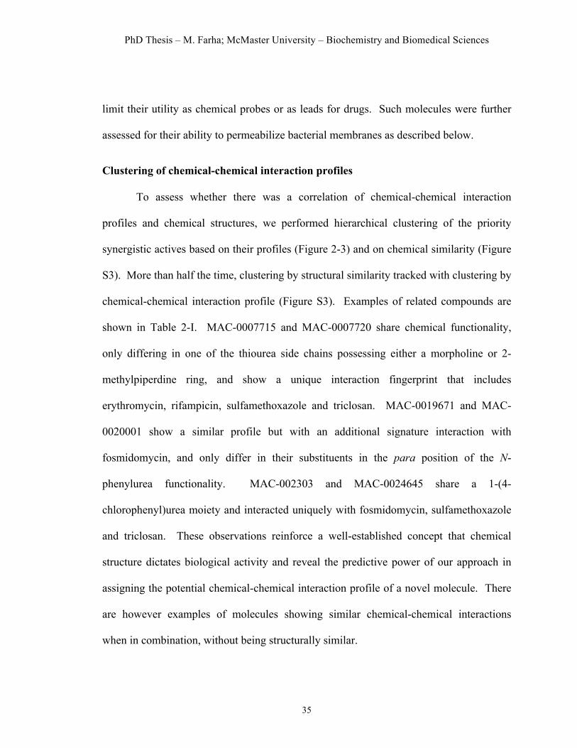
PhD Thesis – M. Farha; McMaster University – Biochemistry and Biomedical Sciences
35
limit their utility as chemical probes or as leads for drugs. Such molecules were further
assessed for their ability to permeabilize bacterial membranes as described below.
Clustering of chemical-chemical interaction profiles
To assess whether there was a correlation of chemical-chemical interaction
profiles and chemical structures, we performed hierarchical clustering of the priority
synergistic actives based on their profiles (Figure 2-3) and on chemical similarity (Figure
S3). More than half the time, clustering by structural similarity tracked with clustering by
chemical-chemical interaction profile (Figure S3). Examples of related compounds are
shown in Table 2-I. MAC-0007715 and MAC-0007720 share chemical functionality,
only differing in one of the thiourea side chains possessing either a morpholine or 2-
methylpiperdine ring, and show a unique interaction fingerprint that includes
erythromycin, rifampicin, sulfamethoxazole and triclosan. MAC-0019671 and MAC-
0020001 show a similar profile but with an additional signature interaction with
fosmidomycin, and only differ in their substituents in the para position of the N-
phenylurea functionality. MAC-002303 and MAC-0024645 share a 1-(4-
chlorophenyl)urea moiety and interacted uniquely with fosmidomycin, sulfamethoxazole
and triclosan. These observations reinforce a well-established concept that chemical
structure dictates biological activity and reveal the predictive power of our approach in
assigning the potential chemical-chemical interaction profile of a novel molecule. There
are however examples of molecules showing similar chemical-chemical interactions
when in combination, without being structurally similar.
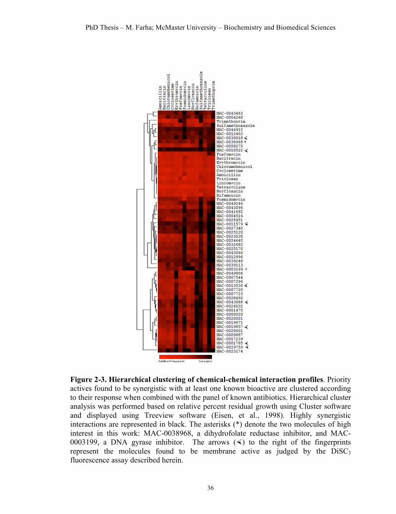
PhD Thesis – M. Farha; McMaster University – Biochemistry and Biomedical Sciences
36
Figure 2-3. Hierarchical clustering of chemical-chemical interaction profiles. Priority actives found to be synergistic with at least one known bioactive are clustered according to their response when combined with the panel of known antibiotics. Hierarchical cluster analysis was performed based on relative percent residual growth using Cluster software and displayed using Treeview software (Eisen, et al., 1998). Highly synergistic interactions are represented in black. The asterisks (*) denote the two molecules of high interest in this work: MAC-0038968, a dihydrofolate reductase inhibitor, and MAC-0003199, a DNA gyrase inhibitor. The arrows (×) to the right of the fingerprints represent the molecules found to be membrane active as judged by the DiSC3 fluorescence assay described herein.
PhD Thesis – M. Farha; McMaster University – Biochemistry and Biomedical Sciences
37
Table 2-1. Examples of molecules with similar structures and biological fingerprints.
Compound Structure
Biological Fingerprints
MAC-0007715
MAC-0007720
MAC-0019671
MAC-0020001
MAC-0023030
MAC-0024645
Biological fingerprints derived from the combination profiling screen with a panel of known antibiotics for the various molecules are shown, where highly synergistic combinations are represented in black. The biological fingerprints were taken out of the heat map in Figure 2-3 to illustrate the correlation between activity and chemical structures.

PhD Thesis – M. Farha; McMaster University – Biochemistry and Biomedical Sciences
38
In this work, we were especially interested in following up on compound
combinations that showed unique synergies. Two high interest compounds, MAC-
0038968 and MAC-0003199, were selected for follow-up experiments to characterize
cellular targets and mechanisms of action. We also investigated the activity of a subset of
priority actives that were found to be promiscuously synergistic.
MAC-0038968 is synergistic with sulfamethoxazole and active against dihydrofolate reductase (DHFR)
One compound of interest uncovered in our screen was MAC-0038968 (Figure 2-
4A). This small molecule was found to be uniquely synergistic with sulfamethoxazole,
an antibiotic that inhibits tetrahydrofolate biosynthesis in bacteria by acting as an analog
to one of the pathway intermediates (Walsh, 2003). A detailed checkerboard analysis of
the compound combination confirmed strong synergy between MAC-0038968 with
sulfamethoxazole with a fractional inhibitory index (∑FIC) of 0.187 (Figure 2-4B).
Interestingly, we previously reported on this compound in a biochemical screen to
identify inhibitors of dihydrofolate reductase (DHFR), the enzyme responsible for the
reduction of dihydrofolate to tetrahydrofolate but had not investigated its cellular activity
(Zolli-Juran, et al., 2003). The observed synergy served to validate the proposed method,
as MAC-0038968, which was found to be highly synergistic with sulfamethoxazole,
inhibits a later step in a common cellular pathway. To investigate the cellular target of
MAC-0038968, we sought to suppress the action of this compound by expression of the
putative target DHFR at high copy. Indeed, we have previously found high copy
suppression to be a valuable chemical-genetic tool to probe mechanism of novel

PhD Thesis – M. Farha; McMaster University – Biochemistry and Biomedical Sciences
39
antibacterial compounds (Li, et al., 2004; Pathania, et al., 2009). Figure 2-4C reveals that
increased expression of DHFR led to a steady increase in the MIC for MAC-0038968 and
trimethoprim without any impact on the MIC of the control compound tetracycline
(Figure 2-4C). These results are consistent with the conclusion that the growth inhibition
by MAC-0038968 was due to inhibition of DHFR. Trimethoprim, one of the known
bioactives in this study is of course a celebrated DHFR inhibitor (Baccanari and Kuyper,
1993). The lack of synergy with trimethoprim (Figure 2-3A) suggests that MAC-
0038968 and trimethoprim bind to similar sites on DHFR; otherwise a synergistic
interaction would be expected if the two bound to separate sites on the target (Krogstad
and Moellering Jr., 1986). In fact, we have previously shown that this DHFR inhibitor,
like trimethoprim, is competitive with the substrate dihyrofolate and inhibits the enzyme
with a reasonable potency (Ki = 65 nM) (Zolli-Juran, et al., 2003). Importantly MAC-
0038968 represents a new chemical class of DHFR inhibitors with cellular activity.
MAC-0003199 is a DNA gyrase inhibitor, uncovered through synergy with norfloxacin
From the combinatorial screen the quinoline carboxylic acid, MAC-0003199, was
found to have a unique interaction fingerprint that included lincomycin, triclosan and
norfloxacin. The latter interaction was particularly noteworthy as this compound had a
selective and profound interaction with this DNA gyrase inhibitor. MAC-0003199
represents a novel structure but is reminiscent of the quinolone family of synthetic
antibiotics that are well known for their inhibitory action on bacterial DNA gyrase and
topoisomerase IV and associated lethal impact on DNA replication and transcription.

PhD Thesis – M. Farha; McMaster University – Biochemistry and Biomedical Sciences
40
Figure 2-4. MAC-0038968 is uniquely synergistic with sulfamethoxazole and active against dihydrofolate reductase (DHFR). (A) Chemical structure of MAC-0038968. (B) Checkerboard analysis of MAC-0038968 in combination with sulfamethoxazole, showing a strong synergistic interaction with a ƩFIC=0.187. (C) Suppression of growth inhibition by MAC-0038968 on increasing expression of DHFR encoded in the folA gene. Here expression is manipulated by the arabinose-inducible promoter of copy of pBAD18-folA in E. coli strain MC1061. Arabinose dependence of the MIC is shown for the positive control trimethoprim (circles, top panel) and the test compound MAC-0038968 (circles, bottom panel). Also shown is the negative control tetracycline (squares).

PhD Thesis – M. Farha; McMaster University – Biochemistry and Biomedical Sciences
41
A detailed checkerboard analysis of the interaction between norfloxacin and
MAC-0003199 confirmed the synergistic interaction with a ∑FIC of 0.312 (Figure 2-5B).
In an effort to assess the capacity of MAC-0003199 to induce DNA damage we
characterized the in vivo phenotypic response of E. coli in response to the compound,
employing a DNA damage-inducible reporter construct that is based on LexA repression
for regulation of gfp transcription (Dwyer, et al., 2007). Thus an output in fluorescence is
indicative of RecA-stimulated autocleavage of LexA following recognition of DNA-
damage (Dwyer, et al., 2007). The control compound norfloxacin led to the induction of
high levels of fluorescence (Figure 2-5C). Norfloxacin is known to stabilize a tripartite
interaction with DNA gyrase and cleaved DNA thus stimulating the formation of DNA
breaks (Walsh, 2003). We also observed a large shift in fluorescence upon treatment of
the cells with MAC-0003199 (Figure 2-5C), as compared to the negative control,
tetracycline (Figure 2-5C). We next sought to determine whether MAC-0003199’s mode
of action in the cell was similar to that of norfloxacin, acting by inhibiting DNA gyrase’s
ability to supercoil DNA (Drlica and Zhao, 1997). MAC-0003199 inhibited DNA gyrase
at a relatively high concentration, compared to the potent inhibitor norfloxacin (Figure 2-
5D) but nevertheless was consistent with the relative cellular potencies of these
compounds (6.4 µg/mL for MAC-0003199 compared to 0.025 µg/mL for norfloxacin).
Interestingly, the synergistic nature of the interaction between MAC-0003199 and
norfloxacin suggests that the two molecules do not work with exactly the same
mechanism. For example, it seems unlikely that the two compounds bind to exactly the
same site on DNA gyrase as this would lead to an antagonistic interaction (Krogstad and

PhD Thesis – M. Farha; McMaster University – Biochemistry and Biomedical Sciences
42
Moellering Jr., 1986). Further, MAC-0003199 was found to be synergistic with triclosan
and lincomycin, while norfloxacin showed no such interaction. Triclosan is known to
disrupt the inner membrane of bacteria (Schweizer, 2001) and this interaction may relate
to inherent permeability problems for MAC-0003199. Lincomycin is an inhibitor of
protein synthesis that binds the 50S ribosome and inhibits peptidyl transferase activity,
thus preventing translation termination (Lin, et al., 1997; Walsh, 2003). The mechanistic
connection between lincomycin and MAC-0003199 remains unclear but suggests
nevertheless that MAC-0003199 may not function simply as an inhibitor of DNA gyrase.
Its striking similarity in structure to the quinolone class of molecules, however strongly
suggests that MAC-0003199 is likely an inhibitor of DNA gyrase, such that its interaction
with norfloxacin would expectedly be antagonistic in nature. However, quinolones have
a unique mechanism of action in that they inhibit both bacterial DNA gyrase and
topoisomerase IV, with varying target preference, depending on the organism in question.
In E. coli, the inhibition of topoisomerase IV becomes more apparent when DNA gyrase
is mutated to be resistant to the quinolone (Khodursky, et al., 1995). Moreover,
quinolones can in fact prefer topoisomerase IV over DNA gyrase, and vice-versa, or
target both enzymes at nearly the same level (Takei, et al., 2001). This dual-targeting
property of quinolones may help explain the synergy between MAC-0003199 and
norfloxacin, in that the observed antibacterial activities are involved not only in DNA
gyrase inhibition but also in topoisomerase IV inhibition.

PhD Thesis – M. Farha; McMaster University – Biochemistry and Biomedical Sciences
43
Figure 2-5. MAC-0003199, synergistic with norfloxacin, inhibits DNA gyrase. (A) Chemical structure of MAC-0003199. (B) Checkerboard analysis of MAC-0003199 in combination with norfloxacin, showing synergy with a ƩFIC=0.312 (C). E. coli MC1061 harbouring pL(lexO)-GFP, a promoter-GFP reporter gene construct used to report on DNA lesion formation, was grown to an OD600 of 0.3-0.4 and treated with various concentration of norfloxacin (positive control), MAC-0003199, and tetracycline (negative control). Optical density at 600 nm and fluorescence at 535 nm were monitored for 7 hours. Induction of DNA damage was evident for norfloxacin and MAC-0003199. (D) Inhibition of supercoiling of DNA by MAC-0003199 and norfloxacin. E. coli gyrase was incubated with relaxed pBR322 in the presence of various concentrations of norfloxaxin (top panel) and MAC-0003199 (bottom panel).The lane labelled –G is relaxed pBR322 in the absence of E. coli gyrase . R and S represent relaxed and supercoiled pBR322, respectively.

PhD Thesis – M. Farha; McMaster University – Biochemistry and Biomedical Sciences
44
Promiscuously synergistic molecules
As discussed above, a particular advantage of systematic screening for chemical-
chemical interactions was the identification of promiscuously synergistic molecules. In an
effort to understand the mode of action of such molecules, we investigated the possibility
that these were membrane active and resulted in a breach in bacterial membrane potential.
This was assessed with a membrane potential-sensitive fluorescent probe, 3,3’-
dipropylthiacarbocyanine (DiSC3) (Sims, et al., 1974). Once inside the cell, this dye
becomes concentrated and self-quenches its own fluorescence. Membrane active
compounds that depolarize the ∆ψ component of the proton motive force will lead to the
release of the dye and a consequent increase in fluorescence. Normally, this dye is
incapable of penetrating Gram-negative bacteria; however, we were able to establish this
assay using our screening strain (E. coli strain MC1061) because of its documented
hyperpermeability. In fact, we tested all compounds found to be synergistic in our screen
in our combination profiling screen for their capacity to alter membrane potential at MIC
concentrations. All 14 of the known bioactives tested negatively in this assay, as
expected, whereas polymyxin B, a lipopeptide known to be membrane active
(Daugelavicius, et al., 2000), led to a significant increase in DiSC3 fluorescence in our
assay (Figure S4A). Of the 45 synergistic priority actives, 8 molecules caused a
significant increase in DiSC3 fluorescence, suggesting that these molecules have the
capacity to depolarize the cytoplasmic membrane. These are highlighted with an arrow in
Figure 2-3 to the right of their respective chemical-chemical interaction profiles and the
data are presented in Figure S4B.
PhD Thesis – M. Farha; McMaster University – Biochemistry and Biomedical Sciences
45
It is tempting to conclude that these eight molecules represent non-selective
compounds that are unsuitable probes of biology or as leads for new drugs. Membrane
active molecules, such as the peptide-drugs, daptomycin and polymyxin B, have proven
invaluable in treating drug resistant infections (Evans, et al., 1999; Fuchs, et al., 2002;
Giamarellou and Poulakou, 2009; King and Phillips, 2001). Regardless of the
disposition toward membrane-active and promiscuous chemical matter, the combination
profiling approach used here was quite successful in identifying strong candidates for
membrane depolarizing activity among priority actives.
Discussion
The systematic determination of the mechanism of actives derived from
phenotype-based small molecule screening remains a very significant challenge to the
discovery of useful probes of biology or leads for new drugs. Chemical-genetic
approaches have dominated for this purpose in recent years and, while the success of
these has been tangible, there remain some significant drawbacks. Forward chemical-
genetic methods rely on enhancement or suppression of growth phenotypes associated
with novel actives by genetic perturbation. The latter has the potential to inform on
mechanism and provide testable hypotheses particularly when there is depth of
knowledge on the interacting genes. Classical chemical-genetic approaches are powerful
but require the tedious isolation and characterization of suppressing or enhancing mutants
(Eggert, et al., 2001; Gitai, et al., 2005; Heitman, et al., 1991). Modern chemical-
genomic approaches have typically relied on genome-scale deletion or overexpression
clone-sets in model microbes such as yeast or E. coli to screen for suppressing or

PhD Thesis – M. Farha; McMaster University – Biochemistry and Biomedical Sciences
46
enhancing genotypes (Baetz, et al., 2004; Giaever, et al., 1999; Li, et al., 2004; Lum, et
al., 2004; Pathania, et al., 2009). Such clonesets are currently unavailable in all but the
most tractable model microbes.
Herein, we describe an alternative and complementary approach where a small
library of known bioactives is systematically combined with novel actives to detect
chemical-chemical interactions. Almost 200 growth inhibitory and novel active
compounds were systematically combined with a library of some 14 well-known
antibiotics of diverse chemical class and mechanism. Combination profiles so discovered
were examined in the context of a deep knowledge of the mechanism of interacting
chemicals. These profiles generated hypotheses that were testable with biochemical and
physiological experiments and revealed new probes of folate metabolism, DNA gyrase
and the cell membrane. As such, chemical-chemical interaction profiles can be generated
for any phenotype-inducing compound in any biological system without a need for
genetic manipulations provided there are existing well characterized chemical perturbants
available.
The work reported here suggests that phenotypic responses to combinations of
known and novel bioactives can reveal information about the pathways and targets
affected by the latter. The approach was data-rich but nevertheless allowed for efficient
testing of combinations of chemicals, making it readily adaptable as a secondary
screening approach in high-throughput screening efforts. In a validating discovery, the
bi-guanidine-containing compound MAC-0038968 was found to be synergistic with
sulfamethoxazole. Sulfamethoxazole limits the supply of PABA precursor available to

PhD Thesis – M. Farha; McMaster University – Biochemistry and Biomedical Sciences
47
subsequent steps in the tetrahydrofolate biosynthesis pathway and is well known for its
synergy with trimethoprim, an inhibitor of DHFR (Rubin and Swartz, 1980). Further, we
demonstrated that MAC-0038968, previously shown to be a potent inhibitor of DHFR in
vitro (Zolli-Juran, et al., 2003), could be suppressed by high copy DHFR, demonstrating
that his compound had cellular actvitiy and was on target. Interestingly, MAC-0038968
showed no synergy with trimethoprim, presumably because these compounds bind to the
same site on DHFR. MAC-0003199, a quinoline carboxylic acid, was shown to be a
DNA-damaging agent that targets DNA gyrase. This molecule, similar in structure to
quinolones, was suitably found to be highly synergistic with norfloxacin, a potent
inhibitor of DNA gyrase. The two novel inhibitors of bacterial physiology uncovered
through the use of chemical-chemical interactions highlight the utility of the proposed
approach in facilitating the search for cellular targets of novel biological probes.
In cases where a novel compound shows promiscuous interactions with multiple
known bioactives, this non-selective behaviour can be used as a filter to eliminate
nuisance compounds. Other methods to identify such molecules include computational
means based on chemical functionality (Roche, et al., 2002; Walters, et al., 1999).
Another is a biochemical approach that uses a detergent-based assay to counter-screen for
non-selective aggregating compounds (Feng, et al., 2005; McGovern, et al., 2002; Seidler,
et al., 2003). There are however few systematic counter screens for non-selective
compounds that are cell-based. Interestingly, further study of such compounds in the
work reported here led to the discovery of a subset of compounds capable of dissipating
the transmembrane potential of E. coli. Given recent clinical success of the antibiotic
PhD Thesis – M. Farha; McMaster University – Biochemistry and Biomedical Sciences
48
daptomycin, also know to depolarize membranes (Silverman, et al., 2003), promiscuous
behaviour might well be viewed as both a strength and a weakness. Nevertheless,
combination profiling proved to be a powerful tool in identifying these molecules.
Interactions between bioactive chemicals can be synergistic or antagonistic
(Krogstad and Moellering Jr., 1986). In this study, we limited our focus to synergistic
interactions. In the same manner that synergies were specifically used to elucidate
mechanisms of action, a screen looking for antagonistic interactions with known
bioactives could also yield rich information about associated cellular pathways. Briefly,
antagonistic interactions are most often observed when two compounds compete between
binding sites or when one alters the binding site of the other (Krogstad and Moellering Jr.,
1986). Antagonism might also be the result of complex genetic interactions propagated
through the cellular genetic network (D'Elia, et al., 2009; Motter, et al., 2008). Thus,
looking for antagonistic interactions among novel and know bioactives, though rare,
could surely also provide useful mechanistic insights and testable hypotheses regarding
the mechanism of action of novel actives.
Herein we have demonstrated real promise for chemical-chemical interactions in
understanding mechanism of the interacting components. So characterized, these
interactions also have the potential to perturb the complex and redundant nature of
biological pathways. It is increasingly becoming evident that biological systems are
composed of dense networks of interacting components that are characterized by
redundancy (Ho, et al., 2002; Tong, et al., 2004). Thus, the simultaneous use of multiple
perturbants will ultimately be required to adequately describe this complex cellular matrix
PhD Thesis – M. Farha; McMaster University – Biochemistry and Biomedical Sciences
49
(Lehár, et al., 2008). As the compendium of interacting combinations grows, these will
become an increasingly powerful tool for assessing mechanism of action.
Significance
Phenotype based screening provides a powerful tool for the discovery and
characterization of new probes of biology and leads for new drugs, however significant
challenges remain for connecting compounds to their targets. Here, we report the
application of systematic chemical-chemical combination profiling to understand the
mechanism of action of novel antibacterial compounds. The interaction of newly
discovered compounds with a panel of known bioactives of diverse mechanism and
chemical class was used to gain insight into mode of action. Combination profiling for
synergistic chemical-chemical interactions resulted in a data-rich map of interactions and
led to testable hypotheses regarding the mechanism of action of the novel bioactive
molecules. One such molecule was found to be a novel inhibitor of folate biosynthesis,
(MAC-0038968) and another was shown to be a new DNA gyrase inhibitor (MAC-
0003199). The approach also proved to be useful in identifying non-selective molecules
that showed promiscuous interaction behaviour where a subset of these compounds was
shown to depolarize the bacterial membrane.

PhD Thesis – M. Farha; McMaster University – Biochemistry and Biomedical Sciences
50
Experimental Procedures Bacterial strains, reagents and general methods. E. coli MC1061 was used for all
experiments and follow-up work. Growth media was liquid or agar Luria-Bertani Broth
(LB). The small molecule library was purchased from Maybridge (Cornwall, England)
and was dissolved in DMSO at a concentration of 6.4 mg/ml. All chemicals were
purchased from Sigma Aldrich (Oakville, ON).
Determination of minimum inhibitory concentrations of chemical compounds using
E. coli strain MC1061. The minimum inhibitory concentrations of priority actives and
known antibiotics were determined to characterize their growth inhibition. Typically, E.
coli MC1061 was grown overnight in 5 ml of LB media. The cells were then diluted
1:100 in fresh media and allowed to grow until the OD600 reached 0.4. The cells were then
diluted 1:100,000 and exposed to two-fold serial dilutions of the compounds at final
concentrations ranging from 0-204.8 µg/mL from stock solutions of 6.4 mg/mL. These
were tested in 96-well microwell plates with a total volume of 200 µl and incubated at
37°C with 80% humidity for 18 hours before determining optical density at 600 nm. The
concentration where the optical density was less than 0.05 was deemed the MIC of the
test compound.
Combination profiling screen. To screen the various combinations, 80-fold stock
solutions of chemicals were placed into polypropylene 96-well master plates in two
consecutive columns. Aliquots of 2.5 µl from both master plates were dispensed into the
assay plate using a Biomek FX liquid handler (Beckman Coulter Inc., Fullerton, CA) in
the McMaster High Throughput Screening Laboratory. Subsequently, 195 µl of a mid-

PhD Thesis – M. Farha; McMaster University – Biochemistry and Biomedical Sciences
51
log subculture of E. coli MC1061 cells diluted 1:100,000 were added. These plates were
incubated for 18 hours at 37˚C before being read for optical density at 600 nm. All
combinations were done in triplicate, allowing the assignment of a standard error to all
percent growth values.
Checkerboard analysis of synergy. A mid-log subculture of E. coli MC1061 cells was
diluted 1:100,000 before adding to an 8 x 8 matrix of priority actives and known
antibiotics. The plates were incubated for 18 hours at 37 ˚C before reading for optical
density at 600 nm. All combinations were done in duplicate. Heat maps of the averages
were created in Excel and used to illustrate growth compared to the high controls (DMSO
only). To evaluate the effect of the combinations, the fractional inhibitory concentrations
(∑FIC) indices were calculated. This metric is defined as the sum of the MIC of each
drug when used in combination divided by the MIC when used alone. Chemical-
chemical interactions with ∑FIC of less than 0.5 were deemed synergistic.
High copy suppression of growth inhibition. E. coli MC1061 harbouring pBAD18-
folA was grown overnight in LB supplemented with 100 µg/mL ampicillin, subcultured
the following day (1:100 dilution in the same media) and grown to mid log (OD600 = 0.4)
with aeration at 250 rpm at 37ºC. The cells were then diluted 1:100,000 and exposed to
two-fold serial dilutions of the compounds ranging from 0-102.4 µg/mL in the presence
of various concentration of arabinose (0-3.2%). These were tested in 96-well microwell
plates (total volume of 200 µl) and incubated at 37°C with 80% humidity for 18 hours
before determining optical density at 600 nm. The concentration where the optical density
was less than 0.05 was deemed the MIC.

PhD Thesis – M. Farha; McMaster University – Biochemistry and Biomedical Sciences
52
Promoter-reporter construct experiments. The DNA-damage reporter construct was a
kind gift from Dr. James Collins (Boston University). In all experiments, E. coli MC1061
harbouring pL(lexO)-GFP were grown overnight and then diluted 1:100 in 50 mL LB
supplemented with 100 mg/ml ampicillin. Cells were grown at 37˚C, 250 rpm, until an
OD600 of 0.3-0.4. Cells were aliquoted into black clear bottom 96-well plates (Corning
Life Sciences, New York, USA) and drug, previously serially diluted in stock plates, was
added at various concentrations. Optical density at 600 nm and fluorescence at 535 nm
were monitored for 7 hours using the EnVision from Perkin Elmer (Massachusetts, USA).
DNA supercoiling assay. E. coli gyrase was incubated with 0.5 µg of relaxed pBR322
DNA in a 30 µl reaction mixture at 37˚C for 1 h under the following conditions: 35 mM
Tris-HCL (pH 7.5), 24 mM KCl, 4mM MgCl2, 2 mM DTT, 1.75 mM ATP, 5 mM
spermidine, 0.1 mg/mL BSA, and 6.5% glycerol. Reactions were analyzed by
electrophoresis through a 0.8% agarose gel (run at 130V for 1.5 h).
Dissipation of transmembrane potential. The transmembrane potential was determined
with the fluorescent probe , 3’-dipropylthiacarbocyanine (DiSC3(5)). E. coli MC1061
cells were washed twice with Tris buffer (10 mM, pH 7.5), and then resuspended to an
optical density at 600 nm of 0.35. DiSC3(5) was added at a final concentration of 0.4 µM,
and the cells were incubated, with constant stirring to let the dye stabilize. Compounds
were then injected. Fluorescent traces were measured in a fluorimeter (Photon
Technology International) at the excitation and emission wavelengths of 622 and 660 nm,
respectively.
PhD Thesis – M. Farha; McMaster University – Biochemistry and Biomedical Sciences
53
Acknowledgements We thank James Collins (Boston University) for providing the pL(lexO)-GFP reporter
construct. We thank the staff of the McMaster High Throughput Screening Laboratory for
technical assistance. We also thank Soumaya Zlitni for helpful suggestions and comments
on the manuscript. This work was supported by an operating grant from the Canadian
Institutes of Health Research (MOP-81330) and by a Canada Research Chair Award to
E.D.B.
References Alaimo, P.J., Shogren-Knaak, M.A., and Shokat, K.M. (2001). Chemical genetic approaches for the elucidation of signaling pathways. Curr Opin Chem Biol. 5, 360-367. Baccanari, D.P., and Kuyper, L.F. (1993). Basis of selectivity of antibacterial diaminopyrimidines. J Chemother 5, 393-399. Baetz, K., McHardy, L., Gable, K., Tarling, T., Reberioux, D., Bryan, J., Andersen, R.J., Dunn, T., Hieter, P., and Roberge, M. (2004). Yeast genome-wide drug-induced haploinsufficiency screen to determine drug mode of action. Proc Natl Acad Sci U S A 101, 4525-4530. Boucher, H.W., Talbot, G.H., Bradley, J.S., Edwards, J.E., Gilbert, D., Rice, L.B., Scheld, M., Spellberg, B., and Bartlett, J. (2009). Bad bugs, no drugs: no ESKAPE! An update from the Infectious Diseases Society of America. Clin Infect Dis 48, 1-12. Burdine, L., and Kodadek, T. (2004). Target identification in chemical genetics: the (often) missing link. Chem Biol 11, 593-597. Casabadan, M.J., and Cohen, S.N. (1980). Analysis of gene control signals by DNA fusion and cloning in Escherichia coli. . J Mol Biol. 138, 179-207. D'Elia, M.A., Pereira, M.P., and Brown, E.D. (2009). Are essential genes really essential? Trends Microbiol 17, 433-438. Daugelavicius, R., Bakiene, E., and Bamford, D.H. (2000). Stages of polymyxin B interaction with the Escherichia coli cell envelope. Antimicrob Agents Chemother 44, 2969-2978. Donald, R.G., Skwish, S., Forsyth, R.A., Anderson, J.W., Zhong, T., Burns, C., Lee, S., Meng, X., LoCastro, L., Jarantow, L.W., et al. (2009). A Staphylococcus aureus fitness test platform for mechanism-based profiling of antibacterial compounds. Chem Biol 16, 826-836. Drlica, K., and Zhao, X. (1997). DNA gyrase, topoisomerase IV, and the 4-quinolones. Microbiol Mol Biol Rev 61, 377-392.
PhD Thesis – M. Farha; McMaster University – Biochemistry and Biomedical Sciences
54
Dwyer, D.J., Kohanski, M.A., Hayete, B., and Collins, J.J. (2007). Gyrase inhibitors induce an oxidative damage cellular death pathway in Escherichia coli. . Mol Syst Biol. 3, 1-15. Eggert, U.S., Ruiz, N., Falcone, B.V., Branstrom, A.A., Goldman, R.C., Silhavy, T.J., and Kahne, D. (2001). Genetic basis for activity differences between vancomycin and glycolipid derivatives of vancomycin. Science 294, 361-364. Eisen, M.B., Spellman, P.T., Brown, P.O., and Botstein, D. (1998). Cluster analysis and display of genome-wide expression patterns. Proc Natl Acad Sci U S A 95, 14863-14868. Evans, M.E., Feola, D.J., and Rapp, R.P. (1999). Polymyxin B sulfate and colistin: old antibiotics for emerging multiresistant gram-negative bacteria. Ann Pharmacother 33, 960-967. Feng, B.Y., Shelat, A., Doman, T.N., Guy, R.K., and Shoichet, B.K. (2005). High-throughput assays for promiscuous inhibitors. Nat Chem Biol 1, 146-148. Fuchs, P.C., Barry, A.L., and Brown, S.D. (2002). In vitro bactericidal activity of daptomycin against staphylococci. J Antimicrob Chemother 49, 467-470. Giaever, G., Shoemaker, D.D., Jones, T.W., Liang, H., Winzeler, E.A., Astromoff, A., and Davis, R.W. (1999). Genomic profiling of drug sensitivities via induced haploinsufficiency. Nat Genet 21, 278-283. Giamarellou, H., and Poulakou, G. (2009). Multidrug-resistant Gram-negative infections: what are the treatment options? Drugs 69, 1879-1901. Gitai, Z., Dye, N.A., Reisenauer, A., Wachi, M., and Shapiro, L. (2005). MreB actin-mediated segregation of a specific region of a bacterial chromosome. Cell 120, 329-341. Heitman, J., Movva, N.R., and Hall, M.N. (1991). Targets for cell cycle arrest by the immunosuppressant rapamycin in yeast. Science 253, 905-909. Hillenmeyer, M.E., Fung, E., Wildenhain, J., Pierce, S.E., Hoon, S., Lee, W., Proctor, M., St Onge, R.P., Tyers, M., Koller, D., et al. (2008). The chemical genomic portrait of yeast: uncovering a phenotype for all genes. Science 320, 362-365. Ho, Y., Gruhler, A., Heilbut, A., Bader, G.D., Moore, L., Adams, S.L., Millar, A., Taylor, P., Bennett, K., Boutilier, K., et al. (2002). Systematic identification of protein complexes in Saccharomyces cerevisiae by mass spectrometry. Nature 415, 180-183. Khodursky, A.B., Zechiedrich, E.L., and Cozzarelli, N.R. (1995). Topoisomerase IV is a target of quinolones in Escherichia coli. Proc Natl Acad Sci U S A 92, 11801-11805. King, A., and Phillips, I. (2001). The in vitro activity of daptomycin against 514 Gram-positive aerobic clinical isolates. J Antimicrob Chemother 48, 219-223. Krogstad, D.J., and Moellering Jr., R.C. (1986). Antibiotics in Laboratory Medicine. (Baltimore: Williams and Wilkins). Lehar, J., Krueger, A., Zimmermann, G., and Borisy, A. (2008). High-order combination effects and biological robustness. Mol Syst Biol 4, 215. Lehar, J., Stockwell, B.R., Giaever, G., and Nislow, C. (2008). Combination chemical genetics. Nat Chem Biol 4, 674-681. Lehár, J., Stockwell, B.R., Giaver, G., and Nislow, C. (2008). Combination chemical genetics. Nat. Chem. Biol. 4, 674-681.
PhD Thesis – M. Farha; McMaster University – Biochemistry and Biomedical Sciences
55
Li, X., Zolli-Juran, M., Cechetto, J.D., Daigle, D.M., Wright, G.D., and Brown, E.D. (2004). Multicopy suppressors for novel antibacterial compounds reveal targets and drug efflux susceptibility. Chem Biol. 11, 1423-1430. Lin, A.H., Murray, R.W., Vidmar, T.J., and Marotti, K.R. (1997). The oxazolidinone eperezolid binds to the 50S ribosomal subunit and competes with binding of chloramphenicol and lincomycin. . Antimicrob Agents Chemother 41, 2127-2131. Lum, A.M., Huang, J., Hutchinson, C.R., and Kao, C.M. (2004). Reverse engineering of industrial pharmaceutical-producing actinomycete strains using DNA microarrays. Metab Eng 6, 186-196. Lum, P.Y., Armour, C.D., Stepaniants, S.B., Cavet, G., Wolf, M.K., Butler, J.S., Hinshaw, J.C., Garnier, P., Prestwich, G.D., Leonardson, A., et al. (2004). Discovering modes of action for therapeutic compounds using a genome-wide screen of yeast heterozygotes. Cell 116, 121-137. McGovern, S.L., Caselli, E., Grigorieff, N., and Shoichet, B.K. (2002). A common mechanism underlying promiscuous inhibitors from virtual and high-throughput screening. J Med Chem 45, 1712-1722. Moellering, R.C., Jr. (1983). Rationale for use of antimicrobial combinations. Am J Med 75, 4-8. Motter, A.E., Gulbahce, N., Almaas, E., and Barabasi, A.L. (2008). Predicting synthetic rescues in metabolic networks. Mol Syst Biol 4, 168. Neu, H.C., and Kamimura, T. (1982). Synergy of fosmidomycin (FR-31564) and other antimicrobial agents. Antimicrob Agents Chemother 22, 560-563. Parsons, A.B., Brost, R.L., Ding, H., Li, Z., Zhang, C., Sheikh, B., Brown, G.W., Kane, P.M., Hughes, T.R., and Boone, C. (2004). Integration of chemical-genetic and genetic interaction data links bioactive compounds to cellular target pathways. Nat Biotechnol 22, 62-69. Pathania, R., Zlitni, S., Barker, C., Das, R., Gerritsma, D.A., Lebert, J., Awuah, E., Melacini, G., Capretta, F.A., and Brown, E.D. (2009). Chemical genomics in Escherichia coli identifies an inhibitor of bacterial lipoprotein targeting. Nat Chem Biol 5, 849-856. Peterson, R.T. (2008). Chemical biology and the limits of reductionism. Nat Chem Biol 4, 635-638. Rand, K.H., Houck, H.J., Brown, P., and Bennett, D. (1993). Reproducibility of the microdilution checkerboard method for antibiotic synergy. Antimicrob Agents Chemother 37, 613-615. Roche, O., Schneider, P., Zuegge, J., Guba, W., Kansy, M., Alanine, A., Bleicher, K., Danel, F., Gutknecht, E.M., Rogers-Evans, M., et al. (2002). Development of a virtual screening method for identification of "frequent hitters" in compound libraries. J Med Chem 45, 137-142. Rubin, R.H., and Swartz, M.N. (1980). Trimethoprim-sulfamethoxazole. N Engl J Med 303, 426-432. Schweizer, H.P. (2001). Triclosan: a widely used biocide and its link to antibiotics. . FEMS Microbiol Lett. 202, 1-7.

PhD Thesis – M. Farha; McMaster University – Biochemistry and Biomedical Sciences
56
Seidler, J., McGovern, S.L., Doman, T.N., and Shoichet, B.K. (2003). Identification and prediction of promiscuous aggregating inhibitors among known drugs. J Med Chem 46, 4477-4486. Silverman, J.A., Perlmutter, N.G., and Shapiro, H.M. (2003). Correlation of daptomycin bactericidal activity and membrane depolarization in Staphylococcus aureus. Antimicrob Agents Chemother 47, 2538-2544. Sims, P.J., Waggoner, A.S., Wang, C.H., and Hoffman, J.F. (1974). Studies on the mechanism by which cyanine dyes measure membrane potential in red blood cells and phosphatidylcholine vesicles. Biochemistry 13, 3315-3330. Stockwell, B.R. (2000). Chemical genetics: ligand-based discovery of gene function. Nat Rev Genet 1, 116-125. Takei, M., Fukuda, H., Kishii, R., and Hosaka, M. (2001). Target preference of 15 quinolones against Staphylococcus aureus, based on antibacterial activities and target inhibition. Antimicrob Agents Chemother 45, 3544-3547. Tong, A.H., Lesage, G., Bader, G.D., Ding, H., Xu, H., Xin, X., Young, J., Berriz, G.F., Brost, R.L., Chang, M., et al. (2004). Global mapping of the yeast genetic interaction network. Science 303, 808-813. Walsh, C. (2003). Antibiotics: Actions, Origins, Resistance. ((Washington: ASM Press). Walters, W.P., Ajay, and Murcko, M.A. (1999). Recognizing molecules with drug-like properties. Curr Opin Chem Biol 3, 384-387. Yeh, P., and Kishony, R. (2007). Networks from drug-drug surfaces. Mol Syst Biol 3, 85. Yeh, P., Tschmu, A., and Kishony, R. (2006). Functional classification of drugs by properties of their pairwise interactions. . Nat. Gen. 38, 489-494. Zimmermann, G.R., Lehar, J., and Curtis, K.T. (2007). Multi-target therapeutics: when the whole is greater than the sum of the parts. . Drug Discov Today 12, 34-42. Zlitni, S., Blanchard, J.E., and Brown, E.D. (2009). High-throughput screening of model bacteria. Methods Mol Biol 486, 13-27. Zolli-Juran, M., Cechetto, J.D., Hartlen, R., D.M., D., and Brown, E.D. (2003). High throughput screening identifies novel inhibitors of Escherichia coli dihydrofolate reductase that are competitive with dihydrofolate. . Bioorg. Med. Chem. Lett. 13, 2493-2496.
PhD Thesis – M. Farha; McMaster University – Biochemistry and Biomedical Sciences
57
CHAPTER THREE - Inhibition of WTA Synthesis Blocks the Cooperative Action of
PBPs and Sensitizes MRSA to ß-lactams

PhD Thesis – M. Farha; McMaster University – Biochemistry and Biomedical Sciences
58
Preface The work presented in this chapter was previously published in: Farha, MA, Leung, A, Sewell, EW, D’Elia, MA, Allison, SE, Ejim, L, Pereira, PM, Pinho, MG, Wright, GD, and Brown, ED. (2013) Inhibition of WTA synthesis blocks the cooperative action of PBPs and sensitizes MRSA to ß-lactams. ACS Chemical Biology, 8:226-33. Reproduced with permission. © 2013 American Chemical Society For this work, I performed all experiments with technical assistance from Leung A, with the exception of: creation of tarO deletion strains and PG analysis (D’Elia, MA and Pereira, PM), creation of the tarH conditional deletion strain (Allison, SE) and TarO in vitro assay (Sewell, EW). Ejim L provided technical assistance with the G. mellonella model. I wrote the manuscript with edits provided by Brown, ED.

PhD Thesis – M. Farha; McMaster University – Biochemistry and Biomedical Sciences
59
Abstract Rising drug resistance is limiting treatment options for infections by methicillin-resistant
Staphylococcus aureus (MRSA). Herein we provide new evidence that wall teichoic acid
(WTA) biogenesis is a remarkable antibacterial target with the capacity to destabilize the
cooperative action of penicillin-binding proteins (PBPs) that underlie ß-lactam resistance
in MRSA. Deletion of gene tarO, encoding the first step of WTA synthesis, resulted in
the restoration of sensitivity of MRSA to a unique profile of β-lactam antibiotics with a
known selectivity for penicillin binding protein 2 (PBP2). Of these, cefuroxime was used
as a probe to screen for previously-approved drugs with a cryptic capacity to potentiate its
activity against MRSA. Ticlopidine, the antiplatelet drug Ticlid®, strongly potentiated
cefuroxime and this synergy was abolished in strains lacking tarO. The combination was
also effective in a Galleria mellonella model of infection. Using both genetic and
biochemical strategies, we determined the molecular target of ticlopidine as the N-
acetylglucosamine-1-phosphate transferase encoded in gene tarO and provide evidence
that WTA biogenesis represents an Achilles heel supporting the cooperative function of
PBP2 and PBP4 in creating highly cross-linked muropeptides in the peptidoglycan of S.
aureus. This approach represents a new paradigm to tackle MRSA infection.

PhD Thesis – M. Farha; McMaster University – Biochemistry and Biomedical Sciences
60
Introduction
Since their first appearance in the early 1960s, methicillin-resistant
Staphylococcus aureus (MRSA) strains have spread worldwide and have become one of
the most menacing of human pathogens (1-2). For much of this period, outbreaks of
MRSA were confined to hospitals, however, over the last decade, the prevalence of
MRSA in the community has increased alarmingly. USA300 and USA400 isolates now
represent the most predominant cause of community-acquired infections in the United
States, Canada and Europe (3-4). While penicillin and other β-lactams such as
methicillin were once very effective antibiotics in treating staphylococcal infections, the
widespread resistance of MRSA to this class of antibiotics has made treatment
increasingly difficult. Besides common resistance to methicillin and β-lactams in
general, S. aureus has also become resistant to so-called ‘drugs of last resort’ including
vancomycin, daptomycin and linezolid (5-7).
β-lactams target the synthesis of peptidoglycan (PG), a stress-bearing cell wall
polymer of a disaccharide of N-acetylmuramic acid and N-acetylglucosamine. The
former is bonded to a short amino acid stem (L-alanine-D-iso-glutamine-L-lysine-D-
alanine-D-alanine) that is bridged to neighboring stems via a pentaglycyl segment in S.
aureus (Supplementary Figure 1). Transglycosylases and transpeptidases mediate the
final assembly of peptidoglycan, forming glycosyl bonds between the disaccharides and
cross-links between the neighboring stem peptides using pentaglycine bridges,
respectively. These enzymes are collectively known as penicillin-binding proteins

PhD Thesis – M. Farha; McMaster University – Biochemistry and Biomedical Sciences
61
(PBPs), with their transpeptidase region making up the cellular target of β-lactam
antibiotics. β-lactam resistance in MRSA involves the acquisition of PBP2A, encoded in
mecA, to the complement of the four native staphylococcal PBPs. The function of the
transpeptidase domain of PBP2A is similar to that of the bifunctional
transglycosylase/transpeptidase PBP2 but has a remarkably low affinity for β-lactam
antibiotics (8). PBP2A is thought to take over the biosynthetic role of β-lactam-inhibited
PBP2 in cross-linking peptidoglycan when assisted by the transglycosylase domain of
native PBP2 (9).
The bacterial cell wall remains a prime target for anti-MRSA drug discovery and
the β-lactam class of antibiotics continues to be explored, albeit with limited success, for
example, with the development of analogues capable of inhibiting PBP2A (10-11).
Others strategies to treat MRSA have centered on the development of new drug classes
with orthogonal mechanisms to the β-lactams such as linezolid, daptomycin and new
glycopeptides (12). Nevertheless resistance to these novel classes has already been
reported (5-7). With limited treatment options for MRSA, there is a pressing need for
new agents that will avoid existing resistance mechanisms.
To this end, there is a growing interest in identifying auxiliary genes that are
required for β-lactam resistance as potential novel targets. Previous work has suggested
that PBP2A is not the sole determinant for β-lactam resistance (13-16). Specifically,
although not essential for viability, PBP4 has been shown to play a key role in β-lactam
resistance in strains of community-acquired MRSA (CA-MRSA), which was linked to its
unique function in producing highly-cross linked peptidoglycan species during cell wall

PhD Thesis – M. Farha; McMaster University – Biochemistry and Biomedical Sciences
62
synthesis (17). Thus cooperative functions among PBPs, in particular PBP2 and PBP2A
(9) as well as PBP2 and PBP4 (17-18), during cell wall synthesis are also critical to the
expression of β-lactam resistance in MRSA. Moreover, growing evidence suggests that a
complex network of gene products involved in PG metabolism (14), synthesis (19-20)
and regulation (21-22) play key roles in the expression of β-lactam resistance in strains of
MRSA. Most recently, antisense technology was used to systematically probe 245
essential genes in MRSA for their role in β-lactam resistance (23). Several non-obvious
genes were noted including ftsZ, an important cell division protein. A follow up study
revealed that the known FtsZ inhibitor, PC190723, was in fact synergistic with imipenem
and effective against MRSA (24).
There is likewise emerging evidence that wall teichoic acids (WTAs), anionic
polyol phosphate polymers covalently attached to PG, have a role in the expression of β-
lactam resistance in MRSA. In 1994, a transposon mutant (llm) that suppressed
methicillin resistance in S. aureus was isolated and mapped to the 3'-terminal region of
tarO, which encodes the first step of WTA synthesis (25). In S. aureus, the assembly of
WTA polymers begins with two non-essential steps for cell viability, encoded in tarO
and tarA, and continues with late-acting gene products that are indispensible upon
initiation of polymer synthesis (26). Specifically, the TarO protein initiates the assembly
of WTAs with the transfer of N-acetylglucosamine-1-phosphate to a membrane-anchored
undecaprenyl-phosphate carrier lipid while TarA successively adds an N-
acetylmannosamine moiety. This product is ultimately elaborated into a long polymer
containing ribitol phosphate repeats that is attached to PG (26) (Supplementary Figure 1).

PhD Thesis – M. Farha; McMaster University – Biochemistry and Biomedical Sciences
63
The function of WTAs is incompletely understood but shown to be important in processes
such as cell division (27-29), virulence (30) and, more recently, in the expression of β-
lactam resistance. In the aforementioned antisense study, gene tarL, encoding a late
acting WTA biosynthetic enzyme was identified to lead to β-lactam sensitivity upon
depletion (23). In addition, recent evidence has shown that TarS, a glycosyltransferase
that attaches β-O-N-acetyl-D-glucosamine residues to WTA polymers, is also required in
maintaining β-lactam resistance in MRSA (31). Finally, tunicamycin, a common probe of
WTA synthesis, has been shown to sensitize MRSA to β-lactam antibiotics (28). Here,
sensitization was attributed to defects in the assembly of PG machinery, due to possible
mislocalization of either PBP2 or PBP2A (28). While tunicamycin is an imperfect
inhibitor of wall teichoic acid synthesis – it is a non-selective glycosyltransferase
inhibitor that also inhibits PG synthesis and has significant eukaryotic toxicity (32) –
these observations are intriguing and consistent with a role for WTA in β-lactam
resistance.
Herein we provide new evidence that wall teichoic acid (WTA) biogenesis is
indeed a remarkable antibacterial target in MRSA. We found that deletion of gene tarO
restored the sensitivity of MRSA to cefuroxime and other β-lactam antibiotics with
signature selectivity for PBP2. In addition, we have discovered a novel inhibitor of wall
teichoic acid synthesis in S. aureus that strongly potentiates β-lactam antibiotics against
MRSA in vitro and in vivo. Where previous studies have suggested that WTA has an
important role in localizing PBP4, we provide evidence here that WTA biogenesis plays a
key role among the cooperative function of PBPs 2 and 4. The present work offers new
PhD Thesis – M. Farha; McMaster University – Biochemistry and Biomedical Sciences
64
insights into the complex biology underlying cell wall synthesis in S. aureus and provides
a promising example of how antibiotic drug resistance may be targeted with existing
drugs.
Results and Discussion
Deletion of tarO sensitizes MRSA to β-lactams
Given their intimate link to β-lactam resistance, we sought to gain a better
understanding of the precise mechanism by which WTA polymers mediate β-lactam
resistance. We generated a deletion of the tarO gene in MRSA strains, both community-
(CA-) and hospital-acquired (HA-), to investigate their sensitivity to β-lactams.
Phosphate analysis of isolated cell wall of the epidemic strains CA-MRSA USA300
ΔtarO and HA-MRSA EMRSA 15 ΔtarO, which measures the levels of phosphate-rich
WTA polymers, confirmed that the strains were devoid of WTA (Supplementary Table
1). Comparison of the parental strains to their respective ΔtarO deletion strains following
treatment with an extensive panel of antibiotics revealed a high sensitivity to β-lactams,
while the activity of other classes of antibiotics remained unaffected (Figure 3-1).
Interestingly, only certain β-lactams were highly sensitized in the deletion background,
while others retained their resistant phenotype. For example, we observed a greater than
64-fold change in the CA-MRSA ΔtarO strain with cefuroxime and oxacillin and as high
as a 512-fold change in the HA-MRSA ΔtarO strain with ceftizoxime. Very minor
changes in MIC values were obtained with β-lactams such as cefsulodin and meropenem.
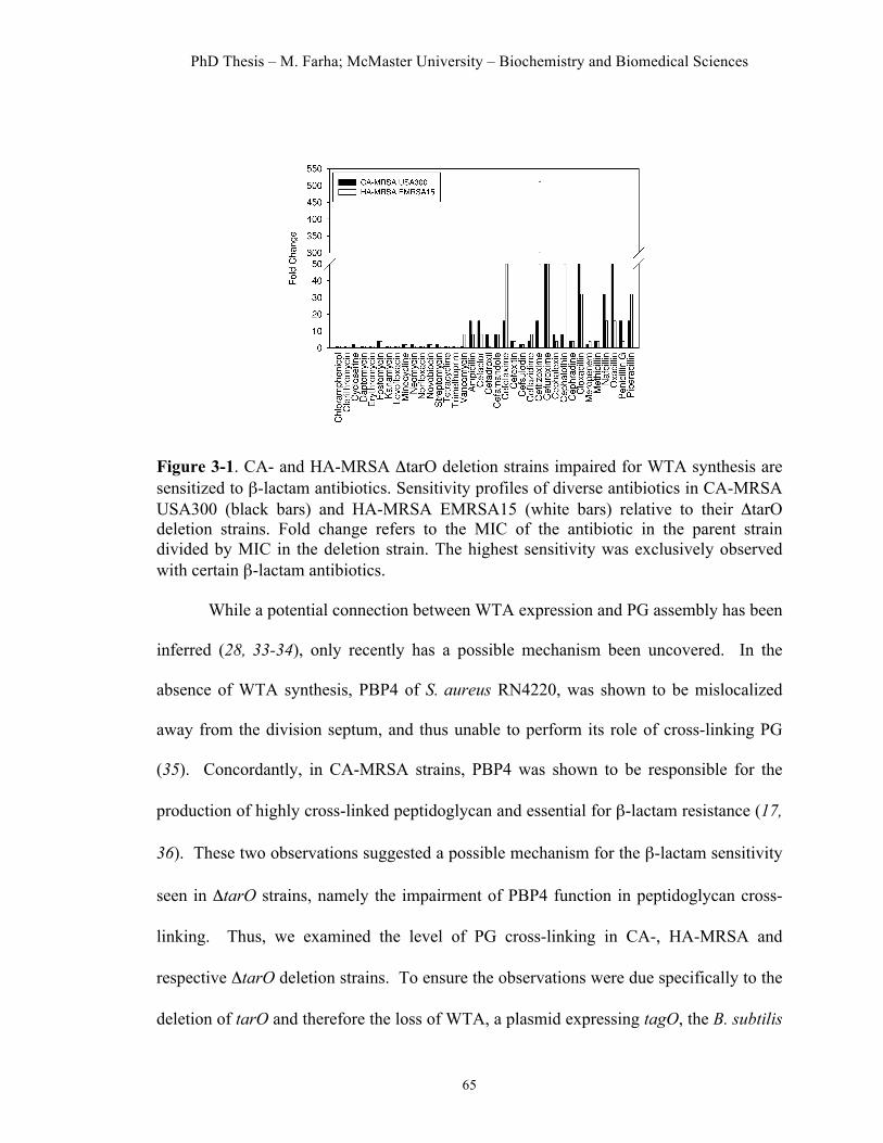
PhD Thesis – M. Farha; McMaster University – Biochemistry and Biomedical Sciences
65
Figure 3-1. CA- and HA-MRSA ΔtarO deletion strains impaired for WTA synthesis are sensitized to β-lactam antibiotics. Sensitivity profiles of diverse antibiotics in CA-MRSA USA300 (black bars) and HA-MRSA EMRSA15 (white bars) relative to their ΔtarO deletion strains. Fold change refers to the MIC of the antibiotic in the parent strain divided by MIC in the deletion strain. The highest sensitivity was exclusively observed with certain β-lactam antibiotics.
While a potential connection between WTA expression and PG assembly has been
inferred (28, 33-34), only recently has a possible mechanism been uncovered. In the
absence of WTA synthesis, PBP4 of S. aureus RN4220, was shown to be mislocalized
away from the division septum, and thus unable to perform its role of cross-linking PG
(35). Concordantly, in CA-MRSA strains, PBP4 was shown to be responsible for the
production of highly cross-linked peptidoglycan and essential for β-lactam resistance (17,
36). These two observations suggested a possible mechanism for the β-lactam sensitivity
seen in ΔtarO strains, namely the impairment of PBP4 function in peptidoglycan cross-
linking. Thus, we examined the level of PG cross-linking in CA-, HA-MRSA and
respective ΔtarO deletion strains. To ensure the observations were due specifically to the
deletion of tarO and therefore the loss of WTA, a plasmid expressing tagO, the B. subtilis

PhD Thesis – M. Farha; McMaster University – Biochemistry and Biomedical Sciences
66
orthologue, was used to complement the HA-MRSA EMRSA15 ΔtarO deletion strain
(Supplementary Figure 2). Indeed, the ΔtarO strain was found to have decreased levels
(approximately 30%) of highly-cross linked muropeptide species as compared to the
parental strain and a higher amount of monomeric, dimeric and trimeric muropeptides
(Supplementary Figure 2), establishing a link between WTA synthesis and β-lactam
sensitivity. We posit that a strain devoid of WTA leads to the mislocalization of PBP4,
compromising its role as a transpeptidase in PG cross-linking and, specifically in the case
of CA-MRSA USA300, results in sensitivity to certain β-lactams.
Combination screening identifies ticlopidine
Given the therapeutic potential of blocking WTA biogenesis in restoring the
efficacy of β-lactams against MRSA, we set out to identify a novel inhibitor of WTA
synthesis. As a source of chemical matter we employed a library of approximately 2,080
previously-approved drugs (PADs). There has been considerable interest in recent years
in the concept of screening for new uses for previously-approved drug molecules. The
interest stems from a growing understanding that small molecules with proven therapeutic
activity for a particular use often have uncharacterized potential for alternate therapeutic
uses (37-38). Implicit is the advantageous potential of any such molecule to rapidly
advance into clinical development by leveraging a proven track record in humans and a
deep history of study. Importantly, potentiators of β-lactam activity would be highly
desirable components of therapeutic combinations against MRSA.

PhD Thesis – M. Farha; McMaster University – Biochemistry and Biomedical Sciences
67
Thus, we mounted a screen of 2,080 previously-approved drugs (Supplementary
Table 2) for compounds capable of potentiating the activity of cefuroxime, a broad
spectrum β-lactam that was highly sensitized in the ΔtarO strain, against the clinically
relevant CA-MRSA strain USA300 (Supplementary Figure 3a). Our screening efforts
yielded several active compounds (Supplementary Figure 3b). To identify potential
inhibitors of WTA, active combinations were tested against the CA-MRSA USA300
ΔtarO strain. Here, we were looking for suppression of synergy since this strain lacks
WTA and is not susceptible to WTA inhibitors. One compound, ticlopidine (Figure 3-
2a), an antiplatelet drug (Ticlid®) that inhibits the binding of adenosine 5’-phosphate to
its platelet receptor in humans, was effective in restoring the efficacy of cefuroxime
against MRSA (Figure 3-2b) and this synergistic interaction was reversed in the ΔtarO
strain (Figure 3-2c). Ticlopidine, while not active on its own as an antibiotic, was
potently synergistic with cefuroxime. Indeed, the Fractional Inhibitory Concentration
(FIC) Index, a common measure of synergy (39), for this pair against CA-MRSA
USA300 was ≤0.063 (Figure 3-2b). Further, ticlopidine dramatically potentiated the
activity of cefuroxime against 9 of 10 MRSA strains (40), including CA-MRSA strain
USA300 (64-fold) and HA-MRSA strain USA200 (32-fold) (Table 3-1). Notably, we did
not observe synergy in the common lab strain of S. aureus, RN4220 (Table 3-1),
consistent with a lack of sensitivity to β-lactams in a ΔtarO deletion of this strain
(Supplementary Table 3). Strong synergies with cefuroxime were also observed with
commercially available on-patent analogs of ticlopidine, namely clopidogrel (Plavix®)
and prasugrel (Effient®), against CA-MRSA USA300 (FIC Index ≤0.125 and ≤0.5,

PhD Thesis – M. Farha; McMaster University – Biochemistry and Biomedical Sciences
68
respectively) (Supplementary Table 4). To assess in vivo efficacy, we administered
ticlopidine and cefuroxime in a Galleria mellonella model of MRSA infection. The
larval stage of the Greater Wax Moth is a widely used model to assess S. aureus virulence
and allows for a testing throughput that is otherwise impossible with small mammals but
nevertheless representative of in vivo activity (41-42). In this model, a significantly
(P<0.001) higher fraction of larvae survived MRSA infection following combined
treatment, compared to that with cefuroxime and ticlopidine alone (Figure 3-2d). After
14 days, the survival rate increased from 3.3% in untreated larvae to 53.3% when treated
with the combination; treatment with cefuroxime and ticlopidine alone led to 16.7% and
10% survival, respectively (Figure 3-2d). Thus, cefuroxime and ticlopidine at sub-
efficacious doses acted synergistically to provide G. mellonella protection from bacterial
infection.
Table 3-1. In vitro interactions between ticlopidine and cefuroxime in S. aureus species
Strain a MIC
cefuroxime (µg/mL)
FICb cefuroxime
MIC ticlopidine (µg/mL)
FICb ticlopidine
FIC Indexc
Newman d 2 0.125 >256 0.125 ≤0.250 HA-MRSA USA600 e ≥1024 0.008 >256 0.032 ≤0.040 HA-MRSA USA100/800/NY e 1024 0.125 >256 0.125 ≤0.250 HA-MRSA e >2048 0.250 >256 0.032 ≤0.282 HA-MRSA USA200/EMRSA16 e 1024 0.032 >256 0.063 ≤0.095 HA-MRSA USA500 e 32 1 >256 1 ≤2 HA-MRSA e >2048 0.250 >256 0.016 ≤0.266 CA-MRSA USA400/MW2 e 256 0.063 >256 0.063 ≤0.125 HA-MRSA EMRSA15 e 512 0.063 >256 0.063 ≤0.125 HA-MRSA e 2048 0.125 >256 0.063 ≤0.188 CA-MRSA USA300 e 512 0.032 >256 0.032 ≤0.063 RN4220 f 0.5 1 >128 1 ≤2 SA178R1 g 0.5 1 >128 1 ≤2
a HA, hospital-associated isolate, CA, community-associated isolate b FIC = [X]/MICX, where [X] is the lowest inhibitory concentration of drug in the presence of the co-drug. c FIC index = FICcefuroxime + FICticlopidine d Ref (50) e Ref (40) f Ref (51) g Ref (26)

PhD Thesis – M. Farha; McMaster University – Biochemistry and Biomedical Sciences
69
Figure 3-2. Ticlopidine potentiates the activity of the β-lactam antibiotic cefuroxime against CA-MRSA USA300, but not CA-MRSA USA300 ΔtarO. (a) Chemical structure of ticlopidine. (b) Microdilution checkerboard analysis showing the combined effect of cefuroxime and ticlopidine against CA-MRSA USA300 where the extent of inhibition is shown as a heat plot. Synergistic effects are evident as both molecules alone have MICs that exceed 256 µg mL-1 and result in an FIC Index of ≤0.063. (c) Suppression of the synergy in CAMRSA USA300 ΔtarO, leading to an additive interaction with FIC Index of ≤2. (d) Galleria mellonella virulence assay. Survival curve of G. Mellonella infected with CA-MRSA USA300 receiving no drug treatment control (CTRL) or a treatment with 0.3 mg/kg cefuroxime (CEF) or 0.3 mg/kg ticlopidine (TIC), or a combination of both at 0.3 mg/kg each (CEF+TIC). After 14 days, treatment with the combination lead to significantly increased survival, compared to no drug control or antibiotic treatment alone (P<0.001).
Characterization of the mode of action of ticlopidine To further probe the mechanism of action of ticlopidine, we investigated its interactions
with an extensive panel of diverse antibiotics using CA-MRSA USA300. Ticlopidine
restored the efficacy of several β-lactam antibiotics, while the activity of other classes of

PhD Thesis – M. Farha; McMaster University – Biochemistry and Biomedical Sciences
70
antibiotics remained unaffected (Supplementary Table 5). Indeed, the sensitization
profile was strikingly similar to that seen in the ΔtarO strains (Figure 3-1), although the
sensitivity was generally higher in the deletion strain, likely because it was completely
devoid of WTA (as shown by phosphate analysis in Supplementary Table 1). Most
remarkable was the lack of antibacterial activity of ticlopidine on its own and its capacity
to render MRSA highly susceptible to a number of β-lactams. We reasoned that if
ticlopidine inhibited WTA synthesis, its lack of antibacterial activity would be consistent
with inhibition of the early steps in the pathway catalyzed by TarO or TarA. WTA
biosynthetic genes exhibit complex dispensability patterns (26, 43). WTA genes
encoding the initiating enzymes, TarO and TarA, are dispensable for growth in vitro
while the downstream late-acting genes have an essential phenotype. Idiosyncratically,
the late-acting genes become dispensable in strains with a deletion in either tarO or tarA,
presumably because accumulation of WTA intermediates is toxic to the cell (26, 29, 33,
43).
To test the hypothesis that ticlopidine inhibited either of these initiating enzymes
we initially employed genetic and physiological approaches. First, we took advantage of
the conditional dispensability patterns of late steps in WTA assembly. As an early step
inhibitor, ticlopidine should have an antagonistic interaction with the late step (TarG)
inhibitor, targosil. (44) TarG, an essential gene product, is the transmembrane component
of the ABC transporter that exports WTAs to the cell surface. Indeed, ticlopidine
rendered targosil completely inactive against both CA-MRSA USA300 and the S. aureus
lab strain RN4220 (Figure 3-3a and Supplementary Figure 4). In additional genetic

PhD Thesis – M. Farha; McMaster University – Biochemistry and Biomedical Sciences
71
experiments, we tested the capacity of ticlopidine to suppress the lethal phenotype
associated with late gene tarH. Using a strain where tarH expression was under the
control of a xylose-induced promoter, we found that ticlopidine could partially rescue of
growth in the absence of xylose (40% relative to growth in 2% xylose) at the highest
concentration tested (Figure 3-3b). We next investigated ticlopidine`s ability to directly
decrease WTA synthesis by measuring phosphate content in the cell wall of S. aureus
with increasing concentrations of ticlopidine. In both CA-MRSA USA300 (Figure 3-3c)
and SA1781 (a RN4220 derivative) (Supplementary Figure 5), WTA incorporation began
to decrease at ticlopidine concentrations of 64 µg/mL, similar to the concentration at
which it begins to potentiate the activity of cefuroxime. Greater inhibition was noted at
200 µg/mL of ticlopidine but residual WTA remained (~50%), while ΔtarO strains were
completely devoid of phosphate. We note here that the partial suppression seen was
consistent with the partial inhibition of WTA synthesis evident in our cell wall phosphate
content assays. Furthermore, bacteriophage Ø11, which uses WTA as receptor-binding
sites, infected S. aureus RN450 but not ticlopidine- or tunicamycin-treated RN450
(Supplementary Figure 6). WTA is known to contribute to the resistance of PG against
lysozyme (45). We found that wild-type PG was completely resistant to degradation by
lysozyme while PG from ticlopidine-treated cells and ΔtarO cells were indeed sensitive to
degradation (Supplementary Figure 7). In sum, our genetic and physiological
experiments were consistent with ticlopidine targeting either TarO or TarA.
To test directly the hypothesis that ticlopidine inhibited either TarO or TarA, we
used in vitro biochemical assays. While ticlopidine showed no impact on the transferase

PhD Thesis – M. Farha; McMaster University – Biochemistry and Biomedical Sciences
72
activity of TarA (Supplementary Figure 9) we found substantial inhibition of TarO
activity (Figure 3-3d). Further, the relatively weak inhibition constant (IC50 of 238 µM or
71 µg/mL) was consistent with the partial phenotypes noted in our phenotypic and genetic
assays.
Figure 3-3. Ticlopidine inhibits the initiation of wall teichoic acid biosynthesis in S. aureus. (a) At concentrations of 8 µg mL-1, ticlopidine begins to antagonize the activity of targosil, a late-stage inhibitor, against CA-MRSA USA300. (b) Ticlopidine can suppress the lethality associated with late WTA stage deletion. Shown is the percent growth of a tarH conditional deletion strain normalized to the growth upon induction with 2% xylose in the presence of increasing concentrations of ticlopidine. Ticlopidine can recover approximately 40% of the growth at the highest concentration of 256 µg mL-1. (c) Ticlopidine shows a dose-dependent decrease in the phosphate content of cell wall isolated from CA-MRSA USA300, with approximately 50% less phosphate when ticlopidine is present at 200 µg mL-1. CA-MRSA USA300 ΔtarO is completely devoid of WTA compared to the parental strain. (d) Membrane-based in vitro assay following the generation of a radiolabeled reaction product of TarO, undecapreny-P-P-[14C]GlcNAc, as a result of the incorporation of [14C]GlcNAc onto undecaprenyl-P-P. Assay was performed on membranes derived from E. coli cells (ΔwecA) expressing recombinant TarO from S. aureus. The reaction product was monitored by thin layer chromatography (TLC) and shown to be dependent on the presence of recombinant TarO (Supplementary Figure 8). Ticlopidine inhibited the activity of TarO, yielding an IC50 value of 238 ± 14 µM.

PhD Thesis – M. Farha; McMaster University – Biochemistry and Biomedical Sciences
73
A basis for the synergy among ticlopidine and ß-lactams
To further investigate the basis of synergy of ticlopidine and cefuroxime we
sought to understand the characteristic β-lactam sensitivity profiles that were shared on
treatment of MRSA in combination with ticlopidine or alone using the ∆tarO MRSA
strains. A survey of the PBP selectivity of β-lactams tested in our studies revealed that
sensitization was seen particularly with those having high binding affinities for PBP2 of
S. aureus (Figure 3-1 and Supplementary Table 5). Given the impact of the loss of WTA
on PBP4 localization (35), PBP2 selectivity can be rationalized based on previous reports
of cooperativity between PBP2 and PBP4 in creating highly cross-linked PG in MRSA
strains contributing to β-lactam resistance (18) (Figure 3-4, panel i). A strain lacking
WTA, such as with treatment with ticlopidine, would be compromised in its PG cross-
linking due to mislocalized PBP4 and be highly sensitive to β-lactams such as cefuroxime
that target its partner protein PBP2 (Figure 3-4, panel iii). Resistance would persist if the
β-lactam in combination with ticlopidine targets PBP1 or PBP3, as sufficient cross-
linking would maintain the resistant phenotype (Figure 3-4, panel ii). Further, this
rationale is consistent with the idea that PBP2A is not the sole determinant for β-lactam
resistance in MRSA (9, 15, 17). It suggests that the synergy between ticlopidine and
cefuroxime might also be observed in a wild-type, methicillin-sensitive strain such as S.
aureus Newman, which lacks PBP2A. Indeed, synergy is evident in this strain (Table 3-
1). Remarkably, this work also suggests that β-lactam resistance might be reversed by
targeting PBP2 and PBP4 with combinations of existing β-lactams. We tested this using

PhD Thesis – M. Farha; McMaster University – Biochemistry and Biomedical Sciences
74
the PBP4-selective β-lactam cefoxitin (46). When cefoxitin was combined with the
PBP2-selective β-lactams cefuroxime, ceftizoxime, oxacillin and penicillin, the MICs of
these antibiotics decreased 32 to 128-fold. Conversely, combining cefoxitin with β-
lactams having a low affinity for PBP2 led to only a 2-8 fold change in the MIC
(Supplementary Table 6).
Figure 3-4. A synthetic lethal interaction when targeting TarO and native PBP2. Indirect Inhibition of PBP4, via inhibition of TarO, and inhibition of PBP2 function account for the synergistic interaction among ticlopidine and cefuroxime. i. When not challenged with β-lactams, WTA synthesis will guide the proper localization of PBP4, and together with PBP2, will provide highly cross-linked muropeptide species (thick arrow) that contribute to high-level β-lactam resistance. ii. Treatment with a β-lactam with low affinity for PBP2 and ticlopidine leads to an additive interaction as sufficient highly cross-linked peptidoglycan are present to maintain β-lactam resistance (one thick arrow), even when PBP4 function is affected by the lack of WTA (one thin arrow). iii. Due to their cooperative function, PBP4 (when challenged with ticlopidine) and PBP2 (when challenged with β-lactam with high affinity for PBP2), will be impaired in their capacities to produce a highly cross-linked cell wall (two thin arrows), contributing to enhanced β-lactam susceptibility and thus a synergistic interaction.

PhD Thesis – M. Farha; McMaster University – Biochemistry and Biomedical Sciences
75
Conclusions
The WTA biosynthetic pathway has long been speculated as a viable target for
antibacterial intervention. While dispensable for viability, WTA is known to be a critical
determinant of cell shape in Bacillus subtilis (27, 29), virulence in S. aureus (30) and
recently, for proper cell division (28). Furthermore, the essential phenotypes of late WTA
biosynthetic genes, although paradoxical, suggest that these enzymes may well be
reasonable targets for new antibiotics. Emerging evidence of a role for wall teichoic
acids in β-lactam resistance in MRSA has fueled further interest in targeting this pathway,
including a renewed interest in tunicamycin, a natural product nucleoside antibiotic that
has been shown to inhibit TarO (28). Unfortunately, tunicamycin is a promiscuous
inhibitor of bacterial and eukaryotic phosphosugar transferases, hindering its use as a
selective probe and therapeutic. In the work reported here, we have carried out a
meticulous chemical genetic study of the importance of wall teichoic acids to community-
and hospital-acquired MRSA strains to reveal signature interactions with β-lactam
antibiotics having a known selectivity for PBP2. These findings led to the discovery that
ticlopidine, a well-known antiplatelet drug, had the cryptic capacity to block WTA
synthesis through the inhibition of TarO. Importantly, synergistic interactions of
ticlopidine and PBP2-binding β-lactams provide for efficacious combinations to limit the
growth of MRSA strains in vitro and in vivo. Thus ticlopidine represents a promising
new lead with a strong record of examination in humans but also provides an exciting

PhD Thesis – M. Farha; McMaster University – Biochemistry and Biomedical Sciences
76
new probe of WTA synthesis. In our hands, ticlopidine was an extremely useful probe
along with existing β-lactam antibiotics to further elaborate the role of WTA in
supporting the cooperative role of PBP2 and 4 in the expression of β-lactam resistance in
MRSA. As such, this work offers new insights into the complex biology underlying cell
wall synthesis in S. aureus and provides a promising example of how antibiotic drug
resistance might be targeted with existing drugs.
Methods Peptidoglycan purification and analysis. Peptidoglycan from CA-MRSA USA300,
HA-MRSA EMRSA15, their ΔtarO deletion and HA-MRSA EMRSA15ΔtarO pLI50-
tagO was prepared from exponentially growing cells as previously described (47). The
purified peptidoglycan was digested with mutanolysin (Sigma), an N-acetylmuramidase
that cuts glycan strands between the N-acetylmuramic and N-acetylglucosamine residues.
The resulting muropeptides were reduced with sodium borohydride (Sigma) and analyzed
by reverse-phase HPLC using a Hypersil ODS column (Thermo Electron Corporation).
The eluted muropeptides were detected and quantified by determination of their
ultraviolet absorption at 206 nm, using the Shimadzu LC solution software. The area of
eluted UV-absorbing peaks, corresponding to the different muropeptides, was quantified
and shown as a percentage of the total area of the chromatogram.
Combination screening. CA-MRSA USA300 was screened against the PAD library in
the presence of cefuroxime. The screening protocol was based on CLSI guidelines.
Screening was carried out in 96-well plates, in duplicate, using Mueller Hinton Broth
(MHB) with 2% DMSO and a library compound concentration of 10 µM. The

PhD Thesis – M. Farha; McMaster University – Biochemistry and Biomedical Sciences
77
concentration of cefuroxime was 16 µg ml-1, a quarter of its MIC value obtained under
the same conditions. Background controls (8 wells per plate) contained only media and
DMSO and growth controls, also 8 wells per plate, contained media, DMSO and
inoculum. Plates were incubated at 37°C for 20 hours and optical density read at 600nm
using an EnVision plate reader (Perkin Elmer). The percentage growth for each test well
was calculated as (OD – mean background) / (mean growth – mean background) * 100
and normalized to the percent growth attributed by the PAD alone to obtain a growth ratio
such that a ratio of 1.0 is indicative of no difference.
Checkerboard analyses and FIC index determination. FICs were determined by
setting up standard checkerboard broth microdilution assays with serially diluted 8 (or 9)
concentrations of each drug, using the same conditions as screening. At least 3 replicates
were done for each combination and the means used for calculation. The MIC for each
drug was the lowest [drug] showing <10% growth. The FIC for each drug was calculated
as the [drug in the presence of co-drug] for a well showing <10% growth, divided by the
MIC for that drug. The FIC index is the sum of the two FICs. Interactions with FIC
Index of less than 0.5 were deemed synergistic.
Phosphate analysis. Strains were inoculated from an overnight culture and grown in 100
ml MHB to OD600 0.8-0.9 at 37°C, 250 rpm with various compound treatment. Isolation
of cell wall and phosphate analysis were performed as previously described (48).
In vitro TarO inhibition assay. For this assay, UDP-[14C]GlcNAc (0.1 mCi mL-1) was
purchased from American Radiolabelled Chemicals, Ultima Gold liquid scintillation
cocktail from Perkin Elmer and Silica gel 60 TLC plates from EMD Chemicals. TarO

PhD Thesis – M. Farha; McMaster University – Biochemistry and Biomedical Sciences
78
activity was assayed in 100 µL reactions containing Reaction Buffer (50 mM Tris pH = 8,
10 mM MgCl2, 1 mM EDTA), 300 µM UDP-GlcNAc, 0.1 µCi UDP-[14C]GlcNAc, 0.1 %
(w/v) TritonX-100, 0.8 % (v/v) DMSO and varied amounts of membranes (75 µg - 1 mg
of protein). Reactions were quenched with the addition of 1250 µL CHCl3:MeOH (3:2).
Lipid-linked products were extracted according to the following method: Quenched
reactions were incubated for 2.5 hrs followed by vortexing for 3 min. Insoluble material
was removed by centrifugation (13 000 x g, 2 minutes), and 150 µL of 40 mM MgCl2 was
added to the supernatant. Samples were vortexted for 5 min, and the upper aqueous layer
was removed. The lower, organic layer was washed twice with 400 µL of pure solvent
upper phase (CHCl3:MeOH:H2O:1 M MgCl2 in H2O [18:294:282:1]). The final product
was analyzed by either TLC or scintillation counting. TLC analysis was preformed as in
reference (49).
Acknowledgments
We would like to thank N. Magarvey for providing bacteriophage Ø11. The work was supported by a Canada Research Chair Award to E.D.B, the Canadian Cystic Fibrosis Foundation and an operating grant from CIHR to E.D.B (MOP-81330). Work in the laboratory of GDW was supported by a Canada Research Chair Award to G.D.W and CIHR operating grant to G.D.W (MT-13536). Work in the laboratory of M.G.P was funded by FCT grants PTDC/BIA-BCM/099152/2008 and PEst-OE/EQB/LA0004/2011. P.M.P was supported by FCT fellowship SFRH/BD/41119/2007.
References 1. Boucher, H. W., Talbot, G. H., Bradley, J. S., Edwards, J. E., Gilbert, D., Rice, L.
B., Scheld, M., Spellberg, B., and Bartlett, J. (2009) Bad bugs, no drugs: no ESKAPE! An update from the Infectious Diseases Society of America, Clin Infect Dis 48, 1-12.
2. Klevens, R. M., Morrison, M. A., Nadle, J., Petit, S., Gershman, K., Ray, S., Harrison, L. H., Lynfield, R., Dumyati, G., Townes, J. M., Craig, A. S., Zell, E. R., Fosheim, G. E., McDougal, L. K., Carey, R. B., and Fridkin, S. K. (2007)

PhD Thesis – M. Farha; McMaster University – Biochemistry and Biomedical Sciences
79
Invasive methicillin-resistant Staphylococcus aureus infections in the United States, JAMA 298, 1763-1771.
3. Baba, T., Takeuchi, F., Kuroda, M., Yuzawa, H., Aoki, K., Oguchi, A., Nagai, Y., Iwama, N., Asano, K., Naimi, T., Kuroda, H., Cui, L., Yamamoto, K., and Hiramatsu, K. (2002) Genome and virulence determinants of high virulence community-acquired MRSA, Lancet 359, 1819-1827.
4. Diep, B. A., Gill, S. R., Chang, R. F., Phan, T. H., Chen, J. H., Davidson, M. G., Lin, F., Lin, J., Carleton, H. A., Mongodin, E. F., Sensabaugh, G. F., and Perdreau-Remington, F. (2006) Complete genome sequence of USA300, an epidemic clone of community-acquired meticillin-resistant Staphylococcus aureus, Lancet 367, 731-739.
5. Howden, B. P., Johnson, P. D., Charles, P. G., and Grayson, M. L. (2004) Failure of vancomycin for treatment of methicillin-resistant Staphylococcus aureus infections, Clin Infect Dis 39, 1544; author reply 1544-1545.
6. Mangili, A., Bica, I., Snydman, D. R., and Hamer, D. H. (2005) Daptomycin-resistant, methicillin-resistant Staphylococcus aureus bacteremia, Clin Infect Dis 40, 1058-1060.
7. Tsiodras, S., Gold, H. S., Sakoulas, G., Eliopoulos, G. M., Wennersten, C., Venkataraman, L., Moellering, R. C., and Ferraro, M. J. (2001) Linezolid resistance in a clinical isolate of Staphylococcus aureus, Lancet 358, 207-208.
8. Lim, D., and Strynadka, N. C. (2002) Structural basis for the beta lactam resistance of PBP2a from methicillin-resistant Staphylococcus aureus, Nat Struct Biol 9, 870-876.
9. Pinho, M. G., de Lencastre, H., and Tomasz, A. (2001) An acquired and a native penicillin-binding protein cooperate in building the cell wall of drug-resistant staphylococci, Proc Natl Acad Sci U S A 98, 10886-10891.
10. Llarrull, L. I., Fisher, J. F., and Mobashery, S. (2009) Molecular basis and phenotype of methicillin resistance in Staphylococcus aureus and insights into new beta-lactams that meet the challenge, Antimicrob Agents Chemother 53, 4051-4063.
11. Turk, S., Verlaine, O., Gerards, T., Zivec, M., Humljan, J., Sosic, I., Amoroso, A., Zervosen, A., Luxen, A., Joris, B., and Gobec, S. (2011) New noncovalent inhibitors of penicillin-binding proteins from penicillin-resistant bacteria, PLoS One 6, e19418.
12. Aksoy, D. Y., and Unal, S. (2008) New antimicrobial agents for the treatment of Gram-positive bacterial infections, Clin Microbiol Infect 14, 411-420.
13. Berger-Bachi, B. (1999) Genetic basis of methicillin resistance in Staphylococcus aureus, Cell Mol Life Sci 56, 764-770.
14. de Lencastre, H., and Tomasz, A. (1994) Reassessment of the number of auxiliary genes essential for expression of high-level methicillin resistance in Staphylococcus aureus, Antimicrob Agents Chemother 38, 2590-2598.
15. De Lencastre, H., Wu, S. W., Pinho, M. G., Ludovice, A. M., Filipe, S., Gardete, S., Sobral, R., Gill, S., Chung, M., and Tomasz, A. (1999) Antibiotic resistance as a stress response: complete sequencing of a large number of chromosomal loci in

PhD Thesis – M. Farha; McMaster University – Biochemistry and Biomedical Sciences
80
Staphylococcus aureus strain COL that impact on the expression of resistance to methicillin, Microb Drug Resist 5, 163-175.
16. Labischinski, H., Ehlert, K., and Berger-Bachi, B. (1998) The targeting of factors necessary for expression of methicillin resistance in staphylococci, J Antimicrob Chemother 41, 581-584.
17. Memmi, G., Filipe, S. R., Pinho, M. G., Fu, Z., and Cheung, A. (2008) Staphylococcus aureus PBP4 is essential for beta-lactam resistance in community-acquired methicillin-resistant strains, Antimicrob Agents Chemother 52, 3955-3966.
18. Leski, T. A., and Tomasz, A. (2005) Role of penicillin-binding protein 2 (PBP2) in the antibiotic susceptibility and cell wall cross-linking of Staphylococcus aureus: evidence for the cooperative functioning of PBP2, PBP4, and PBP2A, J Bacteriol 187, 1815-1824.
19. Gardete, S., Ludovice, A. M., Sobral, R. G., Filipe, S. R., de Lencastre, H., and Tomasz, A. (2004) Role of murE in the Expression of beta-lactam antibiotic resistance in Staphylococcus aureus, J Bacteriol 186, 1705-1713.
20. Sobral, R. G., Ludovice, A. M., Gardete, S., Tabei, K., De Lencastre, H., and Tomasz, A. (2003) Normally functioning murF is essential for the optimal expression of methicillin resistance in Staphylococcus aureus, Microb Drug Resist 9, 231-241.
21. Berger-Bachi, B., and Rohrer, S. (2002) Factors influencing methicillin resistance in staphylococci, Arch Microbiol 178, 165-171.
22. Kuroda, M., Kuroda, H., Oshima, T., Takeuchi, F., Mori, H., and Hiramatsu, K. (2003) Two-component system VraSR positively modulates the regulation of cell-wall biosynthesis pathway in Staphylococcus aureus, Mol Microbiol 49, 807-821.
23. Lee, S. H., Jarantow, L. W., Wang, H., Sillaots, S., Cheng, H., Meredith, T. C., Thompson, J., and Roemer, T. (2011) Antagonism of chemical genetic interaction networks resensitize MRSA to beta-lactam antibiotics, Chem Biol 18, 1379-1389.
24. Tan, C. M., Therien, A. G., Lu, J., Lee, S. H., Caron, A., Gill, C. J., Lebeau-Jacob, C., Benton-Perdomo, L., Monteiro, J. M., Pereira, P. M., Elsen, N. L., Wu, J., Deschamps, K., Petcu, M., Wong, S., Daigneault, E., Kramer, S., Liang, L., Maxwell, E., Claveau, D., Vaillancourt, J., Skorey, K., Tam, J., Wang, H., Meredith, T. C., Sillaots, S., Wang-Jarantow, L., Ramtohul, Y., Langlois, E., Landry, F., Reid, J. C., Parthasarathy, G., Sharma, S., Baryshnikova, A., Lumb, K. J., Pinho, M. G., Soisson, S. M., and Roemer, T. (2012) Restoring Methicillin-Resistant Staphylococcus aureus Susceptibility to beta-Lactam Antibiotics, Sci Transl Med 4, 126ra135.
25. Maki, H., Yamaguchi, T., and Murakami, K. (1994) Cloning and characterization of a gene affecting the methicillin resistance level and the autolysis rate in Staphylococcus aureus, J Bacteriol 176, 4993-5000.
26. D'Elia, M. A., Pereira, M. P., Chung, Y. S., Zhao, W., Chau, A., Kenney, T. J., Sulavik, M. C., Black, T. A., and Brown, E. D. (2006) Lesions in teichoic acid biosynthesis in Staphylococcus aureus lead to a lethal gain of function in the otherwise dispensable pathway, J Bacteriol 188, 4183-4189.

PhD Thesis – M. Farha; McMaster University – Biochemistry and Biomedical Sciences
81
27. Bhavsar, A. P., Beveridge, T. J., and Brown, E. D. (2001) Precise deletion of tagD and controlled depletion of its product, glycerol 3-phosphate cytidylyltransferase, leads to irregular morphology and lysis of Bacillus subtilis grown at physiological temperature, J Bacteriol 183, 6688-6693.
28. Campbell, J., Singh, A. K., Santa Maria, J. P., Jr., Kim, Y., Brown, S., Swoboda, J. G., Mylonakis, E., Wilkinson, B. J., and Walker, S. (2011) Synthetic lethal compound combinations reveal a fundamental connection between wall teichoic acid and peptidoglycan biosyntheses in Staphylococcus aureus, ACS Chem Biol 6, 106-116.
29. D'Elia, M. A., Millar, K. E., Beveridge, T. J., and Brown, E. D. (2006) Wall teichoic acid polymers are dispensable for cell viability in Bacillus subtilis, J Bacteriol 188, 8313-8316.
30. Weidenmaier, C., and Peschel, A. (2008) Teichoic acids and related cell-wall glycopolymers in Gram-positive physiology and host interactions, Nat Rev Microbiol 6, 276-287.
31. Brown, S., Xia, G., Luhachack, L. G., Campbell, J., Meredith, T. C., Chen, C., Winstel, V., Gekeler, C., Irazoqui, J. E., Peschel, A., and Walker, S. (2012) Methicillin resistance in Staphylococcus aureus requires glycosylated wall teichoic acids, Proc Natl Acad Sci U S A.
32. Price, N. P., and Tsvetanova, B. (2007) Biosynthesis of the tunicamycins: a review, J Antibiot (Tokyo) 60, 485-491.
33. D'Elia, M. A., Millar, K. E., Bhavsar, A. P., Tomljenovic, A. M., Hutter, B., Schaab, C., Moreno-Hagelsieb, G., and Brown, E. D. (2009) Probing teichoic acid genetics with bioactive molecules reveals new interactions among diverse processes in bacterial cell wall biogenesis, Chem Biol 16, 548-556.
34. Schlag, M., Biswas, R., Krismer, B., Kohler, T., Zoll, S., Yu, W., Schwarz, H., Peschel, A., and Gotz, F. (2010) Role of staphylococcal wall teichoic acid in targeting the major autolysin Atl, Mol Microbiol 75, 864-873.
35. Atilano, M. L., Pereira, P. M., Yates, J., Reed, P., Veiga, H., Pinho, M. G., and Filipe, S. R. (2010) Teichoic acids are temporal and spatial regulators of peptidoglycan cross-linking in Staphylococcus aureus, Proc Natl Acad Sci U S A 107, 18991-18996.
36. Henze, U. U., and Berger-Bachi, B. (1995) Staphylococcus aureus penicillin-binding protein 4 and intrinsic beta-lactam resistance, Antimicrob Agents Chemother 39, 2415-2422.
37. Chong, C. R., and Sullivan, D. J., Jr. (2007) New uses for old drugs, Nature 448, 645-646.
38. Ejim, L., Farha, M. A., Falconer, S. B., Wildenhain, J., Coombes, B. K., Tyers, M., Brown, E. D., and Wright, G. D. (2010) Combinations of antibiotics and nonantibiotic drugs enhance antimicrobial efficacy, Nat Chem Biol 7, 348-350.
39. Odds, F. C. (2003) Synergy, antagonism, and what the chequerboard puts between them, J Antimicrob Chemother 52, 1.
PhD Thesis – M. Farha; McMaster University – Biochemistry and Biomedical Sciences
82
40. Christianson, S., Golding, G. R., Campbell, J., and Mulvey, M. R. (2007) Comparative genomics of Canadian epidemic lineages of methicillin-resistant Staphylococcus aureus, J Clin Microbiol 45, 1904-1911.
41. Gao, W., Chua, K., Davies, J. K., Newton, H. J., Seemann, T., Harrison, P. F., Holmes, N. E., Rhee, H. W., Hong, J. I., Hartland, E. L., Stinear, T. P., and Howden, B. P. (2010) Two novel point mutations in clinical Staphylococcus aureus reduce linezolid susceptibility and switch on the stringent response to promote persistent infection, PLoS Pathog 6, e1000944.
42. Purves, J., Cockayne, A., Moody, P. C., and Morrissey, J. A. (2010) Comparison of the regulation, metabolic functions, and roles in virulence of the glyceraldehyde-3-phosphate dehydrogenase homologues gapA and gapB in Staphylococcus aureus, Infect Immun 78, 5223-5232.
43. D'Elia, M. A., Henderson, J. A., Beveridge, T. J., Heinrichs, D. E., and Brown, E. D. (2009) The N-acetylmannosamine transferase catalyzes the first committed step of teichoic acid assembly in Bacillus subtilis and Staphylococcus aureus, J Bacteriol 191, 4030-4034.
44. Swoboda, J. G., Meredith, T. C., Campbell, J., Brown, S., Suzuki, T., Bollenbach, T., Malhowski, A. J., Kishony, R., Gilmore, M. S., and Walker, S. (2009) Discovery of a small molecule that blocks wall teichoic acid biosynthesis in Staphylococcus aureus, ACS Chem Biol 4, 875-883.
45. Bera, A., Biswas, R., Herbert, S., Kulauzovic, E., Weidenmaier, C., Peschel, A., and Gotz, F. (2007) Influence of wall teichoic acid on lysozyme resistance in Staphylococcus aureus, J Bacteriol 189, 280-283.
46. Nair, S. R., and Cherubin, C. E. (1978) Use of cefoxitin, new cephalosporin-like antibiotic, in the treatment of aerobic and anaerobic infections, Antimicrob Agents Chemother 14, 866-875.
47. Filipe, S. R., Tomasz, A., and Ligoxygakis, P. (2005) Requirements of peptidoglycan structure that allow detection by the Drosophila Toll pathway, EMBO Rep 6, 327-333.
48. Bhavsar, A. P., Erdman, L. K., Schertzer, J. W., and Brown, E. D. (2004) Teichoic acid is an essential polymer in Bacillus subtilis that is functionally distinct from teichuronic acid, J Bacteriol 186, 7865-7873.
49. Schaffer, C., Wugeditsch, T., Messner, P., and Whitfield, C. (2002) Functional expression of enterobacterial O-polysaccharide biosynthesis enzymes in Bacillus subtilis, Appl Environ Microbiol 68, 4722-4730.
50. Duthie, E. S., and Lorenz, L. L. (1952) Staphylococcal coagulase; mode of action and antigenicity, J Gen Microbiol 6, 95-107.
51. Kreiswirth, B. N., Lofdahl, S., Betley, M. J., O'Reilly, M., Schlievert, P. M., Bergdoll, M. S., and Novick, R. P. (1983) The toxic shock syndrome exotoxin structural gene is not detectably transmitted by a prophage, Nature 305, 709-712.

PhD Thesis – M. Farha; McMaster University – Biochemistry and Biomedical Sciences
83
CHAPTER FOUR – Collapsing the Proton Motive Force to Identify Synergistic Combinations Against Staphylococcus aureus

PhD Thesis – M. Farha; McMaster University – Biochemistry and Biomedical Sciences
84
Preface The work presented in this chapter was previously published in:
Farha, MA, Verschoor, CP, Bowdish D, and Brown, ED. (2013) Collapsing the proton motive force to identify novel synergistic combinations against Staphylococcus aureus. Chemistry & Biology, 20:1168-1178. Permission has been granted from the publisher to reproduce the material here as prescribed by Chemistry and Biology copyright permissions.
For this work, I performed all of the experiments (with the exception of the cytotoxicity assays, performed by Verschoor, CP) and wrote the manuscript, with edits provided by Brown, ED.
PhD Thesis – M. Farha; McMaster University – Biochemistry and Biomedical Sciences
85
Summary
Pathways of bacterial energy metabolism, such as the proton motive force, have
largely remained unexplored as drug targets, owing to toxicity concerns. Here, we
elaborate a methodical and systematic approach for targeting PMF using chemical
combinations. We began with a high-throughput screen to identify molecules that
selectively dissipate either component of the PMF, ΔΨ or ΔpH, in S. aureus. We
uncovered six perturbants of PMF, three (I1, I2 and I3) that countered ΔΨ and three (D1,
D2 and D3) that selectively dissipated ΔpH. Combinations of dissipators of ΔΨ with
dissipators of ΔpH were highly synergistic against MRSA. Cytotoxicity analyses on
mammalian cells revealed that the dose-sparing effect of the observed synergies could
significantly reduce toxicity. The discovery and rational combination of modulators of
ΔΨ and ΔpH may represent a promising strategy for combating microbial pathogens.

PhD Thesis – M. Farha; McMaster University – Biochemistry and Biomedical Sciences
86
Introduction
Emergence and spread of multidrug-resistant bacteria represents an increasingly
serious problem and an unmet medical need due to the lack of therapeutic options. The
situation is especially dire for Staphylococcus aureus for which methicillin-resistant
(MRSA) and vancomycin intermediate resistant (VISA) strains have emerged (Chopra,
2003). The escalating concern of antibiotic resistance coupled with the dwindling
antibiotic pipeline has prompted drug developers to turn to unconventional strategies for
the discovery of novel drugs to combat bacterial infections, both in exploring novel
compound classes and validating new target pathways.
The cell membrane of S. aureus, in particular, is a relatively unexplored target for
the development of novel antimicrobials. This essential macromolecular structure plays a
critical role in cellular activities such as active transport of substances into the cell,
cellular respiration, the establishment of the proton motive force and cell-cell
communication (Hurdle, et al., 2011). Yet, over the years, discovery efforts have steered
clear of membrane-active agents that either disrupt the membrane’s physical integrity or
target its energetics, mostly given challenges with associated toxicity in mammals. In
fact, a consensus has emerged in the field that active compounds discovered in whole
cells screens that act primarily on the cell membrane should be excluded from further
study (O'Neill and Chopra, 2004; Silver, 2011). Indeed, a number of assays have been
explored to detect these undesirable compounds early enough to evade follow-up studies
(Gentry, et al., 2010; Prakash Singh, 2006).

PhD Thesis – M. Farha; McMaster University – Biochemistry and Biomedical Sciences
87
Paradoxically, this unconventional antibacterial target has gained attention, in part
because of studies showing its importance in non-growing bacteria (Coates and Hu, 2008;
Hu, et al., 2010; Hurdle, et al., 2011) and out of recognition of the recent success of the
antibiotic medicines daptomycin (Hawkey, 2008), telavancin (Zhanel, et al., 2010), and
HT61 (Hu, et al., 2010), the latter currently in clinical trials for topical use, that work by
permeabilizing and depolarizing the cytoplasmic membrane of S. aureus. Also
noteworthy is the compound TMC107, which disrupts membrane energetics by targeting
the ATP synthase and is currently in phase IIb trials for the treatment of drug-resistant
Mycobacterium tuberculosis (Balemans, et al., 2012). Nevertheless, the energetic
pathways of the bacterial membrane remain largely unexplored and relatively unpopular
drug targets.
In bacteria, the extrusion of protons by the electron transport chain results in an
electrochemical gradient of protons, known as the proton motive force (PMF), generated
across the cell membrane. The PMF is subsequently necessary for ATP synthesis by the
F1F0-ATPase and for transport of various solutes (Mitchell, 1966). The PMF is made up
of the sum of two parameters: the electric potential (ΔΨ) and the transmembrane proton
gradient (ΔpH). Critical to bacterial survival, the PMF has nevertheless been largely
disregarded as a target for antimicrobials, owing to toxicity concerns. Still, ionophores
such as the natural product monensin, that dissipate the PMF by disrupting the flow of
ions across the membrane, have found extensive use as antibacterial agents in the cattle
and poultry industries (Russell and Houlihan, 2003). Interestingly, bacteria exercise
exquisite control over ΔΨ and ΔpH in order to maintain a constant value of PMF.
PhD Thesis – M. Farha; McMaster University – Biochemistry and Biomedical Sciences
88
Dissipation in either component is compensated for by a counteracting increase in the
other (Bakker and Mangerich, 1981). We sought in this work to exploit this
compensation mechanism as an Achilles heel in bacteria that would be exquisitely
susceptible to a combination of perturbants, one that specifically disrupted the electric
potential (ΔΨ) and another, the transmembrane proton gradient (ΔpH).
We set out to identify specific disrupters of the electrical and chemical
components of PMF through a fluorescence-based high-throughput screen. In particular,
the screen identified three compounds that specifically dissipated ΔΨ and three that
selectively disrupted ΔpH. We demonstrated profound synergy between these two classes
of compounds when used in combination against methicillin resistant S. aureus (MRSA).
Thus the concerted dissipation of ΔΨ and ΔpH had a synergistic effect in collapsing the
PMF and, importantly, this synergy spares the doses that are necessary for each of the
individual agents. We show that this dose-sparing phenomenon likewise reduces the
toxicity of the respective agents. Indeed, drug combination therapy is gaining favour
(Keith, et al., 2005) on account of increased efficacy, lower toxicity and reduced
propensity for resistance (Cottarel and Wierzbowski, 2007). Herein, we report on the first
systematic effort in discovering synergistic antibacterial chemical combinations that
exploit the interdependence of the electric potential and chemical gradient of the
membrane in maintaining the proton motive force. These findings suggest new
opportunities to target energetic systems within bacterial pathogens as a means to develop
novel therapeutic strategies.

PhD Thesis – M. Farha; McMaster University – Biochemistry and Biomedical Sciences
89
Results
Development of a high-throughput screen for alterations in PMF.
A widely used probe to study the effect of small molecules on membrane potential
is the cationic, membrane potential-sensitive dye, 3,3’-dipropylthiacarbocyanine iodide
(DiSC3(5)) (Wu, et al., 1999). In this fluorescence-based assay, bacteria are grown and
loaded with dye, which accumulates in the cytoplasmic membrane in response to ΔΨ and
self-quenches its own fluorescence. When ΔΨ is disrupted or the membrane
permeabilized upon treatment with a small molecule, the dye is released into the medium
resulting in an increase in fluorescence. Interestingly, this dye can also inform on
dissipations in the other component of PMF, the transmembrane ΔpH, through observed
decreases in fluorescence. Upon dissipation of their pH gradient, bacterial cells will
compensate by increasing ΔΨ to maintain a constant PMF. As such, this increased
membrane potential further concentrates DiSC3(5) dye in the membrane, such that high
local concentrations lead to decreased fluorescence intensity due to further quenching.
As validation for the assay, we measured the effect of valinomycin (in the presence of K+)
and nigericin. Valinomcyin, a K+ ionophore which specifically dissipates the ΔΨ
component of the PMF, led to a significant increase in fluorescence (Figure 4-1A), while
nigericin, an electroneutral antiporter for H+ and K+, caused a decrease in fluorescence as
it selectively dissipates ΔpH (Figure 4-1A). Solvent (DMSO)-treated DiSC3(5) loaded
cells did not lead to changes in fluorescence consistent with an intact membrane potential
(Figure 4-1A). Having established DiSC3(5) as a suitable tool to identify molecules that

PhD Thesis – M. Farha; McMaster University – Biochemistry and Biomedical Sciences
90
selectively dissipate both components of the PMF, we adapted the methodology to a
microwell plate format to allow screening of large numbers of compounds. In this assay,
after loading S. aureus cells with DiSC3(5) and treating with compound, dissipation of
either ΔΨ or ΔpH was immediate, but stable for at least 30 minutes, which allowed a
sufficient window of time for recording fluorescence intensity. Overall, the optimized
DiSC3(5) microwell assay had robust signal and noise characteristics, with average Z’
values (Zhang, et al., 1999) of 0.88 and 0.80, using known probes valinomycin and
nigericin, for increases and decreases in fluorescence intensity, respectively.
We carried out a high-throughput screen (HTS) for DiSC3(5) fluorescence using a
diverse collection of synthetic chemicals and known bioactive compounds (30,000
compounds) assayed at 10 µM against S. aureus (strain Newman). Results of the screen
are shown in Figure 4-1B, plotted as a normalized fluorescence ratio (Experimental
Procedures) such that a ratio of 1.0 represents no change in fluorescence. We set
stringent, arbitrary cut-offs for both increases and decreases in fluorescence as ratios of
1.4 and 0.25, respectively. This translated to 79 compounds that increased fluorescence
and 272 compounds that decreased fluorescence (Figure 4-1C). A counterscreen of the
active compounds was conducted to account for fluorescence artifacts, which could arise
from intrinsically fluorescent compounds or compounds that quench DiSC3(5)
fluorescence, independent of the proton motive force (Figure S1). The latter effect,
assessed by reading the fluorescence intensity in the absence of cells but in the presence
of dye, was a very important counterscreen and reduced the number of active compounds
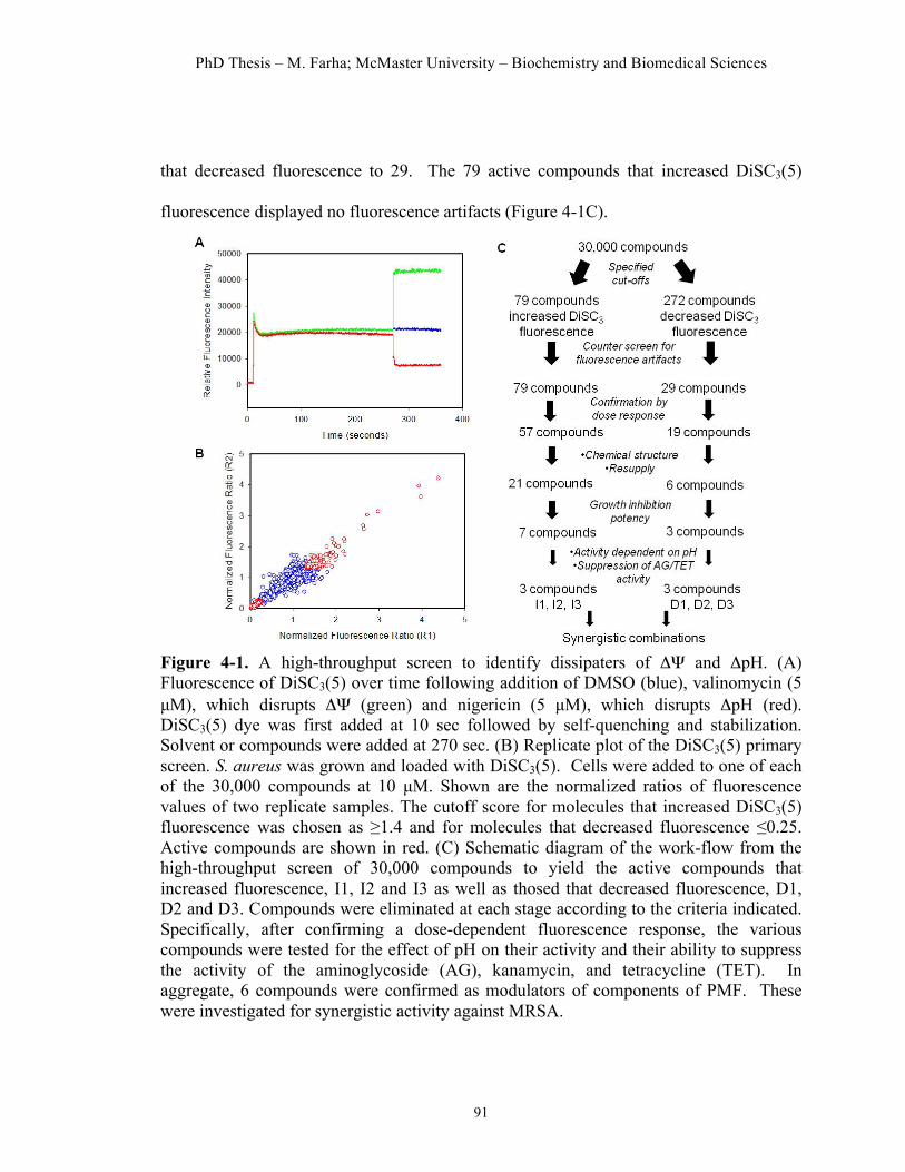
PhD Thesis – M. Farha; McMaster University – Biochemistry and Biomedical Sciences
91
that decreased fluorescence to 29. The 79 active compounds that increased DiSC3(5)
fluorescence displayed no fluorescence artifacts (Figure 4-1C).
Figure 4-1. A high-throughput screen to identify dissipaters of ΔΨ and ΔpH. (A) Fluorescence of DiSC3(5) over time following addition of DMSO (blue), valinomycin (5 µM), which disrupts ΔΨ (green) and nigericin (5 µM), which disrupts ΔpH (red). DiSC3(5) dye was first added at 10 sec followed by self-quenching and stabilization. Solvent or compounds were added at 270 sec. (B) Replicate plot of the DiSC3(5) primary screen. S. aureus was grown and loaded with DiSC3(5). Cells were added to one of each of the 30,000 compounds at 10 µM. Shown are the normalized ratios of fluorescence values of two replicate samples. The cutoff score for molecules that increased DiSC3(5) fluorescence was chosen as ≥1.4 and for molecules that decreased fluorescence ≤0.25. Active compounds are shown in red. (C) Schematic diagram of the work-flow from the high-throughput screen of 30,000 compounds to yield the active compounds that increased fluorescence, I1, I2 and I3 as well as thosed that decreased fluorescence, D1, D2 and D3. Compounds were eliminated at each stage according to the criteria indicated. Specifically, after confirming a dose-dependent fluorescence response, the various compounds were tested for the effect of pH on their activity and their ability to suppress the activity of the aminoglycoside (AG), kanamycin, and tetracycline (TET). In aggregate, 6 compounds were confirmed as modulators of components of PMF. These were investigated for synergistic activity against MRSA.

PhD Thesis – M. Farha; McMaster University – Biochemistry and Biomedical Sciences
92
Follow-up to the HTS
In order to confirm active molecules from the HTS, we generated dose response
analyses using a range of compound concentrations to assess the dose dependence of the
fluorescence readout. Of the 79 compounds that increased fluorescence in the primary
screen, 57 dissipated the membrane potential in a concentration-dependent manner
(Figure 4-1C). We eliminated 22 compounds whose effects on DiSC3(5) fluorescence did
not occur in a clear dose-dependent fashion or showed high variability in fluorescence
readings among three replicates (examples in Figure S2A). Among the 29 compounds
that decreased fluorescence, 19 compounds confirmed as causing a dose-dependent
reduction in the pH gradient. Similarly, here, we eliminated 10 compounds that did not
display clear dose-dependence or exhibited high variability among the three replicates
(examples in Figure S2B). Subsequently, we narrowed our list of molecules based on
conventional filters, such as visual inspection of chemical structures, ability to resupply
and growth inhibitory potency (detailed process in Figure S3) against S. aureus. While
not all dissipaters of components of the PMF have antibacterial activity, we focused on
molecules that were inhibitory against S. aureus. Importantly, we also observed that
antibacterial activity of the molecules against S. aureus correlated well with their ability
to dissipate either ΔΨ or ΔpH. Overall, we narrowed our working set of molecules to 10
compounds, 7 of which caused an increase in fluorescence (‘I’ molecules) and potential
dissipaters of ΔΨ and 3 causing a decrease in dye fluorescence (‘D’ molecules),
potentially dissipating ΔpH (Figure 4-1C).

PhD Thesis – M. Farha; McMaster University – Biochemistry and Biomedical Sciences
93
Uncovering agents that selectively dissipate components of PMF
The nature of the screen not only allows for the discovery of molecules that
selectively dissipate components of the PMF, but also agents that permeabilize the
membrane allowing DiSC3(5) dye to be released. Further, since PMF is generated by the
electron transport chain, inhibitors of this respiration process may also be uncovered in
the screen. It was therefore important to further investigate the mode of action of the
active compounds to ensure selective disruption of components of the PMF. To this end,
we first assessed the impact of pH on the activity of these compounds. Because the two
components of the PMF, the membrane potential and the transmembrane pH gradient (the
difference between the intracellular and extracellular pH) are interdependent, a shift in the
extracellular pH to alkaline values leads to a decrease in the pH gradient across the
membrane, such that the compensatory component, ΔΨ, accounts for a greater share of
the PMF (Booth, 1985). In contrast, a shift to acidic values leads to an increase in ΔpH
across the membrane resulting in a compensatory fall in ΔΨ, such that ΔpH becomes the
dominant component (Booth, 1985). A shift in pH to alkaline values led to potentiation
of the antibacterial activity of I1, I2 and I3 but did not enhance the activity of the 4
remaining ‘I’ molecules, suggesting the former molecules selectively modulate ΔΨ. The
chemical structures of I1, I2 and I3 are shown in Figure 4-2A along with their DiSC3(5)
dose-response curves in Figure 4-2B. Specifically, a range of pH 5.5 to pH 9.5 led to a
32-fold change in the minimum inhibitory concentration (MIC) of molecule I1 (Figure 4-
2Ci), a 64-fold shift for I2 (Figure 4-2Cii) and a 16-fold change for I3 (Figure 4-2Ciii).
Conversely, the antibacterial activity of D1, D2 and D3, for which chemical structures
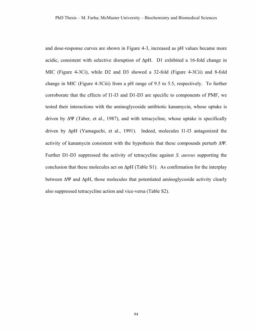
PhD Thesis – M. Farha; McMaster University – Biochemistry and Biomedical Sciences
94
and dose-response curves are shown in Figure 4-3, increased as pH values became more
acidic, consistent with selective disruption of ΔpH. D1 exhibited a 16-fold change in
MIC (Figure 4-3Ci), while D2 and D3 showed a 32-fold (Figure 4-3Cii) and 8-fold
change in MIC (Figure 4-3Ciii) from a pH range of 9.5 to 5.5, respectively. To further
corroborate that the effects of I1-I3 and D1-D3 are specific to components of PMF, we
tested their interactions with the aminoglycoside antibiotic kanamycin, whose uptake is
driven by ΔΨ (Taber, et al., 1987), and with tetracycline, whose uptake is specifically
driven by ΔpH (Yamaguchi, et al., 1991). Indeed, molecules I1-I3 antagonized the
activity of kanamycin consistent with the hypothesis that these compounds perturb ΔΨ.
Further D1-D3 suppressed the activity of tetracycline against S. aureus supporting the
conclusion that these molecules act on ΔpH (Table S1). As confirmation for the interplay
between ΔΨ and ΔpH, those molecules that potentiated aminoglycoside activity clearly
also suppressed tetracycline action and vice-versa (Table S2).

PhD Thesis – M. Farha; McMaster University – Biochemistry and Biomedical Sciences
95
Figure 4-2. Hit compounds I1, I2 and I3 dissipate ΔΨ of S. aureus. (A) Chemical structures of (i) I1 (logP = 4.02), (ii) I2 (logP = 2.65) and (iii) I3 (logP = 1.56) . (B) Concentration-dependent effect of (i) I1, (ii) I2 and (iii) I3 on DiSC3(5) fluorescence. All three compounds caused a dose-dependent increase in fluorescence, indicative of dissipation of ΔΨ. (C) Effect of altering the external pH on the minimum inhibitory concentration of (i) I1, (ii) I2 and (iii) I3. The growth inhibitory activity of all three compounds is enhanced in when the external pH is shifted to an alkaline environment, where ΔΨ becomes the main component of PMF.
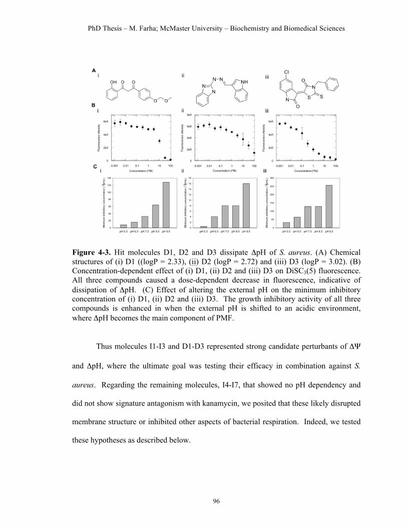
PhD Thesis – M. Farha; McMaster University – Biochemistry and Biomedical Sciences
96
Figure 4-3. Hit molecules D1, D2 and D3 dissipate ΔpH of S. aureus. (A) Chemical structures of (i) D1 ((logP = 2.33), (ii) D2 (logP = 2.72) and (iii) D3 (logP = 3.02). (B) Concentration-dependent effect of (i) D1, (ii) D2 and (iii) D3 on DiSC3(5) fluorescence. All three compounds caused a dose-dependent decrease in fluorescence, indicative of dissipation of ΔpH. (C) Effect of altering the external pH on the minimum inhibitory concentration of (i) D1, (ii) D2 and (iii) D3. The growth inhibitory activity of all three compounds is enhanced in when the external pH is shifted to an acidic environment, where ΔpH becomes the main component of PMF.
Thus molecules I1-I3 and D1-D3 represented strong candidate perturbants of ΔΨ
and ΔpH, where the ultimate goal was testing their efficacy in combination against S.
aureus. Regarding the remaining molecules, I4-I7, that showed no pH dependency and
did not show signature antagonism with kanamycin, we posited that these likely disrupted
membrane structure or inhibited other aspects of bacterial respiration. Indeed, we tested
these hypotheses as described below.

PhD Thesis – M. Farha; McMaster University – Biochemistry and Biomedical Sciences
97
Characterizing I1, I2 and I3, potential dissipaters of ΔΨ
To further characterize the action of I1, I2 and I3, the potential modulators of ΔΨ,
we first assessed their impact on the energetic condition of S. aureus cells by measuring
their effect on the reduction of iodonitrotetrazolium chloride (INT). INT, a tetrazolium
salt, can be reduced to a red insoluble formazan (INF) by components of the prokaryotic
respiratory chain (Altman, 1976). Since PMF and the ETC are closely interlinked,
whereby generation of an electrochemical proton gradient occurs as a result of the
extrusion of protons by the electron transport chain, we reasoned that dissipation of this
gradient across the cytoplasmic membrane should ultimately affect electron transport
across the respiratory chain. Overall, when tested at concentrations equivalent to their
MICs, all three molecules led to significant decreases in the reduction of INT to its
formazan product as compared to the untreated control, suggesting that the electron
transport across the cytoplamsic membrane was impaired (Figure 4-4A).
We next investigated the extent to which I1, I2 and I3, by dissipating the
membrane potential, interfere with respiratory ATP production. The proton motive force
is essential for the production of ATP via the F0F1-ATPase, such that dissipation of this
electrochemical gradient should halt ATP production. Intracellular ATP levels were
measured by a luciferin-luciferase bioluminescence assay at the MIC values of the
various compounds. As depicted in Figure 4-4B, upon addition of I1, intracellular ATP
levels drastically decreased by approximately 84% compared to DMSO-treated control.
Similarly, I2 decreased ATP levels by approximately 41%, while I3 by 81% compared to
the control. This is consistent with a mode of action of dissipating ΔΨ; upon treatment

PhD Thesis – M. Farha; McMaster University – Biochemistry and Biomedical Sciences
98
with the various molecules, the intracellular ATP pool is depleted in an attempt to
maintain the PMF. These results also suggest that interference with ATP synthesis or
depletion of the cellular ATP pool may be the cause of bacterial killing of these
molecules on their own.
We further assessed whether treatment with I1, I2 and I3 results in the efflux of
cell constituents, by measuring for any loss of materials absorbing at 260 nm. Leakage of
cellular constituents, such as potassium ions, amino acids and ATP, is a common
mechanism shared by many small molecules that dissipate PMF. While some cause pores
(Garcera, et al., 1993; McAuliffe, et al., 1998), others integrate in the membrane
(Silverman, et al., 2003; Ultee, et al., 1999), similar to an ionophore-like inhibitor, and
cause dissipations of the ion gradients. In this assay, molecules I2 and I3 did not cause a
leak of cellular constituents, as compared to the DMSO control (Figure 4-4C). Nisin, a
pore-forming lantibiotic (Garcera, et al., 1993), served as a positive control and led to a
significant increase in absorbance, consistent with its mode of action. I1 caused an
intermediate absorbance at a concentration equivalent to its MIC (Figure 4-4C). This
increased absorbance is likely attributable to I1’s high hydrophobicity, which may favor
its partitioning into the cytoplasmic membrane, consequently dissipating the
transmembrane ion gradient. Nucleic acids, which were leaked through a damaged
cytoplasmic membrane, were more abundant with increased concentrations of I1 (data not
shown), presumably as more I1 dissolves into the membrane. It should be noted that, at
the pertinent tested concentration, there was no decrease in the optical density of the I1-
treated cell suspensions, indicating that cell lysis did not occur. The same can be said of

PhD Thesis – M. Farha; McMaster University – Biochemistry and Biomedical Sciences
99
treatment with I2 and I3. Thus, we suggest that the collapse of ΔΨ at the concentrations
tested was caused by compound-mediated effects on the cell membrane rather than
having occurred as a result of general cell lysis. While I1, I2 and I3 all act on the
membrane, only I1was disruptive to its structural integrity, causing leakage of large
nucleotides, while I2 and I3 likely only cause small ions to be transported or released
across its surface. Overall, molecules I1, I2 and I3 all decreased membrane potential,
which in turn inhibited ATP production and depleted energy in the cell, leading to
reduced viability.
Figure 4-4. Effect of potential dissipaters of ΔΨ at MIC concentrations (I1: 8 µg/mL, I2: 2 µg/mL, I3: 2 µg/mL) on cellular respiration, ATP levels and membrane integrity. (A) Effects of I1, I2 and I3 on the reduction of INT to its formazan product, which can be detected at an absorbance of 490 nm. (B) Effects of compounds on intracellular ATP levels, measured by a luciferin-luciferase bioluminescence assay. Values are relative to DMSO control. (C) Measure of leakage of cellular constituents at 260 nm with nisin as a positive control.
Characterizing D1, D2 and D3, potential dissipaters of ΔpH
Similar to molecules I1-I3, we first assessed the effect of molecules D1, D2 and
D3 on the reduction of INT (Figure 4-5A). All three molecules had adverse effects on
cellular respiration, decreasing the production of formazan product, consistent with an
action of dissipating PMF.

PhD Thesis – M. Farha; McMaster University – Biochemistry and Biomedical Sciences
100
Additionally, D1, D2 and D3 also depleted cellular energy stores by causing a
decrease in intracellular levels of ATP, however, to a lesser extent than ‘I’ molecules.
Since membrane potential, but not the pH gradient, is essential for the synthesis of ATP
by the F1F0-ATPase (Kaim and Dimroth, 1998), it is not surprising that D1, D2, and D3
only decreased ATP levels by 37%, 16% and 46%, respectively (Figure 4-5B). Finally,
none of the dissipaters of ΔpH caused a significant leakage of cell constituents when
tested at MIC values (Figure 4-5C), suggesting that they do not cause damage to the
physical integrity of the membrane.
Figure 4-5. Effect of potential dissipaters of ΔpH at MIC values (D1: 32 µg/mL, D2: 8 µg/mL, D3: 128 µg/mL) on cellular respiration, ATP levels and membrane integrity. (A) Effects of D1, D2 and D3 on the reduction of INT to its formazan product, which can be detected at an absorbance of 490 nm. (B) Effects of compounds on intracellular ATP levels, measured by a luciferin-luciferase bioluminescence assay. Values are relative to DMSO control. (C) Measure of leakage of cellular constituents at 260nm. Here, nisin is a positive control.
Characterizing I4, I5, I6 and I7: other molecules that caused an increase in DiSC3(5) fluorescence
A number of molecules were uncovered in the HTS causing an increase in
DiSC3(5) fluorescence but whose activity was not suppressed by an acidic external pH.
We suspected that such molecules might inhibit cellular respiration, ultimately affecting

PhD Thesis – M. Farha; McMaster University – Biochemistry and Biomedical Sciences
101
membrane potential, or simply disrupt the membrane, allowing DiSC3(5) dye to be
released.
Molecule I4 (Figure S4A) caused a dose-dependent increase in DiSC3(5)
fluorescence (Figure S4B) but its activity was not potentiated upon a shift of the external
pH to alkaline values. I4 did, however, suppress the activity of kanamycin against S.
aureus by 8-fold (data not show), suggesting that I4 may inhibit cellular respiration. In
fact, aminoglycoside bacterial uptake not only requires intact ΔΨ, but also electron flow
through the membrane-associated respiratory chain (Taber, et al., 1987). Expectedly, I4
led to decreased INT reduction as well as decreased intracellular ATP levels, as intact
cellular respiration is required for both (Figures S4C and S4D). We also assessed its
potential membrane-damaging properties by measuring for any loss of 260-nm-absorbing
material. I4 did not cause any significant release of cell constituents, suggesting it does
not inhibit growth by permeablizing the membrane of S. aureus (data not shown).
Finally, cells treated with I4 had a significant reduction in their amount of
staphyloxanthin pigment compared to untreated cells (Figure S4E). Staphyloxanthin
synthesis has been shown to require intact cellular respiration (von Eiff, et al., 2006). I4
may be driving S. aureus into fermentative growth, which would explain the decrease in
ATP yield. Additionally, since pyruvate is shunted to support fermentation, less acetyl-
CoA would enter the mevalonate pathway necessary for staphyloxanthin synthesis (Pelz,
et al., 2005). Interestingly, I4 synergized with I1-I3 and D1-D3 (Table S3), molecules
that inhibit the generation of PMF, lending further support to the notion that I4 targets
cellular respiration as a mechanism of action. In combination with these molecules, we

PhD Thesis – M. Farha; McMaster University – Biochemistry and Biomedical Sciences
102
would expect a respiration inhibitor to further deplete energy reserves leading to reduced
viability.
Other active compounds, I5, I6 and I7 (Figure S5A) induced similar effects on S.
aureus. All three caused significant increases in DiSC3 fluorescence (Figure S5B).
Unlike our specific dissipaters of ΔΨ, their antibacterial activities were not potentiated
upon a pH shift to alkaline values and they enhanced the activity of the kanamycin,
instead of suppressing it. All three molecules did however cause a gross leakage of UV-
absorbing material (Figure S5C), similar to nisin, suggesting that they may lead to
structural disruption of the membrane. This permeabilization is likely the cause of the
increase in DiSC3(5) dye, which is released in the medium and may also explain the
observed enhanced activity of kanamycin, whose uptake would be accordingly facilitated.
Interestingly, molecule I7 was structurally a bile salt, compounds known to disrupt
cellular membranes (Ross, et al., 2004). By disrupting membrane integrity, I5, I6 and I7
expectedly caused decreases in INT reduction and ATP levels (Table S4), as the
respiratory chain and metabolic pathways within the cytoplasmic membrane would be
destroyed. Further, the membrane damage caused by I5, I6 and I7 could be large enough
to allow the leakage of large compounds, such as ATP. Finally, all three compounds
were also synergistic with I1-I3 and D1-D3, the selective dissipaters of ΔΨ and ΔpH,
again likely due to their facilitated uptake into the cell and likely unrelated to an energetic
mechanism.

PhD Thesis – M. Farha; McMaster University – Biochemistry and Biomedical Sciences
103
Combinations of molecules I1-I3 and D1-D3 lead to synergistic interactions Our overriding objective for the discovery of molecules that selectively dissipate
ΔΨ and ΔpH was their combination. We hypothesized that combining antibacterial
compounds belonging to these two mechanistic classes would circumvent the
compensatory responses in ΔΨ and ΔpH that maintain PMF. Accordingly, we predicted
synergistic, dose-sparing interactions from such combinations. We performed all possible
combinations among molecules I1-I3 and molecules D1-D3 using antibacterial
checkerboard methodology (White, et al., 1996) against the highly problematic
community-acquired methicillin-resistant strain of S. aureus, USA 300 (CA-MRSA
USA300) (Figure 4-6). Here, we define synergy as having an FIC Index ≤0.5, additivity
as 1-2 and antagonism as ≥2. In all cases, synergy was observed with FIC indices equal
to or below 0.5. The most potent synergies were observed for combinations of I2 with D2
and I3 with D3, each resulting in an FIC index of 0.25 (Figure 4-6). As controls, we also
performed all combinations among the 'I' molecules and on the other hand, among the 'D'
molecules; all combinations led to FIC indices ranging between 0.75-2 (data not shown),
indicative of additive interactions.
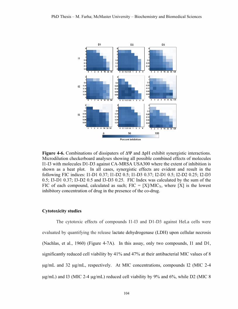
PhD Thesis – M. Farha; McMaster University – Biochemistry and Biomedical Sciences
104
Figure 4-6. Combinations of dissipaters of ΔΨ and ΔpH exhibit synergistic interactions. Microdilution checkerboard analyses showing all possible combined effects of molecules I1-I3 with molecules D1-D3 against CA-MRSA USA300 where the extent of inhibition is shown as a heat plot. In all cases, synergistic effects are evident and result in the following FIC indices: I1-D1 0.37; I1-D2 0.5; I1-D3 0.37; I2-D1 0.5; I2-D2 0.25; I2-D3 0.5; I3-D1 0.37; I3-D2 0.5 and I3-D3 0.25. FIC Index was calculated by the sum of the FIC of each compound, calculated as such; FIC = [X]/MICX, where [X] is the lowest inhibitory concentration of drug in the presence of the co-drug.
Cytotoxicity studies
The cytotoxic effects of compounds I1-I3 and D1-D3 against HeLa cells were
evaluated by quantifying the release lactate dehydrogenase (LDH) upon cellular necrosis
(Nachlas, et al., 1960) (Figure 4-7A). In this assay, only two compounds, I1 and D1,
significantly reduced cell viability by 41% and 47% at their antibacterial MIC values of 8
µg/mL and 32 µg/mL, respectively. At MIC concentrations, compounds I2 (MIC 2-4
µg/mL) and I3 (MIC 2-4 µg/mL) reduced cell viability by 9% and 6%, while D2 (MIC 8

PhD Thesis – M. Farha; McMaster University – Biochemistry and Biomedical Sciences
105
µg/mL) and D3 (MIC 128 µg/mL) by 2% and 5%, respectively. Beyond the MIC values
of the compounds, however, all but D3 showed significant cytotoxic effects on HeLa
cells.
We next assessed whether toxicity at concentrations that lead to synergistic
interactions could be minimized compared to toxicity of the compounds as antibacterial
agents alone (Figure 4-7B). Overall, it was evident that the observed synergistic
combinations would allow for reduced doses to be used, indeed minimizing cytotoxicity.
We tested 3 of the 9 combinations, I1+D1, I2+D2, and I3+D3, at the concentration of
each molecule that led to synergy for cytotoxicity effects (Figure 4-7B). Overall, toxicity
of each compound could be reduced in combination, thus allowing for dose-sparing
effects. For example, molecule I1 (which causes 41% toxicity alone) when combined
with D1 (47% toxicity alone) at their effective synergistic concentrations led to 19%
toxicity (Figure 4-7B). In combination, the activity of I1 is potentiated 4-fold by D1,
reducing its cytotoxicity at that concentration to <25%, while D1 is potentiated 8 fold by
I1, reducing its toxicity by over 20%. In other cases, such as with I2 and D2, displaying
9% and 2% cytotoxicity as single agents, respectively, resulted in very minimal
cytotoxicity in combination (5%) (Figure 4-7B). Finally, as a single agent, I3 exhibited
10% toxicity at its MIC concentration and D3, 5% cytotoxicity. In combination,
however, the total cytotoxicity at their effective synergistic concentrations was 7%, thus
remaining almost negligible (Figure 4-7B). Overall, while all six compounds exhibited
some moderate cytotoxicity, their combinations to achieve enhanced antibacterial efficacy
allowed the ability to reduce cytotoxicity effects.
PhD Thesis – M. Farha; McMaster University – Biochemistry and Biomedical Sciences
106
Figure 4-7. Cytotoxic effects of compounds I1, I2 and I3 as well as D1, D2 and D3 and their combinations on a HeLa cell line. (A) Cytotoxic effects of compounds I1-I3 and D1-D3 following exposure to different concentrations of compounds. Cell viability was assessed using LDH release method. All values are relative to the maximum LDH release control. Effect by the solvent DMSO was subtracted. (B) Cytotoxic effects of individual compounds at MIC (antibacterial) concentrations and at concentrations that are effective (antibacterial) when used in the synergistic combinations indicated.
Discussion
Bacteria establish and maintain an electrochemical gradient of protons across their
cytoplasmic membrane, known as the proton motive force (Mitchell, 1966). This PMF is
essential for a variety of critical bacterial processes, such as ATP synthesis, flagellar
motility and nutrient import. Collapse of the PMF inhibits these important functions and
thus results in the loss of bacterial viability. A unique feature of the PMF is its
composition of two components that continually work together to maintain homeostasis
(Bakker and Mangerich, 1981). Dissipation of one component of the gradient is buffered
by a counteracting increase in the other in order to maintain a constant value for the PMF.
Nevertheless, most modern antibacterial discovery efforts purposely deprioritize
molecules that disrupt membrane energetics. In the work reported here, we explore the

PhD Thesis – M. Farha; McMaster University – Biochemistry and Biomedical Sciences
107
potential of combining small molecules that perturb the ΔΨ and ΔpH components of the
PMF, with the underlying hypothesis that such agents should show advantageous synergy
in combination.
Having established DiSC3(5) fluorescence as a robust assay to identify dissipaters
of ΔΨ and ΔpH, we screened a chemical library for small molecules modulators of ΔΨ
and ΔpH. Compounds specific to ΔΨ and ΔpH were subsequently identified by their
ability to suppress the activity of aminoglycosides or tetracyclines. Such molecules could
also be modulated with changes in the external pH. This effort resulted in the
identification of molecules I1, I2 and I3, which dissipate membrane potential, and D1, D2
and D3, which dissipate the pH gradient across the cell membrane of S. aureus. As
expected, I1-I3 and D1-D3 all reduced the pool of intracellular ATP. We also uncovered
a potential inhibitor of respiration, I4, as well as three membrane-damaging agents, I5, I6
and I7, which all led to increases in DiSC3(5) fluorescence.
Interestingly, Oyamada et al. have previously reported on molecule I1, discovered
in an annucleate cell blue assay performed to identify type II topoisomerase inhibitors
against Escherichia coli (Oyamada, et al., 2006). In their in vitro follow-up, however, we
note that I1 exhibited a very high IC50 towards purified gyrase and topoisomerase IV,
namely 50 µg/mL (171µM) and >100 µg/ml (>342 µM), respectively. Further, molecule
I3 is a natural product, known as agelasine found in marine sponges (Agelas sp.).
Notably, no previous characterization of its antibacterial activity has been reported, but it
has been characterized as a putative inhibitor of human Na/K-ATPase (Trachtenberg, et
al., 1981).
PhD Thesis – M. Farha; McMaster University – Biochemistry and Biomedical Sciences
108
Importantly, in combination, synergistic interactions among our dissipaters of ΔΨ
and dissipaters of ΔpH were observed and proved efficacious against MRSA. One
advantage afforded by combination therapy over monotherapies is the ability to
circumvent toxicity with dose-sparing concentrations of two-component therapy. Indeed,
in the work presented here, cytotoxicity was significantly reduced through reductions in
dose that were made possible by the synergies of 'I' and 'D' molecules when used in
combination.
While molecules that act on the membrane are often associated with toxicities, it
is nevertheless clear that bacterial specificity is in fact attainable. The success stories of
antibacterials such as daptomycin and telavancin, which depolarize the cytoplasmic
membrane, reinforce the point that selective toxicity is achievable (Hurdle, et al., 2011).
In our case, molecules I1-I3 and D1-D3 displayed relatively low cytotoxicity at pertinent
concentrations i.e. those relating to antibacterial activity. Only some displayed high
cytotoxicity against HeLa cells at very high concentrations, in many cases well above 32-
fold their antibacterial inhibitory activity. Importantly, however, cytotoxicity was often
negligible at MIC concentrations. In eukaryotic cells, the respiratory chain and resulting
PMF is in the mitochondrial membrane instead of the cytoplasmic membrane. This
distinct localization of the respiratory chain between eukaryotic and bacterial cells could
partly explain the observed low toxicity (Wu, et al., 2010). Nevertheless, their
combinations allowed for dose-sparing effects and further lowered cytotoxicities.
A number of molecules impart bactericidal activity by collapsing the PMF,
including a variety of antimicrobial peptides (Peters, et al., 2010; Wilmes, et al., 2011),

PhD Thesis – M. Farha; McMaster University – Biochemistry and Biomedical Sciences
109
which do so by forming pores, and uncoupling agents, which inhibit oxidative
phosphorylation (Terada, 1990); fewer, however, selectively dissipate either component
of the PMF (McAuliffe, et al., 1998). Discriminating between decreases in the individual
components of the PMF allows for a greater control of bacterial membrane energetics and
gives an opportunity for collapsing the PMF via chemical combinations. In fact, targeting
redundancy and homeostasis is a critical feature of combinatorial therapy and at the root
of successful combination therapies (Cottarel and Wierzbowski, 2007; Fitzgerald, et al.,
2006).
We speculate that collapsing the PMF might be ideal as an antibacterial strategy
against a variety of species. Compounds that collapse the PMF inhibit flagellar motility
(Paul, et al., 2008), preventing invasion (Nan, et al., 2011) and swarming activities that
eventually lead to biofilm formation (Ikonomidis, et al., 2008). The PMF also plays a
necessary role in protein excretion and the export of many toxic secreted metabolites
(Geller, 1991), which could be prevented by perturbing the PMF. Additionally, PMF-
targeted molecules and their combinations could be expected to inhibit more efficiently
dormant bacteria, a subpopulation of slow-growing or non-growing microorganisms that
play important roles in many persistent infections (Lewis, 2007). Dormant bacteria
already have a reduced cellular metabolism, with a lowered but adequate proton motive
force (Hurdle, et al., 2011) and could thus be more susceptible to modulators of the PMF.
Finally, many antibiotic efflux pumps are driven by the PMF (Paulsen, et al., 1996), thus
dissipaters of PMF could find utility as novel adjuvants to conventional antibiotics.
PhD Thesis – M. Farha; McMaster University – Biochemistry and Biomedical Sciences
110
In the work reported here, we have presented a proof of principle effort for a
rational and methodical strategy for the discovery of chemical combinations that block
PMF in a synergistic manner. This method of achieving efficacy while minimizing
potential toxicities by targeting two interlinked components of a common pathway
provides a foundation for subsequent development of highly potent combinations and for
the application to other compensatory pathways in the cell.
Significance Recently, we have seen a groundswell of concern over the lack of effective antibiotics to
treat bacterial infection. The growing number of multi-drug resistant pathogens coupled
with the retreat of the pharmaceutical sector from new antibiotic development has
exacerbated this problem such that new, perhaps unconventional, strategies are needed to
meet these health challenges. Here, we report on a new approach to uncover synergistic
combinations effective against resistant pathogens such as methicillin-resistant
Staphylococcus aureus. Using a fluorescence-based high-throughput screen of S. aureus,
we have identified selective dissipaters of either ΔΨ or ΔpH, the components that make
up the proton motive force (PMF). We reasoned that combinations of selective ΔΨ and
ΔpH perturbants would be uniquely synergistic because of a requirement in bacteria to
maintain a constant PMF across the cytoplasmic membrane by balancing both
components. We have identified 3 novel dissipaters of ΔΨ and 3 novel dissipaters of
ΔpH and provided evidence that strategically modulating ΔΨ and ΔpH in concert via
combinations leads to profound synergy with potent activity against MRSA.
Additionally, we have shown that the synergy among these membrane-active compounds

PhD Thesis – M. Farha; McMaster University – Biochemistry and Biomedical Sciences
111
allows for considerable dose-sparing and as a result, lower toxicity. Indeed, modern
antibacterial drug discovery has largely avoided membrane-active agents because of
toxicity challenges. This work represents a new paradigm to tackle infectious pathogens
and provides a methodical strategy to uncover novel and synergistic antibacterial drug
combinations.
Experimental procedures Materials and bacterial strains. 3,3’-Dipropylthiacarbocyanine iodide and all antibiotics used in the study were purchased from Sigma Aldrich. The strains used in the study are S. aureus (strain Newman strain) and CA-MRSA USA 300. Mueller Hinton Broth was used as the growth medium. High-throughput screening for molecules that modulate PMF of S. aureus. Effects of compounds on the bioenergetics of the cytoplasmic membrane of S. aureus (strain Newman) were determined using the membrane potential-sensitive cyanine dye DiSC3(5) by a modification of the method of Epand et al. (Epand, et al.). Briefly, cultures of S. aureus were grown to exponential phase and washed 3 times and resuspended in a buffer containing 10 mM potassium phosphate, 5 mM MgSO4, 250 mM sucrose (pH 7.0). After the final wash, pellets were resuspended in the same buffer to an OD600 of 0.085. The cells were loaded with 1 µM DiSC3(5) and allowed to stabilize before adding to assay plates containing compounds. Compounds were dispensed into 96 well assay plates (final concentration of 10 µM) using a Biomek FX liquid handler (Beckman Coulter Inc., Fullerton, CA). Cells were added using a µfill system (Biotek). Fluorescence was monitored at one time point using an EnVision plate reader (Perkin Elmer) at an excitation wavelength of 620 nm and an emission wavelength of 685 nm. The screen was completed in duplicate and each assay plate contained controls (16 wells total) including background DMSO controls, valinomycin and nigericin controls. All raw data points were normalized to the mean of the middle two quartiles computed on an individual plate basis, to account for plate-to-plate variation, resulting in a ratio, where a high ratio indicates dissipation of ΔΨ and a low ratio, dissipation of ΔpH. Positive and negative controls on each plate were used as checkpoints that the assay was running correctly. Hit compounds were selected based on an arbitrary cut-off of 1.4 for molecules that caused an increase in fluorescence and 0.25 for molecules that caused a decrease in fluorescence. Counter screen for fluorescence artifacts. Fluorescence artifacts were accounted for by measuring the fluorescence of the compounds, which caused an increase in fluorescence, on their own in assay buffer relative to DMSO controls, while compounds that caused a
PhD Thesis – M. Farha; McMaster University – Biochemistry and Biomedical Sciences
112
decrease in fluorescence were screened for their ability to quench the fluorescence of DiSC3(5) in assay buffer in the absence of cells. Dose response studies. Half-log serial dilutions of the hit compounds were prepared and aliquoted into assay plates. Fluorescence values were obtained in a similar manner as the screen. Curves were plotted on GraFit; any molecules with deviations from a clear dose-response relationship were eliminated. MIC determinations. Protocol for MIC determinations was based on CLSI guidelines. Plates were incubated at 37°C for 18 hours and optical density read at 600 nm. The MIC for the drug was the lowest concentration showing <10% growth. INT reduction assay. S.aureus cells were grown from an overnight culture to exponential phase and washed and resuspended in 0.1M potassium phosphate buffer (pH 7.5) to an optical density of 0.3 and kept on ice. In glass tubes, compound was added in buffer with 1 mM INT and 1mL of cell in a total volume of 3mL. Tubes were mixed vigorously and absorbance at 490 nm was read at 10 minute intervals. Tubes were incubated at 30°C between reads. ATP levels. Protocol was adapted from (Patton, et al., 2006). ATP concentrations were determined using an ATP bioluminescent assay kit (Sigma) according to the manufacturer’s instructions. Bioluminescence was measured using an EnVision plate reader (PerkinElmer). Staphyloxanthin levels. S. aureus cell culture at an OD 0.1 was treated with compound and grown at 37°C for 24 hours. Cells were centrifuged and pellets washed twice with PBS. Pigment was extracted in methanol, placing the samples in a 40°C water bath for 20 mins. Samples were cooled and the absorbance of the extracted staphyloxanthin pigment was read at 450 nm and normalized to OD at 600 nm of the initial culture. Checkerboard analyses and FIC index determination. FICs were determined by setting up standard checkerboard broth microdilution assays with serially diluted 8 concentrations of each drug, using the conditions based on CLSI guidelines. At least 3 replicates were done for each combination and the means used for calculation. The FIC for each drug was calculated as the [drug in the presence of co-drug] for a well showing <10% growth, divided by the MIC for that drug. The FIC index is the sum of the two FICs. Chemical-chemical interactions with an FIC index ≤0.5 were deemed synergistic. Cytotoxicity assays. Cytotoxicity experiments were performed using the CytoTox 96 Non-Radioactive Cytotoxicity Assay (Promega, WI, USA), which measures the release of lactate dehydrogenase (LDH) upon cellular necrosis. Briefly, HeLa cells were seeded at 104 cells per well of a 96-well plate in DMEM supplemented with 10% fetal calf serum and 1% Pen-Strep, and incubated for 24-26 hours at 37°C/5% CO2. Upon reaching 90-95% confluency, cells were washed and stimulated with each compound in duplicate or triplicate at a concentration of 1 in 100 in complete DMEM. After 18 hours, LDH release was measured according to manufacturer's instructions. Each experiment was performed in triplicate, and all values are presented as relative to the maximum LDH release control, achieved by incubating unstimulated cells with 1x lysis buffer for 45 minutes prior to the measurement of LDH release.

PhD Thesis – M. Farha; McMaster University – Biochemistry and Biomedical Sciences
113
Acknowledgements We thank the staff of the Centre for Microbial Chemical Biology for technical assistance. This work was supported by an operating grant from the Canadian Institutes of Health Research (MOP-81330) and by a Canada Research Chair Award to E.D.B.
References Altman, F.P. (1976). Tetrazolium salts and formazans. Prog Histochem Cytochem 9, 1-56. Bakker, E.P., and Mangerich, W.E. (1981). Interconversion of components of the bacterial proton motive force by electrogenic potassium transport. J Bacteriol 147, 820-826. Balemans, W., Vranckx, L., Lounis, N., Pop, O., Guillemont, J., Vergauwen, K., Mol, S., Gilissen, R., Motte, M., Lancois, D., et al. (2012). Novel antibiotics targeting respiratory ATP synthesis in Gram-positive pathogenic bacteria. Antimicrob Agents Chemother 56, 4131-4139. Booth, I.R. (1985). Regulation of cytoplasmic pH in bacteria. Microbiol Rev 49, 359-378. Chopra, I. (2003). Antibiotic resistance in Staphylococcus aureus: concerns, causes and cures. Expert Rev Anti Infect Ther 1, 45-55. Coates, A.R., and Hu, Y. (2008). Targeting non-multiplying organisms as a way to develop novel antimicrobials. Trends Pharmacol Sci 29, 143-150. Cottarel, G., and Wierzbowski, J. (2007). Combination drugs, an emerging option for antibacterial therapy. Trends Biotechnol 25, 547-555. Epand, R.F., Pollard, J.E., Wright, J.O., Savage, P.B., and Epand, R.M. Depolarization, bacterial membrane composition, and the antimicrobial action of ceragenins. Antimicrob Agents Chemother 54, 3708-3713. Fitzgerald, J.B., Schoeberl, B., Nielsen, U.B., and Sorger, P.K. (2006). Systems biology and combination therapy in the quest for clinical efficacy. Nat Chem Biol 2, 458-466. Garcera, M.J., Elferink, M.G., Driessen, A.J., and Konings, W.N. (1993). In vitro pore-forming activity of the lantibiotic nisin. Role of protonmotive force and lipid composition. Eur J Biochem 212, 417-422. Geller, B.L. (1991). Energy requirements for protein translocation across the Escherichia coli inner membrane. Mol Microbiol 5, 2093-2098. Gentry, D.R., Wilding, I., Johnson, J.M., Chen, D., Remlinger, K., Richards, C., Neill, S., Zalacain, M., Rittenhouse, S.F., and Gwynn, M.N. (2010). A rapid microtiter plate assay for measuring the effect of compounds on Staphylococcus aureus membrane potential. J Microbiol Methods 83, 254-256. Hawkey, P.M. (2008). Pre-clinical experience with daptomycin. J Antimicrob Chemother 62 Suppl 3, iii7-14. Hu, Y., Shamaei-Tousi, A., Liu, Y., and Coates, A. (2010). A new approach for the discovery of antibiotics by targeting non-multiplying bacteria: a novel topical antibiotic for staphylococcal infections. PLoS One 5, e11818.
PhD Thesis – M. Farha; McMaster University – Biochemistry and Biomedical Sciences
114
Hurdle, J.G., O'Neill, A.J., Chopra, I., and Lee, R.E. (2011). Targeting bacterial membrane function: an underexploited mechanism for treating persistent infections. Nat Rev Microbiol 9, 62-75. Ikonomidis, A., Tsakris, A., Kanellopoulou, M., Maniatis, A.N., and Pournaras, S. (2008). Effect of the proton motive force inhibitor carbonyl cyanide-m-chlorophenylhydrazone (CCCP) on Pseudomonas aeruginosa biofilm development. Lett Appl Microbiol 47, 298-302. Kaim, G., and Dimroth, P. (1998). ATP synthesis by the F1Fo ATP synthase of Escherichia coli is obligatorily dependent on the electric potential. FEBS Lett 434, 57-60. Keith, C.T., Borisy, A.A., and Stockwell, B.R. (2005). Multicomponent therapeutics for networked systems. Nat Rev Drug Discov 4, 71-78. Lewis, K. (2007). Persister cells, dormancy and infectious disease. Nat Rev Microbiol 5, 48-56. McAuliffe, O., Ryan, M.P., Ross, R.P., Hill, C., Breeuwer, P., and Abee, T. (1998). Lacticin 3147, a broad-spectrum bacteriocin which selectively dissipates the membrane potential. Appl Environ Microbiol 64, 439-445. Mitchell, P. (1966). Chemiosmotic coupling in oxidative and photosynthetic phosphorylation. Biol Rev Camb Philos Soc 41, 445-502. Nachlas, M.M., Margulies, S.I., Goldberg, J.D., and Seligman, A.M. (1960). The determination of lactic dehydrogenase with a tetrazolium salt. Anal Biochem 1, 317-326. Nan, B., Chen, J., Neu, J.C., Berry, R.M., Oster, G., and Zusman, D.R. (2011). Myxobacteria gliding motility requires cytoskeleton rotation powered by proton motive force. Proc Natl Acad Sci U S A 108, 2498-2503. O'Neill, A.J., and Chopra, I. (2004). Preclinical evaluation of novel antibacterial agents by microbiological and molecular techniques. Expert Opin Investig Drugs 13, 1045-1063. Oyamada, Y., Ito, H., Fujimoto-Nakamura, M., Tanitame, A., Iwai, N., Nagai, K., Yamagishi, J., and Wachi, M. (2006). Anucleate cell blue assay: a useful tool for identifying novel type II topoisomerase inhibitors. Antimicrob Agents Chemother 50, 348-350. Patton, T.G., Yang, S.J., and Bayles, K.W. (2006). The role of proton motive force in expression of the Staphylococcus aureus cid and lrg operons. Mol Microbiol 59, 1395-1404. Paul, K., Erhardt, M., Hirano, T., Blair, D.F., and Hughes, K.T. (2008). Energy source of flagellar type III secretion. Nature 451, 489-492. Paulsen, I.T., Brown, M.H., and Skurray, R.A. (1996). Proton-dependent multidrug efflux systems. Microbiol Rev 60, 575-608. Pelz, A., Wieland, K.P., Putzbach, K., Hentschel, P., Albert, K., and Gotz, F. (2005). Structure and biosynthesis of staphyloxanthin from Staphylococcus aureus. J Biol Chem 280, 32493-32498. Peters, B.M., Shirtliff, M.E., and Jabra-Rizk, M.A. (2010). Antimicrobial peptides: primeval molecules or future drugs? PLoS Pathog 6, e1001067. Prakash Singh, M. (2006). Rapid test for distinguishing membrane-active antibacterial agents. J Microbiol Methods 67, 125-130.
PhD Thesis – M. Farha; McMaster University – Biochemistry and Biomedical Sciences
115
Ross, B.P., Braddy, A.C., McGeary, R.P., Blanchfield, J.T., Prokai, L., and Toth, I. (2004). Micellar aggregation and membrane partitioning of bile salts, fatty acids, sodium dodecyl sulfate, and sugar-conjugated fatty acids: correlation with hemolytic potency and implications for drug delivery. Mol Pharm 1, 233-245. Russell, J.B., and Houlihan, A.J. (2003). Ionophore resistance of ruminal bacteria and its potential impact on human health. FEMS Microbiol Rev 27, 65-74. Silver, L.L. (2011). Challenges of antibacterial discovery. Clin Microbiol Rev 24, 71-109. Silverman, J.A., Perlmutter, N.G., and Shapiro, H.M. (2003). Correlation of daptomycin bactericidal activity and membrane depolarization in Staphylococcus aureus. Antimicrob Agents Chemother 47, 2538-2544. Taber, H.W., Mueller, J.P., Miller, P.F., and Arrow, A.S. (1987). Bacterial uptake of aminoglycoside antibiotics. Microbiol Rev 51, 439-457. Terada, H. (1990). Uncouplers of oxidative phosphorylation. Environ Health Perspect 87, 213-218. Trachtenberg, M.C., Packey, D.J., and Sweeney, T. (1981). In vivo functioning of the Na+, K+-activated ATPase. Curr Top Cell Regul 19, 159-217. Ultee, A., Kets, E.P., and Smid, E.J. (1999). Mechanisms of action of carvacrol on the food-borne pathogen Bacillus cereus. Appl Environ Microbiol 65, 4606-4610. von Eiff, C., McNamara, P., Becker, K., Bates, D., Lei, X.H., Ziman, M., Bochner, B.R., Peters, G., and Proctor, R.A. (2006). Phenotype microarray profiling of Staphylococcus aureus menD and hemB mutants with the small-colony-variant phenotype. J Bacteriol 188, 687-693. White, R.L., Burgess, D.S., Manduru, M., and Bosso, J.A. (1996). Comparison of three different in vitro methods of detecting synergy: time-kill, checkerboard, and E test. Antimicrob Agents Chemother 40, 1914-1918. Wilmes, M., Cammue, B.P., Sahl, H.G., and Thevissen, K. (2011). Antibiotic activities of host defense peptides: more to it than lipid bilayer perturbation. Nat Prod Rep 28, 1350-1358. Wu, G., Wu, H., Fan, X., Zhao, R., Li, X., Wang, S., Ma, Y., Shen, Z., and Xi, T. (2010). Selective toxicity of antimicrobial peptide S-thanatin on bacteria. Peptides 31, 1669-1673. Wu, M., Maier, E., Benz, R., and Hancock, R.E. (1999). Mechanism of interaction of different classes of cationic antimicrobial peptides with planar bilayers and with the cytoplasmic membrane of Escherichia coli. Biochemistry 38, 7235-7242. Yamaguchi, A., Ohmori, H., Kaneko-Ohdera, M., Nomura, T., and Sawai, T. (1991). Delta pH-dependent accumulation of tetracycline in Escherichia coli. Antimicrob Agents Chemother 35, 53-56. Zhanel, G.G., Calic, D., Schweizer, F., Zelenitsky, S., Adam, H., Lagace-Wiens, P.R., Rubinstein, E., Gin, A.S., Hoban, D.J., and Karlowsky, J.A. (2010). New lipoglycopeptides: a comparative review of dalbavancin, oritavancin and telavancin. Drugs 70, 859-886. Zhang, J.H., Chung, T.D., and Oldenburg, K.R. (1999). A Simple Statistical Parameter for Use in Evaluation and Validation of High Throughput Screening Assays. J Biomol Screen 4, 67-73.

PhD Thesis – M. Farha; McMaster University – Biochemistry and Biomedical Sciences
116
CHAPTER FIVE – Future Directions and Conclusions

PhD Thesis – M. Farha; McMaster University – Biochemistry and Biomedical Sciences
117
Summary and Future Directions
Work in this thesis highlights the powerful utility of chemical-chemical
combinations as probes of biology and as routes to novel drug discovery. Specifically, all
three high-throughput screening campaigns discussed herein offer new methodologies
that can be used in early stages of antimicrobial drug discovery. The first tackles the
challenge of target identification and can be universally applicable to any pathogen under
study. Indeed, the modes of action of uncharted compounds can be inferred based on
interactions, whether additive, synergistic or antagonistic, with known probes of
physiology. This simple approach has proven its utility in various phenotype-based
screens in a variety of ways, such as identifying direct cellular target (Farha and Brown
2010), understanding unique modes of action (Ejim, Farha et al. 2011; Farha, Verschoor
et al. 2013) and aiding in mapping out novel connections within biological systems
(Farha, Leung et al. 2013). The potential of this strategy is only limited by the number of
probes used. In this respect, the approach would benefit from the assembly of a
comprehensive compendium of all probes of bacterial physiology that can be routinely
screened in combination with hit compounds following phenotype-based screens. The
potential to combine probes of all major cellular processes awaits the discovery of small
molecule probes to all physiological pathways. Another consideration, as with all high-
throughout studies, would be the need for effective methods to analyze and interpret
generated profiles.
The last two screening efforts described in this study offer unique methodologies
for therapeutic discovery via combination therapy to combat drug-resistant pathogens.

PhD Thesis – M. Farha; McMaster University – Biochemistry and Biomedical Sciences
118
The first was a search for adjuvants among previously-approved drugs that can restore the
activity of β-lactams, antibiotics that have lost their effectiveness with the rise of MRSA.
This screening strategy has been previously established and validated, yielding another
novel combination effective against P. aeruginosa (Ejim, Farha et al. 2011). With the
evolution of antibiotic resistance, the majority of antibiotics are no longer pertinent to
current practice; adjuvants can expand chemical space and effectively restore their utility.
Here, the use of previously-approved drugs as adjuvants provides the added benefit of
accelerating clinical development, as pharmacology and toxicology are well studied.
Indeed, ticlopidine is a well-established antiplatelet drug, however, factors such as
metabolic conversion limit its immediate use. To this end, SAR studies were undertaken
to design analogs that can evade enzymatic conversion (Farha, Koteva et al. 2014).
The last study in this thesis represents a new paradigm in the field of antibacterial
drug discovery. Modern drug discovery has largely avoided agents that act on the
membrane because of toxicity challenges. In this study, identified combinations of
compounds were very effective while minimizing potential toxicities. This strategy of
targeting two interlinked components of a common pathway provides a foundation for
subsequent development of potent combinations and can be extended towards other
compensatory pathways in the cell.
Overall, development of combination therapies is not without challenges. A large
limitation of combination studies is the number of possible combinations to be screened
among even a modestly sized compound library. Strategies are increasingly being devised
and implemented to aid in managing the overwhelming numbers (Feala, Cortes et al.
PhD Thesis – M. Farha; McMaster University – Biochemistry and Biomedical Sciences
119
2010). Further, designs of appropriate clinical trials and efficient regulatory paths need to
be established. Drug approval processes risk being further extended with combinations,
whereby drugs will have to be approved individually and then tested as a combination. A
sound approach for clinical approval needs to be established and will ultimately dictate
the future of combination therapy.
Given the power of pairwise combinations in probing functional biological
connections and their benefits as therapeutics, there is reason to believe that high-order
combinations will have even greater potential in capturing biological robustness and
clinical efficacy. Early research involving third order combinations have tested pairwise
combinations against single mutants (Haggarty, Clemons et al. 2003) or conversely,
double mutants for sensitivity to drug treatments (St Onge, Mani et al. 2007). To date,
rare have been studies that investigate purely chemical combinations. The growing
appreciation that biological systems are best manipulated by modulating a set of nodes
that ultimately control viability predicts that higher-order combinations will exert an even
more selective control of complex systems. As such, high-order combinations as probes
of biology have the potential to provide a more integrated understanding of the biological
system under study. From a drug discovery perspective, the benefits remain; potency,
toxicity, specificity and drug resistance would all be adequately addressed. In fact, in
vitro studies of resistant bacteria have shown that third-order combinations are more
effective than drug pairs (Bhusal, Shiohira et al. 2005). Efforts to move to higher-order
combinations will likely be very challenging. The complex nature of high-order

PhD Thesis – M. Farha; McMaster University – Biochemistry and Biomedical Sciences
120
combinations will certainly bring forth technical and data management challenges, but it
is, nonetheless, an area of potential activity in the foreseeable future.
Concluding remarks
While there is a long history of use of chemical-chemical combinations, only
more recently have they been appreciated for their power as probes of biology and their
benefits as therapeutics. Indeed, combination chemical genetics is a growing field that is
benefiting from advances in systems biology and high-throughput technologies. The work
reported here has identified a high-throughput approach for target identification
irrespective of the organism under study. Further, two separate unique screening
strategies were taken to identify novel synergistic pairs effective against highly
problematic pathogens in the clinic and community. In addition to exhibiting in vitro and
in vivo efficacy, the first combination has shed light on the intricate connections that
underlie teichoic acid and cell wall syntheses in S. aureus. This adjuvant to β-lactam
antibiotics shows great promise in clinical value and highlights the ability of adjuvants to
repurpose existing chemical matter. The last high-throughput screen led to the
identification of synergistic pairs that effectively modulate two compensatory pathways
that make up the proton motive force across the bacterial cell wall. This study reinforces
the power of pairwise combinations in perturbing systems and opens up the possibility to
dually control other interconnected aspects of physiology via chemical-chemical
combinations. Overall, the prospect of novel and better drug combinations is enticing,
and screening strategies, as described herein, hold great promise in uncovering novel
therapeutics.

PhD Thesis – M. Farha; McMaster University – Biochemistry and Biomedical Sciences
121
References Bhusal, Y., C. M. Shiohira, et al. (2005). "Determination of in vitro synergy when three
antimicrobial agents are combined against Mycobacterium tuberculosis." International journal of antimicrobial agents 26(4): 292-297.
Ejim, L., M. A. Farha, et al. (2011). "Combinations of antibiotics and nonantibiotic drugs enhance antimicrobial efficacy." Nature chemical biology 7(6): 348-350.
Farha, M. A. and E. D. Brown (2010). "Chemical probes of Escherichia coli uncovered through chemical-chemical interaction profiling with compounds of known biological activity." Chemistry & biology 17(8): 852-862.
Farha, M. A., K. Koteva, et al. (2014). "Designing analogs of ticlopidine, a wall teichoic acid inhibitor, to avoid formation of its oxidative metabolites." Bioorganic & medicinal chemistry letters 24(3): 905-910.
Farha, M. A., A. Leung, et al. (2013). "Inhibition of WTA synthesis blocks the cooperative action of PBPs and sensitizes MRSA to beta-lactams." ACS chemical biology 8(1): 226-233.
Farha, M. A., C. P. Verschoor, et al. (2013). "Collapsing the proton motive force to identify synergistic combinations against Staphylococcus aureus." Chemistry & biology 20(9): 1168-1178.
Feala, J. D., J. Cortes, et al. (2010). "Systems approaches and algorithms for discovery of combinatorial therapies." Wiley interdisciplinary reviews. Systems biology and medicine 2(2): 181-193.
Haggarty, S. J., P. A. Clemons, et al. (2003). "Chemical genomic profiling of biological networks using graph theory and combinations of small molecule perturbations." Journal of the American Chemical Society 125(35): 10543-10545.
St Onge, R. P., R. Mani, et al. (2007). "Systematic pathway analysis using high-resolution fitness profiling of combinatorial gene deletions." Nature genetics 39(2): 199-206.

![A novel chemical-combination screen in zebrafish identifies ......screening and evaluating small molecule therapies [42, 43]. Zebrafish eggs can be rapidly produced in large numbers,](https://img.pdfslide.net/doc/110x75/60f82944aef91d30c2285407/a-novel-chemical-combination-screen-in-zebrafish-identifies-screening-and.jpg)