Embed Size (px)
Citation preview
OpenStax-CNX module: m46501 1
Chemical Digestion and Absorption:
A Closer Look∗
OpenStax College
This work is produced by OpenStax-CNX and licensed under the
Creative Commons Attribution License 3.0†
Abstract
By the end of this section, you will be able to:
• Identify the locations and primary secretions involved in the chemical digestion of carbohydrates,
proteins, lipids, and nucleic acids
• Compare and contrast absorption of the hydrophilic and hydrophobic nutrients
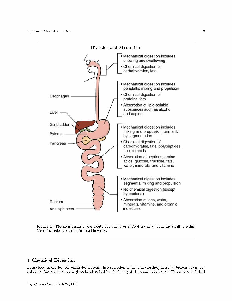
As you have learned, the process of mechanical digestion is relatively simple. It involves the physicalbreakdown of food but does not alter its chemical makeup. Chemical digestion, on the other hand, is acomplex process that reduces food into its chemical building blocks, which are then absorbed to nourish thecells of the body (Figure 1 (Digestion and Absorption )). In this section, you will look more closely at theprocesses of chemical digestion and absorption.
∗Version 1.3: Jun 4, 2013 3:11 pm -0500†http://creativecommons.org/licenses/by/3.0/
http://cnx.org/content/m46501/1.3/
OpenStax-CNX module: m46501 2
Digestion and Absorption
Figure 1: Digestion begins in the mouth and continues as food travels through the small intestine.
Most absorption occurs in the small intestine.
1 Chemical Digestion
Large food molecules (for example, proteins, lipids, nucleic acids, and starches) must be broken down intosubunits that are small enough to be absorbed by the lining of the alimentary canal. This is accomplished
http://cnx.org/content/m46501/1.3/
OpenStax-CNX module: m46501 3
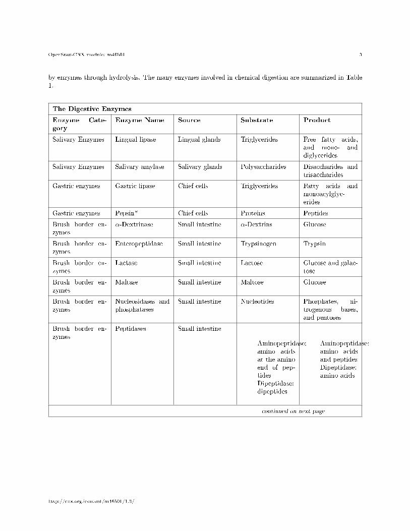
by enzymes through hydrolysis. The many enzymes involved in chemical digestion are summarized in Table1.
The Digestive Enzymes
Enzyme Cate-gory
Enzyme Name Source Substrate Product
Salivary Enzymes Lingual lipase Lingual glands Triglycerides Free fatty acids,and mono- anddiglycerides
Salivary Enzymes Salivary amylase Salivary glands Polysaccharides Disaccharides andtrisaccharides
Gastric enzymes Gastric lipase Chief cells Triglycerides Fatty acids andmonoacylglyc-erides
Gastric enzymes Pepsin* Chief cells Proteins Peptides
Brush border en-zymes
α-Dextrinase Small intestine α-Dextrins Glucose
Brush border en-zymes
Enteropeptidase Small intestine Trypsinogen Trypsin
Brush border en-zymes
Lactase Small intestine Lactose Glucose and galac-tose
Brush border en-zymes
Maltase Small intestine Maltose Glucose
Brush border en-zymes
Nucleosidases andphosphatases
Small intestine Nucleotides Phosphates, ni-trogenous bases,and pentoses
Brush border en-zymes
Peptidases Small intestine
Aminopeptidase:amino acidsat the aminoend of pep-tidesDipeptidase:dipeptides
Aminopeptidase:amino acidsand peptidesDipeptidase:amino acids
continued on next page
http://cnx.org/content/m46501/1.3/
OpenStax-CNX module: m46501 4
Brush border en-zymes
Sucrase Small intestine Sucrose Glucose and fruc-tose
Pancreatic en-zymes
Carboxy-peptidase*
Pancreatic acinarcells
Amino acids at thecarboxyl end ofpeptides
Amino acids andpeptides
Pancreatic en-zymes
Chymotrypsin* Pancreatic acinarcells
Proteins Peptides
Pancreatic en-zymes
Elastase* Pancreatic acinarcells
Proteins Peptides
Pancreatic en-zymes
Nucleases Pancreatic acinarcells
Ribonuclease:ribonucleicacids
Deoxyribonuclease:deoxyri-bonucleicacids
Nucleotides
Pancreatic en-zymes
Pancreatic amy-lase
Pancreatic acinarcells
Polysaccharides(starches)
α-Dextrins, dis-accharides (mal-tose), trisaccha-rides (maltotriose)
Pancreatic en-zymes
Pancreatic lipase Pancreatic acinarcells
Triglycerides thathave been emulsi-�ed by bile salts
Fatty acids andmonoacylglyc-erides
Pancreatic en-zymes
Trypsin* Pancreatic acinarcells
Proteins Peptides
Table 1: *These enzymes have been activated by other substances.
1.1 Carbohydrate Digestion
The average American diet is about 50 percent carbohydrates, which may be classi�ed according to thenumber of monomers they contain of simple sugars (monosaccharides and disaccharides) and/or complexsugars (polysaccharides). Glucose, galactose, and fructose are the three monosaccharides that are commonlyconsumed and are readily absorbed. Your digestive system is also able to break down the disaccharidesucrose (regular table sugar: glucose + fructose), lactose (milk sugar: glucose + galactose), and maltose(grain sugar: glucose + glucose), and the polysaccharides glycogen and starch (chains of monosaccharides).Your bodies do not produce enzymes that can break down most �brous polysaccharides, such as cellulose.While indigestible polysaccharides do not provide any nutritional value, they do provide dietary �ber, whichhelps propel food through the alimentary canal.
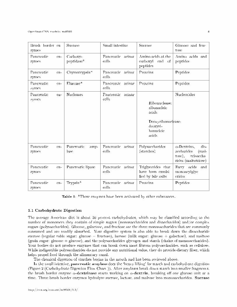
The chemical digestion of starches begins in the mouth and has been reviewed above.In the small intestine, pancreatic amylase does the `heavy lifting' for starch and carbohydrate digestion
(Figure 2 (Carbohydrate Digestion Flow Chart )). After amylases break down starch into smaller fragments,the brush border enzyme α-dextrinase starts working on α-dextrin, breaking o� one glucose unit at atime. Three brush border enzymes hydrolyze sucrose, lactose, and maltose into monosaccharides. Sucrase
http://cnx.org/content/m46501/1.3/
OpenStax-CNX module: m46501 5
splits sucrose into one molecule of fructose and one molecule of glucose; maltase breaks down maltose andmaltotriose into two and three glucose molecules, respectively; and lactase breaks down lactose into onemolecule of glucose and one molecule of galactose. Insu�cient lactase can lead to lactose intolerance.
Carbohydrate Digestion Flow Chart
Figure 2: Carbohydrates are broken down into their monomers in a series of steps.
1.2 Protein Digestion
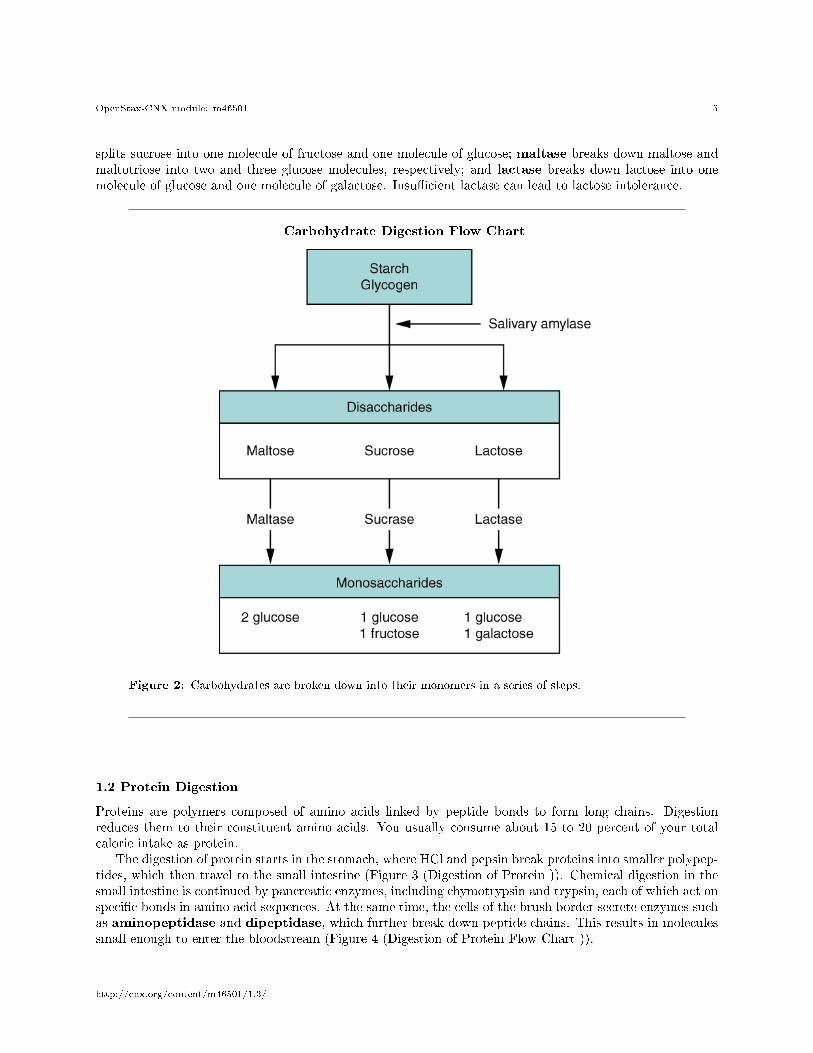
Proteins are polymers composed of amino acids linked by peptide bonds to form long chains. Digestionreduces them to their constituent amino acids. You usually consume about 15 to 20 percent of your totalcalorie intake as protein.
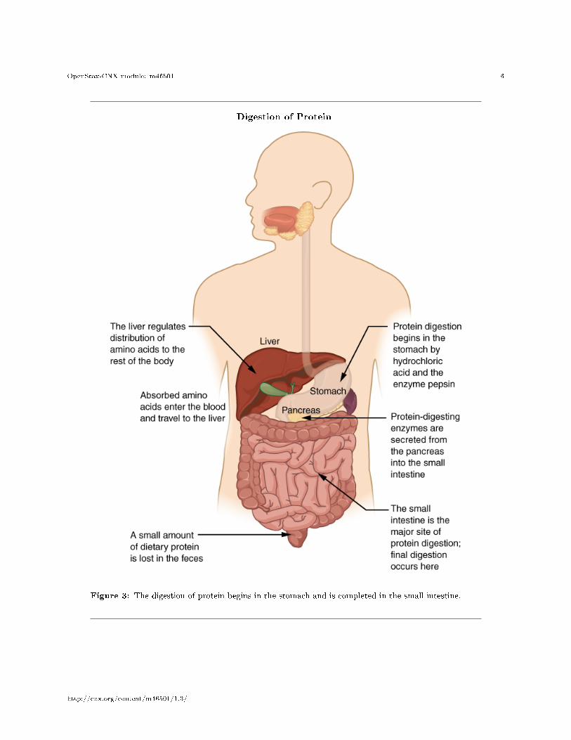
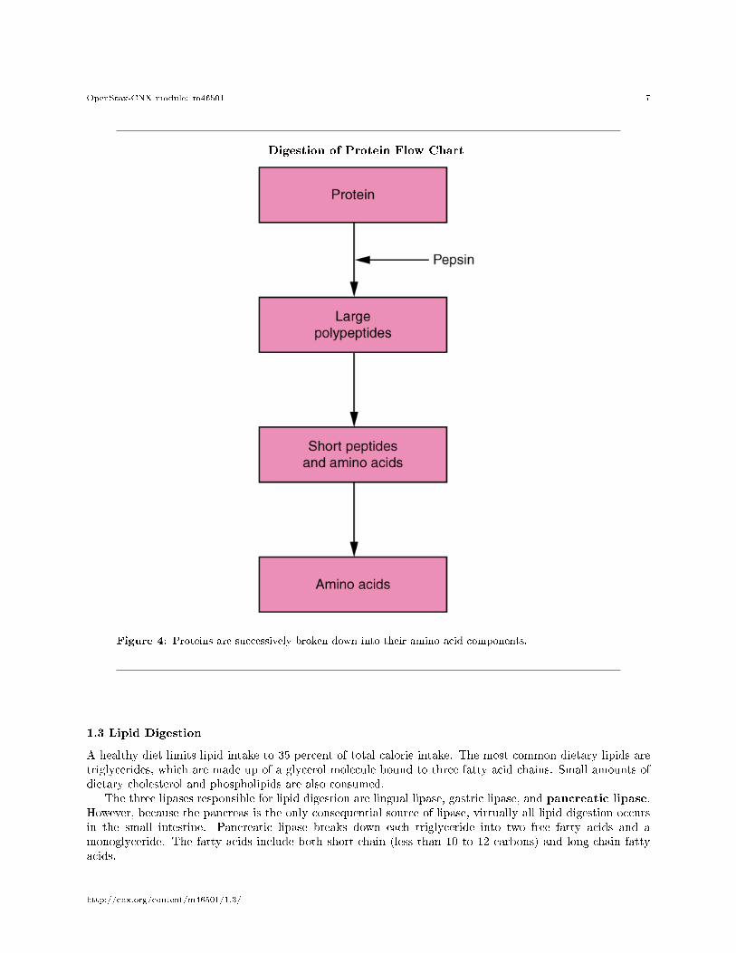
The digestion of protein starts in the stomach, where HCl and pepsin break proteins into smaller polypep-tides, which then travel to the small intestine (Figure 3 (Digestion of Protein )). Chemical digestion in thesmall intestine is continued by pancreatic enzymes, including chymotrypsin and trypsin, each of which act onspeci�c bonds in amino acid sequences. At the same time, the cells of the brush border secrete enzymes suchas aminopeptidase and dipeptidase, which further break down peptide chains. This results in moleculessmall enough to enter the bloodstream (Figure 4 (Digestion of Protein Flow Chart )).
http://cnx.org/content/m46501/1.3/
OpenStax-CNX module: m46501 6
Digestion of Protein
Figure 3: The digestion of protein begins in the stomach and is completed in the small intestine.
http://cnx.org/content/m46501/1.3/
OpenStax-CNX module: m46501 7
Digestion of Protein Flow Chart
Figure 4: Proteins are successively broken down into their amino acid components.
1.3 Lipid Digestion
A healthy diet limits lipid intake to 35 percent of total calorie intake. The most common dietary lipids aretriglycerides, which are made up of a glycerol molecule bound to three fatty acid chains. Small amounts ofdietary cholesterol and phospholipids are also consumed.
The three lipases responsible for lipid digestion are lingual lipase, gastric lipase, and pancreatic lipase.However, because the pancreas is the only consequential source of lipase, virtually all lipid digestion occursin the small intestine. Pancreatic lipase breaks down each triglyceride into two free fatty acids and amonoglyceride. The fatty acids include both short-chain (less than 10 to 12 carbons) and long-chain fattyacids.
http://cnx.org/content/m46501/1.3/
OpenStax-CNX module: m46501 8
1.4 Nucleic Acid Digestion
The nucleic acids DNA and RNA are found in most of the foods you eat. Two types of pancreaticnuclease are responsible for their digestion: deoxyribonuclease, which digests DNA, and ribonuclease,which digests RNA. The nucleotides produced by this digestion are further broken down by two intestinalbrush border enzymes (nucleosidase and phosphatase) into pentoses, phosphates, and nitrogenous bases,which can be absorbed through the alimentary canal wall. The large food molecules that must be brokendown into subunits are summarized Table 2
Absorbable Food Substances
Source Substance
Carbohydrates Monosaccharides: glucose, galactose, and fructose
Proteins Single amino acids, dipeptides, and tripeptides
Triglycerides Monoacylglycerides, glycerol, and free fatty acids
Nucleic acids Pentose sugars, phosphates, and nitrogenous bases
Table 2
2 Absorption
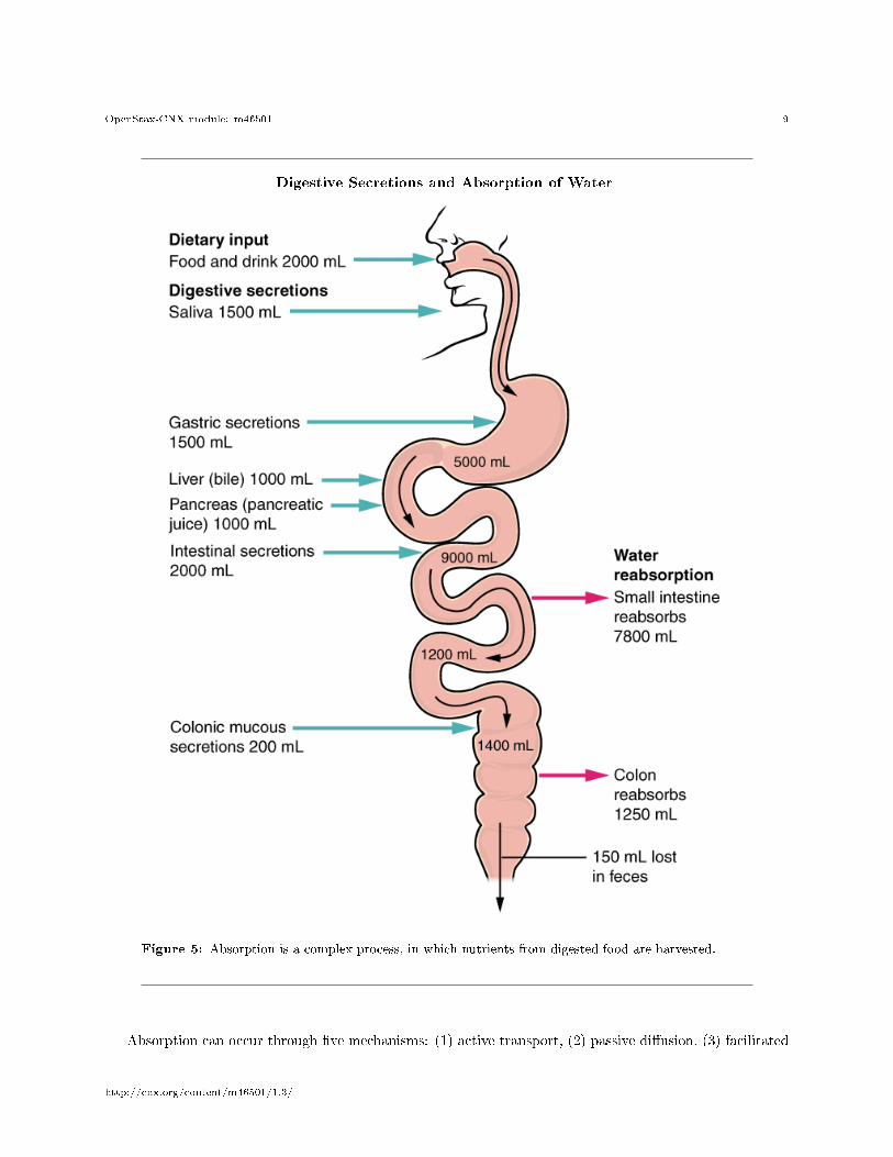
The mechanical and digestive processes have one goal: to convert food into molecules small enough to beabsorbed by the epithelial cells of the intestinal villi. The absorptive capacity of the alimentary canal isalmost endless. Each day, the alimentary canal processes up to 10 liters of food, liquids, and GI secretions,yet less than one liter enters the large intestine. Almost all ingested food, 80 percent of electrolytes, and90 percent of water are absorbed in the small intestine. Although the entire small intestine is involved inthe absorption of water and lipids, most absorption of carbohydrates and proteins occurs in the jejunum.Notably, bile salts and vitamin B12 are absorbed in the terminal ileum. By the time chyme passes from theileum into the large intestine, it is essentially indigestible food residue (mainly plant �bers like cellulose),some water, and millions of bacteria (Figure 5 (Digestive Secretions and Absorption of Water )).
http://cnx.org/content/m46501/1.3/
OpenStax-CNX module: m46501 9
Digestive Secretions and Absorption of Water
Figure 5: Absorption is a complex process, in which nutrients from digested food are harvested.
Absorption can occur through �ve mechanisms: (1) active transport, (2) passive di�usion, (3) facilitated
http://cnx.org/content/m46501/1.3/
OpenStax-CNX module: m46501 10
di�usion, (4) co-transport (or secondary active transport), and (5) endocytosis. As you will recall fromChapter 3, active transport refers to the movement of a substance across a cell membrane going from anarea of lower concentration to an area of higher concentration (up the concentration gradient). In this typeof transport, proteins within the cell membrane act as �pumps,� using cellular energy (ATP) to move thesubstance. Passive di�usion refers to the movement of substances from an area of higher concentration to anarea of lower concentration, while facilitated di�usion refers to the movement of substances from an area ofhigher to an area of lower concentration using a carrier protein in the cell membrane. Co-transport uses themovement of one molecule through the membrane from higher to lower concentration to power the movementof another from lower to higher. Finally, endocytosis is a transportation process in which the cell membraneengulfs material. It requires energy, generally in the form of ATP.
Because the cell's plasma membrane is made up of hydrophobic phospholipids, water-soluble nutrientsmust use transport molecules embedded in the membrane to enter cells. Moreover, substances cannot passbetween the epithelial cells of the intestinal mucosa because these cells are bound together by tight junctions.Thus, substances can only enter blood capillaries by passing through the apical surfaces of epithelial cellsand into the interstitial �uid. Water-soluble nutrients enter the capillary blood in the villi and travel to theliver via the hepatic portal vein.
In contrast to the water-soluble nutrients, lipid-soluble nutrients can di�use through the plasma mem-brane. Once inside the cell, they are packaged for transport via the base of the cell and then enter thelacteals of the villi to be transported by lymphatic vessels to the systemic circulation via the thoracic duct.The absorption of most nutrients through the mucosa of the intestinal villi requires active transport fueledby ATP. The routes of absorption for each food category are summarized in Table 3.
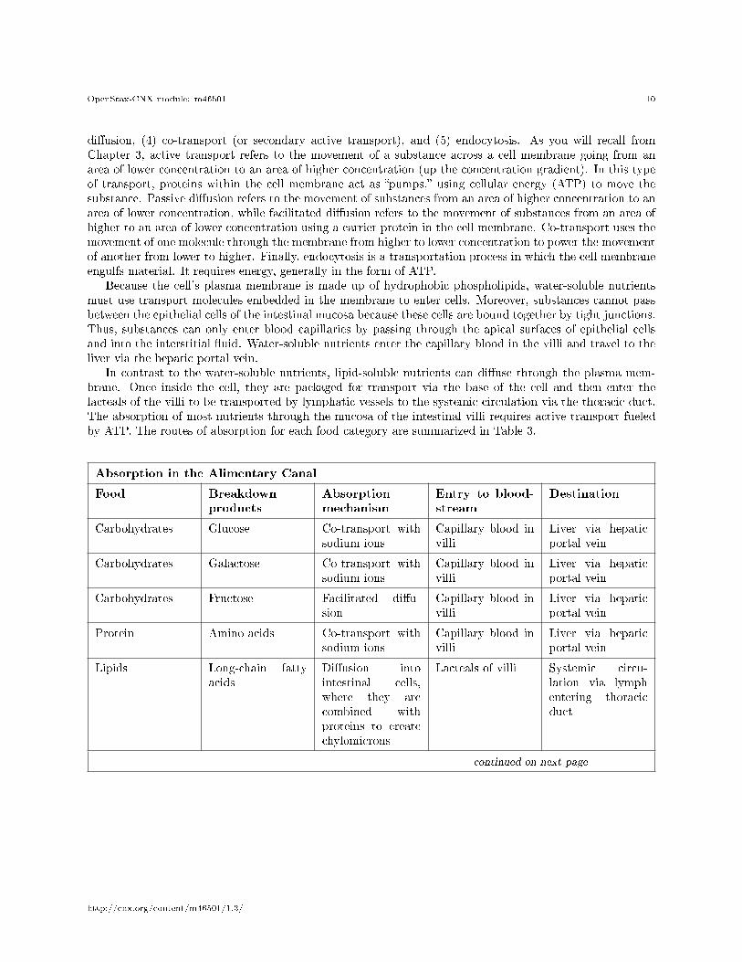
Absorption in the Alimentary Canal
Food Breakdownproducts
Absorptionmechanism
Entry to blood-stream
Destination
Carbohydrates Glucose Co-transport withsodium ions
Capillary blood invilli
Liver via hepaticportal vein
Carbohydrates Galactose Co-transport withsodium ions
Capillary blood invilli
Liver via hepaticportal vein
Carbohydrates Fructose Facilitated di�u-sion
Capillary blood invilli
Liver via hepaticportal vein
Protein Amino acids Co-transport withsodium ions
Capillary blood invilli
Liver via hepaticportal vein
Lipids Long-chain fattyacids
Di�usion intointestinal cells,where they arecombined withproteins to createchylomicrons
Lacteals of villi Systemic circu-lation via lymphentering thoracicduct
continued on next page
http://cnx.org/content/m46501/1.3/
OpenStax-CNX module: m46501 11
Lipids Monoacylglycerides Di�usion intointestinal cells,where they arecombined withproteins to createchylomicrons
Lacteals of villi Systemic circu-lation via lymphentering thoracicduct
Lipids Short-chain fattyacids
Simple di�usion Capillary blood invilli
Liver via hepaticportal vein
Lipids Glycerol Simple di�usion Capillary blood invilli
Liver via hepaticportal vein
Lipids Nucleic acid diges-tion products
Active transportvia membranecarriers
Capillary blood invilli
Liver via hepaticportal vein
Table 3
2.1 Carbohydrate Absorption
All carbohydrates are absorbed in the form of monosaccharides. The small intestine is highly e�cient atthis, absorbing monosaccharides at an estimated rate of 120 grams per hour. All normally digested dietarycarbohydrates are absorbed; indigestible �bers are eliminated in the feces. The monosaccharides glucose andgalactose are transported into the epithelial cells by common protein carriers via secondary active transport(that is, co-transport with sodium ions). The monosaccharides leave these cells via facilitated di�usion andenter the capillaries through intercellular clefts. The monosaccharide fructose (which is in fruit) is absorbedand transported by facilitated di�usion alone. The monosaccharides combine with the transport proteinsimmediately after the disaccharides are broken down.
2.2 Protein Absorption
Active transport mechanisms, primarily in the duodenum and jejunum, absorb most proteins as their break-down products, amino acids. Almost all (95 to 98 percent) protein is digested and absorbed in the smallintestine. The type of carrier that transports an amino acid varies. Most carriers are linked to the activetransport of sodium. Short chains of two amino acids (dipeptides) or three amino acids (tripeptides) arealso transported actively. However, after they enter the absorptive epithelial cells, they are broken downinto their amino acids before leaving the cell and entering the capillary blood via di�usion.
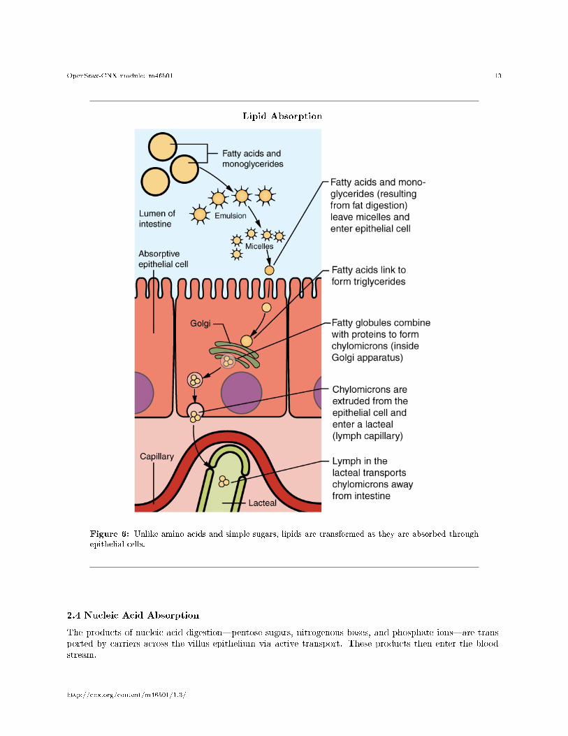
2.3 Lipid Absorption
About 95 percent of lipids are absorbed in the small intestine. Bile salts not only speed up lipid digestion,they are also essential to the absorption of the end products of lipid digestion. Short-chain fatty acids arerelatively water soluble and can enter the absorptive cells (enterocytes) directly. Despite being hydrophobic,the small size of short-chain fatty acids enables them to be absorbed by enterocytes via simple di�usion, andthen take the same path as monosaccharides and amino acids into the blood capillary of a villus.
The large and hydrophobic long-chain fatty acids and monoacylglycerides are not so easily suspendedin the watery intestinal chyme. However, bile salts and lecithin resolve this issue by enclosing them in amicelle, which is a tiny sphere with polar (hydrophilic) ends facing the watery environment and hydrophobictails turned to the interior, creating a receptive environment for the long-chain fatty acids. The core alsoincludes cholesterol and fat-soluble vitamins. Without micelles, lipids would sit on the surface of chyme andnever come in contact with the absorptive surfaces of the epithelial cells. Micelles can easily squeeze between
http://cnx.org/content/m46501/1.3/
OpenStax-CNX module: m46501 12
microvilli and get very near the luminal cell surface. At this point, lipid substances exit the micelle and areabsorbed via simple di�usion.
The free fatty acids and monoacylglycerides that enter the epithelial cells are reincorporated into triglyc-erides. The triglycerides are mixed with phospholipids and cholesterol, and surrounded with a protein coat.This new complex, called a chylomicron, is a water-soluble lipoprotein. After being processed by the Golgiapparatus, chylomicrons are released from the cell (Figure 6 (Lipid Absorption )). Too big to pass throughthe basement membranes of blood capillaries, chylomicrons instead enter the large pores of lacteals. Thelacteals come together to form the lymphatic vessels. The chylomicrons are transported in the lymphaticvessels and empty through the thoracic duct into the subclavian vein of the circulatory system. Once in thebloodstream, the enzyme lipoprotein lipase breaks down the triglycerides of the chylomicrons into freefatty acids and glycerol. These breakdown products then pass through capillary walls to be used for energyby cells or stored in adipose tissue as fat. Liver cells combine the remaining chylomicron remnants withproteins, forming lipoproteins that transport cholesterol in the blood.
http://cnx.org/content/m46501/1.3/
OpenStax-CNX module: m46501 13
Lipid Absorption
Figure 6: Unlike amino acids and simple sugars, lipids are transformed as they are absorbed through
epithelial cells.
2.4 Nucleic Acid Absorption
The products of nucleic acid digestion�pentose sugars, nitrogenous bases, and phosphate ions�are trans-ported by carriers across the villus epithelium via active transport. These products then enter the blood-stream.
http://cnx.org/content/m46501/1.3/
OpenStax-CNX module: m46501 14
2.5 Mineral Absorption
The electrolytes absorbed by the small intestine are from both GI secretions and ingested foods. Sinceelectrolytes dissociate into ions in water, most are absorbed via active transport throughout the entire smallintestine. During absorption, co-transport mechanisms result in the accumulation of sodium ions insidethe cells, whereas anti-port mechanisms reduce the potassium ion concentration inside the cells. To restorethe sodium-potassium gradient across the cell membrane, a sodium-potassium pump requiring ATP pumpssodium out and potassium in.
In general, all minerals that enter the intestine are absorbed, whether you need them or not. Ironand calcium are exceptions; they are absorbed in the duodenum in amounts that meet the body's currentrequirements, as follows:
Iron�The ionic iron needed for the production of hemoglobin is absorbed into mucosal cells via activetransport. Once inside mucosal cells, ionic iron binds to the protein ferritin, creating iron-ferritin complexesthat store iron until needed. When the body has enough iron, most of the stored iron is lost when worn-out epithelial cells slough o�. When the body needs iron because, for example, it is lost during acute orchronic bleeding, there is increased uptake of iron from the intestine and accelerated release of iron into thebloodstream. Since women experience signi�cant iron loss during menstruation, they have around four timesas many iron transport proteins in their intestinal epithelial cells as do men.
Calcium�Blood levels of ionic calcium determine the absorption of dietary calcium. When blood levelsof ionic calcium drop, parathyroid hormone (PTH) secreted by the parathyroid glands stimulates the releaseof calcium ions from bone matrices and increases the reabsorption of calcium by the kidneys. PTH alsoupregulates the activation of vitamin D in the kidney, which then facilitates intestinal calcium ion absorption.
2.6 Vitamin Absorption
The small intestine absorbs the vitamins that occur naturally in food and supplements. Fat-soluble vitamins(A, D, E, and K) are absorbed along with dietary lipids in micelles via simple di�usion. This is why you areadvised to eat some fatty foods when you take fat-soluble vitamin supplements. Most water-soluble vitamins(including most B vitamins and vitamin C) also are absorbed by simple di�usion. An exception is vitaminB12, which is a very large molecule. Intrinsic factor secreted in the stomach binds to vitamin B12, preventingits digestion and creating a complex that binds to mucosal receptors in the terminal ileum, where it is takenup by endocytosis.
2.7 Water Absorption
Each day, about nine liters of �uid enter the small intestine. About 2.3 liters are ingested in foods andbeverages, and the rest is from GI secretions. About 90 percent of this water is absorbed in the smallintestine. Water absorption is driven by the concentration gradient of the water: The concentration of wateris higher in chyme than it is in epithelial cells. Thus, water moves down its concentration gradient from thechyme into cells. As noted earlier, much of the remaining water is then absorbed in the colon.
3 Chapter Review
The small intestine is the site of most chemical digestion and almost all absorption. Chemical digestionbreaks large food molecules down into their chemical building blocks, which can then be absorbed throughthe intestinal wall and into the general circulation. Intestinal brush border enzymes and pancreatic enzymesare responsible for the majority of chemical digestion. The breakdown of fat also requires bile.
Most nutrients are absorbed by transport mechanisms at the apical surface of enterocytes. Exceptionsinclude lipids, fat-soluble vitamins, and most water-soluble vitamins. With the help of bile salts and lecithin,the dietary fats are emulsi�ed to form micelles, which can carry the fat particles to the surface of theenterocytes. There, the micelles release their fats to di�use across the cell membrane. The fats are thenreassembled into triglycerides and mixed with other lipids and proteins into chylomicrons that can pass into
http://cnx.org/content/m46501/1.3/
OpenStax-CNX module: m46501 15
lacteals. Other absorbed monomers travel from blood capillaries in the villus to the hepatic portal vein andthen to the liver.
4 Review Questions
Exercise 1 (Solution on p. 16.)
Where does the chemical digestion of starch begin?
a. mouthb. esophagusc. stomachd. small intestine
Exercise 2 (Solution on p. 16.)
Which of these is involved in the chemical digestion of protein?
a. pancreatic amylaseb. trypsinc. sucrased. pancreatic nuclease
Exercise 3 (Solution on p. 16.)
Where are most fat-digesting enzymes produced?
a. small intestineb. gallbladderc. liverd. pancreas
Exercise 4 (Solution on p. 16.)
Which of these nutrients is absorbed mainly in the duodenum?
a. glucoseb. ironc. sodiumd. water
5 Critical Thinking Questions
Exercise 5 (Solution on p. 16.)
Explain the role of bile salts and lecithin in the emulsi�cation of lipids (fats).
Exercise 6 (Solution on p. 16.)
How is vitamin B12 absorbed?
http://cnx.org/content/m46501/1.3/
OpenStax-CNX module: m46501 16
Solutions to Exercises in this Module
to Exercise (p. 15)Ato Exercise (p. 15)Bto Exercise (p. 15)Dto Exercise (p. 15)Bto Exercise (p. 15)Bile salts and lecithin can emulsify large lipid globules because they are amphipathic; they have a nonpolar(hydrophobic) region that attaches to the large fat molecules as well as a polar (hydrophilic) region thatinteracts with the watery chime in the intestine.to Exercise (p. 15)Intrinsic factor secreted in the stomach binds to the large B12 compound, creating a combination that canbind to mucosal receptors in the ileum.
Glossary
De�nition 1: α-dextrinbreakdown product of starch
De�nition 2: α-dextrinasebrush border enzyme that acts on α-dextrins
De�nition 3: aminopeptidasebrush border enzyme that acts on proteins
De�nition 4: chylomicronlarge lipid-transport compound made up of triglycerides, phospholipids, cholesterol, and proteins
De�nition 5: deoxyribonucleasepancreatic enzyme that digests DNA
De�nition 6: dipeptidasebrush border enzyme that acts on proteins
De�nition 7: lactasebrush border enzyme that breaks down lactose into glucose and galactose
De�nition 8: lipoprotein lipaseenzyme that breaks down triglycerides in chylomicrons into fatty acids and monoglycerides
De�nition 9: maltasebrush border enzyme that breaks down maltose and maltotriose into two and three molecules ofglucose, respectively
De�nition 10: micelletiny lipid-transport compound composed of bile salts and phospholipids with a fatty acid andmonoacylglyceride core
De�nition 11: nucleosidasebrush border enzyme that digests nucleotides
De�nition 12: pancreatic amylaseenzyme secreted by the pancreas that completes the chemical digestion of carbohydrates in thesmall intestine
http://cnx.org/content/m46501/1.3/
OpenStax-CNX module: m46501 17
De�nition 13: pancreatic lipaseenzyme secreted by the pancreas that participates in lipid digestion
De�nition 14: pancreatic nucleaseenzyme secreted by the pancreas that participates in nucleic acid digestion
De�nition 15: phosphatasebrush border enzyme that digests nucleotides
De�nition 16: ribonucleasepancreatic enzyme that digests RNA
De�nition 17: sucrasebrush border enzyme that breaks down sucrose into glucose and fructose
http://cnx.org/content/m46501/1.3/