Embed Size (px)
Citation preview

Chemical Modification of Alginates in Organic Media
Siddhesh Nitin Pawar
Dissertation submitted to the faculty of the Virginia Polytechnic Institute and State University in
partial fulfillment of the requirements for the degree of
Doctor of Philosophy
In
Macromolecular Science and Engineering
Kevin J. Edgar, Committee Chair
S. Richard Turner
Judy S. Riffle
Charles E. Frazier
Maren Roman
April 30, 2013
Blacksburg, VA
Keywords: Alginate, derivatization, regioselective, acetate, modification, DS, gelling,
microbeads, esters, drug delivery, naringenin, solubility, enhancement
Copyright 2013, Siddhesh N. Pawar
Chemical Modification of Alginates in Organic Media
Siddhesh Nitin Pawar
Abstract
Alginates are (1→4) linked linear copolysaccharides composed of β-D-mannuronic acid
(M) and its C-5 epimer, α-L-guluronic acid (G). Several strategies to synthesize organically
modified alginate derivatives have been reported, but almost all chemistries are performed in
either aqueous or aqueous-organic media. The ability to react alginates homogeneously in
organic solvents would open up access to a wide range of new chemistries and derivatives.
However, past attempts have been restricted by the absence of methods for alginate dissolution
in organic media. We therefore report a strategy to solubilize tetrabutylammonium (TBA) salts
of alginic acid in polar aprotic solvents containing tetrabutylammonium fluoride (TBAF).
Acylation of TBA-alginate was performed in DMSO/TBAF to get products with DSacetyl
up to ≈ 1.0. We further report that by using appropriate solvent conditions, placement of acyl
groups can be controlled to achieve either random or M-selective substitution. Alginate acetates
synthesized in an M-selective fashion were used to study the ability of these derivatives to form
Ca-crosslinked hydrogels. Detailed structure-property analyses were performed to identify
acetylation reaction conditions and product properties that may be ideal for hydrogel formation.
Furthermore, alginate esters were synthesized via modification of carboxylate groups on the
backbone. These derivatives dissolved in polar aprotic solvents without the need to add TBAF. A
proof of concept study showed their utility in the solubility enhancement of the poorly water
soluble flavonoid naringenin.
iii
Acknowledgements
First and foremost, I would like to thank my graduate advisor, Prof. Kevin J. Edgar,
whose expertise, support and guidance enabled me to bring our research goals to fruition. His
research and managements styles have truly brought out the best in me, and have influenced me
deeply through my graduate career. His penchant for hard work, high thinking, kindheartedness
and generosity are life’s lessons, for which I will forever be grateful.
I wish to express deep gratitude towards my graduate committee members, Prof. Riffle,
Prof. Turner, Prof. Frazier and Prof. Roman. Their guidance and suggestions have proven most
valuable through my research. More importantly, I wish to thank them for being remarkable
educators in this noble profession; you will be a constant source of inspiration to me in my life.
I would like to acknowledge our collaborators from Norway, Prof. Gudmund Skjåk-Bræk
and Dr. Anne Tøndervik for sending us generous quantities of specialized alginates which are
not commercially available. I would also like to thank our collaborator Prof. Emmanuel Opara
from Wake Forest Institute of Regenerative Medicine (WFIRM) for all our joint efforts in
successfully forming Ca-crosslinked alginate acetates. I wish to thank FMC Biopolymer for
providing us with food grade alginates for our studies. I thank Dr. Hugo Azurmendi for
assistance with NMR, Ms. Suzanne Barnes for performing and analyzing aqueous SEC data, and
Mr. Joshua Moore for his assistance in the laboratory. I wish to give a big thanks to all my past
and present colleagues in the cellulose research group – Junia Pereira, Haoyu Liu, Joyann Marks,
Xueyan Zheng, Ruoran Zhang, Xiangtao Meng, Thomas Simerly, Dr. Daiqiang Xu, Dr. Carter
Fox, Dr. Bin Li and Dr. Nilanjana Kar.

iv
I would like to extend my deep gratitude towards organizations that funded this work –
the Sustainable Engineered Materials Institute (SEMI) at Virginia Tech and USDA (Grant No.
2011-67009-20090). A special thanks to the Institute for Critical Technology and Applied
Science (ICTAS) for providing top-class research facilities, and Macromolecules and Interfaces
Institute (MII) for academic support.
Last but not the least, this accomplishment is dedicated to the people in my life who
matter most and have inspired me to become who I am. I thank my parents for the blood, sweat
and tears they have spent in successfully raising me. I thank my grandparents, sister, uncles,
aunts and their families for their unending love. I wish to thank Ms. Sneha Kelkar for being a
solid support through this journey. Last but not least, I would like to thank Prof. Thomas W.
Smith, my Master’s thesis advisor for being an inspiring mentor throughout my graduate school
career.

v
Table of Contents
Abstract ........................................................................................................................................................ iii
Acknowledgements ...................................................................................................................................... iii
List of Figures ............................................................................................................................................ viii
List of Schemes ........................................................................................................................................... xiv
List of Tables .............................................................................................................................................. xvi
Chapter 1: Dissertation Overview ................................................................................................................. 1
Chapter 2: Literature Review on the Derivatization of Alginates ................................................................. 3
2.1 Abstract ............................................................................................................................................... 3
2.2 Introduction ......................................................................................................................................... 4
2.3 Description of alginates ...................................................................................................................... 5
2.3.1 Biosynthesis ................................................................................................................................. 5
2.3.2 Manufacture ................................................................................................................................. 6
2.3.3 Structure determination ................................................................................................................ 8
2.3.4 Physical properties ..................................................................................................................... 10
2.3.5 Chemical properties ................................................................................................................... 13
2.4 Alginate modification ....................................................................................................................... 16
2.4.1 Acetylation of alginates ............................................................................................................. 17
2.4.2 Phosphorylation of alginates ...................................................................................................... 29
2.4.3 Sulfation of alginates ................................................................................................................. 31
2.4.4 Hydrophobic modification ......................................................................................................... 35
2.4.5 Attachment of cell signaling molecules ..................................................................................... 45
2.4.6 Covalent crosslinking of alginates ............................................................................................. 54
2.4.7 Graft copolymerization of alginates ........................................................................................... 64
2.5 Conclusions ....................................................................................................................................... 71
2.6 References ......................................................................................................................................... 73
2.7 Copyright Authorization ................................................................................................................... 83
Chapter 3: Organic Dissolution of Alginates and Epimer-selective Acetylation Thereof .......................... 84
3.1 Abstract ............................................................................................................................................. 84
3.2 Introduction ....................................................................................................................................... 84
vi
3.3 Experimental ..................................................................................................................................... 87
3.3.1 Materials and methods ............................................................................................................... 87
3.3.2 Synthesis of TBA-alginate ......................................................................................................... 91
3.3.3 Homogeneous synthesis of alginate acetate in DMSO/TBAF using acetic anhydride .............. 91
3.3.4 Heterogeneous synthesis of alginate acetate in DMSO ............................................................. 92
3.3.5 Homogeneous synthesis of alginate acetate in DMSO/TBAF using acetyl chloride ................. 93
3.3.6 Homogeneous synthesis of alginate propionate in DMSO/TBAF using propionic anhydride .. 94
3.3.7 Synthesis of anhydrous DMSO/TBAF solvent system .............................................................. 95
3.4 Results and discussion ...................................................................................................................... 96
3.5 Conclusions ..................................................................................................................................... 111
3.6 Supporting information ................................................................................................................... 112
3.7 References ....................................................................................................................................... 118
3.8 Copyright Authorization ................................................................................................................. 122
Chapter 4: Ca+2
-Induced Gelation of Alginate Acetates for Tailored Hydrogels ..................................... 123
4.1 Abstract ........................................................................................................................................... 123
4.2 Introduction ..................................................................................................................................... 124
4.3 Experimental ................................................................................................................................... 127
4.3.1 Materials .................................................................................................................................. 127
4.3.2 Synthesis of TBA-alginate ....................................................................................................... 128
4.3.3 Synthesis of alginate acetate .................................................................................................... 129
4.3.4 Synthesis of Ca-alginate beads ................................................................................................ 130
4.3.5 DSNMR measurement ................................................................................................................ 130
4.3.6 DSTitration measurement ............................................................................................................. 131
4.3.7 FTIR measurement ................................................................................................................... 132
4.3.8 SEC analysis ............................................................................................................................ 133
4.3.9 Viscosity measurement ............................................................................................................ 133
4.3.10 Imaging of Microbeads .......................................................................................................... 134
4.3.11 Mechanical Strength Testing of Microbeads ......................................................................... 134
4.3.12 Statistical evaluation of data .................................................................................................. 135
4.4 Results and Discussion ................................................................................................................... 135
4.5 Conclusions ..................................................................................................................................... 153
4.6 Supporting information ................................................................................................................... 156
vii
4.7 References ....................................................................................................................................... 174
Chapter 5: Synthesis of Alginate Esters via Carboxyl Group Modification ............................................. 179
5.1 Abstract ........................................................................................................................................... 179
5.2 Introduction ..................................................................................................................................... 180
5.3 Experimental ................................................................................................................................... 184
5.3.1 Materials .................................................................................................................................. 184
5.3.2 Synthesis of TBA-alginate ....................................................................................................... 185
5.3.3 Synthesis of benzyl alginate ..................................................................................................... 186
5.3.4 Synthesis of methyl alginate .................................................................................................... 187
5.3.5 Synthesis of ethyl alginate ....................................................................................................... 188
5.3.6 Synthesis of butyl alginate ....................................................................................................... 188
5.3.7 Saponification of esters ............................................................................................................ 189
5.3.8 NMR Analysis ......................................................................................................................... 189
5.3.9 FTIR measurement ................................................................................................................... 191
5.3.10 Elemental analysis.................................................................................................................. 191
5.3.11 DS measurement by titration .................................................................................................. 191
5.3.12 Synthesis of solid dispersions ................................................................................................ 192
5.3.13 Alginate ester solubility in water ........................................................................................... 192
5.3.14 Nar calibration curves ............................................................................................................ 193
5.3.15 Measurement of % drug loading ............................................................................................ 193
5.3.16 Nar release studies ................................................................................................................. 193
5.4 Results and Discussion ................................................................................................................... 194
5.5 Conclusion ...................................................................................................................................... 209
5.6 Supporting information ................................................................................................................... 211
5.7 References ....................................................................................................................................... 228
Chapter 6: Summary and Future Work ..................................................................................................... 231
6.1 References ....................................................................................................................................... 237

viii
List of Figures
Figure 2.1: Bacterial alginate biosynthesis pathway9 ................................................................................... 6
Figure 2.2: Schematic showing alginate extraction procedure from algae ................................................... 8
Figure 2.3: Representative alginate structure: (a) chain conformation and (b) block distribution ............... 9
Figure 2.4: Anomeric region in the 1H-NMR spectra of alginates containing varying M contents. Peak
assignments as described by Grasdalen, H.27 .............................................................................................. 10
Figure 2.5: Possible junction points in alginates. (a) GG/GG junctions, (b) MG/MG junctions, and (c)
GG/MG junctions.30 .................................................................................................................................... 12
Figure 2.6: Important parameters governing alginate derivatization .......................................................... 17
Figure 2.7: Viscosity of alginate acetate plotted against corresponding DS value (represented as ‘degree
of acetylation’)46 ......................................................................................................................................... 19
Figure 2.8: Dependence of the modulus of rigidity (G) on DS (represented as "D.a.") in acetylated Ca-
alginate gels53 .............................................................................................................................................. 24
Figure 2.9: Partial dissolution of TBA-alginate in DMSO (left) and complete dissolution in DMSO/TBAF
(right) at a TBAF concentration of 1.1% (w/v). Alginate concentration in both vials was 15 mg/mL28 .... 26
Figure 2.10: Shift in the SEC chromatograms to longer elution times for alginate acetates shown in Table
2.4, recorded over increasing reaction time28 .............................................................................................. 29
Figure 2.11: (a) Viscosity vs. concentration (C) plot for PGA (empty squares) and PGA-C12 (half-filled
squares) at a shear rate of 0.06 (1/s) and (b) Viscosity vs. NaCl concentration (Cs) plot for PGA (empty
squares) and PGA-C12 (half-filled squares) at a fixed polymer concentration of 1.2 g/dL and shear rate of
0.06 (1/s)68 ................................................................................................................................................... 39
Figure 2.12: (a) Alg-C12 adsorption at the air water interface and (b) Alg-C12+DTAB adsorption at the air
water interface73 .......................................................................................................................................... 41

ix
Figure 2.13: Schematic illustration of the encapsulation of the hydrophobic moieties on Alg-CONH-C8 by
the β-CD truncated cone76 ........................................................................................................................... 45
Figure 2.14: Schematic describing the synthesis of Amylose-g-Alg132 ...................................................... 68
Figure 2.15: Schematic showing the temperature dependent behavior in PNIPAAm-g-Alg hydrogels133 . 69
Figure 2.16: Schematic showing the effect of pH and ionic strength on PDMAEMA-g-OAlg hydrogels134
.................................................................................................................................................................... 71
Figure 3.1: Partial dissolution of alginate M063 in DMSO (left) and complete dissolution in
DMSO/TBAF (right) at a TBAF concentration of 1.1% (w/v). Alginate concentration in both vials was 15
mg/mL ......................................................................................................................................................... 99
Figure 3.2: 1H-NMR spectrum of alginate acetate synthesized from alginate M063 ............................... 101
Figure 3.3: FTIR spectrum of alginate acetate synthesized from alginate M063 ..................................... 101
Figure 3.4: 1H-NMR spectrum of alginate acetate synthesized from alginate M100. Peak A represents H-3
from M residues monoacetylated at position 3. Peak B represents H-3 from M residues diacetylated at
positions 2 and 3. Peaks A and B assigned according to previously published literature49 ...................... 105
Figure 3.5: 1H-NMR spectra of homogeneously acetylated alginates from Table 3.3. Peak A represents H-
3 from M residues monoacetylated at position 3. Peak B represents H-3 from M residues diacetylated at
positions 2 and 3. Peaks A and B assigned according to previously published literature49 ...................... 106
Figure 3.6: 1H-NMR spectra of heterogeneously acetylated alginates from Table 3.3. Peak A represents
H-3 from M residues monoacetylated at position 3. Peak B represents H-3 from M residues diacetylated
at positions 2 and 3. Peak C represents H-1 from unreacted GGG, MGG and GGM triad sequences. Peak
D represents H-5 from unreacted GGG triad sequences. Peaks A, B, C and D were assigned according to
previously published literature49-50 ............................................................................................................ 108
Figure 3.7: Right-shift in the SEC chromatograms obtained using light scattering for the alginate acetates
shown in Table 3.4, recorded over increasing reaction time ..................................................................... 111
Figure 3.8: 1H-NMR of M063 TBA-alginate ............................................................................................ 112

x
Figure 3.9: FTIR of M063 TBA-alginate .................................................................................................. 113
Figure 3.10: 1H-NMR for product 2 shown in Table 3.4 .......................................................................... 113
Figure 3.11: FTIR for product 2 shown in Table 3.4 ................................................................................ 114
Figure 3.12: 1H-NMR for product 7 shown in Table 3.2 .......................................................................... 114
Figure 3.13: FTIR for product 7 shown in Table 3.2 ................................................................................ 115
Figure 3.14: 1H-NMR for product 8 shown in Table 3.2 .......................................................................... 115
Figure 3.15: FTIR for product 8 shown in Table 3.2 ................................................................................ 116
Figure 3.16: (a) 1H-NMR spectra and (b) DLS intensity plots for TBA-alginate partially dissolved in
DMSO and completely dissolved in DMSO/TBAF. 1H-NMR spectra show integration of the DMSO-d6
solvent peak at 2.5 ppm against the polymer backbone region between 3.3-5.0 ppm. ............................. 117
Figure 4.1: 1H-NMR spectrum of alginate acetate A.P10.Ac20 from Table 4.2. HDO peak suppression
used during acquisition ............................................................................................................................. 139
Figure 4.2: FTIR spectrum of alginate acetate A.P10.Ac20 from Table 4.2 ............................................ 140
Figure 4.3: 1H-NMR spectrum of alginate acetate A.P10.Ac20 from Table 4.2. HDO peak suppression
carried out post acquisition using NMR processing software ................................................................... 141
Figure 4.4: Potentiometric titration [pH] plot for alginate acetate A.P10.Ac20 from Table 4.2 .............. 142
Figure 4.5: Potentiometric titration derivative [d(pH)] plot for alginate acetate A.P10.Ac20 from Table
4.2 ............................................................................................................................................................. 143
Figure 4.6: SEC chromatograms of alginate acetates in Series I, II and III from Table 4.2 ..................... 145
Figure 4.7: Microscopic images of capsules formed using alginate acetates from Table 4.2 ................... 147
Figure 4.8: Viscosity vs. shear rate plots for alginate acetates from Table 4.2 at various concentrations,
measured at room temperature .................................................................................................................. 150
Figure 4.9: SEC chromatograms of alginate acetates in Series IV from Table 4.2 .................................. 151
Figure 4.10: (A) Osmotic and (B) mechanical stress effects, measured as % of broken microbeads. For
osmotic stress test: 1.5% LVM vs. 5% A-Na, p < 0.001; 1.5% LVM vs. 5% A.P10.Ac2, no significant
xi
difference; 5% A-Na vs. 5% A.P10.Ac2, p < 0.001. For mechanical stress test: 1.5% LVM vs. 5% A-Na,
no significant difference; 5% A-Na vs. 5% A.P10.Ac2, p < 0.05; 1.5% LVM vs. 5% A.P10.Ac2, p < 0.05.
.................................................................................................................................................................. 153
Figure 4.11: 1H-NMR of TBA-alginate ‘A’ .............................................................................................. 156
Figure 4.12: FTIR of TBA-alginate ‘A’ .................................................................................................... 157
Figure 4.13: 1H-NMR spectrum of alginate acetate A.P2.Ac20 from Table 4.2. HDO peak suppression
pulse applied during acquisition ............................................................................................................... 159
Figure 4.14: FTIR spectrum of alginate acetate A.P2.Ac20 from Table 4.2 ............................................ 160
Figure 4.15: 1H-NMR spectrum of alginate acetate A.P2.Ac20 from Table 4.2. HDO peak suppression
pulse applied during acquisition ............................................................................................................... 161
Figure 4.16: FTIR spectrum of alginate acetate A.P2.Ac20 from Table 4.2 ............................................ 162
Figure 4.17: 1H-NMR spectrum of alginate acetate A.P10.Ac2 from Table 4.2. HDO peak suppression
pulse applied during acquisition ............................................................................................................... 163
Figure 4.18: FTIR spectrum of alginate acetate A.P10.Ac2 from Table 4.2 ............................................ 164
Figure 4. 19: 1H-NMR spectrum of alginate acetate A.P10.Ac5 from Table 4.2. HDO peak suppression
pulse applied during acquisition ............................................................................................................... 165
Figure 4.20: FTIR spectrum of alginate acetate A.P10.Ac5 from Table 4.2 ............................................ 166
Figure 4.21: 1H-NMR spectrum of alginate acetate A.P10.Ac10 from Table 4.2. HDO peak suppression
pulse applied during acquisition ............................................................................................................... 167
Figure 4.22: FTIR spectrum of alginate acetate A.P10.Ac10 from Table 4.2 .......................................... 168
Figure 4.23: 1H-NMR spectrum of alginate acetate B.P10.Ac10 from Table 4.2. HDO peak suppression
pulse applied during acquisition ............................................................................................................... 169
Figure 4.24: FTIR spectrum of alginate acetate B.P10.Ac10 from Table 4.2 .......................................... 170
Figure 4.25: 1H-NMR spectrum of alginate acetate C.P10.Ac10 from Table 4.2. HDO peak suppression
pulse applied during acquisition ............................................................................................................... 171
xii
Figure 4.26: FTIR spectrum of alginate acetate C.P10.Ac10 from Table 4.2 .......................................... 172
Figure 4.27: 1H-NMR spectrum of alginate acetate D.P10.Ac10 from Table 4.2. HDO peak suppression
pulse applied during acquisition ............................................................................................................... 173
Figure 4.28: FTIR spectrum of alginate acetate D.P10.Ac10 from Table 4.2 .......................................... 174
Figure 5.1: 1H and
13C-NMR spectra of benzyl alginate with DSbenzyl = 1.0 ......................................... 197
Figure 5.2: FTIR spectrum of benzyl alginate with DSbenzyl = 1.0 ............................................................ 198
Figure 5.3: 1H-NMR spectra of benzyl alginate pre (top) and post hydrolysis (bottom) .......................... 200
Figure 5.4: Potentiometric titration [left] and d(pH) [right] plots for benzyl alginate .............................. 201
Figure 5.5: Stacked 1H-NMR spectra of butyl alginates of varying M contents....................................... 202
Figure 5.6: Stacked 13
C-NMR spectra of butyl alginates of varying M contents ..................................... 203
Figure 5.7: Nar release profiles studied at pH 1.2 (top) and pH 6.8 (bottom) .......................................... 207
Figure 5.8: 1H-NMR of TBA-alginate ...................................................................................................... 212
Figure 5.9: FTIR of TBA-alginate ............................................................................................................ 212
Figure 5.10: 1H-NMR spectrum of benzyl alginate with DS 0.44 synthesized using BzBr ..................... 213
Figure 5.11: DQF-COSY spectrum of benzyl alginate M000 .................................................................. 213
Figure 5.12: HMQC spectrum of benzyl alginate M000 .......................................................................... 214
Figure 5.13: DQF-COSY spectrum of benzyl alginate M060 .................................................................. 215
Figure 5.14: HMQC spectrum of benzyl alginate M060 .......................................................................... 216
Figure 5.15: DQF-COSY spectrum of benzyl alginate M063 .................................................................. 217
Figure 5.16: HMBC spectrum of benzyl alginate M063........................................................................... 218
Figure 5.17: DQF-COSY spectrum of benzyl alginate M100 .................................................................. 219
Figure 5.18: HMQC spectrum of benzyl alginate M100 .......................................................................... 220
Figure 5.19: 1H and
13C-NMR of methyl alginate .................................................................................... 221
Figure 5.20: FTIR of methyl alginate ....................................................................................................... 222
xiii
Figure 5.21: 1H and
13C-NMR of ethyl alginate ........................................................................................ 223
Figure 5.22: FTIR of ethyl alginate .......................................................................................................... 224
Figure 5.23: 1H and
13C-NMR of butyl alginate ....................................................................................... 225
Figure 5.24: FTIR of butyl alginate .......................................................................................................... 226
Figure 5.25: 1H-NMR of partially esterified alginates acquired in D2O ................................................... 227
Figure 5.26: Nar calibration curves obtained in pH 1.2 (left) and pH 6.8 (right) buffer .......................... 227
Figure 6.1: Proposed routes to synthesis of alginate acetoacetates ........................................................... 235
Figure 6.2: 1H-NMR of alginate acetoacetate ........................................................................................... 236

xiv
List of Schemes
Scheme 2.1: Acid-catalyzed hydrolytic degradation of alginates ............................................................... 14
Scheme 2.2: Alkaline degradation of alginates by β-elimination ............................................................... 15
Scheme 2.3: Acetylation of alginate using pyridine/acetic anhydride. For acetylation in gel form, M =
Ca+2
and M = Na+.52
For homogeneous acetylation in DMSO/TBAF, M = TBA+ and M” = -H or Na
+.28 . 22
Scheme 2.4: Phosphorylation of alginate56 ................................................................................................. 30
Scheme 2.5: (a) Alginate sulfation using chlorosulfonic acid and (b) quaternization of alginate sulfate
using 2,3-epoxypropyl trimethylammonium chloride................................................................................. 32
Scheme 2.6: Sulfation of alginates using DCC-sulfuric acid route60 .......................................................... 33
Scheme 2.7: Sulfation of alginates using sodium bisulfite-sodium nitrite reagents61 ................................. 35
Scheme 2.8: Synthesis of BzlO-TEG-NH-Alg by periodate oxidation of sodium alginate followed by
reductive amination using BzlO-TEG-NH2 ................................................................................................ 36
Scheme 2.9: Synthesis of alginate amides by reaction of propylene glycol esters of alginate with
dodecylamine .............................................................................................................................................. 38
Scheme 2.10: Reaction scheme for the synthesis of Alg-C12 ...................................................................... 40
Scheme 2.11: Reaction scheme for the synthesis of Alg-CONH-C8 .......................................................... 43
Scheme 2.12: Mechanism of carbodiimide mediated coupling of carboxylic acids to amines ................... 44
Scheme 2.13: Reaction scheme for the synthesis of galactose substituted alginate ................................... 47
Scheme 2.14: Reaction scheme for the synthesis of galactosylated alginate (GA) using lactobionic lactone
.................................................................................................................................................................... 52
Scheme 2.15: Reaction scheme for the synthesis of GRGDY containing alginates ................................... 53
Scheme 2.16: Mechanism for alginate crosslinking using epichlorohydrin ............................................... 55

xv
Scheme 2.17: (a) mechanism of acid catalyzed acetalization of vicinal hydroxyl groups, (b) reaction
scheme for covalent crosslinking of Na-alginate using glutaraldehyde ...................................................... 57
Scheme 2.18: (a) mechanism for the synthesis of alginate amide derivative using CMPI activating agent,
and (b) alginate crosslinking reaction using bifunctional amine reagent .................................................... 61
Scheme 2.19: Sodium alginate crosslinking using adipic dihydrazide via reaction with PAG .................. 63
Scheme 2.20: Synthesis of PDMAEMA-g-OAlg ....................................................................................... 70
Scheme 3.1: Acetylation of TBA-alginate ................................................................................................ 100
Scheme 5.1: General reaction scheme for esterification of alginates using alkyl halides ........................ 196
Scheme 5.2: Postulated side reaction of DMSO with benzyl bromide leading to the formation of
benzyldimethyloxosulfonium salts of alginate ......................................................................................... 199
Scheme 6.1: Suggested route for acylation of benzyl alginate and subsequent hydrogenation to remove
benzyl groups ............................................................................................................................................ 233

xvi
List of Tables
Table 2.1: Solubility of alginates in various solvents at a concentration of 15 mg/mL .............................. 11
Table 2.2: Relative acetyl substitution ratios on M and G residues for alginates52..................................... 23
Table 2.3: Homogeneous and heterogeneous acetylation of alginates and the corresponding DS values .. 27
Table 2.4: Molecular weight degradation of TBA-alginate during acetylation .......................................... 28
Table 2.5: Substitution selectivity for alginates at various DS values, measured using 1H-NMR
spectroscopy80 ............................................................................................................................................. 48
Table 2.6: Monomer and dyad compositions of galactosylated polymannuronan derivatives after
epimerization82 ............................................................................................................................................ 50
Table 3.1: Solubility of alginates in various solvents at a concentration of 15 mg/mL .............................. 96
Table 3.2: Acylation of alginates and the corresponding DS values ........................................................ 103
Table 3.3: Homogeneous and heterogeneous acetylation of alginates and the corresponding DS values 104
Table 3.4: Molecular weight degradation of TBA-alginate during acetylation ........................................ 110
Table 3.5: Experimental and theoretically calculated % elemental compositions of alginate acetates shown
in Table 3.4 ............................................................................................................................................... 116
Table 4.1: Molecular weights, PDI, DPn and M/G compositions of alginates A-D used in the study ..... 136
Table 4.2: Molar ratios used for acetylation, DS values as measured by titration, and aqueous SEC results
for corresponding products ....................................................................................................................... 137
Table 4.3: Viscosities of alginate acetates from Table 4.2 at various concentrations ............................... 149
Table 4.4: Osmotic and mechanical stress effects, measured as % of broken microbeads ....................... 153
Table 4.5: Elemental compositions and percent salt contents of TBA-alginates A-D .............................. 158
Table 4.6: Percent TBA contents of alginate acetates, corresponding average repeat unit molar masses and
DS values, as measured by 1H-NMR and titration ................................................................................... 158
Table 5.1: Comparison of DSbenzyl values measured using 1H NMR and titration ............................... 201

xvii
Table 5.2: Alginate esters, their DS values and corresponding solubility properties ............................... 204
Table 5.3: SD compositions, along with theoretical and actual drug loading ........................................... 206
Table 5.4: Maximum percent Nar released and solubility enhancement factors for the SDs studied ....... 209
Table 5.5: 1H and
13C-NMR peak shifts in butyl alginates ....................................................................... 220

1
Chapter 1: Dissertation Overview
Alginate is an important commercial polysaccharide that is widely used in the food,
textile and paper industries, and is having a growing impact in the biomedical applications area.
The biosynthetic pathways used by nature to make alginates afford tremendous variability in
their size, structure and function. Furthermore, identification of the genes involved in
biosynthesis and isolation of various enzymes has enabled in vitro control over these parameters.
However, the idea of being able to dissolve alginates in organic media to perform modifications
has not been explored to its full potential. We believe that alginate derivatization in organic
media, if made possible, could be coupled with the biochemical advancements to open up new
and high impact areas of applications. The primary goal of this dissertation is therefore to study
the organic dissolution of alginates followed by their derivatization in the solvents to make novel
functional biomacromolecules.
A brief overview of this dissertation can be expressed as follows. In Chapter 2, a detailed
literature review of alginate derivatization will be presented. In Chapter 3, we will examine
results from basic dissolution studies, which prove that the tetrabutylammonium (TBA) salts of
alginate can be made soluble in two component polar aprotic solvent systems such as
DMSO/TBAF. Based on these studies, we will then describe methods to acetylate alginates in
both random and M-selective fashions by –OH group modification. This will be followed by
Chapter 4, wherein we will show that Ca-crosslinking of M-selective alginate acetates is possible
to prepare tailored hydrogel beads. The effect of acetylation reaction conditions on the degree of
substitution (DS) and molecular weight (MW) will be discussed, and conditions that are most
suitable to form stable crosslinked beads will be identified. In Chapter 5, we will describe

2
methods to synthesize alginate esters via –COOH group modification. Using SN2 substitution
reactions in the presence of alkyl halides, alginate derivatives that are capable of dissolution in
single component polar aprotic solvents can be prepared. The use of these derivatives in
enhancing aqueous solubility of poorly soluble flavonoids, such as naringenin will be reported.
Finally, in Chapter 6, we will provide closing arguments with a summary and future work
discussion.

3
Chapter 2: Literature Review on the Derivatization of Alginates
(Used with permission of Elsevier: Pawar S.N., Edgar K.J. Biomaterials 2012; 33(11), 3279-
3305)
2.1 Abstract
Alginates have become an extremely important family of polysaccharides because of
their utility in preparing hydrogels at mild pH and temperature conditions, suitable for the
encapsulation of sensitive biomolecules like proteins and nucleic acids, and even for living cells
such as islets of Langerhans. In addition, the complex monosaccharide sequences of alginates,
and our growing ability to create controlled sequences by the action of isolated epimerases upon
the alginate precursor poly(mannuronic acid), create remarkable opportunities for understanding
the relationship of properties to sequence in natural alginates (control of monosaccharide
sequence being perhaps the greatest synthetic challenge in polysaccharide chemistry). There is
however a trend in recent years to create “value-added” alginates, by performing derivatization
reactions on the polysaccharide backbone. For example, chemical derivatization may enable
alginates to achieve enhanced hydroxyapetite (HAP) nucleation and growth, heparin-like
anticoagulation properties, improved cell-surface interactions, degradability, or tuning of the
hydrophobic-hydrophilic balance for optimum drug release. The creation of synthetic derivatives
therefore has the potential to empower the next generation of applications for alginates. Herein
we review progress towards controlled synthesis of alginate derivatives, and the properties and
applications of these derivatives.

4
2.2 Introduction
Alginates are unbranched polysaccharides consisting of 1→4 linked β-D-mannuronic
acid (M) and its C-5 epimer α-L-guluronic acid (G). The natural copolymer is an important
component of algae such as kelp, and is also an exopolysaccharide of bacteria including
Pseudomonas aeruginosa. It is comprised of sequences of M (M-blocks) and G (G-blocks)
residues interspersed with MG sequences (MG-blocks). While it is possible to obtain alginates
from both algal and bacterial sources, commercially available alginates currently come only from
algae. The copolymer composition, sequence and molecular weights vary with the source and
species that produce the copolymer. Due to the abundance of algae in water bodies, there is a
large amount of alginate material present in nature. Industrial alginate production is
approximately 30,000 metric tons annually, and is estimated to comprise less than 10% of the
bio-synthesized alginate material.1 Therefore there is significant additional potential to design
sustainable biomaterials based on alginates. The combination of chemical and biochemical
techniques provide considerable potential for creating modified alginic acid derivatives with
control over monosaccharide sequence and nature, location and quantity of substituents. This in
turn enables the tailoring of alginate derivative properties such as solubility, hydrophobicity,
affinity for specific proteins, and many others. Such modifications are complicated by key
alginic acid properties including solubility, pH sensitivity, and complexity (which can make both
synthetic control and analysis difficult). Both the promise and the difficulty of alginate
modification have attracted much effort towards controlled derivatization.
The significance of alginates as natural polysaccharides in biomedicine can hardly be
overstated. Alginates are currently used as wound dressing materials for the treatment of acute or
chronic wounds.2 They also play a crucial part in the progression of cystic fibrosis, wherein

5
bacterial biofilms formed from alginate gels are secreted by Pseudomonas aeruginosa.3 More
importantly, use of alginate crosslinking to make hydrogels for cell encapsulation has proved to
be most advantageous for biomedical applications.4-6
Worthy of mention is the role played by
alginates gels in encapsulating islets of Langerhans for diabetes treatment.7 Chemical
modification of alginates is used as a tool to attain one of two ends – (A) enhance existing
properties (example: improvement of ionic gel strength by additional covalent crosslinking,
increase hydrophobicity of the backbone, improve biodegradation, or achieve greater HAP
nucleation and growth), or (B) introduce completely new properties otherwise not existing in
unmodified alginates (example: afford anticoagulant properties, provide chemical/biochemical
anchors to interact with cell surfaces or bestow temperature dependent characteristics such as
lower critical solution temperature). In short, alginate derivatization is the most convenient way
to achieve both inherent property enhancement and new property introduction. We herein review
the chemical routes to such modifications, and the resulting properties achieved.
2.3 Description of alginates
2.3.1 Biosynthesis
Recent progress in biosynthesis of bacterial alginates has been reviewed in several
excellent articles.8-11
Alginate biosynthesis (Figure 2.1) involves the oxidation of a carbon
source to acetyl-CoA, which enters the TCA cycle to be converted to fructose-6-phosphate via
gluconeogenesis. Fructose-6-phosphate then undergoes a series of biosynthetic transformations
to be eventually converted to GDP-mannuronic acid, which acts as a precursor to alginate
synthesis. In general, the biosynthetic operation can be broken down into four stages: (1) GDP-
mannuronic acid precursor synthesis; (2) cytoplasmic membrane transfer and polymerization to

6
polymannuronic acid; (3) periplasmic transfer and modification; and (4) export through the outer
membrane. Post-polymerization modification of alginates occurs at stage (3), where
polymannuronic acid is acetylated at the O-2 and/or O-3 positions by several transacetylases.12-
14. Epimerization is then performed by a family of epimerase enzymes to convert some non-
acetylated M residues to G residues.14-17
. Finally, alginate is released from the cell through
transmembrane porins.
Figure 2.1: Bacterial alginate biosynthesis pathway9
2.3.2 Manufacture
Current commercial production of alginates is based entirely on algal sources, with upto
30,000 metric tons produced annually. Alginates occur in brown algae in the intracellular matrix
as gels containing sodium, calcium, magnesium, strontium and barium ions, such that the
counterion composition is determined by the ion-exchange equilibrium with sea water. A

7
schematic of the alginate extraction procedure is represented in Figure 2.2. The first step in the
extraction process is removal of the counterions by proton exchange using 0.1-0.2 M mineral
acid. In the second step, insoluble alginic acid is solubilized by neutralization with alkali such as
sodium carbonate or sodium hydroxide to form sodium alginate. Rigorous separation processes
such as sifting, flotation, centrifugation and filtration follow in order to remove particulate
matter. Sodium alginate is then precipitated directly by alcohol, calcium chloride or a mineral
acid. The product is dried and milled. Alginates obtained using the described procedure contains
several mitogens (agents that induce cell mitosis) and cytotoxic impurities making them
unsuitable for biomedical applications. Ultrapure, amitogenic and biocompatible alginates which
are suitable for biomedical purposes have therefore been prepared using more rigorous extraction
processes. Free flow electrophoresis was applied as one technique to remove mitogenic
impurities from commercial alginates.18
This method was however not suitable for large scale
processing because it was time consuming and required expensive electrophoresis equipment. A
chemical extraction method was therefore described using Ba-alginate gels.19
Ba+2
ions show
higher affinity towards alginates compared to Ca+2
ions. Ba-alginate gels are therefore stable in
acidic and neutral pH environments, but disintegrate under alkaline pH conditions. Mitogenic
contaminants were first eluted from Ba-alginate beads by treatment with various solutions
followed by ethanol extraction, after which the pure alginate beads were dissolved in alkaline
solutions. Finally the solution was dialyzed, Ba+2
exchanged for Na+ ions and pure,
biocompatible alginate precipitated from ethanol.

8
Figure 2.2: Schematic showing alginate extraction procedure from algae
2.3.3 Structure determination
The first report on the chemical structure of alginates appeared as early as 1966. Larsen et
al. described in detail the partial hydrolysis of alginates followed by fractionation to obtain
alginates containing different copolymer compositions.20
Fractionation yielded a soluble
(hydrolysable) fraction and an insoluble (resistant) fraction. The resistant fractions consisted of
molecules which had either mainly M rich or mainly G rich residues, while the hydrolysable
fractions consisted of a high proportion of alternating MG residues. A structure was therefore
proposed which consisted of M-blocks, G-blocks, and hydrolysable MG alternating blocks. A
representative structure of the alginate backbone is shown in Figure 2.3 where (a) shows the
chain conformation and (b) shows the typical block distribution. Larsen et al. later employed free
boundary electrophoresis to examine the hydrolysis products isolated by fractionation.21
The
results confirmed the presence of M-blocks, G-blocks and alternating MG blocks. Computer
driven mathematical models were also used in understanding the alginate microstructure.22-24
The

9
clearest understanding of alginate backbone structure was however achieved only using 1H and
13C-NMR spectroscopy.
Figure 2.3: Representative alginate structure: (a) chain conformation and (b) block
distribution
Penman et al. described a method for the determination of M/G ratio in alginates using
peak ratios obtained from the 1H-NMR spectrum.
25 Grasdalen et al. later showed that
1H-NMR
can also yield fractions of the four dyads MM, MG, GM and GG.26
Figure 2.4 shows the
anomeric region in the 1H-NMR spectra of alginates with varying M contents. The monomer and
dyad fractions were obtained using the following equations, where IA, IB and IC are the intensities
of the three peaks labeled A, B and C respectively.

10
Figure 2.4: Anomeric region in the 1H-NMR spectra of alginates containing varying M
contents. Peak assignments as described by Grasdalen, H.27
2.3.4 Physical properties
2.3.4.1 Solubility
The solubility of alginates in water is governed by four parameters – a) pH of the solvent,
b) ionic strength of the medium, c) presence of gelling ions in the solvent, and d) molecular
either. To make alginates soluble it is essential that the pH be above a certain critical value and
the carboxylic acid groups be deprotonated. Changing the ionic strength of the medium affects
solution properties such as polymer conformation, chain extension, viscosity and therefore
solubility. Alginates gel in the presence of divalent cations such as Ca+2
, Sr+2
and Ba+2
. It is
therefore necessary to have an aqueous solvent free of crosslinking ions to enable dissolution.
The solubility of alginates in organic media requires formation of a tetrabutylammonium (TBA)

11
salt. We recently reported the complete dissolution of TBA-alginate in polar aprotic solvents
containing tetrabutylammonium fluoride (TBAF).28
The solubility of alginates depends strongly
on the state of the backbone carboxylic acid groups. Alginic acid with its carboxylic acid groups
in their protonated form was not fully soluble in any solvent system examined, including water.
Na-alginate dissolved in water, but was not entirely soluble in any organic medium examined.
TBA-alginate was completely soluble in water, ethylene glycol and polar aprotic solvents
containing TBAF, but did not dissolve in any other solvent systems under consideration. Table
2.1 shows the results of a detailed solubility study.28
Table 2.1: Solubility of alginates in various solvents at a concentration of 15 mg/mL
H2O EG DMAc DMF DMSO DMAc/
LiCl
DMF/
TBAF
DMSO/
TBAF
DMAc/
TBAF
DMI/
TBAF
Alginic
acid - - - - - - - - - -
Na-
alginate + - - - - - - - - -
TBA-
alginate + + - - - - + + + +
(+) complete solubility and (-) partial or no solubility; EG = ethylene glycol
2.3.4.2 Ionic crosslinking
Alginate chelates with divalent cations to form hydrogels. Gel formation is driven by the
interactions between G-blocks which associate to form tightly held junctions in the presence of
divalent cations.29
In addition to G-blocks, MG-blocks also participate, forming weak
junctions.30
Thus, alginates with high G contents yield stronger gels. The affinity of alginates
towards divalent ions decreases in the following order: Pb > Cu > Cd > Ba > Sr > Ca > Co, Ni,
Zn > Mn.31
Ca+2
, however, is the most commonly used cation to induce alginate gel formation. A

12
pictorial representation of the three junction types possible in Ca-alginate gels is shown in
Figure 2.5.
Figure 2.5: Possible junction points in alginates. (a) GG/GG junctions, (b) MG/MG
junctions, and (c) GG/MG junctions.30
Calcium crosslinking of alginates can be performed by two methods. The first is a
“diffusion” method, wherein crosslinking ions diffuse into the alginate solution from an outside
reservoir. The second is the “internal setting” method, where the ion source is located within the
alginate solution and a controlled trigger (typically pH or solubility of the ion source) sets off the
release of crosslinking ions into solution. The diffusion method yields gels having a Ca+2
ion
concentration gradient across the thickness, while internal setting gives gels with uniform ion
concentrations throughout.32
Diffusion set gels are typically made by dropping a Na-alginate
solution into a CaCl2 bath. Internal set gels typically use insoluble calcium salts such as CaCO3
as a calcium source. A change in the pH caused by a slowly hydrolyzing lactone such as D-
glucono-δ-lactone (GDL) triggers the release of Ca+2
ions internally and leads to gel formation.

13
The encapsulation of cells within alginate gels is a preferred technique for immunoprotection and
has been reviewed in several publications.4-6
2.3.4.3 Alginic acid gels
When the pH of alginate solutions is lowered below the pKa of the uronic acids in a
highly controlled fashion, acid gels are formed. Such gels are stabilized by an intermolecular
hydrogen bonding network. Two methods are generally used to make acid gels.33
In the first
method, a slowly hydrolyzing lactone such as GDL is added to a solution of Na-alginate. In the
second method, pre-formed Ca-alginate gels are converted to acid gels by proton exchange.
2.3.5 Chemical properties
Polysaccharides undergo hydrolytic cleavage under acidic conditions. The mechanism of
acid hydrolysis of the glycosidic bond has been described by Timell.34
It involves three steps: (1)
protonation of the glycosidic oxygen to give the conjugate acid; (2) heterolysis of the conjugate
acid forming a non-reducing end group and a carbonium-oxonium ion; and (3) rapid addition of
water to the carbonium-oxonium ion, forming a reducing end group. The acid hydrolysis
mechanism for alginates is illustrated in Scheme 2.1. Sodium alginate in the form of dry powder
can be stored without degradation in a cool, dry place and away from sunlight for several
months. The shelf life can be extended to several years by storing it in the freezer. Alginic acid
degrades more rapidly than the sodium salt form. The reason for this enhanced degradation rate
is thought to be intramolecular catalysis by the C-5 carboxyl groups.35

14
Scheme 2.1: Acid-catalyzed hydrolytic degradation of alginates
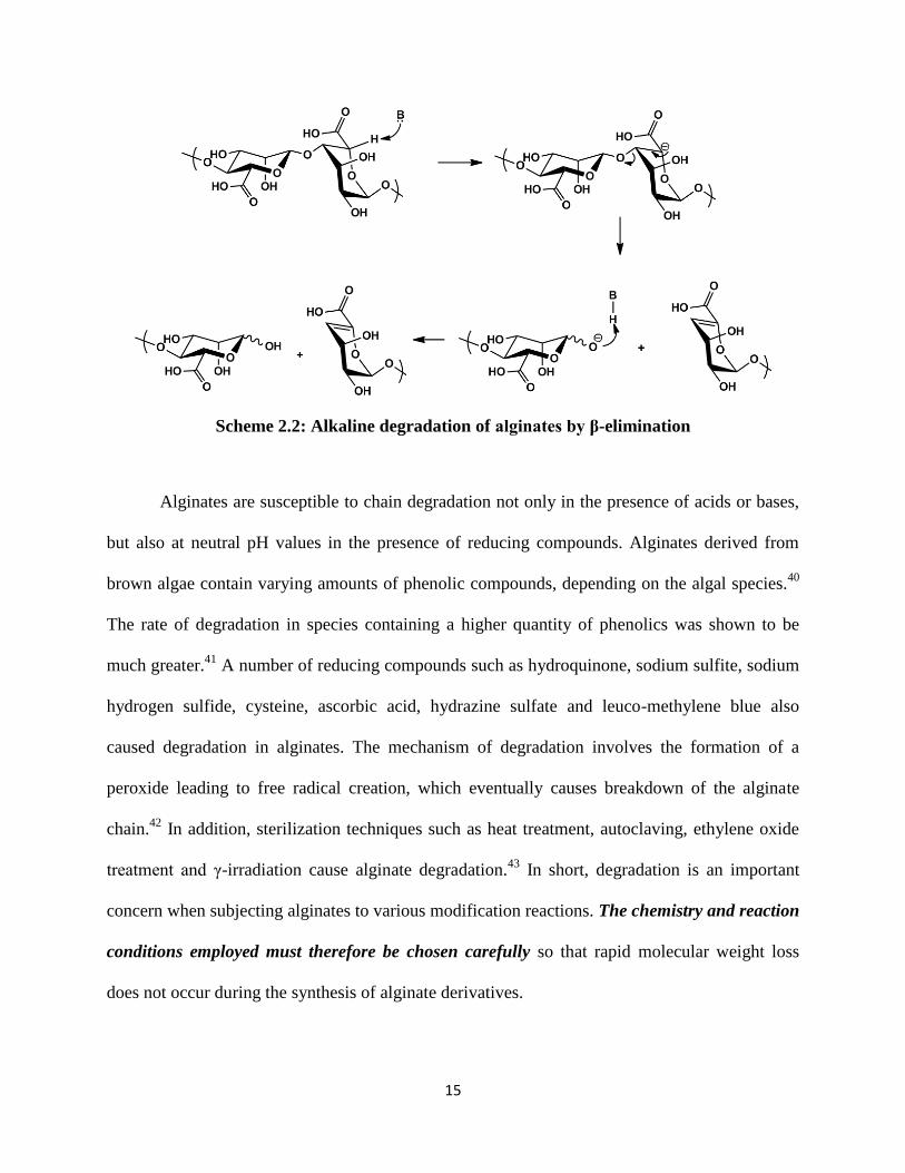
The enzymatic degradation of alginates by lyase occurs by a β-elimination mechanism
resulting in unsaturated compounds (Scheme 2.2).36-37
A similar degradation route is followed
when they are subjected to strongly alkaline environments. The rate of degradation increases
rapidly above pH 10.0 and below pH 5.0. Above a pH of 10.0, the degradation arises mostly
from the β-elimination mechanism, while below 5.0 the degradation is mostly due to acid
catalyzed hydrolysis.38
The mechanism of β-elimination involves abstraction of the proton at C-5
position, which is enhanced by the electron-withdrawing effect of the carbonyl group at C-6
position. When the C-6 carboxyl group is ionized, the electron-withdrawing effect is moderated
and abstraction of the C-5 proton is not as facile as when the carboxyl group is protonated.39
However, the rate of abstraction is still sufficiently high to cause relatively rapid degradation at
high pH.
15
Scheme 2.2: Alkaline degradation of alginates by β-elimination
Alginates are susceptible to chain degradation not only in the presence of acids or bases,
but also at neutral pH values in the presence of reducing compounds. Alginates derived from
brown algae contain varying amounts of phenolic compounds, depending on the algal species.40
The rate of degradation in species containing a higher quantity of phenolics was shown to be
much greater.41
A number of reducing compounds such as hydroquinone, sodium sulfite, sodium
hydrogen sulfide, cysteine, ascorbic acid, hydrazine sulfate and leuco-methylene blue also
caused degradation in alginates. The mechanism of degradation involves the formation of a
peroxide leading to free radical creation, which eventually causes breakdown of the alginate
chain.42
In addition, sterilization techniques such as heat treatment, autoclaving, ethylene oxide
treatment and γ-irradiation cause alginate degradation.43
In short, degradation is an important
concern when subjecting alginates to various modification reactions. The chemistry and reaction
conditions employed must therefore be chosen carefully so that rapid molecular weight loss
does not occur during the synthesis of alginate derivatives.

16
2.4 Alginate modification
The derivatization and design strategies for alginates depend on three important
parameters: solubility, reactivity and characterization (Figure 2.6). (A) Solubility: alginates may
be dissolved in aqueous, organic or mixed aqueous-organic media for derivatization. The choice
of solvent system can dictate the type of reagents that may be used for modification. In addition,
the degree of alginate solubility in the solvent system can impact derivative substitution pattern.
(B) Reactivity: alginates can be modified at the two secondary –OH positions (C-2 and C-3) or
the one –COOH (C-6) position. The difference in reactivity between the two functional group
types can be easily used to selectively modify either one of the two types. Selective modification
of either the C-2 or C-3 hydroxyl group is challenging due to their minor reactivity differences.
In addition, the reaction may be controlled in terms of selective modification of M or G residues.
This may be achieved by taking advantage of the selective chelation of G-residues in Ca-alginate
gels, or by using the partial solubility properties of alginates in certain solvent systems. More
importantly, the reactivity of alginates towards acids, bases and reducing agents cannot be
overlooked when performing derivatization reactions. Competitive degradation reactions can
cause rapid molecular weight loss in short time periods. (C) Characterization: in order to
understand the substitution patterns obtained for the alginate derivatives, it is often essential to
have multiple alginate samples with a range of M/G ratios. In addition, derivatization of alginates
enriched in M, G or MG blocks may be required to achieve a detailed understanding of the
substitution patterns. A lack of commercial availability of alginates with controlled sequences
may impede complete structural characterization of the derivatives. Due to the complex nature of
the alginate copolysaccharide backbone, use of advanced analytical techniques is often
necessary.

17
Figure 2.6: Important parameters governing alginate derivatization
In view of the aspects of alginate derivatization just described, we herein review the
progress made in modification of these useful polysaccharides. We highlight important
chemistries that have been reported in the literature to date, and discuss the properties attained
and applications envisioned for the synthesized derivatives. Our goal is to present a clear and
complete picture of the available methods for alginate modification, enabling the creation of
successful strategies for synthesis of target alginate derivatives, and pointing out the areas where
synthetic advances are still needed, through a solid understanding of the chemistry of this
important polysaccharide.
2.4.1 Acetylation of alginates
Alginates as biosynthesized by bacteria are partially acetylated.12-14
The acetylation
patterns govern both biosynthesis (by preventing epimerization of acetylated monosaccharides)

18
and biological functions of bacterial alginates. In vitro acetylation of alginates is therefore
valuable to the understanding of structure-property relationships of bacterial alginates. The
earliest known report addressing the chemical modification of alginates was published by
Chamberlain et al., wherein acetylation of the hydroxyl groups of alginic acid was described.44
The hydroxyl groups present in alginic acid yarn could not be reacted with acetic anhydride in
the dry state due to strong H-bonding. However when swollen with water, the hydroxyl groups
were available for reaction. Following the swelling process, solvent exchange was performed to
replace water with glacial acetic acid. The alginic acid yarn swollen with glacial acetic acid was
subjected to a mixture consisting of benzene, acetic anhydride and sulfuric acid catalyst. The
yarn acetylated in this manner yielded alginic acid with 97.3% di-acetate, as determined from the
total acetyl content. However, severe degradation was reported to occur during the reaction.
Wassermann reported the acetylation of alginic acid using ketene, a gaseous reagent.44-45
The reactive small molecule ketene was used as an acetylating agent, probably in order to avoid
the alginate degradation caused by exposure to acetic acid or anhydride, and catalysts such as
pyridine or strong acids. Acetylation was performed by swelling alginic acid fully in acetone,
and then reacting with ketene at room temperature to form alginic acid acetate. The product was
insoluble in water as well as in common organic solvents. Alginic acid acetate was converted to
its Na- or Ca-salt forms by treatment with sodium or calcium hydroxide solutions respectively.
Alternatively, Na- or Ca-alginate was directly reacted with ketene to form Na- or Ca-alginate
acetates. Approximately one acetyl group was introduced per monosaccharide unit of the
alginate chain, as determined by a titration method. Degradation during acetylation was assessed
using viscosity measurements. While Na-alginate acetate and alginic acid acetate showed lower
viscosities due to more rapid molecular weight loss, Ca-alginate acetate did not degrade to a very
19
large extent. Regioselectivity of the reaction and composition of the products were not reported,
most likely due to the limited analytical methods available at that time.
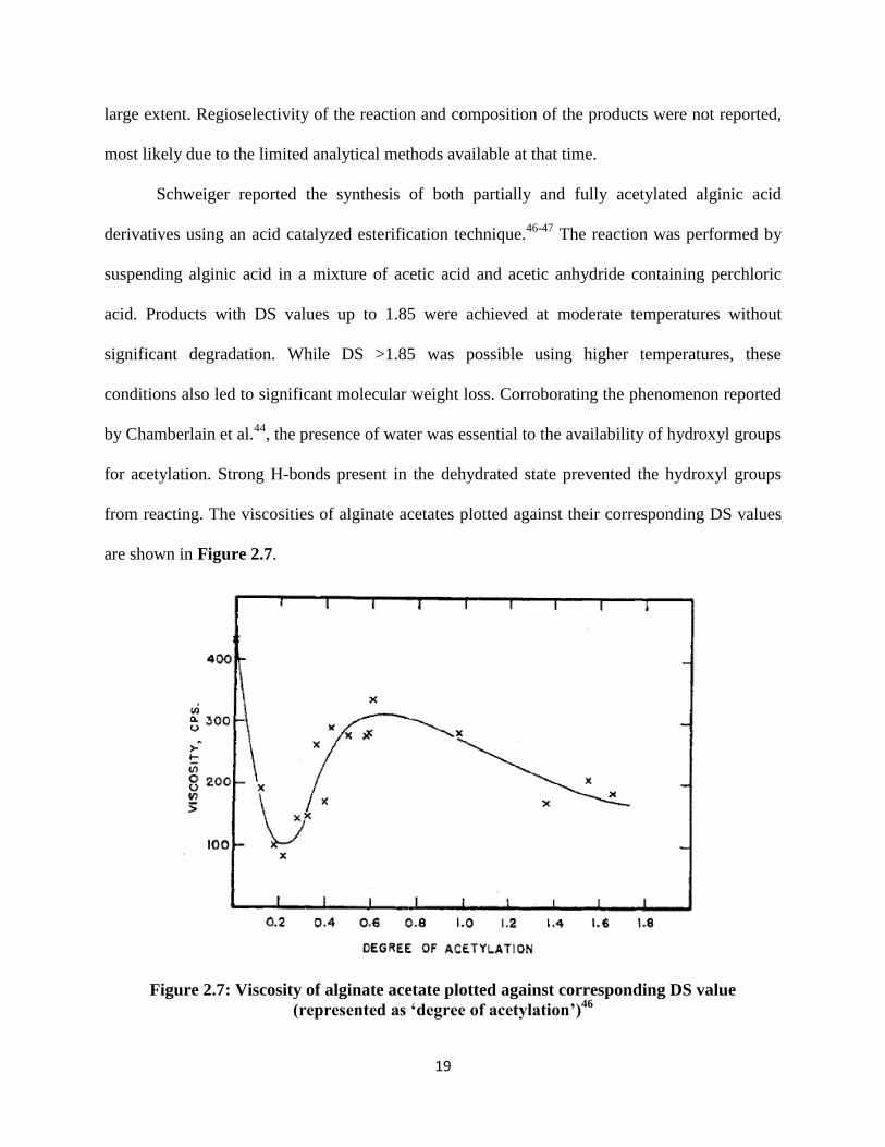
Schweiger reported the synthesis of both partially and fully acetylated alginic acid
derivatives using an acid catalyzed esterification technique.46-47
The reaction was performed by
suspending alginic acid in a mixture of acetic acid and acetic anhydride containing perchloric
acid. Products with DS values up to 1.85 were achieved at moderate temperatures without
significant degradation. While DS >1.85 was possible using higher temperatures, these
conditions also led to significant molecular weight loss. Corroborating the phenomenon reported
by Chamberlain et al.44
, the presence of water was essential to the availability of hydroxyl groups
for acetylation. Strong H-bonds present in the dehydrated state prevented the hydroxyl groups
from reacting. The viscosities of alginate acetates plotted against their corresponding DS values
are shown in Figure 2.7.
Figure 2.7: Viscosity of alginate acetate plotted against corresponding DS value
(represented as ‘degree of acetylation’)46
20
A hypothesis was presented based on the assumption that an increase in the solution
viscosity is caused by the presence of free, unsubstituted and non-hydrogen bonded hydroxyl
groups owing to their ability to associate with water molecules. A decrease in the solution
viscosity on the other hand results from either (a) acid hydrolysis or (b) substitution of the free
hydroxyls with acetyl groups. The initial decrease in viscosity up to DS ~0.2 is evident in Figure
9, and was attributed to a combined effect of (a) and (b). However, as the reaction progressed, H-
bonding between the vicinal hydroxyl groups was overcome and an alginate monoacetate
product was formed. An increase in the total number of free hydroxyl groups occurred as a
result of more vicinal H-bonds being cleaved, resulting in a spike in the solution viscosity. A
viscosity maximum was reached at DS ~ 0.7. Beyond this maximum, the monoacetate sugars
were acetylated further to form diacetates. This led to a reduction in the number of free
hydroxyls and a corresponding decrease in viscosity. The presence of vicinal H-bonds was
therefore thought to be an important factor causing alginate hydroxyl groups to have low
reactivity. However, the hydroxyl group reactivity in monoacetylated alginate products was
higher compared to unreacted alginates. This phenomenon was substantiated by kinetic data
which indicated to a rapid increase in the reaction rate at DS values between 0.7-1.5. The
increase in the reaction rate was associated with increasing availability of non H-bonded
hydroxyl groups for acetyl substitution.
The partially and fully acetylated alginates were used as tools to enhance understanding
of the chelate structure formed in ionically crosslinked alginate gels.47
The addition of divalent
ions to a solution of alginate diacetate (DS = 2.0) did not lead to gel formation. Furthermore,
alginates with DS = 1.4 which contained monoacetylated and diacetylated, but no non-acetylated

21
saccharide residues resisted gel formation. This suggested that the presence of non-acetylated
saccharide units was a must for chelate formation. It also indicated that the presence of hydroxyl
groups was essential for ionic crosslinking, and that carboxylate anions were only partially
responsible for gelation. Therefore, based on the data acquired a chelate structure was proposed
wherein a single Ca+2
ion co-ordinated with two carboxylate groups and two vicinal hydroxyl
groups belonging to the same sugar to form a gel. This proposed chelate structure was further
supported by the fact that the tendency towards gelation was shown to be greater for alginates
containing a higher fraction of unreacted uronic acid sugar residues.
While the early efforts to acetylate alginate were successful, they were limited in terms of
structural characterization of the synthesized derivatives. These investigators were hampered by
a lack of detailed knowledge about the composition and sequence of the alginate backbone,
which was not achieved until a series of fundamental papers published in the 1960s-1980s
illuminated the alginate copolysaccharide structure.20-23, 25, 27, 48-51
Early studies were also
restricted by the absence of advanced techniques for structural analysis. DS values in alginate
acetates were determined predominantly in these early papers using deacylation and titration
methods to measure the acetyl content. Insight about the acetyl substitution variation between M
and G monosaccharide residues, and the regioselectivity between the two secondary OH-groups
on each, was possible only later through powerful characterization techniques such as NMR
spectroscopy. Modern researchers are also advantaged by the availability of alginates with
different M/G contents and sequences, by isolation from various organisms and by enzymatic
preparation in vitro. Alginate variants having different block structures and sequences were not
available until the biochemistry of alginates was better understood.8-11

22
Acetylation of alginates coupled with a detailed analysis of the acetyl substitution pattern
along the backbone was first reported by Skjåk-Bræk et al.52
The reaction was carried out by
preparing Ca-alginate gel beads in aqueous media, then replacing water with pyridine via solvent
exchange. The beads were then suspended in a mixture of pyridine-acetic anhydride at 38 ºC to
perform the acetylation reaction. Scheme 2.3 shows the reaction scheme. Acetylated Ca-alginate
beads were washed thoroughly with water, Ca+2
ions removed and exchanged with Na+ ions, and
the final product dialyzed and freeze dried. Alternatively, acetylation was also performed using
alginic acid. In all cases, the presence of water was essential for reaction to occur and the DS was
found to depend on the amount of water present. The DS value reached maximum at a water
content of ~20%. The reaction rate was fast for the first 40 min of reaction time, and then
dropped off over the next 20 hours.
Scheme 2.3: Acetylation of alginate using pyridine/acetic anhydride. For acetylation in gel
form, M = Ca+2
and M = Na+.52
For homogeneous acetylation in DMSO/TBAF, M = TBA+
and M” = -H or Na+.28
The alginate acetate derivatives so prepared were characterized using 1H-NMR
spectroscopy. The anomeric and high field resonances overlapped, making accurate peak
assignments difficult. The splitting patterns resulting from the O-acetyl peaks around ~2.0-2.2
were therefore used for substituent analysis. In order to make accurate peak assignments
23
possible, it was essential to have alginates enriched in certain block sequences and acetylate
them to identify the peaks arising from their respective structures. By comparing the 1H-NMR
spectra of acetylated alginates enriched in G-blocks and M-blocks, the resonances at 2.04-2.06
ppm were assigned to the acetyl groups from monoacetylated G-residues. Using the relative
changes in the peak intensities observed at varying DS values, signals arising from
monoacetylated G-residues were identified and distinguished from diacetylated G-residues. 1H-
NMR spectra of acetates synthesized from alginates containing varying MG block sequences
were compared, and the peaks arising from acetylated G-residues adjacent to G residues were
distinguished from acetylated G-residues adjacent to M residues.
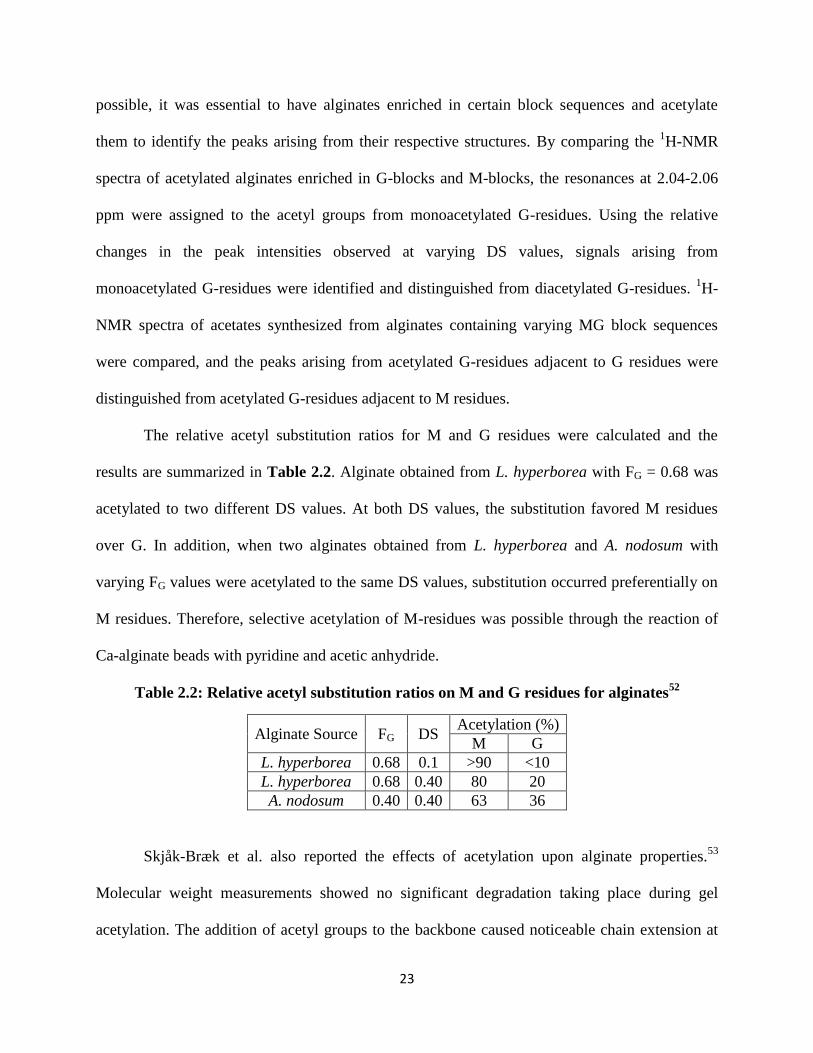
The relative acetyl substitution ratios for M and G residues were calculated and the
results are summarized in Table 2.2. Alginate obtained from L. hyperborea with FG = 0.68 was
acetylated to two different DS values. At both DS values, the substitution favored M residues
over G. In addition, when two alginates obtained from L. hyperborea and A. nodosum with
varying FG values were acetylated to the same DS values, substitution occurred preferentially on
M residues. Therefore, selective acetylation of M-residues was possible through the reaction of
Ca-alginate beads with pyridine and acetic anhydride.
Table 2.2: Relative acetyl substitution ratios on M and G residues for alginates52
Alginate Source FG DS Acetylation (%)
M G
L. hyperborea 0.68 0.1 >90 <10
L. hyperborea 0.68 0.40 80 20
A. nodosum 0.40 0.40 63 36
Skjåk-Bræk et al. also reported the effects of acetylation upon alginate properties.53
Molecular weight measurements showed no significant degradation taking place during gel
acetylation. The addition of acetyl groups to the backbone caused noticeable chain extension at

24
0.1M ionic strength. Even a small proportion of acetyl groups on the backbone played an
important role in defining the polymer conformation. When Ca-alginate gels were formed,
acetylation was found to severely diminish the ability of Ca+2
ions to induce conformational
ordering. Consequently, lower strength gels resulted from acetylated alginates. On drying and re-
swelling the Ca-alginate beads, the degree of swelling increased 500-fold from DS 0 to DS 0.65.
This was a result of enhancement in the positive osmotic pressure resulting from a higher
number of dissociating counterions per polymer chain, due to impairment of co-operative
binding by the added acetyl groups. A plot of the moduli (G) of gels made from acetylated
alginates vs. DS is shown in Figure 2.8. Even at low DS values where M residues were
acetylated selectively, a large drop in modulus was recorded. Thus, to form Ca+2
junctions, it was
important that both G- as well as M-residues remained unsubstituted.
Figure 2.8: Dependence of the modulus of rigidity (G) on DS (represented as "D.a.") in
acetylated Ca-alginate gels53

25
We recently reported a strategy for acetylation of alginates by homogeneous dissolution
of TBA-alginate in two-component organic solvent systems.28
TBA-alginate dissolved
completely in DMSO solutions containing TBAF. In the absence of TBAF, a heterogeneous
mixture was formed with partial dissolution of TBA-alginate, as confirmed by 1H-NMR and
dynamic light scattering (DLS). Figure 2.9 shows an image of the two mixtures. In addition to
DMSO, dipolar aprotic solvents such as DMF, DMAc and DMI fully dissolved TBA-alginate in
the presence of TBAF. Partial acetylation of alginates was achieved in all solvent systems using
pyridine/acetic anhydride reagent under homogeneous conditions with DS ranging between 0.74-
0.85. A maximum DS of ~1.0 was however achieved for acetylation under anhydrous conditions
wherein TBAF was synthesized in situ54
, or by adding a second portion of acetic anhydride to the
reaction mixture. The DS value for alginate acetates could not be increased significantly beyond
~1.0 under any attempted conditions. A potential explanation for this limited DS was based on
monosaccharide ring electronic effects. The uronate ring in unreacted TBA-alginate contained
one electron-withdrawing substituent at the C-5 position. During acetylation, the monoacetate
product was formed first by reaction at one of the two hydroxyl groups. The monoacetylated
uronate ring now had two electron-withdrawing substituents; the first was the C-5 carboxylate
and the second was the acetyl substituent. It is likely that with two electron-withdrawing
substituents present, the reactivity of the second hydroxyl group was significantly reduced. The
DS value may therefore be limited to ~1.0 when pyridine/anhydride is used as the acylating
reagent system.

26
Figure 2.9: Partial dissolution of TBA-alginate in DMSO (left) and complete dissolution in
DMSO/TBAF (right) at a TBAF concentration of 1.1% (w/v). Alginate concentration in
both vials was 15 mg/mL28
An alternate theory was based on the stereochemistry of the monosaccharide residues. In
M residues, the 2-OH group is axial and can interact with the C-4 proton through 1,3-diaxial
interactions, thus favoring substitution at the 3-OH position. In G residues the 3-OH group is
axial and therefore interacts with the C-5 proton through 1,3-diaxial interactions. Substitution of
the 2-OH position was hence favored on G units. Thus, in each monosaccharide, stereochemistry
favored reaction of one out of the two hydroxyl groups, limiting the DS to ~ 1.0. While these
possible causes for an upper DS limit were postulated, they were not corroborated
experimentally. In addition to acetates, propionylation of alginates was also reported using
propionic anhydride and pyridine to obtain a product with DS = 0.31.
To better understand the selectivity of alginate acetylation, the reaction was performed
using alginates containing varying M/G ratios. Under homogeneous reaction conditions, the DS
values obtained were nearly identical (~0.8) across the entire range of alginate M/G ratios. This
proved that the reaction was not selective for M vs. G, and that both M and G residues could be
acetylated. 1H-NMR peak assignments showed that both 2- and 3-OH positions on M residues
could be acetylated under homogeneous conditions. Regioselectivity between the 2-OH and 3-

27
OH positions on G residues was however difficult to determine with certainty due to severe
overlap of signals in the 1H-NMR spectrum.
Acetylation of TBA alginates performed in DMSO without added TBAF, that is, under
partly heterogeneous conditions, afforded more variable DS values compared to homogeneous
acetylation. The products of heterogeneous reactions yielded DS values that varied with the M/G
ratios of the starting polymers. The 1H-NMR spectra of heterogeneously acetylated alginates
showed a predominance of two peaks at shifts of 5.08 and 4.47 ppm respectively. This
predominance was not observed in the case of homogeneous reaction products. Previous
assignments for alginates have shown that the peak at ~5.08 ppm arises from the H-1 signal of G
residues in GGG, MGG and GGM triads. The peak centered at ~4.47 ppm on the other hand
arises from the H-5 signal of G-residues in GGG triads.27
Thus, the predominance of the two
peaks in heterogeneously acetylated alginates suggested that GGG triad sequences remained
relatively unreacted. G-blocks were therefore preserved, resulting in selective acetylation of M
residues. Table 2.3 shows the DS values obtained for alginates with various M contents, under
both homogeneous as well as heterogeneous conditions.
Table 2.3: Homogeneous and heterogeneous acetylation of alginates and the corresponding
DS values
Alginate DSHomogeneous DSHeterogeneous
100% M 0.80 0.91
63% M 0.80 0.48
29% M 0.78 0.63
13% M 0.78 0.54
0% M 0.82 0.72
DS values measured using 1H-NMR. All
reactions employed TBA alginate at 100
mg/mL DMSO using alginate
monosaccharide unit:pyridine:acetic
anhydride molar ratio of 1:17:15;
homogeneous reactions also employed 100
mg TBAF/mL DMSO.

28
The difference in DS values and distribution observed between homogeneous and
heterogeneous reaction products was likely related to the partial solubility of TBA-alginate in
DMSO. A possible explanation may be that the M-blocks have a higher affinity for DMSO, thus
facilitating the selective acetylation of M residues. A contrasting picture was seen for
homogeneous reactions in DMSO/TBAF, where all polymer chains were fully solubilized. Thus,
there was equal accessibility to G-blocks, M-blocks and MG-blocks, which resulted in a
statistical distribution of acetyl groups.
Table 2.4: Molecular weight degradation of TBA-alginate during acetylation
Reaction time DS Mn (kDa) Mw (kDa) PDI DP
0 min 0 56.37 98.18 1.74 144
15 min 0.65 20.36 48.88 2.40 66
30 min 0.75 13.23 33.32 2.52 43
1 h 0.51 8.13 19.61 2.41 28
2 h 0.90 2.78 10.91 3.93 9
DS values measured using 1H-NMR. All reactions were
performed in 1% (w/v) DMSO/TBAF using M063 sample
at 40 ˚C for 2 hours. Ac2O was used as the acyl reagent
with an Alg:Pyr:Acyl ratio of 1:17:15
As previously described in this review, alginates degrade via β-elimination at higher pH
values.38-39
Pyridine is a mild base which nonetheless can degrade alginates. Furthermore,
fluoride from TBAF is also known to be a strong base55
. Molecular weight measurements were
therefore performed on alginate acetates that were reacted for different time periods, and
compared to unreacted TBA-alginate. Table 2.4 shows the obtained results, which clearly point
to significant chain degradation. The starting DP (0 min) of 144 is reduced to 66 within the first
15 minutes of reaction. DP continues to decrease throughout the two hours of reaction time.
Figure 2.10 shows shifts in the SEC chromatograms to longer elution times for alginate acetate

29
as degradation proceeds. Thus in order to preserve the alginate molecular weight, it was desirable
to minimize reaction times and temperatures.
Figure 2.10: Shift in the SEC chromatograms to longer elution times for alginate acetates
shown in Table 2.4, recorded over increasing reaction time28
2.4.2 Phosphorylation of alginates
The synthesis of phosphorylated alginate derivatives was recently described to evaluate
their ability to induce hydroxyapatite (HAP) nucleation and growth.56
Scheme 2.4 shows the
reaction scheme. Phosphorylation was performed using urea/phosphoric acid reagent by creating
a suspension of alginate in DMF. A maximum DS of 0.26 was achieved using an
alginate:H3PO4:urea mole ratio of 1:20:70; the heterogeneous nature of the reaction apparently
limited the maximum DS attainable. Phosphoric acid as a strong acid is likely to cause alginate
molecular weight degradation. Accordingly, a 2-4 fold reduction in the value of Mw was reported
for phosphorylated alginates compared to unreacted alginates.

30
Scheme 2.4: Phosphorylation of alginate56
The regioselectivity of phosphorylation with respect to four possible substitution sites (2-
OH and 3-OH positions of each residue, G and M) was studied using NMR spectroscopy. A
combination of NMR techniques including one-dimensional 1H and
31P,
1H-
1H COSY,
1H-
31P
HMBC, 1H-
31P HMQC-TOCSY and
1H-
13C HSQC was used to carry out detailed peak
assignments. The analysis pointed towards phosphate substitution on M residues such that a
higher degree of phosphorylation occurred on the 3-OH group compared to the 2-OH group. This
regioselectivity was a result of the higher reactivity of the equatorial 3-OH. The equatorial sites
were more accessible compared to axial during phosphorylation. NMR spectroscopy also showed
evidence for substitution occurring on G residues. However, the regioselectivity between 3-OH
and 2-OH groups for G residues could not be cleanly measured because the correlations observed
for G residues in a 1H,
31P-HMQC-TOCSY experiment were much weaker compared to those for
M residues. Interestingly, these phosphorylated alginates were found to be incapable of forming
Ca-crosslinked ionic gels. The reduction of molecular weight during reaction in addition to
conformational changes resulting from phosphorylation were cited as causes for the inability of
these alginate derivatives to form gels. However, Ca-crosslinked gels could be formed by
blending unreacted alginate with phosphorylated alginate. Such gels formed using alginate
blends showed higher resistance to calcium extraction compared to gels formed using only

31
unreacted alginates. The authors pointed out that the phosphate groups were likely to have
participated in the chelation process, thus making the gels more stable.
2.4.3 Sulfation of alginates
Sulfation of polysaccharides, both enzymatically in nature as well as by chemical
methods is known to provide blood-compatibility and anticoagulant activity.57
Heparin is the
most widely used naturally sulfated polysaccharide because of its ability to prevent blood from
clotting.58
It is therefore of interest to see whether alginate sulfates would possess similar
anticoagulation properties. Yumin et al. first reported the sulfation of sodium alginate using
chlorosulfonic acid in formamide.59
The reaction scheme is depicted in Scheme 2.5(a). Sulfates
with DS up to 1.41 were obtained, and the values were measured using the %C to %S ratio from
elemental analysis. The anticoagulant activity of alginate sulfates was measured using activated
partial thrombosis time (APTT), thrombin time (TT) and prothrombin time (PT). Using APTT,
anticoagulant activity of alginate sulfate was found to be comparable to heparin. But using PT,
the activity was very low. Since APTT is based on an intrinsic coagulation pathway and PT is
based on an external coagulation pathway, alginate sulfates were shown to have a greater
influence on the intrinsic coagulation pathway.
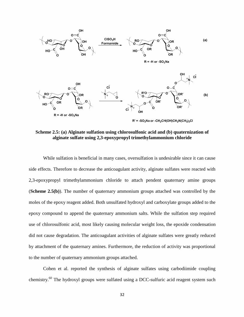
32
Scheme 2.5: (a) Alginate sulfation using chlorosulfonic acid and (b) quaternization of
alginate sulfate using 2,3-epoxypropyl trimethylammonium chloride
While sulfation is beneficial in many cases, oversulfation is undesirable since it can cause
side effects. Therefore to decrease the anticoagulant activity, alginate sulfates were reacted with
2,3-epoxypropyl trimethylammonium chloride to attach pendent quaternary amine groups
(Scheme 2.5(b)). The number of quaternary ammonium groups attached was controlled by the
moles of the epoxy reagent added. Both unsulfated hydroxyl and carboxylate groups added to the
epoxy compound to append the quaternary ammonium salts. While the sulfation step required
use of chlorosulfonic acid, most likely causing molecular weight loss, the epoxide condensation
did not cause degradation. The anticoagulant activities of alginate sulfates were greatly reduced
by attachment of the quaternary amines. Furthermore, the reduction of activity was proportional
to the number of quaternary ammonium groups attached.
Cohen et al. reported the synthesis of alginate sulfates using carbodiimide coupling
chemistry.60
The hydroxyl groups were sulfated using a DCC-sulfuric acid reagent system such

33
that the carboxylate groups remained unreacted. The reaction mechanism (Scheme 2.6) is
marked by the formation of a DCC-H2SO4 adduct which reacts with the hydroxyl nucleophile.
The products were characterized using 13
C-NMR, and the spectra revealed that the C-1 and C-6
peak shifts did not change upon sulfation. The C-2 and C-3 peaks however shifted downfield,
thus proving that the sulfate groups were added to either one or both hydroxyl positions. The use
of a strong acid reagent caused substantial molecular weight loss. The average molecular weight
decreased from 100 to 10 kDa during reaction, but was not affected by the M/G ratio.
Scheme 2.6: Sulfation of alginates using DCC-sulfuric acid route60
The binding of alginate sulfates to both heparin-binding and non-binding proteins was
compared to unmodified alginates. While the sulfates bound strongly to heparin-binding
proteins, unmodified alginates did not bind. Furthermore, both sulfates and unmodified alginates
showed no affinity for heparin non-binding proteins. In nature, specific binding of growth factors

34
to heparin and heparan sulfate protects them from denaturation and proteolytic degradation. The
ability of alginate sulfates to provide protection to growth factors and sustain their release was
therefore studied. Ca-alginate microspheres were formed by blending alginate sulfates with
unreacted alginates. A much more sustained release of bFGF growth factor was achieved from
alginate/alginate sulfate microspheres compared to that from pure alginate microspheres. The
sustained in vivo release of bFGF afforded enhanced angiogenic activity compared to non-
specifically bound alginate.
Fan et al. reported the sulfation of alginates by an uncommon reagent prepared using
sodium bisulfite and sodium nitrite in an aqueous solvent.61
Traditional sulfation reagents such as
sulfuric acid, chlorosulfonic acid, sulfuryl chloride, sulfur trioxide and sulfamic acid cause
hydrolytic degradation of alginates. The new synthetic route was therefore used to overcome this
limitation of traditional reagents. The reaction was performed in water, wherein the sulfation
reagent was first adjusted to the desired pH followed by addition of sodium alginates (Scheme
2.7). DS values for the derivatives were measured using barium sulfate nephelometry. An
optimum DS value of 1.87 was achieved at pH 9.0, 40 °C and 2:1 mole ratio of reagent:uronic
acid residue. Anticoagulant activities for the sulfated alginate derivatives were measured and the
DS, molecular weight and concentration were found to be influential factors.

35
Scheme 2.7: Sulfation of alginates using sodium bisulfite-sodium nitrite reagents61
While the sulfation chemistries described thus far yield alginate sulfates, control over the
placement of sulfate groups along the alginate backbone has not yet been achieved. It is known
that most biological functions in sulfated polysaccharides are related to their sulfation pattern
and sequence.62-63
Therefore, advances in controlled placement of sulfate groups along the
alginate backbone may eventually translate into greater understanding and perhaps control of
structure-property relationships in alginate sulfates.
2.4.4 Hydrophobic modification
Alginates are hydrophilic polysaccharides. In addition to hydrophilicity resulting from
the two hydroxyl groups, the carboxylate ion enhances water solubility at pH 5 and above. The
motivation for hydrophobic modification of alginates is therefore to transform the polysaccharide
from its predominantly hydrophilic nature to a molecule with amphiphilic or hydrophobic
characteristics. The most straightforward way to achieve this transformation is by covalent

36
attachment of hydrophobic moieties such as long alkyl chains or aromatic groups to the polymer
backbone. Hubert et al. attempted the hydrophobic modification of sodium alginate by covalent
attachment of short polyether chains to the alginate backbone.64
Sodium alginate was first
activated using sodium metaperiodate, which oxidized the two secondary hydroxyl groups to
aldehyde groups. The aldehydes were then reacted with α-amino, ω-benzyloxy tetraoxyethylene
(C6H5(OCH2CH2)4NH2, abbreviated as BzlO-TEG-NH2) by reductive amination to yield a
tetraoxyethylene functionalized amine derivative of alginate (BzlO-TEG-NH-Alg; Scheme 2.8).
Through oxidization, alginate hydroxyl groups having low reactivity were converted to
aldehydes having significantly higher reactivity. The carboxylate groups were preserved in the
process, thus retaining the ability of the modified alginate to form ionic gels.
Scheme 2.8: Synthesis of BzlO-TEG-NH-Alg by periodate oxidation of sodium alginate
followed by reductive amination using BzlO-TEG-NH2
The amount of periodate reagent used for the reaction was calculated to be sufficient to
oxidize 20% of the available uronate residues. A significant reduction in the viscosity was
recorded on completion of this step due to molecular weight degradation and extension of chain
conformation by ring opening. Breakdown of the alginate molecular weight in solutions
containing periodate, as previously mentioned, depends on the sequence and distribution of M
and G residues.65
Alginates containing longer MG block sequences were found to degrade more
rapidly than alginates containing shorter MG blocks. Partial oxidation of alginates may be
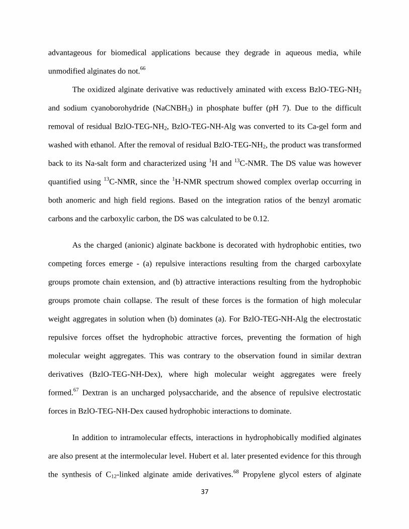
37
advantageous for biomedical applications because they degrade in aqueous media, while
unmodified alginates do not.66
The oxidized alginate derivative was reductively aminated with excess BzlO-TEG-NH2
and sodium cyanoborohydride (NaCNBH3) in phosphate buffer (pH 7). Due to the difficult
removal of residual BzlO-TEG-NH2, BzlO-TEG-NH-Alg was converted to its Ca-gel form and
washed with ethanol. After the removal of residual BzlO-TEG-NH2, the product was transformed
back to its Na-salt form and characterized using 1H and
13C-NMR. The DS value was however
quantified using 13
C-NMR, since the 1H-NMR spectrum showed complex overlap occurring in
both anomeric and high field regions. Based on the integration ratios of the benzyl aromatic
carbons and the carboxylic carbon, the DS was calculated to be 0.12.
As the charged (anionic) alginate backbone is decorated with hydrophobic entities, two
competing forces emerge - (a) repulsive interactions resulting from the charged carboxylate
groups promote chain extension, and (b) attractive interactions resulting from the hydrophobic
groups promote chain collapse. The result of these forces is the formation of high molecular
weight aggregates in solution when (b) dominates (a). For BzlO-TEG-NH-Alg the electrostatic
repulsive forces offset the hydrophobic attractive forces, preventing the formation of high
molecular weight aggregates. This was contrary to the observation found in similar dextran
derivatives (BzlO-TEG-NH-Dex), where high molecular weight aggregates were freely
formed.67
Dextran is an uncharged polysaccharide, and the absence of repulsive electrostatic
forces in BzlO-TEG-NH-Dex caused hydrophobic interactions to dominate.
In addition to intramolecular effects, interactions in hydrophobically modified alginates
are also present at the intermolecular level. Hubert et al. later presented evidence for this through
the synthesis of C12-linked alginate amide derivatives.68
Propylene glycol esters of alginate

38
(PGA) with ~ 30% carboxylic acid groups esterified (DSPG ~0.3) were substrates for nucleophilic
displacement by dodecylamine (Scheme 2.9) to form the corresponding amides (PGA-C12) with
a DSAmide ~ 0.09 (calculated based on nitrogen content analysis). The reaction was performed in
the heterogeneous state by making a suspension of PGA in DMF.
Scheme 2.9: Synthesis of alginate amides by reaction of propylene glycol esters of alginate
with dodecylamine
The change in viscosity of PGA solutions after C12 modification was studied. Figure
2.11(a) shows the viscosity vs. concentration plots for both PGA and PGA-C12. A large increase
in PGA-C12 viscosity with concentration was observed compared to that in PGA. This increase in
PGA-C12 viscosity was attributed to a combined effect of chain entanglements and
intermolecular interactions between the hydrophobic dodecyl chains. Interactions at an
intermolecular level lead to the formation of physically crosslinked domains. This effect was
similar to that observed in hydrophobically modified hydroxyethyl cellulose (HMHEC)
derivatives, wherein hydrophobically driven association between individual molecules leads to
the formation of a three-dimensional network.69
The viscosity increase in unmodified PGA
resulted only from chain entanglements. The absence of intermolecular interactions and
physically crosslinked domains prevented a large viscosity spike. The presence of intermolecular
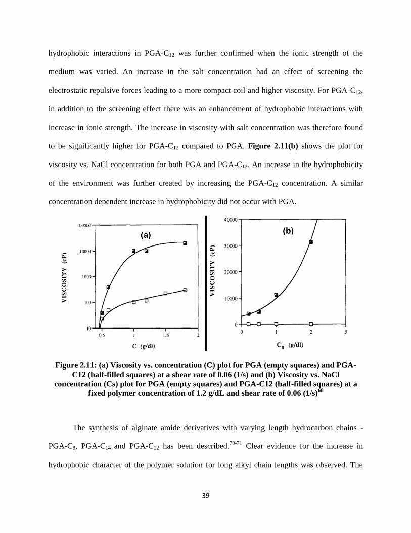
39
hydrophobic interactions in PGA-C12 was further confirmed when the ionic strength of the
medium was varied. An increase in the salt concentration had an effect of screening the
electrostatic repulsive forces leading to a more compact coil and higher viscosity. For PGA-C12,
in addition to the screening effect there was an enhancement of hydrophobic interactions with
increase in ionic strength. The increase in viscosity with salt concentration was therefore found
to be significantly higher for PGA-C12 compared to PGA. Figure 2.11(b) shows the plot for
viscosity vs. NaCl concentration for both PGA and PGA-C12. An increase in the hydrophobicity
of the environment was further created by increasing the PGA-C12 concentration. A similar
concentration dependent increase in hydrophobicity did not occur with PGA.
Figure 2.11: (a) Viscosity vs. concentration (C) plot for PGA (empty squares) and PGA-
C12 (half-filled squares) at a shear rate of 0.06 (1/s) and (b) Viscosity vs. NaCl
concentration (Cs) plot for PGA (empty squares) and PGA-C12 (half-filled squares) at a
fixed polymer concentration of 1.2 g/dL and shear rate of 0.06 (1/s)68
The synthesis of alginate amide derivatives with varying length hydrocarbon chains -
PGA-C8, PGA-C14 and PGA-C12 has been described.70-71
Clear evidence for the increase in
hydrophobic character of the polymer solution for long alkyl chain lengths was observed. The

40
rheological properties of PGA, PGA-C12 and PGA-C14 derivatives were studied in the semi-dilute
aqueous regime, and a marked change was observed for the hydrophobically modified
derivatives when compared to parent PGA.72
The intermolecular hydrophobic interactions
between long alkyl chains were disrupted reversibly under high shear.
Hubert et al. also reported the synthesis of hydrophobically modified alginate derivatives
starting from Na-alginate.73
Dodecyl chains were covalently linked to the carboxylate groups on
the backbone. TBA-alginate was first synthesized by converting Na-alginate to alginic acid and
subsequent neutralization with TBA hydroxide. TBA-alginate was reported to be soluble in
DMSO, but experiments in our laboratory have shown that TBA-alginate synthesized using a
similar procedure was only partially soluble in DMSO.28
The reaction was carried out using
dodecyl bromide reagent via nucleophilic displacement (Scheme 2.10). The product (Alg-C12)
was purified by dialysis and subsequently freeze dried, with hydrophobe DS 0.12 as determined
by gas chromatography after alkaline hydrolysis of the sample.
Scheme 2.10: Reaction scheme for the synthesis of Alg-C12
The surface tension isotherms for Alg-C12 and unreacted Na-alginate were compared by
measuring the change in surface tension (σ) with concentration. The σ value for Na-alginate was
surprisingly unchanged after C12 modification. It was proposed that collapsed micellar structures
formed due to intramolecular hydrophobic interactions. The dodecyl chains were buried inside

41
the micelles, protecting them from exposure to the adsorption interface. In addition, the charged
alginate backbone made it incapable of folding at short distances and restricted orientation of the
hydrophobic chains at the surface. Figure 2.12(a) shows a schematic of the adsorption of Alg-
C12 at the air water interface in the presence of a high concentration of covalently attached long
alkyl chains.
Figure 2.12: (a) Alg-C12 adsorption at the air water interface and (b) Alg-C12+DTAB
adsorption at the air water interface73
While Alg-C12 did not show markedly enhanced adsorption activity, the addition of a
cationic surfactant, dodecyltrimethylammonium bromide (DTAB) with Na-alginate showed a
remarkable improvement in the surface adsorption. The surfactant possesses a C12 hydrophobic
tail and a cationic head group which interacted ionically with the negative charges on the
alginate backbone. Therefore, while the hydrophobic C12 chain was covalently linked in Alg-C12,
the C12 chain was bonded ionically to the alginate backbone in a mixture of Na-Alg + DTAB. At
identical C12 molar concentrations, the ionically linked Na-Alg + DTAB showed almost a
thousand-fold higher surface adsorption activity compared to the covalently linked Alg-C12. In

42
other words, the electrostatically linked C12 alkyl chains in Alg+DTAB were more densely
aligned at the air-water interface than the covalently attached C12 chains in Alg-C12. Figure
2.12(b) shows a schematic of Alg +DTAB adsorption at the air-water interface. The labile nature
of electrostatically attached C12 alkyl chains allowed a free re-organization at the interface, while
such re-organization was not possible when the chains were covalently linked to the alginate
backbone. Dellacherie et al. later reported the physico-chemical properties of hydrophobically
modified alginates in aqueous solution.74
Fluorescence spectroscopy studies were performed
which showed an increase in the hydrophobic character with increasing alkyl chain lengths.
Production of microspheres based on hydrophobically modified alginates by a dispersion
gelation method was reported by Leonard et al.75
Gelation was induced in an aqueous solution of
Alg-C12 by the addition of NaCl. A dispersive shear stress was first applied to overcome the
initially high solution viscosity of Alg-C12. The solution was then dispersed in aq. NaCl to afford
gel microparticles. Microparticle size was controlled by varying the applied shear stress. While
the particles were stable in aqueous solutions for several days, they aggregated upon
centrifugation. A small amount of CaCl2 was therefore used during particle formation to
strengthen the physical network by ion crosslinking. Increasing the polymer concentration in the
starting mixture caused a slight increase in the particle size. Furthermore, at high DS values,
small particle sizes could be achieved at low salt concentrations; while at low DS values, high
salt concentrations were necessary to achieve the same size. Bovine serum albumin (BSA) was
encapsulated within the Alg-C12 gel microspheres. In water, the protein was released from
unmodified alginate within a few hours, but BSA encapsulated in Alg-C12 microspheres was not
released for up to 5 days. In citrate solutions, no release of BSA encapsulated in Alg-C12
microspheres was observed, whereas BSA encapsulated in unmodified alginate gel was released

43
instantaneously by dissolution of Ca-alginate beads. Sodium citrate is a Ca+2
chelating agent, and
likely caused a rapid Na+-Ca
+2 exchange causing the beads to dissolve. The authors further cited
that no BSA release occurred from Alg-C12 microspheres into the surrounding aqueous
environment due to the stability of hydrophobic junctions within the gels.
Scheme 2.11: Reaction scheme for the synthesis of Alg-CONH-C8
Nyström et al. reported the synthesis of hydrophobically modified alginate (Alg-CONH-
C8) by coupling n-octylamine to the backbone carboxylic acid groups using 1-ethyl-3-(3-
dimethylaminopropyl)carbodiimide hydrochloride (EDC-HCl) 76
(Scheme 2.11). A DS value up
to 0.30 was measured using 1H-NMR, using the ratio of methyl (from the C8 chain) to anomeric
protons, and was confirmed by elemental analysis. The effect of the M/G ratio and the sequence
of the two monosaccharides on substitution pattern were however not reported. The mechanism
of carbodiimide mediated coupling of carboxylic acids to amines is depicted in Scheme 2.12.

44
Scheme 2.12: Mechanism of carbodiimide mediated coupling of carboxylic acids to amines
The hydrophobic interactions occurring in Alg-CONH-C8 were reversible in the presence
of β-cyclodextrin (β-CD). β-CD was shown to have a noticeable effect on the Alg-CONH-C8
chain length. Small-Angle Neutron Scattering (SANS) was used to calculate two physical
parameters – persistence length (L) and correlation length (ξ). Persistence length is a measure of
the stiffness of long polymer chains, whereas correlation length defines the average spatial extent
of polymer concentration fluctuations resulting from thermal changes. When β-CD was added to
a solution of Alg-CONH-C8, the value of L increased while that of ξ decreased. The truncated
cone structures in β-CD encapsulated the hydrophobic C8 chains on Alg-CONH-C8 causing a
decrease in hydrophobic interactions. This in turn led to an increase in the net repulsive
electrostatic forces, resulting in local stretching of polymer chains and higher L values. The
decrease in ξ was a result of re-organization of the network, whereby the hydrophobic
interactions fell apart to yield a homogeneous system devoid of junction points. Figure 2.13
shows a schematic illustrating the encapsulation of hydrophobic C8 moieties on Alg-CONH-C8
by the β-CD truncated cone.

45
Figure 2.13: Schematic illustration of the encapsulation of the hydrophobic moieties on
Alg-CONH-C8 by the β-CD truncated cone76
Nyström et al. later reported complex formation between Alg-CONH-C8 and
hydroxypropyl-β-CD (HP-β-CD) or polymerized β-CD (poly-β-CD).77
On addition of HP-β-CD
to a solution of Alg-CONH-C8, a reduction in viscosity was observed. This viscosity reduction
was an effect of encapsulation of the hydrophobic moieties by the HP-β-CD truncated cone, thus
deactivating the hydrophobic interactions. Addition of poly-β-CD to a solution of Alg-CONH-C8
showed more complex behavior, where the solution viscosities depended on the both poly-β-CD
and Alg-CONH-C8 concentrations.
2.4.5 Attachment of cell signaling molecules
Alginates are important biomaterials in the area of bioengineering.78
A critical
requirement of such biomaterials is their ability to provide an environment that is both physically
and chemically favorable to the presence of biological species such as living cells. In order to
enhance the chemical interactions of alginate matrices with cells, they are functionalized with
cell-specific ligands or extracellular signaling molecules. In addition to enhancement of the
cellular interactions, functionalization may also play a role in controlling the growth,

46
differentiation and behavior of cells in culture. Alginates as biomaterials offer several advantages
including hydrophilicity, biocompatibility and non-immunogenicity.79
The ability of alginates to
form gels capable of encapsulating cells, drugs and other biological entities is another important
advantage for biological applications. However, a possible drawback is that cells do not adhere
to alginates naturally. For that reason Mooney et al. have recently described alginates as “blank
slates”.6 Chemical functionalization with cell signaling moieties is therefore crucial in order to
overcome the low affinity of alginates to cell surfaces.
Skjåk-Bræk et al. reported the covalent attachment of galactose moieties to the alginate
backbone for enhancement of hepatocyte cell recognition.80
Hepatocytes perform a variety of
metabolic functions in the normal liver. However, outside the liver they lose their function and
are viable for only short time periods. The presence of an anchor material that provides
mechanical support and immunoprotection to the cells prevents such loss of functionality and
cell death. Alginates with their unique gel forming ability are considered very good matrix
materials, providing a suitable environment for sustaining hepatocytes outside the organism. The
surface of hepatocyte cell membranes possesses an asialoglycoprotein receptor (ASGPR), which
binds and internalizes glycoproteins with terminal β-galactosyl residues. Galactose modified
alginates capable of ionic gelation were therefore synthesized to be used as biomaterials for
improved encapsulation and adhesion of hepatocytes.
The synthesis was performed by reaction of the alginate carboxylic acid groups with 1-
amino-1-deoxy-β-D-galactose (Gal-1-NH2) using aqueous carbodiimide chemistry (Scheme
2.13). An EDC/N-hydroxysulfosuccinimide (NHS) coupling reagent was used. The EDC/NHS
coupling mechanism is similar to that depicted in Scheme 2.12, except the O-acylated urea

47
derivative formed in the first step is stabilized by conversion into an amine reactive sulfo-NHS
ester, thus increasing the efficiency of EDC mediated coupling.
Scheme 2.13: Reaction scheme for the synthesis of galactose substituted alginate
The maximum achievable DS (measured by 1H-NMR) was 0.36 using 1.5 equivalents of
Gal-1-NH2 per monosaccharide unit at pH 4.5. 1H-NMR spectroscopy was also used to identify
the positions at which galactose substitution occurred on the alginate backbone. For accurate
assignments of the new peaks that appeared in the anomeric region after derivatization, it was
essential to obtain 1H-NMR spectra of polymannuronate, G-enriched alginate and polyalternating
MG, and their corresponding galactose substituted derivatives. Three new signals appeared in the
anomeric region upon galactosylamine modification; signal (A) was assigned to the H-1 proton
of galactosylamine linked to G-residues, signal (B) was assigned to the H-1 proton of a
galactosylamine substituents linked to M residues neighboring G residues, and signal (C)
resulted from a non-anomeric proton which experienced a peculiar environment like that of
galactose moieties linked to diaxial GG dyads. The areas of these peaks corresponded to the
fraction of galactosylamine linked to G-residues (A), the fraction of galactosylamine linked to M
residues in alternating sequences (B), and the amount of galactosylamine residues linked to G
residues neighboring G residues (C). The difference between peaks A and C was then used to
calculate the fraction of substitution on G residues in alternating sequences. The fraction of
48
substituted M residues neighboring M residues could not be calculated due to overlapping
signals in the 4.60-4.80 ppm region.
The difficult nature of the analytical problem at hand is clearly evident through this
example.80
The alginate derivatives studied in this case were modified only at the carboxylic acid
position on the backbone. Increasing complexity arises in the NMR spectra when substitution
occurs on the two hydroxyl groups in addition to the carboxyl group. For the galactosylamine
substituted alginates, the positional selectivity present at various DS values is shown in Table
2.5. Based on the data, it was concluded that at low DS values, the reaction was more selective to
G residues; as higher DS values were attained; M residues participated in the reaction. In
addition, G residues neighboring M residues were more reactive than G residues neighboring G
residues.
Table 2.5: Substitution selectivity for alginates at various DS values, measured using 1H-
NMR spectroscopy80
DS
Total
Substitution
on G (%)
Total
Substitution
on M (%)
Substitution
on G
adjacent to
M
(%)
Substitution
on G
adjacent to
G
(%)
Substitution
on M
adjacent to
G
(%)
Substitution
on M
adjacent to
M
(%)
0.07 7 - 7 - - -
0.19 19 - 7.2 11.8 - -
0.36 29 7 7.3 21.7 7 -
The ability of galactose substituted alginate derivatives to form gel beads was studied.
Substitution of galactose sugar residues on the alginate backbone caused a reduction in the
number of available carboxylic acid groups, and impacted the bead size. Having galactose side
chains caused conformational disordering within gels, whereby the junctions formed were held
less tightly compared to unmodified alginates. A result of this effect was that larger bead
volumes were achieved due to a net gain in hydration. Galactose moieties also interfered with the

49
polymer chain packing process during gel formation. When the gel beads were dried and
rehydrated, the kinetics and thermodynamics of swelling were affected. Larger volume increases
during rehydration resulted at high DS values. An increase in the DS was also found to decrease
the affinity of alginates towards Ca+2
ions. Cytotoxicity studies were performed which showed
that all of these Gal-substituted alginate derivatives were non-toxic.
The conformational effects resulting from the introduction of galactose moieties on the
alginate backbone were studied using intrinsic viscosity, radius of gyration and chiro-optical
property measurements.81
Galactose substitution on the alginate backbone led to a complex
conformational change in solution. The presence of a fairly large moiety such as galactose in
close proximity to the alginate backbone altered the conformational space available to the
backbone. Additionally, when G residues in alternating sequences were substituted, the chain
adopted a less extended conformation compared to the substitution of G residues in
homopolymeric sequences. A decrease in the hydrodynamic radius was therefore observed at
low DS values, when G residues neighboring M residues were substituted. As the DS value
increased, G residues neighboring other G residues were substituted, and an increase in the
hydrodynamic properties occurred.
Although galactose moieties were covalently attached to the alginate backbone,
substitution on both M and G units was disadvantageous due to the loss of mechanical properties
for the alginate gels. Skjåk-Bræk et al. therefore reported a method combining a chemical and
enzymatic approach to achieve selective substitution on M residues.82
Since G units were not
galactosylated, the mechanical properties of the ionotropic gels formed were not altered. A
reaction scheme similar to that shown in Scheme 2.13 was used wherein polymannuronan (with
no G residues) was galactosylated first. Galactosylated polymannuronan was then subjected to
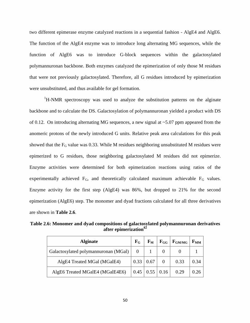
50
two different epimerase enzyme catalyzed reactions in a sequential fashion - AlgE4 and AlgE6.
The function of the AlgE4 enzyme was to introduce long alternating MG sequences, while the
function of AlgE6 was to introduce G-block sequences within the galactosylated
polymannuronan backbone. Both enzymes catalyzed the epimerization of only those M residues
that were not previously galactosylated. Therefore, all G residues introduced by epimerization
were unsubstituted, and thus available for gel formation.
1H-NMR spectroscopy was used to analyze the substitution patterns on the alginate
backbone and to calculate the DS. Galactosylation of polymannuronan yielded a product with DS
of 0.12. On introducing alternating MG sequences, a new signal at ~5.07 ppm appeared from the
anomeric protons of the newly introduced G units. Relative peak area calculations for this peak
showed that the FG value was 0.33. While M residues neighboring unsubstituted M residues were
epimerized to G residues, those neighboring galactosylated M residues did not epimerize.
Enzyme activities were determined for both epimerization reactions using ratios of the
experimentally achieved FG, and theoretically calculated maximum achievable FG values.
Enzyme activity for the first step (AlgE4) was 86%, but dropped to 21% for the second
epimerization (AlgE6) step. The monomer and dyad fractions calculated for all three derivatives
are shown in Table 2.6.
Table 2.6: Monomer and dyad compositions of galactosylated polymannuronan derivatives
after epimerization82
Alginate FG FM FGG FGM/MG FMM
Galactosylated polymannuronan (MGal) 0 1 0 0 1
AlgE4 Treated MGal (MGalE4) 0.33 0.67 0 0.33 0.34
AlgE6 Treated MGalE4 (MGalE4E6) 0.45 0.55 0.16 0.29 0.26

51
To better understand the effect of the presence of galactose moieties on the epimerization
reaction, galactosylated polymannuronan with a spacer group present between the galactose unit
and mannuronan residue was synthesized. Carboxylic acid groups of mannuronic acid were
coupled to p-aminophenyl-β-D-galactopyranoside (pNH2PhβGal) using EDC/NHS coupling. The
rigid spacer group did not significantly alter the epimerization of unmodified M residues. In
addition, the amide bond was intact after exposure to both enzymes AlgE4 and AlgE6. Gel
studies were performed with the galactosylated alginates. When the only source of G residues
was from MG alternating sequences, the gels formed were based on the formation of interchain
junctions. However, when the source of G residues was both MG alternating sequences and G-
block sequences, “egg-box” (Figure 2.5(A)) structures were formed. The introduction of
galactose units selectively on M residues caused faster gelling and higher gel strength compared
to when galactose units were attached to both G and M residues. For gels that contained
galactose units only on M residues, the alginate chains associated laterally with each other. This
was contrary to gels made from alginates bearing galactosyl groups on both M and G residues,
where the bulky galactose units on G residues sterically hinder lateral association among chains.
Selective galactosylation of M residues also provided gels that were more stable to saline
solutions, critical to in vivo performance.
Akaike et al. reported a strategy to galactosylate alginate using lactobionic lactone and
ethylenediamine83
(Scheme 2.14). Lactobionic acid was dehydrated to give lactobionic lactone
in the first step. Then the lactone was reacted with excess ethylenediamine to form the
monoamine terminated lactobionic lactone (L-NH2). The free primary amine groups of L-NH2
were then coupled with the carboxylic acid groups on the alginate backbone mediated by
EDC/NHS. This afforded galactosylated alginates (GA) with DS 0.26 (elemental analysis).

52
Scheme 2.14: Reaction scheme for the synthesis of galactosylated alginate (GA) using
lactobionic lactone
The attachment of hepatocytes on to a GA coated polystyrene (PS) dish was studied.
Hepatocyte attachment to GA was 55%, compared to 3% for PS coated with unmodified
alginate. This clearly demonstrated that enhancement in hepatocyte attachment was caused by
the linking of galactose residues to alginate. Furthermore, the number of hepatocytes attached
was greater when the density of galactose residues on the GA coated surface was higher. Specific
interaction of hepatocyte surfaces with galactose moieties was verified by poly-N-p-vinylbenzyl-
D-lactoneamide (PVLA) competitive binding assay. The hepatocyte cells were encapsulated
within calcium crosslinked GA gels. In order to maintain cell stability and liver-specific
functions, the formation of three-dimensional hepatocyte spheroids within the anchoring matrix
is essential. For calcium alginate gels formed using GA, more than 80% of the cells were found
to aggregate forming uniformly distributed multicellular spheroids. In addition, aggregation
increased as epidermal growth factor (EGF) and insulin were added. For calcium alginate gels
formed using unmodified alginates, the hepatocyte cells did not aggregate. Hepatocytes are
anchorage sensitive cells, which means that their functions in vitro and in vivo are regulated by

53
cell anchorage and cell aggregation.84
GA thus enhanced liver related functions in encapsulated
hepatocyte cells. Gels made from GA also showed higher cell viability compared to unmodified
alginates.
Mooney et al. reported the linking of GRGDY cell adhesion peptide to alginates for
enhancement of their interactions with mouse skeletal myoblasts. 85
Amine functional peptides
were attached to the alginate carboxylic acid groups using EDC/NHS coupling (Scheme 2.15).
The interaction of C2C12 mouse skeletal myoblasts with GRGDY containing alginates was
studied. Myoblast cells seeded on to GRGDY-coupled alginate hydrogels attached and spread on
the surface, while no attachment was observed on unmodified alginate control surfaces. The
observed myoblast adhesion was a result of specific interactions with the GRGDY peptide
sequences. Even though significant cell spreading occurred, the number of cells did not increase
within the first 24 hours of seeding. At day 3, the number of cells increased slightly and reached
an upper limit. Thereafter the number of cells decreased. This decrease was a result of the fusion
of myoblasts into multinucleated microfibrils, an important step in the pathway to skeletal
muscle differentiation.
Scheme 2.15: Reaction scheme for the synthesis of GRGDY containing alginates
Mooney et al. also reported the effect of variation of GRGDY ligand density and alginate
M/G ratio on proliferation and differentiation of C2C12 skeletal myoblast cells.86
With an
increase in the alginate G-content, cell proliferation increased significantly. In addition, alginates

54
with high G-contents promoted extensive fusion of the myoblasts causing a corresponding
increase in the levels of muscle creatine kinase (MCK) activity over time. Cell proliferation and
differentiation was also dependent on the ligand density present on the alginate backbone. As the
ligand density increased, the proliferation and spreading of myoblast cells improved and an
increase in the cell count was recorded.
2.4.6 Covalent crosslinking of alginates
Ionic crosslinking of alginates in the presence of divalent cations is well known.87
Such
physical gels are used in a variety of areas including textile printing, paper coating, wound
dressing, food additives and biotechnology.88-89
While the ionic gels are of great interest,
covalent crosslinking of alginates yields matrices with improved mechanical properties. Ionic
alginate gels are known to have limited stability in vivo. In physiological media, ion exchange
with monovalent ions causes destabilization and rupture of the gel.90
Chemical crosslinking is an
attractive strategy for overcoming this drawback by creating more stable and robust networks.
The methods used for covalent crosslinking of alginates and their applications are therefore
reviewed.
Cascone et al. reported chemical crosslinking of alginates to make beads for affinity and
ion exchange chromatography.91
In an ion exchange material, availability of the carboxylic acid
groups on the alginate backbone is necessary. The carboxylic acid groups in Ca-alginate gels
participate in ion crosslinking and are thus are not available for ion exchange. Covalent
crosslinking by reaction of the alginate hydroxyl groups for network formation is therefore
essential for this application. This was achieved by reacting Ca-alginate beads with
epichlorohydrin in the presence of aqueous NaOH. Calcium was later removed by multiple
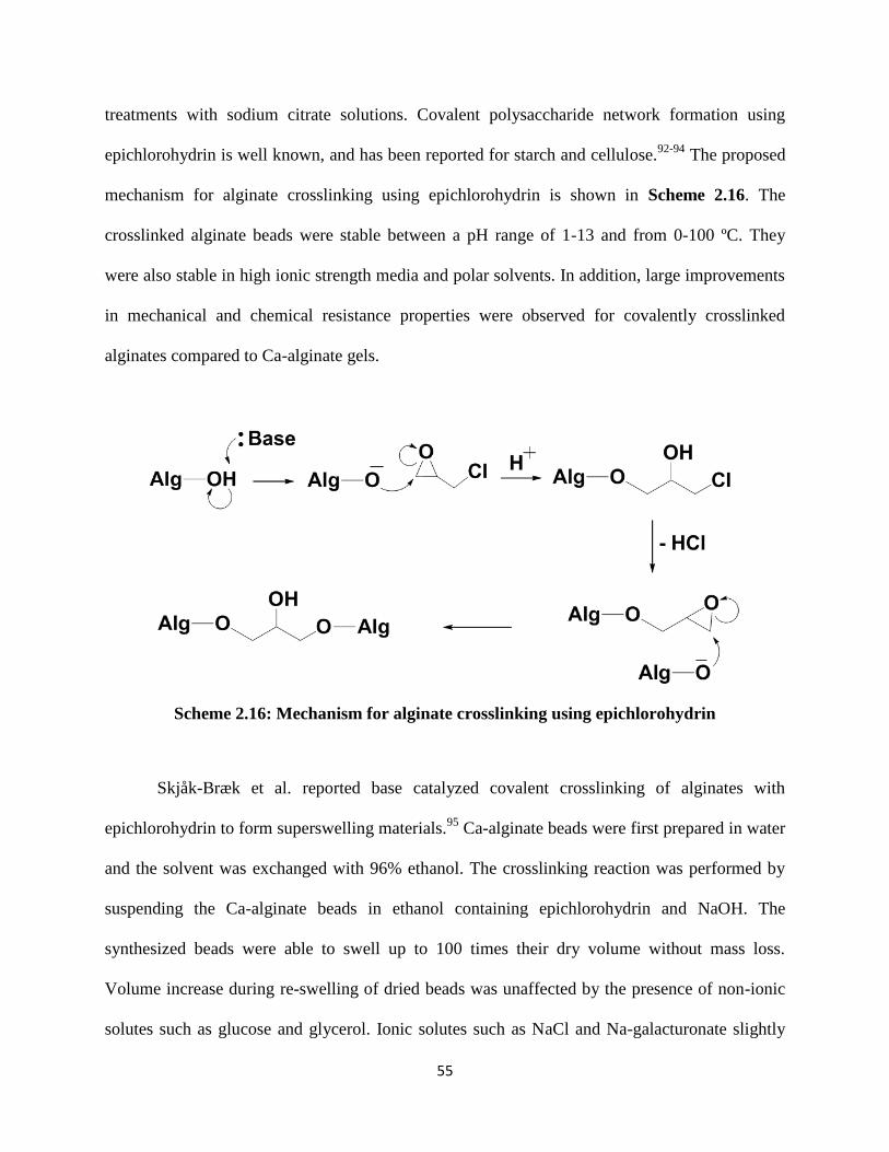
55
treatments with sodium citrate solutions. Covalent polysaccharide network formation using
epichlorohydrin is well known, and has been reported for starch and cellulose.92-94
The proposed
mechanism for alginate crosslinking using epichlorohydrin is shown in Scheme 2.16. The
crosslinked alginate beads were stable between a pH range of 1-13 and from 0-100 ºC. They
were also stable in high ionic strength media and polar solvents. In addition, large improvements
in mechanical and chemical resistance properties were observed for covalently crosslinked
alginates compared to Ca-alginate gels.
Scheme 2.16: Mechanism for alginate crosslinking using epichlorohydrin
Skjåk-Bræk et al. reported base catalyzed covalent crosslinking of alginates with
epichlorohydrin to form superswelling materials.95
Ca-alginate beads were first prepared in water
and the solvent was exchanged with 96% ethanol. The crosslinking reaction was performed by
suspending the Ca-alginate beads in ethanol containing epichlorohydrin and NaOH. The
synthesized beads were able to swell up to 100 times their dry volume without mass loss.
Volume increase during re-swelling of dried beads was unaffected by the presence of non-ionic
solutes such as glucose and glycerol. Ionic solutes such as NaCl and Na-galacturonate slightly
56
reduced the extent of volume increase. However, the reduction in degree of swelling due to the
presence of ionic solutes was significantly lower in magnitude compared to synthetic
superswelling materials like crosslinked polyacrylic acid.
Skjåk-Bræk et al. also studied the swelling behavior of covalently crosslinked alginate
beads in the presence of monovalent and divalent cations.96
On swelling the beads in water, ion
concentration in the interior of the bead was high compared to the exterior. When the beads were
swelled with monovalent salt solutions, the difference in ion concentration between the exterior
and interior was lower, and caused a marked decrease in bead volume with increasing salt
concentrations. Swelling with salt solutions of divalent (crosslinking) ions like Ca+2
, Sr+2
and
Ba+2
showed hysteresis in the swelling curves - a feature displayed due to cation binding in
alginate gels. This proved that alginates retain their ion-binding properties in the covalently
linked state. By introducing divalent cations, physical crosslink sites were introduced in addition
to the existing covalent linking sites, making stronger gels. Furthermore, the bead volume
decreased as the pH of the swelling solution was reduced. Protonation of the alginate carboxylate
groups at lower pH values reduced the difference between interior and exterior ion
concentrations. This led to gel shrinkage. The change in gel volume as a function of ethanol
addition showed that above a certain ethanol concentration, the gel transformed from a
“dissolved” voluminous state to a “solid” compact state. A large volume decrease at high ethanol
concentrations occurred due to precipitation-like behavior.
Yeom et al. reported the use of covalently crosslinked sodium alginate membranes for
pervaporation separation.97
Alginates are considered to be good membrane materials for
dehydration of ethanol-water mixtures due to their ability to incorporate water into the
hydrophilic membrane structure.98
However, excess hydrophilicity may be disadvantageous as it

57
can cause low selectivity and poor membrane stability. Thus, chemical modification via covalent
crosslinking was performed for membrane property enhancement. The cis-oriented vicinal
hydroxyl groups at the C-2 and C-3 positions can react with glutaraldehyde to form acetal-linked
alginate networks. Scheme 2.17a shows the mechanism for acid catalyzed acetal formation,
while Scheme 2.17b shows the reaction scheme for glutaraldehyde crosslinking of alginates. Na-
alginate films were first cast from aqueous solutions. The films were dried and reacted in an
acetone solution containing glutaraldehyde and HCl. Crosslinking density in alginate films was
controlled using the concentration of glutaraldehyde present in the reactive solution. Swelling
measurements for the crosslinked films as well as Na-alginate films were performed in solutions
containing varying ethanol:water ratios. The liquid volume fractions in Na-alginate films were
higher compared to crosslinked films. In addition, crosslinked alginate films contained a higher
fraction of ethanol compared to Na-alginate after absorption.
Scheme 2.17: (a) mechanism of acid catalyzed acetalization of vicinal hydroxyl groups, (b)
reaction scheme for covalent crosslinking of Na-alginate using glutaraldehyde

58
Several reports on glutaraldehyde crosslinking of alginates for pervaporation separation
have been described, wherein inorganic compounds such as zeolite, silicotungstic acid,
aluminum-containing mesoporous silica and molecular sieves have been incorporated into the
crosslinked network.99-103
Ko et al. demonstrated the use of glutaraldehyde crosslinked alginates
to form superabsorbent fibers for use in disposable diapers and sanitary napkins.104
Alginates
crosslinked with divalent cations such as Ca+2
have low absorbency due to tightly packed
junction points. Chemical crosslinking using glutaraldehyde enables high absorbency because
linkers of adjustable lengths can be used to create crosslink junctions allowing more liquid to be
absorbed. Saline absorption showed that an increase in the glutaraldehyde concentration caused a
decrease in saline absorbency. Saline absorbency values were also dependent on the number of
ionic groups present on the alginate. Deprotonation of the alginate acid groups increased saline
absorbency by creation of a more hydrophilic environment. An important factor for application
as superabsorbent materials is the extraction of uncrosslinked or partially crosslinked alginate
chains into solution causing mass loss and reduction in absorbency. Reduced mass loss in
synthetic urine solutions was reported compared to saline. Synthetic urine contains Ca+2
ions
which result in the formation of ionic crosslinks in alginates. Ionic crosslinking in addition to the
already exisiting covalent crosslinking helps in preventing the loss of alginate chains into
solution.
Jegal et al. described the use of glutaraldehyde crosslinked Na-alginate membranes to
separate the optical isomers of water-soluble α-amino acids (for example, mixtures of D-
tryptophan and L-tryptophan).105
Two important characteristics are required to create a
membrane material for effective isomer separation – hydrophilicity and the presence of a large
number of chiral centers. Alginates satisfy both requirements and are therefore suitable for

59
isomer separation. The degrees of alginate crosslinking and swelling govern the separation
efficiency. Swelling increases the intermolecular distances of the molecules and makes larger
chiral spaces. Such large chiral spaces are unsuitable as they allow the isomers to pass through
without interaction with the chiral environment, resulting in low separation efficiency.
Riyajan et al. reported the use of a glutaraldehyde crosslinked alginate gel used for the
encapsulation and controlled release of neem Azadirachtin-A (Aza-A).106
Aza-A, an insecticidal
tetranortriterpenoid, is highly unstable and degrades or isomerizes when exposed to light. One
way to protect Aza-A from degradation is through microencapsulation within crosslinked
alginate. The Aza-A entrapment efficiency was found to be a function of crosslinking time,
wherein the efficiency decreased with increasing time. Beads crosslinked for shorter times
swelled faster compared to the longer crosslinking times. The higher degree of crosslinking
achieved by longer reaction times resulted in better control over the release properties of Aza-A.
Entrapped Aza-A was released completely within 5, 10 and 25 hours in the case of beads
crosslinked for 10, 20 and 30 min respectively. Controlled release from covalently crosslinked
alginate matrices has been reported for other active molecules such neem seed oil (NSO).
Neufeld et al. studied the reaction kinetics of alginate crosslinking by glutaraldehyde
using equilibrium swelling measurements.108
An equilibrium swelling model was used to
calculate median pore sizes for the gels. The reaction rate was zero-order with respect to the
alginate concentration, and independent of the alginate M/G composition and sequences. Thus,
both M and G were equally reactive towards glutaraldehyde. However, the reaction rate
increased with an increase in the alginate molecular weight. Higher molecular weights caused
lower mobility of chains, thus improving the probability of collision with glutaraldehyde.
Reaction rate was second-order with respect to glutaraldehyde concentration and first-order with

60
respect to the acid catalyst concentration. Lower temperatures slowed the rate of network
formation due to the high activation energy associated with acid catalyzed acetalization
reactions.
Neufeld et al. later reported the synthesis of thermodynamically controlled alginate
network gels having pH-responsive properties.109
Thermodynamic gels were synthesized by
allowing the crosslinking reaction to reach equilibrium. The reaction rate was zero-order with
respect to the initial alginate concentration, but the critical gel time decreased at higher alginate
concentrations. Higher alginate fraction in mixtures led to a corresponding increase in the
hydroxyl group concentration. This improved the probability of collision with the crosslinking
reagent to form the gel. The reaction kinetics were also affected by the addition of a cosolvent to
the aqueous media. Up to 10-20% water-miscible organic solvents were added as cosolvents
without precipitation. Dipolar aprotic cosolvents such as acetone, dioxane and DMSO increase
the rate of acetalization compared to pure water. The rate increase was an effect of the
differential solubility of glutaraldehyde between external solvent and the solvent present in the
interior of gel beads. Dipolar protic cosolvents such as 1-propanol, ethanol and methanol
decreased the rate of acetalization compared to pure water. Alcohol groups from the cosolvent
reacted with glutaraldehyde, resulting in a competitive side reaction and causing a decrease in
crosslinking reaction rate. The pH-responsive properties of the crosslinked alginate gels were
studied by subjecting them to stepwise pH-stimuli, and measuring the corresponding dynamic
swelling response. At pH = 7.8, the carboxylic acid groups were deprotonated leading to
increased hydrophilicity and electrostatic repulsion. This caused expansion of the network into a
swollen state. At pH = 1.2 the carboxylic acid groups were protonated leading to charge

61
neutralization and limited solubility. This caused a rapid chain rearrangement and gel contraction
by the expulsion of water from the network.
Barbucci et al. reported covalent crosslinking of alginate by amide bond formation.110
Covalently crosslinked hydrogels thus formed were used for the treatment of traumatic disorders
of the intervertebral disc (IVD). An important component of IVD is nucleus pulposus (NP), a
type II collagen matrix consisting of chondrocyte cells along with proteoglycans such as versican
and hyaluronan. The typically encountered hyal-based hydrogels have a disadvantage due to
rapid enzymatic degradation. Therefore, alginate hydrogel materials were examined as potential
materials for NP replacement. Na-alginate was first converted to TBA-alginate and then
dissolved in DMF for reaction. 2-Chloro-N-methyl pyridinium iodide (CMPI) was used to
activate the carboxyl groups. A reactive diamine was then used along with triethylamine catalyst
to yield crosslinked alginates. Scheme 2.18 shows the reaction mechanism for synthesis of the
alginate amide derivative using CMPI as an activating agent, and (b) shows the crosslinking
reaction using a diamine.
Scheme 2.18: (a) mechanism for the synthesis of alginate amide derivative using CMPI
activating agent, and (b) alginate crosslinking reaction using bifunctional amine reagent

62
DS values were controlled using the CMPI stoichiometry. Water uptake and hydration
kinetics revealed that the alginate hydrogel swelled up to 250%, while NP swelled up to 200%.
Rheology studies showed a reversible sol-gel transition at a stress of 1270 Pa. This made the gels
suitable for injection applications. Additionally, the modulus values for crosslinked alginate
hydrogels were very close to those found in NP. Chondrocyte cells proliferated and maintained
their ability to produce type II collagen within the alginate hydrogel matrix.
Johnson et al. reported the synthesis of crosslinked alginate hydrogels using water soluble
carbodiimide chemistry.111
Carboxylic acid groups on the alginate backbone reacted with
hydroxyl groups to form a covalently crosslinked matrix. Na-alginate membranes were first cast
from aqueous solutions, dried and then swollen in ethanol-water mixtures containing
carbodiimide coupling reagent. An optimum water:ethanol ratio was essential for the reaction. At
very low ethanol concentrations, sodium alginate membranes dissolved in the solvent mixture.
At very low water concentrations, the carbodiimide coupling reagent was insoluble. The pH of
crosslinking medium had a significant effect on final membrane swelling properties. At low pH
values the carboxylic acid groups were protonated. Since the carbodiimide mediated coupling
mechanism involves a protonated carboxylic acid, the crosslinking reaction was most efficient at
low pH values. The degree of crosslinking increased with the carbodiimide concentration of the
mixture and reached a stable value above 60 mM. The heats of fusion (ΔHm) and melting points
(Tm) of membranes crosslinked at various carbodiimide concentrations were measured. The ΔHm
values for crosslinked membranes were considerably larger than those of non-crosslinked
membranes. Thus, crosslinking increased the crystalline character of the membranes.

63
Mooney et al. reported the synthesis of covalently crosslinked alginates by partial
oxidation of the vicinal hydroxyl groups at the C-2 and C-3 positions.112
Low molecular weight
poly(guluronate) segments were partially oxidized using sodium periodate to form poly(aldehyde
guluronate) [PAG], which was subsequently crosslinked using adipic dihydrazide (Scheme
2.19). The acyl hydrazone bonds linking the alginate chains were cleaved hydrolytically under
aqueous conditions, making the gel degradable.
Scheme 2.19: Sodium alginate crosslinking using adipic dihydrazide via reaction with PAG
The degree of oxidation of alginate was controlled by varying the molar ratio of sodium
periodate to uronate monosaccharides. With increasing dihydrazide concentration, a higher
number of crosslinks were formed. However, the crosslinks involved both intermolecular and
64
intramolecular junctions. At an adipic dihydrazide concentration of 150 mM, an optimum
number of intermolecular crosslink junctions was formed which maximized modulus value. The
pH of the crosslinking mixture had a significant effect on the mechanical properties of the final
gels. Crosslinking at pH 4.5 yielded gels with higher compressive moduli compared to pH 7.4.
This was a result of optimal hydrazide reactivity towards aldehydes at low pH values.
Compressive modulus values of the final gels were also controlled using the degree of oxidation
of PAG, where an increased number of aldehyde groups on the PAG backbone yielded gels with
higher modulus values.
2.4.7 Graft copolymerization of alginates
Graft copolymerization is a powerful method for modification of the physical and
chemical properties of alginates. Grafting synthetic polymer chains to the alginate backbone can
introduce hydrophobicity and steric bulkiness, which help protect the polysaccharide backbone
from rapid dissolution and erosion. This in turn translates into a sustained release of active
molecules from alginate matrices. Trivedi et al. reported the ceric ammonium nitrate (CAN)-
induced grafting of poly(acrylonitrile) (PAN), poly(methyl acrylate) (PMA) or poly(methyl
methacrylate) (PMMA) onto alginates.113
Homopolymer by-products were separated from the
grafted alginates by solvent extraction. A comparison of the three different acrylate monomers
showed that the grafting efficiency was highest for AN followed by MA followed by MMA. The
homopolymer fractions formed showed a reverse trend where MMA polymerized to a greater
extent compared to MA and AN. Similarly, Trivedi et al. reported alginate grafting using
Fenton’s reagent, another redox initiator system for grafting synthetic polymers onto
alginates.114-115
An argument was presented wherein proton abstraction on the alginate backbone

65
was reported to have occurred by cleavage of the Alg-O-H bond to form Alg-O• radical. It may
however be equally or more likely for proton abstraction to occur by cleavage of the ring C-H
bonds giving rise to carbon centered radicals. It should be noted that for alginates as with other
polysaccharides, grafting using a free radical initiator like CAN or Fenton’s reagent is rather
unselective, since all C-H bonds of the monosaccharides are labile due to the attached oxygen
functionality. Therefore it can be expected that graft initiation points will occur at all
monosaccharide carbons.
Singh et al. described the grafting of polyacrylamide (PAAm) onto alginates using a
ceric-induced system.116-117
The advantage of using acrylamide (AAm) compared to AN, MA or
MMA for radical grafting using CAN was that the homopolymer PAAm did not form in the
mixture. In the case of AAm, radical formation takes place only on the alginate backbone leading
exclusively to grafted products. PAAm grafted alginate (PAAm-g-Alg) was evaluated for its use
as a commercial flocculant. The flocculation efficiency of the PAAm-g-Alg was studied for
suspensions of kaolin, iron ore and coal fines, and its performance was found to be marginally
better than that of commercial flocculants. Alginates containing longer PAAm grafts performed
better than short grafts. Hydrolysis of the amide groups of PAAm-g-Alg was described through
aqueous KOH treatment.118
When in contact with KOH under certain controlled conditions, the
amide groups of PAAm-g-Alg can hydrolyze without causing alginate degradation. Hydrolyzed
PAAm-g-Alg possessed enhanced flocculation and thickening characteristics vs. commercial
flocculants.
Sa et al. reported the preparation of alginate based sustained release interpenetrating
network (IPN) tablets.119
IPNs are formed when one polymer is crosslinked in the presence of
another one. Such networks impart additional rigidity to simple crosslinked polymer matrices.

66
Alginate based IPNs were formed by mixing together Na-alginate with PAAm-g-Alg followed
by Ca-crosslinking. The release of Diltiazem-HCl (DTZ) (a model water soluble drug) from the
IPNs was studied. DTZ is a calcium channel blocker that is widely used for the treatment of
angina pectoris, arrhythmia and hypertension. The DTZ release rate decreased as the ratio of
PAAm-g-Alg:Na-alginate increased. Reduced swelling of the matrix and increased gel viscosity
caused slow drug diffusion and hence sustained release.
Aminabhavi et al. described the glutaraldehyde crosslinking of PAAm-g-Alg to form
membranes for pervaporation separation of water-isopropanol mixtures as well as water-
tetrahydrofuran mixtures.120-121
Li et al. reported the use of PMMA-g-Alg in forming composites
with hydroxyapatite for the study of properties of the interface between the organic polymer and
the inorganic material.122
Mollah et al. described an approach where 3-(trimethoxysilyl)propyl
methacrylate was grafted onto Na-alginate to form films through casting. The films were soaked
in a silane monomer mixture and were subsequently photocrosslinked to study the physico-
mechanical properties of the networks formed.123
Sa et al. described the synthesis of electrically
responsive alginate based hydrogels by grafting PAAm to the backbone and subsequently
hydrolyzing the amide.124
Similar electro-responsive hydrogels were also described by Yang et
al. by grafting poly(acrylic acid) (PAA) onto the alginate backbone.125
The derivatives were used
as transdermal drug delivery systems for ketoprofen, a model non-steroidal anti-inflammatory
drug. On applying an electrical stimulus across the hydrogel, the positive counterion moved
towards the negative electrode, but the negative carboxylate groups remained immobile.
Consequently, an increased osmotic pressure near the positive electrode led to deswelling of the
hydrogel, causing release of ketoprofen from the matrix. Kulkarni et al. also reported the use of
hydrolyzed PAAm-g-Alg in forming IPNs with carboxymethyl cellulose (CMC) for the pH

67
dependent release of ketoprofen.126
Pal et al. reported the use of microwave assisted synthesis for
preparing PAAm-g-Alg polymers.127
Liu et al. described the synthesis of PMMA-g-Alg using
potassium ditelluratoargenate (III) as initiator.128
Işıklan et al. reported the grafting of itaconic
acid onto alginates using CAN as well as benzoyl peroxide initiators.129-130
Xiao et al. described
the synthesis of polyvinyl alcohol grafted alginates (PVOH-g-Alg) to prepare ferromagnetic
particles.131
Vinyl acetate (VAc) was first grafted to create PVAc-g-Alg which was subsequently
hydrolyzed to yield PVOH-g-Alg. The resulting graft copolymer was physically crosslinked
using Fe (II) ions. Fe (II) was subsequently oxidized to Fe (III), creating ferromagnetic
microparticles containing entrapped Fe3O4.
Kadokawa et al. described a chemoenzymatic route to prepare amylose-grafted-alginates,
and disintegrable beads thereof.132
Amylose can be prepared by a phosphorylase-catalyzed
enzymatic polymerization initiated from a maltooligosaccharide primer. Thus, an amine
functional maltooligosaccharide primer was first synthesized and attached to the alginate
backbone via aqueous EDC/NHS coupling. Phosphorylase-catalyzed polymerization was then
performed to propagate amylose chains from the alginate backbone (Figure 2.14). Amylose-g-
Alg thus synthesized was used to form Ca-crosslinked gel beads containing entrapped
fluorescein (a model dye). Release of the dye from the beads was studied as they were exposed
to β-amylase, an enzyme that catalyzes amylose hydrolysis. Attachment of amylose chains
impacted Ca-gel formation and for very long amylose chain lengths, gel formation did not occur.
On β-amylase treatment, the gel beads disintegrated causing dye release.

68
Figure 2.14: Schematic describing the synthesis of Amylose-g-Alg132
Lee et al. described the grafting of poly(N-isopropylacrylamide) (PNIPAAm) to the
alginate backbone to make temperature/pH responsive hydrogels.133
PNIPAAm hydrogels are
well known for their ability to show lower critical solution temperature (LCTS) behavior in
aqueous solutions at 32 °C. Alginates on the other hand show pH responsive behavior due to the
presence of carboxylic acid groups on the backbone. The temperature and pH responsive
properties of PNIPAAm and alginates were combined by a grafting technique. Amine terminal
PNIPAAm (PNIPAAm-NH2) was first synthesized and reacted with the carboxyl groups on the
alginate backbone using EDC/NHS coupling. Hydrogel properties of bulk grafted PNIPAAm-g-
Alg were compared to surface grafting of PNIPAAm onto Ca-hydrogel surfaces. Temperature
dependent behavior of PNIPAAm-g-Alg hydrogels was evident through a noticeable decrease in

69
the swelling ratio above 30-35 °C. Such behavior was not observed in the case of non-grafted
alginate gels. Figure 2.15 shows a schematic representing the temperature dependent behavior of
PNIPAAm-g-Alg hydrogels. The pH responsive behavior was evident through higher swelling
ratios at increasing pH values due to chain expansion from the presence of ionic carboxylate
groups on the backbone.
Figure 2.15: Schematic showing the temperature dependent behavior in PNIPAAm-g-Alg
hydrogels133
Liu et al. reported the grafting of poly((2-dimethylamino)ethyl methacrylate)
(PDMAEMA) onto oxidized Na-alginate (OAlg) to study the in vitro controlled release behavior
of BSA protein from the hydrogels.134
PDMAEMA is a water soluble polymer with tertiary
amine groups containing side chains, and exhibits LCST behavior. To use PDMAEMA for
biomedical applications, the synthesis of PDMAEMA-g-OAlg hydrogel beads was described. As
mentioned previously in this paper¸ Na-alginates do not degrade in aqueous media, but partially
oxidized alginates do.66
OAlg was therefore first synthesized from Na-alginate by periodate
oxidation, wherein the vicinal 2-OH and 3-OH groups on the backbone were converted to
aldehydes. The aldehyde groups were then reacted with amine terminal PDMAEMA to form a
stable Schiff’s base in the form of an imine (Scheme 2.20). Grafting of PDMAEMA chains to

70
the alginate chain allowed control over the equilibrium swelling ratio of hydrogels employing pH
and ionic strength of the media. Figure 2.16 shows an illustration of the swelling behavior. As
the pH of the medium is increased above 3.0, ionization of the carboxylic acid groups takes place
leading to the formation of polyelectrolyte complexes with the PDMAEMA cations. This causes
a decrease in the equilibrium swelling ratio. Increasing the ionic strength (NaCl concentration)
initially causes an increase in the swelling ratio due to disruption of the polyelectrolyte
complexes, and later decreases the swelling ratio as the gel network is destroyed by Na-Ca ion
exchange.
Scheme 2.20: Synthesis of PDMAEMA-g-OAlg

71
Figure 2.16: Schematic showing the effect of pH and ionic strength on PDMAEMA-g-OAlg
hydrogels134
2.5 Conclusions
Advances in the biochemistry of the complex alginate polysaccharides have provided
opportunities to control monosaccharide sequence that are unique in polysaccharide
chemistry135
. This provides insight into the structure-property relationships of alginates that can
illuminate its important natural functions, for example in structural gels in kelp and in protective
bacterial films made by Pseudomonas aeruginosa (which play a critical part in the progression of
cystic fibrosis3). The developing ability to chemically modify alginate creates significant
potential for tailoring alginate materials for particular applications. The ability of alginates to
form hydrogels under mild pH and temperature conditions, along with the inherent
biocompatibility of most alginates, makes these materials particularly attractive for biomedical

72
applications including drug delivery, tissue engineering, and implantation of protected living
cells. More importantly, chemical derivatization is the most preferred method to create next
generation alginate biomaterials having enhanced or altogether new properties. A thorough
understanding of the chemistry of alginates is vital to this goal. Preparation of such tailored
derivatives however has been limited by two main factors; the complete lack of organic
solubility of alginic acid, and the proclivity for alginic acid to decompose under acidic, alkaline,
and reductive conditions. Even so, the advent of carbonyl activating reagents that work in water,
for example DCC and sulfonated N-hydroxysuccinimide, has permitted selective modification of
the carboxyl groups of alginic acid. This permits not only modification of bulk properties like
solubility, but also attachment of functional substituents like targeting ligands and fluorophores.
Modification of the hydroxyl groups in aqueous media is more challenging, but has been
accomplished under heterogeneous conditions, for example by gel acylation, and using
techniques that can work in water, for example by acetal formation. The discovery of organic
solvents for the tetrabutylammonium salts of alginic acid (TBAF in polar aprotic solvents)
greatly expands the potential for alginate modification, encompassing a much wider variety of
reagents. By analogy with cellulose136
, these new solvent systems should also enable enhanced
exploration of chemoselectivity (e.g., OH vs. COOH reaction) and regioselectivity (M vs. G, 2-
OH vs. 3-OH within each monosaccharide), because of the more selective reagents and milder
conditions that are now possible. Such regio- and chemoselectivity should enable combined
biochemical and chemical routes to alginate derivatives with exquisite control over both
monosaccharide sequence and substitution position, to a degree unmatched in polysaccharide
chemistry to date (the only comparable examples involve de novo synthesis of polysaccharides
by laborious, standard carbohydrate chemistry involving multiple protection, deprotection and

73
glycosylation steps62
). In addition to the anticipation of progress against the important
challenges of overcoming regiocontrol and chemoselectivity issues in alginate derivative
synthesis, we can anticipate that researchers in the near future will be addressing analytical
issues. The ability to use techniques like multidimensional NMR experiments, partial and full
polysaccharide hydrolysis, combined LC/MS and GC/MS, and other spectroscopic and
chromatographic techniques to give a more complete picture of substitution position and
sequence will be essential accompaniment to the anticipated synthetic advances. In this way the
great promise of alginates as biomaterials for biomedical and other applications can be fully
realized.
2.6 References
1. Draget, K. I., Alginates. In Handbook of Hydrocolloids, Phillips, G. O., Williams, P.A.,
Ed. 2009; pp 379-395.
2. Boateng, J. S.; Matthews, K. H.; Stevens, H. N. E.; Eccleston, G. M., J Pharm Sci-Us
2008, 97 (8), 2892-2923.
3. Ramsey, D. M.; Wozniak, D. J., Molecular Microbiology 2005, 56 (2), 309-22.
4. Zimmermann, H.; Shirley, S.; Zimmermann, U., Current Diabetes Reports 2007, 7 (4),
314-320.
5. Ghidoni, I.; Chlapanidas, T.; Bucco, M.; Crovato, F.; Marazzi, M.; Vigo, D.; Torre, M.;
Faustini, M., Cytotechnology 2008, 58 (1), 49-56.
6. Lee, K. Y.; Mooney, D. J., Prog. Polym. Sci. 2012, 37 (1), 106-126.

74
7. Soonshiong, P.; Feldman, E.; Nelson, R.; Heintz, R.; Yao, Q.; Yao, Z. W.; Zheng, T. L.;
Merideth, N.; Skjåk-Bræk, G.; Espevik, T.; Smidsrød, O.; Sandford, P., P Natl Acad Sci USA
1993, 90 (12), 5843-5847.
8. Hay, I. D.; Ur Rehman, Z.; Ghafoor, A.; Rehm, B. H. A., J Chem Tech Biotech 2010, 85
(6), 752-759.
9. Remminghorst, U.; Rehm, B. H., Biotechnol Lett 2006, 28 (21), 1701-12.
10. Gacesa, P., Microbiology 1998, 144 ( Pt 5), 1133-43.
11. Rehm, B. H. A.; Valla, S., Appl Microbiol Biotechnol 1997, 48 (3), 281-288.
12. Franklin, M. J.; Ohman, D. E., J Bacteriol 2002, 184 (11), 3000-7.
13. Franklin, M. J.; Douthit, S. A.; McClure, M. A., J Bacteriol 2004, 186 (14), 4759-73.
14. Franklin, M. J.; Ohman, D. E., J Bacteriol 1993, 175 (16), 5057-65.
15. Gimmestad, M.; Sletta, H.; Ertesvag, H.; Bakkevig, K.; Jain, S.; Suh, S. J.; Skjåk-Bræk,
G.; Ellingsen, T. E.; Ohman, D. E.; Valla, S., J Bacteriol 2003, 185 (12), 3515-23.
16. Chitnis, C. E.; Ohman, D. E., J Bacteriol 1990, 172 (6), 2894-900.
17. Franklin, M. J.; Chitnis, C. E.; Gacesa, P.; Sonesson, A.; White, D. C.; Ohman, D. E., J
Bacteriol 1994, 176 (7), 1821-30.
18. Zimmermann, U.; Klöck, G.; Federlin, K.; Hannig, K.; Kowalski, M.; Bretzel, R. G.;
Horcher, A.; Entenmann, H.; Sieber, U.; Zekorn, T., ELECTROPHORESIS 1992, 13 (1), 269-
274.
19. Klöck, G.; Frank, H.; Houben, R.; Zekorn, T.; Horcher, A.; Siebers, U.; Wöhrle, M.;
Federlin, K.; Zimmermann, U., Applied Microbiology and Biotechnology 1994, 40 (5), 638-643.
20. Haug, A.; Larsen, B.; Smidsrød, O., Acta Chem Scand 1966, 20, 183-90.
21. Haug, A.; Larsen, B.; Smidsrød, O., Acta Chem Scand 1967, 21, 691-704.

75
22. Painter, T. J.; Smidsrød, O.; Haug, A., Acta Chem Scand 1968, 22, 1637-48.
23. Larsen, B.; Smidsrød, O.; Painter, T. J.; Haug, A., Acta Chem Scand 1970, 24, 726-8.
24. Smidsrod, O.; Whittington, S. G., Macromolecules 1969, 2 (1), 42-44.
25. Penman, A.; Sanderson, G. R., Carbohyd Res 1972, 25 (2), 273-282.
26. Grasdalen, H.; Larsen, B.; Smidsrød, O., Carbohydr Res 1979, 68 (1), 23-31.
27. Grasdalen, H., Carbohydr. Res. 1983, 118 (Jul), 255-260.
28. Pawar, S. N.; Edgar, K. J., Biomacromolecules 2011, 12 (11), 4095-4103.
29. Sikorski, P.; Mo, F.; Skjåk-Bræk, G.; Stokke, B. r. T., Biomacromolecules 2007, 8 (7),
2098-2103.
30. Donati, I.; Holtan, S.; Mørch, Y. A.; Borgogna, M.; Dentini, M.; Skjåk-Bræk, G.,
Biomacromolecules 2005, 6 (2), 1031-40.
31. Morch, Y. A.; Donati, I.; Strand, B. L.; Skjak-Braek, G., Biomacromolecules 2006, 7 (5),
1471-80.
32. Skjåk-Bræk, G.; Grasdalen, H.; Smidsrød, O., Carbohydrate Polymers 1989, 10 (1), 31-
54.
33. Draget, K. I.; Skjåk-Bræk, G.; Smidsrød, O., Carbohydrate Polymers 1994, 25 (1), 31-
38.
34. Timell, T. E., Can. J. Chem. 1964, 42, 1456.
35. Smidsrød, O. H., A.; Larsen, B., Acta Chem Scand 1966, 20, 1026-34.
36. Tsujino, I.; Saito, T., Nature 1961, 192, 970-1.
37. Preiss, J.; Ashwell, G., J Biol Chem 1962, 237 (2), 309-316.
38. Haug, A. L., B.; Smidsrød, O., Acta Chem. Scand. 1963, 17, 1466-1468.
39. Haug, A. L., B.; Smidsrød, O., Acta Chem. Scand. 1967, 21, 2859-2870.

76
40. Haug, A. L., B., Acta Chem Scand 1958, 12, 650.
41. Smidsrød, O. H., A.; Larsen, B., Acta Chem Scand 1963, 17, 1473.
42. Smidsrød, O. H., A.; Larsen, B., Acta Chem Scand 1963, 17, 2628-2637.
43. Leo, W. J.; Mcloughlin, A. J.; Malone, D. M., Biotechnol Progr 1990, 6 (1), 51-53.
44. Chamberlain, N. H., Cunningham, G.E., Speakman, J.B., Nature 1946, 158, 553.
45. Wassermann, A., J. Chem. Soc. 1948, 197.
46. Schweiger, R. G., J. Org. Chem. 1962, 27 (5), 1786-1789.
47. Schweiger, R. G., J. Org. Chem. 1962, 27 (5), 1789-1791.
48. Smidsrød, O.; Whittington, S. G., Macromolecules 1969, 2 (1), 42-44.
49. Grasdalen, H.; Larsen, B.; Smidsrod, O., Carbohydrate Research 1979, 68 (1), 23-31.
50. Grasdalen, H.; Larsen, B.; Smidsrød, O., Carbohydr Res 1977, 56 (2), C11-C15.
51. Grasdalen, H.; Larsen, B.; Smidsrød, O., Carbohydr Res 1981, 89 (2), 179-191.
52. Skjåk-Bræk, G.; Paoletti, S.; Gianferrara, T., Carbohyd. Res. 1989, 185 (1), 119-129.
53. Skjåk-Bræk, G.; Zanetti, F.; Paoletti, S., Carbohydrate Research 1989, 185 (1), 131-138.
54. Köhler, S.; Heinze, T., Macromol. Biosci. 2007, 7 (3), 307-314.
55. Sun, H.; DiMagno, S. G., J. Am. Chem. Soc. 2005, 127 (7), 2050-1.
56. Coleman, R. J.; Lawrie, G.; Lambert, L. K.; Whittaker, M.; Jack, K. S.; Grøndahl, L.,
Biomacromolecules 2011, 12 (4), 889-897.
57. Alban, S.; Schauerte, A.; Franz, G., Carbohydrate Polymers 2002, 47 (3), 267-276.
58. Linhardt, R. J., J Med Chem 2003, 46 (13), 2551-2564.
59. Ronghua, H.; Yumin, D.; Jianhong, Y., Carbohydrate Polymers 2003, 52 (1), 19-24.
60. Freeman, I.; Kedem, A.; Cohen, S., Biomaterials 2008, 29 (22), 3260-3268.

77
61. Fan, L.; Jiang, L.; Xu, Y.; Zhou, Y.; Shen, Y.; Xie, W.; Long, Z.; Zhou, J., Carbohydrate
Polymers 2011, 83 (4), 1797-1803.
62. Gama, C. I.; Tully, S. E.; Sotogaku, N.; Clark, P. M.; Rawat, M.; Vaidehi, N.; Goddard,
W. A.; Nishi, A.; Hsieh-Wilson, L. C., Nat Chem Biol 2006, 2 (9), 467-473.
63. Rogers, C. J.; Clark, P. M.; Tully, S. E.; Abrol, R.; Garcia, K. C.; Goddard, W. A.; Hsieh-
Wilson, L. C., P Natl Acad Sci USA 2011, 108 (24), 9747-9752.
64. Carré, M.-C.; Delestre, C.; Hubert, P.; Dellacherie, E., Carbohyd. Polym. 1991, 16 (4),
367-379.
65. Scott, J. E.; Tigwell, M. J.; Phelps, C. F.; Nieduszynski, I. A., Carbohydrate Research
1976, 47 (1), 105-117.
66. Bouhadir, K. H.; Lee, K. Y.; Alsberg, E.; Damm, K. L.; Anderson, K. W.; Mooney, D. J.,
Biotechnol Progr 2001, 17 (5), 945-950.
67. Marc Duval, J.; Delestre, C.; Carré, M.-C.; Hubert, P.; Dellacherie, E., Carbohydrate
Polymers 1991, 15 (3), 233-242.
68. Sinquin, A.; Hubert, P.; Dellacherie, E., Langmuir 1993, 9 (12), 3334-3337.
69. Maestro, A.; Gonzalez, C.; Gutierrez, J. M., Journal of Rheology 2002, 46 (1), 127-143.
70. Sinquin, A.; Hubert, P.; Dellacherie, E., Polymer 1994, 35 (16), 3557-3560.
71. Sinquin, A.; Houzelle, M. C.; Hubert, P.; Choplin, L.; Viriot, M. L.; Dellacherie, E.,
Langmuir 1996, 12 (16), 3779-3782.
72. Sinquin, A.; Hubert, P.; Marchal, P.; Choplin, L.; Dellacherie, E., Colloids and Surfaces
A: Physicochemical and Engineering Aspects 1996, 112 (2-3), 193-200.
73. Babak, V. G.; Skotnikova, E. A.; Lukina, I. G.; Pelletier, S.; Hubert, P.; Dellacherie, E.,
J. Colloid Interface Sci. 2000, 225 (2), 505-510.

78
74. Pelletier, S.; Hubert, P.; Lapicque, F.; Payan, E.; Dellacherie, E., Carbohyd. Polym. 2000,
43 (4), 343-349.
75. Leonard, M.; Rastello de Boisseson, M.; Hubert, P.; Dellacherie, E., Journal of
Biomedical Materials Research Part A 2004, 68A (2), 335-342.
76. Galant, C.; Kjoniksen, A. L.; Nguyen, G. T. M.; Knudsen, K. D.; Nystrom, B., J. Phys.
Chem. B 2006, 110 (1), 190-195.
77. Burckbuchler, V.; Kjoniksen, A. L.; Galant, C.; Lund, R.; Amiel, C.; Knudsen, K. D.;
Nystrom, B., Biomacromolecules 2006, 7 (6), 1871-1878.
78. Augst, A. D.; Kong, H. J.; Mooney, D. J., Macromol Biosci 2006, 6 (8), 623-633.
79. Shapiro, L.; Cohen, S., Biomaterials 1997, 18 (8), 583-590.
80. Donati, I.; Vetere, A.; Gamini, A.; Coslovi, A.; Campa, C.; Paoletti, S.,
Biomacromolecules 2003, 4 (3), 624-631.
81. Donati, I.; Coslovi, A.; Gamini, A.; Vetere, A.; Campa, C.; Paoletti, S.,
Biomacromolecules 2004, 5 (1), 186-196.
82. Donati, I.; Draget, K. I.; Borgogna, M.; Paoletti, S.; Skjåk-Bræk, G., Biomacromolecules
2005, 6 (1), 88-98.
83. Yang, J.; Goto, M.; Ise, H.; Cho, C.-S.; Akaike, T., Biomaterials 2002, 23 (2), 471-479.
84. Irwin, M. A., M.D., The Liver: Biology and Pathobiology. Fifth Edition ed.; 2009.
85. Rowley, J. A.; Madlambayan, G.; Mooney, D. J., Biomaterials 1999, 20 (1), 45-53.
86. Rowley, J. A.; Mooney, D. J., J. Biomed. Mater. Res. 2002, 60 (2), 217-223.
87. Mørch, Y. A.; Donati, I.; Strand, B. L.; Skjåk-Bræk, G., Biomacromolecules 2006, 7 (5),
1471-80.

79
88. Draget, K. I. S., O.; Skjåk-Bræk, G., Alginates from Algae. In Polysaccharides and
Polyamides in the Food Industry: Properties, Production, and Patents, Steinbüchel A.; Rhee, S.
K., Ed. 2005; Vol. 1, pp 1-30.
89. Lee, K. Y.; Mooney, D. J., Progress in Polymer Science (0).
90. Thu, B.; Bruheim, P.; Espevik, T.; Smidsrød, O.; SoonShiong, P.; SkjakBraek, G.,
Biomaterials 1996, 17 (11), 1069-1079.
91. Grasselli, M.; Diaz, L. E.; Cascone, O., Biotechnol. Tech. 1993, 7 (10), 707-712.
92. Chang, C.; Zhang, L.; Zhou, J.; Zhang, L.; Kennedy, J. F., Carbohydrate Polymers 2010,
82 (1), 122-127.
93. Zhou, J.; Chang, C.; Zhang, R.; Zhang, L., Macromol Biosci 2007, 7 (6), 804-809.
94. Kartha, K. P. R.; Srivastava, H., Starch/Stärke 1985, 37, 297-306.
95. Moe, S. T.; Skjåk-Bræk, G.; Smidsrød, O., Food Hydrocolloids 1991, 5 (1-2), 119-123.
96. Moe, S. T.; Skjåk-Bræk, G.; Elgsaeter, A.; Smidsroed, O., Macromolecules 1993, 26
(14), 3589-3597.
97. Yeom, C. K.; Lee, K.-H., J. Appl. Polym. Sci. 1998, 67, 209-219.
98. Yeom, C. K.; Jegal, J. G.; Lee, K. H., J Appl Polym Sci 1996, 62 (10), 1561-1576.
99. Patil, M. B.; Veerapur, R. S.; Bhat, S. D.; Madhusoodana, C. D.; Aminabhavi, T. M.,
Desalination and Water Treatment 2009, 3 (1-3), 11-20.
100. Adoor, S. G.; Manjeshwar, L. S.; Bhat, S. D.; Aminabhavi, T. M., Journal of Membrane
Science 2008, 318 (1-2), 233-246.
101. Teli, S. B.; Gokavi, G. S.; Sairam, M.; Aminabhavi, T. M., Separation and Purification
Technology 2007, 54 (2), 178-186.
80
102. Patil, M. B.; Veerapur, R. S.; Patil, S. A.; Madhusoodana, C. D.; Aminabhavi, T. M.,
Separation and Purification Technology 2007, 54 (1), 34-43.
103. Bhat, S. D.; Mallikarjuna, N. N.; Aminabhavi, T. M., Journal of Membrane Science
2006, 282 (1-2), 473-483.
104. Kim, Y.-J.; Yoon, K.-J.; Ko, S.-W., J. Appl. Polym. Sci. 2000, 78 (10), 1797-1804.
105. Kim, J. H.; Jegal, J.; Kim, J. H.; Lee, K. H.; Lee, Y., J. Appl. Polym. Sci. 2003, 89 (11),
3046-3051.
106. Riyajan, S.-A.; Sakdapipanich, J., Polym. Bull. 2009, 63 (4), 609-622.
107. Kulkarni, A. R.; Soppimath, K. S.; Aminabhavi, T. M.; Dave, A. M.; Mehta, M. H.,
Journal of Controlled Release 2000, 63 (1-2), 97-105.
108. Chan, A. W.; Whitney, R. A.; Neufeld, R. J., Biomacromolecules 2008, 9 (9), 2536-45.
109. Chan, A. W.; Whitney, R. A.; Neufeld, R. J., Biomacromolecules 2009.
110. Leone, G.; Torricelli, P.; Chiumiento, A.; Facchini, A.; Barbucci, R., J. Biomed. Mater.
Res.-A 2008, 84A (2), 391-401.
111. Xu, J. B.; Bartley, J. P.; Johnson, R. A., J. Appl. Polym. Sci. 2003, 90 (3), 747-753.
112. Bouhadir, K. H.; Hausman, D. S.; Mooney, D. J., Polymer 1999, 40 (12), 3575-3584.
113. Shah, S. B.; Patel, C. P.; Trivedi, H. C., Carbohydrate Polymers 1995, 26 (1), 61-67.
114. Shah, S. B.; Patel, C. P.; Trivedi, H. C., Journal of Applied Polymer Science 1994, 51 (8),
1421-1426.
115. Shah, S. B.; Patel, C. P.; Trivedi, H. C., Journal of Applied Polymer Science 1994, 52 (7),
857-860.
116. Tripathy, T.; Pandey, S. R.; Karmakar, N. C.; Bhagat, R. P.; Singh, R. P., Eur Polym J
1999, 35 (11), 2057-2072.

81
117. Tripathy, T.; Singh, R. P., Journal of Applied Polymer Science 2001, 81 (13), 3296-3308.
118. Biswal, D. R.; Singh, R. P., Journal of Applied Polymer Science 2004, 94 (4), 1480-1488.
119. Mandal, S.; Basu, S. K.; Sa, B., Carbohydrate Polymers 2010, 82 (3), 867-873.
120. Toti, U. S.; Aminabhavi, T. M., Journal of Applied Polymer Science 2004, 92 (3), 2030-
2037.
121. Kurkuri, M. D.; Kumbar, S. G.; Aminabhavi, T. M., Journal of Applied Polymer Science
2002, 86 (2), 272-281.
122. Yang, W.; Zhang, L.; Wu, L.; Li, J.; Wang, J.; Jiang, H.; Li, Y., Carbohydrate Polymers
2009, 77 (2), 331-337.
123. Mollah, M. Z. I.; Khan, M. A.; Hoque, M. A.; Aziz, A., Carbohydrate Polymers 2008, 72
(2), 349-355.
124. Kulkarni, R. V.; Setty, C. M.; Sa, B., Journal of Applied Polymer Science 2010, 115 (2),
1180-1188.
125. Yang, S.; Liu, G.; Cheng, Y.; Zheng, Y., Journal of Macromolecular Science, Part A
2009, 46 (11), 1078-1082.
126. Kulkarni, R. V. S., B., Current Drug Delivery 2008, 5, 256-264.
127. Sen, G.; Singh, R. P.; Pal, S., Journal of Applied Polymer Science 2010, 115 (1), 63-71.
128. Liu, Y.; Yang, L.; Li, J.; Shi, Z., Journal of Applied Polymer Science 2005, 97 (4), 1688-
1694.
129. Işıklan, N.; Kurşun, F.; İnal, M., Journal of Applied Polymer Science 2009, 114 (1), 40-
48.
130. Işıklan, N.; Kurşun, F.; İnal, M., Carbohydrate Polymers 2010, 79 (3), 665-672.
131. Ma, P.; Xiao, C.; Li, L.; Shi, H.; Zhu, M., Eur Polym J 2008, 44 (11), 3886-3889.
82
132. Omagari, Y.; Kaneko, Y.; Kadokawa, J.-i., Carbohydrate Polymers 2010, 82 (2), 394-
400.
133. Kim, J. H.; Lee, S. B.; Kim, S. J.; Lee, Y. M., Polymer 2002, 43 (26), 7549-7558.
134. Gao, C.; Liu, M.; Chen, S.; Jin, S.; Chen, J., Int J Pharm 2009, 371 (1-2), 16-24.
135. Morch, Y. A.; Donati, I.; Strand, B. L.; Skjak-Braek, G., Biomacromolecules 2007, 8 (9),
2809-14.
136. Fox, S. C.; Li, B.; Xu, D.; Edgar, K. J., Biomacromolecules 2011, 12, 1956-1972.

83
2.7 Copyright Authorization

84
Chapter 3: Organic Dissolution of Alginates and Epimer-selective Acetylation
Thereof
(Used with permission of The American Chemical Society: Pawar S.N., Edgar K.J.
Biomacromolecules 2011; 12(11):4095-4103)
3.1 Abstract
Alginates are (1→4) linked linear copolysaccharides composed of β-D-mannuronic acid
(M) and its C-5 epimer, α-L-guluronic acid (G). Several strategies to synthesize organically
modified alginate derivatives have been reported, but almost all chemistries are performed in
either aqueous or aqueous-organic media. The ability to react alginates homogeneously in
organic solvents would open up access to a wide range of new chemistries and derivatives.
However, past attempts have been restricted by the absence of methods for alginate dissolution
in organic media. We therefore report a strategy to dissolve tetrabutylammonium (TBA) salts of
alginic acid in polar aprotic solvents containing tetrabutylammonium fluoride (TBAF). Acylation
of TBA-alginate was performed under homogeneous conditions, such that both M and G residues
were acetylated up to a total degree of substitution (DS) ≈ 1.0. Performing the same reaction
under heterogeneous conditions resulted in selective acylation of M residues. Regioselectivity in
the acylated alginate products was studied and degradation under basic reaction conditions was
probed.
3.2 Introduction

85
Alginates are unbranched polysaccharide copolymers consisting of 1→4 linked β-D-
mannuronic acid (M) and its C-5 epimer α-L-guluronic acid (G). The natural copolymer is an
important component of algae such as kelp, and is also an exopolysaccharide of bacteria
including Pseudomonas aeruginosa. It is comprised of sequences of M (M-blocks) and G (G-
blocks) residues interspersed with MG sequences (MG-blocks). Alginates are derived from either
algal or bacterial sources. The copolymer composition, sequence and molecular weights vary
with the source and species that produce the copolymer. Due to the abundance of algae in water
bodies, there is a large amount of alginate material present in nature. While industrial alginate
production is approximately 30,000 metric tons annually, it is estimated to comprise less than
10% of the bio-synthesized alginate material.1 Therefore there is significant additional potential
to use designed sustainable biomaterials based on alginates.
The advent of organic solvents with the power to dissolve hitherto recalcitrant
polysaccharides has greatly enabled the synthesis of novel derivatives with unique properties,
including those with highly regioselective substitution2. The chemical modification of alginates
can be performed at the two secondary C-2 and C-3 hydroxyl groups, and the C-6 carboxylic
acid group; almost all such modifications to date have of necessity involved aqueous solutions or
dispersions of alginate in organic solvents. Several chemistries have been reported, taking
advantage of these functional groups to synthesize alginate derivatives.3 Syntheses of acetylated
alginate derivatives were among the earliest attempts at their chemical modification. Acid
catalyzed esterification was used to obtain alginic acetates, wherein partial or complete
substitution of hydroxyl groups was reported.4-6
Alternatively, acetylation was performed using
ketene as a gaseous reagent to react with alginates in a swollen state.7-8
Selective acetylation of
M residues was reported at low degree of substitution (DS) values, by reaction of Ca-alginate

86
beads swollen in a pyridine-acetic anhydride reagent mixture.9 As higher DS values were
attained, G residues were also reacted.
The ease of dissolution of hydrophilic alginate salts in aqueous media has prompted the
use of aqueous carbodiimide chemistry for synthesis of several alginate derivatives. For example,
carboxylic acid groups on the alginate backbone were reacted with long chain alkylamines to
synthesize hydrophobically substituted carboxamides.10
Carbodiimide coupling in aqueous
media was also used for attachment of cell signaling molecules such as galactose moieties11-13
and RGD containing peptides,14-15
and for covalent crosslinking of alginates.16
Several articles
have reported the synthesis of amphiphilic alginate derivatives by attaching hydrophobic
molecules to the backbone. Among these reports is oxidation of the hydroxyl groups to form
aldehydes, and their subsequent reaction via reductive amination.17
Hydrophobic alginates were
also prepared starting from commercially available propylene glycol esters of alginic acid, by
nucleophilic displacement of the esters using long chain alkylamines.18-20
In addition, long chain
alkyl bromides were shown to react with tetrabutylammonium (TBA) carboxylates of alginates
by SN2 chemistry for the synthesis of hydrophobic esters of the alginate C-6 carboxyl.21-22
Chemical modification of alginates in aqueous media also has been explored for the
synthesis of covalently crosslinked matrices. Crosslinked alginates useful for chromatography
and as superabsorbent materials were prepared by reacting the OH groups with epichlorohydrin
in the presence of alkali.23-24
Crosslinking using glutaraldehyde, through the formation of acetal
linkages, has also been reported.25-30
In addition, alginate gels were prepared by formation of
amide linkages via reaction of the backbone carboxylic acid groups,31
and by partial oxidation
followed by subsequent crosslinking using adipic dihydrazide.32
In an alternate strategy,
87
photocrosslinking of alginates bearing methacrylate groups was reported for the preparation of
hydrogel beads.33
We have found only two reports describing the reaction of alginates in organic reaction
media. The first reported dissolution of the TBA salt of alginic acid (TBA-alginate) in
dimethylsulfoxide (DMSO),21
while the second described dissolution of the TBA-alginate in
N,N-dimethylformamide (DMF).31
In our hands, TBA-alginate is only partially soluble in
DMSO. However, in the presence of a dissolution promoter such as TBAF, the TBA salts of
alginic acid were completely soluble in DMSO, DMF, DMAc and DMI. These solvent systems
afforded clear and transparent solutions of TBA-alginate with complete dissolution in a few
minutes.
In the current paper, we report data supporting partial dissolution of TBA-alginate in
DMSO, and complete dissolution in DMSO, DMF, DMAc and DMI in the presence of TBAF.
Furthermore, we report chemical modification (acylation) of TBA-alginates in solution, both in
the presence and absence of TBAF. We report on the type, extent, and pattern of acylation
attainable vs. solvent system. Molecular weight analysis was performed to determine the
degradation of the polysaccharide under basic conditions.
3.3 Experimental
3.3.1 Materials and methods
Five alginate samples were used for the study. Each was represented as ‘Mxxx’, where M
stands for mannuronate and xxx denotes the % of M residues in the sample. For example, M063
signifies a sample containing 63% M and 37% G residues. M100 alginate was isolated from an

88
epimerase-negative mutant of Pseudomonas fluorescens, and was graciously provided by Dr.
Gudmund Skjåk-Bræk at the Norwegian University of Science and Technology, Trondheim,
Norway. M063 alginate, an alginic acid sodium salt from brown algae (low viscosity, Na-
alginate) was acquired from Sigma. M029, M013 and M000 alginates were kindly provided by
Dr. Anne Tøndervik at SINTEF Materials and Chemistry, Department of Biotechnology,
Trondheim, Norway. Sample M029 was isolated from Laminaria Hyperborea. M013 was
mannuronan (polyM) epimerised in vitro with recombinant AlgE6. M000 consisted of G-blocks
with an average DPn ~ 20.
Water used for the experiments was deionized. All other chemicals were used as
supplied, unless otherwise specified. Hydrochloric acid (technical grade, HCl), ethanol (95%
denatured), ethyl acetate (HPLC grade), DMSO (HPLC grade), DMAc (HPLC grade), DMF and
sodium sulfate (granular, 10-60 mesh) were obtained from Fisher. DMSO, DMAc and DMF
were dried using 4 Å molecular sieves. 1,3-Dimethyl-2-imidazolidinone (98%; DMI),
tetrabutylammonium fluoride trihydrate (99%; TBAF), pyridine (99+%; dried using 4A˚
molecular sieves), sodium carbonate (99.5%, extrapure, anhydrous; Na2CO3), hexanoic
anhydride (99%, Hex2O) and acetic anhydride (99+%; Ac2O) were purchased from Acros
Organics. Tetrabutylammonium hydroxide (1.0 M in water; TBAOH) was received from Fluka
Analytical. Ethylene glycol was obtained from BDH. Lithium chloride (99%; LiCl), anhydrous
DMSO (≥99.9%), tetrabutylammonium cyanide (TBACN), hexafluorobenzene (99.5+%, NMR
grade; C6F6), propionic anhydride (97%, Pr2O) and deuterium oxide (99.9 atom% D; D2O)
containing 0.75% 3-(trimethylsilyl)propionic-2,2,3,3-d4 acid, sodium salt for NMR were
acquired from Sigma-Aldrich. KBr used for FTIR analysis was obtained from International
Crystal Labs.

89
For 1H-NMR, sample preparation was carried out by dissolving 8-10 mg of polymer in
1.5 mL of D2O. The solution was filtered using a 1-µ syringe filter into a standard 5-mm NMR
tube to remove unwanted particulates. Spectra were acquired using a Varian Unity 400 MHz
spectrometer with manual shimming. A standard two-pulse sequence was applied to suppress the
residual HDO peak, wherein an observation pulse width (pw) of 11.0 s, second pulse width (p1)
of 22.0 s, first relaxation delay (d1) of 2.0 s and second relaxation delay (d2) of 4.2 s were used.
The acquisition temperature was 85 ˚C for all samples, and the number of scans obtained was 64.
FTIR spectra were acquired using a Thermo Electron Nicolet 8700 instrument in
transmission mode. Samples were prepared using the KBr pellet method. Alginate (1 mg) was
mixed with 99 mg of KBr using a mortar and pestle. The mixture was compressed in the sample
holder between two screws to form a KBr disc. 64 scans were obtained for each spectrum.
Dynamic light scattering (DLS) data was obtained using a Malvern Instruments Zetasizer
Nano-ZS. Sample preparation was performed by adding 1 mg of M063 TBA-alginate to 1 mL
each of DMSO (I) and 1 mg/mL DMSO/TBAF solution (II). I was passed directly through a 0.2-
µ syringe filter prior to measurement. II was diluted 10X to get a TBA-alginate concentration of
0.1 mg/mL in 0.1 mg/mL DMSO/TBAF. The solution was passed through a 0.2-µ syringe filter
prior to measurement. A glass cuvette with square aperture was used for both experiments.
DS value calculation by 1H-NMR was carried out by integrating the O-acetyl multiplet
peak centered at ~2.2 ppm (IAC) against the polymer backbone region ~3.5-5.5 ppm (IBB). IAC
accounts for 3X protons, assigned to the acetyl –CH3 group. IBB accounts for (7-DS(Ac))X
protons. The signals observed in the backbone region of the 1H-NMR spectrum were assumed to
comprise of 5 sugar ring protons and 2 hydroxyl (total of 7) protons. Since the 1H-NMR
spectrum was obtained in D2O, part of the –OH groups undergo deuterium exchange to form –

90
OD groups. The extent of this deuterium exchange defines the “proton fraction” and depends
strongly on the polysaccharide chain conformation.34
The proton fraction value for alginate
acetates was unknown, and therefore it was assumed that all of the two –OH proton signals were
visible in the 1H-NMR spectrum. The following formula was used to calculate the DS value.
DS(Ac) = (7IAC)/(3IBB+IAC)
Elemental analysis was performed using %CHN analyzer with - combustion/thermal
conductivity detector (TCD) and %O analyzer with pyrolysis. Freeze drying of alginates was
performed using Labconco® Freezone 4.5 lyophilizer. Dialysis was performed against water in a
4 L beaker using dialysis tubing (MWCO 3500 Da) for 3 days, by replacing the water twice each
24 hrs.
Molecular weight measurements were performed using SEC-MALS-dRI technique. A
solvent and sample delivery system consisting of solvent reservoir, on-line degasser, isocratic
pump and auto-injector from Agilent Technologies, Santa Clara, CA, USA was used for the
experiments. A TSKgel Guard Alpha column (6.0 mm ID × 4.0 cm, 13 μm) was connected in
series with a TSKgel Alpha-M column (7.8 mm ID × 30 cm, 13 μm) for size exclusion. The
column outlet was connected to a DAWN HELEOS-II MALS detector and to an OPTILAB T-
rEX dRI detector, both obtained from Wyatt Technologies, Santa Barbara, CA, USA. Both
detectors were calibrated using 5k and 20k molecular weights PEG standards. The mobile phase
used for alginate dissolution was 0.05 M Na2SO4/0.01 M EDTA, pH 6 containing 2 ppm NaN3.
An alginate concentration of 1 mg/mL was used with a flow rate of 0.5 mL/min and an injection
volume of 100 μL. All solutions were passed through 0.2μ nylon syringe filters prior to injection.
Data from light scattering and the differential refractometer were processed using Astra (v.
5.3.4.20) software supplied by Wyatt Technologies.

91
3.3.2 Synthesis of TBA-alginate
A procedure described by Babak et al.21
was followed, with modifications. HCl (0.6 N,
30 mL) was added to ethanol (30 mL) in a beaker. Na-alginate (2 g) was added to the mixture
and stirred overnight at 4 ˚C. The mixture was filtered under vacuum using coarse filter paper.
The solid obtained, alginic acid was washed thoroughly with ethanol and acetone, and dried
overnight in the vacuum oven model at 40 ˚C. Thoroughly dried alginic acid thus obtained was
dispersed in 100 mL water. Aqueous TBAOH was added dropwise with continuous stirring until
the polymer was fully dissolved and the pH was adjusted between 7.0-10.0. The solution
obtained was dialyzed, filtered to remove any unwanted particulates and freeze dried to yield
white TBA-alginate polymer. The 1H-NMR and FTIR spectra for TBA-alginate are shown in the
Supporting Information as Figures 3.8 and 3.9.
1H NMR (400 MHz, D2O) δ 5.14 – 3.61 (m, alginate backbone), 3.22 – 3.13 (t,
NCH2CH2CH2CH3 of TBA), 1.72 – 1.59 (m, NCH2CH2CH2CH3 of TBA), 1.45 – 1.32 (m,
NCH2CH2CH2CH3 of TBA), 0.96 (t, NCH2CH2CH2CH3). FTIR (KBr pellet method): 3400 cm-1
,
hydroxyl O–H stretching; 2850-2960 cm-1
, aliphatic C–H stretching, 1610 cm-1, carboxylate C-
O stretching.
3.3.3 Homogeneous synthesis of alginate acetate in DMSO/TBAF using acetic anhydride
TBAF (1 g, 3.82 mmol) was dissolved in DMSO (10 mL) to give a 10% (w/v) solution.
The DMSO/TBAF solvent mixture was added to TBA-alginate (150 mg) in a 3-neck flask
equipped with a condenser under N2 atmosphere, and stirred at room temperature until a clear

92
solution was obtained. The temperature of the solution was raised to 40 ˚C, and pyridine (0.5
mL, 6.14 mmol) and acetic anhydride (0.5 mL, 5.29 mmol) were added. The reaction was stirred
for 2 h at 40 °C, then the polymer was precipitated in ~150 mL of ethyl acetate. The solid was
filtered under gravity using fluted coarse porosity filter paper and dried in the vacuum oven
overnight. The dried crude product was added to ~30 mL of water and stirred, while 0.1 N
Na2CO3 solution was added dropwise until the polymer dissolved and the pH was adjusted
between 7.0-10.0. The solution thus obtained was dialyzed, filtered to remove any unwanted
particulates and freeze dried to yield white TBA alginate acetate. The 1H-NMR and FTIR spectra
for TBA-alginate acetate are shown in Figures 3.2 and 3.3.
1H NMR (400 MHz, D2O) δ 5.56 – 3.51 (m, alginate backbone), 3.30 – 3.08 (t,
NCH2CH2CH2CH3 of TBA), 2.39 – 1.91 (m, COCH3 of acetate), 1.75 – 1.57 (m,
NCH2CH2CH2CH3 of TBA), 1.48 – 1.32 (m, NCH2CH2CH2CH3 of TBA), 0.96 (t,
NCH2CH2CH2CH3). FTIR (KBr pellet method): 1740 cm-1, ester C=O stretching vibration; 1250
cm-1, ester C–O stretching vibration; 3400 cm-1
, hydroxyl O–H stretching; 2850-2960 cm-1
,
aliphatic C–H stretching, 1610 cm-1, carboxylate C-O stretching. DS for product 12 in Table 3.2
by 1H-NMR = 0.76. Elemental composition for product 12 in Table 3.2: Calculated theoretically
using DS 0.76 and assuming 3 moles water of hydration, C, 35.64%; H, 5.78%; N, 0.14%; O,
58.44%; Calculated by 1H-NMR assuming 3 moles water of hydration, C, 38.26%; H, 6.27%; N,
0.42%; O, 55.04%; Found by elemental analysis, C, 35.59%; H, 5.18%; N, 0.14%; O, 54.36%.
3.3.4 Heterogeneous synthesis of alginate acetate in DMSO
DMSO (10 mL) was added to TBA-alginate (150 mg) in a 3-neck flask equipped with a
condenser under N2 atmosphere, and stirred at room temperature until the polymer fibers were

93
well dispersed in the solvent. The temperature of the solution was raised to 40 ˚C, and pyridine
(0.5 mL, 6.14 mmol) and acetic anhydride (0.5 mL, 5.29 mmol) were added. The reaction was
stirred for 2 h at 40 °C, then the polymer was precipitated in ~150 mL of ethyl acetate. The solid
was filtered under gravity using fluted coarse porosity filter paper and dried in the vacuum oven
overnight. The dried crude product was added to ~30 mL of water and stirred, while 0.1 N
Na2CO3 solution was added dropwise until the polymer dissolved and the pH was adjusted
between 7.0-10.0. The solution thus obtained was dialyzed, filtered to remove any unwanted
particulates and freeze dried to yield white alginate acetate. The 1H-NMR and FTIR spectra for
heterogeneously acetylated TBA-alginate are shown in the Supporting Information as Figures
3.10 and 3.11.
1H NMR (400 MHz, D2O) δ 5.44 – 3.50 (m, alginate backbone), 5.29 – 2.00 (m, COCH3
of acetate). FTIR (KBr pellet method): 1740 cm-1, ester C=O stretching vibration; 1250 cm-1,
ester C–O stretching vibration; 3400 cm-1
, hydroxyl O–H stretching; 2850-2960 cm-1
, aliphatic
C–H stretching, 1610 cm-1, carboxylate C-O stretching. DS for product 2 in Table 3.4 by 1H-
NMR = 0.48.
3.3.5 Homogeneous synthesis of alginate acetate in DMSO/TBAF using acetyl chloride
TBAF (1 g, 3.82 mmol) was dissolved in DMSO (10 mL) to give a 10% (w/v) solution.
The DMSO/TBAF solvent mixture was added to TBA-alginate (150 mg) in a 3-neck flask
equipped with a condenser under N2 atmosphere, and stirred at room temperature until a clear
solution was obtained. The temperature of the solution was raised to 70 ˚C, and pyridine (0.5
mL, 6.14 mmol) and acetyl chloride (0.5 mL, 7.03 mmol) were added. The reaction was stirred
for 2 h at 70 °C, then the polymer was precipitated in ~150 mL of ethyl acetate. The solid was

94
filtered under gravity using fluted coarse porosity filter paper and dried in the vacuum oven
overnight. The dried crude product was added to ~30 mL of water and stirred, while 0.1 N
Na2CO3 solution was added dropwise until the polymer dissolved and the pH was adjusted
between 7.0-10.0. The solution thus obtained was dialyzed, filtered to remove any unwanted
particulates and freeze dried to yield white alginate acetate. The 1H-NMR and FTIR spectra for
TBA-alginate acetate synthesized using acetyl chloride are shown in the Supporting Information
as Figures 3.12 and 3.13.
1H NMR (400 MHz, D2O) δ 5.31 – 3.51 (m, alginate backbone), 3.24 – 3.11 (t,
NCH2CH2CH2CH3 of TBA), 2.26 – 2.05 (m, COCH3 of acetate), 1.73 – 1.59 (m,
NCH2CH2CH2CH3 of TBA), 1.46 – 1.32 (m, NCH2CH2CH2CH3 of TBA), 1.01 – 0.90 (t,
NCH2CH2CH2CH3 of TBA). FTIR (KBr pellet method): 1740 cm-1, ester C=O stretching
vibration; 1250 cm-1, ester C–O stretching vibration; 3400 cm-1
, hydroxyl O–H stretching; 2850-
2960 cm-1
, aliphatic C–H stretching, 1610 cm-1, carboxylate C-O stretching. DS for product 7 in
Table 3.2 by 1H-NMR = 0.08.
3.3.6 Homogeneous synthesis of alginate propionate in DMSO/TBAF using propionic anhydride
TBAF (1 g, 3.82 mmol) was dissolved in DMSO (10 mL) to give a 10% (w/v) solution.
The DMSO/TBAF solvent mixture was added to TBA-alginate (150 mg) in a 3-neck flask
equipped with a condenser under N2 atmosphere, and stirred at room temperature until a clear
solution was obtained. The temperature of the solution was raised to 40 ˚C, and pyridine (0.5
mL, 6.14 mmol) and propionic anhydride (0.5 mL, 3.9 mmol) were added. The reaction was
stirred for 2 h at 40 °C, then the polymer was precipitated in ~150 mL of ethyl acetate. The solid
was filtered under gravity using fluted coarse porosity filter paper and dried in the vacuum oven

95
overnight. The dried crude product was added to ~30 mL of water and stirred, while 0.1 N
Na2CO3 solution was added dropwise until the polymer dissolved and the pH was adjusted
between 7.0-10.0. The solution thus obtained was dialyzed, filtered to remove any unwanted
particulates and freeze dried to yield white alginate propionate. The 1H-NMR and FTIR spectra
for homogeneously synthesized TBA-alginate propionate are shown in the Supporting
Information as Figures 3.14 and 3.15.
1H NMR (400 MHz, D2O) δ 5.54 – 3.43 (m, alginate backbone), 3.24 – 3.12 (t,
NCH2CH2CH2CH3 of TBA), 2.67 – 2.27 (broad s, COCH2CH3 of propionate), 1.73 – 1.59 (m,
NCH2CH2CH2CH3 of TBA), 1.44 – 1.32 (m, NCH2CH2CH2CH3 of TBA), 1.20 – 1.03 (t,
COCH2CH3 of propionate), 1.00 – 0.91 (t, NCH2CH2CH2CH3 of TBA). FTIR (KBr pellet
method): 1740 cm-1, ester C=O stretching vibration; 1250 cm-1, ester C–O stretching vibration;
3400 cm-1
, hydroxyl O–H stretching; 2850-2960 cm-1
, aliphatic C–H stretching, 1610 cm-1,
carboxylate C-O stretching. DS for product 8 in Table 3.2 by 1H-NMR = 0.31.
3.3.7 Synthesis of anhydrous DMSO/TBAF solvent system
A procedure described by Heinze et al.35
was adapted, wherein TBACN (1.026 g, 3.82
mmol) was purged with N2 gas in a round bottom flask. Anhydrous DMSO (10 mL) was added
to the flask to dissolve the TBACN at room temperature. C6F6 (73.2 µL, 0.636 mmol) was added
to the TBACN solution to give an intense red solution which was stirred at room temperature for
1 h, after which it was used to dissolve TBA-alginate. The solubility of the polymer in the
anhydrous DMSO/TBAF mixture was confirmed by visual inspection, as the freeze dried solid
TBA-alginate dissolved over about one hour.

96
3.4 Results and discussion
We began our studies with a systematic evaluation of the solubility of alginic acid and its
sodium and TBA salts in common solvents. The solubility of alginates depends strongly on the
state of the backbone carboxylic acid groups. Results from our solubility study are shown in
Table 3.1. Alginic acid with its carboxylic acid groups in their protonated form was not fully
soluble in any solvent system examined, including water. Neutralization of alginic acid with Na+
or TBA+ ions yielded Na-alginate and TBA-alginate respectively. Na-alginate dissolved fully in
water, but was not fully soluble in any organic medium examined. TBA-alginate was fully
soluble in water, ethylene glycol and polar aprotic solvents containing TBAF, but did not
dissolve in any other solvent systems under consideration.
Table 3.1: Solubility of alginates in various solvents at a concentration of 15 mg/mL
H2O EG DMAc DMF DMSO DMAc/
LiCl
DMF/
TBAF
DMSO/
TBAF
DMAc/
TBAF
DMI/
TBAF
Alginic
acid - - - - - - - - - -
Na-
alginate + - - - - - - - - -
TBA-
alginate + + - - - - + + + +
(+) complete solubility and (-) partial or no solubility; EG = ethylene glycol
The difference in solubility between alginic acid and its two salt forms most likely arises
from their differing abilities to H-bond to other alginate molecules. The inability to fully dissolve
alginic acid in all media may be explained by substantial H-bonding interactions resulting from
the presence of a carboxylic acid H-donor in addition to the two hydroxyl groups per
monosaccharide unit. In the case of Na- and TBA-alginate, only two hydroxyl group donors are
present, and the polymer-polymer H-bond interactions are significantly reduced. Furthermore,
97
the highly polar carboxylate anions enhance water solubility. The solubility of TBA-alginate in
organic media likely results from an increased hydrophobicity due to the N-butyl chains and
disruption of H-bonding by the bulky TBA ion (which also is likely to lead to a larger interionic
distance, enhancing affinity of the polyanion for water). Na+ ions are more hydrophilic and
smaller in size compared to TBA+ ions, which makes Na-alginate insoluble or partially soluble in
organic media.
The extent to which a solvent can dissolve alginates depends on its ability to overcome
the intra- and intermolecular H-bonds present in the polymer. Since water is highly effective at
breaking H-bond interactions, it can easily dissolve both Na- and TBA-alginate. Single
component dipolar aprotic solvents such and DMSO, DMF, DMAc and DMI were incapable of
fully dissolving alginates in any form. However, in the presence of the dissolution promoter
TBAF, all four solvents solubilized TBA-alginate fully at room temperature. DMSO/TBAF is an
excellent solvent system for cellulose, providing fast dissolution at room temperature.35-42
In the
present study, a 15 mg/mL solution of M063 TBA-alginate could be prepared by room
temperature dissolution in DMSO or DMF at TBAF concentrations of 1% (w/v). DMAc and
DMI required higher concentrations of TBAF, wherein complete dissolution was achieved at
concentrations of 10% TBAF (w/v). It is known that both the strongly electronegative fluoride
anion and the bulky TBA cation are both important for the dissolution of polysaccharides in
DMSO/TBAF.36
The fluoride ion is a strong H-bond acceptor and is highly effective in
competing with the hydroxyl and acetal oxygen atoms in polysaccharides, thus reducing
interactions between polymer chains.43
TBAF exists as a trihydrate, and the bound water has also
been thought to play a key role in the dissolution mechanism. DMAc/LiCl is the most popular
solvent system for cellulose dissolution, since it is well-suited for a wide variety of derivative-

98
forming chemistries.44
The most commonly used procedure for cellulose dissolution requires
thermal activation of the polymer in DMAc, followed by the addition of LiCl and overnight
stirring.45-46
We examined the utility of the DMAc/LiCl system for alginate dissolution,
following similar procedures to those used in cellulose dissolution. We found however that
DMAc/LiCl is incapable of fully dissolving alginates in either protonated or salt (Na+, TBA
+)
form.
The impact of added TBAF on solubility of TBA-alginate in DMSO was studied in
further detail, by adding equal amounts of the TBA salt of M063 to pure DMSO, and separately
to 1.1% (w/v) TBAF in an equal amount of DMSO. Figure 3.1 shows an image of the two
mixtures. It was observed that the alginate fibers in DMSO (left) were incompletely dissolved,
but in the presence of TBAF (right), the TBA-alginate dissolved completely. However, the
polymer did partly dissolve in DMSO alone, in spite of the cloudy appearance of the mixture, as
confirmed by 1H-NMR and DLS measurements. Figures 3.16(a) and (b) in the Supplemental
Information show the data obtained. In the 1H-NMR spectra, the ratio of integration of the
polymer backbone protons (3.3-5.3 ppm) to that of the DMSO-d6 solvent peak at 2.5 ppm was
calculated. The backbone:solvent ratio obtained in pure DMSO was ~1:25, whereas a ratio of
~1:5 was obtained for DMSO/TBAF. Thus, a higher concentration of the polymer in DMSO
could be achieved by the addition of TBAF as a dissolution promoter. The intensity size
distribution plots obtained using DLS support the conclusion that a fraction of the polymer
chains dissolved in DMSO in the absence of TBAF. However, the intensity average sizes
obtained for TBA-alginate solutions in DMSO and DMSO/TBAF were greater than 100 nm.
These values were very high compared to what is typically observed for fully solvated polymer
chains. This may indicate that TBA-alginate chains aggregate in DMSO.
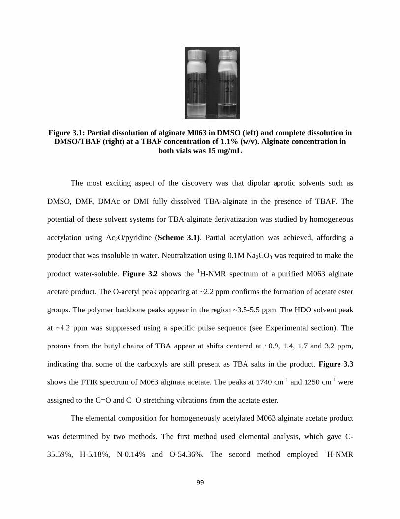
99
Figure 3.1: Partial dissolution of alginate M063 in DMSO (left) and complete dissolution in
DMSO/TBAF (right) at a TBAF concentration of 1.1% (w/v). Alginate concentration in
both vials was 15 mg/mL
The most exciting aspect of the discovery was that dipolar aprotic solvents such as
DMSO, DMF, DMAc or DMI fully dissolved TBA-alginate in the presence of TBAF. The
potential of these solvent systems for TBA-alginate derivatization was studied by homogeneous
acetylation using Ac2O/pyridine (Scheme 3.1). Partial acetylation was achieved, affording a
product that was insoluble in water. Neutralization using 0.1M Na2CO3 was required to make the
product water-soluble. Figure 3.2 shows the 1H-NMR spectrum of a purified M063 alginate
acetate product. The O-acetyl peak appearing at ~2.2 ppm confirms the formation of acetate ester
groups. The polymer backbone peaks appear in the region ~3.5-5.5 ppm. The HDO solvent peak
at ~4.2 ppm was suppressed using a specific pulse sequence (see Experimental section). The
protons from the butyl chains of TBA appear at shifts centered at ~0.9, 1.4, 1.7 and 3.2 ppm,
indicating that some of the carboxyls are still present as TBA salts in the product. Figure 3.3
shows the FTIR spectrum of M063 alginate acetate. The peaks at 1740 cm-1
and 1250 cm-1
were
assigned to the C=O and C–O stretching vibrations from the acetate ester.
The elemental composition for homogeneously acetylated M063 alginate acetate product
was determined by two methods. The first method used elemental analysis, which gave C-
35.59%, H-5.18%, N-0.14% and O-54.36%. The second method employed 1H-NMR

100
spectroscopy by integration of the TBA proton peaks against the backbone proton signals. This
method, assuming DS = 0.76 and 3 moles water of hydration yielded C-38.26%, H-6.27%, N-
0.42% and O-55.04%. These methods were compared with theoretical percentages, calculated by
assuming a DS = 0.76 and 3 moles of water of hydration. The composition obtained using this
technique was C-35.64%, H-5.78%, N-0.14% and O-58.44%. The 1H-NMR technique results
may be less reliable based on the knowledge that the solvent suppression pulse sequence
employed affects the intensities of other signals in the spectra, thus changing the integration
values.47
The experimental and theoretically calculated elemental composition values were in
fairly close agreement when ~3 moles water of hydration was accounted for. When the water of
hydration was not taken into consideration, the values obtained using the two methods were quite
different. The % of saccharide units existing as TBA salts can be calculated using the %N value
obtained by elemental analysis. For example, in a product with acetyl DS of 0.76, the maximum
possible %N value is 3.13 for 100% monosaccharide units in TBA salt form. Therefore, a %N
value of 0.14 (as obtained by elemental analysis for product 12 in Table 3.2) corresponds to
about 2.6% saccharide units present as their TBA salts. The partial TBA salts of alginic acid may
be converted to 100% Na-salts using ion-exchange. Converting the polymer from its salt form to
the protonated form using ion-exchange may be more challenging due to precipitation of alginic
acid acetate from water.
Scheme 3.1: Acetylation of TBA-alginate

101
Figure 3.2: 1H-NMR spectrum of alginate acetate synthesized from alginate M063
Figure 3.3: FTIR spectrum of alginate acetate synthesized from alginate M063
45
55
65
75
85
95
105
115
5001000150020002500300035004000
% T
ran
smit
tan
ce
Wavenumber (cm-1)
Ester C=O stretching(1740cm-1)
Ester C–O stretching (1250cm-1)
Aliphatic C-H stretching(2850-2960 cm-1)
Carboxylate C–O stretching (1610cm-1)Hydroxyl O-H stretching
(3400cm-1)

102
Acylation reactions of alginate M063 are summarized in Table 3.2. Homogeneous
acetylation performed in DMF, DMAc, DMI and DMSO with excess Ac2O afforded DS values
ranging between 0.74-0.85, as indicated for products 1-4. Acetylation using acetic anhydride in
DMSO may lead to side reactions through Pummerer rearrangement.48
Since products 1-4 gave
fairly similar DS values, it was concluded that Pummerer rearrangement did not significantly
impact the acylation reaction. In an effort to increase the DS value obtained, a second addition of
Ac2O was carried out and the reaction continued for 2 h; this afforded DS(Ac) of 1.01 (product
5). We thought the water of hydration of the commercial TBAF might be restricting the
maximum DS(Ac) obtained, so performed the reaction under anhydrous conditions by
synthesizing TBAF in situ.35
Alginate acetate synthesized under anhydrous conditions yielded a
DS value of 1.05 (product 6). Synthesis of alginate acetate using acetyl chloride (product 7)
afforded only a DS(Ac) of 0.08. This result was surprising since acid chlorides are typically
more reactive than the corresponding acid anhydrides. Reaction of TBA alginate with propionic
anhydride afforded alginate propionate (product 8) of DS(Pr) 0.31. The reduction in reactivity
with alginate as chain length of the anhydride increases is striking; indeed, hexanoic anhydride
did not appear to react with TBA alginate (product 9) at all. This is surely due in part to steric
reasons, but perhaps is also caused by the difficulty of mixing the highly polar, polyanionic
alginate and the rather hydrophobic longer chain anhydrides.

103
Table 3.2: Acylation of alginates and the corresponding DS values
Product Acyl
Reagent
Molar ratio
(Alg:Pyr:Acyl) Solvent
TBAF
% (w/v) DS
1 Ac2O 1:17:15 DMF 10 0.74
2 Ac2O 1:17:15 DMAc 10 0.85
3 Ac2O 1:17:15 DMI 10 0.80
4 Ac2O 1:17:15 DMSO 10 0.80 a5 Ac2O 1:17:15 DMSO 10 1.01
b6 Ac2O 1:17:15 DMSO 10 1.05
7 AcCl 1:17:19 DMSO 10 0.08
8 Pr2O 1:17:11 DMSO 10 0.31
9 Hex2O 1:17:6 DMSO 10 0.00
10 Ac2O 1:34:29 DMSO 1 0.98
11 Ac2O 1:20:20 DMSO 1 0.89
12 Ac2O 1:10:10 DMSO 1 0.76
13 Ac2O 1:4:4 DMSO 1 0.70
14 Ac2O 1:2:2 DMSO 1 0.29
15 Ac2O 1:1:1 DMSO 1 0.13
DS values measured using 1H-NMR. All reactions performed in
DMSO using M063 sample at 40 ˚C for 2 hours. asecond addition of Ac2O and reaction continued for 2 more hours.
breaction performed in anhydrous DMSO/TBAF solvent.
A recent article on cellulose derivatization in DMSO/TBAF reported the effect of TBAF
concentration on the DS value achieved.36
A high concentration of TBAF in the reaction mixture
was shown to have a limiting effect on the DS values for cellulose derivatives. To probe the
effect of TBAF concentration on alginate esterification, acetylation was performed in 1% (w/v)
DMSO/TBAF solution. A DS value of 0.98 was achieved for product 10. Products 11-15
resulted from acetylation under varying molar ratios of acetylation reagent to alginate. The DS
could be influenced by the molar ratio of the added reagent to alginate, but only up to an
apparent limit of DS(Ac) ~1.0 (Table 3.2).
We hypothesized two possible causes for this apparent upper limit on attainable alginate
acetate DS. One cause could be that the reaction is highly regioselective, reacting only with

104
either the 2-OH or 3-OH on the alginate monosaccharides. The second possibility could be
greater reactivity of either the M or G monosaccharide units towards acetylation; given the
significant conformational change that occurs upon epimerization of M to G in the natural
alginate, this seemed a possibility. To better understand the selectivity issues, acetylation was
performed using alginates containing varying M/G ratios. Table 3.3 shows the DS values
obtained for TBA alginates acetylated both homogeneously in DMSO/TBAF and (partly)
heterogeneously in DMSO.
Table 3.3: Homogeneous and heterogeneous acetylation of alginates and the corresponding
DS values
Product Alginate DSHomogeneous DSHeterogeneous
1 M100 0.80 0.91
2 M063 0.80 0.48
3 M029 0.78 0.63
4 M013 0.78 0.54
5 M000 0.82 0.72
DS values measured using 1H-NMR. All reactions
employed TBA alginate at 100 mg/mL DMSO using
alginate monosaccharide unit:pyridine:acetic
anhydride molar ratio of 1:17:15; homogeneous
reactions also employed 100 mg TBAF/mL DMSO.
Under homogeneous reaction conditions, the DS values obtained were nearly identical
(~0.8) across the entire range of alginate M/G ratios. This proves that the reaction was not
selective for M vs. G, and that both M and G residues could be acetylated. Figure 3.4 shows the
1H-NMR spectrum obtained for M100 alginate acetate. Peak assignments for partially acetylated
alginates containing 100% M were previously reported by Skjåk-Bræk et al.49
Accordingly, peak
A was assigned as the H-3 signal from M residues monoacetylated at the 3-OH position and peak
B was assigned as the H-3 signal from M residues acetylated at both 2-OH and 3-OH positions.
Thus, both 2- and 3-OH positions on M residues could be acetylated under homogeneous

105
conditions. Figure 3.5 shows stacked 1H-NMR spectra of homogeneously acetylated alginate
samples represented in Table 3.3. While peaks A and B were assigned for M100 alginate, peak
assignments for samples containing G residues were not possible due to severe overlap of
signals. 2D-COSY studies of alginate acetates containing G residues failed to identify the H-H
correlations in the backbone region due to the broad nature of the peaks. Therefore,
regioselectivity between the 2-OH and 3-OH positions on G residues was difficult to determine
with certainty.
Figure 3.4: 1H-NMR spectrum of alginate acetate synthesized from alginate M100. Peak A
represents H-3 from M residues monoacetylated at position 3. Peak B represents H-3 from
M residues diacetylated at positions 2 and 3. Peaks A and B assigned according to
previously published literature49

106
Figure 3.5: 1H-NMR spectra of homogeneously acetylated alginates from Table 3.3. Peak A
represents H-3 from M residues monoacetylated at position 3. Peak B represents H-3 from
M residues diacetylated at positions 2 and 3. Peaks A and B assigned according to
previously published literature49
Acetylation of TBA alginates performed in DMSO without added TBAF, that is under
partly heterogeneous conditions, afforded more variable DS values compared to homogeneous
acetylation, as shown in Table 3.3. While homogeneous acetylation gave similar DS values for
alginates across all M/G ratios, the products of heterogeneous reactions yielded DS values that
varied with the M/G ratios of the starting polymers. This variance in heterogeneous reaction
product DS may result from the differing extent of partial solubility among various TBA-alginate
samples in DMSO, which may in turn depend on factors including molecular weight, M/G ratios,
and block copolymer structure.

107
Figure 3.6 shows stacked 1H-NMR spectra of heterogeneously acetylated alginate
samples from Table 3.3. The spectrum for M100 alginate acetate shows the two peaks A and B,
previously assigned as the H-3 signal from M residues monoacetylated at the 3-OH position, and
H-3 signal from M residues diacetylated at both 2-OH and 3-OH positions respectively.
Therefore, heterogeneous reactions could yield products where both hydroxyl positions on M
residues were substituted. However, in the case of alginates containing G residues,
heterogeneous reactions yielded products where the two peaks C and D, with shifts of 5.08 and
4.47 ppm respectively were predominant. The predominance of these peaks was not observed in
the case of homogeneous reaction products shown in Figure 3.5. Previous assignments for
alginates made using 1H-NMR have shown that the peak at ~5.08 ppm arises from the H-1 signal
of G residues in GGG, MGG and GGM triads. The peak centered at ~4.47 ppm on the other hand
arises from the H-5 signal in GGG triads.50
Thus, the predominance of peaks C and D in
heterogeneously acetylated alginates suggests that GGG triad sequences remained relatively
unreacted. G-blocks were therefore preserved, resulting in selective acetylation of M residues.
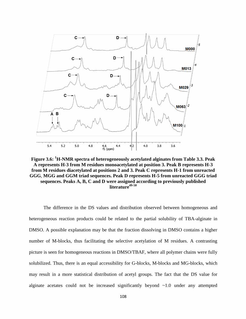
108
Figure 3.6: 1H-NMR spectra of heterogeneously acetylated alginates from Table 3.3. Peak
A represents H-3 from M residues monoacetylated at position 3. Peak B represents H-3
from M residues diacetylated at positions 2 and 3. Peak C represents H-1 from unreacted
GGG, MGG and GGM triad sequences. Peak D represents H-5 from unreacted GGG triad
sequences. Peaks A, B, C and D were assigned according to previously published
literature49-50
The difference in the DS values and distribution observed between homogeneous and
heterogeneous reaction products could be related to the partial solubility of TBA-alginate in
DMSO. A possible explanation may be that the fraction dissolving in DMSO contains a higher
number of M-blocks, thus facilitating the selective acetylation of M residues. A contrasting
picture is seen for homogeneous reactions in DMSO/TBAF, where all polymer chains were fully
solubilized. Thus, there is an equal accessibility for G-blocks, M-blocks and MG-blocks, which
may result in a more statistical distribution of acetyl groups. The fact that the DS value for
alginate acetates could not be increased significantly beyond ~1.0 under any attempted

109
conditions is still a conundrum. A potential explanation for this limited DS is based on
monosaccharide ring electronic effects. The uronate ring in unreacted TBA-alginate contains one
electron-withdrawing substituent at the C-5 position. During acetylation, the monoacetate
product is formed first by reaction at one of the two hydroxyl groups. The monoacetylated
uronate ring now bears two electron-withdrawing substituents; the first is the C-5 carboxylate
and the second is the acetyl substituent. It is likely that with two electron-withdrawing
substituents present, the reactivity of the second hydroxyl group is significantly lowered. The DS
value may therefore be limited to ~1.0 when pyridine/anhydride is used as the acyl reagent
system. An alternate theory may be based on the stereochemistry of the monosaccharide
residues. In an M residue, the 2-OH group is axial and may possibly interact with the C-4 proton
through 1,3-diaxial interactions, thus favoring substitution at the 3-OH position. In a G residue,
an exactly opposite scenario is likely, wherein the 3-OH group is axial and could interact with
the C-5 proton through 1,3-diaxial interactions. Substitution of the 2-OH position may therefore
be favored on G units. Thus, in either of the two residues, stereochemistry could favor the
reaction of one out of the two hydroxyl groups, therefore limiting the DS to ~ 1.0. At this time
we cannot be certain whether one of these mechanisms, or some other mechanism is the source
of the apparent acyl DS limitation.
Finally, uronic acids are known to be base sensitive and degrade via β-elimination at
higher pH values.51-52
Although pyridine is a mild base, it may still degrade the polymer during
reaction. Furthermore, fluoride from TBAF is also known to be a strong base53
. Molecular
weight measurements were therefore performed on M063 alginate acetates that were reacted for
different time periods, and compared to unreacted TBA-alginate. PDI was calculated using the
Mn and Mw values. Elemental analysis was performed for each sample to determine the %N
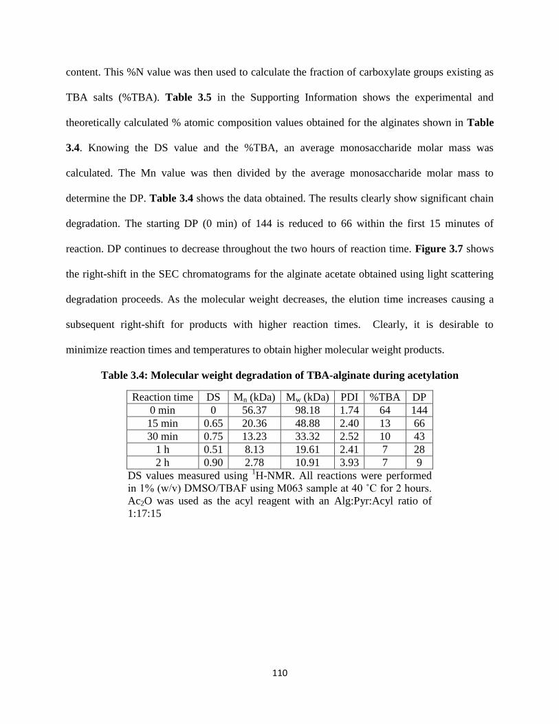
110
content. This %N value was then used to calculate the fraction of carboxylate groups existing as
TBA salts (%TBA). Table 3.5 in the Supporting Information shows the experimental and
theoretically calculated % atomic composition values obtained for the alginates shown in Table
3.4. Knowing the DS value and the %TBA, an average monosaccharide molar mass was
calculated. The Mn value was then divided by the average monosaccharide molar mass to
determine the DP. Table 3.4 shows the data obtained. The results clearly show significant chain
degradation. The starting DP (0 min) of 144 is reduced to 66 within the first 15 minutes of
reaction. DP continues to decrease throughout the two hours of reaction time. Figure 3.7 shows
the right-shift in the SEC chromatograms for the alginate acetate obtained using light scattering
degradation proceeds. As the molecular weight decreases, the elution time increases causing a
subsequent right-shift for products with higher reaction times. Clearly, it is desirable to
minimize reaction times and temperatures to obtain higher molecular weight products.
Table 3.4: Molecular weight degradation of TBA-alginate during acetylation
Reaction time DS Mn (kDa) Mw (kDa) PDI %TBA DP
0 min 0 56.37 98.18 1.74 64 144
15 min 0.65 20.36 48.88 2.40 13 66
30 min 0.75 13.23 33.32 2.52 10 43
1 h 0.51 8.13 19.61 2.41 7 28
2 h 0.90 2.78 10.91 3.93 7 9
DS values measured using 1H-NMR. All reactions were performed
in 1% (w/v) DMSO/TBAF using M063 sample at 40 ˚C for 2 hours.
Ac2O was used as the acyl reagent with an Alg:Pyr:Acyl ratio of
1:17:15
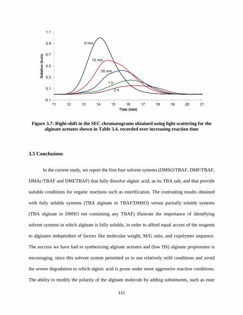
111
Figure 3.7: Right-shift in the SEC chromatograms obtained using light scattering for the
alginate acetates shown in Table 3.4, recorded over increasing reaction time
3.5 Conclusions
In the current study, we report the first four solvent systems (DMSO/TBAF, DMF/TBAF,
DMAc/TBAF and DMI/TBAF) that fully dissolve alginic acid, as its TBA salt, and that provide
suitable conditions for organic reactions such as esterification. The contrasting results obtained
with fully soluble systems (TBA alginate in TBAF/DMSO) versus partially soluble systems
(TBA alginate in DMSO not containing any TBAF) illustrate the importance of identifying
solvent systems in which alginate is fully soluble, in order to afford equal access of the reagents
to alginates independent of factors like molecular weight, M/G ratio, and copolymer sequence.
The success we have had in synthesizing alginate acetates and (low DS) alginate propionates is
encouraging, since this solvent system permitted us to use relatively mild conditions and avoid
the severe degradation to which alginic acid is prone under more aggressive reaction conditions.
The ability to modify the polarity of the alginate molecule by adding substituents, such as ester
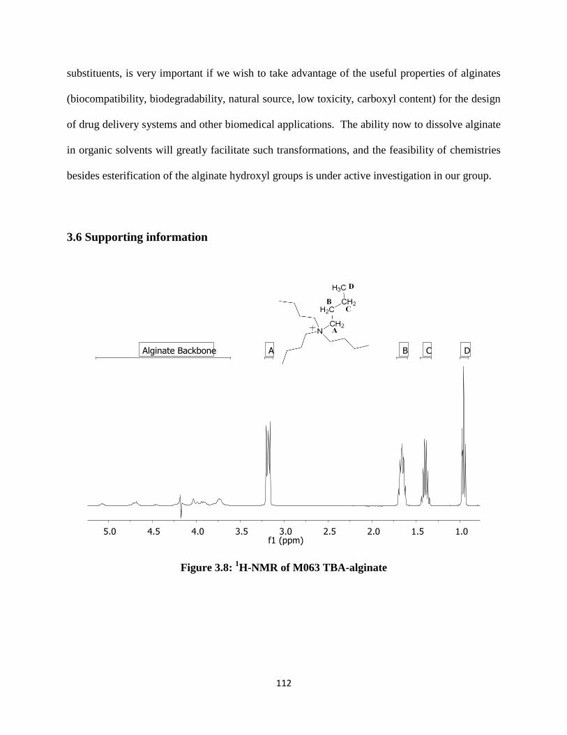
112
substituents, is very important if we wish to take advantage of the useful properties of alginates
(biocompatibility, biodegradability, natural source, low toxicity, carboxyl content) for the design
of drug delivery systems and other biomedical applications. The ability now to dissolve alginate
in organic solvents will greatly facilitate such transformations, and the feasibility of chemistries
besides esterification of the alginate hydroxyl groups is under active investigation in our group.
3.6 Supporting information
Figure 3.8: 1H-NMR of M063 TBA-alginate

113
Figure 3.9: FTIR of M063 TBA-alginate
Figure 3.10: 1H-NMR for product 2 shown in Table 3.4
40
45
50
55
60
65
70
75
80
5001000150020002500300035004000
% T
ran
smit
tan
ce
Wavenumber (cm-1)
Aliphatic C-H stretching(2850-2960 cm-1) Carboxylate C–O stretching (1610cm-1)
Hydroxyl O-H stretching(3400cm-1)

114
Figure 3.11: FTIR for product 2 shown in Table 3.4
Figure 3.12: 1H-NMR for product 7 shown in Table 3.2
0
10
20
30
40
50
60
70
80
90
5001000150020002500300035004000
% T
ran
smit
tan
ce
Wavenumber (cm-1)
Ester C=O stretching(1740cm-1)
Ester C–O stretching (1250cm-1)
Aliphatic C-H stretching(2850-2960 cm-1)
Carboxylate C–O stretching (1610cm-1)Hydroxyl O-H stretching (3400cm-1)
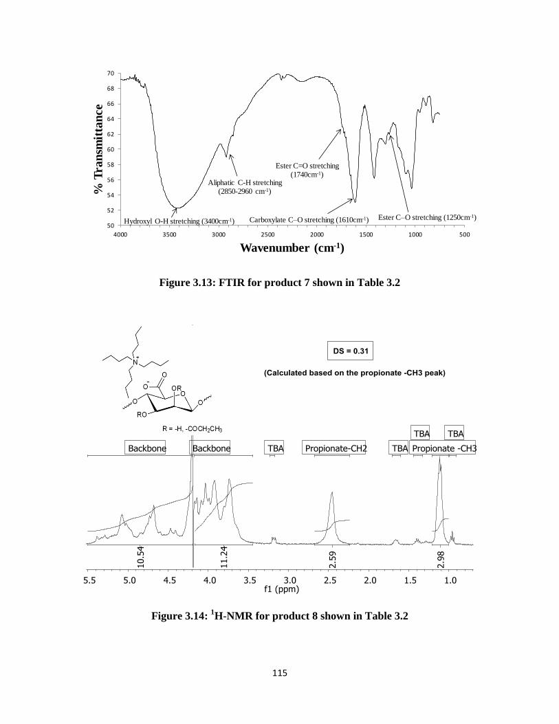
115
Figure 3.13: FTIR for product 7 shown in Table 3.2
Figure 3.14: 1H-NMR for product 8 shown in Table 3.2
50
52
54
56
58
60
62
64
66
68
70
5001000150020002500300035004000
% T
ran
smit
tan
ce
Wavenumber (cm-1)
Ester C=O stretching(1740cm-1)
Ester C–O stretching (1250cm-1)
Aliphatic C-H stretching(2850-2960 cm-1)
Carboxylate C–O stretching (1610cm-1)Hydroxyl O-H stretching (3400cm-1)

116
Figure 3.15: FTIR for product 8 shown in Table 3.2
Table 3.5: Experimental and theoretically calculated % elemental compositions of alginate
acetates shown in Table 3.4
Reaction time %C %H %O %N
0 min Experimental 48.49 9.20 37.98 2.33
Theoretical 50.77 9.39 37.51 2.33
15 min Experimental 35.75 5.77 52.73 0.62
Theoretical 39.03 6.58 53.77 0.62
30 min Experimental 35.14 5.71 53.11 0.49
Theoretical 38.36 6.36 54.79 0.49
1 h Experimental 34.66 5.56 53.03 0.36
Theoretical 36.48 6.17 56.99 0.36
2 h Experimental 34.19 5.32 53.58 0.36
Theoretical 37.81 6.12 55.71 0.36
30
35
40
45
50
55
60
5001000150020002500300035004000
% T
ra
nsm
itta
nce
Wavenumber (cm-1)
Ester C=O stretching(1740cm-1)
Ester C–O stretching (1250cm-1)
Aliphatic C-H stretching(2850-2960 cm-1)
Carboxylate C–O stretching (1610cm-1)
Hydroxyl O-H stretching (3400cm-1)
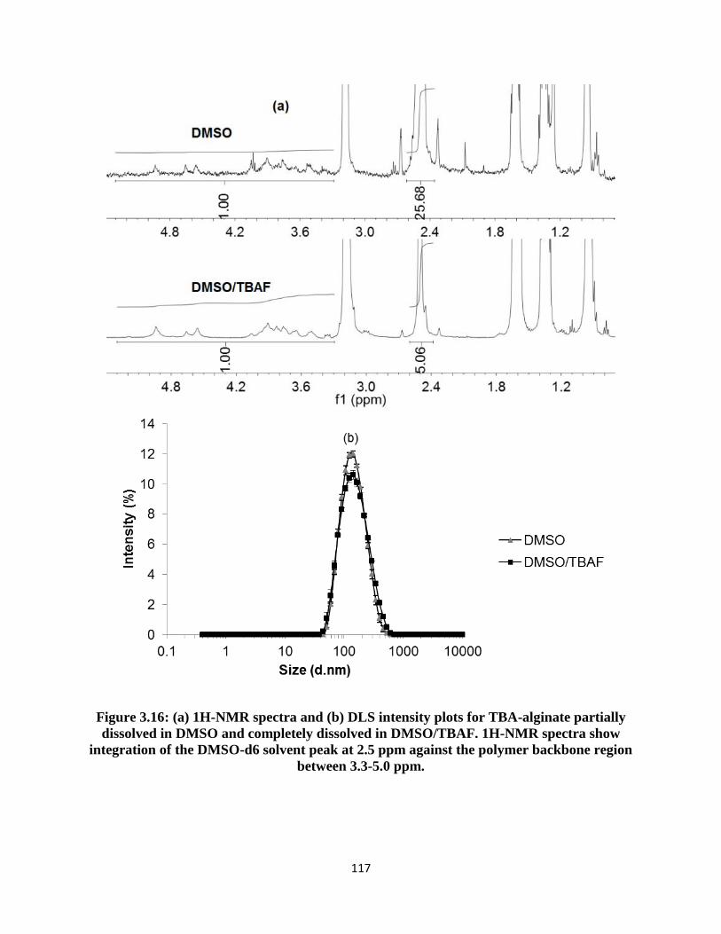
117
Figure 3.16: (a) 1H-NMR spectra and (b) DLS intensity plots for TBA-alginate partially
dissolved in DMSO and completely dissolved in DMSO/TBAF. 1H-NMR spectra show
integration of the DMSO-d6 solvent peak at 2.5 ppm against the polymer backbone region
between 3.3-5.0 ppm.

118
3.7 References
1. Draget, K. I., Alginates. In Handbook of Hydrocolloids, Phillips, G. O., Williams, P.A.,
Ed. 2009; pp 379-395.
2. Fox, S. C.; Li, B.; Xu, D.; Edgar, K. J., Biomacromolecules 2011, 12, 1956-1972.
3. Cathell, M. D.; Szewczyk, J. C.; Schauer, C. L., Mini-Reviews in Organic Chemistry
2010, 7(1), 61-67.
4. Chamberlain, N. H., Cunningham, G.E., Speakman, J.B., Nature 1946, 158, 553.
5. Schweiger, R. G., J. Org. Chem. 1962, 27 (5), 1786-1789.
6. Schweiger, R. G., J. Org. Chem. 1962, 27 (5), 1789-1791.
7. Wassermann, A., Nature 1946, 158, 271.
8. Wassermann, A., J. Chem. Soc. 1948, 197.
9. Skjåk-Bræk, G.; Paoletti, S.; Gianferrara, T., Carbohyd. Res. 1989, 185 (1), 119-129.
10. Galant, C.; Kjoniksen, A. L.; Nguyen, G. T. M.; Knudsen, K. D.; Nystrom, B., J. Phys.
Chem. B 2006, 110 (1), 190-195.
11. Donati, I.; Coslovi, A.; Gamini, A.; Vetere, A.; Campa, C.; Paoletti, S.,
Biomacromolecules 2004, 5 (1), 186-196.
12. Donati, I.; Vetere, A.; Gamini, A.; Coslovi, A.; Campa, C.; Paoletti, S.,
Biomacromolecules 2003, 4 (3), 624-631.
13. Yang, J.; Goto, M.; Ise, H.; Cho, C.-S.; Akaike, T., Biomaterials 2002, 23 (2), 471-479.
14. Rowley, J. A.; Madlambayan, G.; Mooney, D. J., Biomaterials 1999, 20 (1), 45-53.
15. Rowley, J. A.; Mooney, D. J., J. Biomed. Mater. Res. 2002, 60 (2), 217-223.
16. Xu, J. B.; Bartley, J. P.; Johnson, R. A., J. Appl. Polym. Sci. 2003, 90 (3), 747-753.

119
17. Carré, M.-C.; Delestre, C.; Hubert, P.; Dellacherie, E., Carbohyd. Polym. 1991, 16 (4),
367-379.
18. Sinquin, A.; Hubert, P.; Dellacherie, E., Langmuir 1993, 9 (12), 3334-3337.
19. Sinquin, A.; Houzelle, M. C.; Hubert, P.; Choplin, L.; Viriot, M. L.; Dellacherie, E.,
Langmuir 1996, 12 (16), 3779-3782.
20. Sinquin, A.; Hubert, P.; Dellacherie, E., Polymer 1994, 35 (16), 3557-3560.
21. Babak, V. G.; Skotnikova, E. A.; Lukina, I. G.; Pelletier, S.; Hubert, P.; Dellacherie, E.,
J. Colloid Interface Sci. 2000, 225 (2), 505-510.
22. Pelletier, S.; Hubert, P.; Lapicque, F.; Payan, E.; Dellacherie, E., Carbohyd. Polym. 2000,
43 (4), 343-349.
23. Grasselli, M.; Diaz, L. E.; Cascone, O., Biotechnol. Tech. 1993, 7 (10), 707-712.
24. Moe, S. T.; Skjåk-Bræk, G.; Smidsrød, O., Food Hydrocolloids 1991, 5 (1-2), 119-123.
25. Yeom, C. K.; Lee, K.-H., J. Appl. Polym. Sci. 1998, 67, 209-219.
26. Kim, J. H.; Jegal, J.; Kim, J. H.; Lee, K. H.; Lee, Y., J. Appl. Polym. Sci. 2003, 89 (11),
3046-3051.
27. Kim, Y.-J.; Yoon, K.-J.; Ko, S.-W., J. Appl. Polym. Sci. 2000, 78 (10), 1797-1804.
28. Riyajan, S.-A.; Sakdapipanich, J., Polym. Bull. 2009, 63 (4), 609-622.
29. Chan, A. W.; Whitney, R. A.; Neufeld, R. J., Biomacromolecules 2008, 9 (9), 2536-45.
30. Chan, A. W.; Whitney, R. A.; Neufeld, R. J., Biomacromolecules 2009, 10 (3), 609-616.
31. Leone, G.; Torricelli, P.; Chiumiento, A.; Facchini, A.; Barbucci, R., J. Biomed. Mater.
Res.-A 2008, 84A (2), 391-401.
32. Bouhadir, K. H.; Hausman, D. S.; Mooney, D. J., Polymer 1999, 40 (12), 3575-3584.

120
33. Rokstad, A. M.; Donati, I.; Borgogna, M.; Oberholzer, J.; Strand, B. L.; Espevik, T.;
Skjåk-Bræk, G., Biomaterials 2006, 27 (27), 4726-4737.
34. Hills, B. P.; Cano, C.; Belton, P. S., Macromolecules 1991, 24 (10), 2944-2950.
35. Köhler, S.; Heinze, T., Macromol. Biosci. 2007, 7 (3), 307-314.
36. Ass, B. A. P.; Frollini, E.; Heinze, T., Macromol. Biosci. 2004, 4 (11), 1008-1013.
37. Fidale, L. C.; Kohler, S.; Prechtl, M. H. G.; Heinze, T.; El Seoud, O. A., Cellulose 2006,
13 (5), 581-592.
38. Heinze, T.; Dicke, R.; Koschella, A.; Kull, A. H.; Klohr, E. A.; Koch, W., Macromol.
Chem. Phys. 2000, 201 (6), 627-631.
39. Heinze, T.; Lincke, T.; Fenn, D.; Koschella, A., Polym. Bull. 2008, 61 (1), 1-9.
40. Hussain, M. A.; Liebert, T.; Heinze, T., Macromol. Rapid. Commun. 2004, 25 (9), 916-
920.
41. Ramos, L.; Frollini, E.; Koschella, A.; Heinze, T., Cellulose 2005, 12 (6), 607-619.
42. Ramos, L. A.; Frollini, E.; Heinze, T., Carbohyd. Polym. 2005, 60 (2), 259-267.
43. Ostlund, A.; Lundberg, D.; Nordstierna, L.; Holmberg, K.; Nyden, M.,
Biomacromolecules 2009, 10 (9), 2401-2407.
44. McCormick, C. L.; Callais, P. A., Polymer 1987, 28 (13), 2317-2323.
45. Edgar, K. J.; Arnold, K. M.; Blount, W. W.; Lawniczak, J. E.; Lowman, D. W.,
Macromolecules 1995, 28 (12), 4122-4128.
46. Liebert, T. F.; Heinze, T. J., Biomacromolecules 2001, 2 (4), 1124-32.
47. Salomonsen, T. J., H.M.; Larsen, F.H.; Engelsen, S.B., The Quantitative Impact of Water
Suppression on NMR Spectra for Compositional Analysis of Alginates. In Magnetic Resonance

121
in Food Science: Challenges in a Changing World, Gujonsdottir, M.; Belton, P.; Webb, G., Eds.
The Royal Society of Chemistry: 2009; pp 12-19.
48. Johnson, C. R.; Phillips, W. G., J. Am. Chem. Soc 1969, 91 (3), 682-687.
49. Skjåk-Bræk, G.; Grasdalen, H.; Larsen, B., Carbohydr. Res. 1986, 154, 239-50.
50. Grasdalen, H., Carbohydr. Res. 1983, 118 (Jul), 255-260.
51. Haug, A. L., B.; Smidsrød, O., Acta Chem. Scand. 1967, 21, 2859-2870.
52. Haug, A. L., B.; Smidsrød, O., Acta Chem. Scand. 1963, 17, 1466-1468.
53. Sun, H.; DiMagno, S. G., J. Am. Chem. Soc. 2005, 127 (7), 2050-1.

122
3.8 Copyright Authorization
123
Chapter 4: Ca+2
-Induced Gelation of Alginate Acetates for Tailored
Hydrogels
(Adapted from Pawar, S.N., Bowden, S., McQuilling, J.P., Opara, E.C., Edgar, K.J.; Manuscript
under review)
4.1 Abstract
Alginates are (1→4) linked linear copolysaccharides composed of β-D-mannuronic acid
(M) and its C-5 epimer, α-L-guluronic acid (G). Several routes to synthesize alginate acetates
have previously been described. We recently reported on methods to acetylate alginates either
non-selectively or M-selectively. We hypothesized that while the non-selective acetates would be
incapable of forming Ca++
-gels, the M-selective acetates would be more amenable to
crosslinking. We herein report on our attempts to synthesize Ca+2
-crosslinked alginate
microbeads after acetylation. As we expected, it was not possible to form beads using randomly
acetylated alginates within practical concentration ranges. M-selective alginate acetates however
formed beads at relevant concentrations, thus highlighting the role of G blocks in alginate gelling
by Ca+2
crosslinking. The effects of reagent stoichiometry (pyridine and acetic anhydride) on
degree of substitution (DS) and molecular weight were studied. Their influence upon solution
viscosities, and the formation and mechanical strength of microbeads were studied with the
intention of drawing conclusions about what reaction conditions may be best suited to bead
formation. Furthermore, we discuss the relative merits of using 1H-NMR or titration for DS
measurement.

124
4.2 Introduction
Alginates are unbranched copolysaccharides consisting of 1→4 linked β-D-mannuronic
acid (M) and its C-5 epimer α-L-guluronic acid (G). The natural copolymer is an important
component of algae such as kelp, and is also a biofilm-forming exopolysaccharide of bacteria
including Pseudomonas aeruginosa. It is comprised of sequences of M (M-blocks) and G (G-
blocks) residues interspersed with MG sequences (MG-blocks)1. Based on the desired
functionality, alginate properties are tuned in nature by enzymatically (C-5 epimerase) varying
composition (M/G ratio) and sequence2, and also by adjusting molar mass. Alginate is an
important industrial polysaccharide and is isolated from kelp in quantities up to 30,000 metric
tons annually. However, this is estimated to comprise less than 10% of the biosynthesized
alginate materials3. The large abundance of algae in water bodies (and the potential for algae
farming) therefore offers significant additional potential to use designed sustainable biomaterials
based on alginates.
Development of powerful organic solvent systems capable of dissolving recalcitrant
polysaccharides has enabled routes to achieve regioselective transformations, leading to
derivatives with novel properties.4 We have previously shown that in the case of alginates,
selection of the appropriate medium for performing a particular reaction can greatly influence
regioselectivity.5 Acylation of tetrabutylammonium (TBA) salts of alginate by complete
solubilization in DMSO/TBAF (tetrabutylammonium fluoride) solvent provided derivatives with
non-selective substitution of both epimers (M/G). In contrast, acylation under conditions of
partial alginate solubilization in DMSO provided derivatives with preferential substitution on M
residues. Prior to our report, several methods had been described for the synthesis of acetylated
alginates.6 Acid-catalyzed acetylation was described as leading to partially and fully substituted

125
derivatives.7-9
An alternate route to acetylation was developed using ketene, a gaseous reagent
which reacted with alginates in the swollen state.10, 11
However, the selectivity of this ketene
chemistry was not reported. Selective acetylation of M residues was described by forming Ca-
alginate beads and swelling them in a pyridine-acetic anhydride reagent mixture.12
M residues
reacted in preference to G residues at low degrees of substitution (DS). As the DS approached its
limiting value, G residues reacted and epimer selectivity was compromised.
Alginates are most well known for their ability to form hydrogels by crosslinking with
bivalent cations. Gel formation is driven by G-block sequences which associate via multidentate
chelation to form tightly held junction points.13
In addition, MG-block sequences provide
assistance by forming relatively weaker junctions.14
Hydrogel formation is not possible in the
absence of G residues. The importance of these physically crosslinked systems in medicine and
biology is immense.15
Alginates are currently used as dressing materials in the treatment of acute
or chronic wounds.16
They play a central role in facilitating the progression of cystic fibrosis
wherein alginate biofilms are secreted by bacteria for immunoprotection17
; conversely, alginate
oligomers have been proposed for therapy of cystic fibrosis patients, by competitively inhibiting
alginate-mucin binding.18
One of the more impactful applications of alginates is in diabetes
therapy.19
Diabetes is a globally prevalent disease, currently affecting about 285 million people
worldwide.20
Type 1 diabetes is a significant cause of morbidity and mortality in young adults. In
addition, secondary diabetic complications may arise in the form of increased risk of heart attack
and stroke, and decreased life expectancy.20, 21
Current standard treatment regimens for Type 1
diabetes include daily insulin injections to control blood glucose. While intensive insulin therapy
can adequately control blood glucose levels so as to permit most normal activities, it cannot
duplicate the continuous monitoring and response of a functional pancreas, and does not totally

126
eradicate secondary diabetic complications and adverse side effects such as unwanted weight
gain and hypoglycemia.22, 23
An alternative treatment modality for Type 1 diabetes is replacement of the missing β-
cells via transplantation of the pancreas.24, 25
This treatment method is capable of achieving
normal glycemic levels along with prevention, and even reversal of secondary complications.
The islets of Langerhans are the most preferred source of insulin secreting β-cells.26
It is however
essential to prevent immune responses that may result from transplantation of foreign islet cells.
Microencapsulation within alginate beads is a convenient way to accomplish this goal. We have
previously described the principle of immunoisolation by islet encapsulation within alginates.25
Upon cell encapsulation within alginate microbeads, a permselective poly-L-ornithine (PLO)
coating is applied which is permeable to small molecules such as nutrients, oxygen and water,
but impermeable to large entities such as immune cells and antibodies. Finally, an outer alginate
layer is applied to prevent interactions between positively charged PLO and negatively charged
proteins and cells in vivo.
While alginates play a very important role in the design of the bioartificial pancreas, there
appear to be no reports on the use of hydrophobically modified alginates for this application.
Hydrophobic modifications may be envisioned as an excellent tool to fine-tune the
diffusion/transport properties of alginates encapsulating β-cells. In addition, gel instabilities
arising from Na+-Ca
+ ion exchange may be suppressed by making the alginate gel more
hydrophobic. However, to be able to use modified alginates for encapsulation, a primary
criterion is the preservation of its ability to form ionic gels. In the current article, we report on
the potential of acetylated alginates to form Ca+2
induced gels. The effects of variables including
acetylation reaction conditions, corresponding DS values, molecular weights and M/G ratios and

127
viscosities were studied to establish clear correlations with the gelling abilities of the modified
alginates. In addition, we noticed in following up our earlier alginate acetylation studies that
there were unexplained variations in DS values, and herein we describe investigation of the
cause of those variations, and development of a superior method for determination of DS(acetyl).
Furthermore, we describe our investigations of the relative mechanical strength of microbeads
made either from alginate or acetylated alginate.
4.3 Experimental
4.3.1 Materials
Four alginates were used in the study and are represented as A-D. ‘A’ and ‘B’, were high-
mannuronic alginic acid sodium salts extracted from brown algae with viscosities of ≥ 2,000 cP
and 100-300 cP respectively (measured as 2% aqueous solutions). ‘C’ and ‘D’ were alginic acid
sodium salts with high M and high G contents respectively. The molecular weights and M/G
compositions of A-D are listed in Table 4.1. In addition, an ultrapure grade of alginate ‘LVM’
with a low viscosity and high M content commonly used for islet encapsulation was also studied
for comparative analyses.
Water used in the experiments was deionized. Hydrochloric acid, reagent alcohol
(histological grade), hexanes (HPLC grade), dimethylsulfoxide (HPLC grade), 5 N standardized
NaOH solution, 1 N standardized HCl solution, ethyl acetate and calcium chloride (CaCl2) were
purchased from Fisher Scientific (Fair Lawn, NJ). Acetic anhydride (Ac2O; 99+%), pyridine
(Pyr; anhydrous, 99.0%), tetrabutylammonium hydroxide (TBAOH; 40 wt%, 1.5 M solution in
water) and sodium carbonate (99.8% anhydrous powder) were obtained from Acros. Deuterium

128
oxide (99.9 atom% D; D2O) containing 0.75% 3-(trimethylsilyl)propionic-2,2,3,3-d4 acid,
sodium salt for NMR, NaCl, 4-(2-hydroxyethyl)-1-piperazineethanesulfonic acid (HEPES), and
alginates ‘A’ and ‘B’ were acquired from Sigma-Aldrich (St. Louis, MO). Alginates ‘C’ and ‘D’
were acquired from FMC BioPolymer (Philadelphia, PA). KBr used for FTIR analysis was
purchased from International Crystal Labs. Ultrapure low-viscosity high mannuronic acid
(LVM) alginate was obtained from Novamatrix (Sandvika, Norway).
4.3.2 Synthesis of TBA-alginate
For conversion of Na-alginate to its protonated form, a procedure described by Babak et
al.27
was followed with modifications. HCl (0.6 N, 120 mL) was mixed with ethanol (120 mL) in
a beaker. Na-alginate (8 g) was added to the mixture and stirred overnight at 4 ˚C. The mixture
was filtered, the solid (alginic acid) washed thoroughly with ethanol followed by acetone, and
dried overnight in vacuo at 60 ˚C. Thoroughly dried alginic acid was then dispersed in 200 mL
water. TBAOH was added dropwise with continuous stirring at room temperature until the
polymer was fully dissolved and the pH was adjusted to 10.0. If the solution was too viscous,
small amounts of water were added until a final viscosity was reached at which the solution
could be easily stirred. The aqueous TBA-alginate solution was then premixed with reagent
alcohol. The premix was precipitated in a mixture of ethyl acetate-hexane. The solvent ratios
used were aqueous alginate acetate : ethanol : ethyl acetate : hexane = 1:3:12:3. The precipitate
was then filtered under vacuum and dried in vacuo at 60 ˚C. The 1H-NMR and FTIR spectra for
TBA-alginate are shown in the Supporting Information as Figures 4.11 and 4.12 respectively.
1H NMR (500 MHz, D2O) δ 5.82 – 3.46 (m, alginate backbone), 3.19 (t,
NCH2CH2CH2CH3 of TBA), 1.67 (m, NCH2CH2CH2CH3 of TBA), 1.40 (m, NCH2CH2CH2CH3

129
of TBA), 0.97 (t, NCH2CH2CH2CH3). FTIR (KBr pellet method): 3400 cm-1
, hydroxyl O–H
stretching; 2850-2960 cm-1
, aliphatic C–H stretching, 1610 cm-1
, carboxylate C-O stretching.
Elemental composition TBA-alginate ‘A’ measured by elemental analysis: C, 57.30%; H, 9.76%;
N, 2.82%. This composition corresponds to roughly 78% TBA and 22% Na salt.
4.3.3 Synthesis of alginate acetate
DMSO (350 mL) was added to TBA-alginate (7 g) in a 3-neck flask. The mixture was
blended using a mechanical stirrer at room temperature until the polymer was fully dispersed.
The temperature of the mixture was raised to 40 ˚C. Ac2O was added followed by pyridine at the
desired molar ratios. The suspension was stirred for 30 min, and then the polymer was
precipitated in 3500 mL of ethyl acetate. The solid was filtered under gravity using fluted filter
paper and dried in vacuo at 60 °C. The dried crude solid was crushed into a powder using mortar
and pestle, and the powder was added to ~50-250 mL of water. While the mixture was stirring, 1
M Na2CO3 solution was added dropwise to dissolve the polymer. The pH was adjusted to 10.0.
The solution was added dropwise to 500 mL ethanol to precipitate the sodium salt of alginate
acetate. The resulting solid was filtered under vacuum and dried in vacuo at 60 ˚C. A second
purification was carried out by dissolving the dry product in water and re-precipitating in ethanol
before drying in vacuo. The 1H-NMR and FTIR spectra for alginate acetate A.P10.Ac20 from
Table 4.2 are shown in Figures 4.1 and 4.2.
Analytical results for A.P10.Ac20 are as follows. 1H NMR (500 MHz, D2O) δ 5.69 – 3.31
(m, alginate backbone), 2.45-1.80 (m, COCH3 of acetate). FTIR (KBr pellet method): 1740 cm-1
,
ester C=O stretching vibration; 1250 cm-1
, ester C–O stretching vibration; 3400 cm-1
, hydroxyl
O–H stretching; 2850-2960 cm-1
, aliphatic C–H stretching, 1610 cm-1
, carboxylate C-O

130
stretching. DSNMR = 2.0. DSTitration = 0.85. Elemental composition, as measured experimentally
using elemental analyzer: C, 35.00%; H, 4.51%; N, 0.09%; O, 44.05%. Theoretical compositions
were determined using a spreadsheet in which %TBA values were varied from 0-100% and the
corresponding elemental percentages were calculated. By inserting the DS value (DSTitration =
0.85 for this example) and assuming an appropriate number of moles of water of hydration (1 for
this example), theoretical compositions that were closest matching to the experimental values
were determined. The %TBA corresponding to that composition was then used for average molar
mass calculations. For A.P10.Ac20, the theoretically calculated composition was C, 37.39%; H,
4.41%; N, 0.09%; O, 49.24%. This corresponded to a %TBA value of 1.6. For all alginate
acetates listed in Table 4.2, the %TBA values and their calculated average repeat unit molar
masses are shown in Table 4.6 of Supporting Information.
4.3.4 Synthesis of Ca-alginate beads
Solutions of M-acetylated alginate (3%, 5%, 7.5% w/v) and a solution of 1.5% non-
acetylated sodium alginate (sample A) were prepared in deionized water and placed on a shaker
in a warm room (37oC) for 24 hours. Each alginate solution (3 mL) was placed in a 5 mL syringe
and passed through a 2-nozzle microfluidic encapsulator28
at a flow rate of 0.2 mL/min and fixed
air pressure (0.5, 1, 1.5 or 2 psi). The capsules were dropped into 45 mL of cross-linking solution
(1.1% w/v CaCl2) in a 100/20 mm Petri dish with continuous stirring.
4.3.5 DSNMR measurement

131
For 1H-NMR, sample preparation was carried out by dissolving ~10 mg of polymer in 0.6
mL D2O containing TMS standard. Spectra were acquired using a JEOL 500 MHz spectrometer
with automatic shimming. An automatic water suppression pulse was applied to suppress the
residual HDO peak. The acquisition temperature was 85 ˚C for all samples, and 64 scans were
obtained for each. All spectra were processed using MestReNova version: 6.2.1-7569. DS value
calculation was carried out by evaluating the ratio of the integral of the O-acetyl peak centered at
~2.2 ppm (IAC) to that of the polymer backbone region ~3.5-5.5 ppm (IBB). IAC accounts for 3X
protons, assigned to the acetyl –CH3 group. IBB accounts for (7-DS(Ac))X protons. The following
formula was used to calculate DS(acetyl) from these integrals:
( )
4.3.6 DSTitration measurement
Titrations were performed by reacting alginate acetates with excess NaOH to hydrolyze
the esters. NaOH molecules remaining after hydrolysis were then back-titrated with HCl to
measure the moles of acetate present. Specifically, 50 mg alginate acetate was weighed into a 25
mL 3-neck flask. To this, 10.0 mL of standardized 0.1 N NaOH solution was added and stirred
overnight at room temperature. Potentiometric titrations were performed using a pH electrode
(Inlab 413, Mettler-Toledo International, Inc.). The electrode was inserted into the 3-neck flask,
0.1 mL aliquots of standardized 0.1 N HCl solution added, the solution allowed to stir for 30
seconds and the pH measured. Total volume of HCl added was 15 mL. Two plots were made
using the recorded data. pH was first plotted against the volume of HCl added. Derivative d(pH)

132
was plotted in a second graph against the volume of HCl added. Inflexion point was recorded as
the volume at which peak maximum was attained in the d(pH) plot.
Moles of acetate present in the alginate acetate used for titration (50 mg) were calculated
as follows:
( )
DS was then calculated as:
( ) Equation (1)
Where,
( ) ( ) ( ) Equation (2)
‘x’ refers to the fraction of alginate acetate existing as TBA-salts. Correspondingly, (1-x) is the
fraction existing as Na-salts. The value of ‘x’ can be expressed in terms on ‘DS’ as follows:
( ( ))
Equation (3)
‘A’ is the % elemental composition of nitrogen measured using %CHN analyzer with
combustion/thermal conductivity detector (TCD) and %O analyzer with pyrolysis. Equation (3)
was substituted in Equation (2), which was in turn plugged in to Equation (1) to calculate
DSTitration.
4.3.7 FTIR measurement
FTIR spectra were acquired using a Thermo Electron Nicolet 8700 instrument in
transmission mode. Samples were prepared using the KBr pellet method. Alginate (1 mg) was
mixed with 99 mg of KBr in a mortar-pestle. The mixture was compressed in the sample holder
between two screws to form a KBr disc. 64 scans were obtained for each spectrum.
133
4.3.8 SEC analysis
Molecular weight measurements were performed using SEC-MALS-dRI technique. A
solvent and sample delivery system consisting of solvent reservoir, on-line degasser, isocratic
pump and auto-injector from Agilent Technologies, Santa Clara, CA, USA was used for the
experiments. Three Waters columns were connected in series - UltrahydrogelTM
Linear (7.8 ×
300 mm), UltrahydrogelTM
500 (7.8 × 300 mm) and UltrahydrogelTM
120 (7.8 × 300 mm). The
columns were preceded by an UltrahydrogelTM
Guard column (6 × 40 mm). The column outlet
was connected to a DAWN HELEOS-II MALS detector and to an OPTILAB rEX dRI detector,
both obtained from Wyatt Technologies, Santa Barbara, CA, USA. The mobile phase used for
alginate dissolution was 0.05 M Na2SO4/0.01 M EDTA, pH 6 containing 2 ppm NaN3. An
alginate concentration of 1-2 mg/mL was used with a flow rate of 0.8 mL/min and an injection
volume of 100 μL. All solutions were passed through 0.2μ nylon syringe filters prior to injection.
Data from light scattering and the differential refractometer were processed using Astra (v.
6.0.6.13) software supplied by Wyatt Technologies.
4.3.9 Viscosity measurement
A Brookfield LVDV-II+PRO CP viscometer (Middleboro, MA) with a CP-52 spindle
attached was connected to a 23°C water bath and RheoCalc program was used to collect the
viscosity data. Various solutions of non-acetylated alginate A, the M-acetylated alginates (3%,
5.5%, 6%, 6.5%, 7% and 7.5% w/v) and a solution of 1.5% non-acetylated LVM were placed on
a shaker in a warm room (37oC) for 24 hours. A 0.5 mL sample aliquot was placed in the spindle

134
plate of the Brookfield viscometer and spun for two minutes at <99% torque to remove air
bubbles and spread the sample evenly. Each sample was run for five 60-second loops, each
separated by one second, decreasing in shear rate with each subsequent loop. The shear rate
values corresponded to a range of 10% to 99% torque in order to maximize the accuracy of the
readings. Viscosity data were collected at one second intervals during each loop. Three 0.5 mL
samples were run for each alginate preparation. Each sample was run three times. The viscosity
data were averaged for each concentration of alginate examined. Viscosity, measured in Pa.s,
was plotted against shear rate.
4.3.10 Imaging of Microbeads
Microbeads were placed in a 24-well plate containing approximately 0.2 mL of cross-
linking solution. Images were taken at 4x and 10x using an Olympus IX-71 Inverted Microscope.
Scale bars were added to the image to determine microbead size.
4.3.11 Mechanical Strength Testing of Microbeads
The mechanical strength of the microbeads was assessed using the osmotic stress and
mechanical stress procedures described previously29
. In the osmotic stress test, briefly
microbeads measuring 500-600 µM in diameter were generated using a 2-channel air jacket
microencapsulator. Osmotic stress test was conducted by placing 100 microbeads/well in a 48
well plate with 1 mL distilled H2O and then incubating at 37 °C for 2 h. The microbeads were
then washed with a mixed solution of 0.9% saline and 0.25% CaCl2 prior to assessment of the
numbers of broken and intact microbeads using an inverted light microscope. In the mechanical

135
stress test, groups of 350 microbeads were placed in T-25 flasks containing approximately 6.5 g
of 3 mm inert soda lime beads (VWR Scientific Products Corporation) and 5 mL of the mixed
0.9% saline and 0.25% CaCl2 solution. Microbeads from each group were then subjected to
mechanical stress for 48 h by shaking at 100 rpm using a Max Q 3000 Shaker (Thermo
Scientific). The percentages of broken and unbroken capsules were then determined by visual
analysis under an inverted microscope.
4.3.12 Statistical evaluation of data
Quantitative data for mechanical strength studies of microbeads are presented as mean +
standard deviation and were statistically evaluated using One-Way ANOVA with Tukey-Kramer
multiple comparisons test. Differences between data were considered significant at p < 0.05.
4.4 Results and Discussion
In order to perform a systematic study correlating gel-forming ability to acetylation
reaction conditions, DS, molecular weight and M/G ratio, careful selection of starting alginates
was essential. Table 4.1 shows molar masses and compositions of alginates A-D used in this
study. ‘A’ and ‘D’ represent alginates with the lowest and highest FG (fraction of G residues)
values respectively. ‘B’ and ‘C’ possess intermediate FG values. While FG indicates the total
fraction of G units within the alginate chain, it includes those G units in G-blocks, MG-blocks
and interspersed residues. For the study of gelling properties, perhaps a more significant
parameter would be FGG (fraction of G residues neighboring another G residue) − the fraction
representing G residues involved in G-block formation. ‘D’ therefore possesses the largest
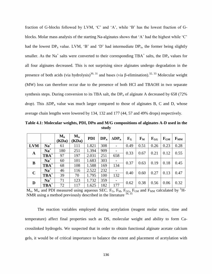
136
fraction of G-blocks followed by LVM, ‘C’ and ‘A’, while ‘B’ has the lowest fraction of G-
blocks. Molar mass analysis of the starting Na-alginates shows that ‘A’ had the highest while ‘C’
had the lowest DPn value. LVM, ‘B’ and ‘D’ had intermediate DPn, the former being slightly
smaller. As the Na+ salts were converted to their corresponding TBA
+ salts, the DPn values for
all four alginates decreased. This is not surprising since alginates undergo degradation in the
presence of both acids (via hydrolysis)30, 31
and bases (via β-elimination).32, 33
Molecular weight
(MW) loss can therefore occur due to the presence of both HCl and TBAOH in two separate
synthesis steps. During conversion to its TBA salt, the DPn of alginate A decreased by 658 (72%
drop). This ΔDPn value was much larger compared to those of alginates B, C and D, whose
average chain lengths were lowered by 134, 132 and 177 (44, 57 and 49% drops) respectively.
Table 4.1: Molecular weights, PDI, DPn and M/G compositions of alginates A-D used in the
study
Mn
(KDa)
Mw
(KDa) PDI DPn ΔDPn FG FM FGG FGM FMM
LVM Na+ 61 111 1.821 308 - 0.49 0.51 0.26 0.23 0.28
A Na
+ 180 251 1.394 909 -
0.33 0.67 0.21 0.12 0.55 TBA
+ 97 197 2.031 251 658
B Na
+ 60 101 1.683 303 -
0.37 0.63 0.19 0.18 0.45 TBA
+ 68 108 1.588 169 134
C Na
+ 46 116 2.522 232 -
0.40 0.60 0.27 0.13 0.47 TBA
+ 39 70 1.795 100 132
D Na
+ 71 123 1.732 359 -
0.62 0.38 0.56 0.06 0.32 TBA
+ 72 117 1.625 182 177
Mn, Mw and PDI measured using aqueous SEC. FG, FM, FGG, FGM and FMM calculated by 1H-
NMR using a method previously described in the literature 34, 35
The reaction variables employed during acetylation (reagent molar ratios, time and
temperature) affect final properties such as DS, molecular weight and ability to form Ca-
crosslinked hydrogels. We suspected that in order to obtain functional alginate acetate calcium
gels, it would be of critical importance to balance the extent and placement of acetylation with

137
the amount of molecular weight loss occurring during acylation. We were interested specifically
in the effects of pyridine (because of its potential catalysis of base-catalyzed chain degradation)
and Ac2O molar equivalents (mainly impacting product DSacetyl and the location of acetyl groups,
we thought) on final properties. Table 4.2 lists the experiments conducted to probe these effects,
and the corresponding results. In Series I, pyridine molar equivalents were varied from 2-20
while using a large excess (20 equivalents) of Ac2O. This series therefore illuminated the effect
of pyridine molar excess on final properties at a high concentration of Ac2O. Similarly, Series II
showed the effect of varying the pyridine molar equivalents on final properties at low
concentrations of Ac2O. In Series III, the Ac2O molar equivalents were varied from 2-20 while
keeping the pyridine equivalents constant at 10, thus providing insight into the effect of Ac2O
molar excess on final properties. Finally, Series IV shed light on the effects of changes in the
alginate composition and sequence.
Table 4.2: Molar ratios used for acetylation, DS values as measured by titration, and
aqueous SEC results for corresponding products
Series Sample ID Molar
Ratio DSTitration
Mn
(KDa)
Mw
(KDa) PDI DPn
I
A.P2.Ac20 1:2:20 0.98 15 58 3.867 57
A.P10.Ac20 1:10:20 0.85 27 66 2.444 106
A.P20.Ac20 1:20:20 0.92 17 49 2.882 65
II
A.P2.Ac2 1:2:2 0.40 35 99 2.829 145
A.P10.Ac2 1:10:2 0.30 60 140 2.333 259
A.P20.Ac2 1:20:2 0.40 36 79 2.194 151
III
A.P10.Ac2 1:10:2 0.30 60 140 2.333 259
A.P10.Ac5 1:10:5 0.69 41 156 3.805 166
A.P10.Ac10 1:10:10 0.80 26 91 3.500 103
A.P10.Ac20 1:10:20 0.85 27 66 2.444 106
IV
B.P10.Ac10 1:10:10 0.96 39 141 3.615 151
C.P10.Ac10 1:10:10 0.90 32 81 2.531 125
D.P10.Ac10 1:10:10 0.97 36 79 2.194 138
UAR:Uronic acid residue; Molar ratios expressed as UAR:Pyridine:Ac2O

138
We have previously described a method to prepare TBA-salts of alginic acid for
dissolution in organic solvents and further reaction in those solvents5. Since commercially
available alginates are typically Na-salts, it is essential first to protonate the salts using ethanolic
HCl to form alginic acids. TBAOH can then be added to form the corresponding TBA-salts. Our
previously described TBA-alginate isolation procedure used dialysis and freeze-drying as the
final purification steps. However for processing larger (gram) quantities of material on a lab
scale, these purification steps may require several weeks for final product recovery. In addition,
we noticed that freeze drying of extremely viscous solutions caused inconsistencies in physical
appearance, for example between portions of the same solution dried in separate flasks. We
therefore developed a faster precipitation workup procedure which yielded products that were
free of physical inconsistencies. The aqueous TBA-alginate solution was first premixed with
ethanol. The premix was then added dropwise to an ethyl acetate-hexane mixture. Ethanol was
necessary since water and ethyl acetate are immiscible without a compatibilizing solvent. The
presence of hexane in the non-solvent aided filtration by yielding a harder precipitate which was
easier to filter. Figures 4.11 and 4.12 in the supporting information show the 1H-NMR and FTIR
spectra of TBA-alginate (A) respectively. The percentage of uronic acid residues existing as
TBA salts was quantified using elemental analysis. Table 4.5 in the Supporting Information
shows the elemental compositions and % TBA contents of TBA-alginates A-D.
We have previously described the heterogeneous acetylation of TBA-alginate in DMSO
as a method to achieve selective reaction of M residues on the backbone 5. Water-soluble
alginate acetates were recovered as final products. Figures 4.1 and 4.2 depict the 1H-NMR and
FTIR spectra of product A.P10.Ac20 shown in Table 4.2. The O-acetyl peak appearing at ~2.2
ppm confirms the presence of acetate esters. The polymer backbone peaks appear in the region

139
~3.5-5.5 ppm. The HDO solvent peak at ~4.2 ppm was eliminated using a water suppression
experiment. In the FTIR spectrum, the peaks at 1740 cm-1
and 1250 cm-1
were assigned to the
C=O and C–O stretching vibrations from the acetate ester. These peaks were absent in the FTIR
spectrum of TBA-alginate shown in Figure 4.12 of supporting information.
Figure 4.1: 1H-NMR spectrum of alginate acetate A.P10.Ac20 from Table 4.2. HDO peak
suppression used during acquisition
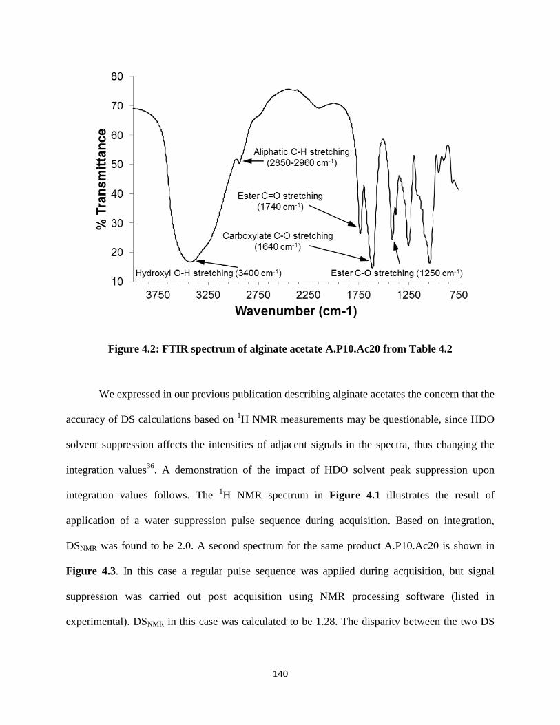
140
Figure 4.2: FTIR spectrum of alginate acetate A.P10.Ac20 from Table 4.2
We expressed in our previous publication describing alginate acetates the concern that the
accuracy of DS calculations based on 1H NMR measurements may be questionable, since HDO
solvent suppression affects the intensities of adjacent signals in the spectra, thus changing the
integration values36
. A demonstration of the impact of HDO solvent peak suppression upon
integration values follows. The 1H NMR spectrum in Figure 4.1 illustrates the result of
application of a water suppression pulse sequence during acquisition. Based on integration,
DSNMR was found to be 2.0. A second spectrum for the same product A.P10.Ac20 is shown in
Figure 4.3. In this case a regular pulse sequence was applied during acquisition, but signal
suppression was carried out post acquisition using NMR processing software (listed in
experimental). DSNMR in this case was calculated to be 1.28. The disparity between the two DS

141
values clearly demonstrates how water suppression can change peak integration ratios and lead
to inaccuracy.
Figure 4.3: 1H-NMR spectrum of alginate acetate A.P10.Ac20 from Table 4.2. HDO peak
suppression carried out post acquisition using NMR processing software
In order to circumvent this problem, we developed a method to determine DS values
based on titration. DS measurement by saponification of polysaccharide esters using aqueous
NaOH, and back titration of the excess base is an old, reliable and accurate technique37
. We
therefore saponified known quantities of alginate acetates using standardized NaOH solution,
and back-titrated the excess base using standardized HCl solutions. The moles of NaOH
consumed were equivalent to the moles of acetate present in the known quantity of sample.
DSTitration values were then calculated using Equations 1-3 described in the Experimental section.
Figures 4.4 and 4.5 show the potentiometric titration [pH] and derivative [d(pH)] plots for
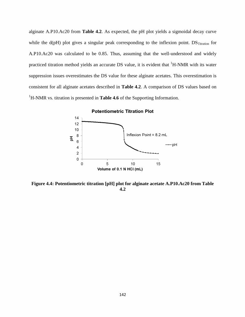
142
alginate A.P10.Ac20 from Table 4.2. As expected, the pH plot yields a sigmoidal decay curve
while the d(pH) plot gives a singular peak corresponding to the inflexion point. DSTitration for
A.P10.Ac20 was calculated to be 0.85. Thus, assuming that the well-understood and widely
practiced titration method yields an accurate DS value, it is evident that 1H-NMR with its water
suppression issues overestimates the DS value for these alginate acetates. This overestimation is
consistent for all alginate acetates described in Table 4.2. A comparison of DS values based on
1H-NMR vs. titration is presented in Table 4.6 of the Supporting Information.
Figure 4.4: Potentiometric titration [pH] plot for alginate acetate A.P10.Ac20 from Table
4.2

143
Figure 4.5: Potentiometric titration derivative [d(pH)] plot for alginate acetate A.P10.Ac20
from Table 4.2
A comparison of DSTitration values for alginate acetates shown in Table 4.2 provides
insight into the effect of reagent molar equivalents. In Series I the pyridine molar equivalents
were varied while a large excess of Ac2O was added, examining the impact of pyridine molar
excess at high Ac2O molar equivalents. DSTitration values ranged between 0.85-0.98; they did not
change drastically with increasing pyridine molar excess. This supports our previous findings
that the maximum possible DS value in alginates acetylated using this method tends to be
limited, for reasons that are not well-understood, to 1.0. Series II looked at the impact of
pyridine molar excess at lower Ac2O molar equivalents; the products in this series had acetate
DS values in the range of 0.30-0.40, again displaying little or no sensitivity to pyridine molar
excess. Series III represents experiments in which the pyridine molar equivalents were held
constant, but the Ac2O equivalents were varied. With increasing molar excess of Ac2O, acetate
DStitration values increased as expected; this method can therefore be used to control the acetate
DS of alginate acetates.

144
MW analysis reveals that it is challenging to completely shut down the loss of molar
mass during acetylation of the labile alginates. This is clear from comparison of the DPn of the
alginate starting material A-TBA (251) to those of the reaction products listed in Table 4.2.
When a large excess of Ac2O (20 equivalents) was used, MW loss was most pronounced. DPn
values for Series I were measured as 57, 106 and 65 for 2, 10 and 20 equivalents of pyridine
respectively. Figure 4.6 shows the shifts in the SEC chromatograms obtained using a refractive
index detector for alginate acetates listed in Table 4.2. Consistent with the strong influence of
the molar equivalent ratio of acetic anhydride per M/G monosaccharide, alginates from Series I
show the shortest elution times, signifying strong MW degradation. In contrast, Series II in
which 2 equivalents of Ac2O were employed instead of 20, measured DPn values were 145 and
151. A reduction in the loss of MW at lower Ac2O/monosaccharide equivalents is supported by
the positions of the peaks on the chromatograms, wherein A.P2.Ac2 and A.P20.Ac2 appear to
shorter elution times compared to Series I. In Series III, measured DPn values were 259, 166, 103
and 106 for 2, 5, 10 and 20 molar equivalents of Ac2O respectively. Correspondingly, the SEC
chromatograms showed longer elution times with higher Ac2O equivalents used in the reaction.
This analysis clearly indicates that acetic anhydride had a stronger effect on chain degradation
compared to pyridine. This was surprising to us; we expected that pyridine-catalyzed elimination
reactions (loss of the C-5 proton followed by elimination of the linkage hydroxyl from C-4)
would be a significant cause of DP and MW loss and would become even more important at
higher molar excesses of pyridine. The lack of consistent impact (Series I) upon DPn makes it
clear that the pyridine molar excess is not a critical factor. We are not sure of the cause of the
large impact from increasing acetic anhydride equivalents upon DPn loss. Certainly pyridinium
acetate, the by-product of acetylation, could be a catalyst for alginate chain cleavage. If this were

145
the primary cause for DPn loss, then the higher the DS (acetyl) achieved, the higher the degree of
chain cleavage that would be expected, since increasing DS (acetyl) causes concomitant increase
in pyridinium acetate by-product. The entries of Series III neither contradict this notion, nor
prove it.
Figure 4.6: SEC chromatograms of alginate acetates in Series I, II and III from Table 4.2
Changes in DS and loss in molecular weight with changing acetylation conditions appear
to significantly impact the ability of alginate acetates to form Ca-crosslinked gels. Our
experiments show that when alginates were acetylated homogeneously in DMSO/TBAF (thereby
acetylating both M and G residues), the products were incapable of forming Ca-crosslinked
microspheres even at concentrations as high as 25%. We therefore focused exclusively on the
method by which alginates were acetylated heterogeneously in DMSO, the results from which
are described in Table 4.2.
Figure 4.7 shows the optical microscopy images of microbeads prepared starting with
alginate concentrations of 3, 5 and 7.5%, and an air pressure of 0.5 psi. For unmodified sodium

146
alginate A-Na (DPn = 909), while spherical microbeads with minimal imperfections were made
at 3%; at 5% and 7.5% concentrations, tailing was observed due to the high viscosity of the
alginate solutions. When ultrapure LVM (DPn = 308) was used, spherical microbeads with
minimal imperfections were prepared with 3 and 5% solutions. At 7.5% tailing was observed.
As a general observation, ultrapure alginates formed smoother and more transparent microbeads.
This is advantageous since only ultrapure grades of alginate are medically relevant in order to
prevent toxicity issues. The alginate acetates of Series I, which had relatively high DS (acetyl)
due to the high excesses of acetic anhydride used, did not form microbeads at any
concentration/air pressure combination. This series consists of products that not only have high
degrees of acetylation, but also are most strongly degraded. This combination appears to prevent
bead formation altogether. The bead-forming abilities of alginates in Series III, synthesized using
increasing molar equivalents of acetic anhydride were then compared. Results using A.P10.Ac2
showed that at, DS (acetyl) 0.3 and DPn = 259 (2 molar equiv Ac2O), a 3% solution yielded
spherical beads with no tailing. At 5%, uniform beads with small tails were observed. At 7.5%,
the microbeads contained both tails and occurrences of polymorphism. At higher DS (0.69) and
lower DPn (166) (5 molar equiv Ac2O), overall beads were not as well shaped as those seen with
2 equivalents Ac2O. Microbeads were malformed at both 3% and 5% concentrations. At 7.5%,
spherical beads were observed, but with high incidence of polymorphism. In the case of the
highest DS alginate acetate (0.80, with lowest DPn (103)), made with 10 equiv Ac2O per
monosaccharide, no bead formation was observed at 3 and 5% concentrations since alginate
droplets were unable to break the surface tension of the calcium chloride crosslinking solution.
Clearly in this case, DS (acetate) (and the resulting hydrophobicity) was an influential factor. At
7.5 %, beads could be prepared, but were malformed.

147
% Solution by Weight
3 5 7.5
A-Na
LVM
A.P10.Ac2
A.P10.Ac5
A.P10.Ac10
B.P10.Ac10
C.P10.Ac10
D.P10.Ac10
Figure 4.7: Microscopic images of capsules formed using alginate acetates from Table 4.2

148
The results shown above highlight the existence and position of a window of alginate
acetate structure and preparation conditions, in which uniform and perfectly spherical alginate
acetate microbeads can be synthesized. On the one hand, unmodified alginates with very high
molecular weights (providing high solution viscosities) show undesirable tailing effects. On the
other hand, alginate acetates with high DS (acetate) values that are significantly degraded may
have difficulty forming microbeads altogether. In addition, malformation and polymorphism may
be observed as undesirable side effects. From the examples demonstrated above, A.P10.Ac2 (DS
= 0.3 and DPn = 259) seems to be within the operating window; 3% solutions of this alginate
acetate yielded perfectly shaped microbeads with no undesirable features such as tailing or
polymorphism.
Figure 4.8 shows the viscosity vs. shear rate plots for alginates used to generate the
microbeads shown in Figure 4.7. Since Series I did not show any microbead formation, viscosity
studies of these samples were not undertaken. The data shown in Figure 4.8 is summarized
numerically in Table 4.3, wherein the viscosity of solutions at concentrations of 3, 5 and 7.5%
has been reported. Viscosity is measured in Pa.s at a shear rate of 1 s-1
. Clearly, unmodified A-
Na and LVM show high viscosities at the concentrations being studied. Acetylation causes a
definite drop in viscosity, as noted from the viscosity values of reacted alginates. This drop in
viscosity may result from a loss in MW as well as disruption of intra- and interchain H-bonding.
Considering the specific example of A.P10.Ac2, even at a fairly low acetyl DStitration value of 0.3
and DPn of 259, there is a >90% reduction in viscosity compared to A-Na at all three
concentrations. This clearly demonstrates the manner in which even small DS(acetate) values
can have a significant impact of the physical properties of alginates. Since viscosity of alginate
solutions in an important criterion in the preparation of microbeads, it is evident that one would

149
have to account for changes occurring in the physical nature, and adjust the microbead
processing parameters accordingly.
Table 4.3: Viscosities of alginate acetates from Table 4.2 at various concentrations
Viscosity at shear rate = 1 s-1
(Pa.s)
3% 5% 7.5%
A-Na 4709 17146 21314
LVM 3803 6996 18705
A.P10.Ac2 89 342 1409
A.P10.Ac5 84 254 715
A.P10.Ac10 25 62 102
B.P10.Ac10 123 229 1983
C.P10.Ac10 276 407 637
D.P10.Ac10 2001 4410 1203
*Measurements performed at room temperature

150
Figure 4.8: Viscosity vs. shear rate plots for alginate acetates from Table 4.2 at various
concentrations, measured at room temperature

151
Figure 4.9: SEC chromatograms of alginate acetates in Series IV from Table 4.2
Figure 4.9 shows the SEC chromatograms of alginates A, B, C and D acetylated under
identical conditions. The final DPn values were measured as 103, 151, 125 and 138 respectively.
As mentioned previously, microbead formation using A.P10.Ac10 (DStitration = 0.80, DPn = 103
and FGG = 0.21) was successful only at a concentration of 7.5%; the beads were however
malformed. Lower concentration solutions were not suitable. In the case of alginate B.P10.Ac10
(DStitration = 0.96, DPn = 151 and FGG = 0.19), 3 and 5% solutions yielded malformed microbeads.
At a concentration of 7.5% however, spherical beads were obtained at an air pressure of 0.5 psi.
Nearly identical results were achieved using C.P10.Ac10 (DStitration = 0.90, DPn = 125 and FGG =
0.27), wherein 3 and 5% solutions yielded malformed microbeads but 7.5% solutions produced
perfectly spherical beads. For alginate D.P10.Ac10 (DStitration = 0.97, DPn = 138 and FGG = 0.56),
no bead formation was achieved at any concentration.
Comparing the DPn values of alginates in Series IV, A.P10.Ac10 was more degraded
compared to its B, C and D counterparts. This may be why A.P10.Ac10 could at best form
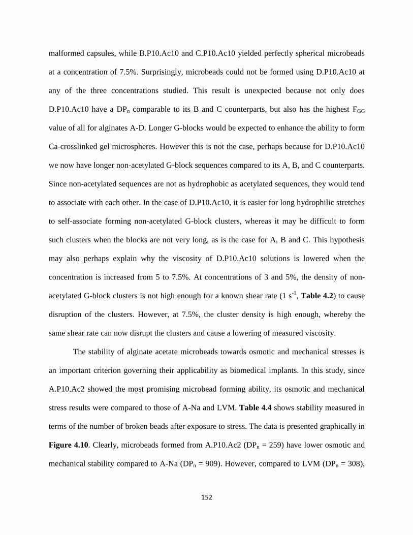
152
malformed capsules, while B.P10.Ac10 and C.P10.Ac10 yielded perfectly spherical microbeads
at a concentration of 7.5%. Surprisingly, microbeads could not be formed using D.P10.Ac10 at
any of the three concentrations studied. This result is unexpected because not only does
D.P10.Ac10 have a DPn comparable to its B and C counterparts, but also has the highest FGG
value of all for alginates A-D. Longer G-blocks would be expected to enhance the ability to form
Ca-crosslinked gel microspheres. However this is not the case, perhaps because for D.P10.Ac10
we now have longer non-acetylated G-block sequences compared to its A, B, and C counterparts.
Since non-acetylated sequences are not as hydrophobic as acetylated sequences, they would tend
to associate with each other. In the case of D.P10.Ac10, it is easier for long hydrophilic stretches
to self-associate forming non-acetylated G-block clusters, whereas it may be difficult to form
such clusters when the blocks are not very long, as is the case for A, B and C. This hypothesis
may also perhaps explain why the viscosity of D.P10.Ac10 solutions is lowered when the
concentration is increased from 5 to 7.5%. At concentrations of 3 and 5%, the density of non-
acetylated G-block clusters is not high enough for a known shear rate (1 s-1
, Table 4.2) to cause
disruption of the clusters. However, at 7.5%, the cluster density is high enough, whereby the
same shear rate can now disrupt the clusters and cause a lowering of measured viscosity.
The stability of alginate acetate microbeads towards osmotic and mechanical stresses is
an important criterion governing their applicability as biomedical implants. In this study, since
A.P10.Ac2 showed the most promising microbead forming ability, its osmotic and mechanical
stress results were compared to those of A-Na and LVM. Table 4.4 shows stability measured in
terms of the number of broken beads after exposure to stress. The data is presented graphically in
Figure 4.10. Clearly, microbeads formed from A.P10.Ac2 (DPn = 259) have lower osmotic and
mechanical stability compared to A-Na (DPn = 909). However, compared to LVM (DPn = 308),

153
there does not seem to be a statistically significant difference in the percentage of broken beads.
Since LVM is commonly used for encapsulation, we can safely state that the stability of alginate
acetates is in the workable range. The reduced stability of alginate acetate compared to unreacted
Na-alginate may be caused by the DP loss during TBA salt formation and acetylation, and/or due
to changes in crosslink density because of the acetate substituents in alginate acetate.
Table 4.4: Osmotic and mechanical stress effects, measured as % of broken microbeads
Sample Conc. (%)
Osmotic Stress Test
% Broken Capsules
Mechanical Stress Test
% Broken Capsules
Average Std. Dev. Average Std. Dev.
A.P10.Ac2 5 27.74 11.33 31.42 16.16
A-Na 5 2.25 3.82 12.02 2.85
LVM 1.5 19.61 4.59 15.36 9.88
For osmotic stress test: 1.5% LVM vs. 5% A-Na, p < 0.001; 1.5% LVM
vs. 5% A.P10.Ac2, no significant difference; 5% A-Na vs. 5%
A.P10.Ac2, p < 0.001. For mechanical stress test: 1.5% LVM vs. 5% A-
Na, no significant difference; 5% A-Na vs. 5% A.P10.Ac2, p < 0.05;
1.5% LVM vs. 5% A.P10.Ac2, p < 0.05.
Figure 4.10: (A) Osmotic and (B) mechanical stress effects, measured as % of broken
microbeads. For osmotic stress test: 1.5% LVM vs. 5% A-Na, p < 0.001; 1.5% LVM vs. 5%
A.P10.Ac2, no significant difference; 5% A-Na vs. 5% A.P10.Ac2, p < 0.001. For
mechanical stress test: 1.5% LVM vs. 5% A-Na, no significant difference; 5% A-Na vs. 5%
A.P10.Ac2, p < 0.05; 1.5% LVM vs. 5% A.P10.Ac2, p < 0.05.
4.5 Conclusions

154
Acylation chemistry is one of the most widely used methods for polysaccharide
modification. The ability to acylate alginates without significant degradation can be a useful tool
for fine-tuning physical properties to meet the needs of specific applications. Chemical
modification of hydrogel-forming alginates has the potential to allow the attachment of various
functional moieties to the polysaccharide backbone. The nature and function of these moieties
could range from those that make alginate significantly more hydrophobic, to those that allow it
to interact specifically in biological environments, to those that can enable covalent crosslinking
of chains in addition to the ionic crosslinks. The current study looks in detail at how control of
the degree of acetylation, position of acetylation (M vs. G), and the concomitant chain cleavage
reaction impact the ability of alginate acetates to form functional, partly hydrophobic Ca-gels.
In order to use acylated alginates as Ca-crosslinked hydrogels, it is important that
acylation be selective for M residues. Indeed, we show herein that homogeneous acetylation of
alginates provides randomly substituted alginate acetates, for which the solution concentrations
required for microbead formation were so high that they were out of the practical working range.
However, M-selective acetylation products afforded microbeads in a practically feasible
concentration range of 3-7.5%. When acetylated using the pyridine/acetic anhydride reagent
system, the molar equivalents of acetic anhydride used per monosaccharide had a direct impact
on DS (acetyl) and DPn, and thereby on the ability of final products to form Ca+2
-crosslinked
microbeads. While DS values could be controlled using the amount of Ac2O added, there
appeared to be a ceiling (DS ≈ 1.0) beyond which no more acetate substitution took place on the
alginate backbone, consistent with our earlier study of alginate dissolution in organic solvents
(TBAF/polar aprotic solvent such as DMSO) followed by hydroxyl esterification 5, 38
. It is
possible that this is related to our recent observation that TBAF catalyzes deacylation of

155
cellulose esters 38
, but the cause of the apparent limit on alginate acylation in this system will
require further investigation. Since DS (acetyl) has such a powerful impact on alginate acetate
gelation and other physical properties, accurate measurement of this parameter is crucial. We
demonstrate herein the successful development of a titration method to determine DS(acetyl) that
replaces the former 1H NMR method, which was plagued by the need to suppress the water peak,
strongly influencing DS results. This more reliable method for DS(acetyl) determination is a
crucial element as we attempt to realize the potential of alginate acetates as delivery systems for
cells and molecules.
Molecular weight loss during acetylation seems to be unavoidable, given the sensitivity
of alginate to both acidic and basic reaction conditions. While both acetic anhydride and pyridine
have the potential to cause chain degradation, we report here the surprising discovery that the
presence of excess acetic anhydride accelerates degradation much more effectively than does
excess pyridine. Furthermore, the data presented herein on the gelling abilities of alginate
acetates clearly proves that products that are more degraded have difficulty forming microbeads.
Therefore, if one desires to synthesize Ca-crosslinked alginate acetate gel microspheres, the
amount of acetic anhydride added in the reaction needs to be considered as a key factor. While
high DS values can be attained only in the presence of large excesses of Ac2O, these excesses
also lead to more extensive DPn loss. Fortunately, these studies clearly demonstrate that alginate
acetates with only a relatively low DS (acetyl) (ca. 0.3) show excellent ability to gel and form
capsules in the presence of Ca+2
ions. Since these alginate acetates can be made using only small
excesses of acetic anhydride, this opens the door to synthesis of materials for encapsulation and
delivery by the process described herein. Once acetylated, the viscosity of the alginate solution
drops significantly. Because of a drop in viscosity, microbead synthesis is directly affected.

156
While very high viscosities may be undesirable as they cause artifacts such as tailing, very low
viscosities prevent the alginate droplets from breaking the surface tension of CaCl2 bath solution,
thereby preventing bead formation. We demonstrate in this work a processing window for
forming perfectly spherical microbeads from alginate acetates of the proper DP and DS,
accessible by the methods we have described. Furthermore, we have shown that while the
osmotic and mechanical stability may be reduced due to acetylation, it is still in the workable
range by analogy to the similar stability of LVM microbeads routinely used for encapsulation.
4.6 Supporting information
Figure 4.11: 1H-NMR of TBA-alginate ‘A’

157
Figure 4.12: FTIR of TBA-alginate ‘A’
40
45
50
55
60
65
70
75
80
5001000150020002500300035004000
% T
ran
smit
tan
ce
Wavenumber (cm-1)
Aliphatic C-H stretching(2850-2960 cm-1) Carboxylate C–O stretching (1610cm-1)
Hydroxyl O-H stretching(3400cm-1)
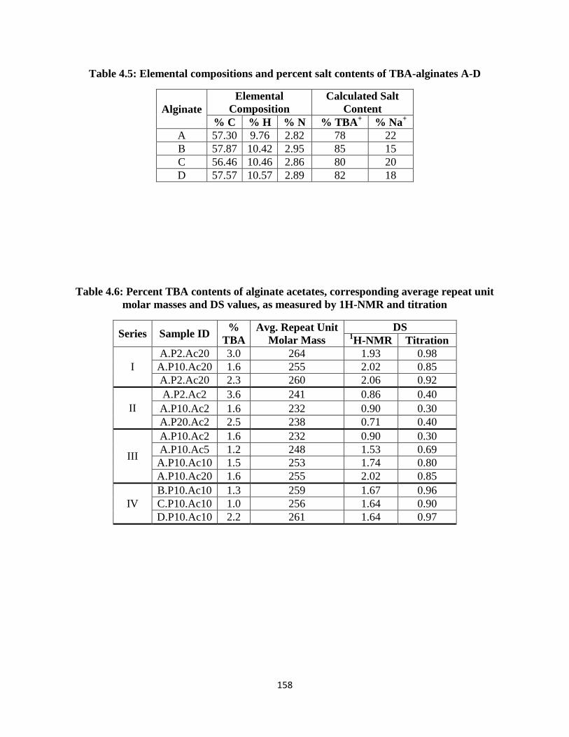
158
Table 4.5: Elemental compositions and percent salt contents of TBA-alginates A-D
Alginate
Elemental
Composition
Calculated Salt
Content
% C % H % N % TBA+ % Na
+
A 57.30 9.76 2.82 78 22
B 57.87 10.42 2.95 85 15
C 56.46 10.46 2.86 80 20
D 57.57 10.57 2.89 82 18
Table 4.6: Percent TBA contents of alginate acetates, corresponding average repeat unit
molar masses and DS values, as measured by 1H-NMR and titration
Series Sample ID %
TBA
Avg. Repeat Unit
Molar Mass
DS 1H-NMR Titration
I
A.P2.Ac20 3.0 264 1.93 0.98
A.P10.Ac20 1.6 255 2.02 0.85
A.P2.Ac20 2.3 260 2.06 0.92
II
A.P2.Ac2 3.6 241 0.86 0.40
A.P10.Ac2 1.6 232 0.90 0.30
A.P20.Ac2 2.5 238 0.71 0.40
III
A.P10.Ac2 1.6 232 0.90 0.30
A.P10.Ac5 1.2 248 1.53 0.69
A.P10.Ac10 1.5 253 1.74 0.80
A.P10.Ac20 1.6 255 2.02 0.85
IV
B.P10.Ac10 1.3 259 1.67 0.96
C.P10.Ac10 1.0 256 1.64 0.90
D.P10.Ac10 2.2 261 1.64 0.97

159
Figure 4.13: 1H-NMR spectrum of alginate acetate A.P2.Ac20 from Table 4.2. HDO peak
suppression pulse applied during acquisition

160
Figure 4.14: FTIR spectrum of alginate acetate A.P2.Ac20 from Table 4.2

161
Figure 4.15: 1H-NMR spectrum of alginate acetate A.P2.Ac20 from Table 4.2. HDO peak
suppression pulse applied during acquisition

162
Figure 4.16: FTIR spectrum of alginate acetate A.P2.Ac20 from Table 4.2

163
Figure 4.17: 1H-NMR spectrum of alginate acetate A.P10.Ac2 from Table 4.2. HDO peak
suppression pulse applied during acquisition

164
Figure 4.18: FTIR spectrum of alginate acetate A.P10.Ac2 from Table 4.2

165
Figure 4. 19: 1H-NMR spectrum of alginate acetate A.P10.Ac5 from Table 4.2. HDO peak
suppression pulse applied during acquisition

166
Figure 4.20: FTIR spectrum of alginate acetate A.P10.Ac5 from Table 4.2

167
Figure 4.21: 1H-NMR spectrum of alginate acetate A.P10.Ac10 from Table 4.2. HDO peak
suppression pulse applied during acquisition

168
Figure 4.22: FTIR spectrum of alginate acetate A.P10.Ac10 from Table 4.2

169
Figure 4.23: 1H-NMR spectrum of alginate acetate B.P10.Ac10 from Table 4.2. HDO peak
suppression pulse applied during acquisition

170
Figure 4.24: FTIR spectrum of alginate acetate B.P10.Ac10 from Table 4.2

171
Figure 4.25: 1H-NMR spectrum of alginate acetate C.P10.Ac10 from Table 4.2. HDO peak
suppression pulse applied during acquisition

172
Figure 4.26: FTIR spectrum of alginate acetate C.P10.Ac10 from Table 4.2
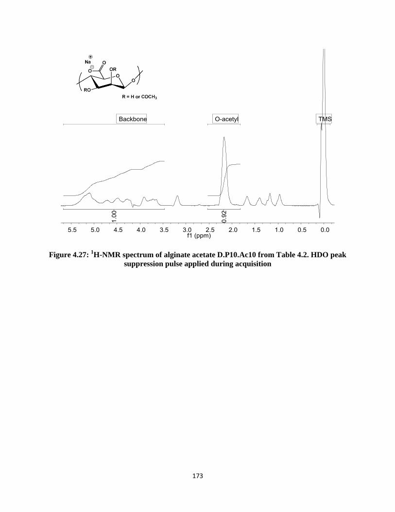
173
Figure 4.27: 1H-NMR spectrum of alginate acetate D.P10.Ac10 from Table 4.2. HDO peak
suppression pulse applied during acquisition
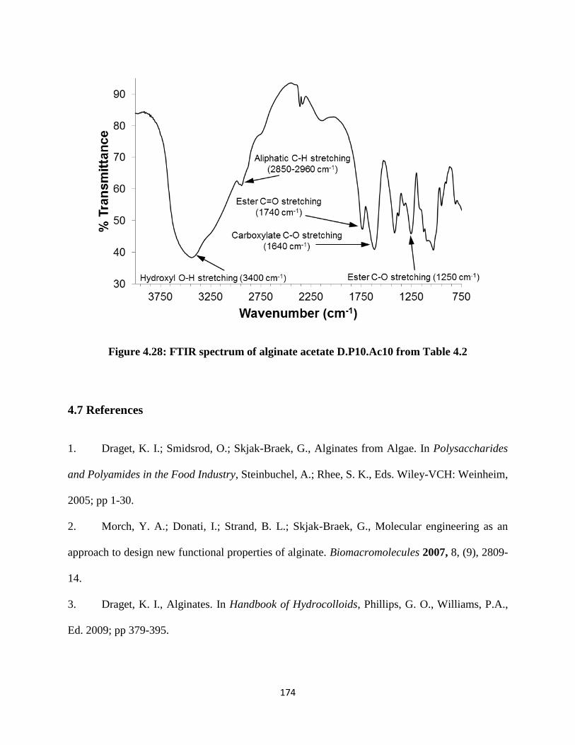
174
Figure 4.28: FTIR spectrum of alginate acetate D.P10.Ac10 from Table 4.2
4.7 References
1. Draget, K. I.; Smidsrod, O.; Skjak-Braek, G., Alginates from Algae. In Polysaccharides
and Polyamides in the Food Industry, Steinbuchel, A.; Rhee, S. K., Eds. Wiley-VCH: Weinheim,
2005; pp 1-30.
2. Morch, Y. A.; Donati, I.; Strand, B. L.; Skjak-Braek, G., Molecular engineering as an
approach to design new functional properties of alginate. Biomacromolecules 2007, 8, (9), 2809-
14.
3. Draget, K. I., Alginates. In Handbook of Hydrocolloids, Phillips, G. O., Williams, P.A.,
Ed. 2009; pp 379-395.
175
4. Fox, S. C.; Li, B.; Xu, D.; Edgar, K. J., Regioselective esterification and etherification of
cellulose: a review. Biomacromolecules 2011, 12, (6), 1956-72.
5. Pawar, S. N.; Edgar, K. J., Chemical Modification of Alginates in Organic Solvent
Systems. Biomacromolecules 2011, 12, (11), 4095-4103.
6. Pawar, S. N.; Edgar, K. J., Alginate derivatization: A review of chemistry, properties and
applications. Biomaterials 2012, 33, (11), 3279-3305.
7. Chamberlain, N. H., Cunningham, G.E., Speakman, J.B., Alginic acid diacetate. Nature
1946, 158, 553.
8. Schweiger, R. G., Acetylation of Alginic Acid. I. Preparation and Viscosities of Algin
Acetates. J. Org. Chem. 1962, 27, (5), 1786-1789.
9. Schweiger, R. G., Acetylation of Alginic Acid. II. Reaction of Algin Acetates with
Calcium and Other Divalent Ions. J. Org. Chem. 1962, 27, (5), 1789-1791.
10. Wassermann, A., Alginic Acid-acetate. Nature 1946, 158, 271.
11. Wassermann, A., Alginic Acid-acetate. J. Chem. Soc. 1948, 197.
12. Skjåk-Bræk, G.; Paoletti, S.; Gianferrara, T., Selective acetylation of mannuronic acid
residues in calcium alginate gels. Carbohyd. Res. 1989, 185, (1), 119-129.
13. Sikorski, P.; Mo, F.; Skjåk-Bræk, G.; Stokke, B. r. T., Evidence for Egg-Box-Compatible
Interactions in Calcium Alginate Gels from Fiber X-ray Diffraction. Biomacromolecules 2007, 8,
(7), 2098-2103.
14. Donati, I.; Holtan, S.; Mørch, Y. A.; Borgogna, M.; Dentini, M.; Skjåk-Bræk, G., New
hypothesis on the role of alternating sequences in calcium-alginate gels. Biomacromolecules
2005, 6, (2), 1031-40.

176
15. Lee, K. Y.; Mooney, D. J., Alginate: Properties and biomedical applications. Prog.
Polym. Sci. 2012, 37, (1), 106-126.
16. Boateng, J. S.; Matthews, K. H.; Stevens, H. N. E.; Eccleston, G. M., Wound healing
dressings and drug delivery systems: A review. J Pharm Sci-Us 2008, 97, (8), 2892-2923.
17. Ramsey, D. M.; Wozniak, D. J., Understanding the control of Pseudomonas
aeruginosaalginate synthesis and the prospects for management of chronic infections in cystic
fibrosis. In Mol. Microbiol., 2005; Vol. 56, pp 309-322.
18. Khan, S.; Tondervik, A.; Sletta, H.; Klinkenberg, G.; Emanuel, C.; Onsoyen, E.;
Myrvold, R.; Howe, R. A.; Walsh, T. R.; Hill, K. E.; Thomas, D. W., Overcoming Drug
Resistance with Alginate Oligosaccharides Able To Potentiate the Action of Selected
Antibiotics. Antimicrob Agents Chemother 2012, 56, (10), 5134-5141.
19. Soonshiong, P.; Feldman, E.; Nelson, R.; Heintz, R.; Yao, Q.; Yao, Z. W.; Zheng, T. L.;
Merideth, N.; Skjåk-Bræk, G.; Espevik, T.; Smidsrød, O.; Sandford, P., Long-Term Reversal of
Diabetes by the Injection of Immunoprotected Islets. P Natl Acad Sci USA 1993, 90, (12), 5843-
5847.
20. Zhang, P.; Zhang, X.; Brown, J.; Vistisen, D.; Sicree, R.; Shaw, J.; Nichols, G., Global
healthcare expenditure on diabetes for 2010 and 2030. Diabetes Res Clin Pract 2010, 87, (3),
293-301.
21. Chukwuma, C., Sr., Type I diabetic nephropathy: clinical characteristics and economic
impact. J Diabetes Complicat. 1993, 7, (1), 15-27.
22. The Diabetes Control and Complications Trial Research Group. Weight gain associated
with intensive therapy in the diabetes control and complications trial. . Diabetes Care 1988, 11,
(7), 567-73.

177
23. The Diabetes Control and Complications Trial Research Group. The effect of intensive
treatment of diabetes on the development and progression of long-term complications in insulin-
dependent diabetes mellitus. . N Engl J Med 1993, 329, (14), 977-86.
24. White, S. A.; Shaw, J. A.; Sutherland, D. E., Pancreas transplantation. Lancet 2009, 373,
(9677), 1808-17.
25. Opara, E. C.; Mirmalek-Sani, S. H.; Khanna, O.; Moya, M. L.; Brey, E. M., Design of a
bioartificial pancreas(+). J Investig Med 2010, 58, (7), 831-7.
26. Harlan, D. M.; Kenyon, N. S.; Korsgren, O.; Roep, B. O., Current advances and travails
in islet transplantation. Diabetes 2009, 58, (10), 2175-84.
27. Babak, V. G.; Skotnikova, E. A.; Lukina, I. G.; Pelletier, S.; Hubert, P.; Dellacherie, E.,
Hydrophobically associating alginate derivatives: Surface tension properties of their mixed
aqueous solutions with oppositely charged surfactants. J. Colloid Interface Sci. 2000, 225, (2),
505-510.
28. Tendulkar, S.; Mirmalek-Sani, S. H.; Childers, C.; Saul, J.; Opara, E. C.;
Ramasubramanian, M. K., A three-dimensional microfluidic approach to scaling up
microencapsulation of cells. Biomed Microdevices 2012, 14, (3), 461-9.
29. Darrabie, M. D.; Kendall, W. F., Jr.; Opara, E. C., Characteristics of Poly-L-Ornithine-
coated alginate microcapsules. Biomaterials 2005, 26, (34), 6846-52.
30. Timell, T. E., The acid hydrolysis of glycosides: I. general conditions and the effect of
the nature of the aglycone. Can. J. Chem. 1964, 42, 1456.
31. Smidsrød, O. H., A.; Larsen, B., The influence of pH on the Rate of Hydrolysis of Acidic
Polysaccharides. Acta Chem Scand 1966, 20, 1026-34.
178
32. Tsujino, I.; Saito, T., A new unsaturated uronide isolated from alginase hydrolysate.
Nature 1961, 192, 970-1.
33. Preiss, J.; Ashwell, G., Alginic Acid Metabolism in Bacteria. J Biol Chem 1962, 237, (2),
309-316.
34. Penman, A.; Sanderson, G. R., A method for the determination of uronic acid sequence in
alginates. Carbohyd Res 1972, 25, (2), 273-282.
35. Grasdalen, H.; Larsen, B.; Smidsrød, O., PMR Study of the Composition and Sequence
of Uronate Residues in Alginates. Carbohydr Res 1979, 68, (1), 23-31.
36. Salomonsen, T. J., H.M.; Larsen, F.H.; Engelsen, S.B., The Quantitative Impact of Water
Suppression on NMR Spectra for Compositional Analysis of Alginates. In Magnetic Resonance
in Food Science: Challenges in a Changing World, Gujonsdottir, M.; Belton, P.; Webb, G., Eds.
The Royal Society of Chemistry: 2009; pp 12-19.
37. Heinze, T.; Liebert, T.; Koschella, A., Structure Analysis of Polysaccharide Esters. In
Esterification of Polysaccharides, Springer Berlin Heidelberg: 2006; pp 143-167.
38. Xu, D. G.; Edgar, K. J., TBAF and Cellulose Esters: Unexpected Deacylation with
Unexpected Regioselectivity. Biomacromolecules 2012, 13, (2), 299-303.

179
Chapter 5: Synthesis of Alginate Esters via Carboxyl Group Modification
(Adapted from Pawar, S.N., Edgar, K.J.; Manuscript under preparation)
5.1 Abstract
Alginates are (1→4) linked linear copolysaccharides composed of β-D-mannuronic acid
(M) and its C-5 epimer, α-L-guluronic acid (G). Several strategies to synthesize carboxyl
modified alginate derivatives exist in the literature. A large majority of the findings however take
advantage of aqueous chemistries, such as carbodiimide coupling reactions. Based on our
previous report on the homogeneous dissolution of TBA-alginate, we now describe the use of
TBAF based two component solvent systems as media for the synthesis of carboxyl modified
alginate esters. Partially and fully esterified derivatives such as benzyl, butyl ethyl and methyl
alginate were synthesized via reaction with the corresponding alkyl halides. The newly
synthesized derivatives were soluble in polar aprotic solvents without the addition of TBAF.
Saponification was performed to demonstrate that -COOH groups were selectively reacted over –
OH groups to form esters. Furthermore, DS was measured using both NMR and titration to show
that the two methods yielded similar values. Later, we demonstrated the utility of the alginate
esters to enhance aqueous solubility of the flavonoid naringenin by formation of solid
dispersions.

180
5.2 Introduction
Alginates are unbranched polysaccharides consisting of 1→4 linked β-D-mannuronic
acid (M) and its C-5 epimer α-L-guluronic acid (G). They are isolated from one of two major
sources - algae such as kelp, or as an exopolysaccharide of bacteria such as Pseudomonas
aeruginosa. The alginate microstructure is unique and comprises M (M-blocks) and G (G-
blocks) residues interspersed with MG sequences (MG-blocks). Source and species dependent
variability exists between alginates in terms of their copolymer composition, sequence and
molecular weights. This variance is often used as a tool to control final properties in a number of
natural functions. For example, in Laminaria hyperborea, the stipe and holdfast require high
strength and rigidity and therefore contain alginates having higher fractions of G residues. In
contrast, the leaves of the plant which require flexibility for flotation in streaming water possess
alginates with lower fractions of G residues. Due to the abundance of algae in water bodies, there
is a large amount of alginate material present in nature. Industrial alginate production is
approximately 30,000 metric tons annually, and is estimated to comprise less than 10% of the
biosynthesized alginate material.1 Therefore there is significant additional potential to design
sustainable biomaterials based on alginates.
One of the most obvious and effective ways to design high performance biomaterials is
by chemically reacting the functional groups available on the alginate backbone. Alginate being
a water soluble polysaccharide in salt form, the use of aqueous chemistries such as EDC/NHS
coupling reactions to make esters/amides is a widely used strategy.2 While aqueous phase
reactions can be a good way to modify polysaccharides, they do not match the versatility that
organic phase reactions can offer. This is clearly demonstrated through the rich body of literature
published on organic dissolution and derivatization of cellulose.3 A major factor that

181
differentiates alginates from cellulose is the presence of ionizable carboxylic acid functionalities
on the former’s backbone. The charges play a crucial role in dictating organic solubility of
alginates; in fact, until recently no organic solvent systems had been described that would permit
full dissolution of alginate. Addressing this key problem, we have recently shown that it is
possible to dissolve alginates in two-component organic media consisting of polar aprotic
solvents (such as DMSO, DMF, DMAc or DMI) and the salt tetrabutylammonium fluoride
(TBAF).4 Furthermore, water soluble alginate acetates up to DSacetyl ~1.0 were synthesized by
reaction of alginate hydroxyl groups.
A disadvantage with the TBAF based two-component solvent systems is that the salt is
hydrated and there is always a small percentage of water present in the mixture. Furthermore, it
has been shown by our group that TBAF is useful as a reagent for regioselective deacylation of
cellulose esters.5 Although not experimentally confirmed, a similar deacylation reaction
occurring in situ could be the source of the limitation we have observed in alginate acylations,
where alginate acetate product DSacetyl has not exceeded ~1.0. We would therefore like to
develop alginate derivatives that would dissolve in organic media without the addition of TBAF.
From our past studies, no single component organic solvent (including ionic liquids) is capable
of fully dissolving (tetrabutylammonium) TBA-salts of alginic acid. The ability to dissolve
alginates in water-free organic media will be advantageous in two ways. Firstly, it will permit the
use of organic reagents that are incompatible with water, thereby enabling new pathways to
alginate functionalization. Secondly, it will enable solvent-based processing methods, which may
extend the applicability of alginates beyond its current scope (which involves mostly aqueous
processing). Substitution of alginate at the carboxyl group to form an ester is one potential
approach to the preparation of derivatives with such properties. Within our scope of knowledge,

182
we are aware of two prior instances where alginate esters have been synthesized via carboxyl
group modification.2 The first derivative is a commercially available alginate ester – propylene
glycol alginate, synthesized by reaction with propylene oxide. The second known set of reports
has detailed the reaction of TBA-alginate with dodecyl bromide to prepare dodecyl alginate in
DMSO.6-8
However the maximum DSdodecyl value reported was 0.12. Through this work we
intend to study the reaction of TBA-alginate with alkyl halides by homogeneous dissolution in
TBAF containing solvent systems. Furthermore, we shall study the study the impact of this
derivatization method on enhancing the organic solubility of alginates.
In order to demonstrate the potential utility of organo-soluble alginate derivatives, we
chose to test the performance of alginate carboxylate esters for amorphous solid dispersion of
naringenin (Nar). Nar is the aglycone of naringin, a flavonoid that confers a bitter taste to citrus
fruits. Among several potential Nar health benefits, it is well known to possess hypolipidemic
and anti-diabetic properties, and is therefore of great interest in the treatment of high cholesterol
and diabetes.9-10
In addition, like many other flavonoids, it is an antioxidant and is of importance
in cancer prevention and treatment.11
Recently, it was shown that Nar inhibits the secretion of
hepatitis C virus (HCV) in infected patients, and is a regulator of cytochrome P450 enzymes,
which are critical elements in drug metabolism.12-13
While Nar possesses favorable activity
against a host of diseases, it suffers from poor water solubility, which is a cause of its low oral
bioavailability. The Nar molecule is hydrophobic by nature (logP = 2.628), is highly crystalline
with a melting point of 250 °C, and has low reported aqueous solubility.14
To be able to use Nar
effectively in therapeutic formulations or dietary supplements, improving its aqueous solubility
and bioavailability is critical.
183
One approach that has been of interest for enhancing the solubility of poorly water
soluble drugs is through formation of amorphous solid dispersions (ASD).15-16
ASD formation is
a method by which drug crystallinity can be suppressed by trapping the drug molecules in a
metastable amorphous state via molecular dispersions with high glass transition temperature (Tg)
polymers. Polymer matrices chosen by previous ASD designers have been selected from
polymers that had already been included in drug formulations approved by the FDA for some
other purpose (so by definition not designed for ASDs). Polyvinylpyrrolidone (PVP) for
example, is a synthetic polymer that is well suited for SD formulations.17
Another class of
polymers that has shown promise is carboxyl-containing cellulose derivatives. These cellulose
derivatives have high Tg values, availability of OH and COOH groups for secondary interactions
with the drug, and the ability to tailor hydrophobicity using the type and DS of other substituents.
As a result of these properties, properly designed cellulose derivatives make excellent ASD
polymers. Most well-known among other cellulose derivatives are
hydroxypropylmethylcellulose acetate succinate (HPMCAS),18
carboxymethylcellulose acetate
butyrate (CMCAB),19-20
and more recently from our laboratory, cellulose ω-carboxy alkanoates
including cellulose acetate adipate propionate (CAAdP).21-24
Thus far, within our scope of knowledge, alginate derivatives have not yet been explored
for SD formulations. A major reason behind this is that it has never before been made soluble in
single-component organic media for ease of processing. In the current manuscript, we describe
new routes to synthesizing alginate derivatives that are soluble in single component polar aprotic
solvents as well as certain ionic liquids. Using bimolecular substitution nucleophilic (SN2)
reactions, the alginate carboxylate groups were esterified, whereby fully substituted (DS = 1.0)
derivatives were possible. The solubility properties of the derivatives were studied and peak

184
assignments were performed using alginates enriched in M and G residues. Furthermore, the use
of derivatives having partial DS in enhancing aqueous solubility of Nar was demonstrated.
5.3 Experimental
5.3.1 Materials
Four alginates were used for the study. We represented each as ‘Mxxx’, where M stands
for mannuronate and xxx denotes the % of M residues in the sample. For example, M063
signifies a sample containing 63% M and 37% G residues. M100 alginate was isolated from an
epimerase-negative mutant of Pseudomonas fluorescens in the laboratory of , Prof. Gudmund
Skjåk-Bræk.25
M063 alginate, an alginic acid sodium salt (algal source undisclosed) was from
Sigma. M060 alginate (algal source undisclosed) was from FMC BioPolymer. M000 was an
alginate containing G-blocks with an average DPn ~ 20 from the laboratory of Dr. Anne
Tøndervik.
Water used in the experiments was deionized. Reagent alcohol (histological grade),
hexanes (HPLC grade), DMSO (HPLC grade), DMF (HPLC grade), 5 N standardized NaOH
solution, 1 N standardized HCl solution, sodium iodide [NaI], potassium chloride [KCl],
potassium phosphate monobasic [KH2PO4] and ethyl acetate were purchased from Fisher
Scientific (Fair Lawn, NJ). Benzyl bromide (98%) [BzBr], iodoethane (98%) [EtI], 1-iodobutane
(98%) [BuI], 1-methyl-2-pyrrolidone (99.5%, extra dry) [NMP] and tetrabutylammonium
hydroxide (TBAOH; 40 wt%, 1.5 M solution in water) were obtained from Acros (Fair Lawn,
NJ). Deuterium oxide (99.9 atom% D; D2O) containing 0.75% 3-(trimethylsilyl)propionic-
2,2,3,3-d4 acid sodium salt for NMR, (±)-naringenin, and iodomethane [MeI] were obtained from

185
Sigma-Aldrich (St. Louis, MO). KBr used for FTIR analysis was purchased from International
Crystal Labs (Garfield, NJ). DMSO-d6 for NMR was acquired from Cambridge Isotope
Laboratories, Inc. (Andover, MA).
5.3.2 Synthesis of TBA-alginate
For conversion of Na-alginate to its protonated form, a procedure described by Babak et
al.6 was followed with modifications. HCl (0.6 N, 120 mL) was mixed with reagent alcohol (120
mL) in a beaker. Na-alginate (8 g) was added to the mixture and stirred overnight at 4 ˚C to
convert Na-alginate to alginic acid. The percentage of Na-salts protonated to the acid form was
not determined since the purpose was to subsequently synthesize TBA-alginate from alginic
acid. The mixture was filtered, then the solid (alginic acid) was washed thoroughly, sequentially
with alcohol and acetone. The solid product was dried overnight in vacuo at 60 ˚C. Fully dried
alginic acid was then dispersed in 200 mL water. TBAOH was added dropwise with continuous
stirring at room temperature until the polymer was dissolved and the pH was adjusted to 10.0. If
the solution was too viscous, small amounts of water were added until a final viscosity was
reached at which the solution could be easily stirred. The aqueous TBA-alginate solution was
then premixed with reagent alcohol. The premix was precipitated in a mixture of ethyl acetate-
hexane. The solvent ratios used were aqueous alginate acetate : ethanol : ethyl acetate : hexane =
1:3:12:3. The precipitate was then filtered under vacuum and dried in vacuo at 60 ˚C. The 1H-
NMR and FTIR spectra for TBA-alginate are shown in the Supporting Information as Figures
5.8 and 5.9 respectively.
1H NMR (500 MHz, D2O) δ 5.82 – 3.46 (m, alginate backbone), 3.19 (t,
NCH2CH2CH2CH3 of TBA), 1.67 (m, NCH2CH2CH2CH3 of TBA), 1.40 (m, NCH2CH2CH2CH3

186
of TBA), 0.97 (t, NCH2CH2CH2CH3). FTIR (KBr pellet method): 3400 cm-1
, hydroxyl O–H
stretching; 2850-2960 cm-1
, aliphatic C–H stretching, 1610 cm-1
, carboxylate C-O stretching.
Elemental composition TBA-alginate M060 measured by elemental analysis: C, 56.46%; H,
10.46%; N, 2.86%. This composition corresponds to roughly 80% TBA and 20% Na salt.
5.3.3 Synthesis of benzyl alginate
TBAF (0.1 g, 0.32 mmol) was dissolved in DMF (10 mL) at room temperature. TBA-
alginate (0.15 g) was then added and the mixture stirred until the polymer was dissolved. The
temperature was then raised to 80 °C. Benzylation was carried out using either BzBr or benzyl
iodide (BzI). In case of BzBr, the necessary equivalents of reagent were directly added to the
alginate solution. For BzI, the reagent was synthesized starting from BzBr and then added to the
alginate solution. Upon reagent addition, the solution was stirred at 80 °C for 30 min, after which
it was precipitated in ethyl acetate (~150 mL). The precipitate was recovered and dried in vacuo
at 60 °C. Dry crude benzyl alginate was then dissolved in DMF (~1.5 mL), precipitated in ethyl
acetate, filtered and dried in vacuo; this step was repeated one more time. The dry solid obtained
thereafter was benzyl alginate. The synthesis of BzI reagent is described as follows.
Benzyl iodide: NaI (0.535 g, 3.6 mmol) was dissolved in 5 mL of acetone (dried over 3 Å
molecular sieves) under N2 pressure at room temperature. To this solution BzBr (0.43 mL, 3.6
mmol) was added. Upon addition, NaBr precipitated as a white solid, which was filtered out and
discarded. To the filtrate, 1 mL of DMF (solvent for benzyl alginate synthesis) was added, and
acetone removed from the mixture using a rotary evaporator. BzI solution of DMF was thus
obtained, and was immediately used for benzyl alginate synthesis.

187
For benzyl alginate shown in Figures 5.1 and 5.2: 1H NMR (500 MHz, DMSO-d6) δ 7.48
– 7.23 (m, H9-13), δ 5.43 – 3.16 (m, H1-5), 5.20 (m, H7). 13
C NMR (500 MHz, DMSO-d6) δ
169.15 – 166.33 (m, C6), δ 135.24 – 125.33 (m, C8-13), δ 102.94 – 91.41 (m, C1), δ 80.88-61.39
(m, C2-5), δ 65.62 (m, C7). FTIR (KBr pellet method): 3400 cm-1
, hydroxyl O–H stretching;
2850-2960 cm-1
, aliphatic C–H stretching; 1740 cm-1
, ester C=O stretching; 1250, ester C–O
stretching. Elemental composition: C, 46.62%; H, 4.70%; N, 0.04%; O, 41.99%.
5.3.4 Synthesis of methyl alginate
TBAF (0.1 g, 0.32 mmol) was dissolved in DMF (10 mL) at room temperature. TBA-
alginate (0.15 g) was then added and the mixture stirred until the polymer was dissolved. The
temperature was then raised to 40 °C. Methyl iodide was added at once (molar equivalents varied
by experiment) to the alginate solution. Upon reagent addition, the solution was stirred at 40 °C
for 30 min, after which it was precipitated in ethyl acetate (~150 mL). The precipitate was
recovered and dried in vacuo at 60 °C. Dry crude methyl alginate was then dissolved in DMF
(~1.5 mL), precipitated in ethyl acetate, filtered and dried in vacuo; this step was repeated one
more time. The dry solid obtained thereafter was methyl alginate.
For methyl alginate shown in Figures 5.19 and 5.20 of Supporting Information: 1H NMR
(500 MHz, DMSO-d6) δ 6.00 – 3.49 (m, H1-5), δ 3.73 (m, H7). 13
C NMR (500 MHz, DMSO-d6)
δ 169.36 – 166.43 (m, C6), δ 114.918 – 92.09 (m, C1), δ 80.91-61.63 (m, C2-5), δ 51.53 (m, C7).
FTIR (KBr pellet method): 3400 cm-1
, hydroxyl O–H stretching; 2850-2960 cm-1
, aliphatic C–H
stretching; 1740 cm-1
, ester C=O stretching; 1250, ester C–O stretching. Elemental composition:
C, 40.38%; H, 5.20%; N, 0.11%; O, 49.24%.

188
5.3.5 Synthesis of ethyl alginate
TBAF (0.1 g, 0.32 mmol) was dissolved in DMF (10 mL) at room temperature. TBA-
alginate (0.15 g) was then added and the mixture stirred until the polymer was dissolved.
Temperature was then raised to 60 °C. Ethylation was carried out using either EtI wherein the
necessary equivalents of reagent were directly added to the alginate solution. Upon reagent
addition, the solution was stirred at 60 °C for 30 min, after which it was precipitated in ethyl
acetate (~150 mL). The precipitate was recovered and dried in vacuo at 60 °C. Dry crude ethyl
alginate was then dissolved in DMF (~1.5 mL), precipitated in ethyl acetate, filtered and dried in
vacuo; this step was repeated one more time. The dry solid obtained thereafter was ethyl
alginate.
For ethyl alginate shown in Figures 5.21 and 5.22 of Supporting Information: 1H NMR
(500 MHz, DMSO-d6) δ 6.00 – 3.49 (m, H1-5), δ 3.73 (s, H7). 13
C NMR (500 MHz, DMSO-d6)
δ 169.36 – 166.43 (m, C6), δ 114.918 – 92.09 (m, C1), δ 80.91-61.63 (m, C2-5), δ 39.50 (m, C7).
FTIR (KBr pellet method): 3400 cm-1
, hydroxyl O–H stretching; 2850-2960 cm-1
, aliphatic C–H
stretching; 1740 cm-1
, ester C=O stretching; 1250, ester C–O stretching. Elemental composition:
C, 40.31%; H, 5.37%; N, 0.11%; O, 48.98%.
5.3.6 Synthesis of butyl alginate
TBAF (0.1 g, 0.32 mmol) was dissolved in DMF (10 mL) at room temperature. TBA-
alginate (0.15 g) was then added and the mixture stirred until the polymer was dissolved.
Temperature was then raised to 80 °C. Butylation was carried out using either BuI wherein the
necessary equivalents of reagent were directly added to the alginate solution. Upon reagent
189
addition, the solution was stirred at 80 °C for 30 min, after which it was precipitated in ethyl
acetate (~150 mL). The precipitate was recovered and dried in vacuo at 60 °C. Dry crude butyl
alginate was then dissolved in DMF (~1.5 mL), precipitated in ethyl acetate, filtered and dried in
vacuo; this step was repeated one more time. The dry solid obtained thereafter was butyl
alginate.
For butyl alginate shown in Figures 5.23 and 5.24 of Supporting Information: 1H NMR
(500 MHz, DMSO-d6) δ 5.95 – 3.44 (m, H1-5), δ 4.13 (m, H7), δ 1.63 (m, H8), δ 1.38 (m, H9), δ
0.93 (m, H10). 13
C NMR (500 MHz, DMSO-d6) δ 169.57 – 166.60 (m, C6), δ 113.25 – 92.71 (m,
C1), δ 80.21-61.91 (m, C2-5), δ 64.42 (m, C7), δ 29.64 (m, C8), δ 18.04 (m, C9), δ 12.85 (m,
C10). FTIR (KBr pellet method): 3400 cm-1
, hydroxyl O–H stretching; 2850-2960 cm-1
, aliphatic
C–H stretching; 1740 cm-1
, ester C=O stretching; 1250, ester C–O stretching. Elemental
composition: C, 50.52%; H, 6.91%; N, 0.07%; O, 40.72%.
5.3.7 Saponification of esters
Benzyl alginate with DS 1.0 (131 mg) was added to water (13 mL) forming a suspension.
To this suspension, 1M NaOH solution (1.3 mL) was added and stirred for 30 min. Over time,
the alginate dissolved. It was then precipitated in reagent alcohol (~150 mL), filtered, the solid
recovered and dried in vacuo at 60 °C. The dry solid was dissolved in water and re-precipitated
in reagent alcohol, followed by filtration and drying in vacuo at 60 °C.
5.3.8 NMR Analysis

190
For all alginate esters, the solid sample (40-50 mg) was dissolved in DMSO-d6 (~0.7
mL) by applying heat. Spectra were acquired using a JEOL 500 MHz spectrometer with
automatic shimming, and the acquisition temperature was 85 ˚C. Number of scans acquired was
16 for 1H-NMR and DQF-COSY, 250 for HMQC and HSQC, and 13,000 for
13C-NMR. All
spectra were processed using MestReNova version: 8.0.2-11021. For water soluble samples, an
automatic water suppression pulse was applied to suppress the residual HDO peak. For benzyl
alginate, DS value calculation was carried out by integrating the benzyl aromatic peaks
appearing at 7.48 – 7.23 ppm (IAromatic) against the region appearing around 5.43 – 3.16 ppm,
which contains the backbone and benzyl CH2 signals (IBackbone+Methylene). IAromatic accounts for 5X
protons while IBackbone+Methylene accounts for 7+2(X) protons. The following formula was then
used to calculate the DS value.
To calculate the DS values for ethyl and butyl alginate derivatives, the methyl peak of the ester
appearing at ~0.93 ppm (IMethyl) was integrated against the region appearing at 5.95 – 3.44 ppm,
which contains the backbone and ester CH2 signals (IBackbone+Methylene). IMethyl accounts for 3X
protons while IBackbone+Bz,methylene accounts for 7+2(X) protons. The following formula was then
used to calculate the DS value.
DS value of methyl alginate could not be calculated using 1H-NMR due to the overlap of the
methyl ester peak with the backbone region.
191
5.3.9 FTIR measurement
FTIR spectra were acquired using a Thermo Electron Nicolet 8700 instrument in
transmission mode. Samples were prepared using the KBr pellet method. Alginate (1 mg) was
mixed with 99 mg of KBr using a mortar and pestle. The mixture was compressed in the sample
holder between two screws to form a KBr disc. 64 scans were obtained for each spectrum.
5.3.10 Elemental analysis
Measurements were performed using %CHN analyzer with a combustion/thermal
conductivity detector (TCD) and %O analyzer with pyrolysis.
5.3.11 DS measurement by titration
Titrations were performed by reacting alginate esters with excess NaOH to hydrolyze the
ester groups. NaOH molecules remaining after hydrolysis were then back-titrated with HCl to
measure the moles of acetate present. For example in the case of benzyl alginate, 250 mg solid
was weighed into a 50 mL 3-neck flask. To this, 25.0 mL of standardized 0.1 N NaOH solution
was added and stirred overnight at room temperature. The water insoluble polymer dissolved
over time. Potentiometric titrations were performed using a pH electrode (Inlab 413, Mettler-
Toledo International, Inc.). The electrode was inserted into the 3-neck flask, 0.1 mL aliquots of
standardized 0.1 N HCl solution added, the mixture allowed to stir for 30 seconds and the pH
measured. Total volume of HCl added was 30 mL. Two plots were made using the recorded data.
pH was first plotted against the volume of HCl added. Derivative d(pH) was plotted in a second

192
graph against the volume of HCl added. Inflexion point was recorded as the volume at which
peak maximum was attained in the d(pH) plot.
Moles of ester groups present in the sample used for titration (250 mg) was calculated as follows:
( )
DS was then calculated as:
( ) Equation (1)
Where,
( ) ( ( )) Equation (2)
Equation (2) was plugged in to Equation (1) to calculate DSTitration.
5.3.12 Synthesis of solid dispersions
Three solid dispersions were prepared starting from three different alginate ester
derivatives – ethyl, butyl and benzyl. In each case, the alginate derivative (180 mg) was
dissolved in 4 mL NMP by applying heat. Once the solution was cool, naringenin (60 mg) was
added and dissolved at room temperature. The solution was then precipitated in ice-cold water
(100 mL) and filtered to recover the wet solid, which was then placed in the freezer overnight
before lyophilizing.
5.3.13 Alginate ester solubility in water
193
To test the solubility of alginate ester in water, the solid (4 mg) was added to D2O (1
mL), heat applied and stirred using a vortex mixer. The suspension was passed through a syringe
filter to separate the solid, while the filtrate was added to a 5 mm NMR tube for acquisition.
5.3.14 Nar calibration curves
To plot calibration curves, Nar was dissolved in NMP at a concentration of 1 mg/mL.
The NMP solution was then diluted to concentrations of 0.02, 0.015, 0.01 and 0.005 mg/mL
using buffer solution, and UV-Vis spectra were obtained for each dilution. Two buffers solutions
were used in the study: pH 6.8 and 1.2. The absorbance wavelengths monitored were 316 nm in
pH 6.8 buffer, and 291 nm in pH 1.2 buffer.
5.3.15 Measurement of % drug loading
Drug loading was measured by dissolving the solid dispersion in NMP at a concentration
of 1 mg/mL. The NMP solution was then diluted to a concentration of 0.02 mg/mL in pH 6.8
buffer, and UV-Vis spectrum obtained. The absorbance at 316 nm was recorded and the
concentration of Nar in the solution was determined using the Nar calibration plot. Drug loading
was then calculated as follows:
5.3.16 Nar release studies

194
Nar samples (pure or solid dispersion) were dispersed in 100 mL buffer (pH 6.8 or 1.2) in
amber glass flasks at Nar concentrations of 0.07 mg/mL, and stirred at room temperature.
Aliquots (1.5 mL) were withdrawn at appropriate time intervals and replaced with 1.5 mL of
fresh dissolution medium after each sampling to maintain constant volume. UV-Vis absorption
of each aliquot was recorded after centrifugation at 14,000 rcf (relative centrifugal force) for 10
min.
5.4 Results and Discussion
Since alginates are predominantly water soluble polysaccharides, it has been a persistent
effort in our laboratory to find diverse organic solvents capable of dissolving them for
subsequent derivatization. Our previous findings have shown that a minimum requirement
necessary to solubilize alginates in organic media is the exchange of their inorganic counterions
(such as Na+ or Ca
+2) with organic counterions (such TBA
+). However, even after this
requirement is satisfied, TBA-alginate can only dissolve in polar aprotic solvents (DMSO, DMF,
DMI and DMAc) with the addition of the dissolution promoter TBAF. These two-component
solvent systems can serve as homogeneous reaction media, whereby it may now be feasible to
react the available functional groups on alginate backbone to synthesize derivatives with high
degrees of substitution (DS). Our hope was that upon derivatization, the new derivatives will
show improved solubility properties, and perhaps dissolve in single component organic solvents.
Having single component solvents will not only eliminate the use of TBAF (and thereby avoid
the accompanying water of TBAF hydration, as well as the potential for side reactions caused by
the presence of fluoride ion) but also allow easier processing for intended applications.

195
There are two available functional group types on the alginate backbone that may be used
for derivatization. We have previously shown that upon acetylation of the -OH groups up to
DSacetyl = 1.0, the derivatives were water-soluble and showed no single component organic
solubility. Furthermore, propionylation yielded alginate propionate up to DSpropionyl = 0.31, but its
solubility was still limited to water. At this point, we hypothesized that that while hydrophobic
groups could be attached to the alginate backbone via –OH group modification, the charges
imparted by carboxylate moieties presented significant hydrophilic character to the backbone to
prevent organic dissolution, particularly since we were unable to achieve the maximum
theoretical DS of 2.0 upon acylation of alginate hydroxyls in these solvent systems. We therefore
adopted a strategy of carboxyl group esterification as a potential method for synthesis of a new
class of organo-soluble alginate derivatives.
Selective reaction of carboxylate (COO-) over hydroxyl (-OH) groups is advantaged,
since the former is a stronger nucleophile. The difference in nucleophilicity can be used to
selectively react the carboxylate groups with substrates that can undergo SN2 substitution. The
ability to do so in our new alginate solvent systems4 afforded the additional possibility of
complete substitution on the carboxyl groups, as well as the possibility that no added base would
be needed since alginate carboxyls are already ionized in our solvent systems. We therefore
reacted alginate in solution with several halide reagents to prepare the corresponding esters. This
reaction of carboxylates with halides is non-catalyzed and does not entail the use of extreme pH
environments. Such conditions are favorable for preserving the molecular weight of the sensitive
polysaccharide alginate during reaction. It is well known that alginates suffer a loss in molar
mass in both strongly acidic26-27
and strongly alkaline solutions.28-29
Modification of alginates

196
under mild pH conditions is therefore often preferred. A general reaction scheme for the
esterification of alginate carboxylate groups using alkyl halides is shown in Scheme 5.1.
Scheme 5.1: General reaction scheme for esterification of alginates using alkyl halides
We first synthesized the benzyl ester by reaction of TBA-alginate with BzBr. DSbenzyl =
0.44 was achieved when the reaction was performed at 80 °C for 30 min. 1H-NMR spectrum of
the product is shown in Figure 5.10 in the Supporting Information. Knowing that the theoretical
maximum DSbenzyl value is 1.0, this indicates that 44% of the available carboxylate groups were
substituted. What is more significant however is the fact that this is the first demonstration of the
capability to react alginate with a hydrophobic organo-soluble reagent such as BzBr. Since BzBr
is water immiscible, achieving the same level of substitution by performing the reaction in an
aqueous medium could be anticipated as a challenge. In addition, a relatively rapid reaction
under mild pH conditions can allow preservation of molar mass during reaction. This highlights
the benefits of being able to dissolve alginates in organic solvents. We sought to achieve DSbenzyl
> 0.44, preferably full substitution, so took the approach of replacing BzBr with a benzyl reagent
that possesses a better leaving group. For example, when TBA-alginate was reacted with BnI at
80 °C for 30 min, we were able to synthesize fully substituted benzyl alginate with DSbenzyl = 1.0.
This to our knowledge is the first demonstration of complete substitution at the carboxyl group
position to form alginate esters. Figure 5.1 shows the 1H and
13C-NMR spectra of fully
substituted benzyl alginate.

197
Figure 5.1: 1H and
13C-NMR spectra of benzyl alginate with DSbenzyl = 1.0

198
Figure 5.2: FTIR spectrum of benzyl alginate with DSbenzyl = 1.0
The ability to dissolve TBA-alginate in a variety of TBAF based two-component solvent
systems has important implications in the preparation of alginate esters. This can be
demonstrated through an example in which we first used DMSO/TBAF as reaction solvent for
benzyl alginate synthesis. Our results indicate that while alginate carboxyl groups were
benzylated, DMSO participated as a reactive solvent forming benzyldimethyloxosulfonium salts
of alginate. A probable mechanism by which this reaction proceeds is shown in Scheme 5.2. The
1H-NMR spectra of all benzyl alginates synthesized in DMSO/TBAF showed a consistent peak
~2.9 ppm, as indicated by (*) in Figure 5.10 of Supporting Information. The reactivity of DMSO
with alkyl halides is previously documented, wherein the alkyl halide can first react with the
DMSO oxygen to give alkyloxydimethylsulfonium halide, which further rearranges to the more
stable form of the salt yielding alkyldimethyloxosulfonium halide.30
Due to the reactivity of
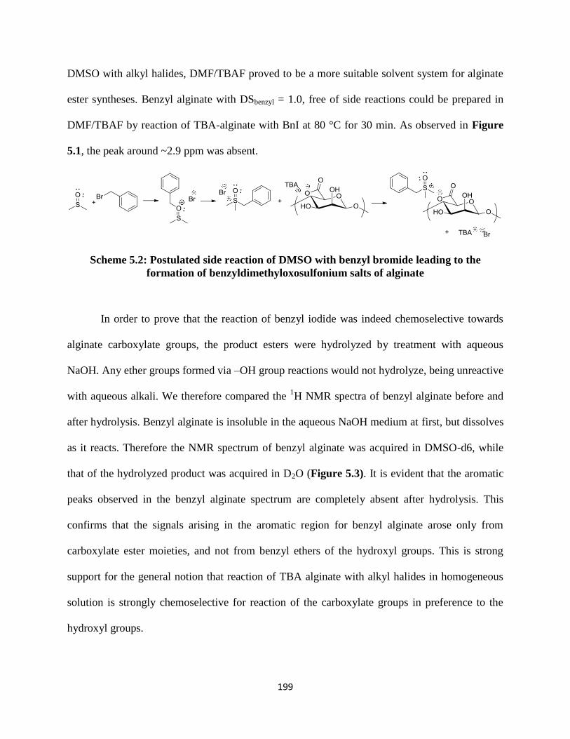
199
DMSO with alkyl halides, DMF/TBAF proved to be a more suitable solvent system for alginate
ester syntheses. Benzyl alginate with DSbenzyl = 1.0, free of side reactions could be prepared in
DMF/TBAF by reaction of TBA-alginate with BnI at 80 °C for 30 min. As observed in Figure
5.1, the peak around ~2.9 ppm was absent.
Scheme 5.2: Postulated side reaction of DMSO with benzyl bromide leading to the
formation of benzyldimethyloxosulfonium salts of alginate
In order to prove that the reaction of benzyl iodide was indeed chemoselective towards
alginate carboxylate groups, the product esters were hydrolyzed by treatment with aqueous
NaOH. Any ether groups formed via –OH group reactions would not hydrolyze, being unreactive
with aqueous alkali. We therefore compared the 1H NMR spectra of benzyl alginate before and
after hydrolysis. Benzyl alginate is insoluble in the aqueous NaOH medium at first, but dissolves
as it reacts. Therefore the NMR spectrum of benzyl alginate was acquired in DMSO-d6, while
that of the hydrolyzed product was acquired in D2O (Figure 5.3). It is evident that the aromatic
peaks observed in the benzyl alginate spectrum are completely absent after hydrolysis. This
confirms that the signals arising in the aromatic region for benzyl alginate arose only from
carboxylate ester moieties, and not from benzyl ethers of the hydroxyl groups. This is strong
support for the general notion that reaction of TBA alginate with alkyl halides in homogeneous
solution is strongly chemoselective for reaction of the carboxylate groups in preference to the
hydroxyl groups.
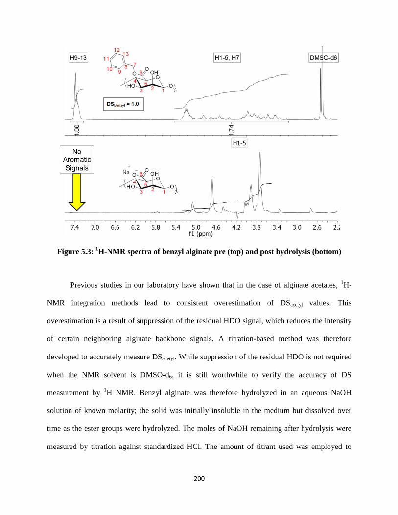
200
Figure 5.3: 1H-NMR spectra of benzyl alginate pre (top) and post hydrolysis (bottom)
Previous studies in our laboratory have shown that in the case of alginate acetates, 1H-
NMR integration methods lead to consistent overestimation of DSacetyl values. This
overestimation is a result of suppression of the residual HDO signal, which reduces the intensity
of certain neighboring alginate backbone signals. A titration-based method was therefore
developed to accurately measure DSacetyl. While suppression of the residual HDO is not required
when the NMR solvent is DMSO-d6, it is still worthwhile to verify the accuracy of DS
measurement by 1H NMR. Benzyl alginate was therefore hydrolyzed in an aqueous NaOH
solution of known molarity; the solid was initially insoluble in the medium but dissolved over
time as the ester groups were hydrolyzed. The moles of NaOH remaining after hydrolysis were
measured by titration against standardized HCl. The amount of titrant used was employed to

201
calculate the moles of ester present, and therefore the DSbenzyl. Figure 5.4 shows the
potentiometric titration and d(pH) plots for benzyl alginate. Table 5.1 shows a comparison of the
DS values as measured using 1H-NMR and titration. It is clear that both methods yield the same
value for DSbenzyl, thus confirming that 1H-NMR can be used as a reliable method for DS
measurement.
Figure 5.4: Potentiometric titration [left] and d(pH) [right] plots for benzyl alginate
Table 5.1: Comparison of DSbenzyl values measured using 1H NMR and titration
DSbenzyl 1H-NMR Titration
1.0 1.0
Peak assignments for alginate esters can be performed using 2D-NMR spectroscopy. As a
general observation, dissolving alginate derivatives in DMSO-d6 for NMR resulted in better peak
resolution compared to D2O, where peak overlap appeared to be more severe. To facilitate
interpretation of NMR spectra and assign signals arising from M and G residues, we took the
approach of esterifying the homopolymers M100 and M000, and using their simplified spectra as
guideposts for interpretation of copolymer spectra. The homopolymeric alginates are not
commercially available, but M100 can be prepared from bacterial culture of epimerase-deficient
bacteria,25
while M000 can be obtained from extensive exposure of natural alginate co-polymers

202
to a series of alginate epimerases in vitro.31-34
We then prepared butyl esters of alginates having
varying M/G compositions in order to perform the necessary peak assignments. Figure 5.5 and
5.6 show the stacked 1H and
13C NMR spectra of butyl alginates.
Figure 5.5: Stacked 1H-NMR spectra of butyl alginates of varying M contents

203
Figure 5.6: Stacked 13
C-NMR spectra of butyl alginates of varying M contents
For peak assignment, we used a strategy wherein the anomeric signals were first
identified based on their shifts in the 13
C-NMR spectrum. Then, using a combination of DQF-
COSY (1H-
1H coupling) and HMQC (
1H-
13C coupling) correlation spectroscopy techniques, all
remaining peaks were assigned. We observed that upon esterification, peak assignment for G
residues was relatively simpler compared to M residues. For example, in the case of the butyl
ester of M000 alginate, only one anomeric peak was observed which was correlated to the
corresponding G backbone peaks. Conversely, for the butyl ester of M100 alginate, at least five

204
separate anomeric signals were identified, and their correlations to corresponding M backbone
peaks were challenging using the techniques chosen. Furthermore, cross peaks obtained in the
2D-NMR spectra were not well resolved in the case of M100. As the fraction of M residues in
the backbone increased, the complexity arising in the NMR spectra increased correspondingly,
and analysis became more challenging. We were nevertheless able to clearly assign signals
arising from G residues in all alginate samples. The DQF-COSY and HMQC spectra for all four
alginates are shown in Figures 5.11 – 5.18 of Supporting Information. In addition, all backbone
peak shifts for G residues, and anomeric peak shifts for M residues are listed in Table 5.5 of
Supporting Information.
Table 5.2: Alginate esters, their DS values and corresponding solubility properties
Derivative Solubility
DMSO, DMF, NMP, DMAc, DMI Methanol, Isopropanol, Acetone Water
Benzyl alginate
DS = 1.0 S I I
Butyl alginate
DS = 1.0 S I I
Ethyl alginate
DS = 1.0 S I I
Methyl alginate
DS = 1.0 S I I
S = soluble, I = insoluble, P = partially soluble
Synthesis of alginate esters using carboxylate modification can afford derivatives with a
range of structures and properties based on the reagent and stoichiometry used. To demonstrate
this, we synthesized methyl and ethyl alginate in addition to the benzyl and butyl ester
derivatives. The 1H,
13C-NMR and FTIR spectra of methyl, ethyl and butyl alginate are shown in
Figures 5.19 – 5.24 of the Supporting Information. Table 5.2 shows the solubility properties of
ester derivatives with different DS values. The utility of alginate esters can be demonstrated
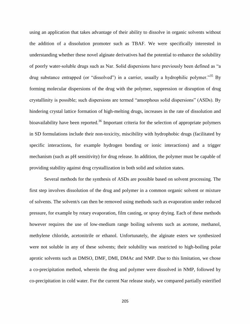
205
using an application that takes advantage of their ability to dissolve in organic solvents without
the addition of a dissolution promoter such as TBAF. We were specifically interested in
understanding whether these novel alginate derivatives had the potential to enhance the solubility
of poorly water-soluble drugs such as Nar. Solid dispersions have previously been defined as “a
drug substance entrapped (or “dissolved”) in a carrier, usually a hydrophilic polymer.”35
By
forming molecular dispersions of the drug with the polymer, suppression or disruption of drug
crystallinity is possible; such dispersions are termed “amorphous solid dispersions” (ASDs). By
hindering crystal lattice formation of high-melting drugs, increases in the rate of dissolution and
bioavailability have been reported.36
Important criteria for the selection of appropriate polymers
in SD formulations include their non-toxicity, miscibility with hydrophobic drugs (facilitated by
specific interactions, for example hydrogen bonding or ionic interactions) and a trigger
mechanism (such as pH sensitivity) for drug release. In addition, the polymer must be capable of
providing stability against drug crystallization in both solid and solution states.
Several methods for the synthesis of ASDs are possible based on solvent processing. The
first step involves dissolution of the drug and polymer in a common organic solvent or mixture
of solvents. The solvent/s can then be removed using methods such as evaporation under reduced
pressure, for example by rotary evaporation, film casting, or spray drying. Each of these methods
however requires the use of low-medium range boiling solvents such as acetone, methanol,
methylene chloride, acetonitrile or ethanol. Unfortunately, the alginate esters we synthesized
were not soluble in any of these solvents; their solubility was restricted to high-boiling polar
aprotic solvents such as DMSO, DMF, DMI, DMAc and NMP. Due to this limitation, we chose
a co-precipitation method, wherein the drug and polymer were dissolved in NMP, followed by
co-precipitation in cold water. For the current Nar release study, we compared partially esterified

206
benzyl, butyl and ethyl alginate derivatives listed in Table 5.2. Partially esterified derivatives
have a fraction of carboxylate groups that remain non-esterified, thus providing a pH dependent
release mechanism. None of the three derivatives in question showed complete water solubility.
However, we imagined that any partial water solubility may affect the percent drug loading of
the resulting solid dispersion, and hence its release performance. The water solubility of the each
derivative was therefore tested using 1H-NMR following exposure to D2O. Figure 5.25 in
Supporting Information shows the spectra, wherein the alginate backbone region has been
shaded grey. It is evident that the ethyl derivative shows partial solubility in D2O, while the butyl
and benzyl derivatives do not show any water solubility. Owing to this partial solubility of ethyl
alginate in water, loss of polymer occurs during SD preparation. A direct result of this is that
actual drug loading is higher compared to the theoretically expected loading. On the contrary,
butyl and benzyl alginate SDs yielded percent drug loadings that were lower than the
theoretically expected values. Table 5.3 shows the compositions of the SDs, and the theoretical
and actual percent drug loading values.
Table 5.3: SD compositions, along with theoretical and actual drug loading
Derivative Drug:Alginate
Wt. Ratio
Theoretical
drug loading
Actual drug
loading
Ethyl alginate 1:3 25% 42%
Butyl alginate 1:3 25% 21%
Benzyl alginate 1:3 25% 19%
We studied the release of Nar from the solid dispersions in aqueous solutions buffered at
pH 1.2 (simulating gastric environment) and 6.8 (simulating small intestinal environment).
Figure 5.7 shows the Nar release curves obtained. Compared to pur Nar (Blank), all three
alginate derivatives showed enhancement in drug solubility at both pH values. Table 5.4 shows
the maximum percent Nar release achieved for all SD formulations and the corresponding

207
solubility enhancement factors. Ethyl and benzyl alginates showed solubility enhancement
factors that were close to ~1.50, while that of butyl alginate ASD were slightly higher (ca. 2.25).
It is worthwhile at this point to compare these factors with the ones reported for carboxyl-
containing cellulose derivatives [Li, Liu, Edgar et al.; manuscript under review]. HPMCAS
clearly yields the best improvement in Nar aqueous solubility with an enhancement factor of 14.
CMCAB and CAAdP have factors of 5 and 4 respectively, and are closer to the values yielded by
alginate esters.
Figure 5.7: Nar release profiles studied at pH 1.2 (top) and pH 6.8 (bottom)

208
In order for alginate derivatives to be competitive as matrix polymers, there is clearly a
need for improvement in their solubility enhancement factors. However, the purpose of this
study is simply to demonstrate that modifying alginates in this fashion can lead to derivatives
capable of solubility enhancement of poorly water-soluble drugs. Going forward, there may be
several avenues possible for performance optimization. Firstly, the ability to dissolve alginates in
low boiling solvents can be expected to have a significant impact on the ease of ASD formation.
Allowing the use of solvent evaporation techniques such as film casting, vacuum evaporation
and spray drying could significantly impact the nature of the solid dispersions formed. Solvent
evaporation is perhaps the most flexible, practical technique for SD formation; melt extrusion is
often preferred when it is practical, but it is restricted to cases where both drug and polymer have
sufficient thermal stability. Extending the solubility profiles of alginate derivatives to permit
dissolution in low-boiling solvents should be possible by functionalization of the available –OH
groups in addition to the –COOH groups. Furthermore, varying the DS and type of both carboxyl
and hydroxyl substituents should allow fine-tuning of the hydrophilicity-hydrophobicity balance
to achieve optimal performance. To realize the best possible solid-state stabilization,
determination of the glass transition temperatures (Tg) of the alginate derivatives will be key. In
addition, XRD studies will be necessary to determine the extent to which alginate derivatives are
able to suppress drug crystallinity. In summary, our data demonstrates the principle that alginate
esters are capable of enhancing solubility of poorly water-soluble drugs, but there is ample room
for performance enhancement using multitude of strategies. We therefore believe that alginates
hold the promise to be interesting candidates for achieving improved dissolution and
bioavailability of poorly water-soluble drugs.

209
Table 5.4: Maximum percent Nar released and solubility enhancement factors for the SDs
studied
SD
pH 1.2 pH 6.8
Maximum
Nar Conc.
(mg/mL)
Solubility
enhancement
factor
Maximum
Nar Conc.
(mg/mL)
Solubility
enhancement
factor
Nar Blank 1.11×10⁻²
- 1.67×10⁻²
-
Nar:Ethyl alginate (1:3) 1.55×10⁻²
1.40 2.89×10⁻²
1.73
Nar:Butyl alginate (1:3) 2.55×10⁻²
2.30 3.65×10⁻²
2.19
Nar:Benzyl alginate (1:3) 1.68×10⁻²
1.52 2.59×10⁻²
1.55
Nar/HPMCAS - - - 14a
Nar/CMCAB - - - 5a
Nar/CAAdP - - - 4a
aData reported by Edgar et al.[ Li, Liu, Edgar et al.; manuscript under review]
5.5 Conclusion
With a growing emphasis on the use of renewable resource materials and the potential to
harvest algae in abundance, alginates have great promise as the source of polysaccharide
derivatives for demanding applications. Herein we have demonstrated chemoselective
modification of the alginate carboxyl group to provide a variety of esters, with DS controllable
by stoichiometry, that broadens the palette of available alginate derivatives and provides a new
route for their synthesis. We build on our previous finding that TBA salts of alginate were
soluble in two component polar aprotic solvent systems such as DMSO/TBAF, showing that
these organosoluble alginate salts react directly with alkyl halide reagents exclusively at the
carboxylate groups on the alginate backbone. A relatively fast reaction of alginates with alkyl
iodides presents the opportunity to achieve high DS products without significant loss of molar
mass. The resulting carboxyl ester derivatives, including benzyl, butyl, ethyl and methyl
alginates, have good solubility in polar aprotic solvents such as (DMSO, DMF, DMAc, DMI and

210
NMP) without addition of the dissolution promoter TBAF. These new derivatives, with enhanced
organic solubility and significantly modified properties, promise to find applications for which
the natural alginate polymer would not be suited.
It is clear from this work that the type of halide reagent used strongly influences
reactivity and hence achievable DS. While alkyl bromides do react with TBA alginate in organic
solution to give partially carboxyl-esterified products, switching to alkyl iodide reagents
permitted synthesis of completely substituted (DS 1.0) alginate esters at the proper
stoichiometry. This work further demonstrates the importance of choosing the correct solvent
system from the several polar aprotic solvents that effectively dissolve TBA alginate in the
presence of TBAF. The use of DMSO afforded side products from reactions of the solvent with
the alkyl halides, while amide solvents like DMF permitted smooth and clean alginate
carboxylate alkylation with no observed side reactions. It proved possible to discount the
possibility of competing hydroxyl alkylation by saponification of the product esters followed by
NMR analysis to demonstrate the absence of residual alkyl ether groups; this saponification
methodology was also a useful tool, coupled with back titration, to confirm the DS values
measured by 1H NMR and to provide an alternative analysis method that might in under some
circumstances be preferred to 1H NMR. 2D-NMR analysis of alginates was also performed for
samples with varying M contents. We were able to show that the complexity of NMR spectra
grew as the percentage of M residues in the backbone increased. Peak assignments were
performed to identify all backbone signals arising from G residues. Because of the complex
spectra with overlapping signals and cross peaks from M residues, only the anomeric signals of
these residues were identified.
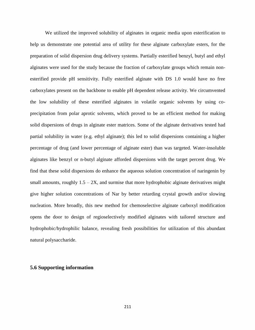
211
We utilized the improved solubility of alginates in organic media upon esterification to
help us demonstrate one potential area of utility for these alginate carboxylate esters, for the
preparation of solid dispersion drug delivery systems. Partially esterified benzyl, butyl and ethyl
alginates were used for the study because the fraction of carboxylate groups which remain non-
esterified provide pH sensitivity. Fully esterified alginate with DS 1.0 would have no free
carboxylates present on the backbone to enable pH dependent release activity. We circumvented
the low solubility of these esterified alginates in volatile organic solvents by using co-
precipitation from polar aprotic solvents, which proved to be an efficient method for making
solid dispersions of drugs in alginate ester matrices. Some of the alginate derivatives tested had
partial solubility in water (e.g. ethyl alginate); this led to solid dispersions containing a higher
percentage of drug (and lower percentage of alginate ester) than was targeted. Water-insoluble
alginates like benzyl or n-butyl alginate afforded dispersions with the target percent drug. We
find that these solid dispersions do enhance the aqueous solution concentration of naringenin by
small amounts, roughly 1.5 – 2X, and surmise that more hydrophobic alginate derivatives might
give higher solution concentrations of Nar by better retarding crystal growth and/or slowing
nucleation. More broadly, this new method for chemoselective alginate carboxyl modification
opens the door to design of regioselectively modified alginates with tailored structure and
hydrophobic/hydrophilic balance, revealing fresh possibilities for utilization of this abundant
natural polysaccharide.
5.6 Supporting information
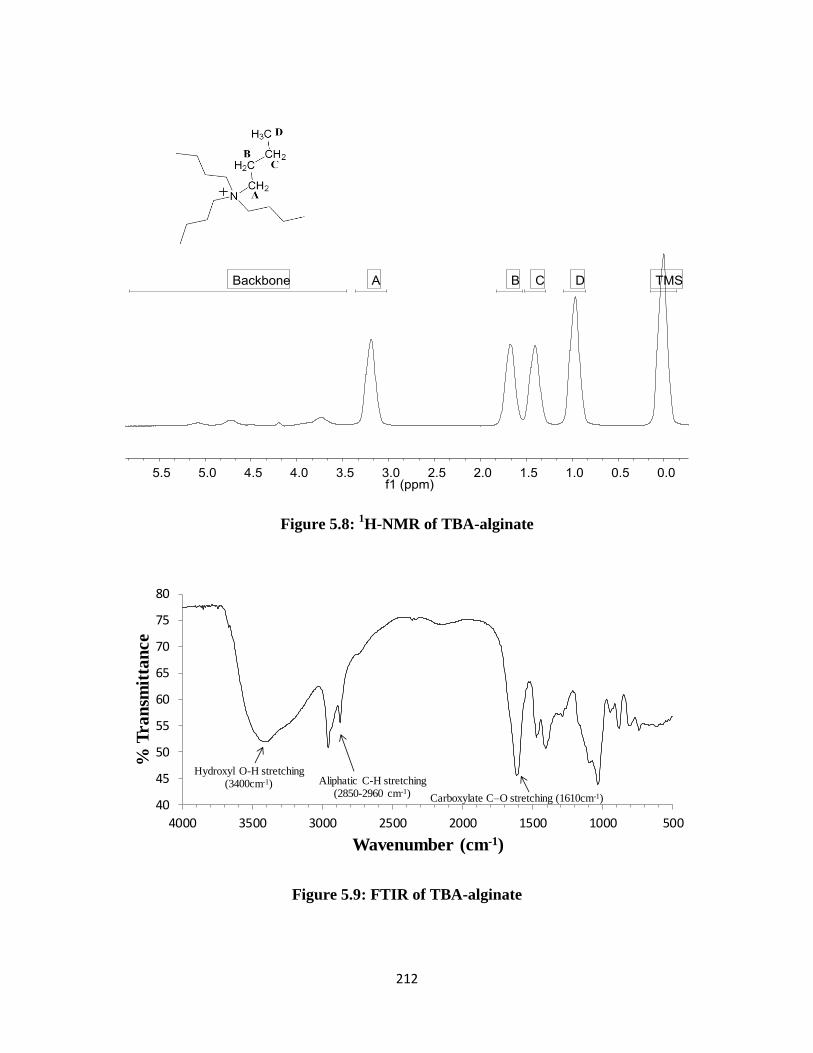
212
Figure 5.8: 1H-NMR of TBA-alginate
Figure 5.9: FTIR of TBA-alginate
40
45
50
55
60
65
70
75
80
5001000150020002500300035004000
% T
ran
smit
tan
ce
Wavenumber (cm-1)
Aliphatic C-H stretching(2850-2960 cm-1) Carboxylate C–O stretching (1610cm-1)
Hydroxyl O-H stretching(3400cm-1)

213
Figure 5.10: 1H-NMR spectrum of benzyl alginate with DS 0.44 synthesized using BzBr
Figure 5.11: DQF-COSY spectrum of benzyl alginate M000

214
Figure 5.12: HMQC spectrum of benzyl alginate M000

215
Figure 5.13: DQF-COSY spectrum of benzyl alginate M060

216
Figure 5.14: HMQC spectrum of benzyl alginate M060

217
Figure 5.15: DQF-COSY spectrum of benzyl alginate M063
218
Figure 5.16: HMBC spectrum of benzyl alginate M063

219
Figure 5.17: DQF-COSY spectrum of benzyl alginate M100

220
Figure 5.18: HMQC spectrum of benzyl alginate M100
Table 5.5: 1H and
13C-NMR peak shifts in butyl alginates
G Residues M Residues
Position 1H-NMR Shift
13C-NMR Shift Position
1H-NMR Shift
13C-NMR Shift
G1 4.86 100.32 M1 5.87 112.46
G2 3.64, 3.67 64.06, 64.49 M1 4.64 100.05
G3 3.82 68.36 M1 4.82 97.49
G4
G4(NR)
3.95
4.46
79.03
71.30 M1 5.22 95.07
G5 4.61 65.99 M1 5.34, 5.01 93.51
G6 - 168.09
NR: non-reducing

221
Figure 5.19: 1H and
13C-NMR of methyl alginate

222
Figure 5.20: FTIR of methyl alginate
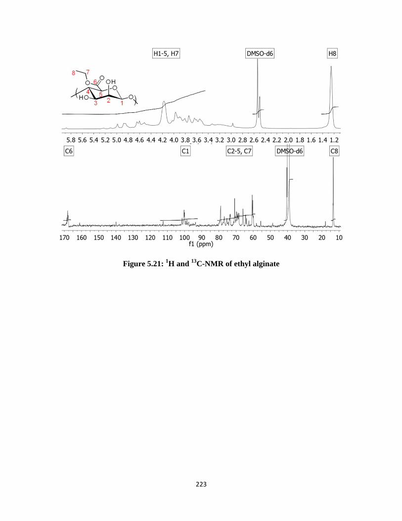
223
Figure 5.21: 1H and
13C-NMR of ethyl alginate
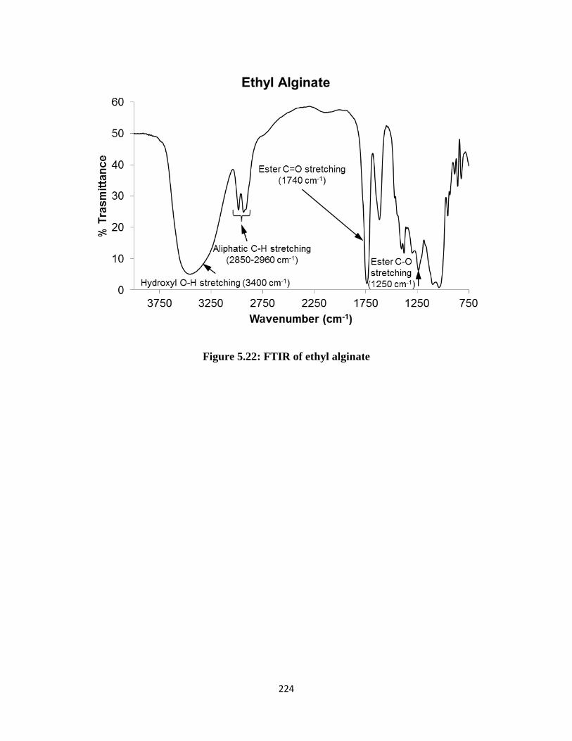
224
Figure 5.22: FTIR of ethyl alginate
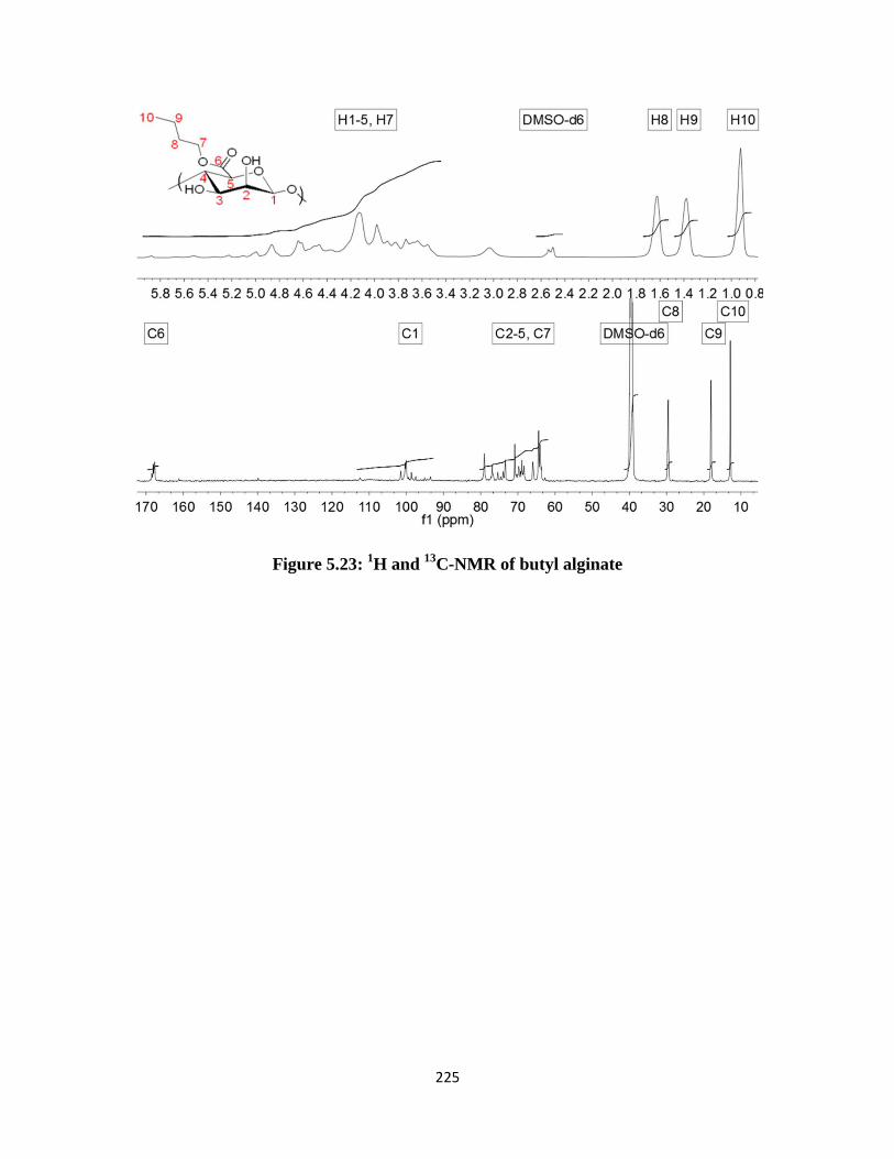
225
Figure 5.23: 1H and
13C-NMR of butyl alginate
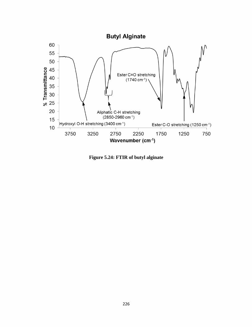
226
Figure 5.24: FTIR of butyl alginate

227
Figure 5.25: 1H-NMR of partially esterified alginates acquired in D2O
Figure 5.26: Nar calibration curves obtained in pH 1.2 (left) and pH 6.8 (right) buffer

228
5.7 References
1. Draget, K. I., Alginates. In Handbook of Hydrocolloids, Phillips, G. O., Williams, P.A.,
Ed. 2009; pp 379-395.
2. Pawar, S. N.; Edgar, K. J., Biomaterials 2012, 33 (11), 3279-3305.
3. Liebert, T.; Heinze, T.; Edgar, K. J., Preface. In Cellulose Solvents: For Analysis,
Shaping and Chemical Modification, American Chemical Society: 2010; Vol. 1033, pp ix-x.
4. Pawar, S. N.; Edgar, K. J., Biomacromolecules 2011, 12 (11), 4095-4103.
5. Xu, D. G.; Edgar, K. J., Biomacromolecules 2012, 13 (2), 299-303.
6. Babak, V. G.; Skotnikova, E. A.; Lukina, I. G.; Pelletier, S.; Hubert, P.; Dellacherie, E.,
J. Colloid Interface Sci. 2000, 225 (2), 505-510.
7. Pelletier, S.; Hubert, P.; Lapicque, F.; Payan, E.; Dellacherie, E., Carbohyd. Polym. 2000,
43 (4), 343-349.
8. Leonard, M.; Rastello de Boisseson, M.; Hubert, P.; Dellacherie, E., Journal of
Biomedical Materials Research Part A 2004, 68A (2), 335-342.
9. Borradaile, N. M.; de Dreu, L. E.; Barrett, P. H.; Behrsin, C. D.; Huff, M. W.,
Biochemistry-Us 2003, 42 (5), 1283-91.
10. Tsai, S. J.; Huang, C. S.; Mong, M. C.; Kam, W. Y.; Huang, H. Y.; Yin, M. C., J Agric
Food Chem 2012, 60 (1), 514-21.
11. Du, G.; Jin, L.; Han, X.; Song, Z.; Zhang, H.; Liang, W., Cancer Res 2009, 69 (7), 3205-
12.
12. Goldwasser, J.; Cohen, P. Y.; Lin, W.; Kitsberg, D.; Balaguer, P.; Polyak, S. J.; Chung,
R. T.; Yarmush, M. L.; Nahmias, Y., J Hepatol 2011, 55 (5), 963-71.
229
13. Tsujimoto, M.; Horie, M.; Honda, H.; Takara, K.; Nishiguchi, K., Biol Pharm Bull 2009,
32 (4), 671-6.
14. Tommasini, S.; Raneri, D.; Ficarra, R.; Calabro, M. L.; Stancanelli, R.; Ficarra, P., J
Pharm Biomed Anal 2004, 35 (2), 379-87.
15. Van Eerdenbrugh, B.; Taylor, L. S., Mol Pharm 2010, 7 (4), 1328-37.
16. Klein, C. E.; Chiu, Y. L.; Awni, W.; Zhu, T.; Heuser, R. S.; Doan, T.; Breitenbach, J.;
Morris, J. B.; Brun, S. C.; Hanna, G. J., Jaids-J Acq Imm Def 2007, 44 (4), 401-410.
17. Kanaze, F. I.; Kokkalou, E.; Niopas, I.; Barmpalexis, P.; Georgarakis, E.; Bikiaris, D.,
Drug Dev Ind Pharm 2010, 36 (3), 292-301.
18. Curatolo, W.; Nightingale, J. A.; Herbig, S. M., Pharm Res 2009, 26 (6), 1419-31.
19. Posey-Dowty, J. D.; Watterson, T. L.; Wilson, A. K.; Edgar, K. J.; Shelton, M. C.;
Lingerfelt, L. R., Cellulose 2007, 14 (1), 73-83.
20. Shelton, M., C.; Posey-Dowty, J., D.; Lingerfelt, L.; Kirk, S., K.; Klein, S.; Edgar, K., J.,
Enhanced dissolution of poorly soluble drugs from solid dispersions in carboxymethylcellulose
acetate butyrate matrices. In Polysaccharide Materials: Performance by Design, American
Chemical Society: 2009; Vol. 1017, pp 93-113.
21. Kar, N.; Liu, H. Y.; Edgar, K. J., Biomacromolecules 2011, 12 (4), 1106-1115.
22. Liu, H. Y.; Kar, N.; Edgar, K. J., Cellulose 2012, 19 (4), 1279-1293.
23. Ilevbare, G. A.; Liu, H. Y.; Edgar, K. J.; Taylor, L. S., Crystengcomm 2012, 14 (20),
6503-6514.
24. Ilevbare, G. A.; Liu, H. Y.; Edgar, K. J.; Taylor, L. S., Cryst Growth Des 2012, 12 (6),
3133-3143.
25. Chitnis, C. E.; Ohman, D. E., J Bacteriol 1990, 172 (6), 2894-900.
230
26. Timell, T. E., Can. J. Chem. 1964, 42, 1456.
27. Smidsrød, O. H., A.; Larsen, B., Acta Chem Scand 1966, 20, 1026-34.
28. Tsujino, I.; Saito, T., Nature 1961, 192, 970-1.
29. Preiss, J.; Ashwell, G., J Biol Chem 1962, 237 (2), 309-316.
30. Smith, S. G.; Winstein, S., Tetrahedron 1958, 3 (3), 317-319.
31. Høidal, H. K.; Ertesvåg, H.; Skjåk-Bræk, G.; Stokke, B. T.; Valla, S., J Biol Chem 1999,
274 (18), 12316-12322.
32. Hartmann, M.; Duun, A. S.; Markussen, S.; Grasdalen, H.; Valla, S.; Skjåk-Bræk, G.,
Biochim Biophys Acta 2002, 1570 (2), 104-12.
33. Hartmann, M.; Holm, O. B.; Johansen, G. A. B.; Skjåk-Bræk, G.; Stokke, B. T.,
Biopolymers 2002, 63 (2), 77-88.
34. Campa, C.; Holtan, S. v.; Nilsen, N.; Bjerkan, T. M.; Stokke, B. r. T.; Skjåk-Braek, G.,
Biochem. J. 2004, 381 (1), 155-164.
35. Shelton, M. C.; Posey-Dowty, J. D.; Lingerfelt, L. R.; Kirk, S. K.; Klein, S.; Edgar, K. J.,
Enhanced dissolution of poorly soluble drugs from solid dispersions in carboxymethylcellulose
acetate butyrate matrices. In Polysaccharide Materials: Performance by Design, American
Chemical Society: 2009; Vol. 1017, pp 93-113.
36. Hancock, B.; Parks, M., Pharmaceutical Research 2000, 17 (4), 397-404.
231
Chapter 6: Summary and Future Work
Sodium alginate in its unmodified form is a water soluble hydrophilic polysaccharide that
shows no organic solubility. Through our knowledge of the literature, we recognized that
homogeneous organic dissolution of alginates was an aspect of its derivatization chemistry that
was not previously explored.[1] We further predicted that if alginates could be dissolved in
organic media, new routes to its functionalization could be unlocked, access to useful
hydrophobic reagents made possible, and derivatives not previously permissible through aqueous
chemistry could be synthesized. Based on this hypothesis, we discovered TBAF based two
component solvent systems capable of dissolving tetrabutylammonium (TBA) salts of alginate.
Polar aprotic solvents such as DMSO, DMF, DMI or DMAc along with TBAF were able to
afford clear solutions of TBA-alginate at room temperature. This was the first ever
demonstration of the unequivocal dissolution of alginates in organic media. This discovery has
opened up numerous possibilities for the derivatization of alginates using a variety of chemistries
and reagents not previously possible. Through our work, we have successfully illustrated some
of the possible routes to alginate functionalization. While this was the first demonstration of the
use of DMSO/TBAF for solubilizing TBA-alginate, it has previously been reported to dissolve
crystalline polysaccharides such as cellulose at room temperature.[2]
Solubilizing alginates in two component solvent systems was made possible by simply
exchanging the small inorganic counterions (Na+ or Ca
+2) with a bulky organic counterion
(TBA+). However, it appears that this counterion exchange is not enough to affect single
component organic solvent solubility. An ideal scenario could be imagined, wherein alginates are
easily soluble in common low-boiling organic solvents such as acetone, methanol, ethanol,
chloroform, dichloromethane and so on. Perhaps the only way to achieve this ideal solubility
232
range is via organic derivatization of the alginate –OH and/or –COOH groups. As of now, we
believe that the best way to achieve this is via dissolution and derivatization of TBA-alginate in
solvents systems such as DMSO/TBAF.
We demonstrated that the alginate –OH groups can be acetylated using Ac2O/pyridine in
DMSO/TBAF.[3] Knowing that the maximum possible achievable DSacetyl is 2.0, we reacted
TBA-alginate with large excesses of Ac2O. Under no circumstances were we able to achieve
DSacetyl values higher than ~1.0. Clearly, there was a ceiling beyond which no further acetylation
occurred when Ac2O/pyridine was used. We speculated on the reasons why such a ceiling may
be observed. One possible explanation relates to the tendency of TBAF to regioselectively
deacylate esters of cellulose.[4] A similar reaction occurring in situ during acetylation of
alginates may be one explanation for limiting the DSacetyl value. In order to further explore the
role played by TBAF during acetylation, it would be valuable to perform acetylation
homogeneously, but in the absence of TBAF. If DSacetyl values higher than 1.0 can be achieved
under such conditions, it would confirm our suspicion that the fluoride ions do indeed limit
DSacetyl. However, the homogeneous dissolution of alginates in organic media without TBAF is
possible only through derivatives prepared via carboxyl esterification (as demonstrated in
Chapter 5). We therefore believe that the alginates esters synthesized in Chapter 5 would be
suitable substrates to study acetylation in the absence of TBAF. Notably, the benzyl derivative
could be more valuable, considering the possibility of subsequent hydrogenation to remove
benzyl groups without hydrogenolysis of acyl esters. Scheme 6.1 shows a general scheme that
may be used for this study.
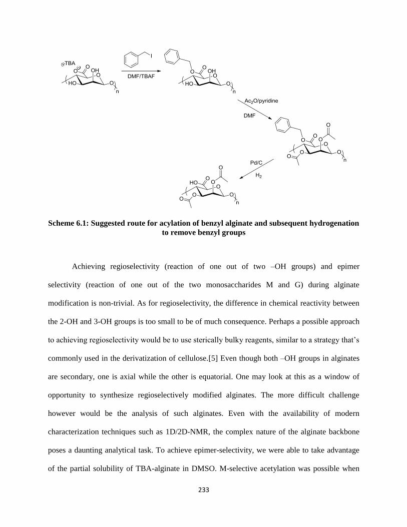
233
Scheme 6.1: Suggested route for acylation of benzyl alginate and subsequent hydrogenation
to remove benzyl groups
Achieving regioselectivity (reaction of one out of two –OH groups) and epimer
selectivity (reaction of one out of the two monosaccharides M and G) during alginate
modification is non-trivial. As for regioselectivity, the difference in chemical reactivity between
the 2-OH and 3-OH groups is too small to be of much consequence. Perhaps a possible approach
to achieving regioselectivity would be to use sterically bulky reagents, similar to a strategy that’s
commonly used in the derivatization of cellulose.[5] Even though both –OH groups in alginates
are secondary, one is axial while the other is equatorial. One may look at this as a window of
opportunity to synthesize regioselectively modified alginates. The more difficult challenge
however would be the analysis of such alginates. Even with the availability of modern
characterization techniques such as 1D/2D-NMR, the complex nature of the alginate backbone
poses a daunting analytical task. To achieve epimer-selectivity, we were able to take advantage
of the partial solubility of TBA-alginate in DMSO. M-selective acetylation was possible when

234
the reaction conditions were heterogeneous (DMSO), and non-selective acetates were prepared
when alginate was dissolved homogeneously (DMSO/TBAF). While we have shown this to be a
possible way to achieve epimer-selective control, other promising routes have been reported for
the same.[6] A chemoenzymatic approach would perhaps be most appealing, considering that it
offers complete control in terms of epimer-selectivity.
An important aspect of alginate derivatization that cannot be ignored is its susceptibility
to molecular weight degradation. Alginates degrade under both acidic and basic pH conditions.
When performing acetylation using Ac2O/pyridine, the acetic acid by product formed had a
much stronger effect on degradation compared to pyridine. We further observed that degradation
during acetylation could only be minimized at best; it could not be prevented. The impact of
molecular weight on the strength of Ca-crosslinked alginate gels is well known. We were further
able to prove that in the case of M-selective alginate acetates, molecular weight had a strong
influence on the ability to form Ca-crosslinked beads. High DSacetyl values coupled with
significant loss in MW severely compromised Ca-crosslinking capacity. This observation is a
clear demonstration of why it may be important to preserve alginate DP during reaction.
To achieve acylation of alginates while preserving the degree of polymerization (DPn),
formation of acetoacetate esters is an attractive option. The synthesis of cellulose acetoacetate
using diketene reagent has previously been reported.[7] Diketene would indeed be the best
choice for alginate acetoacetylation since it requires no catalyst and reacts at low temperatures,
as shown through cellulose acetoacetates. Such a situation would be idea to synthesize
derivatives without significant loss in DPn values. However, the highly reactive and at times
hazardous nature of diketene can be a deterrent to its use. As part of our work, diketene reactions
were not explored due to the fact that it is no longer commercially available and requires
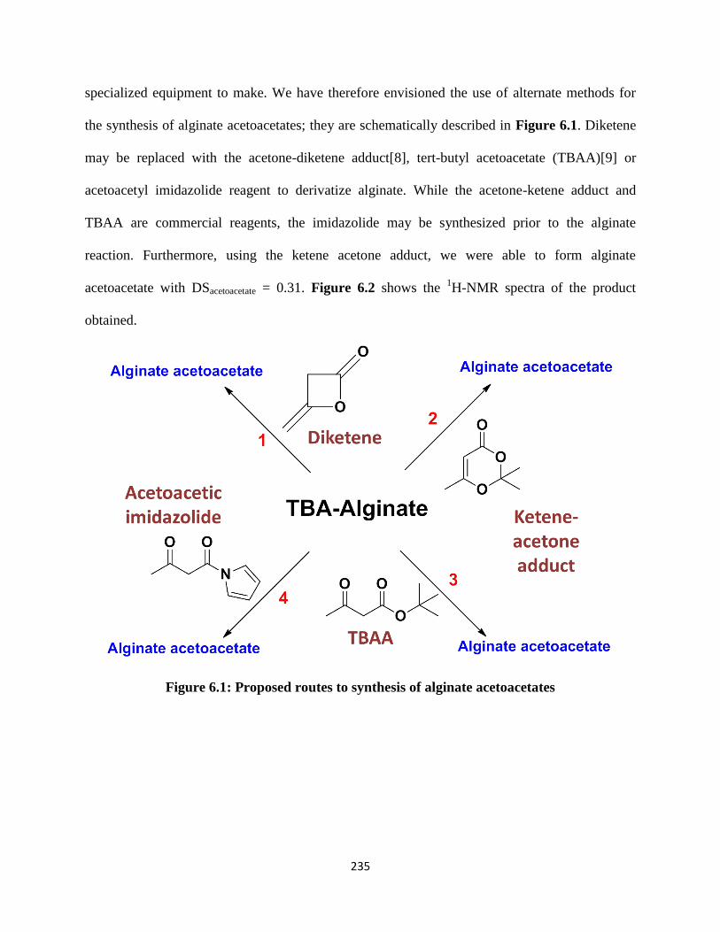
235
specialized equipment to make. We have therefore envisioned the use of alternate methods for
the synthesis of alginate acetoacetates; they are schematically described in Figure 6.1. Diketene
may be replaced with the acetone-diketene adduct[8], tert-butyl acetoacetate (TBAA)[9] or
acetoacetyl imidazolide reagent to derivatize alginate. While the acetone-ketene adduct and
TBAA are commercial reagents, the imidazolide may be synthesized prior to the alginate
reaction. Furthermore, using the ketene acetone adduct, we were able to form alginate
acetoacetate with DSacetoacetate = 0.31. Figure 6.2 shows the 1H-NMR spectra of the product
obtained.
Figure 6.1: Proposed routes to synthesis of alginate acetoacetates

236
Figure 6.2: 1H-NMR of alginate acetoacetate
Based on the accomplishments from this work, some general comments can be made
about its impact on the area of alginate derivatization. In addition to preparing alginate esters, it
is always an attractive option to explore the synthesis of ethers via hydroxyl group modification.
Most etherification reactions necessitate the use of strong bases for deprotonating –OH groups.
Knowing that alginates undergo degradation via β-elimination, a competitive side reaction that
lowers molecular weight is almost guaranteed. This makes the synthesis of alginate ethers
challenging. However, if the MW loss can be controlled under specific conditions, and if the
intended applications do not require high MWs, synthesizing alginate ethers would be a novel
addition to the field. Like ethers, the synthesis of alginate sulfates is marred by similar
difficulties. Alginate sulfates are of interest as heparin analogs due to their anticoagulant
properties.[10, 11] Most sulfate syntheses require the use of reagents based on strong acids such
as H2SO4. In such harsh conditions, a competitive degradation reaction through a hydrolytic
mechanism would be unavoidable. It would therefore be a valuable exercise to consider the use
of reagents that are milder compared to H2SO4. Perhaps a reagent such as SO3.pyridine complex
may lead to sulfation while minimizing loss of molar mass. In conclusion, alginate derivatization
is non-trivial, both in terms of actual syntheses and structural characterization. However,
237
syntheses that use novel chemistries, afford good regioselective control, offer high DS values
and limit the loss in molar mass will be valuable additions to alginate research.
6.1 References
1. Pawar SN, Edgar KJ. Alginate derivatization: A review of chemistry, properties and
applications. Biomaterials 2012;33(11):3279-3305.
2. Ostlund A, Lundberg D, Nordstierna L, Holmberg K, Nyden M. Dissolution and Gelation
of Cellulose in TBAF/DMSO Solutions: The Roles of Fluoride Ions and Water.
Biomacromolecules 2009 Sep;10(9):2401-2407.
3. Pawar SN, Edgar KJ. Chemical Modification of Alginates in Organic Solvent Systems.
Biomacromolecules 2011;12(11):4095-4103.
4. Xu DG, Edgar KJ. TBAF and Cellulose Esters: Unexpected Deacylation with
Unexpected Regioselectivity. Biomacromolecules 2012;13(2):299-303.
5. Fox SC, Li B, Xu D, Edgar KJ. Regioselective esterification and etherification of
cellulose: a review. Biomacromolecules 2011;12(6):1956-1972.
6. Rokstad AM, Donati I, Borgogna M, Oberholzer J, Strand BL, Espevik T, et al. Cell-
compatible covalently reinforced beads obtained from a chemoenzymatically engineered
alginate. Biomaterials 2006;27(27):4726-4737.
7. Edgar KJ, Arnold KM, Blount WW, Lawniczak JE, Lowman DW. Synthesis and
Properties of Cellulose Acetoacetates. Macromolecules 1995 Jun 5;28(12):4122-4128.
8. Clemens RJ, Hyatt JA. Acetoacetylation with "2,2,6-Trimethyl-4h-1,3-Dioxin-4-One - a
Convenient Alternative to Diketene. J Org Chem 1985;50(14):2431-2435.

238
9. Witzeman JS, Nottingham WD. Transacetoacetylation with Tert-Butyl Acetoacetate -
Synthetic Applications. J Org Chem 1991 Mar 1;56(5):1713-1718.
10. Freeman I, Kedem A, Cohen S. The effect of sulfation of alginate hydrogels on the
specific binding and controlled release of heparin-binding proteins. Biomaterials
2008;29(22):3260-3268.
11. Freeman I, Cohen S. The influence of the sequential delivery of angiogenic factors from
affinity-binding alginate scaffolds on vascularization. Biomaterials 2009;30(11):2122-2131.