Embed Size (px)
Citation preview
Chemical Senses
Chemical senses – gustation (taste) and olfaction (smell)
Their chemoreceptors respond to chemicals in aqueous solution
Taste – to substances dissolved in saliva
Smell – to substances dissolved in fluids of the nasal membranes
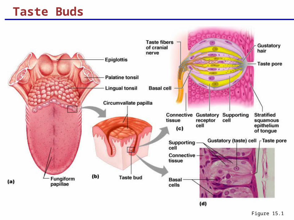
Taste Buds
Most of the 10,000 or so taste buds are found on the tongue
Taste buds are found in papillae of the tongue mucosa
Papillae come in three types: filiform, fungiform, and circumvallate
Fungiform and circumvallate papillae contain taste buds
Taste Buds
Figure 15.1
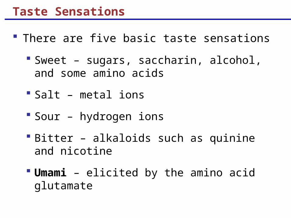
Taste Sensations
There are five basic taste sensations
Sweet – sugars, saccharin, alcohol, and some amino acids
Salt – metal ions
Sour – hydrogen ions
Bitter – alkaloids such as quinine and nicotine
Umami – elicited by the amino acid glutamate
Physiology of Taste
In order to be tasted, a chemical:
Must be dissolved in saliva
Must contact gustatory hairs
Binding of the food chemical:
Depolarizes the taste cell membrane, releasing neurotransmitter
Initiates a generator potential that elicits an action potential
Taste Transduction
The stimulus energy of taste is converted into a nerve impulse by:
Na+ influx in salty tastes
H+ in sour tastes (by directly entering the cell, by opening cation channels, or by blockade of K+ channels)
Gustducin in sweet and bitter tastes
Gustatory Pathway
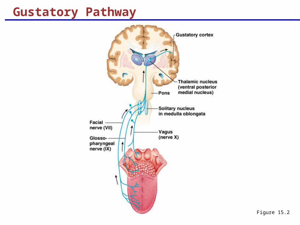
Cranial Nerves VII and IX carry impulses from taste buds to the solitary nucleus of the medulla
These impulses then travel to the thalamus, and from there fibers branch to the:
Gustatory cortex (taste)
Hypothalamus and limbic system (appreciation of taste)
Gustatory Pathway
Figure 15.2
Influence of Other Sensations on Taste
Taste is 80% smell
Thermoreceptors, mechanoreceptors, nociceptors also influence tastes
Temperature and texture enhance or detract from taste
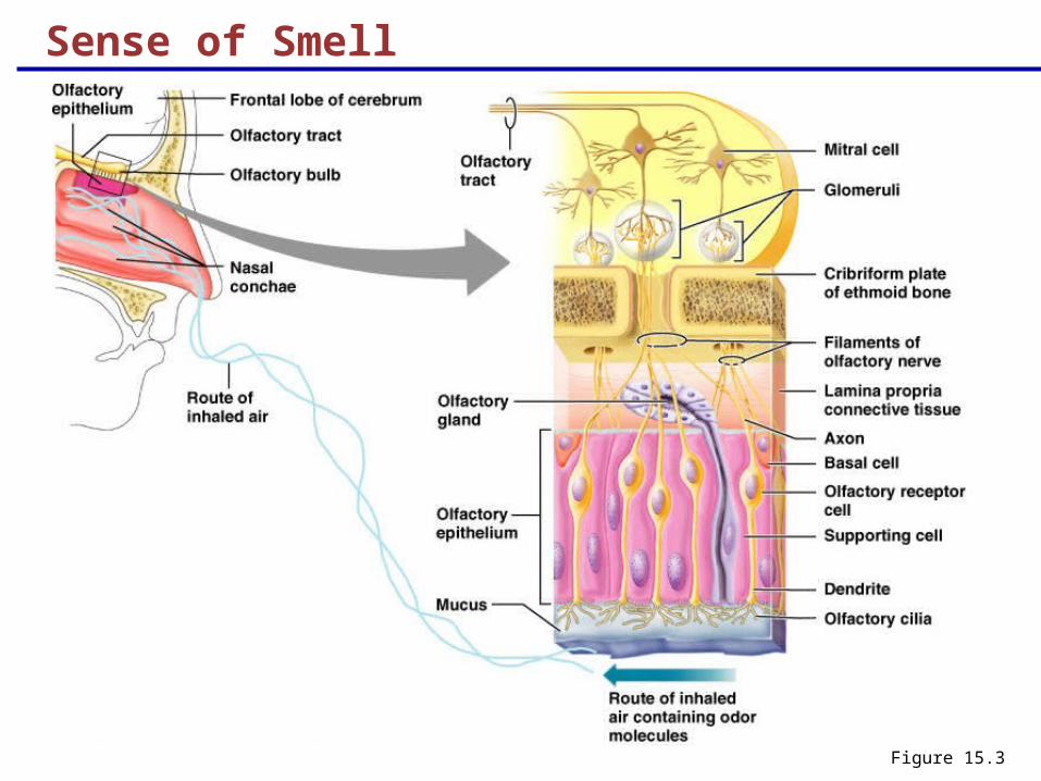
Sense of Smell The organ of smell is the olfactory epithelium,
which covers the superior nasal concha
Olfactory receptor cells are bipolar neurons with radiating olfactory cilia
Olfactory receptors are surrounded and cushioned by supporting cells
Basal cells lie at the base of the epithelium
Sense of Smell
Figure 15.3
Physiology of Smell
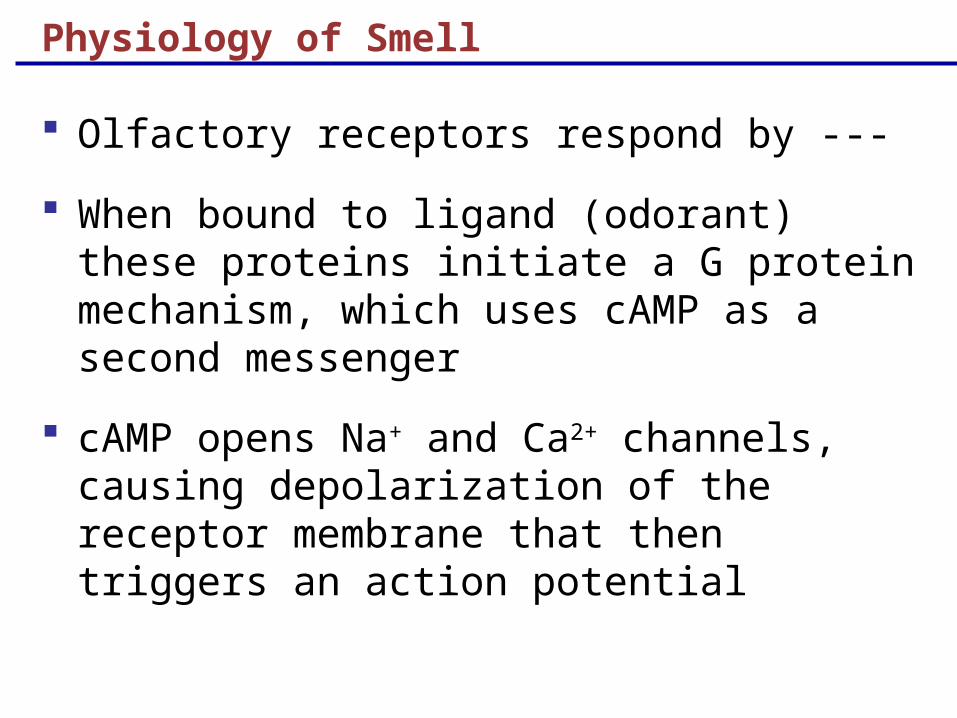
Olfactory receptors respond by ---
When bound to ligand (odorant) these proteins initiate a G protein mechanism, which uses cAMP as a second messenger
cAMP opens Na+ and Ca2+ channels, causing depolarization of the receptor membrane that then triggers an action potential
Olfactory Transduction Process
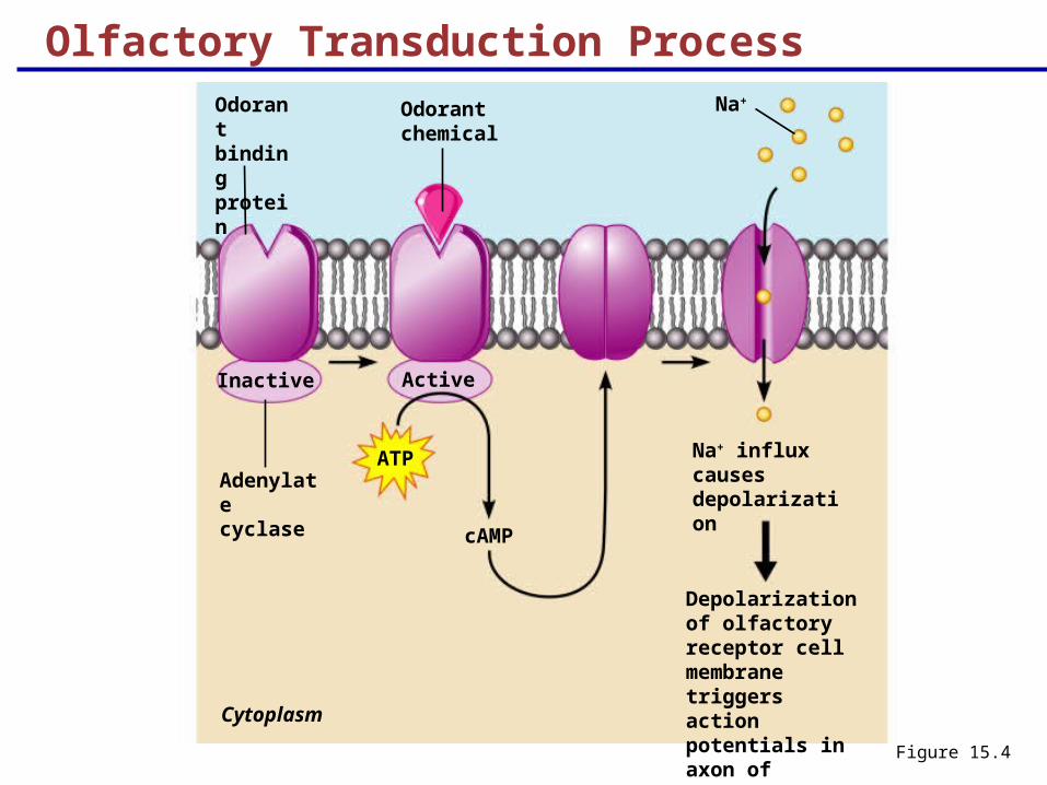
Figure 15.4
Odorant binding protein
Odorant chemical
Na+
Cytoplasm
Inactive Active
Na+ influx causes depolarization
Adenylate cyclase
ATP
cAMP
Depolarization of olfactory receptor cell membrane triggers action potentials in axon of receptor
Olfactory Pathway
Olfactory receptor cells synapse with mitral cells
Glomerular mitral cells “process” odor signals
Mitral cells send impulses to:
The olfactory cortex
The hypothalamus, amygdala, and limbic system

Structure of the Eyeball
Figure 15.8a
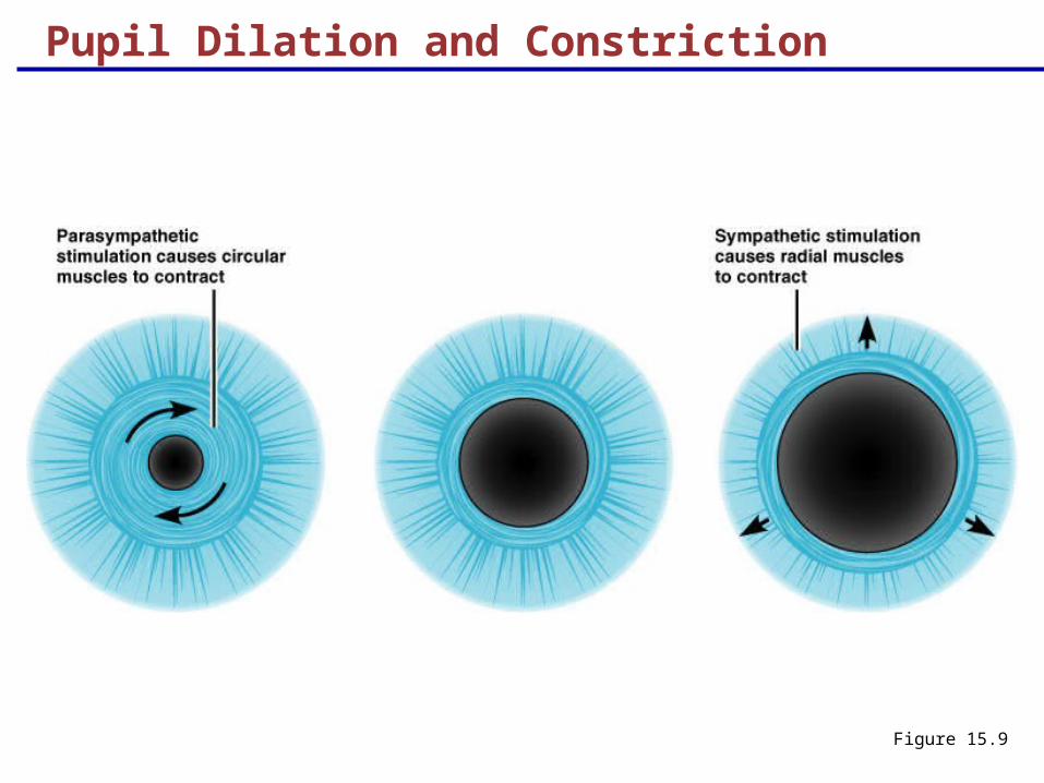
Pupil Dilation and Constriction
Figure 15.9
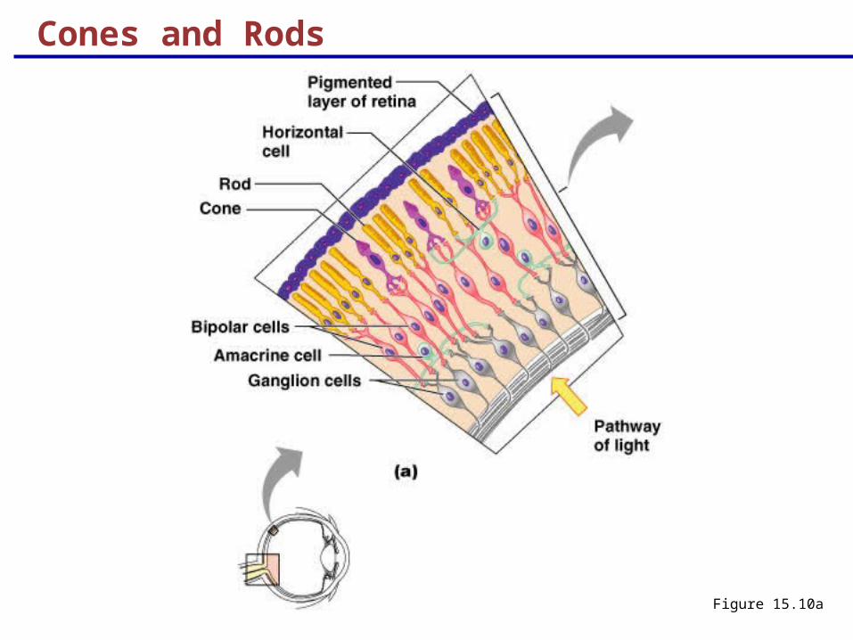
Sensory Tunic: Retina
A delicate two-layered membrane
Pigmented layer – the outer layer that absorbs light and prevents its scattering
Neural layer, which contains:
Photoreceptors that transduce light energy
Bipolar cells and ganglion cells
Amacrine and horizontal cells
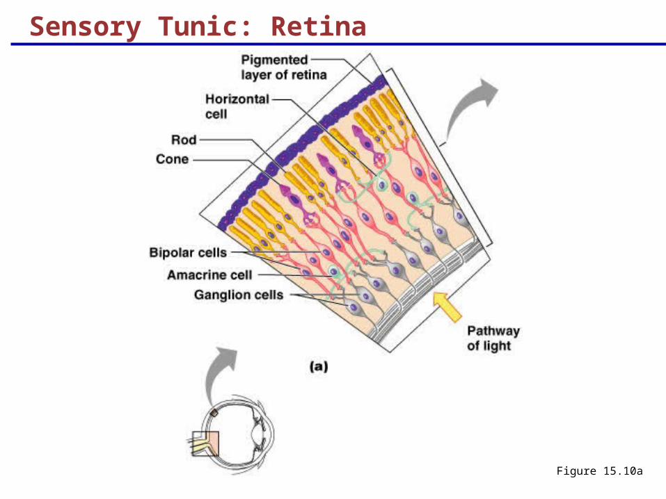
Sensory Tunic: Retina
Figure 15.10a
retina
optic nerve
sclera
pupil
lens
cornea
iris
vitreous humor
RPE
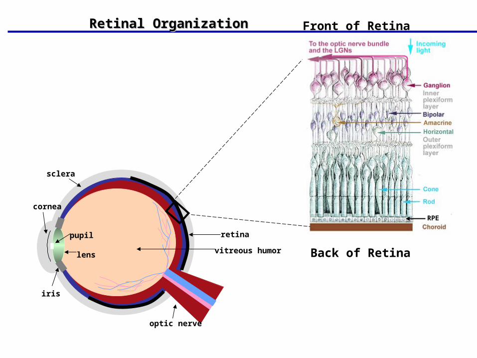
Front of Retina
Back of Retina
Retinal OrganizationRetinal Organization
The Retina: Ganglion Cells and the Optic Disc
Ganglion cell axons:
Run along the inner surface of the retina
Leave the eye as the optic nerve
The optic disc:
Is the site where the optic nerve leaves the eye
Lacks photoreceptors (the blind spot)
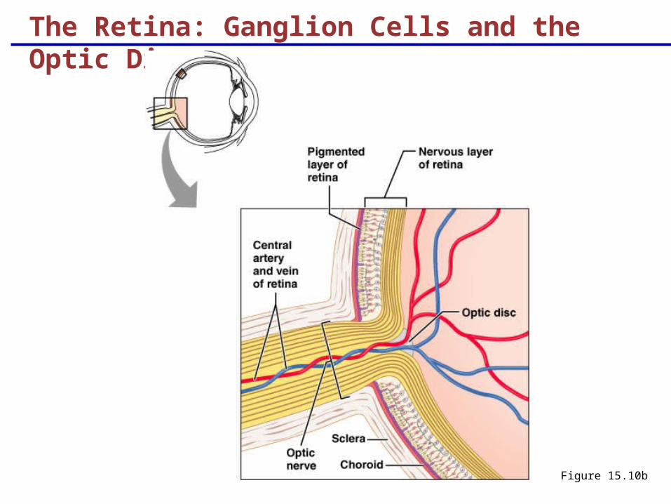
The Retina: Ganglion Cells and the Optic Disc
Figure 15.10b
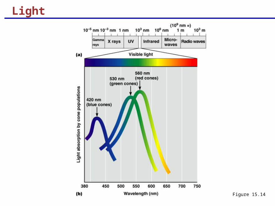
Light
Figure 15.14
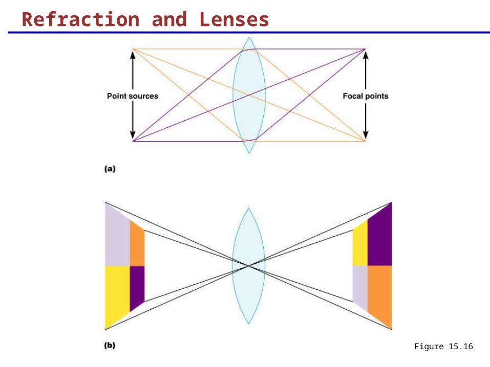
Refraction and Lenses
When light passes from one transparent medium to another its speed changes and it refracts (bends)
Light passing through a convex lens (as in the eye) is bent so that the rays converge to a focal point
When a convex lens forms an image, the image is upside down and reversed right to left
Refraction and Lenses
Figure 15.16
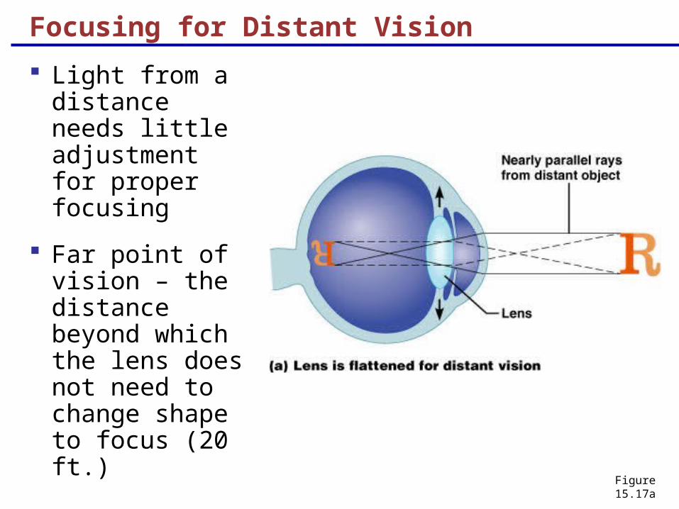
Focusing for Distant Vision
Light from a distance needs little adjustment for proper focusing
Far point of vision – the distance beyond which the lens does not need to change shape to focus (20 ft.)
Figure 15.17a
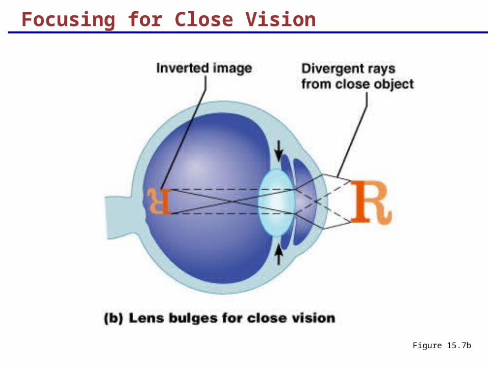
Focusing for Close Vision
Close vision requires:
Accommodation – changing the lens shape by ciliary muscles to increase refractory power
Constriction – the pupillary reflex constricts the pupils to prevent divergent light rays from entering the eye
Convergence – medial rotation of the eyeballs toward the object being viewed
Focusing for Close Vision
Figure 15.7b
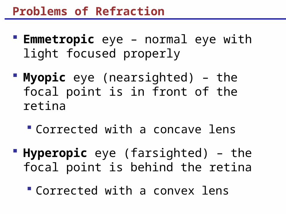
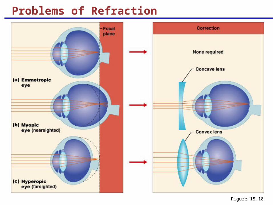
Problems of Refraction
Emmetropic eye – normal eye with light focused properly
Myopic eye (nearsighted) – the focal point is in front of the retina
Corrected with a concave lens
Hyperopic eye (farsighted) – the focal point is behind the retina
Corrected with a convex lens
Problems of Refraction
Figure 15.18
Cones and Rods
Figure 15.10a
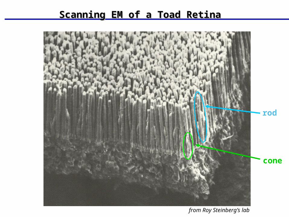
from Roy Steinberg’s lab
rod
cone
Scanning EM of a Toad RetinaScanning EM of a Toad Retina
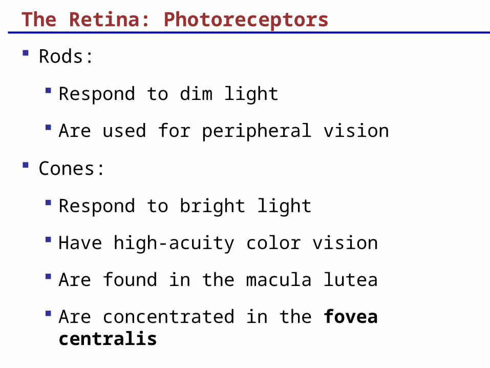
The Retina: Photoreceptors
Rods:
Respond to dim light
Are used for peripheral vision
Cones:
Respond to bright light
Have high-acuity color vision
Are found in the macula lutea
Are concentrated in the fovea centralis
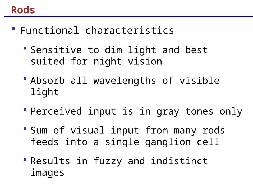
Rods
Functional characteristics
Sensitive to dim light and best suited for night vision
Absorb all wavelengths of visible light
Perceived input is in gray tones only
Sum of visual input from many rods feeds into a single ganglion cell
Results in fuzzy and indistinct images

Cones
Functional characteristics
Need bright light for activation (have low sensitivity)
Have pigments that furnish a vividly colored view
Each cone synapses with a single ganglion cell
Vision is detailed and has high resolution
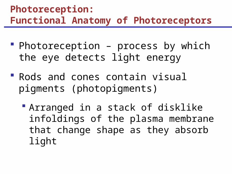
Photoreception – process by which the eye detects light energy
Rods and cones contain visual pigments (photopigments)
Arranged in a stack of disklike infoldings of the plasma membrane that change shape as they absorb light
Photoreception: Functional Anatomy of Photoreceptors
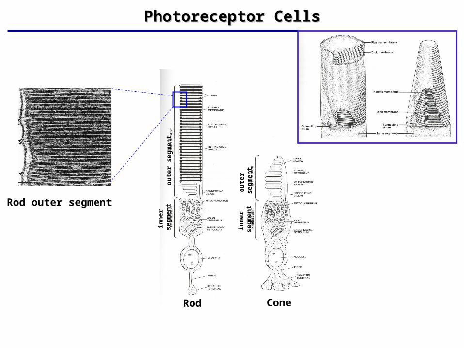
Photoreceptor CellsPhotoreceptor Cells
Rod outer segment
Rod Cone
ou
ter
seg
men
tin
ner
se
gm
ent
inn
er
seg
men
to
ute
r se
gm
ent
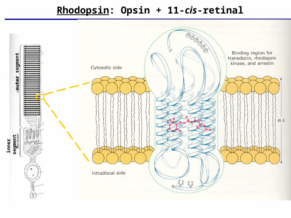
Rhodopsin: Opsin + 11-cis-retinalo
ute
r s
eg
me
nt
inn
er
se
gm
en
t
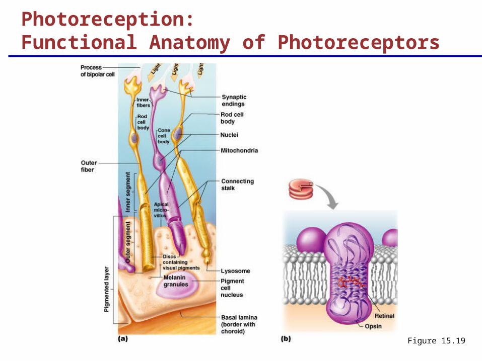
Figure 15.19
Photoreception: Functional Anatomy of Photoreceptors
Chemistry of Visual Pigments
Retinal is a light-absorbing molecule
Combines with opsins to form visual pigments
Similar to and is synthesized from vitamin A
Two isomers: 11-cis and all-trans
Isomerization of retinal initiates electrical impulses in the optic nerve
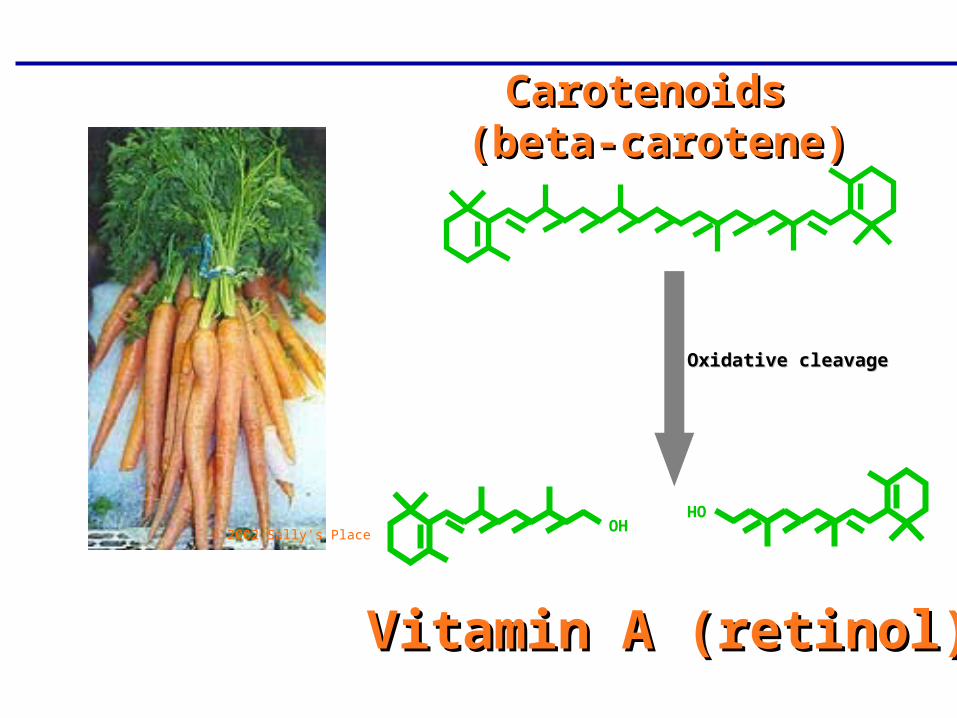
Carotenoids Carotenoids (beta-carotene)(beta-carotene)
© 2002 Sally's Place
Oxidative cleavageOxidative cleavage
Vitamin A (retinol)Vitamin A (retinol)
OH
OH
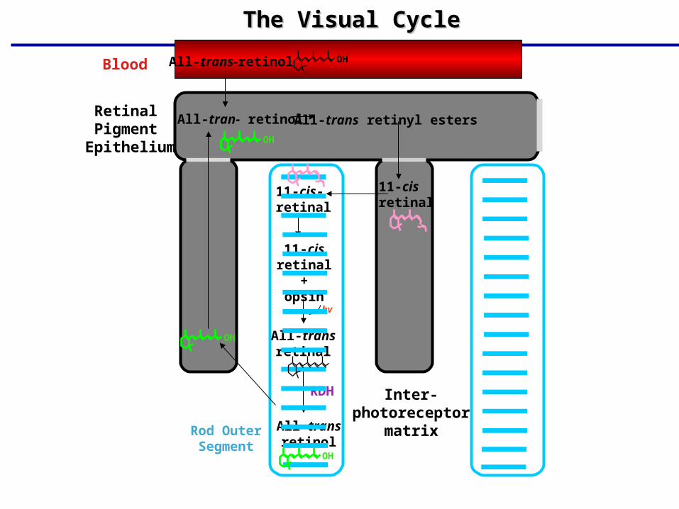
The Visual CycleThe Visual Cycle
Blood
Retinal Pigment
Epithelium
All-tran- retinol
All-trans-retinol
All-trans retinyl esters
11-cisretinal
11-cis-retinal
11-cisretinal
+opsin
All-transretinal
All-transretinol
hv
RDH Inter-photoreceptor
matrixRod OuterSegment
OH
OH
OH
OH
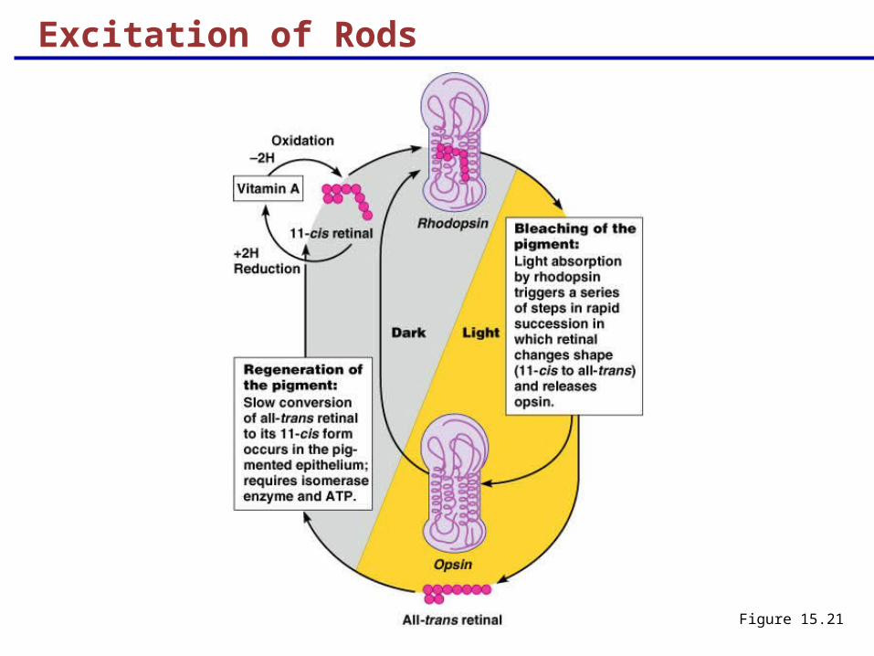
Excitation of Rods
The visual pigment of rods is rhodopsin (opsin + 11-cis retinal)
Light phase
Rhodopsin breaks down into all-trans retinal + opsin (bleaching of the pigment)
Dark phase
All-trans retinal converts to 11-cis form
11-cis retinal is also formed from vitamin A
11-cis retinal + opsin regenerate rhodopsin
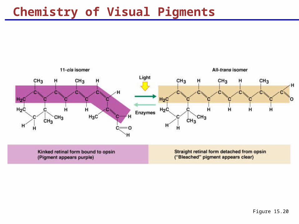
Chemistry of Visual Pigments
Figure 15.20
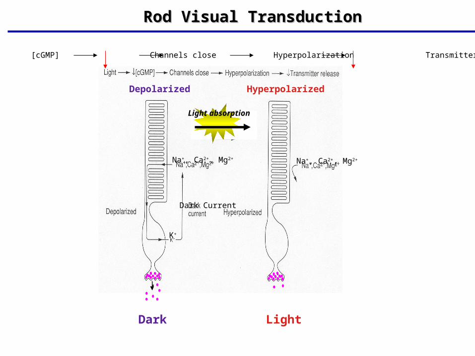
Rod Visual TransductionRod Visual Transduction
Light [cGMP] Channels close Hyperpolarization Transmitter release
Depolarized Hyperpolarized
Na+, Ca2+, Mg2+ Na+, Ca2+, Mg2+
K+
Dark Current
Light absorption
Dark Light
Excitation of Rods
Figure 15.21

Phototransduction
Figure 15.22
Phototransduction
Light energy splits rhodopsin into all-trans retinal, releasing activated opsin
The freed opsin activates the G protein transducin
Transducin catalyzes activation of phosphodiesterase (PDE)
PDE hydrolyzes cGMP to GMP and releases it from sodium channels
Without bound cGMP, sodium channels close, the membrane hyperpolarizes, and neurotransmitter cannot be released
Excitation of Cones
Visual pigments in cones are similar to rods (retinal + opsins)
There are three types of cones: blue, green, and red
Intermediate colors are perceived by activation of more than one type of cone
Method of excitation is similar to rods
Adaptation
Adaptation to bright light (going from dark to light) involves:
Dramatic decreases in retinal sensitivity – rod function is lost
Switching from the rod to the cone system – visual acuity is gained
Adaptation to dark is the reverse
Cones stop functioning in low light
Rhodopsin accumulates in the dark and retinal sensitivity is restored
Visual Pathways
Axons of retinal ganglion cells form the optic nerve
Medial fibers of the optic nerve decussate at the optic chiasm
Most fibers of the optic tracts continue to the lateral geniculate body of the thalamus
Other optic tract fibers end in superior colliculi (initiating visual reflexes) and pretectal nuclei (involved with pupillary reflexes)
Optic radiations travel from the thalamus to the visual cortex
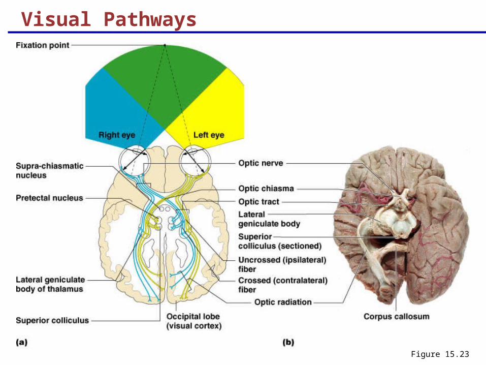
Visual Pathways
Figure 15.23
Visual Pathways
Some nerve fibers send tracts to the midbrain ending in the superior colliculi
A small subset of visual fibers contain melanopsin (circadian pigment) which:
Mediates papillary light reflexes
Sets daily biorhythms
Depth Perception
Achieved by both eyes viewing the same image from slightly different angles
Three-dimensional vision results from cortical fusion of the slightly different images
If only one eye is used, depth perception is lost and the observer must rely on learned clues to determine depth
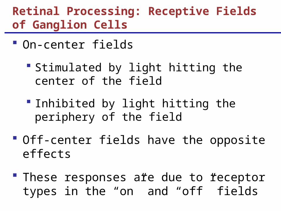
On-center fields
Stimulated by light hitting the center of the field
Inhibited by light hitting the periphery of the field
Off-center fields have the opposite effects
These responses are due to receptor types in the “on” and “off” fields
Retinal Processing: Receptive Fields of Ganglion Cells
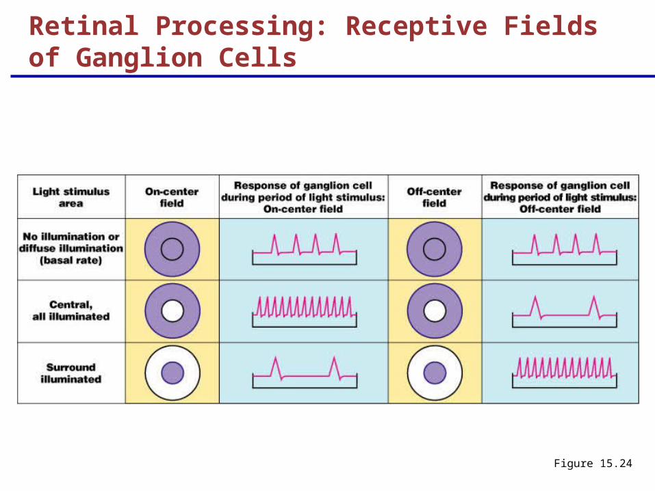
Figure 15.24
Retinal Processing: Receptive Fields of Ganglion Cells
Thalamic Processing
The lateral geniculate nuclei of the thalamus:
Relay information on movement
Segregate the retinal axons in preparation for depth perception
Emphasize visual inputs from regions of high cone density
Sharpen the contrast information received by the retina

Cortical Processing
Striate cortex processes
Basic dark/bright and contrast information
Prestriate cortices (association areas) processes
Form, color, and movement
Visual information then proceeds anteriorly to the:
Temporal lobe – processes identification of objects
Parietal cortex and postcentral gyrus – processes spatial location
The Ear: Hearing and Balance
The three parts of the ear are the inner, outer, and middle ear
The outer and middle ear are involved with hearing
The inner ear functions in both hearing and equilibrium
Receptors for hearing and balance:
Respond to separate stimuli
Are activated independently