Embed Size (px)
Citation preview
www.afm-journal.de
FULL P
APER
© 2014 WILEY-VCH Verlag GmbH & Co. KGaA, Weinheim 4497
www.MaterialsViews.com
wileyonlinelibrary.com
1. Introduction
Boron nitride nanotubes (BNNTs), analo-gous in geometry with carbon nanotubes (CNTs), have exceptional mechanical, thermal, and optical properties. [ 1–3 ] These properties make BNNTs promising mate-rials for many applications, such as polymeric composites [ 4 ] and biological applications. [ 5 ] For example, BNNT-fi lled polymer composites are expected to have high mechanical strength and high thermal conductivity similar to those fi lled with CNTs, but in contrast are much lighter in color (usually white due to light scattering) and electrically insu-lating. [ 1–4 ] These latter characteristics are attributed to the large bandgap (∼6 eV) of BNNTs due to the less delocalized and polarized B–N bond network. BNNTs are also known to be very resistant to thermal and wet oxidations as well as many other chemical modifi cations.
In stark contrast, oxidation of CNTs has been proven very convenient and funda-
mentally important to modify the nanotube structure and mor-phology via controlled corrosive effects. [ 6–11 ] For example, in the early explorations of the chemical properties of multi-walled CNTs (MWCNTs), it was found that air oxidation could lead to end-cap opening of the tubes accompanied by the nanotube tip sharpening and the tube wall thinning. [ 6,7 ] Sonicating single-walled CNTs (SWCNTs) or MWCNTs in strong oxidative acids, such as the mixture of sulfuric and nitric acids, led to nanotube shortening. [ 8–11 ] Recently, oxidation-related treatments have been employed to successfully unzip MWCNTs into graphene nanoribbons (GNRs). [ 12–14 ] For instance, Tour and co-workers extensively explored MWCNT unzipping using a potassium permanganate–sulfuric acid system. [ 12 ] Dai and co-workers son-icated partially air-oxidized MWCNTs in a polymer solution to obtain GNRs. [ 14 ]
The equivalent corrosion and unzipping of BNNTs using chemical methods, however, has never been reported partially due to their strong resistance to oxidation and various chemical treatments. Compared to CNTs that usually start to decompose in air at 400–500 °C, BNNTs usually remain stable to nearly 800 °C. [ 15 ] Thermal oxidation of BNNTs could only be carried out above this temperature when signifi cant damage to the nanotubes might start to occur. [ 16a ] Wet oxidation of BNNTs
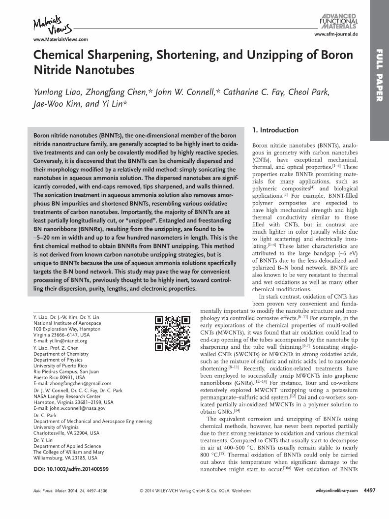
Chemical Sharpening, Shortening, and Unzipping of Boron Nitride Nanotubes
Yunlong Liao , Zhongfang Chen , * John W. Connell , * Catharine C. Fay , Cheol Park , Jae-Woo Kim , and Yi Lin *
Boron nitride nanotubes (BNNTs), the one-dimensional member of the boron nitride nanostructure family, are generally accepted to be highly inert to oxida-tive treatments and can only be covalently modifi ed by highly reactive species. Conversely, it is discovered that the BNNTs can be chemically dispersed and their morphology modifi ed by a relatively mild method: simply sonicating the nanotubes in aqueous ammonia solution. The dispersed nanotubes are signif-icantly corroded, with end-caps removed, tips sharpened, and walls thinned. The sonication treatment in aqueous ammonia solution also removes amor-phous BN impurities and shortened BNNTs, resembling various oxidative treatments of carbon nanotubes. Importantly, the majority of BNNTs are at least partially longitudinally cut, or “unzipped”. Entangled and freestanding BN nanoribbons (BNNRs), resulting from the unzipping, are found to be ∼5–20 nm in width and up to a few hundred nanometers in length. This is the fi rst chemical method to obtain BNNRs from BNNT unzipping. This method is not derived from known carbon nanotube unzipping strategies, but is unique to BNNTs because the use of aqueous ammonia solutions specifi cally targets the B-N bond network. This study may pave the way for convenient processing of BNNTs, previously thought to be highly inert, toward control-ling their dispersion, purity, lengths, and electronic properties.
DOI: 10.1002/adfm.201400599
Y. Liao, Dr. J.-W. Kim, Dr. Y. Lin National Institute of Aerospace 100 Exploration Way , HamptonVirginia 23666–6147 , USA E-mail: [email protected] Y. Liao, Prof. Z. Chen Department of ChemistryDepartment of PhysicsUniversity of Puerto Rico Rio Piedras Campus, San Juan Puerto Rico 00931 , USA E-mail: [email protected] Dr. J. W. Connell, Dr. C. C. Fay, Dr. C. Park NASA Langley Research Center Hampton, Virginia 23681–2199 , USA E-mail: [email protected] Dr. C. Park Department of Mechanical and Aerospace Engineering University of Virginia Charlottesville , VA 22904 , USA Dr. Y. Lin Department of Applied Science The College of William and Mary Williamsburg , VA 23185 , USA
Adv. Funct. Mater. 2014, 24, 4497–4506
FULL
PAPER
4498
www.afm-journal.dewww.MaterialsViews.com
wileyonlinelibrary.com © 2014 WILEY-VCH Verlag GmbH & Co. KGaA, Weinheim
under relatively harsh conditions were also reported, [ 16b ] in which hydrogen peroxide was used to react with the nanotubes at 120 °C for 24 h in an autoclave. Though BNNTs were found chemically active toward various Lewis bases, such as various amines and phosphines, [ 17–19 ] the attachments of these func-tional groups have been considered mostly ionic in nature and seldom impart changes to the BNNT atomic structure. Reports are also available detailing the covalent functionaliza-tion of BNNTs by targeting the residual tube surface amino groups produced under nitrogen-rich conditions. [ 20,21 ] It was suggested that such functionalization was suffi cient to modify the bandgap of BNNTs. [ 22 ] Direct covalent modifi cation of B-N bond network was yet to be reported on BNNTs, but has very recently been achieved on boron nitride nanosheets (BNNSs), the planar relative of the nanotubes. [ 23,24 ] Very reactive species such as nitrenes and oxygen radicals were used therein.
Only a handful of reports are currently available on cutting BNNTs in either circumferential (i.e., shortening) or longi-tudinal (i.e., unzipping) directions. For example, Zhi and co-workers pre-oxidized BNNTs at over 900 °C and sonicated the partially damaged nanotubes in solvents to obtain shortened tubes. [ 16a ] Yap and co-workers were able to shorten BNNTs with noncovalent functionalities simply by sonicating the soluble nanotubes in aqueous solution under ambient conditions. [ 25 ] The shortening was attributed to the high acoustic energy cou-pled with the individual dispersion of the nanotubes. Longitu-dinal unzipping of BNNTs yields boron nitride nanoribbons (BNNRs). The reported BNNT unzipping strategies include plasma etching [ 26 ] and the expansion of intercalated potas-sium. [ 27 ] These physical methods were fi rst demonstrated with the unzipping of CNTs [ 28,29 ] and subsequently applied to BNNTs. However, the known chemical unzipping methods for CNTs often involve oxidation, [ 12–14 ] making them not directly applicable to BNNTs. To date, a chemical method that is spe-cifi cally designed to achieve BNNT unzipping has yet to be reported.
In this article, it was demonstrated that the aqueous ammonia solution is an effective reagent to functionalize and disperse BNNTs with the assistance of low power sonication under ambient conditions. It is not suprising that ammonia, as a small Lewis base molecule, can be used for such purpose since similar chemistry is well known. [ 17–19 ] However, unex-pectedly but more importantly, the nanotube structures were signifi cantly corroded. The observed corrosive effects included nanotube end-cap removal, tip sharpening, sidewall thinning, length shortening, and also partial or even full longitudinal unzipping. While BNNTs are inert to strong oxidative treatment conditions, their structural vulnerability to aqueous ammonia solutions opens up a pathway which may allow their conven-ient processing toward a variety of applications.
2. Results and Discussion
2.1. Pristine BNNTs
To date, techniques to grow BNNTs can be divided into two broad categories. [ 1–3 ] The fi rst category includes high tempera-ture synthesis techniques, which involve vaporizing targets
made of elemental boron or boron nitride, which reacts with nitrogen and condenses into BNNT solids. This method pro-duces BNNTs with few defects and small numbers of walls but is limited to small batches (milligrams). [ 30,31 ] The second category includes various low temperature synthesis methods, where the operating temperatures are typically between 600 and 1700 °C, well below the vaporization temperature of pure boron (over 4000 °C, depending on the applied pressure). These low temperature synthesis methods, including chemical vapor deposition (CVD), [ 32,33 ] CNT substitution reaction, [ 34 ] or ball-milling, [ 35 ] are much more scalable and can routinely produce hundreds of milligrams to grams of BNNTs. Typical diameters of BNNTs from CVD and ball-milling are about an order of magnitude greater than those grown by high tempera-ture synthesis method (∼50 nm vs. ∼5 nm). Furthermore, these nanotubes often have comparably somewhat more defective morphologies due to the lower synthesis temperature.
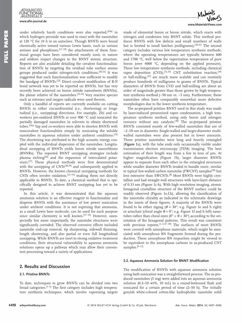
The as-prepared pristine BNNT used in this study was made by laser-induced pressurized vapor condensation, a high tem-perature synthesis method, using only boron and nitrogen resource without any catalysts. [ 30 ] The as-prepared pristine BNNTs consisted mostly of few-walled nanotubes that are of ∼2–10 nm in diameter. Single-walled and larger-diameter multi-walled nanotubes were also present but in lower amounts. These pristine nanotubes were long and heavily entangled ( Figure 1 a), with the tube ends only occasionally visible under transmission electron microscopy (TEM) imaging. The best estimation of their length was from a few to tens of µm. At higher magnifi cation (Figure 1 b), larger diameter BNNTs appear to separate from each other in the entangled structures while smaller diameter BNNTs tended to bundle more, similar to typical few-walled carbon nanotube (FWCNT) samples [ 36 ] but less extensive than SWCNTs. [ 8 ] Most BNNTs were highly crys-talline and had straight wall structures with layer-layer distance of 0.33 nm (Figure 1 c-h). With high resolution imaging, atomic hexagonal crystalline structure of the BNNT surface could be readily observed (Figure 1 e,f,h), allowing the classifi cation of the nanotube chirality as indicated in the schematic drawings in the insets of these fi gures. A majority of the BNNTs were found to be either zigzag ( θ = 30°; e.g. Figure 1 e and h right) or armchair (chiral angle θ = 0°; e.g. Figure 1 f and h left) nano-tubes rather than chiral ones (0° < θ < 30°) according to the ori-entation of the hexagonal patterns. This result was consistent with previous reports. [ 1–3,37–39 ] The surfaces of most BNNTs were covered with amorphous materials, which might be asso-ciated with amorphous BN fragments formed during the pro-duction. These amorphous BN impurities might be viewed to be equivalent to the amorphous carbons in as-produced CNT samples. [ 9–11 ]
2.2. Aqueous Ammonia Solution for BNNT Modifi cation
The modifi cation of BNNTs with aqueous ammonia solution using bath sonication was a straightforward process. The as-pro-duced nanotubes (5 mg) were added into an aqueous ammonia solution (0.3–10 wt%, 10 mL) in a round-bottomed fl ask and sonicated for a certain period of time (2–50 h). The initially greyish white-colored cotton-like hydrophobic nanotube solid
Adv. Funct. Mater. 2014, 24, 4497–4506
FULL P
APER
4499
www.afm-journal.dewww.MaterialsViews.com
wileyonlinelibrary.com© 2014 WILEY-VCH Verlag GmbH & Co. KGaA, Weinheim
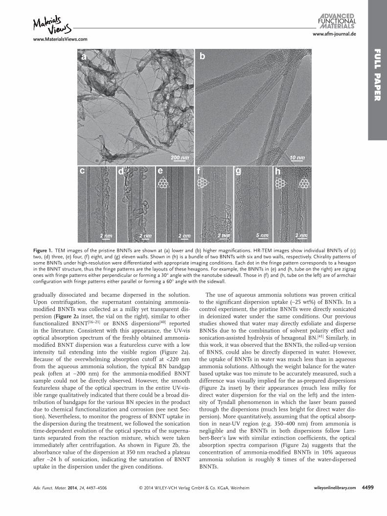
gradually dissociated and became dispersed in the solution. Upon centrifugation, the supernatant containing ammonia-modifi ed BNNTs was collected as a milky yet transparent dis-persion ( Figure 2 a inset, the vial on the right), similar to other functionalized BNNT [ 16–21 ] or BNNS dispersions [ 40 ] reported in the literature. Consistent with this appearance, the UV-vis optical absorption spectrum of the freshly obtained ammonia-modifi ed BNNT dispersion was a featureless curve with a low intensity tail extending into the visible region (Figure 2 a). Because of the overwhelming absorption cutoff at <220 nm from the aqueous ammonia solution, the typical BN bandgap peak (often at ∼200 nm) for the ammonia-modifi ed BNNT sample could not be directly observed. However, the smooth featureless shape of the optical spectrum in the entire UV-vis-ible range qualitatively indicated that there could be a broad dis-tribution of bandgaps for the various BN species in the product due to chemical functionalization and corrosion (see next Sec-tion). Nevertheless, to monitor the progress of BNNT uptake in the dispersion during the treatment, we followed the sonication time-dependent evolution of the optical spectra of the superna-tants separated from the reaction mixture, which were taken immediately after centrifugation. As shown in Figure 2 b, the absorbance value of the dispersion at 350 nm reached a plateau after ∼24 h of sonication, indicating the saturation of BNNT uptake in the dispersion under the given conditions.
The use of aqueous ammonia solutions was proven critical to the signifi cant dispersion uptake (∼25 wt%) of BNNTs. In a control experiment, the pristine BNNTs were directly sonicated in deionized water under the same conditions. Our previous studies showed that water may directly exfoliate and disperse BNNSs due to the combination of solvent polarity effect and sonication-assisted hydrolysis of hexagonal BN. [ 41 ] Similarly, in this work, it was observed that the BNNTs, the rolled-up version of BNNS, could also be directly dispersed in water. However, the uptake of BNNTs in water was much less than in aqueous ammonia solutions. Although the weight balance for the water-based uptake was too minute to be accurately measured, such a difference was visually implied for the as-prepared dispersions (Figure 2 a inset) by their appearances (much less milky for direct water dispersion for the vial on the left) and the inten-sity of Tyndall phenomenon in which the laser beam passed through the dispersions (much less bright for direct water dis-persion). More quantitatively, assuming that the optical absorp-tion in near-UV region (e.g. 350–400 nm) from ammonia is negligible and the BNNTs in both dispersions follow Lam-bert-Beer's law with similar extinction coeffi cients, the optical absorption spectra comparison (Figure 2 a) suggests that the concentration of ammonia-modifi ed BNNTs in 10% aqueous ammonia solution is roughly 8 times of the water-dispersed BNNTs.
Adv. Funct. Mater. 2014, 24, 4497–4506
Figure 1. TEM images of the pristine BNNTs are shown at (a) lower and (b) higher magnifi cations. HR-TEM images show individual BNNTs of (c) two, (d) three, (e) four, (f) eight, and (g) eleven walls. Shown in (h) is a bundle of two BNNTs with six and two walls, respectively. Chirality patterns of some BNNTs under high-resolution were differentiated with appropriate imaging conditions. Each dot in the fringe pattern corresponds to a hexagon in the BNNT structure, thus the fringe patterns are the layouts of these hexagons. For example, the BNNTs in (e) and (h, tube on the right) are zigzag ones with fringe patterns either perpendicular or forming a 30° angle with the nanotube sidewall. Those in (f) and (h, tube on the left) are of armchair confi guration with fringe patterns either parallel or forming a 60° angle with the sidewall.
FULL
PAPER
4500
www.afm-journal.dewww.MaterialsViews.com
wileyonlinelibrary.com © 2014 WILEY-VCH Verlag GmbH & Co. KGaA, Weinheim
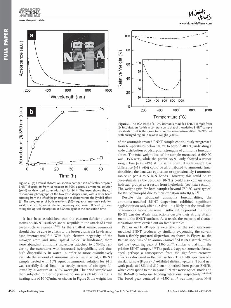
It has been established that the electron-defi cient boron atoms on BNNT surfaces are susceptible to the attack of Lewis bases such as amines. [ 17–19 ] As the smallest amine, ammonia should also be able to attach to the boron atoms via Lewis acid-base interactions. [ 42,43 ] With high electron negativity of the nitrogen atom and small spatial molecular hindrance, there were abundant ammonia molecules attached to BNNTs, ren-dering the nanotubes with increased hydrophilicity and thus high dispersibility in water. In order to more quantitatively evaluate the amount of ammonia molecules attached, a BNNT sample treated with 10% aqueous ammonia solution for 24 h was carefully dried fi rst in a steady stream of nitrogen fol-lowed by in vacuum at ∼60 °C overnight. The dried sample was then subjected to thermogravimetric analysis (TGA) in air at a heating rate of 10 °C/min. As shown in Figure 3 , the weight loss
of the ammonia-treated BNNT sample continuously progressed from temperatures below 100 °C to beyond 400 °C, indicating a wide distribution of adsorption strengths of ammonia function-alities. The total weight loss of the sample measured at 600 °C was ∼15.6 wt%, while the parent BNNT only showed a minor weight loss (∼3.8 wt%) at the same point. If such weight loss difference (∼12 wt%) could be all attributed to ammonia func-tionalities, the data was equivalent to approximately 1 ammonia molecule per 4 to 5 B–N bonds. However, this could be an overestimate as the resultant BNNTs could also contain some hydroxyl groups as a result from hydrolysis (see next section). The weight gain for both samples beyond 750 °C were typical for BN polymorphs due to their oxidation into B 2 O 3 . [ 15 ]
Despite the abundant ammonia functionalities, the ammonia-modifi ed BNNT dispersions exhibited signifi cant agglomeration only after 1–2 days. It is likely that the small size of ammonia molecules were insuffi cient to prevent the inter-BNNT van der Waals interactions despite their strong attach-ment to the BNNT surfaces. As a result, the majority of charac-terizations were carried out on fresh samples.
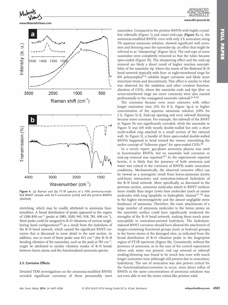
Raman and FT-IR spectra were taken on the solid ammonia-modifi ed BNNT products by similarly evaporating the solvent from a freshly prepared dispersion. As shown in Figure 4 a, the Raman spectrum of an ammonia-modifi ed BNNT sample exhib-ited the typical E 2g peak at 1360 cm −1 , similar to that from the pristine BNNT sample. [ 1–3 ] The peak did appear somewhat broad-ened, perhaps a consequence from the signifi cant corrosive effects as discussed in the next section. The FT-IR spectrum of a similar sample (Figure 4 b) exhibited distinct typical B-N bond net-work peaks at 1383 and 812 cm −1 inherited from parent BNNTs, which correspond to the in-plane B-N transverse optical mode and the B–N–B out-of-plane bending vibrations, respectively. [ 1–3,40,44 ] The broad peak centered at ∼3300 cm −1 was likely from N–H
Adv. Funct. Mater. 2014, 24, 4497–4506
Figure 3. The TGA trace of a 10% ammonia-modifi ed BNNT sample from 24 h sonication (solid) in comparison to that of the pristine BNNT sample (dashed). Inset is the same trace for the ammonia-modifi ed BNNTs but with enlarged region in relative weight (y-axis).
Figure 2. (a) Optical absorption spectra comparison of freshly prepared BNNT dispersion from sonication in 10% aqueous ammonia solution (solid) or deionized water (dashed) for 24 h. The inset shows the cor-responding photograph of the two fresh dispersions, with a laser beam entering from the left of the photograph to demonstrate the Tyndall effect; (b) The progresses of both reactions (10% aqueous ammonia solution: solid, open circle; water: dashed, open square) were followed by moni-toring the optical absorption at 350 nm against the sonication time.
FULL P
APER
4501
www.afm-journal.dewww.MaterialsViews.com
wileyonlinelibrary.com© 2014 WILEY-VCH Verlag GmbH & Co. KGaA, Weinheim
stretching, which may be readily attributed to ammonia func-tionalities. A broad distribution of peaks appeared in the region of 1200–830 cm −1 (peaks at 1085, 1020, 945, 918, 781, 690 cm −1 ). Most peaks could be assigned to B–O vibrations of various neigh-boring bond confi gurations [ 45 ] as a result from the hydrolysis of the B–N bond network, which caused the signifi cant BNNT cor-rosion that is discussed in more detail in the next section. In addition, one or more of these peaks near 811 cm −1 (the B–N–B bending vibration of the nanotube), such as the peak at 781 cm −1 , might be attributed to similar vibration modes of B–N bonds between boron atoms and the functionalized ammonia species.
2.3. Corrosive Effects
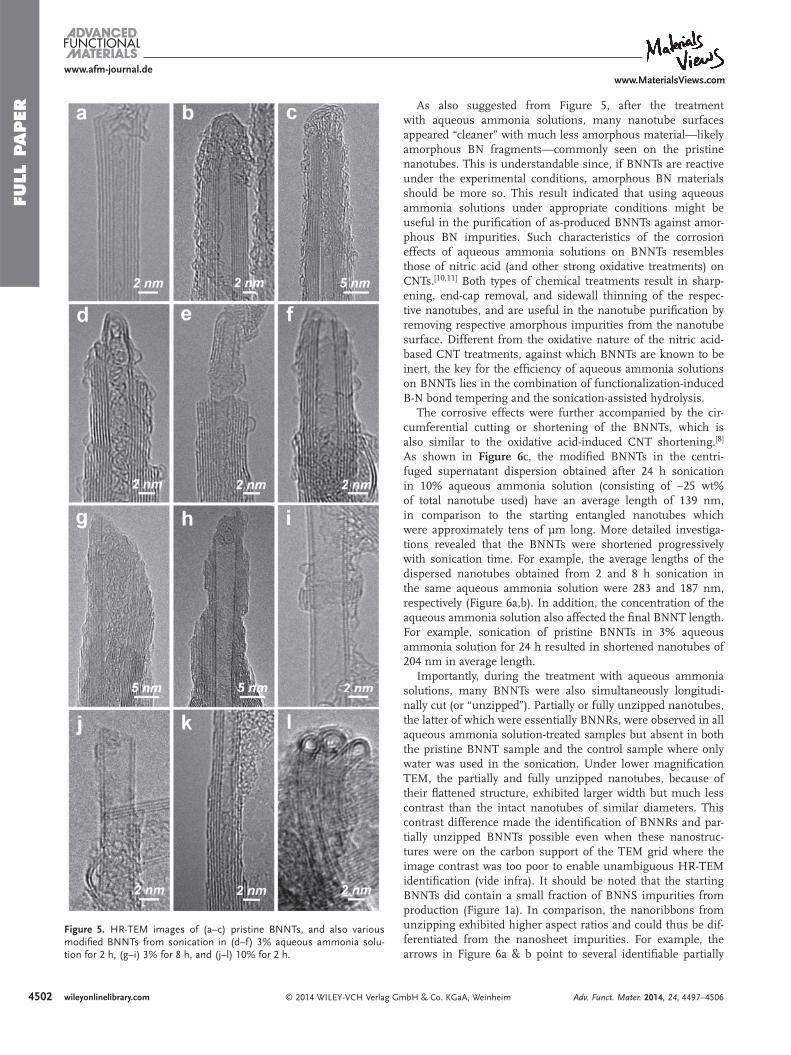
Detailed TEM investigations on the ammonia-modifi ed BNNTs revealed signifi cant corrosion of these presumably inert
nanotubes. Compared to the pristine BNNTs with highly crystal-line sidewalls (Figure 1 ) and intact end-caps ( Figure 5 a–c), the ammonia-modifi ed BNNTs, even with only 2 h sonication using 3% aqueous ammonia solution, showed signifi cant wall corro-sion and thinning near the nanotube tip, an effect that might be referred to as “sharpening” (Figure 5 d,e). The end-caps of some nanotubes were completely removed so that the tubes became open-ended (Figure 5 f). The sharpening effect and the end-cap removal are likely a direct result of higher reaction suscepti-bility of the nanotube tip, where the joints of the fl attened B–N bond network (typically with four- or eight-membered rings for BN polymorphs) [ 1,2 ] exhibits larger curvature and likely more structural strain and discontinuity. This effect is similar to what was observed for the oxidation and other covalent function-alization of CNTs, where the nanotube ends and tips (fi ve- or seven-membered rings are more common) were also reacted preferentially to the conjugated nanotube sidewall. [ 6–8,46 ]
The corrosion became even more extensive with either longer sonication time (3% for 8 h, Figure 5 g–i) or higher concentration of the aqueous ammonia solution (10% for 2 h, Figure 5 j–l). End-cap opening and even sidewall thinning became more common. For example, the sidewall of the BNNT in Figure 5 h was signifi cantly corroded, while the nanotube in Figure 5 i was left with mostly double-walled but only a short multi-walled ring attached to a small section of the external wall. In Figure 5 l, a bundle of three open-ended double-walled BNNTs happened to bend toward the viewer, resembling the earlier concept of “fullerene pipes” for open-ended CNTs. [ 8 ]
In a recent report, gas-phase ammonia plasma was used to functionalize BNNTs, but no nanotube wall corrosion or end-cap removal was reported. [ 47 ] In the experiments reported herein, it is likely that the presence of both ammonia and water was critical to the corrosion of BNNTs under sonication conditions. Mechanistically, the observed corrosive effect can be viewed as a synergistic result from boron-ammonia (Lewis acid-base) interactions and sonication-induced hydrolysis of the B–N bond network. More specifi cally, as discussed in the previous section, ammonia molecules attach to BNNT surfaces more readily than larger Lewis base molecules (such as amine molecules with long lipophilic or hydrophilic chains) [ 17–19 ] due to the higher electronegativity and the almost negligible steric hindrance of ammonia. Therefore, the ionic attachments of a large number of ammonia molecules to the boron atoms on the nanotube surface could have signifi cantly weakened the strengths of the B–N bond network, making them much more susceptible to sonication-assisted hydrolysis. The hydrolysis-induced BNNT corrosion should have allowed the attachment of oxygen-containing functional groups (such as hydroxyl groups) to the boron atoms at the damaged sites, as indicated from the broad distribution of B–O vibration peaks in the fi ngerprint region of FT-IR spectrum (Figure 4 b). Consistently, without the presence of ammonia, as in the case of the control experiment where only water was present, end cap removal or sidewall eroding/thinning was found to be much less even with much longer sonication time (although still present due to sonication/hydrolysis). The use of sonication was also proven critical for the functionalization/corrosion to occur, since direct refl ux of BNNTs in the same concentrations of ammonia solutions was not even able to wet the entire cotton-like pristine solid.
Adv. Funct. Mater. 2014, 24, 4497–4506
Figure 4. (a) Raman and (b) FT-IR spectra of a 10% ammonia-modi-fi ed BNNT sample with 24 h sonication (solid) and the pristine BNNTs (dashed).
FULL
PAPER
4502
www.afm-journal.dewww.MaterialsViews.com
wileyonlinelibrary.com © 2014 WILEY-VCH Verlag GmbH & Co. KGaA, Weinheim
As also suggested from Figure 5 , after the treatment with aqueous ammonia solutions, many nanotube surfaces appeared “cleaner” with much less amorphous material—likely amorphous BN fragments—commonly seen on the pristine nanotubes. This is understandable since, if BNNTs are reactive under the experimental conditions, amorphous BN materials should be more so. This result indicated that using aqueous ammonia solutions under appropriate conditions might be useful in the purifi cation of as-produced BNNTs against amor-phous BN impurities. Such characteristics of the corrosion effects of aqueous ammonia solutions on BNNTs resembles those of nitric acid (and other strong oxidative treatments) on CNTs. [ 10,11 ] Both types of chemical treatments result in sharp-ening, end-cap removal, and sidewall thinning of the respec-tive nanotubes, and are useful in the nanotube purifi cation by removing respective amorphous impurities from the nanotube surface. Different from the oxidative nature of the nitric acid-based CNT treatments, against which BNNTs are known to be inert, the key for the effi ciency of aqueous ammonia solutions on BNNTs lies in the combination of functionalization-induced B-N bond tempering and the sonication-assisted hydrolysis.
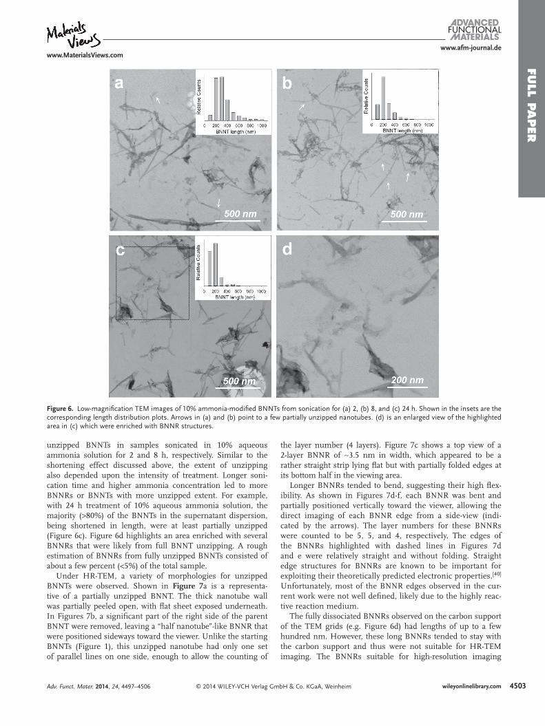
The corrosive effects were further accompanied by the cir-cumferential cutting or shortening of the BNNTs, which is also similar to the oxidative acid-induced CNT shortening. [ 8 ] As shown in Figure 6 c, the modifi ed BNNTs in the centri-fuged supernatant dispersion obtained after 24 h sonication in 10% aqueous ammonia solution (consisting of ∼25 wt% of total nanotube used) have an average length of 139 nm, in comparison to the starting entangled nanotubes which were approximately tens of µm long. More detailed investiga-tions revealed that the BNNTs were shortened progressively with sonication time. For example, the average lengths of the dispersed nanotubes obtained from 2 and 8 h sonication in the same aqueous ammonia solution were 283 and 187 nm, respectively (Figure 6 a,b). In addition, the concentration of the aqueous ammonia solution also affected the fi nal BNNT length. For example, sonication of pristine BNNTs in 3% aqueous ammonia solution for 24 h resulted in shortened nanotubes of 204 nm in average length.
Importantly, during the treatment with aqueous ammonia solutions, many BNNTs were also simultaneously longitudi-nally cut (or “unzipped”). Partially or fully unzipped nanotubes, the latter of which were essentially BNNRs, were observed in all aqueous ammonia solution-treated samples but absent in both the pristine BNNT sample and the control sample where only water was used in the sonication. Under lower magnifi cation TEM, the partially and fully unzipped nanotubes, because of their fl attened structure, exhibited larger width but much less contrast than the intact nanotubes of similar diameters. This contrast difference made the identifi cation of BNNRs and par-tially unzipped BNNTs possible even when these nanostruc-tures were on the carbon support of the TEM grid where the image contrast was too poor to enable unambiguous HR-TEM identifi cation (vide infra). It should be noted that the starting BNNTs did contain a small fraction of BNNS impurities from production (Figure 1 a). In comparison, the nanoribbons from unzipping exhibited higher aspect ratios and could thus be dif-ferentiated from the nanosheet impurities. For example, the arrows in Figure 6 a & b point to several identifi able partially
Adv. Funct. Mater. 2014, 24, 4497–4506
Figure 5. HR-TEM images of (a–c) pristine BNNTs, and also various modifi ed BNNTs from sonication in (d–f) 3% aqueous ammonia solu-tion for 2 h, (g–i) 3% for 8 h, and (j–l) 10% for 2 h.
FULL P
APER
4503
www.afm-journal.dewww.MaterialsViews.com
wileyonlinelibrary.com© 2014 WILEY-VCH Verlag GmbH & Co. KGaA, Weinheim
unzipped BNNTs in samples sonicated in 10% aqueous ammonia solution for 2 and 8 h, respectively. Similar to the shortening effect discussed above, the extent of unzipping also depended upon the intensity of treatment. Longer soni-cation time and higher ammonia concentration led to more BNNRs or BNNTs with more unzipped extent. For example, with 24 h treatment of 10% aqueous ammonia solution, the majority (>80%) of the BNNTs in the supernatant dispersion, being shortened in length, were at least partially unzipped (Figure 6 c). Figure 6 d highlights an area enriched with several BNNRs that were likely from full BNNT unzipping. A rough estimation of BNNRs from fully unzipped BNNTs consisted of about a few percent (<5%) of the total sample.
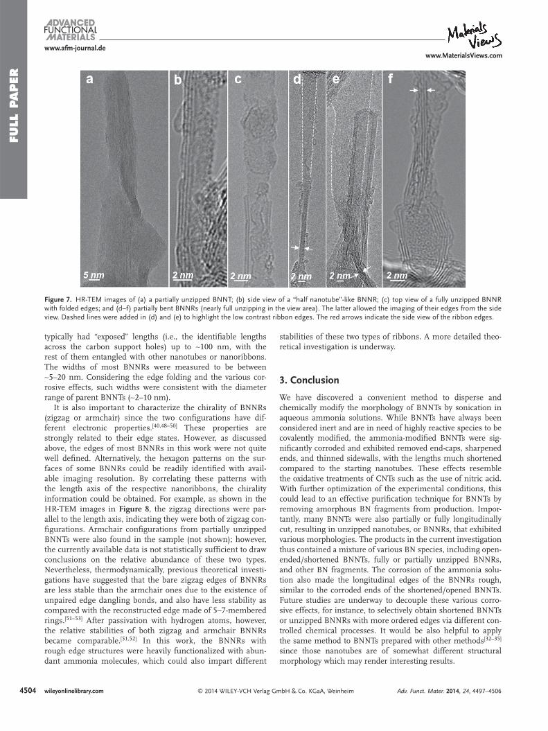
Under HR-TEM, a variety of morphologies for unzipped BNNTs were observed. Shown in Figure 7 a is a representa-tive of a partially unzipped BNNT. The thick nanotube wall was partially peeled open, with fl at sheet exposed underneath. In Figures 7 b, a signifi cant part of the right side of the parent BNNT were removed, leaving a “half nanotube”-like BNNR that were positioned sideways toward the viewer. Unlike the starting BNNTs (Figure 1 ), this unzipped nanotube had only one set of parallel lines on one side, enough to allow the counting of
the layer number (4 layers). Figure 7 c shows a top view of a 2-layer BNNR of ∼3.5 nm in width, which appeared to be a rather straight strip lying fl at but with partially folded edges at its bottom half in the viewing area.
Longer BNNRs tended to bend, suggesting their high fl ex-ibility. As shown in Figures 7 d-f, each BNNR was bent and partially positioned vertically toward the viewer, allowing the direct imaging of each BNNR edge from a side-view (indi-cated by the arrows). The layer numbers for these BNNRs were counted to be 5, 5, and 4, respectively. The edges of the BNNRs highlighted with dashed lines in Figures 7 d and e were relatively straight and without folding. Straight edge structures for BNNRs are known to be important for exploiting their theoretically predicted electronic properties. [ 40 ] Unfortunately, most of the BNNR edges observed in the cur-rent work were not well defi ned, likely due to the highly reac-tive reaction medium.
The fully dissociated BNNRs observed on the carbon support of the TEM grids (e.g. Figure 6 d) had lengths of up to a few hundred nm. However, these long BNNRs tended to stay with the carbon support and thus were not suitable for HR-TEM imaging. The BNNRs suitable for high-resolution imaging
Adv. Funct. Mater. 2014, 24, 4497–4506
Figure 6. Low-magnifi cation TEM images of 10% ammonia-modifi ed BNNTs from sonication for (a) 2, (b) 8, and (c) 24 h. Shown in the insets are the corresponding length distribution plots. Arrows in (a) and (b) point to a few partially unzipped nanotubes. (d) is an enlarged view of the highlighted area in (c) which were enriched with BNNR structures.
FULL
PAPER
4504
www.afm-journal.dewww.MaterialsViews.com
wileyonlinelibrary.com © 2014 WILEY-VCH Verlag GmbH & Co. KGaA, Weinheim Adv. Funct. Mater. 2014, 24, 4497–4506
typically had “exposed” lengths (i.e., the identifi able lengths across the carbon support holes) up to ∼100 nm, with the rest of them entangled with other nanotubes or nanoribbons. The widths of most BNNRs were measured to be between ∼5–20 nm. Considering the edge folding and the various cor-rosive effects, such widths were consistent with the diameter range of parent BNNTs (∼2–10 nm).
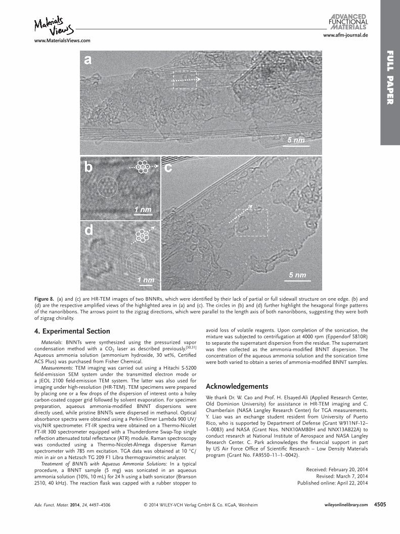
It is also important to characterize the chirality of BNNRs (zigzag or armchair) since the two confi gurations have dif-ferent electronic properties. [ 40,48–50 ] These properties are strongly related to their edge states. However, as discussed above, the edges of most BNNRs in this work were not quite well defi ned. Alternatively, the hexagon patterns on the sur-faces of some BNNRs could be readily identifi ed with avail-able imaging resolution. By correlating these patterns with the length axis of the respective nanoribbons, the chirality information could be obtained. For example, as shown in the HR-TEM images in Figure 8 , the zigzag directions were par-allel to the length axis, indicating they were both of zigzag con-fi gurations. Armchair confi gurations from partially unzipped BNNTs were also found in the sample (not shown); however, the currently available data is not statistically suffi cient to draw conclusions on the relative abundance of these two types. Nevertheless, thermodynamically, previous theoretical investi-gations have suggested that the bare zigzag edges of BNNRs are less stable than the armchair ones due to the existence of unpaired edge dangling bonds, and also have less stability as compared with the reconstructed edge made of 5–7-membered rings. [ 51–53 ] After passivation with hydrogen atoms, however, the relative stabilities of both zigzag and armchair BNNRs became comparable. [ 51,52 ] In this work, the BNNRs with rough edge structures were heavily functionalized with abun-dant ammonia molecules, which could also impart different
stabilities of these two types of ribbons. A more detailed theo-retical investigation is underway.
3. Conclusion
We have discovered a convenient method to disperse and chemically modify the morphology of BNNTs by sonication in aqueous ammonia solutions. While BNNTs have always been considered inert and are in need of highly reactive species to be covalently modifi ed, the ammonia-modifi ed BNNTs were sig-nifi cantly corroded and exhibited removed end-caps, sharpened ends, and thinned sidewalls, with the lengths much shortened compared to the starting nanotubes. These effects resemble the oxidative treatments of CNTs such as the use of nitric acid. With further optimization of the experimental conditions, this could lead to an effective purifi cation technique for BNNTs by removing amorphous BN fragments from production. Impor-tantly, many BNNTs were also partially or fully longitudinally cut, resulting in unzipped nanotubes, or BNNRs, that exhibited various morphologies. The products in the current investigation thus contained a mixture of various BN species, including open-ended/shortened BNNTs, fully or partially unzipped BNNRs, and other BN fragments. The corrosion of the ammonia solu-tion also made the longitudinal edges of the BNNRs rough, similar to the corroded ends of the shortened/opened BNNTs. Future studies are underway to decouple these various corro-sive effects, for instance, to selectively obtain shortened BNNTs or unzipped BNNRs with more ordered edges via different con-trolled chemical processes. It would be also helpful to apply the same method to BNNTs prepared with other methods [ 32–35 ] since those nanotubes are of somewhat different structural morphology which may render interesting results.
Figure 7. HR-TEM images of (a) a partially unzipped BNNT; (b) side view of a “half nanotube”-like BNNR; (c) top view of a fully unzipped BNNR with folded edges; and (d–f) partially bent BNNRs (nearly full unzipping in the view area). The latter allowed the imaging of their edges from the side view. Dashed lines were added in (d) and (e) to highlight the low contrast ribbon edges. The red arrows indicate the side view of the ribbon edges.
FULL P
APER
4505
www.afm-journal.dewww.MaterialsViews.com
wileyonlinelibrary.com© 2014 WILEY-VCH Verlag GmbH & Co. KGaA, WeinheimAdv. Funct. Mater. 2014, 24, 4497–4506
4. Experimental Section Materials : BNNTs were synthesized using the pressurized vapor
condensation method with a CO 2 laser as described previously. [ 30,31 ] Aqueous ammonia solution (ammonium hydroxide, 30 wt%, Certifi ed ACS Plus) was purchased from Fisher Chemical.
Measurements : TEM imaging was carried out using a Hitachi S-5200 fi eld-emission SEM system under the transmitted electron mode or a JEOL 2100 fi eld-emission TEM system. The latter was also used for imaging under high-resolution (HR-TEM). TEM specimens were prepared by placing one or a few drops of the dispersion of interest onto a holey carbon-coated copper grid followed by solvent evaporation. For specimen preparation, aqueous ammonia-modifi ed BNNT dispersions were directly used, while pristine BNNTs were dispersed in methanol. Optical absorbance spectra were obtained using a Perkin-Elmer Lambda 900 UV/vis/NIR spectrometer. FT-IR spectra were obtained on a Thermo-Nicolet FT-IR 300 spectrometer equipped with a Thunderdome Swap-Top single refl ection attenuated total refl ectance (ATR) module. Raman spectroscopy was conducted using a Thermo-Nicolet-Almega dispersive Raman spectrometer with 785 nm excitation. TGA data was obtained at 10 °C/min in air on a Netzsch TG 209 F1 Libra thermogravimetric analyzer.
Treatment of BNNTs with Aqueous Ammonia Solutions : In a typical procedure, a BNNT sample (5 mg) was sonicated in an aqueous ammonia solution (10%, 10 mL) for 24 h using a bath sonicator (Branson 2510, 40 kHz). The reaction fl ask was capped with a rubber stopper to
avoid loss of volatile reagents. Upon completion of the sonication, the mixture was subjected to centrifugation at 4000 rpm (Eppendorf 5810R) to separate the supernatant dispersion from the residue. The supernatant was then collected as the ammonia-modifi ed BNNT dispersion. The concentration of the aqueous ammonia solution and the sonication time were both varied to obtain a series of ammonia-modifi ed BNNT samples.
Acknowledgements We thank Dr. W. Cao and Prof. H. Elsayed-Ali (Applied Research Center, Old Dominion University) for assistance in HR-TEM imaging and C. Chamberlain (NASA Langley Research Center) for TGA measurements. Y. Liao was an exchange student resident from University of Puerto Rico, who is supported by Department of Defense (Grant W911NF-12–1–0083) and NASA (Grant Nos. NNX10AM80H and NNX13AB22A) to conduct research at National Institute of Aerospace and NASA Langley Research Center. C. Park acknowledges the fi nancial support in part by US Air Force Offi ce of Scientifi c Research – Low Density Materials program (Grant No. FA9550–11–1–0042).
Received: February 20, 2014 Revised: March 7, 2014
Published online: April 22, 2014
Figure 8. (a) and (c) are HR-TEM images of two BNNRs, which were identifi ed by their lack of partial or full sidewall structure on one edge. (b) and (d) are the respective amplifi ed views of the highlighted area in (a) and (c). The circles in (b) and (d) further highlight the hexagonal fringe patterns of the nanoribbons. The arrows point to the zigzag directions, which were parallel to the length axis of both nanoribbons, suggesting they were both of zigzag chirality.
FULL
PAPER
4506
www.afm-journal.dewww.MaterialsViews.com
wileyonlinelibrary.com © 2014 WILEY-VCH Verlag GmbH & Co. KGaA, Weinheim Adv. Funct. Mater. 2014, 24, 4497–4506
[1] D. Golberg , Y. Bando , C. C. Tang , C. Y. Zhi , Adv. Mater. 2007 , 19 , 2413 .
[2] D. Golberg , Y. Bando , Y. Huang , T. Terao , M. Mitome , C. Tang , C. Zhi , ACS Nano 2010 , 4 , 2979 .
[3] J. Wang , C. H. Lee , Y. K. Yap , Nanoscale 2010 , 2 , 2028 . [4] C. Zhi , Y. Bando , T. Terao , C. Tang , H. Kuwahara , D. Golberg , Adv.
Funct. Mater. 2009 , 19 , 1857 . [5] G. Ciofani , V. Raffa , A. Menciassi , A. Cuschieri , Nano Today 2009 , 4 ,
8 . [6] S. C. Tsang , P. J. F. Harris , M. L. H. Green , Nature 1993 , 362 , 520 . [7] D. Ugarte , A. Châtelain , W. A. de Heer , Science 1996 , 274 , 1897 . [8] J. Liu , A. G. Rinzler , H. Dai , J. H. Hafner , R. K. Bradley , P. J. Boul ,
A. Lu , T. Iverson , K. Shelimov , C. B. Huffman , F. Rodriguez-Macias , Y.-S. Shon , T. R. Lee , D. T. Colbert , R. E. Smalley , Science 1998 , 280 , 1253 .
[9] A. C. Dillon , T. Gennett , K. M. Jones , J. L. Alleman , P. A. Parilla , M. J. Heben , Adv. Mater. 1999 , 11 , 1354 .
[10] H. Hu , B. Zhao , M. E. Itkis , R. C. Haddon , J. Phys. Chem. B 2003 , 107 , 13838 .
[11] R. C. Haddon , J. Sippel , A. G. Rinzler , F. Papadimitrakopoulos , MRS Bull. 2004 , 29 , 252 .
[12] D. V. Kosynkin , A. L. Higginbotham , A. Sinitskii , J. R. Lomeda , A. Dimiev , B. K. Price , J. M. Tour , Nature 2009 , 458 , 872 .
[13] A. L. Higginbotham , D. V. Kosynkin , A. Sinitskii , Z. Sun , J. M. Tour , ACS Nano 2010 , 4 , 2059 .
[14] L. Jiao , X. Wang , G. Diankov , H. Wang , H. Dai , Nat. Nanotechnol. 2010 , 5 , 321 .
[15] Y. Chen , J. Zou , S. J. Campbell , G. Le Caer , Appl. Phys. Lett. 2004 , 84 , 2430 .
[16] a) C. Zhi , N. Hanagata , Y. Bando , D. Golberg , Chem. Asian J. 2011 , 6 , 2530 ; b) C. Zhi , Y. Bando , T. Terao , C. C. Tang , H. Kuwahara , D. Golberg , Chem. Asian J. 2009 , 4 , 1536 .
[17] S.-Y. Xie , W. Wang , K. A. S. Fernando , X. Wang , Y. Lin , Y.-P. Sun , Chem. Commun. 2005 , 3670 .
[18] S. Pal , S. R. C. Vivekchand , A. Govindaraj , C. N. R. Rao , J. Mater. Chem. 2007 , 17 , 450 .
[19] A. Maguer , E. Leroy , L. Bresson , E. Doris , A. Loiseau , C. Mioskowski , J. Mater. Chem. 2009 , 19 , 1271 .
[20] C. Zhi , Y. Bando , C. Tang , S. Honda , K. Sato , H. Kuwahara , D. Golberg , Angew. Chem. Int. Ed. 2005 , 44 , 7932 .
[21] Z. Sheng-Jun , M. Chun-Yin , M. Ye-Yong , S. Hai-Feng , Z. Zheng , D. Shun-Liu , X. Su-Yuan , Nanotechnology 2012 , 23 , 055708 .
[22] C. Zhi , Y. Bando , C. Tang , D. Golberg , Phys. Rev. B 2006 , 74 , 153413 . [23] T. Sainsbury , A. Satti , P. May , A. O’Neill , V. Nicolosi , Y. K. Gun’ko ,
J. N. Coleman , Chem. Eur. J. 2012 , 18 , 10808 . [24] T. Sainsbury , A. Satti , P. May , Z. Wang , I. McGovern , Y. K. Gun’ko ,
J. Coleman , J. Am. Chem. Soc. 2012 , 134 , 18758 . [25] C. H. Lee , D. Zhang , Y. K. Yap , J. Phys. Chem. C 2011 , 116 , 1798 .
[26] H. Zeng , C. Zhi , Z. Zhang , X. Wei , X. Wang , W. Guo , Y. Bando , D. Golberg , Nano Lett. 2010 , 10 , 5049 .
[27] K. J. Erickson , A. L. Gibb , A. Sinitskii , M. Rousseas , N. Alem , J. M. Tour , A. K. Zettl , Nano Lett. 2011 , 11 , 3221 .
[28] L. Jiao , L. Zhang , X. Wang , G. Diankov , H. Dai , Nature 2009 , 458 , 877 .
[29] D. V. Kosynkin , W. Lu , A. Sinitskii , G. Pera , Z. Sun , J. M. Tour , ACS Nano 2011 , 5 , 968 .
[30] M. W. Smith , K. C. Jordan , C. Park , J.-W. Kim , P. T. Lillehei , R. Crooks , J. S. Harrison , Nanotechnology 2009 , 20 , 505604 .
[31] M. Zheng , C. Ke , I.-T. Bae , C. Park , M. W. Smith , K. Jordan , Nano-technology 2012 , 23 , 095703 .
[32] C. Zhi , Y. Bando , C. Tang , D. Golberg , Solid State Commun. 2005 , 135 , 67 .
[33] C. H. Lee , M. Xie , V. Kayastha , J. S. Wang , Y. K. Yap , Chem. Mater. 2010 , 22 , 1782 .
[34] W.-Q. Han , P. J. Todd , M. Strongin , Appl. Phys. Lett. 2006 , 89 , 173103 .
[35] Y. Chen , J. F. Gerald , J. S. Williams , S. Bulcock , Chem. Phys. Lett. 1999 , 299 , 260 .
[36] Y. Hou , J. Tang , H. Zhang , C. Qian , Y. Feng , J. Liu , ACS Nano 2009 , 3 , 1057 .
[37] A. Loiseau , F. Willaime , N. Demoncy , G. Hug , H. Pascard , Phys. Rev. Lett. 1996 , 76 , 4737 .
[38] M. Menon , D. Srivastava , Chem. Phys. Lett. 1999 , 307 , 407 . [39] R. Arenal , M. Kociak , A. Loiseau , D. J. Miller , Appl. Phys. Lett. 2006 ,
89 , 073104 . [40] a) C. Zhi , Y. Bando , C. Tang , H. Kuwahara , D. Golberg , Adv. Mater.
2009 , 21 , 2889 ; b) Y. Lin , T. V. Williams , J. W. Connell , J. Phys. Chem. Lett. 2010 , 1 , 277 ; c) Y. Lin , J. W. Connell , Nanoscale 2012 , 4 , 6908 .
[41] Y. Lin , T. V. Williams , T.-B. Xu , W. Cao , H. E. Elsayed-Ali , J. W. Connell , J. Phys. Chem. C 2011 , 115 , 2679 .
[42] X. Wu , W. An , X. C. Zeng , J. Am. Chem. Soc. 2006 , 128 , 12001 . [43] Y. Li , Z. Zhou , J. Zhao , J. Chem. Phys. 2007 , 127 , 184705. [44] C. Zhi , Y. Bando , C. Tang , D. Golberg , R. Xie , T. Sekigushi , Appl.
Phys. Lett. 2005 , 86 , 213110 . [45] W.-Q. Han , H.-G. Yu , C. Zhi , J. Wang , Z. Liu , T. Sekiguchi , Y. Bando ,
Nano Lett. 2008 , 8 , 491 . [46] Z. Chen , W. Thiel , A. Hirsch , ChemPhysChem 2003 , 4 , 93 . [47] T. Ikuno , T. Sainsbury , D. Okawa , J. M. J. Fréchet , A. Zettl , Solid
State Commun. 2007 , 142 , 643 . [48] A. J. Du , S. C. Smith , G. Q. Lu , Chem. Phys. Lett. 2007 , 447 , 181 . [49] Z. Zhang , W. Guo , Phys. Rev. B 2008 , 77 , 075403 . [50] C.-H. Park , S. G. Louie , Nano Lett. 2008 , 8 , 2200 . [51] R. Mukherjee , S. Bhowmick , J. Chem. Theory Comput. 2011 , 7 , 720 . [52] B. Huang , H. Lee , B.-L. Gu , F. Liu , W. Duan , Nano Res. 2012 , 5 ,
62 . [53] M. Wu , X. Wu , Y. Pei , X. Zeng , Nano Res. 2011 , 4 , 233 .