Embed Size (px)
Citation preview
Chemistry and Physics o[ Lipids, 26 (1980) 265--278 © Elsevier/North-Holland Scientific Publishers Ltd.
ANALOGS OF CERAMIDE THAT INHIBIT GLUCOCEREBROSIDE SYNTHETASE IN MOUSE BRAIN
RANGA RAO VUNNAM* and NORMAN S. RADIN
Mental Health Research Institute (Department of Psychiatry) and Department of Biological Chemistry, University of Michigan, Ann Arbor, MI 48109 (U.S.A.)
Received October 9th, 1979 accepted January 5th, 1980
In a search for potent inhibitors of glucocerebroside biosynthesis, we synthesized aromatic analogs of the enzyme's substrate, ceramide, many of which have not previously been described in the literature. Mouse brain and spleen, rat brain, and human placenta and spleen were all found to be susceptible to inhibition by a variety of compounds, although to differing extents. The most potent inhibitor was 2-decanoylamino-3-morpholino-l-phenylpropanol. The dehydm version of this compound (2-decanoylamino-3-morpholinopropiophenone) was less effective but it produced inactivation of the enzyme, probably by covalent reaction with the enzyme's active site. Examination of the various effects seen leads us to suggest that the active region of the enzyme contains four recognitional sites:, an anionic moiety that may bind the glucose in activated form, an oxygen-binding region oriented toward the third carbon atom of ceramide, a narrow region that binds the alkyl chain of the fatty acid moiety, and a less narrow region that binds the hydrocarbon chain of the sphingoid base moiety.
Introduction
G l u c o c e r e b r o s i d e is a w i d e s p r e a d s p h i n g o l i p i d t h a t is b o t h a m e m b r a n e
c o m p o n e n t a n d a p r e c u r s o r of m o r e c o m p l e x s p h i n g o l i p i d s . It is s y n t h e s i z e d
f r o m c e r a m i d e a n d U D P - g l u c o s e by a g l u c o s y l t r a n s f e r a s e [1,21. S i n c e t h e
l ip id a c c u m u l a t e s g r e a t l y in G a u c h e r ' s d i s e a s e , d u e to a g e n e t i c l ack of
a d e q u a t e / 3 - g l u c o s i d a s e ac t iv i ty , it s e e m e d p o s s i b l e to us t h a t t h e s y m p t o m s
of t h e d i s o r d e r c o u l d b e a l l e v i a t e d by t r e a t i n g t h e v i c t i m s of t h e d i s e a s e w i t h
a n i n h i b i t o r of t h e t r a n s f e r a s e . L i t t l e is k n o w n of t h e f u n c t i o n a l g r o u p s in
t h e a c t i v e s i t e of g l y c o s y l t r a n s f e r a s e s , so d e s i g n i n g a n e f f i c i en t i n h i b i t o r m u s t
b e d o n e e m p i r i c a l l y .
T e s t s in r a t b r a i n [3] w i t h v a r i o u s a n a l o g s of c e r a m i d e s h o w e d t h a t s e v e r a l
c o m p o u n d s c o u l d ac t as i n h i b i t o r s of t h e g l u c o s y l t r a n s f e r a s e . T h e m o s t
a c t i v e i n h i b i t o r s c o n t a i n e d a b e n z e n e r i ng in p l a c e of t h e l o n g a l k e n e c h a i n
of s p h i n g o s i n e a n d a s h o r t c h a i n f a t t y ac id ( d e c a n o i c ) in p l a c e of t h e
*Present address: Beeton Dickinson Immunodiagnostics, Mountain View Ave.. Oran~eburg. NY 10962 U.S.A.
265
266 R.R. Vunnam and N.S. Radin, Glucocerebroside synthetase inhibitors
normally occurring fatty acid (16---24 carbon atoms). The pr imary hydroxyl normally present at the terminal position of ceramide could be replaced by a hydrogen a tom in the analog while retaining inhibitory activity. The secon- dary hydroxyl group at the C-3 position of ceramide proved to be significant: if it was in the natural (erythro) configuration with respect to the C-2 amine group, it tended to produce activity as a substrate, but if it was absent or in the threo configuration, it resulted in inhibitory activity. From these obser- vations, it may be concluded that the C-1 group was not important with respect to binding to the enzyme but the C-3 substituent played a crucial role in determining whether the enzyme could act catalytically.
To extend these observations, in the hope of synthesizing even more active inhibitors, we prepared and tested additional ceramide analogs, emphasizing substituents on the C-I and C-3 carbon atoms that might be more reactive.
Organic syntheses
Most starting materials were obtained from Aldrich Chemical Co., Mil- waukee, WI or from Eastman Organic, or were described previously [31. The products were shown to yield a single spot (in some cases, one or two trace spots too) by T L C on silica gel plates after spraying with a charring reagent. The spots were at approximately the expected location relative to related compounds (e.g.: longer homologs moved faster than shorter ones, ketones moved faster than the corresponding 2 ° alcohols) and spraying them with ninhydrin or dinitrophenylhydrazine yielded the expected colors. In- frared spectra with the KBr pellet method revealed the expected peaks. Elemental analyses were per formed by Sprang Microanalytical Laboratory and Galbrai th Laboratories to confirm the structures; where necessary the compounds were recrystallized to bring them within the proper range of analytical values. Where structural assignments were in doubt, confirmation was obtained by N M R spectroscopy using tetramethylsilane as internal standard.
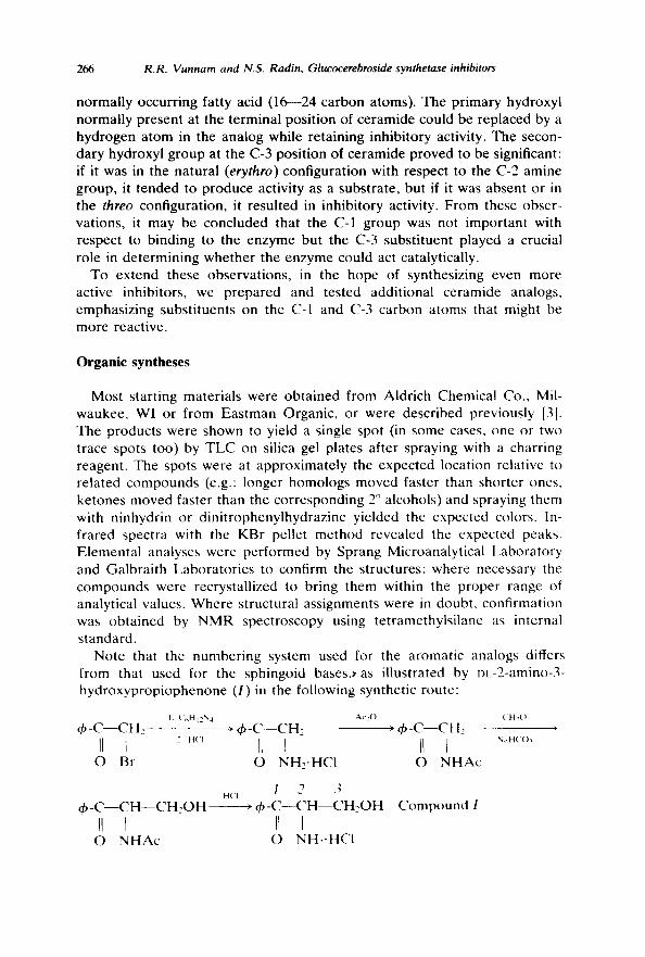
Note that the numbering system used for the aromatic analogs differs from that used for the sphingoid b a s e s ) a s illustrated by DL-2-amino-3- hydroxypropiophenone (I) in the following synthetic route:
I ( 't,}] 12N4 ~ b - C - - C H 2
O Br
¢ b - C - - C H - - C H 2 0 H
O NHAc
Ac~O ('H20 ) ' dp-C--CH2 ) q~-C--CH2
IL t IL I O NH2-HC1 O NHAc
H( "1 / 2 3 • ~ b - C - - C H - - C H 2 O H
11 I O NH2-HCI
Compound I
NaH('()~
R.R. Vunnam and N.S. Radin, Glucocerebroside synthetase inhibitors 267
A. Synthesis of 2-amino-3-hydroxypropiophenones Compound I was prepared as the hydrochloride from 2-bromo-
acetophenone by amination with hexamethylenetetramine, acetylation, hydroxymethylation with formalin, and deacetylation (see above reaction sequence) [4]. p-Substituted derivatives of I were prepared from the cor- responding bromoacetophenones, which were purchased or prepared by bromination. In the case of methoxyacetophenone, bromination was done with methanolic bromine [5].
During the hydroxymethylation of 2-acetylamino-p-methylacetophenone, some dihydroxymethylation occurred at the C-2 position. The product (m.p. 164--165°C from EtOAc) was hydrolyzed with HCI and reacylated with decanoyl chloride (amide m.p. 143---144.5°C from EtOAc).
These and other amine hydrochlorides were acylated with acyl chlorides in tetrahydrofuran and concd, aqueous NaOAc [6,7]. For the synthesis of 2,3-epoxydecanoyl L the free acid was coupled to the amine by a mixed anhydride method. At -10°C, 0.13 ml of isobutyl chloroformate was added to a stirred solution of 1 mmol epoxydecanoic acid [8] and 0.14 ml of Et3N in 10 ml of tetrahydrofuran. After 5 min of stirring, the mixture was added to a suspension of 1 mmol I.HC! and 0.14 ml Et3N in 10 ml tetrahydrofuran, with stirring at -10°C. The mixture was then stirred 1 h at 0°C and 30rain at 22°C. The amide was purified by partitioning between ether and 1% NaHCO3 and crystallizing from benzene/hexane.
In some syntheses, the intermediate 2-aminoacetophenone-HCI was acyl- ated with a long chain acyl chloride instead of acetic anhydride, obviating the need to deacetylate and reacylate.
B. Miscellaneous compounds N-Decanoyl-p-nitro-I was dehydrated with pyridine/acetic anhydride [9]
to produce 2-decanoylamino-p-nitroacrylophenone (m.p. 62--63°C from hexane).
2-Decanoylaminopropiophenone was an oil, prepared from N-acetyl-DL- norephedrine by oxidation of the hydroxyl group with dichromate [10], deacetylation with HCI, and reacylation with decanoyi chloride. It was recrystallized from hexane at -20°C.
2-Acylaminopropiophenones bearing a tertiary amino group at the C-3 carbon atom were prepared by a Mannich reaction between a secondary amine, paraformaldehyde, and 2-acylaminoacetophenone [11].
DL-Erythro-3-phenylserine and its ethyl ester were prepared according to the method of Bolhofer [12]. The compounds gave the reported melting points. Acylation with decanoyl chloride produced the decanoylamino ester (m.p. 69°C from benzene/hexane) and the free acid (m.p. 114 115°C). Treatment of the amido ester for 2 days with ethanol saturated at room temperature with ammonia yielded the carboxamide in 65% yield, m.p. 177°C from methanol. The decanoylamino ester was converted also to the
268 R.R. Vunnam and N.S. Radin, Glucocerebroside synthetase inhibitors
hydrazide by stirring 1 g of ester with 0.5 ml of hydrazine hydrate in 5 ml of methanol for 4h. The product was recrystallized from boiling methanol: m.p. 229-230°C. Like the other inhibitors, it was stored in ch, loro- form/methanol, but a little HCI had to be added to increase its solubility. T h e threo isomer of Dc-phenylserine was purchased from U.S. Biochemicals Corp. and converted to the N-decanoyl ethyl ester (m.p. 64 65°C from benzene/hexane).
N-Bromoacetyl sphingosine, m.p. 84---85°C, was prepared from equi- molar quantities of bromoacetyl bromide and D-sphingosine by the NaOAc method described above. It was purified by silica gel chromatography using chloroform/methanol 99 : I and 99 : 2.
N-Decylthiocarbamyl norephedrine [ C > H e I - - N H - - C ( : S } - - N H - - CH(CH3) - -CHOH--C.Hs] was made by refluxing equimolar amounts of DL-norephedrine and n-decylisothiocyanate in E tOH for 6 h. The solution was evaporated to dryness and the residue rccrystallized.
The tables in the Results section give additional data on the m.p. and solvent of recrystallization for the individual compounds.
Enzyme assays
Most assays for glucosyltransferase were run with lyophilized brain homogenates from 16 day male mice, incubated at 37°C for 90 min [3] with 0.2/zmol of N-octanoyl sphingosine or DDS (N-dodecanoyl-l)l.-decasphin- ganine or lauroyl e r y t h r o - 2 - a m i n o - l , 3 - d e c a n e d i o l ) as glucose acceptors [S]. The compounds tested as inhibitors (generally as 0.3 mM) were evaporated to dryness from solution in the incubation tube with the lipoidal substrate before adding the lyophilized tissue in benzene and evaporating off the solvent. (Because of the toxicity of benzene, the processing must be done in a good hood. However recent work has shown that benzene can be replaced by cyclohexane.) The remaining incubation components (dithioerythritol. Tris-C1 pH 7.4 or 7.8, ATP, EDTA, Mg >, and [3H]UDP-glucose) were then added in water. In some experiments, the lipoidal substrate was added in liposomal form [8]. The cerebroside formed in the incubation was isolated by partitioning between chloroform, methanol, and water. This step [3] was simplified by omitting the filtration step for removal of brain nonlipids. The lipid thus obtained was assayed by liquid scintillation in a water-containing solvent.
Results
Amides of 2-amino-3-hydroxypropiophenone (I) A comparison of different acyi derivatives of 1, tested at 0.3 mM (Table 1),
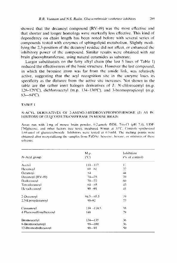
R.R. Vunnarn and N.$. Radin, Glucocerebroside synthetase inhibitors 269
showed that the decanoyl c o m p o u n d (RV-49) was the most effective and that shorter and longer homologs were markedly less effective. This kind of d e p e n d e n c y on chain length has been noted before with several series of c o m p o u n d s tested with enzymes of sphingol ipid metabol i sm. Slightly modi- fying the 2,3-posit ion of the decanoyl residue did not affect, or enhanced the inhibi tory power of the compound . Similar results were ob ta ined with rat
bra in glucosyl t ransferase, using na tura l ceramides as substrate . Larger subs t i tuents on the fatty alkyl chain (the last 5 lines of Table I)
reduced the effectiveness of the basic s tructure. However the last compound , in which the b r o m i n e a tom was far from the amide link, was relatively active, suggest ing that the acyl recogni t ion site in the enzyme loses its
specificity as the dis tance from the active site increases. Not shown in the table are the ra ther inert halogen derivat ives of I : N-chloroacety l (m.p. 128-129°C), d ichloroacetyl (m.p. 134-136°C), and 3 -b romoprop iony l (m.p.
K~-84°C) .
TABLE I
N-ACYL DERIVATIVES OF 2-AMINO-3-HYDROXYPROPIOPHENONE (I) AS IN- HIBITORS OF GLUCOSYLTRANSFERASE IN MOUSE BRAIN
Assay run with l mg of mouse brain powder, 0.2/xmole DDS, Tris-Cl (pH 7.4), UDP- [3H]glucose, and other factors (see text), incubated 90rain at 37°C. Controls synthesized 1.69nmol of glucocerebroside. Inhibitors were tested at 0.3 raM. The melting points were obtained after recrystallizing the samples from EtOAe, benzene, hcxane, or mixtures of these solvents.
M.p. Inhibition N-Acyl group (°C) (%. of control)
Acctyl 114--117 11 Hexanoyl 6(~-61 22 Octanoyl 84 44 Decanoyl (RV-49) 74--75 7(1 Dodecanoyl 7(~-72 60 Tetradecanoyl ~ 4 ~ 5 43 Hcxadecanoyl 90---91 41
2-Decenoyl 94.5--95.5 79 2,3-Epoxydecanoyl 90-92 73
Cinnamoyl 119--119.5 35 4-Fluorosulfonylbenzoyl 14;"; 2;4
Bromoacetyl 134~ 135 36 8- Bromooctanoyl 99-- 100 36 12-Bromododecanoyl 9(~-91 51)
270 R.R. Vunnam and N.S. Radin, Glucocerebroside synthetase inhibitors
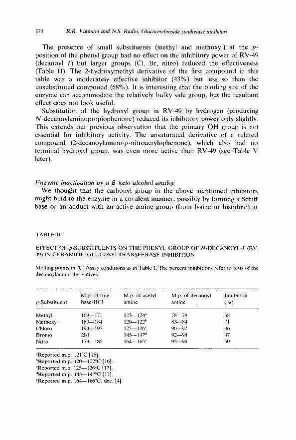
T h e p resence of small subs t i tuen t s (methyl and me thoxy l ) at the p- pos i t ion of the pheny l g roup had no effect on the inh ib i to ry p o w e r of RV-49 (decanoy l I ) but l a rger g roups (CI, Br, n i t ro) r e d u c e d the ef fec t iveness (Table II). The 2 - h y d r o x y m e t h y l de r iva t ive of the first c o m p o u n d in this tab le was a m o d e r a t e l y effect ive inh ib i to r (43%) but less so than the unsubs t i t u t ed c o m p o u n d (68%). It is in te res t ing tha t the b ind ing site of the enzyme can a c c o m m o d a t e the re la t ive ly bu lky side group , but the resu l tan t effect does not l ook useful.
Subs t i tu t ion of the hydroxy l g r o u p in RV-49 by h y d r o g e n (p roduc ing N - d e c a n o y l a m i n o p r o p i o p h e n o n e ) r e d u c e d its inh ib i to ry p o w e r only slightly. This ex t ends ou r p rev ious o b s e r v a t i o n that the p r ima ry O H g roup is not essent ia l for inh ib i to ry act ivi ty. The u n s a t u r a t e d der iva t ive of a r e l a t ed c o m p o u n d ( 2 - d e c a n o y l a m i n o - p - n i t r o a c r y l o p h e n o n e ) , which also had no t e rmina l hydroxyl g roup , was even m o r e act ive than RV-49 (see Tab le V later) .
Enzyme inactivation by a fl-keto alcohol analog W e thought that the ca rbony l g r o u p in the a b o v e m e n t i o n e d inh ib i tors
might b ind to the e n z y m e in a cova len t manne r , poss ib ly by fo rming a Schiff base or an adduc t with an act ive amine g roup (from lysine or h is t id ine) at
TABLE II
EFFEC'T OF p-SUBSTITUENTS ON THE PHENYL GROUP OF N-DECANOYL-I (RV- 49) IN CERAMIDE : GLUCOSYLTRANSFERASE INHIBITION
Melting points in °C. Assay conditions as in Table 1. The percent inhibitions refer to tests of the decanoylamino derivatives.
M.p. of free M.p. of acetyl M.p. of decanoyl Inhibition p-Substituent base.HCI amine amine (%)
Methyl 169-- 171 12.'9--- 124" 78.--79 68 Methoxy 18 ."~- 184 12(~ 122 h 83~-84 71 Chloro 194-- 197 12.~- 126 ~ 9(~-92 46 Bromo 2(XI 145-- 147 d 92--94 47 Nitro 179-- 180 164-- 165 ~ 95--96 50
~Reported m.p. 121°C [15]. bReported m.p. 120~122°C [16]. CReported m.p. 12~%--126°C [17]. dReported m.p. 14.~-147°C [17]. eReported m.p. 164---166°C, dec. [4].
R.R. Vunnam and N.S. Radin, Glucocerebroside synthetase inhibitors 271
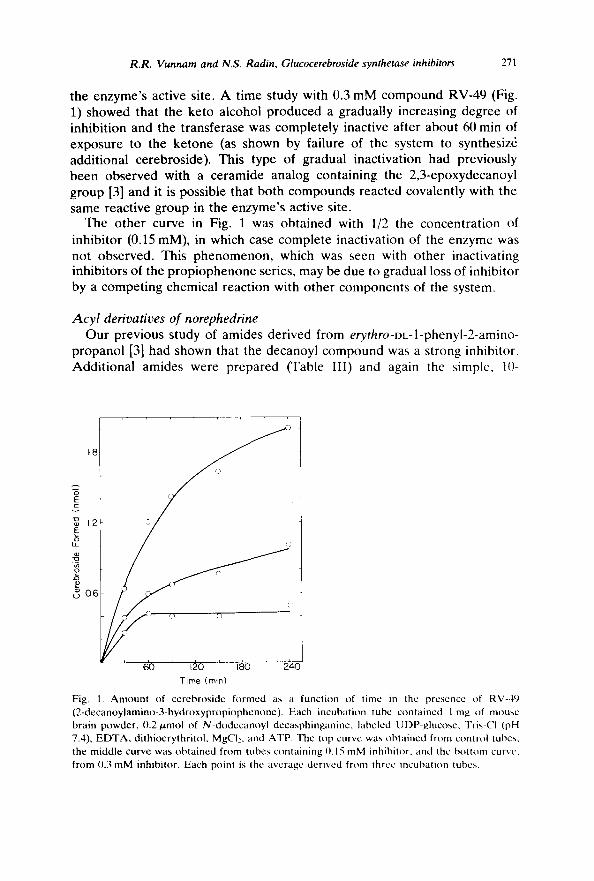
the enzyme's active site. A time study with 0.3 mM compound RV-49 (Fig. 1) showed that the keto alcohol produced a gradually increasing degree of inhibition and the transferase was completely inactive after about 60 min of exposure to the ketone (as shown by failure of the system to synthesiz6 additional cerebroside). This type of gradual inactivation had previously been observed with a ceramide analog containing the 2,3-epoxydecanoyl group [3] and it is possible that both compounds reacted covalently with the same reactive group in the enzyme's active site.
The other curve in Fig. 1 was obtained with 1/2 the concentration of inhibitor (0.15 mM), in which case complete inactivation of the enzyme was not observed. This phenomenon, which was seen with other inactivating inhibitors of the propiophenone series, may be due to gradual loss of inhibitor by a competing chemical reaction with other components of the system.
Acyl derivatives o[ norephedrine Our previous study of amides derived from erythro-DL-l-phenyl-2-amino-
propanol [3] had shown that the decanoyl compound was a strong inhibitor. Additional amides were prepared (Table III) and again the simple, 10-
)
t8
,'.3
I c
v
~ 12
3_ o
,,~ 06
S 60 120 180 24O
Time (rain)
Fig. 1. Amount of cerebroside formed as a function of time in the presence of RV-49 (2-decanoylamino-3-hydroxypropiophenone). Each incubation tube contained 1 mg of mouse brain I~)wder, 0.2 p, mol of N-dodecanoyl decasphinganine, labeled UDP-glucosc, Tris-CI (pH 7.4), EDTA, dithioerythritol, MgCI:, and ATP. The top curve was obtained from control tubes, the middle curve was obtained from tubes containing 0.15 mM inhibitor, and the bottom curve, from 0.3 mM inhibitor. Each point is the average derived from three incubation tubes.
272 R.R. Vunnam and N.S. Radin, Glucocerebroside synthetase inhibitors
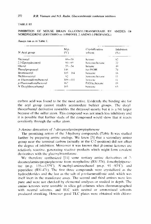
TABLE 111
INHIBITION OF MOUSE BRAIN GLUCOSYLTRANSFERASE BY AMIDES OF NOREPHEDRINE (ER YTHRO-DL- I-PHENYL-2-AMINO- I-PROPANOL)
Assays run as in Table I.
M . p . Crystallization Inhibition N-Acyl group (°C) solvent (%)
Decanoyl 6'4 70 hcxanc ~2 2,3-Epoxydecanoyl 9-t--95 benzene/hexanc 72 Oleoyl 5,~-54 hexane 4'4 Phenylpropynoyl 146 iso-PrOH 24 Bromoacetyl 1113~- 1(14 benzene 16 Dichloroacetyl ~2 benzene/hcxanc I1 m -Fluorosulfonylbc nzoyl 10t) ,_ II1 benzene 11 p- F1 uorosulfonylbenzoyl 1 ~2 EtOAc/hcxanc I S N-Decylthiocarbamyl 105 benzene (~3
ca rbon acid was found to be the most act ive. Ev iden t ly the b inding site for the acyl g roup cannot read i ly a c c o m o d a t e bu lk i e r groups . The decyl- t h i o c a r b a m y l de r iva t ive r e semble s the decanoy l moie ty but it is a bit la rger because of the sulfur a tom. This c o m p o u n d was not much less inh ib i tory and it is poss ib le that fu r the r s tudy of the c o m p o u n d would show that it reacts cova len t ly th rough the sulfur a tom.
3 - A m i n o derivatives of 2-decanoylaminopropiophenone The p romis ing ac t ion of the 3 -hydroxy c o m p o u n d s (Table I) was s tud ied
fu r the r by p r e p a r i n g amine analogs . W e knew [3] that a s econda ry a m i n o g roup near the t e rmina l ca rbon (actual ly at the C-2 pos i t ion) d id not affect the deg ree of inhibi t ion . M o r e o v e r it was known that /3-amino ke tones are re la t ive ly react ive , gene ra t i ng reac t ive p roduc t s which might form cova len t de r iva t ives with the g lucosy l t r ans fe rase .
W e the re fo re syn thes ized [ t l ] some te r t i a ry amine der iva t ives of 2- d e c a n o y l a m i n o p r o p i o p h e n o n c f rom m o r p h o l i n e (RV-378), 4 -me thy lp ipe raz - ine (m.p. 135--137°C), N - m e t h y l - a m i n o e t h a n o l (m.p. 9 l - - t )3°C) , and p ipe r id ine (RV-471). The first three c o m p o u n d s were crys ta l l ized as the h y d r o c h l o r i d e s and the last as the salt of p - t o lue ne su l fon i c acid, which was itself inert in the t r ans fe rase assay. The second and third amines were less pure and were not checked by e l emen ta l ana lyses or s tud ied in dep th . The a m i n o ke tones were uns tab le in sil ica gel co lumns when c h r o m a t o g r a p h e d with neut ra l solvents , and T L C with neut ra l or a m m o n i a c a l so lvents p r o d u c e d s t reaking . H o w e v e r good T L C pla tes were o b t a i n e d with ch loro-
R.R. Vunnam and N.S. Radin, Glucocerebroside synthetase inhibitors 273
form/methanol/acet ic acid (90:5:10) the R F values being 0.68, 0.26, 0.15, and 0.55, respectively.
Tested at 0.3 mM, the amines produced 86 to 95% inhibition. The acetyl homolog of RV-378 yielded only 14% inhibition, showing here too the importance of chain length in the acyl group.
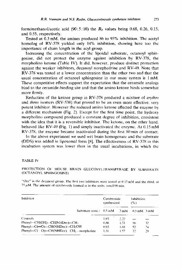
Increasing the concentration of the lipoidal substrate, octanoyl sphin- gosine, did not protect the enzyme against inhibition by RV-378, the morpholino ketone (Table IV). It did, however, produce distinct protection against .the weaker inhibitors, decanoyl norephedrine and RV-49. Note that RV-378 was tested at a lower concentration than the other two and that the usual concentration of octanoyl sphingosine in our assay system is 1 mM. These competitive effects support the expectation that the ceramide analogs bind to the ceramide-binding site and that the amino ketone binds somewhat more firmly.
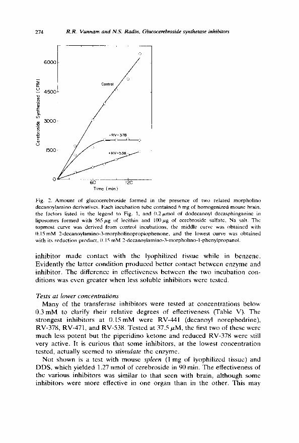
Reduction of the ketone group in RV-378 produced a mixture of erythro and threo isomers (RV-538) that proved to be an even more effective, very potent inhibitor. However the reduced amino ketone affected the enzyme by a different mechanism (Fig. 2). Except for the first time point, the hydroxy morpholino compound produced a constant degree of inhibition, consistent with the idea that it is a reversible inhibitor. The ketone, on the other hand, behaved like RV-49 (Fig. l) and simply inactivated the enzyme. At 0.15 mM RV-378, the enzyme became inactivated during the first 80 min of contact.
In the above experiment we used wet brain homogenate and the substrate (DDS) was added in liposomal form [8]. The effectiveness of RV-378 in this incubation system was lower than in the usual incubations, in which the
TABLE IV
PROTECI ' ION OF MOUSE BRAIN GLUCOSYLTRANSFERASE BY SUBSTRATE (OCTANOYL SPHINGOSINE)
"'Dec" is the decanoyl group. The first two inhibitors were tested at {).15 mM and the third, at 75/.zM. The amount of cerebroside formed is in the units, nmol/90 rain.
Inhibitor Cerebroside Inhibition synthesized (%)
Substrate conc.: {).5 mM 3 mM {I.5 mM 3 mM
Controls 1.93 Phe ny I - -CH(OH)- -CH(NH Dec)~CH3 {I.86 P h e n y I - - C ( = O ) - - C H ( N H D e c ) - - C H 2 O H 0.93 P h e n y l - - C ( = O ) - - C H ( N HDec)---CH ~--morpholine 1.31
2.22 - - - - 1.51 56 32 1.68 52 24 1.57 32 29
274 R.R. Vunnam and N.S. Radin, Glucocerebroside synthetase inhibitors
':3
6000
2 0'3
(...)
150
60 120 Time ( min )
Fig. 2. Amount of glucocerebroside formed in the presence of two related morpholino decanoy/amino derivatives. Each incubation tube contained 6 mg of homogenized mouse brain, the factors listed in the legend to Fig. 1, and 0.2tzmol of dodecanoyl decasphinganine in liposomes formed with 565,~g of lecithin and 100,ttg of cerebroside sulfate, Na salt. The topmost curve was derived from control incubations, the middle curve was obtained with 0.15mM 2-decanoylamino-3-morpholinopropiophenone, and the lowest curve was obtained with its reduction product, 0.15 mM 2-decanoylamino-3-morpholino-l-phenylpropanol.
inh ib i tor made contact with the lyophil ized tissue while in benzene . Eviden t ly the lat ter condi t ion p roduced be t te r contact be tween enzyme and inhibi tor . The difference in effectiveness be tween the two incuba t ion con- di t ions was even greater when less soluble inhibi tors were tested.
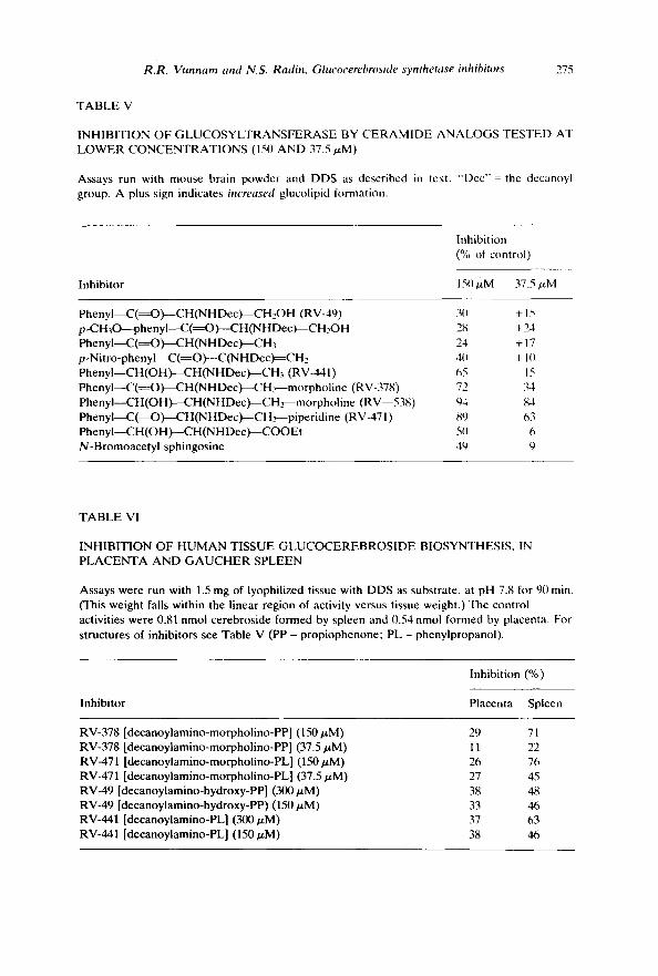
Tests at lower concentrations Many of the t ransferase inhibi tors were tested at concen t ra t ions below
0.3 m M to clarify their relat ive degrees of effectiveness (Table V). The s t rongest inhibi tors at 0 . 1 5 m M were RV-441 (decanoyl norephedr ine) , RV-378, RV-471, and RV-538. Tes ted at 37.5/zM, the first two of these were much less po ten t but the p iper id ino ke tone and reduced RV-378 were still very active. It is curious that some inhibitors , at the lowest concen t ra t ion tested, actually seemed to stimulate the enzyme.
Not shown is a test with mouse spleen (1 mg of lyophil ized tissue) and DDS, which yielded 1.27 nmol of cerebroside in 90 min. The effectiveness of the var ious inhibi tors was similar to that seen with brain , a l though some inhibi tors were more effective in one organ than in the other. This may
R.R. Vunnam and N.S. Radin, Glucocerebroside synthetase inhibitors 275
TABLE V
INHIBITION OF GLUCOSYLTRANSFERASE BY CERAMIDE ANALOGS TESTED AT LOWER CONCENTRATIONS (150 AND 37.5 tzM)
Assays run with mouse brain powder and DDS as described in text. "Dcc"= the decanoyl group. A plus sign indicates increased glucolipid formation.
Inhibition (% of control)
Inhibitor 150/~M 37.5 #M
PhenyI--C(~O)--CH(NHDec)--CH2OH (RV-49) 30 + 15 p-CH30----phenyI--C(~O)---CH(NHDec )---CH2OH 28 +24 PhenyI--C(~O)----CH(NHDec)--CH3 24 + 17 p -Nitro-phe nyI--C(=O)---C(N HDec)==CH2 40 + 1(I PhenyI--CH (OH)--CH(NHDec)--CH3 (RV-441) 65 15 PbenyI--C(~O)--CH(NHDec)--CH~-morpholine (RV-378) 72 34 PhenyI--CH (OH)--CH(NHDec)--CH ~-morpholine (RV--538) 94 84 Phenyl--C(~O)---CH (NHDec)---CH-~-piperidine (RV-471) 89 63 PhenyI--CH(OH)~CH(N HDec)---COO Et 50 6 N-Bromoacetyl sphingosine 49
TABLE VI
INHIBITION OF HUMAN TISSUE GLUCOCEREBROSIDE BIOSYNTHESIS, IN PLACENTA AND GAUCHER SPLEEN
Assays were run with 1.5 mg of lyophilized tissue with DDS as substrate, at pH 7.8 for 90 min. (This weight falls within the linear region of activity versus tissue weight.) The control activities were 0.81 nmol cerebroside formed by spleen and 0.54 nmol formed by placenta. For structures of inhibitors see Table V (PP = propiophenone; PL = phenylpropanol).
Inhibition (%)
Inhibitor Placenta Spleen
RV-378 [decanoylamino-morpholino-PP] (150 p,M) RV-378 [decanoylamino-morpholino-PP] (37.5 p,M) RV-471 [decanoylamino-morpholino-PL] (150/zM) RV-471 [decanoylamino-morpholino-PL] (37.5/,tM) RV-49 [decanoylamino-hydroxy-PP] (300/,tM) RV-49 [decanoylamino-hydroxy-PP) (150/zM) RV-441 [decanoylamino-PL] (300/xM) RV-441 [decanoylamino-PL] (150/zM)
29 71 11 22 26 76 27 45 38 48 33 46 37 63 38 46
276 R.R. Vunnam and N.S, Radin, Glucocerebroside synthetase inhibitors'
TABLE VII
N - D E C A N O Y L E R Y T H R O - P H E N Y L S E R I N E DERIVATIVES AS INHIBITORS OF MOUSE BRAIN GLUCOSYI .TRANSFERASE
Assayed with mouse brain powder and DDS as in the text.
( ' , ,H~- -CH--CH--R
I t OH N H - - C ( = O ) - - ( ' , , H i,
Crystallization M.p. Inhibition R group solvent (o(,) (%)
COOH benzene 114~ 115 34 COOEt bcnzenc/hcxane 60 74 CONH2 methanol 177 14 CONHNH2 methanol 23(1 ,',;
indicate the existence of different types of glucosyltransferasc in the two organs; we had previously seen other differences between the glucosyltrans- ferases of mouse liver and brain [8].
Since the primary goal of this project was to develop inhibitors that wcrc effective in human (Gaucher) tissues, we examined some of the compounds with human placenta and Gaucher spleen (Table VI). All showed promising activity, with greater effectiveness in the spleen. As noted above, such differences could indicate that the two tissues contain transferases with different properties.
Derivatives of N-decanoyl-Dz.-erythro-3-phenylserine The free erythro acid showed some inhibitory activity but the amide and
hydrazide were rather inert (Table VII). The ethyl ester, however, was quite a strong inhibitor. Unexpectedly, the N-decanoyl derivative of the threo amino acid, ethyl ester, was a little less effective than the erythro isomer.
D i s c u s s i o n
The experiments described above contribute additional information about the nature of the active site in ce ramide :UDP-glucose glucosyltransferase. Ceramide analogs seem to bind to the active site that normally binds the lipoidal substrate, ceramide. Replacement of the primary O H of the ceramide analogs with a hydrogen a tom or larger group does not appreci- ably block binding. When the larger group is an amine, the analog's effectiveness increases considerably, suggesting that there is an anionic group in the active site. This could be a thiol, carboxyl, or phosphate group, any of which could form a glucoside of high energy by reaction with UDP-glucose, and then pass on the sugar residue to ceramide.
R.R. Vunnam and N.S. Radin, Glucocerebroside synthetase inhibitors 277
Supporting the idea of an anionic site is the observation (Table VII) that decanoyl phenylserine (the free acid) was a weak inhibitor while its ethyl ester was a strong one. One would expect that an anionic site would repel a negatively-charged analog of ceramide but not a nonionic analog.
The hydroxyl or carbonyl group in the C-3 position of ceramide (C-1 of the aromatic analogs) was also of high significance to the enzyme's binding site. Reduction of the carbonyl group to a hydroxyl considerably enhanced the compound's potency, both in the case of the nonionic RV-49 and the cationic RV-378. Evidently there is a region close to the active site of the enzyme that can form a strong bond with the hydroxyl group. However the ketonic oxygen seems to have a particular role in producing inactivation of the enzyme.
One explanation for this role is that the keto amine or alcohol binds to the active site, then a/3-elimination reaction expels the C-3 group and the C-2 hydrogen, forming water or a secondary amine (e.g.: morpholine) and 2-decanoylaminoacrylophenone. A Michael addition reaction could then occur in which a nucleophilic group in the active site would add to the double bond in the acrylophenone. The proposed nucleophilic group could be the postulated anionic group (SH, COOH, PO4) or an amine ( lysine)or imidazole (histidine). However, the latter groups would probably have a positive charge at the pH used in our incubations and would be expected to repel the highly effective cationic inhibitors.
The above hypothesis explains why RV-49 is less potent than RV-378, since 13-keto alcohols are more stable at neutral pH than /3-keto amines. The ketone lacking a polar group at the 3-position (decanoylamino- propiophenone) was even less effective and did not produce complete enzyme inactivation even when incubated for a long time. This compound would not be expected to spontaneously form an acrylophenone.
Support for the proposed elimination reaction comes from the work of Sch6nenberger et al. [13], who showed that aromatic /3-amino ketones undergo spontaneous elimination of the amine moiety in neutral solution, with formation of the acrylophenone. A wide range of biological activities was exhibited by the compounds studied and the authors proposed a mechanism of action similar to the one above.
These authors also observed a second reaction with the aromatic amino ketones, a hydrolytic cleavage in which formaldehyde and the amine were formed (a reversal of the organic synthesis). Formaldehyde, like acrylo- phenones, is highly reactive and could inactivate the enzyme. Either reaction might be catalyzed by the enzyme, making this a possible example of 'active-site-directed irreversible enzyme inhibition' [14].
The relative bulkiness of the amine group in RV-378 and related com- pounds may act to enhance binding to the enzyme because of the steric resemblance to the glucose residue that becomes attached to that position in the normal mode of enzyme reaction.
278 R.R. Vunnam and N.S. Radin, Glucocerebroside synthetase inhibito~s
One might postulate that the ketonic enzyme inhibitors act, not by forming a covalent linkage with the enzyme's active site, but by binding somewhere else, deforming the enzyme molecule sufficiently to make it thermally unstable. However, in several instances the reduced (hydroxylic) analog was more effective than the original ketone, yet it did not produce inactivation (Fig. 2).
The most active compound we made, RV-538, is probably more active than it seems since it contains the erythro form, which is probably less potent than the threo form. If the compound were to be administered in vivo, part of it might be oxidized enzymatically to the ketone (RV-378), thus giving it dual modes of action. Experiments in progress have shown that RV-378 is active in mice, producing inactivation of liver glucosyltransferase. It is also a relatively effective inhibitor of monoamine oxidase in mice [11], which complicates the therapeutic implications.
Acknowledgements
This work was supported by NIH grants NS-03192 and HD-07406. We are indebted to Inez Mason, Pauline Arisumi, Carol Black, and Dan del Vecchio for laboratory assistance and to Drs. Ruth Heyn and K.M.J. Mennon for their vital help in obtaining human tissues.
References
1 S. Basu, B. Kaufman and S. Roseman, J. Biol. Chem., 248 (1973) 1388. 2 A. Brenkert and N.S. Radin, Brain Res., 36 (1972) 183. 3 K.R. Warren, R.S. Misra, R.C. Arora and N.S. Radin, J. Neurochem., 26 (1976) 1(163. 4 L.M. Long and H.D. Troutman, J. Am. Chem. Soc., 71 (1949) 2473. 5 British Patent No. 607,538 (1948). 6 D. Shapiro and H.M. Flowers, J. Am. Chem. Soc., 83 (1961) 3327. 7 N.S. Radin, Methods Enzymol., 28 (1972) ~10. 8 R.R. Vunnam and N.S. Radin, Biochim. Biophys. Acta, 573 (1979) 73. 9 J. Kollonitsch and A. Hajos, Acta Chim. Acad. Sci. Hung., 18 (19551 271.
10 R.C. Gaver and C.C. Sweeley, J. Am. Chem. Soc., 88 (1966) 3643. 11 R.R. Vunnam, D. Bond, R.A. Schatz, N.S. Radin and N. Narasimhachari, J. Neurochem,
34 (1980) 410. 12 W.A. Bolhofer, J. Am. Chem. Soc., 74 (1952) 5459. 13 H. Sch6nenberger, T. Bastug, L. Bindl, A. Adam, D. Adam, A. Petter and W. Zwez,
Pharm. Acta Helv., 44 (1969) 691. 14 B.R. Baker, Design of Active-Site-Directed Irreversible Enzyme lnhibitors, John Wiley &
Sons Inc., New York, 1967. 15 Ng.Ph. Buu-Hoi, Ng.D. Xuong and Ng.H. Khoi, J. Chem. Soc. (1951) 255. 16 M.C. Rebstock and E.L. PIeitler, J. Am. Chem. Soc., 74 (1952) 3207. 17 L.L. Bambas, H.D. Troutman and L.M. Long, J. Am. Chem. Soc., 72 (195(/) 4445.