Embed Size (px)
Citation preview

CHEMISTRY OF PHOSPHOLIPIDS IN RELATIONTO BIOLOGICAL MEMBRANES
L. L. M. VAN DEENEN
Department of Biochemistry, University of Utrecht,Utrecht, The Netherlands
ABSTRACTThe quantitative determination of molecular species of natural phospholipidsgave new information about the pairing of the fatty acid chains in a given lipidclass. Cells appear to be equipped with enzymes which control the compositionand pairing of hydrocarbon chains of phospholipids and display regulatorymechanism(s) which allow for adaptation of physical properties of membranelipids to alteration in environmental conditions. Phospholipids containingvarious types of fatty acid combinations encountered in membranes havebeen prepared by chemical synthesis. Examination of these compounds inartificial membrane systems demonstrated that chain-length, degree ofunsaturation, and the type of pairing of hydrocarbon chains determine therate of diffusion of non-electrolytes and efficiency of carrier mediatedtransport across the hydrocarbon barrier. Comparison with natural membranesof different lipid composition revealed a close similarity with the modelsystems. This endorses the conclusion that the detailed chemical make-upof the lipid dictates the permeability behaviour of the region of the biologicalinterface.
The diversity of polar headgroups of phospholipids is demonstrated by thepolyglycerol phospholipids of bacterial membranes, Detailed informationabout the structure of amino acyl and glucosamine derivatives of phosphatidylglycerol has been obtained by combination of chemical synthesis and
enzymatic methods.
I. INTRODUCTIONPhospholipids are essential constituents of all living cells. By a combination
of lipophilic and hydrophilic groups within one molecule, physical propertiesare attained which make them particularly suitable compounds to serve asmajor constituents for biological interfaces. Although the basic chemicalstructure of a phospholipid is a relatively simple one, the variations en-countered in the chemical make-up of phospholipids in biological membranesoffer a vast field for investigation not only to chemists of natural products butto biochemists and molecular biologists as well. Confining this discussionto one class of phospholipids (Figure 1), the so-called phosphoglycerides, itcan be stated that nature produces phospholipids with one to at least fourhydrocarbon chains, but that the compounds with two apolar tails dominate.Lipid—lipid association involving London—van der Waals interactions
25

L. L. M. VAN DEENEN
between the apolar residues of the phospholipids contributes to the arrange-ment of a palisade alignment so as to give lipid barriers which separateaqueous compartments. On the other hand, it has been suggested duringrecent years that hydrophobic interactions between the hydrocarbon chainsof lipids and apolar regions of the proteins are of paramount importance
Lipid—lipidCO. 200 variations
interactions 1) Chain length2) Number of double bonds
Lipid—protein 3) Position of double bondsinteractions 4) Ester/ether tinkage
Lipid—proteininteractions ± 40 variationsCation—binding; net chargeternary complexes I(Carrier??) J
Figure 1. Phospholipid skeleton in relation to its functions
for the integrity of the lipoproteins in biological membranes. A bewilderingvariation in chemical nature of the hydrocarbon chains provokes manyquestions with respect to a possible rationale between structure and functionof this part of the phospholipid molecule. Similarly, most conspicuouschemical differences have been found in the hydrophilic moiety of phospho-lipids. Polar headgroups containing e.g. amino alcohols, cyclitols, aminoacids,hexoses, hexosamines and a varying number of phosphate residues havebeen detected. As a result a great variation in net charge of phospholipidmolecules can be encountered in many membranes. Electrostatic forces maybind polar groups of lipids to oppositely charged groups of proteins andperhaps of other lipid species, while in addition, cations may be involved inlinking negatively charged groups of different molecules so as to formternary complexes. Different views have been expressed about the importanceof the electrostatic interactions between lipids and proteins and as a resulthighly conflicting models for biological membranes have been proposed.However, a certain degree of non-uniformity in membrane structure is likelyin view of the multiple functions of biological interfaces. The chemicalheterogeneity of both lipid and protein constituents also favours the opinionthat within the framework of one interface different molecular arrangementsinvolving various types of lipid—lipid and lipid—protein may contribute to thecorrect functioning of the various regions of the membrane concerned.
The chemistry of phospholipids in combination with enzymology andwell-defined artificia.l model systems can contribute towards solving someof the compelling problems around the molecular architecture and dynamicbehaviour of biological interfaces.
26

PHOSPHOLIPIDS AND MEMBRANES
II. MOLECULAR SPECIES OF PHOSPHOGLYCERIDES
The analytical data on the fatty acid composition of phospholipids whichhave been accumulated during the past decade could easily fill a sizeablevolume of an encyclopedia. Some two hundred different fatty acid andaldehyde constituents have been recognized and many facts have beendiscovered about their occurrence in a great variety of cells'. The compositionof the apolar moiety of phospholipids present in a given membrane often is areflection of the capacities for fatty acid biosynthesis of the organismconcerned. In addition it has been clearly demonstrated that environmentalfactors can influence to a significant extent—but not randomly—the make-upof the apolar part of phospholipids. A considerable amount of work wasinvested to separate the various classes of phospholipids containing differentpolar headgroups, and to determine their fatty acid composition. In this way,many preferential combinations between particular headgroups and hydro-carbon chains were detected, which raised many questions not only withrespect to function, but also concerning the biosynthetic machineryresponsible for attaining these assOciations. The next logical step was thequantitative determination of the pairing of fatty acid constituents within onephospholipid class.
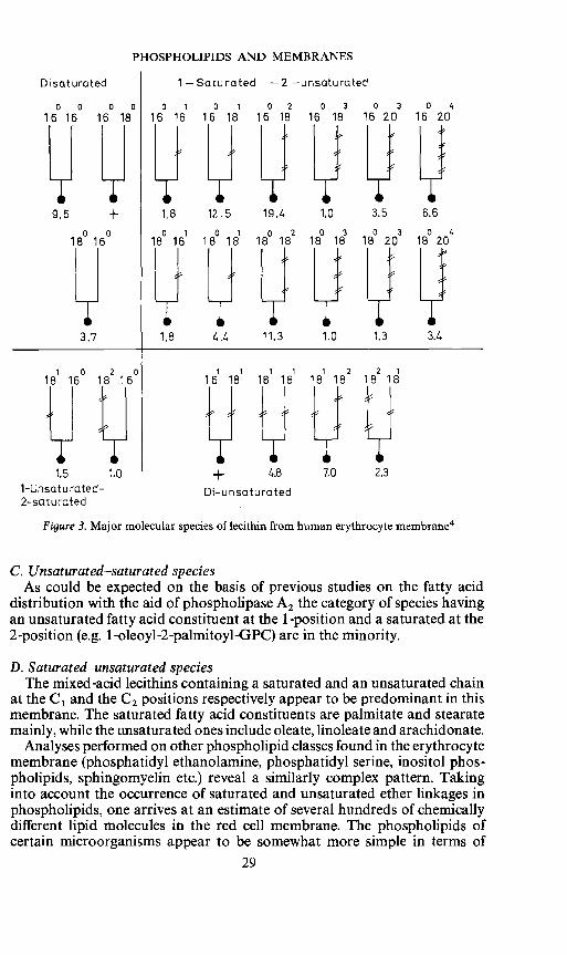
1. Analysis of molecular speciesA phospholipid such as lecithin (phosphatidyicholine) present in a given
membrane cannot be considered as one chemical entity but rather must bethought of as a family of related species which have in common the polarheadgroup but vary with respect to the nature of the apolar tails. Thelecithin preparation isolated from one membrane may contain some tendifferent fatty acid constituents and these can be combined theoretically ina great number of pairs. Phospholipase A2 hydrolysis will furnish informationabout the location of the constituents at the C1 and C2 fatty acid esterposition, but usually the information obtained does not warrant a completemolecular description of the phospholipid (Figure 2). It is possible to unravela phospholipid family by using chromatography on silicic acid impregnatedwith silver nitrate2. Although a certain degree of subfractionation can beachieved by subjecting the phospholipids directly to the chromatographicprocedure a higher degree of resolution can be obtained by abolishing thepolar character of the phospholipid3. For many purposes, this can be easilydone by hydrolyses with phospholipase C and fractionating the diglyceridesproduced in this manner (Figure 2). The separation of the species appears todepend on the degree of unsaturation of the fatty acid constituents, theposition of the double bonds and the distribution of the various acylconstituents among both fatty acid ester positions. Information about thelocalization of the different fatty acid constituents can be obtained byhydrolysing the separated diglyceride fractions with pancreatic lipase, whichenzyme exhibits a specific action on the C1 fatty acid ester position. As anexample, some earlier results are presented on lecithin from humanerythrocyte membrane, which could be analysed in terms of at least twenty
27

L. L. M. VAN DEENEN
major molecular species4 (Figure 3). By further refinements of the analysesmany minor species were identified5.
The lecithin species can be subdivided into four classes, which will bediscussed briefly.
Phospholipase C
R2R1
TLCSi 02/Ag NO3
0.20. 0.3 1.2 1.1 0.1 0.0
• •b• • •J •:• • .. .
Pancreatic Upase
R2 + R1
Figure 2. Principles for the analysis of molecular species of phosphoglycerides
A. Di-saturated speciesAnalysing the data from Figure 3, it can be said that a fully saturated
species abundant in the lecithin of the human erythrocyte is 1,2-dipalmitoyl-sn-glycero-3-phosphorylcholine (dipalmitoyl-GPC). Remarkably, the closelyrelated species such as distearoyl or dimyristoyl-GPC are hardly more thanminor species if present at all. A mixed-acid species viz. 1 -stearoyl-2-palmitoyl-sn-glycero-3-phosphorylcholine appears to be present.
B. Di-unsaturated speciesThe total amount of species containing either two identical or two different
unsaturated fatty acids cannot be ignored. Most conspicuous in this groupare the species containing at least one mono-unsaturated chain, e.g. 1 -oleoyl-2-linoleoyl-GPC and dioleoyl-GPC. However, species containing twopoly-unsaturated fatty acids appear to be extremely rare.
28
R
R1
- PhosphoLipase A
R1
HOHL®R
H
PHOSPHOLIPIDS AND MEMBRANES
Disaturated 1 —Saturated —2 —unsaturated
0 0 0 0 0 1 0 1 0 2 0 3 0 3 0 416 16 16 18 16 16 16 18 16 18 16 18 16 20 16 20
9.5 1.8 12.5 19.4 1.0 3,5 6.6
0 0 0 1 0 1 0 2 0 3 0 3 0 418 16 18 16 18 18 18 18 18 18 18 20 18 20
3.7 1.8 4.4 11.3 1.0 1.3 3.4
1 0 2 0 1 1 1 1 1 2 2 1
18 16 18 16 16 18 18 18 18 18 18 18
1-Unsaturated— D—unsaturated2-sa tu rated
Figure 3. Major molecular species of lecithin from human erythrocyte membrane4
C. Unsaturated—saturated speciesAs could be expected on the basis of previous studies on the fatty acid
distribution with the aid of phospholipase A2 the category of species havingan unsaturated fatty acid constituent at the 1-position and a saturated at the2-position (e.g. 1 -oleoyl-2-palmitoyl-GPC) are in the minority.
D. Saturated—unsaturated speciesThe mixed-acid lecithins containing a saturated and an unsaturated chain
at the C1 and the C2 positions respectively appear to be predominant in thismembrane. The saturated fatty acid constituents are palmitate and stearatemainly, while the unsaturated ones include oleate, linoleate and arachidonate.
Analyses performed on other phospholipid classes found in the erythrocytemembrane (phosphatidyl ethanolamine, phosphatidyl serine, inositol phos-pholipids, sphingomyelin etc.) reveal a similarly complex pattern. Takinginto account the occurrence of saturated and unsaturated ether linkages inphospholipids, one arrives at an estimate of several hundreds of chemicallydifferent lipid molecules in the red cell membrane. The phospholipids ofcertain microorganisms appear to be somewhat more simple in terms of
29

L. L. M. VAN DEENEN
species composition6. Although more work has to be done in this area, thecomparative analyses carried out to date appear to support the conclusionthat with evolution the molecular composition of phospholipids has becomemore complex. As will be discussed later in this paper chemically differentphospholipid molecules may be rather similar with respect to their physicalproperties and may be able to a given extent to fulfil a similar function in abiological membrane.
Limiting this discussion to lecithin from mammalian origin the questioncan be raised whether the data presented on the molecular species of lecithinfrom human erythrocyte membrane are a safe guide line for further explorationof the possible connections between structure and functions. Species analysison some thirty lecithin preparations7 from different tissues of severalmammals enable us to give a positive answer to this question, although somefurther considerations are necessary. It is relevant to note that in the lecithinfamilies significant quantitative differences exist in the ratio of differentlecithin molecules from various organs of one animal species, while inaddition also qualitative variations have been found to occur. As an example,some species analyses of lecithin from a number of organs from pig (Figure 4)are presented. Concerning the disaturated species it can be stated again that
Lung
30 -
10- 11
II fl 11 11 fl 11 fl
Brain
Liver
30
10
Figure4. Variations in molecular species composition of lecithin of several organs from pig7
30
Kidney

PHOSPHOLIPIDS AND MEMBRANES
the most prominent member in this subclass is the dipalmitoyl-GPC. In allsamples tested so far distearoyl-GPC was barely present. This may havesignificant meaning with respect to the properties desired by the membrane ofits phospholipid constituents. The absence of lecithin species such as dilauroyl-and dimyristoyl-GPC is interesting in view of the finding that phospholipidssuch as the former (and even more didecanoyl-GPC) cause a rapid lysis oferythrocytes8' . The significant quantity of dipalmitoyl-GPC found in lungtissue is not unique for pig. This species is also present in a high quantity inlung tissue from other mammals (Figure 5) and this phenomenon may be
30 -
30
10
30
10
30
10
30
10
n nil
Rat
nilRabbit
n n n n fl n fl n n
Pig
i__i1 Il ii n nCow
-[1 n [niin
Sheep
r Ii ii i ii
n iln16° 160 18° 16° 16° 16° 180 181 160 18° 182 16° 180 18' 18' 18114° 16° 16° 18° 16' 18' 181 160 182 182 16° 20° 20h 1 182 20°
Figure 5. Major molecular species of lecithin from lung tissue from different mammals4°
related to the presence of this saturated lecithin in the alveolar lining of thelung. Although notable quantities of species with two identical or twodifferent unsaturated acids were found, the species containing two poly-unsaturated fatty acid constituents seem to be rather rare in phospholipids inmammalian tissues. While in general the make-up of the species found in thelecithins of various organs follows the trends deduced from the analysis of
31
10 - nn

L. L. M. VAN DEENEN
lecithin from red cell membrane, some exception has to be made with respectto the kidney. In this organ notable quantities of the unsaturated-saturatedsubclass (e.g. 1-linoleoyl-palmitoyl-GPC) can be found7'10'11. However, inall lecithins studied the species having saturated fatty acid at the C1 positionand poly-unsaturated fatty acids at the C2 position prevail.
It must be emphasized that the molecular species composition of lecithinsof mammalian organs, particularly of liver, is greatly dependent on environ-mental circumstances. Reference can be made to studies with diets devoid ofessential fatty acids11' 12 Under these conditions the fully saturated lecithinspecies increase to oniy a slight extent but species with linoleate andarachidonate are replaced by species containing eicosatrienoate at the C2position. Furthermore, the absence of unsaturated fat in the diet leads to an
Osmotic swelling Changes in Lecithinmolecular species
:5.Ec 03 -
c4Lfl
EFA—deficien0
x idayp/ a)//!.0.1_ 0
normal '1/18112
Time, mm Period on corn—oiL, days
Figure6. Changes induced in swelling of rat liver mitochondria (left) and in species compositionof lecithin (right) by feeding of corn oil to EFA-deficient rats as a function of time'2 14
augmentation of those species containing mono unsaturated fatty acidconstituents. It appears that the shifts in molecular composition are notrandom and that the organism makes an attempt to maintain as much aspossible certain physical characteristics of the phospholipids. In the case ofEFA deficiency this reaction apparently is not fully adequate. This isdemonstrated also on the level of membrane properties in as much asmitochondria isolated from the liver of EFA-deficient rats exhibited a hightendency to swell13' 14 Feeding of a linoleic acid containing diet brings abouta rapid replacement of the 'abnormal species' by those containing linoleateand arachidonate as well as a normalization of the quantities of various otherspecies (Figure 6). The feeding of corn oil for 48 h to the EFA-deficient rats
32
I..'
18:0/20:4
16:0/18:2
18:0/20:3I15

PHOSPHOLIPIDS AND MEMBRANES
reduced the high rate of swelling of mitochondria nearly to the normal level.Studies of this type indicate the importance of the molecular compositionof phospholipids for membrane properties (see also section 11,4) and indicatethe existence of a metabolic machinery which includes regulatory mechanismsso as to provide the membrane with suitable phospholipids.
2. Metabolic pathways of molecular speciesThe analysis of molecular species of phospholipids and the increasing
notion that there exists an intimate relation with membrane functionchallenges many investigators concerned with lipid metabolism. The ultimatemake-up of the apolar moiety of phospholipids appears to be controlled atthe level of both fatty acid biosynthesis and of that of the enzymes catalysingphospholipid biosynthesis, which have to select from the fatty acyl-CoApool chains of different apolarity in order to attain particular combinations.
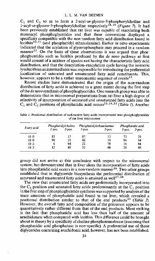
The preferential distribution of saturated and poly-unsaturated fattyacid constituents among the C1- and C2- positions of e.g. rat liver lecitbinshas been the subject of much investigation. Several groups tackled thequestion at which stage of phospholipid metabolism this particular distri-bution of fatty acids was introduced. Various possibilities can be envisaged(Figure 7). The de novo synthesis of lecithins proceeds via acylation ofglycerophosphate so as to give phosphatidic acid1 .After dephosphorylation
HO UU-Cho1ine
0 H
H0Figure 7. Pathway of phosphoglyceride biosynthesis and renewal of fatty acid constituents.Control of positional distribution of saturated (S) and unsaturated (U) fatty acid constituents
of this key-intermediate by phosphatidic acid phosphatase, the diglyceridesaccept phosphorylcholine from cytidine diphosphorylcholine with theparticipation of a choline phosphotransferase'6"7 The lecithins producedby this pathway may be subject to enzymatic hydrolysis by phospholipase A,which cleaves one fatty acid ester linkage so as to give monoacyl-glycero-phosphorylcholine (lysolecithin). It was found that in mammalian tissuessuch as rat liver two lipolytic activities are present denoted as phospholipasesA1 and A2 which are specific in hydrolysing the fatty acid ester linkages at
33
P.A.C.—25/1——C
L. L. M. VAN DEENEN
C1 and C2 so as to form a 2-acyl-sn-glycero-3-phosphorylcholine and1-acyl-sn-glycero-3-phosphorylcholine respectively'8' 19 (Figure 7). It hadbeen previously established that rat liver was capable of reacylating bothmonoacyl phosphoglycerides and that these conversions displayed aspecificity compatible with the non-random fatty acid distribution found inlecithin20'21 (and phosphatidyl ethanolamine). Earlier in vitro experimentsindicated that the acylation of glycerophosphate may proceed in a randommanner22. On the basis of these observations it was argued that phos-phoglycerides such as lecithin produced by the de novo pathway at firstwould consist of a mixture of species not having the characteristic fatty aciddistribution, and that the deacylation—reacylation cycle having the isomericlysolecithins as intermediates was responsible for introducing the preferentiallocalization of saturated and unsaturated fatty acid constituents. This,however, appears to be a rather uneconomic sequence of events23.
Recent studies have demonstrated that the origin of the non-randomdistribution of fatty acids is achieved to a great extent during the first stepof the de novo synthesis of phosphoglycerides. One research group was able todemonstrate that in microsomal preparations from rat liver a high degree ofselectivity of incorporation of saturated and unsaturated fatty acids into theC1 and C2 positions of phosphatidic acid occurs23'24'25 (Table 1). Another
Table 1. Positional distribution of radioactive fatty acids incorporated into phosphoglyceridesof rat liver microsomes
Fatty acid Phosphatidyicholine1-pos. 2-pos.
Phosphatidylethanolamine1-pos. 2-pos.
Phospha1-pos.
tidic acid2-pos.
16:0 83 17 87 13 72 2818:0 90 10 86 14 86 1418:2 6 94 22 78 16 8418:3 12 88 32 68 20 80
group did not arrive at this conclusion with respect to the microsomalsystem, but demonstrated that in liver slices the incorporation of fatty acidsinto phosphatidic acid occurs in a non-random manner26. Two other groupsestablished that in diglyceride biosynthesis the preferential distribution ofsaturated and unsaturated fatty acids is attained as well27' 28
The view that unsaturated fatty acids are preferentially incorporated intothe C2 position and saturated fatty acids predominantly at the C1 positionin the first step of phosphoglyceride synthesis was supported by analysis of thetrace amounts of phosphatidic acid found in rat liver, which revealed apositional distribution similar to that of the end products25 (Table 2).However, the overall fatty acid composition of the precursor appears to bequantitatively rather different from that of the end products. Most strikingis the fact that phosphatidic acid has less than half of the amount ofarachidonate when compared with lecithin. This difference could be broughtabout in theory by a specificity of choline phosphotransferase (assuming thatphosphatidic acid phosphatase is non-specific). A preferential use of thosediglycerides containing arachidonic acid, however, has not been established.
34
PHOSPHOLIPIDS AND MEMBRANES
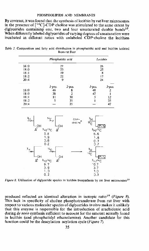
By contrast, it was found that the synthesis of lecithin by rat liver microsomesin the presence of ['4C]-CDP choline was stimulated to the same extent bydiglycerides containing one, two and four unsaturated double bonds29.When differently labeled diglycerides of varying degrees of unsaturation wereincubated in different ratios with unlabeled CDP-choline the lecithins
Table 2. Composition and fatty acid distribution in phosphatidic acid and lecithin isolatedfrom rat liver
Phosphatidic acid Lecithin
16:0 27 2618:0 23 2518:1 19 818:2 22 1720:4 9 24
16:01-pos.
462-pos.
81-pos.
482-pos.
218:0 38 2 47 1
18:1 11 30 5 1218:2 5 35 1 3520:4 — 25 — 47
cho1ine
3H/14C5.8 5.61.9 1.80.6 0.70.2 2
3i14c 2 3H/1C9.7 10.73.9 3.71.0 1.00.2 0.3
Figure 8. Utilization of diglyceride species in lecithin biosynthesis by rat liver microsomes29
produced reflected an identical alteration in isotopic ratio29 (Figure 8).This lack in specificity of choline phosphotransferase from rat liver withrespect to various molecular species of diglycerides in-vitro makes it unlikelythat this enzyme is responsible for the introduction of arachidonic acidduring de novo synthesis sufficient to account for the amount actually foundin lecithin (and phosphatidyl ethanolamine). Another candidate for thisfunction could be the deacylation—acylation cycle (Figure 7).
35

L. L. M. VAN DEENEN
As mentioned before, it was demonstrated that 1-acyl-glycero-3-phosphoryl-choline preferentially reacts with an unsaturated fatty acylgroup20. The acyl residue in the 1-acyl-GPC appeared to have less influenceon the rate of acyl transfer than the nature of the acyl group on the CoAester30' 31• On the other hand, 2-acyl-GPC preferentially stimulates theuptake of saturated acyl chains. Experiments with synthetic32'33'34 2-acyl-GPC and 1-acyl-GPC having differently labeled acyl chains enabled one tofollow their conversion into molecular species of lecithin35' 36• The resultswere in accordance with the view that 1-acyl-GPC is preferentially acylatedwith unsaturated fatty acids, whereas 2-acyl-GPC is better acylated withsaturated fatty acids (Figure 9). The labeled acyl constituents were recovered
HO 18:218:2
18:2__Figure9. Selective acylation of isomeric lysolecithins by rat liver microsomes36
in newly formed lecithins at essentially the same positions as those at whichthey were originally located in the lysolecithins35' '. Theaction of phospho-lipase A2 and A1 in liver will give rise to the formation of endogenous1-acyl-GPC containing predominantly saturated acids and of 2-acyl-GPCwith unsaturated acids mainly9. The selective acylation of these endogenouscompounds will yield lecithin species with a fatty acid distribution comparableto that occurring in the natural lecitbins. Various observations appear tosupport the view that the deacylation—reacylation cycle not only maintainsthe positional fatty acid distribution but that this pathway may make finaladjustments in the molecular species composition. That arachidonate ismore readily introduced into lecithin than into phosphatidic acid species is inagreement with the observation that poly-unsaturated-CoA esters are betterincorporated into 1-acyl-GPC than into 1-acyl-glycerophosphate37. Asregards the acylation of 1-acyl-GPC a most significant conversion intotetraenoic species was observed38. The dietary experiments dealt with in theprevious section suggested that different molecular species participate to adifferent extent in the metabolic conversions under discussion12' 39 It wasargued that replacement of eicosatrienoic species by those containingarachidonate may be accomplished to a great extent by a deacylation—reacylation cycle, while production of linoleate containing species was moredependent on the de novo synthesis. In vitro experiments with several labeledprecursors appear to support this conclusion40.
36
PHOSPHOLIPIDS AND MEMBRANES
The analytical data raise many questions about the contributions ofseveral pathways to the metabolism of individual species of phosphoglyceridesin many tissues. For instance, the high content of dipalmitoyl-lecithinencountered in lung tissue poses an interesting problem. Current studiesindicate that during de novo synthesis an appreciable quantity of this lecithinspecies may be formed. However, it was most striking that rat lung microsomesin contrast to liver microsomes in the presence of 1-acyl-GPC incorporatedpalmitate to at least the same extent as linoleate41. Thus the deacylation—reacylation cycle may be potentially capable of contributing to the 1,2-dipalmitoyl-GPC formation in lung as well.
The study of the enzymatic processes of phospholipid species is not onlyimportant with respect to questions of the design of tailormade membraneconstituents but also intimately related to problems of biogenesis of mem-branes and the ending of their sometimes short life-span. This area ofresearch involves also the transport of lipids from one membrane to another,as well as the translocation of lipids within one single membrane. Further-more, lipids firmly bound at a given membrane may be subject to intermediaryconversions concerning either their polar headgroups or hydrocarbonchains. Such conversions affecting charge distributions and hydrophobicassociations can induce motion at the membrane site concerned and maycontribute to the dynamic behaviour of the biological interface. Thesereactions may be important to the theme of rapid transformations betweendifferent lipid—protein arrangements perhaps overlapping several conflictingproposals, which incorrectly consider the membrane as a static structure.
3. Chemical synthesis of phosphoglyceride speciesThe analytical and biochemical investigations demonstrate that cells
are equipped with enzymes which are responsible for attaining a particularmolecular make-up of membrane phospholipids. The analytical data onphospholipids still have to be interpreted in terms of precise functions ofthese molecules in biological interfaces. If one makes a comparison with thearea of nucleic acids and protein synthesis it seems that we have reached inphospholipid chemistry a stage comparable to that achieved in the field ofnucleic acids around 1950.
Well-defined phospholipids and other complex lipids have to be subjectedto physical examination using a great variety of techniques. Although it ispossible now to analyse all naturally occurring phospholipids in terms ofmolecular species it is not possible to isolate all the individual members froma given phospholipid class in a pure form. This, however, can be achieved bychemical synthesis. This area of research has attracted during severaldecades only relatively few research groups. Various contributions weremade to the preparation of phospholipids with different polar headgroupsbut often the synthesis was limited to fully saturated compounds, which differfrom natural phospholipids and are often less suitable for physical experi-ments. Later, several research groups concentrated on the chemical synthesesof unsaturated phosphoglycerides. During the past decade the so-calledmixed-acid phosphoglycerides having combinations of two different fattyacid constituents as found in natural phosphoglycerides became available bychemical synthesis. As an example, the pathways of the first reliable syntheses
37

L. L. M. VAN DEENEN
of lecithin and phosphatidyl ethanolamine having a saturated fatty acid atC1 and an unsaturated fatty acid constituent at C2 are given42'43'44(Figure 10). Not only lecithins with many combinations of fatty acidconstituents including poly-unsaturated ones have been prepared, but alsophosphatidyl ethanolamines, phosphatidyl serines, phosphatidic acids and
0 0 0II II II
HC—O—C—R1 H2C—O—C—R H2C—O—C—R1
II I ii II IR2-C-O4C'H — R-C-O4CH o —÷R-C-01CPH0
I I ii I ii .4.
H2CI H2C —O—— OAg H2C—0—P-O-Cl—CI—NCH3OBzl
Phosphatidyl choline
00 HC-O-C-R1
II IAg—Q—p—O—CH2—CH2—N(H)X —+ R-C—OlCH o
BzI ,.j _4L -C--CHNA
Phosphatidyl ethanolamine
Figure 10. Principles for the chemical synthesis of mixed-acid phosphoglycerides45'46
phosphatidyl glycerols and various related phospholipids (see also sectionIII) were synthesized in the mixed-acid form. For a detailed discussion of themerits of different methods of synthesis, the use of new protecting groupsand various modifications, reference has to be made to two reviews whichare complimentary in time45' 46• The synthetic phosphoglycerides withdefined localization of two different fatty acid constituents were of greatvalue in the determination of the mode of action of phospholipase A. In theperiod 1954 to 1963 quite different opinions were expressed with respect to thequestion about the site of attack of this enzyme. A distinction between thevarious possibilities, namely a. a positional unspecific hydrolysis whichdepends on the nature of fatty acid constituents b. a specific action on eitherthe C1 or C2 fatty acid ester linkages could be made on the basis of severalisomeric pairs of synthetic mixed-acid phosphoglycerides475 . It was foundthat phospholipase A from snake venom and pancreas hydrolyses exclusivelythe fatty acid ester linkage at C2 of sn-3-phosphoglycerides irrespective of thenature of the fatty acid constituents (Figure 11). The mode of action of thisenzyme—now denoted as phospholipase A2—offered another attractiveroute52'53 towards the synthesis of mixed-acid lecithin which is frequentlyemployed. A synthesis of lecithins containing two identical fatty acidconstituents can be achieved readily by acylation of sn-glycero-3-phosphorylcholine (GPC prepared by deacylation of natural lecithin).Degradation with phospholipase A2 produces 1-acyl-GPC, which then can
38
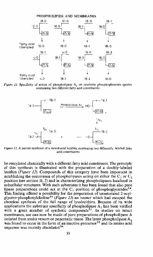
PHOSPHOLIPIDS AND MEMBRANES
18:0 12.0 18:0 18:1
Fatty acid12:0 18:0 18:1 18:0
18:1 4:0 16:0 18:3
NH1INHINHINFatty acidliberated 4:0 18:1 18:3 16:0
Figure 11. Specificity of action of phospholipase A2 on synthetic phosphoglyceride speciescontaining two different fatty acid constituents
18:1 18:1
18:1 Phospholipase HO—
IH2
3H
18:2 i8:O - HO 8:0
Figure 12. A partial synthesis of a mixed-acid lecithin, containing two differently labeled fattyacid constituents
be reacylated chemically with a different fatty acid constituent. The principleof this synthesis is illustrated with the preparation of a doubly-labeledlecithin (Figure 12). Compounds of this category have been important inestablishing the occurrence of phospholipases acting on either the C1 or C2position (see section II, 2) and in characterizing phospholipases localized insubcellular structures. With such substrates it has been found that also purelipase preparations could act at the C1 position of phosphoglycerides54.This finding offered a possibility for the preparation of unsaturated 2-acyl-glycero-phosphorylcholine34 (Figure 13) an isomer which had escaped thechemical synthesis of the full range of lysolecithins. Because of its wideapplications the substrate specificity of phospholipase A2 has been verifiedwith a great number of synthetic compounds51. In studies on intactmembranes, use can now be made of pure preparations of phospholipase Aisolated from snake venom or pancreatic tissue. The latter phospholipase A2was found to occur in the form of an inactive precursor55 and its amino acidsequence was recently elucidated56.
39

L. L. M. VAN DEENEN
18 2__f
Lip7" \<osPhoLiPase A2
18: 2__—[ HO—rFigure 13. Preparation of isomeric lysolecithins by enzymatic hydrolysis of a synthetic lecithin
These examples are quoted to demonstrate briefly that combinations ofmethods of organic chemistry and enzymology are useful also in the fieldof phosphólipids and allow the preparation of molecular species of preciselythe same structure as natural phospholipid families.
4. Properties of phospholipids in model systemsIn order to evaluate the properties of distinct phospholipid species in
relation to their function in biological membranes use can be made ofmethods of surface chemistry, e.g. monomolecular layers and bimolecularlipid membranes which may serve as restricted but in some respects asvaluable models for natural membranes. In this manner, important data canbe provided to the molecular architect attempting to depict functionalmodels of lipid and protein associations for the complex biological interfaces.In this respect it may be useful to include not only the synthetic phospholipids,tailormade according to the pattern provided by nature, but to study relatedstructures not found in nature and to assess why their biosynthesis andincorporation into membranes is avoided by the cell. Conspicuous differencesare often found in the chemical make-up of phospholipids in one membraneunder different conditions as well as between different membranes. Whereasin many cases such structural variations may be reflected by differentproperties of these membrane constituents it is very possible that chemicallydissimilar lipids are rather similar with respect to their physical properties.
The simple system of monomolecular layers of phospholipids which had agreat impact on concepts of membrane structure for more than forty yearsshowed considerable variations in the molecular orientation of differentmolecular species of phospholipids such as lecithin and phosphatidylethanolamine57'58
The mean molecular area occupied by a lecithin molecule at the air/waterinterface appears to increase when the saturated hydrocarbon tails becomeshorter (Figure 14). This shift from a condensed to a more expanded type offilm can be explained by assuming that a decrease in London—van der Waalsinteraction allows a greater mobility of the chains. This is in contrast to thebehaviour of different lysolecithins which revealed little difference in surfacearea, below the collapse pressure59. Introduction of an unsaturated fatty acidconstituent into the lecithin also leads to considerable expansion of the film,and the space occupied by the phospholipid molecule appears to increase
40

PHOSPHOLIPIDS AND MEMBRANES
with increasing unsaturation (Figure 14). In particular, the introduction ofthe first unsaturated fatty acid constituent (or a shorter saturated one)giving an 'asymmetric' molecule appears to have a significant effect on themolecular interactions. In this system there is a fair amount of similaritybetween phosphoglyceride species having at C1 a saturated long-chain fattyacid and at C2 either a mono-unsaturated, a cyclopropane or a saturatedfatty acid constituent of medium chain-length1 (C10, C12, C14). It is intriguing
Figure 14. Force/area characteristics of monomolecular films of synthetic lecithins with differentfatty acid constituents58. (18 :0/18 :1 PC stands for 1-stearoyl-2-oleoyl-sn-glycero-3-phosphoryl-
cho1ine
to note that unicellular organisms have an ability to interchange such fattyacid constituents in their phospholipids provoking the suggestion that underdifferent conditions the physical properties of the membrane phospholipidscan be maintained. Cells appear to avoid the biosynthesis of phospholipidscontaining two saturated fatty acids of medium chain length, e. g. didecanoylor dilauroyl lecithin. It is of interest to note that synthetic lecithins of thistype were found to be highly lytic8 and are apparently not suitable membraneconstituents.
Although some relationships between the behaviour of phospholipids inmonolayer and biological phenomena can be seen, a more relevant approachmay be a study of the permeability properties of bilayers of phospholipidspecies. For this purpose, excellent possibilities are given by liposomes,which consist of a composite array of multiple. concentric closed bimolecularmembranes60. The liposomes behave as osmometers6' allowing one toobtain information on the permeability of non-electrolytes with the samemethods as previously applied to e.g. erythrocytes and mitochondria(Figure 15). As for erythrocyte, the penetration rate into the liposomes ishighly dependent on temperature and on the molecular dimensions of thesolute (e.g. glycol > glycerol > erythritol). Using liposomes made of varioussynthetic lecithin species it could be demonstrated that increase in chain
41
EU
18
a)U0IL
0 20 80
182/18:2 PC
100 120 D
Areo,A /m020 /.0 60 80 100 120 11.0

Erythrocyte
L. L. M. VAN DEENEN
Glycerol
+ H20
LiposorneGlycerol
+ H20
Figure 15. Osmotic behaviour of erythrocytes and liposomes
Figure 16. Effects of chain length and unsaturation on glycerol permeability of liposomes ofsynthetic lecithins. Initial swelling rate in isotonic glycerol as a function of temperature.
The broken line represents the swelling of egg lecithin62
length considerably reduces the penetration rate by glycerol molecules62(Figure 16). It is of interest to note that at physiological temperature aconspicuous difference exists between dipalmitoyl-GPC and distearoyl-GPC, the former is known to be abundant in membranes, whereas thequantity of distearoyl-GPC is very low (section II, 1). One is inclined toconclude that a tight alignment formed by distearoyl-GPC is not appropriate
42
a)000.8
ci)
3U,
D
Temperature, CC

PHOSPHOLIPIDS AND MEMBRANES
for normal membrane function. In addition, a comparison of the permeabilityof the liposomes of 1-stearoyl-2-decanoyl-GPC and 1-stearoyl-2-myristoyl-GPC with those obtained from 1,2-dimyristoyl-GPC and 1,2 dipalmitoyl-GPC respectively, suggests that the model structures of phospholipids withtwo different fatty acid constituents are much more permeable than those oflecithins with two chains of equal length and the same number of paraffincarbon atoms. In accordance with the expectations from the monolayerstudies, introduction of double bonds in the hydrocarbon chains causes anincrease of permeability (Figure 16). It appears that the permeability of theliposomes from lecithins with one saturated and one mono-unsaturatedchain is very close to those of lecithins with a saturated long-chain and asaturated medium-length chain. Liposoms of lecithin species containingtwo polyunsaturated fatty acids, which species are very rare in mammaliancells (section II, 1) appear to be very leaky. The observations on the influenceof the fatty acid constituents of the phospholipids on glycerol and erythritolpermeability are in agreement with data on the glucose leak from liposomes63.
These model experiments on systems composed of phospholipids only,suggest that 'simple diffusion' through a lipid barrier may be highly dependenton the degree of packing and thermal motion of the apolar chains. Althoughin biological membranes the situation is more complex, the variousobservations suggest that in these interfaces a selection of the make-up of thelipid species may contribute so as to regulate the properties of a membrane.For example, the observation made with model systems, that an increase inthe number of unsaturated lipid species enhances permeability, is of interestin relation to the adaptation of organisms towards lower environmentaltemperatures. For E. coli it has been demonstrated that when the temperatureof growth decreases there is a marked decrease in saturated and an increasein unsaturated and cyclopropane fatty acid constituents64' 65• The perme-ability of liposomes of the bacterial phospholipids revealed differences to beexpected from the work with synthetic phospholipids suggesting that thebacteria attempt to counteract a decrease in permeability at lower temperatureby increasing the degree of unsaturation of membrane lipids65 (Figure 17).
Interesting possibilities are offered by the isolation of mutants of E. coliwhich require an unsaturated fatty acid for growth66. Fatty acids of quitedifferent structure can be incorporated into the phospholipids indicatingthat the membrane is rather tolerant in this respect. However, it was recentlydemonstrated that these mutants also control the fatty acid composition oftheir lipids to a notable extent67. When, for instance, the degree of un-saturation of the supplemented cis-fatty acid was increased, relatively moresaturated hydrocarbon chains and fewer unsaturated chains were found to beincorporated into the phospholipids. With a series of cis-mono-unsaturatedfatty acids of different chain length it was found that the percentage ofunsaturated fatty acid constituents of the phospholipids increases withincreasing chain length. The conclusion that there exists a regulatorymechanism which has to maintain the physical properties of the membranelipids within certain limits appears to be supported by current studies onmonolayers and bilayers of the phospholipids from these mutants68.
The impact of the nature of phospholipid species for the physical state andproperties of the natural membrane—as suggested by model studies—in
43

L. L. M. VAN DEENEN
principle should be demonstrated also by direct comparisons between cellsof different lipid composition and the behaviour of liposomes. Goodpossibilities are offered by Mycoplasma laidlawii which has relatively simplespecies composition of the membrane lipids which can be altered to a givenextent by supplementing different fatty acids to the medium69. Measurement
G[ycerol permeability of lipo- Fotty acid composition ofsomes from lipids of F. coil phospolipids from F. coil
0/0
I- 200 30°
_________
I__________
Saturated Unsaturated + cyclopropaneTemperature,0C
Figure17. Effect of growth temperature of E. coli on the fatty acid composition of the phospho-lipids (right) and the permeability behaviour of liposomes65 (left)
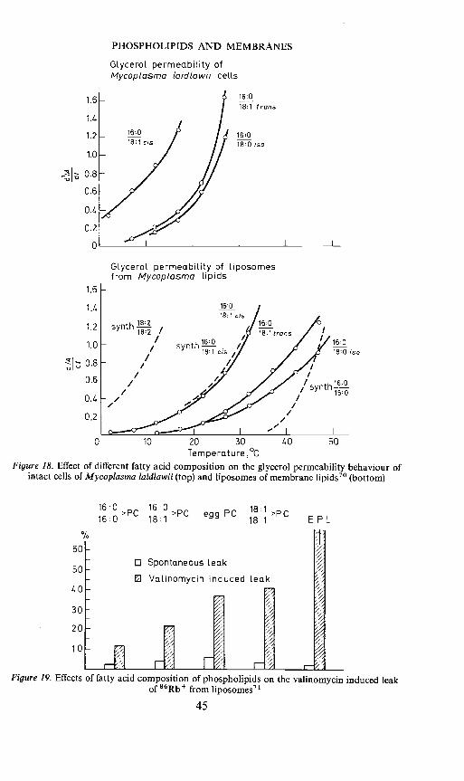
of glycerol diffusion into intact cells and liposomes from the Mycoplasmalipids showed that in both the natural and artificial membranes permeabilityfor glycerol depends on the fatty acid composition of membrane lipids, andthat the shifts in permeability observed in Mycoplasma cells and liposomesare in good agreement7° (Figure 18). These studies confirm that there is acertain degree of tolerance in fatty acid make-up of membrane lipids buton the other hand, the composition can vary only between given limitswithout effecting the viability of the cells. There appear to be restrictions withrespect to the degree of both unsaturation and saturation of the phospho-lipid species.
The barrier properties of phospholipids as dictated by the chemical natureof the apolar chains may not only contribute to the control of permeation ofnon-electrolytes but perhaps also affect the rate of other transport processes.This suggestion is made on the basis of observations on the valinomycininduced Rb leak from liposomes71. It was found that the promoting effectof valinomycin is determined by the degree of unsaturation of the phospho-lipid species present in the bilayers (Figure 19). The concept that within thecell, carrier mediated transport may be dependent on the nature of the lipidconstituents is being tested with biological membranes of different lipidcomposition.
44
80
60 -
20 -
0 10 20 30
PHOSPHOLIPIDS AND MEMBRANES
Glycerol permeability ofMycoplasrna (aid/awl i celLs
Glycerol permeability of liposornesfrom Mycoplasma Lipids
0 10 20 30Temperature ,°c
Figure 18. Effect of different fatty acid composition on the glycerol permeability behaviour ofintact cells of Mycoplasma laidlawii (top) and liposomes of membrane lipids70 (bottom)
16:018:1 cis
16018:1 trans
16:0180 iso
1.6 -
1.4 -
1.2 -
1.0
0.8
0.6
0.4
0.2
0
16:018:1 cis
1.6
1.4
1.2
1.0
0.80.6
0./.
0.2
synth //synth1-/ 18:1 cis///
40 50
16:0 16:0>Pc >Pc160 181 egg t 18:1 Pc
18:1
0 Spontaneous leak
0 VaLinomycin induced Leak
E FL
//
I
60
50
40
30
20
10
Figure 19. Effects of fatty acid composition of phospholipids on the valinomycin induced leakof 86Rb from Iiposomes7'
45
un7///////

L. L. M. VAN DEENEN
Apart from phospholipids, other membrane constituents can control thediffusion across biological interfaces. Experiments on monomolecularlayers57'58 and bilayers62'63'72 of well-defined phospholipid species demon-strated an effect of cholesterol. Liposomes of mixtures of phospholipidand cholesterol normally demonstrate a decrease in permeability which isproportional to the concentration of cholesterol. The presence of cholesterolappears to limit the penetration of glycerol and erythritol, the diffusion ofglucose through the bilayers, but also to reduce valinomycin inducedpermeability of Rb + Extrapolation of this finding to natural membranessuggesting that the prevailing effect will be again a restriction of permeationin cholesterol rich regions of the membrane is supported by experiments witherythrocytes and Mycoplasma. The erythrocyte which has a molar ratio ofphospholipidcho1esterol close to one can be depleted by part of its sterol.After removal of part of the cholesterol the erythrocytes exhibited aconsiderable increase in osmotic fragility and glycerol permeability73.Conversely, cells of Mycoplasma which contained cholesterol in the membranerevealed a decrease in glycerol permeability when compared with cellsdevoid of cholesterol70.
Although many correlations can be made between model systems andnatural membranes such observations do not lead of course to the conclusionthat the membranes can be considered as continuous lipid-bilayers coatedwith protein. While some regions of a membrane may have such structuresit can also be envisaged that a considerable degree of interpenetration of thelipid core by protein exists. The temperature dependence of the glycerolpermeation into liposomes of total lipid (phospholipid plus cholesterol) fromerythrocytes and the behaviour of the intact erythrocytes show somedifferences74 indicating that the proteins also control the degree of mobilityof the structural elements in the natural hydrophobic barrier. Recombinationexperiments with the complex mixture of proteins solubilized from erythrocytemembranes indicated that their association with lipids involves for initialassociation polar interactions followed by the formation of apolar bonds75' 76•These experiments endorse the view that a heterogeneity exists in the bindingof lipids to proteins in the erythrocyte membranes. Attempts to reconstitutein vitro membrane structures containing lipids and proteins from erythrocyteswhich have the same permeability characteristics as intact erythrocytes so farwere not successful. Experiments with single protein components isolatedfrom the membrane may be more rewarding.
Ill. POLYGLYCEROLPHOSPHOLWIDSTo date, the polar headgroups of phospholipids have been thoroughly
investigated in a great number of biological membranes. Considerablevariations in phospholipid composition exist, not only between biologicallydistinct membranes but sometimes also between functionally identicalmembranes77. Most membranes contain a number of phospholipids whichdiffer with respect to the nature of differently charged polar headgroups. Thisheterogeneity in hydrophilic groups has given support to the idea that thepolar headgroups of phospholipids may be involved in a number of functions,
46
PHOSPHOLIPIDS AND MEMBRANES
viz, interaction with charged sidegroups of proteins, binding of cations andmerely speculatively, as carriers in transport phenomena.
In many mammalian membranes the proportions of phospholipids withdifferent headgroups are relatively constant for a given membrane and appearto be genetically determined. More flexibility is often revealed by bacterialmembranes. In recent years, several new phospholipids have been detectedin bacteria and some examples of this area of natural product chemistrywill be discussed briefly.
PhosphatidyiglycerolThis phospholipid which was first detected in algae is an important
constituent ofchloroplasts78. This compound is present in minor amounts inmammalian tissues but is abundant in many bacteria. The stereochemicalconfiguration (Figure 20) was established to be 3-sn-phosphatidyl-1'-sn-glycerol3, this being in agreement with the biosynthetic pathway79 involving
0o H2C—0——R1 H2C—OH
R2-c-01CH o H1C0HH2C—O--P—0—CH20
Phosphatidyl glycerol
oII
o H2C—0-C— R1 H2C —0—P-0—CH2II I I II
R2-C-01CH H4C0H H4C0—C-R2H2C—0—P—0—CH2 H2C—0—C—R1
0
Di—phosphatidyl glycerol(Cardiol ipin)
Figure20. Two major polyglycerol phospholipids
a reaction with CDP-diglyceride and glycerol-3-phosphate producing as anintermediate 3-sn-phosphatidyl-1 '-glycerol-3'-phosphate. The stereochemicalconfiguration was confirmed by chemical synthesis80.
DiphosphatidyiglycerolThis phospholipid usually denoted as cardiolipin is by no means unique
for bacteria. It is well established that it is a quantitatively importantconstituent of the inner membrane of mitochondria, but its precise function
47
L. L. M. VAN DEENEN
is unknown. Cardiolipin has been the subject of many investigations andseveral structures have been proposed. A chemical synthesis of diphosphati-dyiglycerol and several related compounds confirmed that beef heartcardiolipin is identical to diphosphatidylglycerol81 (Figure 20). A conclusiveproof was obtained by breakdown of synthetic and natural phospholipidwith phospholipase C, which gave in both cases, 1,2-diacylglycerol andglycerol-1,3-diphosphoric acid. It is not to rule out, however, the occurrencein nature of other polyglycerol phospholipids closely related to diphosphati-dylglycerol. Some indications were obtained that in a bacterium an acylderivative of diphosphatidyiglycerol may occur82'83
Amino acid esters of phosphatidyiglycerolA class of phospholipids which seems so far to be unique for bacterial
membranes is represented by O-aminoacyl derivatives of phosphatidyl-glycerol84' 85 In gram-positive bacteria, L-lysine and L-alanine appear tobe the predominant amino acids, but in Mycoplasma both D- and L-alanlnewere reported to be linked to phosphatidylglycerol86. The lysine ester ofphosphatidyiglycerol was isolated in a pure form and by chemical andenzymatic hydrolysis, the stereochemical configuration was established(Figure 21). It could be concluded that the compound was 3-sn-phosphatidyl-
or 3' -O-L-lysyl)-sn-glycerol87, but the position of alkaline labile linkage
r0-Ly5 r-Ri rOHI I - I G—3P > 950/0
OH R2 2!_,, HO Dhydrognase CONVERSION
LOH
GIyc2roII
PHOSPHOLIPASE D
1.—R1 —0—LYS rR1 EOH
R2 OH R2 OH + L-Lysinc(DcarboxyIas)
PHOSPHOLIPASE C
0—LYS R1 OHr T r G-3--P NOOH R2 OH Dhydrog2nas REACTION
LOH
Figure 21. Enzymatic degradations utilized for the structural comparison of naturaj andsynthetic O-lysyl-phosphatidylglycerol8791
of the lysyl moiety remained uncertain (Figure 22). This question was tackledby various approaches. Several groups undertook the chemical synthesis ofthis phospholipid class88'89' 90 As an example, the synthesis of 3-sn-phosphatidyl-1'-(3'-O-L-lysyl)-sn-glycerol is gi yen (Figure 23) .Thesynthetic
48

PHOSPHOLIPIDS AND MEMBRANES
o 0ii II
o H2C —o —C —R1 H2C —0—C -CH—(CH2)4— NHII I NHR2-C-01CH , H4C'0H 3
H2C—0—P--O —CH200
o H2C—0——R1 H2C—0H0II I H 4.
R2—C—01CH H4C0—C—CH—(CH2)4—NH3I U I NH
H2C—O—P—0—CH2 3
0Figure 22. Isomers of O-lysyl-phosphatidylglycerol
o o NH—t.BocII II I
o H2C—O—C—R1 H2C — 0—C —CH—(CH2)4—NH—t.Boc
R2-—04CH0 ÷ H1co-tButH2C —O—P—0—Ag I CH
O—BzI
Q 0 NH—t.BocII II I
—
o H2C—O—C — R, H2C—0 —C —CH—(CH2)4—NH--tBoc
o HC.O—t.But.H2C—0—P—O—CH2
O-BzI
o H2C—O--C---R1 H2C—0—c! —CH—(CH2)4—NHII I I
R2-C—O4CH HICOH 3
H2C — 0—P —o CH20
Figure 23. A chemical synthesis of 3-sn-phosphatidyl-1'-(3'-O-L-lysyl)-glycerol9'
compound was compared with the natural product and it was found thatboth substances were completely identical. Of particular interest is the similarenzymatic hydrolysis of both compounds by phospholipases C and D(Figure 22). The first enzyme gave a complete hydrolysis into a 1,2-diglycerideand a water-soluble product identical to synthetic O-L-lysyl-glycerol-phosphate. Phospholipase D action produced phosphatidic acid and onewater-soluble compound identical to synthetic 1-O-lysyl-glycerol91. The
49
L. L. M. VAN DEENEN
latter observation indicates that in the natural product the lysine wasesterified to the primary hydroxyl function just as in the synthetic phospho-lipid. The possibility of a migration of the lysyl residue from the 2' to the 3'position during the isolation of the phospholipid or its enzymatic hydrolysiswas not ruled out. However, one research group active in this field synthesizedisomers having the lysyl moiety linked to either the primary or secondaryhydroxyl function (Figure 22) and found that migration from the 2' to the 3'position does not occur during chromatography on silicic acid in acidic andneutral systems92. Furthermore they found that both isomers displayed adifferent chromatographic behaviour. These joint observations endorsed theview that the phospholipid isolated from St. aureus occurs in the bacteria as3-sn-phosphatidyl-1'-(3'-O-L-lysyl)-sn-glycerol.
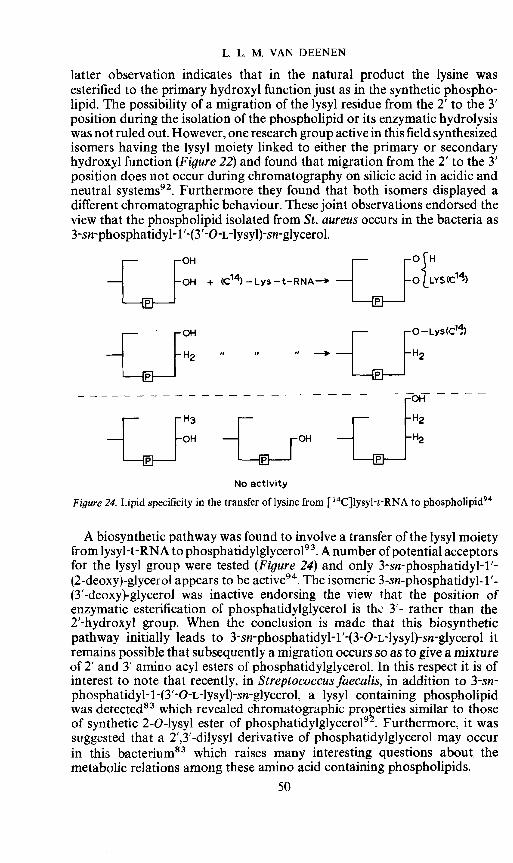
OH + 14 — Lys — t-RNA O{LYS(C14)
fIJ _J
No activity
Figure24. Lipid specificity in the transfer of lysine from [14CJlysyl-t-RNA to phospholipid94
A biosynthetic pathway was found to involve a transfer of the lysyl moietyfrom lysyl-t-RNA to phosphatidylglycerol93. A number of potential acceptorsfor the lysyl group were tested (Figure 24) and only 3-sn-phosphatidyl-1 '-(2-deoxy)-glycerol appears to be active94. The isomeric 3-sn-phosphatidyl-1'-(3'-deoxy)-glycerol was inactive endorsing the view that the position ofenzymatic esterification of phosphatidyiglycerol is thL 3'- rather than the2'-hydroxyl group. When the conclusion is made that this biosyntheticpathway initially leads to 3-sn-phosphatidyl-1'-(3-O-L-lysyl)-sn-glycerol itremains possible that subsequently a migration occurs so as to give a mixtureof 2' and 3' amino acyl esters of phosphatidyiglycerol. In this respect it is ofinterest to note that recently, in Streptococcus faecalis, in addition to 3-sn-phosphatidyl-1 -(3'-O-L-lysyl)-sn-glycerol. a lysyl containing phospholipidwas detected83 which revealed chromatographic properties similar to thoseof synthetic 2-O-lysyl ester of phosphatidylglycerol92. Furthermore, it wassuggested that a 2',3'-dilysyl derivative of phosphatidyiglycerol may occurin this bacterium83 which raises many interesting questions about, themetabolic relations among these amino acid containing phospholipids.
50
PHOSPHOLIPIDS AND MEMBRANES
Ghicosamme derivatives of phosphatidyiglycerolRecent reports on the occurrence of glucosaminyl—phosphatidyiglycerol
in Bacillus megaterium95'96 and Pseudomonas ovalis97 enlarged the seriesof phospholipids derived from phosphatidyiglycerol. A compound isolated
0o H2C—O — C — R1 H2C -OH ,O— OHii I I /HOO-J
R2—C—Q4CH H1CPO_H_,./H2C—O—P—O—CH2 OH
OH
O /01\OHO H2C -0- — R1 H2C —0 —'47
R-C-O1CH o H4COH OH
H2C— O-P—O —CH2OH
Figure 25, Isomers of glucosaminyl-phosphatidyiglycerol
0HC—O-—R1 H2C—O-GIcN--X
R2-C-OCP-H + H4C0LBut.
H2C—0—P—OAg I-CH2OBzt
0o H2C-O-C—R1 H2C-0—GIcN--X
R_C-01C-H H-4C-OtBut.
HC— O—P—OOBzI
i)Ba12I 2)HCL, 3)HN—NH; pH=7.5,5O
0 OH
(j) H2C—O-—R1 H2c—O-_<R-C-O-4C-H o H1C-OH OH
H2C— O—P—O—CH2OH
Figure26. Chemical synthesis of a glucosaminyl-phosphatidylglycerol'°2
51
L L M. VAN DEENEN
from a B. megaterium (MK 1OD) was subjected to a variety of chemicaland enzymatic hydrolysis procedures and was proven to be identicalto 3-sn-phosphatidyl- 1'-[2-(2-amino-2-deoxy-3 -D-glucopyranosyl)]-sn-glycerol98. This structure was confirmed by chemical synthesis99. However,evidence was presented that apart from the 2'-isomer also the 3'-glucosaminylderivative of phosphatidyiglycerol occurs in this bacterium100' 101 (Figure 25).Both compounds were subsequently synthesized102 (compare Figure 26)anda comparison with the pure isolated compounds by chemical and enzymaticprocedures demonstrated their identity103. The ratio of the 2' and 3'-glucosamine derivative of phosphatidyiglycerol appeared to be dependenton the growth conditions. The biosynthesis of these compounds is notelucidated but it can be suggested that a reaction between UDP-glucosamineand phosphatidyiglycerol may be involved.
Functional aspectsAs regards the functions of the amino acid and glucosamine containing
phospholipids one could speculate about a function in transport or a role asdonors in the biosynthesis of macromolecules. However, no conclusiveevidence has come forward to prove that these functions can be attributed tothese phospholipid classes. Recent results even appear to argue against suchfunctions'°4. Alternatively, the possibility exists that these phospholipidsare mainly structural components essential for particular molecular arrange-ments within the plasma membrane or intracellular membranes of bacteria.Particularly the lysine ester(s) of phosphatidyiglycerol are conspicuousbecause these phospholipids are unique in having at normal cell pH a netpositive charge. It may be that the positively charged endgroup is ofparamount importance for interaction with protein constituents or fordonating a given surface charge to the membrane. Both functions are notmutually exclusive. A possible structural function of these lipids finds somesupport in observations that the ratio of amino acyl phosphatidylglycerolto the negatively charged phosphatidyiglycerol can increase under certainenvironmental conditions such as an acidic pH86'87"°4 It can be speculatedthat this is a physiological reaction to counteract the increased protonconcentration at the cell surface so as to preserve the integrity of themembrane. Actually it has been known for several years that in membranemodel systems such as liposomes the presence of positively chargedsurfactants causes a further restriction of the diffusion of cations through thelipid barrier60. Recently, it was reported that bilayer membranes made of lysylphosphatidyiglycerol were more permeable to chloride ions than to protons,whereas, the reverse was true for membranes composed of phosphatidyl-glycerol'°5. This selectivity of thin lipid barriers composed of phospholipidwith different polar headgroups is in agreement with observations made onliposomes composed of various bacterial phospholipids. In addition it wasfound that the valinomycin mediated exchange of cations is strongly reducedin liposomes containing lysyl phosphatidyiglycerol' °'. The suggestion thatthe chemical make-up of the phospholipid headgroup controls not only thesimple diffusion, but also the rate of carrier-mediated transport of ions needsto be verified on natural membranes.
52

PHOSPHOLIPIDS AND MEMBRANES
CONCLUDING COMMENTSProgress has been made with respect to the precise chemical characteriza-
tion of membrane lipids. Details have been elucidated about the lipidcomposition of a great variety of biological membranes. Molecular species ofvarious phospholipid classes have been chemically synthesized. Studies onmonolayers and bilayers continue to contribute to the evaluation of barrierproperties relevant to their function in biological membranes. This approachmay be particularly useful when combined with the induction of chemicalvariation in the lipid components of membranes of cells and measurementsof changes in properties. The chemical nature of the hydrocarbon tails ofphospholipids appears to determine to a significant extent the diffusionprocesses but likely also, the rate of carrier-mediated transport across cellmembranes. Membranes appear to tolerate a certain degree of flexibility inthe chemical nature of the apolar moieties but depending on the cell typethis variation is limited. Enzyme systems involved in the biosynthesis ofindividual molecular species of phospholipids display specificities whichcontrol the physical properties of phospholipids within certain limits. Afunction of the polar headgroups of phospholipids in regulating surfaceproperties and associations with protein constituents is likely but this area ofresearch needs further exploration, also with respect to the understanding ofcatalytically active proteins which depend on the presence of particular lipidconstituents for their activity. The advances made in lipid chemistry andmembrane model systems can be expected to promote in the near future abetter understanding of the electrostatic and hydrophobic associationsbetween lipids and proteins in biological interfaces.
ACKNOWLEDGEMENTSThe author enjoyed and benefited from a stimulating cooperation with
many colleagues in the laboratory as well as in other departments. It isimpossible to give all the names individually but a number are to be found inthe references. I feel most indebted to Dr G. H. de Haas and Dr J. de Gier fortheir continuous contributions to the research programme of this laboratoryduring the past decade.
REFERENCESL. L. M. van Deenen, in Progress in the Chemistry of Fats and other Lipids (Ed: R. T. Holman),Vol. VIII, Part 1, p 1. Pergamon: Oxford and New York (1965).
2 L. J. Morris, J. Lipid Res. 7, 717 (1966).F. Haverkate and L. L. M. van Deenen, Biochim. Biophys. Acta, 106, 78 (1965).L. M. G. van Golde, V. Tomasi and L. L. M. van Deenen, Chem. Phys. Lipids, 1, 282 (1967).L. Marai and A. Kuksis, J. Lipid Res. 10, 141 (1969).
6 L. M. G. van Golde and L. L. M. van Deenen, Chem. Phys. Lipids, 1, 157 (1967).A. Montfoort, L. M. G. van Golde and L. L. M. van Deenen, Paper submitted.
8 F. C. Reman and L. L. M. van Deenen, Biochim. Biophys. Acta, 137, 592 (1967).F. C. Reman, R. A. Demel, J. de Gier, L. L. M. van Deenen, H. Eibl and 0. Westphal,Chem. Phys. Lipids, 3, 221 (1969).'° Th. E. Morgan, D. 0. Tinker and D. J. Hanahan, Arch. Biochem. Biophys. 103, 54 (1963)." L. M. G. van Golde and L. L. M. van Deenen, Biochim. Biophys. Acta, 125, 496 (1966).
53
L. L. M. VAN DEENEN
12 L. M. G. van Golde, W. A. Pieterson and L. L. M. van Deenen, Biochim. Biophys. Acta, 152,84(1968).
13 R. M. Johnson, Exp. Cell. Res. 32, 118 (1963).14 M. Waite and L. M. G. van Golde, Lipids, 5, 449 (1968).
A. Kornberg and W. E. Pricer, J. Biol. Chem. 204, 344 (1953).16 E. P. Kennedy and S. B. Weiss, J. Biol. Chem. 222, 193 (1956).
E. P. Kennedy, Federation Proc. 20, 934 (1961).H. van den Bosch and L. L. M. van Deenen, Biochim. Biophys. Acta, 84, 234 (1964).
19 H. van den Bosch and L. L. M. van Deenen, Biochim. Biophys. Acta, 106, 326 (1965).20 W. E. M. Lands and I. Merki, J. Biol. Chem. 238, 898 (1963).21 Merki and W. E. M. Lands, J. Biol. Chem. 238, 905 (1963).22 W. E. M. Lands and P. Hart, J. Lipid Res. 5, 313 (1964).23 L. L. M. van Deenen, H. van den Bosch, L. M. G. van Golde, G. L. Scherphof and B. M.
Waite, in Cellular Compartrnentalization and Control of Fatty Acid Metabolism. Proceedingsof the Fourth Meeting of the Federation of European Biochemical Societies (Ed: F. C.Gran), p 89 (1968).
24 G. L. Scherphof, Thesis, University of Utrecht (1967).25 F. Possmayer, G. L. Scherphof, T. M. A. R. Dubbelman and L. L. M. van Deenen, Biochim.
Biophys. Acta, 176, 95 (1969).26 E. E. Hill, D. R. Husbands and W. E. M. Lands, J. Biol. Chem. 243, 4440 (1968).27 J Elovson, B. Akesson and G. Arvidson, Biochim. Biophys. Acta, 176, 214 (1969).28 P. K. Raju and R. Reiser, Biochim. Biophys. Acti, 202, 212 (1970).29 J. B. Mudd, L. M. G. van Golde and L. L. M. van Deenen, Biochim. Biophys. Acta, 176,
547 (1969).30 A. E. Brandt and W. E. M. Lands, Biochim. Biophys. Acta, 144, 605 (1967).31 H. van den Bosch, L. M. G. van Golde, H. Eibl and L. L. M. van Deenen, Biochim. Biophys.
Acta, 144, 613 (1967).32 G. H. de Haas and L. L. M. van Deenen, Biochim. Biophys. Acta, 106, 315 (1965).
A. J. Slotboom, G. H. de Haas and L. L. M. van Deenen, Chem. Phys. Lipids, 1, 317 (1967).A. J. Slotboom, G. H. de Haas, G. J. Burbach-Westerhuis and L. L. M. van Deenen, Chem.Phys. Lipids, 4, 30 (1970).H. van den Bosch, L. M. G. van Golde, A. J. Slotboom and L. L. M. van Deenen, Biochim.Biophys. Acta, 152, 694 (1968).
36 H. van den Bosch, A. J. Slotboom and L. L. M. van Deenen, Biochim. Biophys. Acta, 176,632 (1969).E. E. Hill and W. E. M. Lands, Biochim. Biophys. Acta, 152, 144 (1968).
38 H. Kanoh, Biochim. Biophys. Acta, 176, 756(1969).A. Catala and R. R. Brenner, Lipids, 2, 84 (1967).
40 L. M. G. van Golde, G. L. Scherphof and L. L. M. van Deenen, Biochim. Biophys. Acta, 176,635 (1969).
41 A. Montfoort, Thesis, University of Utrecht (1970).42 G. H. de Haas and L. L. M. van Deenen, Rec. 7av. Chim. Pays- &as, 80, 951 (1961).
F. J. M. Daemen, G. H. de Haas and L. L. M. van Deenen, Rec. fl-av. Chim. Pays- Baa, 81,348 (1962).F. J. M. Daemen, G. H. de Haas and L. L. M. van Deenen, Rec. 7Jav. Chim. Pays-Baa, 82,487 (1963).L. L. M. van Deenen and G. H. de Haas, in Advances in Lipid Research (Eds: R. Paolettiand D. Kritchevsky), Vol. II,p 167. Academic Press: New York and London (1964).
46 A. J. Slotboom and P. P. M. Bonsen, Chem. Phys. Lipids, in press (1970).G. H. de Haas, I. Mulder and L. L. M. van Deenen, Biochem. Biophys. Res. Commun. 3,287 (1960).
48 G. H. de Haas and L. L. M. van Deenen, Biochim. Biophys. Acta, 48, 215 (1961).G. H. de Haas, F. J. M. Daemen and L. L. M. van Deenen, Biochim. Biophys. Acta, 65, 260(1962).
50 L. L. M. van Deenen, G. H. de Haas and C. H. T. van Heemskerk, Biochim. Biophys. Acta,67, 295 (1963).
51 L. L. M. van Deenen and G. H. de Haas, Biochim. Biophys. Acta, 70, 538 (1963).52 G. H. de Haas and L. L. M. van Deenen, Tetrahedron Letters, 22, 7 (1960)." D. J. Hanahan and H. Brockerhoff, Arch. Biochem. Biophys. 91, 326 (1960).
G. H. de Haas, L. Sarda and J. Roger, Biochim. Biophys. Acta, 106, 638 (1965).
54
PHOSPHOLIPIDS AND MEMBRANES
0. H. de Haas, N. M. Postema, W. Nieuwenhuizen and L. L. M. van Deenen, Biochim.Biophys. Acta, 159, 118 (1968).
56 s Maroux, A Puigserver, V. Dlouha, P. Desnuelle, 0. H. de Haas, A. J. Slotboom, R P. M.Bonsen, W. Nieuwenhuizen and L. L. M. van Deenen, Biochim. Biophys. Acta, 188, 351(1969)." L. L. M. van Deenen, U. M. T. Houtsmuller, C. H. de Haas and F. Mulder, J. Pharm.Pharmacol. 14, 429 (1962).
58 R. A. Demel, L. L. M. van Deenen and B. A. Pethica, Biochim. Biophys. Acta, 135, 11(1967).H. Eibl, R. A. Demel and L. L. M. van Deenen, J. Colloid Sciences, 29, 381 (1969).
60 A. D. Bangham, in Progress in Biophysics and Molecular Biology (Eds: J. A. V. Butler andD. Noble), p 29. Pergamon: Oxford and New York (1968).
61 A. D. Bangham, J. de Gier and C. D. Greville, Chem. Phys. Lipids, 1, 225 (1967).62 j de Gier, J. G. Mandersloot and L. L. M. van Deenen, Biochim. Biophys. Acta, 150, 666
(1968).63 R. A. Demel, S. C. Kinsky, C. B. Kinsky and L. L. M. van Deenen, Biochim. Biophys. Acta,
150, 655 (1968).64 A. G. Marr and J. L. Ingraham, J. Bacteriol. 84, 1260 (1962).65 c W. M. Haest, J. de. Gier and L. L. M. van Deenen, Chem. Phys. Lipids, 3, 413 (1969).66 D. F. Silbert, F. Ruch and P. R. Vagelos, J. Bacteriol. 95, 1658 (1968).67 M. Esfahani, E. M. Barnes and S. J. Wakil, Proc. Nat. Acad. Sci. Wash. 64, 1057 (1969).68 R. A. Demel, L. L. M. van Deenen, S. 1. Wakil, M. Esfahani and E. M. Barnes, Unpublished
work.69 R. N. McElhaney and M. E. Tourtelotte, Biochim. Biophys. Acta, 202, 120 (1970).70 R. McElhaney, J. de Gier and L. L. M. van Deenen, Paper submitted (1970).71 J.de Gier, C. W. M. Haest, J. G. Mandersloot and L. L. M. van Deenen, Biochim. Thophys.
Acta, in press (1970).72 j de Gier, J. G. Mandersloot and L. L. M. van Deenen, Biochim. Biophys. Acta, 173, 143
(1969).K. R. Bruckdorfer, R. A. Demel, J. de Gier and L. L. M. van Deenen, Biochim. Biophys. Acta,181, 334 (1969).1. de Gier, R. A. Densel and L. L. M. van Deenen. in Surface-active Lipids in Food. S.C.I.Monogr. No. 32, p 39. London (1968).R. F. A. Zwaal and L. L. M. van Deenen, Chem. Phys. Lipids, 4, 80 (1970).
76 V. B. Kamat, D. Chapman, R. F. A. Zwaal and L. L. M. van Deenen, Chem. Phys. Lipids,4, 71(1970).L. L. M. van Deenen and J. de Gier, in The Red Cell (Eds: Ch. Bishop and D. M. Surgenor),Chapter 7, p 243. Academic Press: New York (1964).
78 A. A. Benson and B. Maruo, Biochim. Biophys. Acta, 27, 189 (1959).J. Y. Kiyasu, R. A. Pieringer, H. Paulus and E. P. Kennedy, J. Biol. Chem. 238, 2293 (1963).
80 P. P. M. Bonsen, G. H. de Haas and L. L. M. van Deenen, Chem. Phys. Lipids, 1, 33 (1966).81 G.H. de Haas, P. P. M. Bonsen and L. L. M. van Deenen, Biochim. Biophys. Acta, 116, 114
(1966).82 F. A. Ibbott and A. Abrahams, Biochemistry, 3, 2008 (1964).83 j M. dos Santos Mota, J. A. F. Op den Kamp, H. M. Verheij and L. L. M. van Deenen,
Paper submitted (1970).84 M. G. Macfarlnne, Nature, London, 196, 136 (1962).85 u M. T. Houtsmuller and L. L. M. van Deenen, Biochim. Biophys. Acta, 70, 211 (1963).86 W. L. Koostra and P. F. Smith, Biochemistry, 8, 4794 (1969).87 U. M. T. Houtsmuller and L. L. M. van Deenen, Biochim. Biophys. Acta, 106, 564 (1965).88 P. P. M. Bonsen, G. H. de Haas and L. L. M. van Deenen, Biochim. Biophys. Acta, 106, 93
(1965).89 F. Baer and K. V. Jagannadha Rao, J. Am. Chem. Soc. 87, 135 (1965).90 L. D. Bergelson and J. G. Molotkovsky, Tetrahedron Letters, 1(1966).91 P. P. M. Bonsen, G. H. de Haas and L. L. M. van Deenen, Biochemistry, 6, 1114 (1967).92 j C. Molotkovsky and L. D. Bergelson, Chem. Phys. Lipids, 2, 1 (1968).
W. 3. Lennarz, 3. A. Nesbitt and 3. Reiss, Proc. Nat. Acad. Sci. Wash. 55, 934 (1966).W. J. Lennarz, P. P. M. Bonsen and L. L. M. van Deenen, Biochemistry, 6, 2307 (1967).3. A. F. Op den Kamp, U. M. T. Houtsmuller and L. L. M. van Deenen, Biochim. Biophys.Acta, 106, 438 (1965).
96 3. A. F. Op den Kamp, W. van Iterson and L. L. M. van Deenen, Biochim. Biophys. Acta,135, 862 (1967).
55
L. L. M. VAN DEENEN
P. J. R. Phizackerley, J. C. McDougall and M. J. 0. Francis, Biochem. J. 99, 21C (1966).98 J. A. F. Op den Kamp, P. P. M. Bonsen and L. L. M. van Deenen, Biochim. Biophys. Acta,
176, 298 (1969).M. I. Gurr, P. P. M. Bonsen, J. A. F. Op den Kamp and L. L. M. van Deenen, Bioche,n. J.108, 211 (1968).
100 L. Bertsch, P. P. M. Bonsen and A. Kornberg, J. Bacteriol. 98, 75 (1969).101 J c• McDougall and P. J. R. Phizackerley, Biochem. J. 114, 361 (1969).102 H. M. Verheij, P. P. M. Bonsen and L. L. M. van Deenen, Chein. Phys. lipids, In press (1970).103 J. A. F. Op den Kamp, H. M. Verbeij and L. L. M. van Deenen, Paper submitted (1970).'° R. M. Gould, Thesis, Johns Hopkins University: Baltimore (1970).105 u• Hopfer, A. L. Lehninger and W. J. Lennarz, J. iviembrane Biol. 2, 41(1970).106 c Haest, J. de Gier and L. L. M. van Deenen, Unpublished work.
56