Embed Size (px)
Citation preview
Chest Pain in ED
Dr Steve Costa
Emergency Medicine Training Hub
Ballarat & Grampians Region
20th February 2014

Learning objectives
Focus on Ischaemic heart disease (mainly)
ECGs
Troponins
ED pathways and protocols
Diagnostic reasoning – less well known or alternative diagnoses to IHD
Case discussion
Rationalising and decision making in ED
Pre-reading
Hughes T & Cruickshank J. Adult Emergency Medicine at a Glance.
Chichester, West Sussex, UK : John Wiley & Sons, 2011.
Chapter 34 and 35
Refer to ED lecture series and self directed
workbooks

Introduction
6% ED attendances – about 250’000/yr
Potentially catastrophic consequences
therefore often require admitted for Ix
25% of all acute medical admissions
85% not shown to have MI
Admission costs on average $660 (ED - UK) -
$9000 (Netherlands NSTEMI req. PCI)
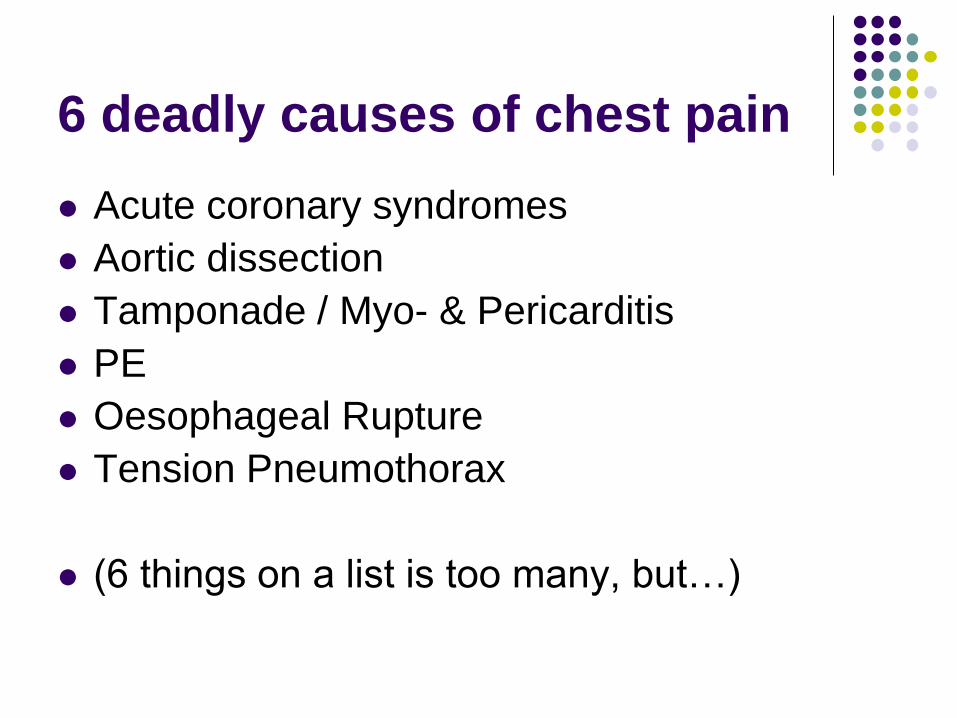
6 deadly causes of chest pain
Acute coronary syndromes
Aortic dissection
Tamponade / Myo- & Pericarditis
PE
Oesophageal Rupture
Tension Pneumothorax
(6 things on a list is too many, but…)
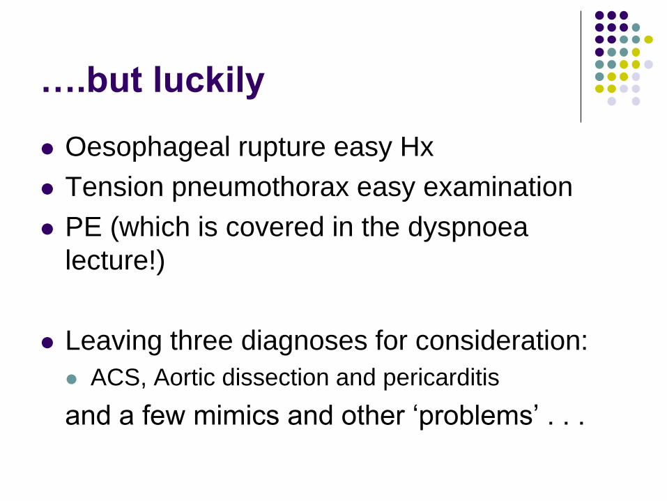
….but luckily
Oesophageal rupture easy Hx
Tension pneumothorax easy examination
PE (which is covered in the dyspnoea
lecture!)
Leaving three diagnoses for consideration:
ACS, Aortic dissection and pericarditis
and a few mimics and other ‘problems’ . . .

Assessing chest pain
Age
Increasing risk
More unreliable symptoms
Risk factors
Part of TIMI scoring criteria
Symptoms and signs
Often non-specific e.g. chest
wall tenderness
ECG
Abnormal ECGs have
increased specificity – often
normal in early disease.
Specific markers of
disease
Few of them reliable in
isolation
Response to therapy
Usually cocktail given
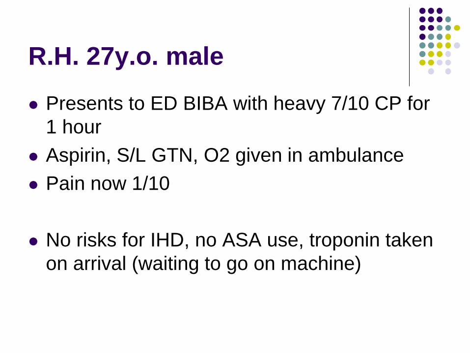
R.H. 27y.o. male
Presents to ED BIBA with heavy 7/10 CP for
1 hour
Aspirin, S/L GTN, O2 given in ambulance
Pain now 1/10
No risks for IHD, no ASA use, troponin taken
on arrival (waiting to go on machine)

R.H. ECG
Presenting the ECG
Obvious anomalies
Rate
Rhythm
Axis
Characteristics of each segment and lead
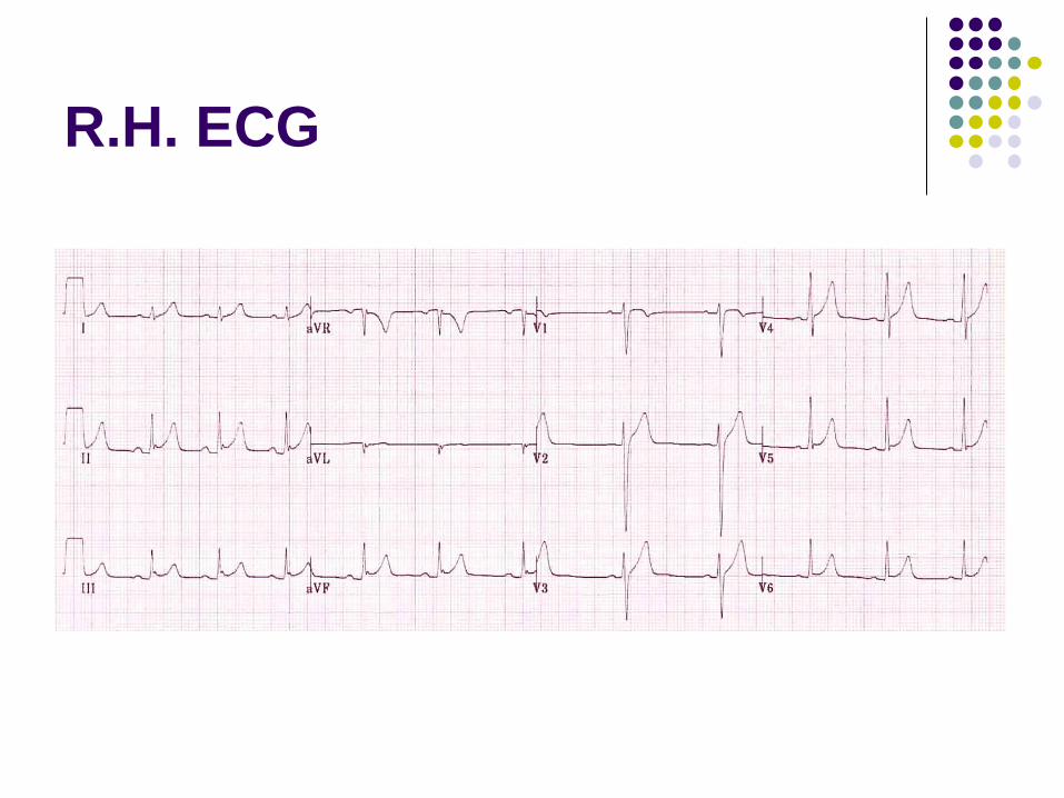
R.H. ECG

R.H. ECG
ST elevation – good or bad?
R.H. ECG
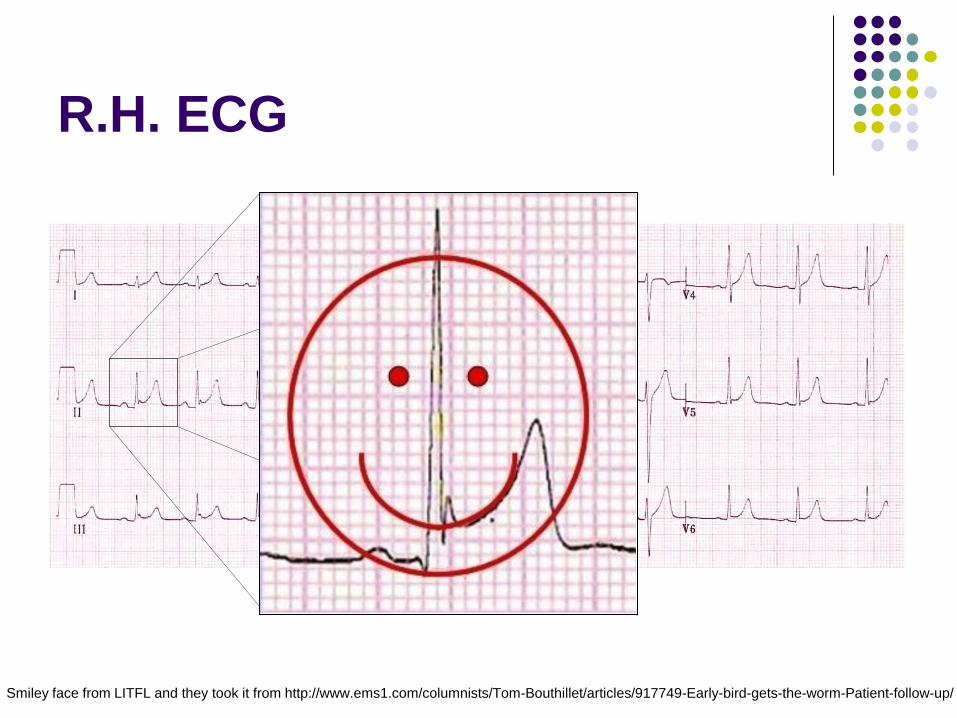
Smiley face from LITFL and they took it from http://www.ems1.com/columnists/Tom-Bouthillet/articles/917749-Early-bird-gets-the-worm-Patient-follow-up/
R.H. ECG

R.H. ECG
Note asymmetrical ‘T’s

R.H. ECG Benign early repolarisation or High take-off

The ST segment-T wave
complex in BER :
There is elevation of the J point
The ST segment and the
ascending limb of the T wave
form an upward concavity
The T wave is peaked and
slightly asymmetrical:The
descending limb of the T wave is
straighter and slightly steeper
than the ascending limb

BER/High take off
Features suggesting BER:
ST elevation concave mainly precordial leads
Absence of PR depression
Prominent T waves
ST segment / T wave ratio < 0.25
Characteristic “fish-hook” appearance in V4
ECG changes relatively
stable over time

R.H. ECG Benign early repolarisation or High take-off

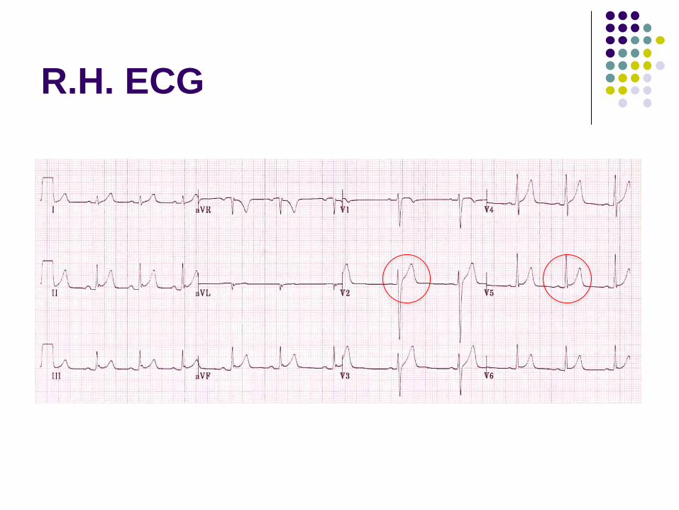
R.H. ECG
J-point ‘notching’

BER/High take off
Features suggesting BER:
ST elevation concave mainly precordial leads
Absence of PR depression
Prominent T waves
ST segment / T wave ratio < 0.25
Characteristic “fish-hook” appearance in V4
ECG changes relatively stable over time

R.H. ECG – likely normal

Can he go home yet?

Can he go home yet?
Pain ongoing?
Troponin result
Repeat ECGs

Troponin
Test normally at arrival for CP and collapse
Repeated at 6 and 12 hours
or at 8 hours from the end of symptoms
or (if high sensitivity) at 1 and 3 hours
Research to consider single high sensitivity
troponin test within 1 hour in ED (Poole, UK)
for very/low risk CP

Troponin assays and
discharge
Rises and falls in troponin levels imply a
myocardial insult
If troponins raised
Likely to require further investigation
Consider non-atherosclerotic insults (e.g. type 2
MI, Renal failure, arrythmias, APO etc.) may
require cause specific treatment first.

Discharging CP patients
Exercise ECG tests
Pharmacological (mainly dipyridamole)
Stress tests - Echo and MIBI★
CT angiogram
Time period – remember TIMI score?
★Tc 99m Sestamibi
Discharging CP patients
Exercise ECG tests - mainly limited to
fit young males
normal ECG (no LBBB, ST abnormalities,
ventricular hypertrophy or strain, paced)
not on digoxin
Pharmacological (mainly dipyridamole)
Stress tests - Echo and MIBI
CT angiogram
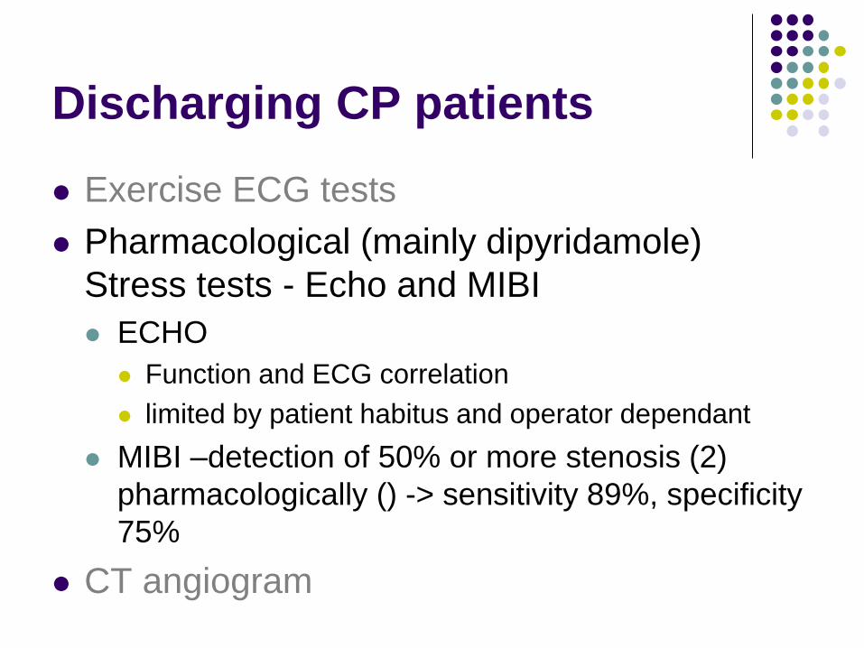
Discharging CP patients
Exercise ECG tests
Pharmacological (mainly dipyridamole)
Stress tests - Echo and MIBI
ECHO
Function and ECG correlation
limited by patient habitus and operator dependant
MIBI –detection of 50% or more stenosis (2)
pharmacologically () -> sensitivity 89%, specificity
75%
CT angiogram

Discharging CP patients
Exercise ECG tests
Pharmacological (mainly dipyridamole)
Stress tests - Echo and MIBI
CT angiogram
non-invasive acquisition of very high-quality
coronary CT angiography
very high radiation exposure, contrast
administration,
need HR @ 60/min or less

P.C. 36 y.o.
Increasing CP for 2 days
Worse on lying flat, better when sits forward
Sharp in middle of chest, feels little SOB
Unwell 2-3 weeks ago
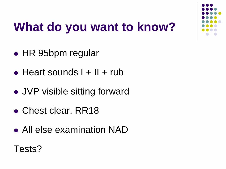
What do you want to know?
HR 95bpm regular
Heart sounds I + II + rub
JVP visible sitting forward
Chest clear, RR18
All else examination NAD
Tests?


Widespread concave ST elevation and PR depression is present
throughout the precordial (V2-6) and limb leads (I, II, aVL, aVF)
There is reciprocal ST depression and PR elevation in aVR


Stages of pericarditic ECG
Stage 1 (1-2/52) – widespread STE and PR depression
with reciprocal changes in aVR
Stage 2 (1-3/52) – normalization of ST changes, T wave
flattening
Stage 3 (3+/52) – Flattened T waves become inverted
Stage 4 – ECG returns to normal

Pericarditis – other ECG
criteria
The ST / T wave ratio > 0.25 is consistent
with pericarditis

BER/Pericarditis
BER pericarditis
ST elevation limited to the precordial
leads
Generalised ST elevation
Absence of PR depression Presence of PR depression
Prominent T waves Normal T wave amplitude
ST segment / T wave ratio < 0.25 ST segment / T wave ratio > 0.25
Characteristic “fish-hook” appearance
in V4 (maybe V5)
Absence of “fish hook” appearance in
V4 (and V5)
ECG changes relatively stable over
time
ECG changes evolve over time

Pulsus Alternans

Discharge for Pericarditis
Appropriate response to NSAIDS
No negative predictors:
Temperature >38
Immunosupression
Traumatic cause
Anticoagulants
No tamponade (pulsus paradoxus) from
pericardial effusion and <20mm

Mr 31 y.o. male
BIBA
CP intermittently for 18 hours
Worse ++ last 2 hours
Sweaty
Pale
Writhing in agony on bed and crying
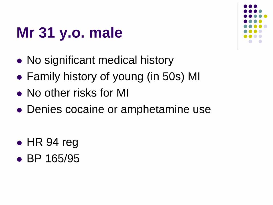
Mr 31 y.o. male
No significant medical history
Family history of young (in 50s) MI
No other risks for MI
Denies cocaine or amphetamine use
HR 94 reg
BP 165/95

ECG

Presenting the ECG
Obvious anomalies
Rate
Rhythm
Axis
Characteristics of each segment and lead

ECG
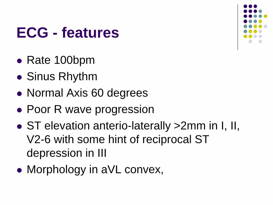
ECG - features
Rate 100bpm
Sinus Rhythm
Normal Axis 60 degrees
Poor R wave progression
ST elevation anterio-laterally >2mm in I, II,
V2-6 with some hint of reciprocal ST
depression in III
Morphology in aVL convex,

Differential diagnosis of STE
AMI
LVH
Paced rhythm
‘normal’
Hyper K+
PE
Coronary spasm
Post cardioversion
Pericarditis
LV aneurysm
Bening early repol.n
Osborne wave
Hypothermia
Brugada’s syndrome
Acute ICH
Wellens’ Syndrome
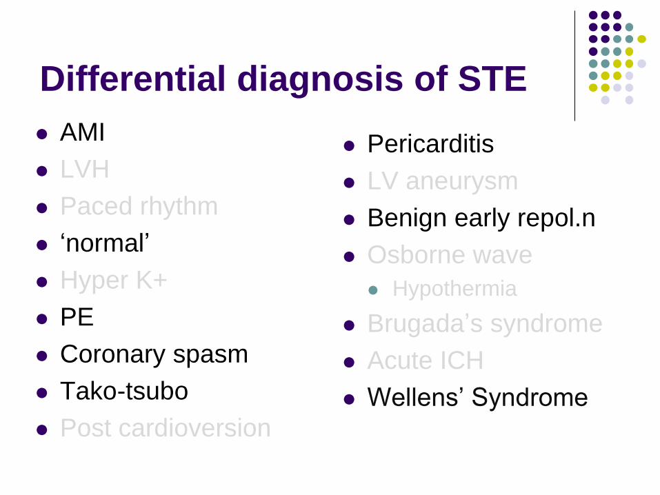
Differential diagnosis of STE
AMI
LVH
Paced rhythm
‘normal’
Hyper K+
PE
Coronary spasm
Tako-tsubo
Post cardioversion
Pericarditis
LV aneurysm
Benign early repol.n
Osborne wave
Hypothermia
Brugada’s syndrome
Acute ICH
Wellens’ Syndrome

ST morphology terminology


ECG

ECG – Left Circumflex

ECG - LAD
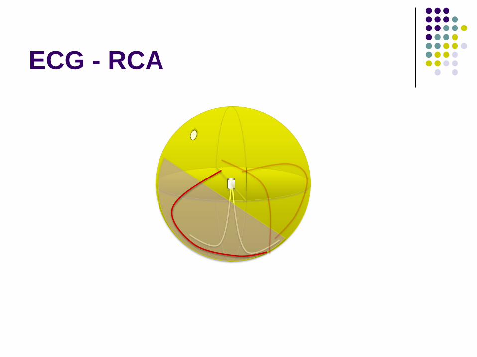
ECG - RCA
ECG
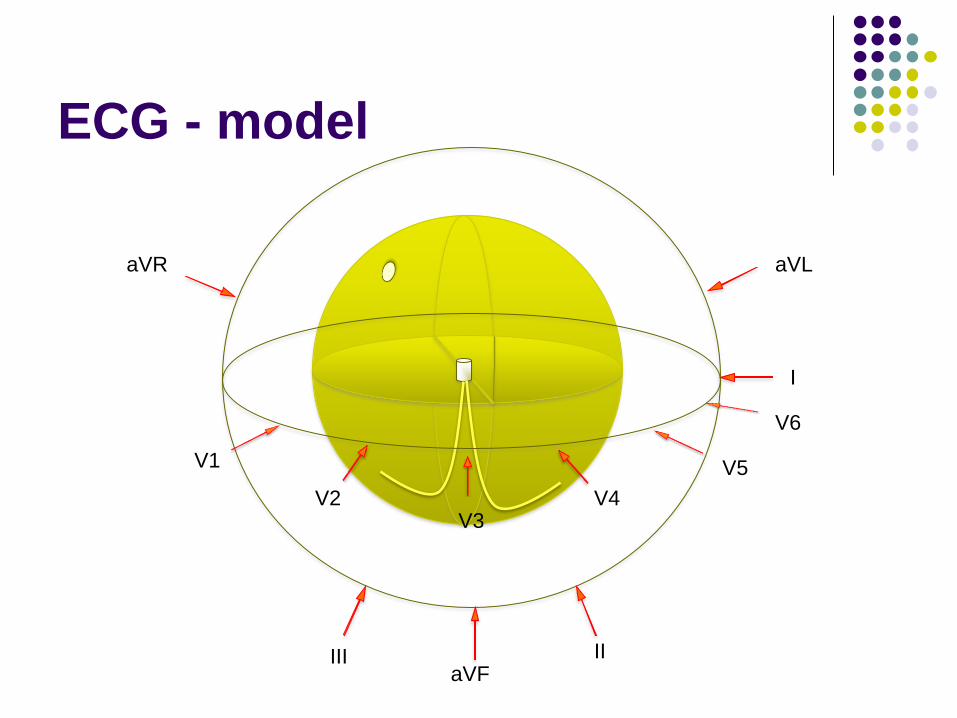
ECG - model
aVR
II aVF
III
V1
V2 V3
V4
V5
V6
I
aVL

ECG - model
aVR
II aVF
III
V1
V2 V3
V4
V5
V6
I
aVL
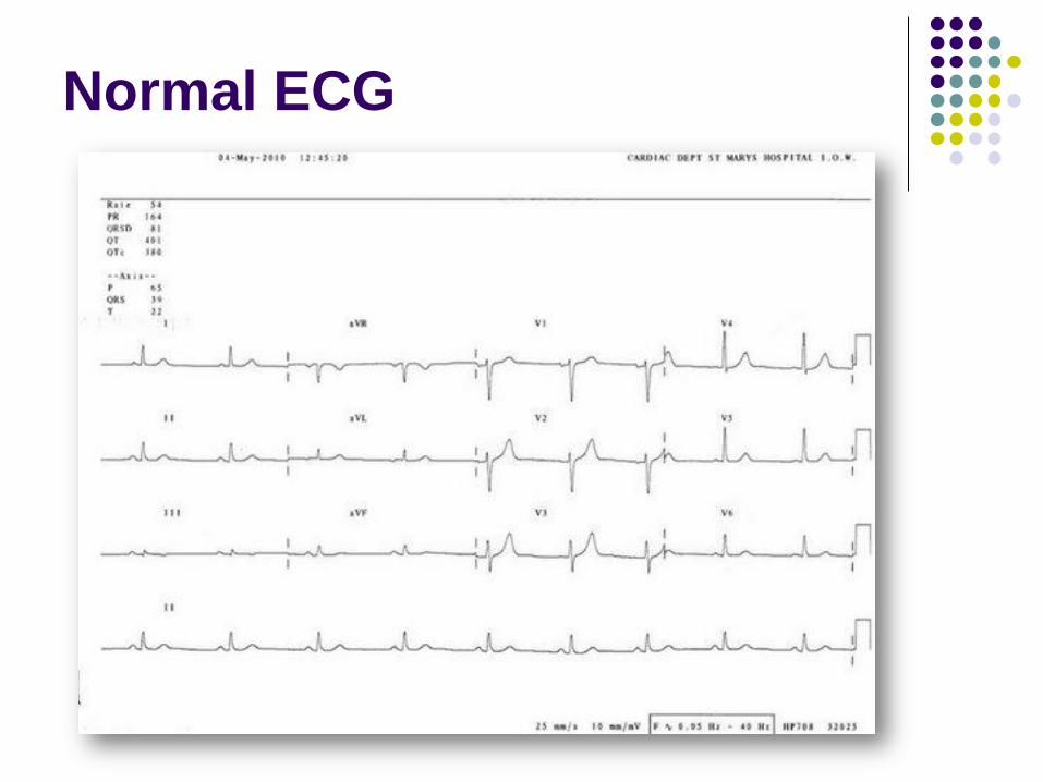
Normal ECG
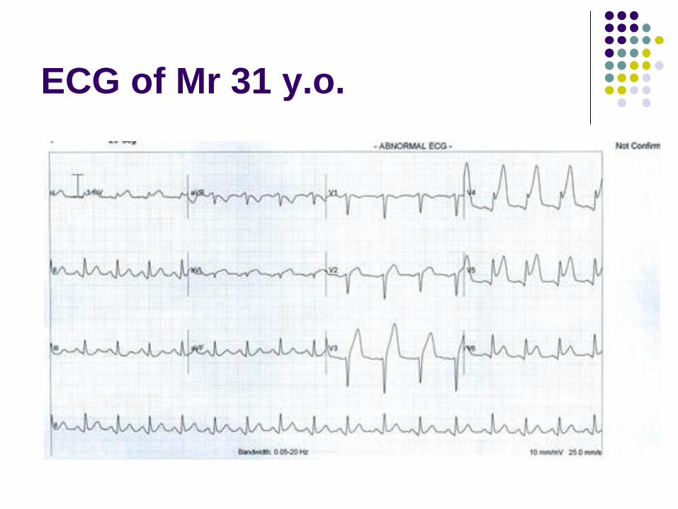
ECG of Mr 31 y.o.


Thrombolytic therapies in MI
Fibrinolysis
Tenecteplase
Percutaneous Coronary Intervention
Angiography and stenting
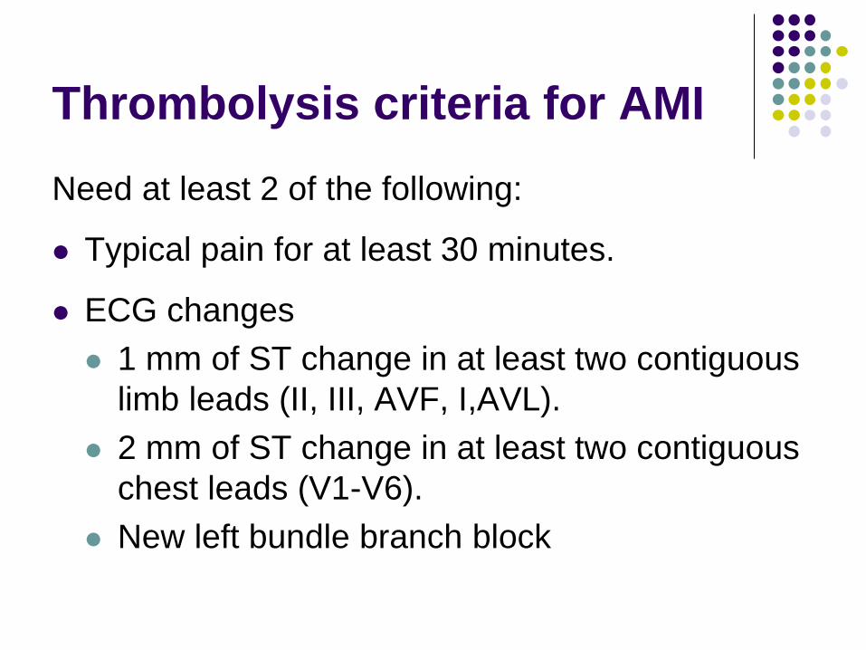
Thrombolysis criteria for AMI
Need at least 2 of the following:
Typical pain for at least 30 minutes.
ECG changes
1 mm of ST change in at least two contiguous
limb leads (II, III, AVF, I,AVL).
2 mm of ST change in at least two contiguous
chest leads (V1-V6).
New left bundle branch block

Thrombolysis (fibrinolysis)
50% flow return rate where indicated
15% re-occlude (and do badly)
1% patients have a major bleed
Usually intra-cranial haemorrhage
PCI considered superior to thrombolysis but
time dependant advantage

PCI protocol


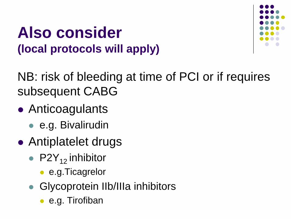
Also consider (local protocols will apply)
NB: risk of bleeding at time of PCI or if requires
subsequent CABG
Anticoagulants
e.g. Bivalirudin
Antiplatelet drugs
P2Y12 inhibitor
e.g.Ticagrelor
Glycoprotein IIb/IIIa inhibitors
e.g. Tirofiban
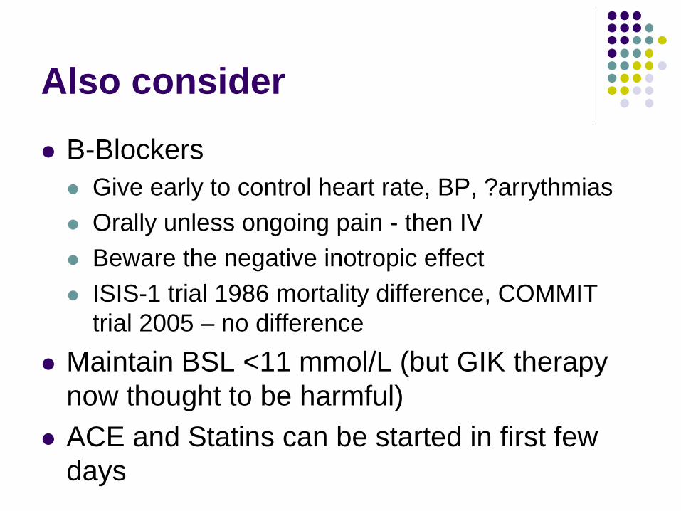
Also consider
B-Blockers
Give early to control heart rate, BP, ?arrythmias
Orally unless ongoing pain - then IV
Beware the negative inotropic effect
ISIS-1 trial 1986 mortality difference, COMMIT
trial 2005 – no difference
Maintain BSL <11 mmol/L (but GIK therapy
now thought to be harmful)
ACE and Statins can be started in first few
days

RCA

LCXA

Occluded LAD


Wire past obstruction
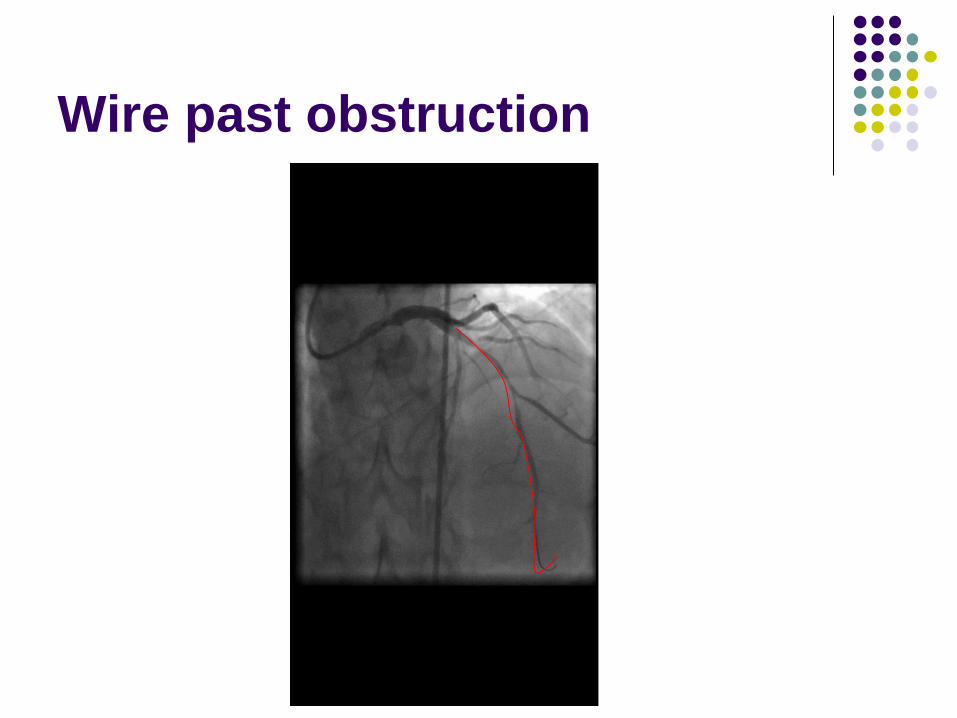
Wire past obstruction
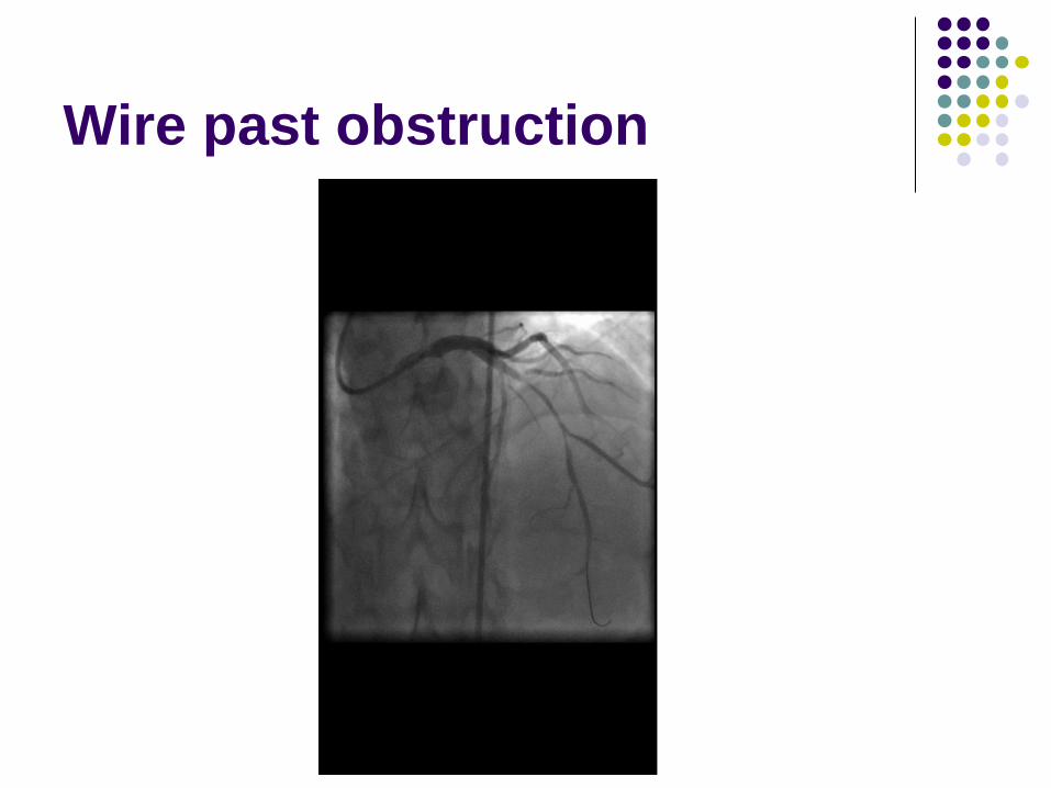
Wire past obstruction

Improved flow after balloon
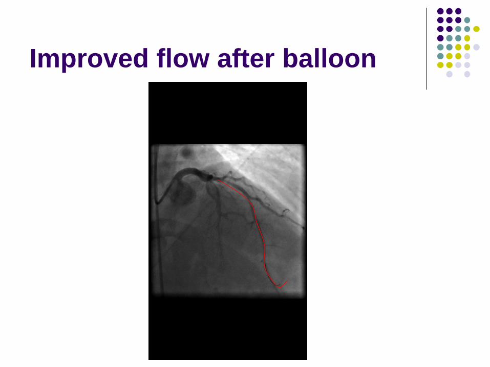
Improved flow after balloon

Improved flow after balloon

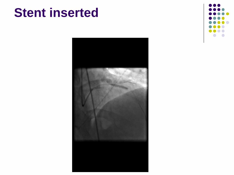
Stent inserted
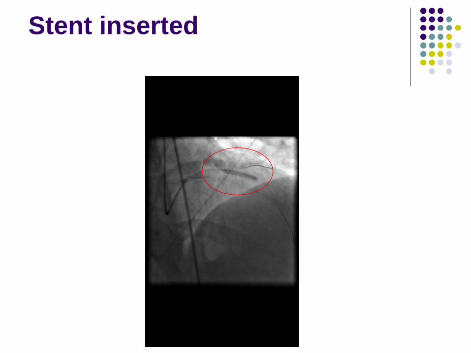
Stent inserted
Stent inserted

flow post stenting
Other ECG presentations of MI
Hyper acute T waves
Reciprocal changes
Posterior infarct
Sgarbossa criteria (in LBBB)
Q waves

Hyper acute T wave

ECG

Reciprocal changes

Posterior infarct
W.A. case
45 y.o. male
Smoker, overweight, FHx hypertension
4 hours of heavy chest pain around midnight
Pain free now
No aspirin use
No previous similar or investigation for IHD
W.A. case
‘Doesn’t use cocaine’
Urine test run in ED suggests no illicit drug
use

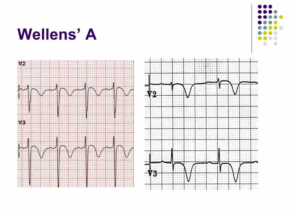
Wellens’ A

Wellens Syndrome
patients presenting with recent history
‘ischaemic’ chest pain
may be pain free by the time the ECG is taken
have normal or minimally elevated cardiac
enzymes
inverted or biphasic T waves in V2-3 that is
highly specific for critical stenosis of the
left anterior descending artery.
extremely high risk for extensive anterior wall
MI within the next 2-3 weeks.

Wellens’ A and B
Type A:
T-waves are deeply and symmetrically
inverted
Type B: T-waves are biphasic, with the initial
deflection positive and the terminal deflection
negative
Wellens’ A
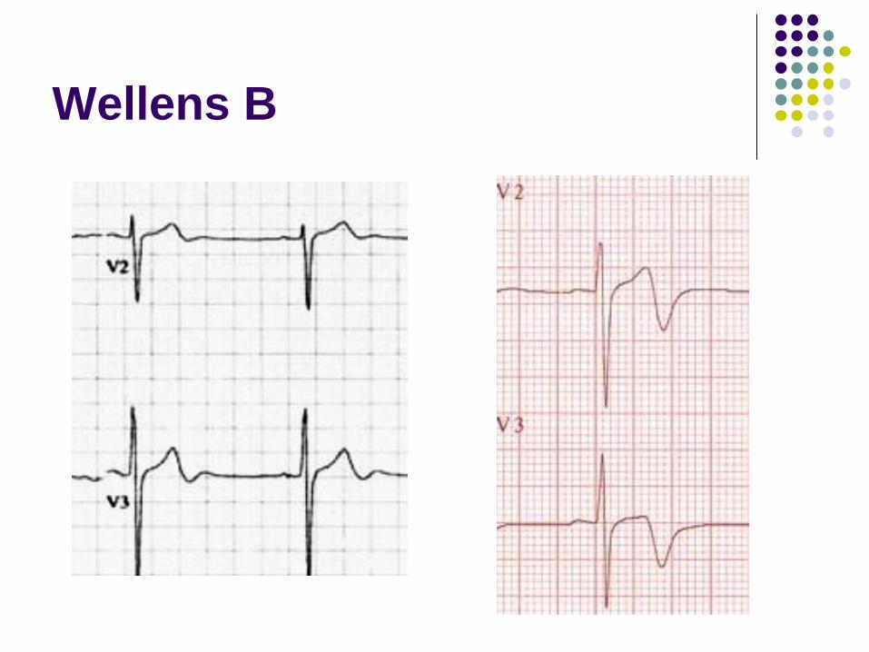
Wellens B

T.T. 56y.o. female
BIBA – 2 hours of crushing heavy CP
In solicitors at time of onset
Cardiac enzyme rise (Trop 0.1) and
widespread borderline ST and T wave
changes (inverted anteriorly V2-4).
No significant risks for IHD
Short run of VT witnessed on monitor

T.T.
What are your thoughts
What shall we do . . .?

SELECTIVE CORONARY
ANGIOGRAPHY AND LEFT
VENTRICULOGRAM
PROCEDURE DETAILS: Coronary angiography was performed
via
the right radial approach using a 6 French sheath and 6
French JL4, JR4 and Pigtail catheters. Significant spasm
requiring intracoronary GTN.
PROCEDURE FINDINGS:
Left Main - Normal.
Left Anterior Descending - Minor proximal disease.
Left Circumflex - Normal.
Ramus Intermediate - Normal
Right Coronary Artery - Dominant and normal.
Left Ventriculogram - Severe regional systolic dysfunction
with only basal contraction suggestive of Takotsubo
cardiomyopathy. There is a small filling defect in the
inferior apex which is opaque and suggestive of a small
thrombus versus trabeculation. No AV pull back gradient.
LVEDP - 25mmHg.
CONCLUSION:
Normal coronary angiogram. Takotsubo cardiomyopathy.
PLAN:
To have echocardiographic correlation of LV thrombus -
commence anticoagulation in the meantime.
Commence heart failure therapy.
Keep monitored overnight.
Cardiology Assoc.Prof E O F

Ventriculogram


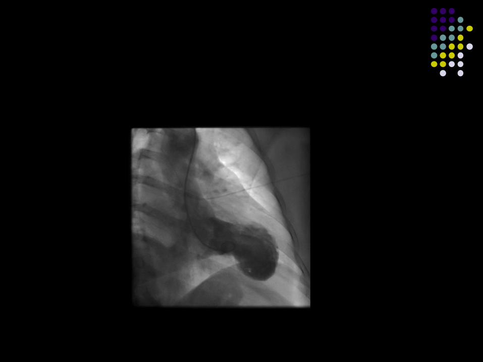


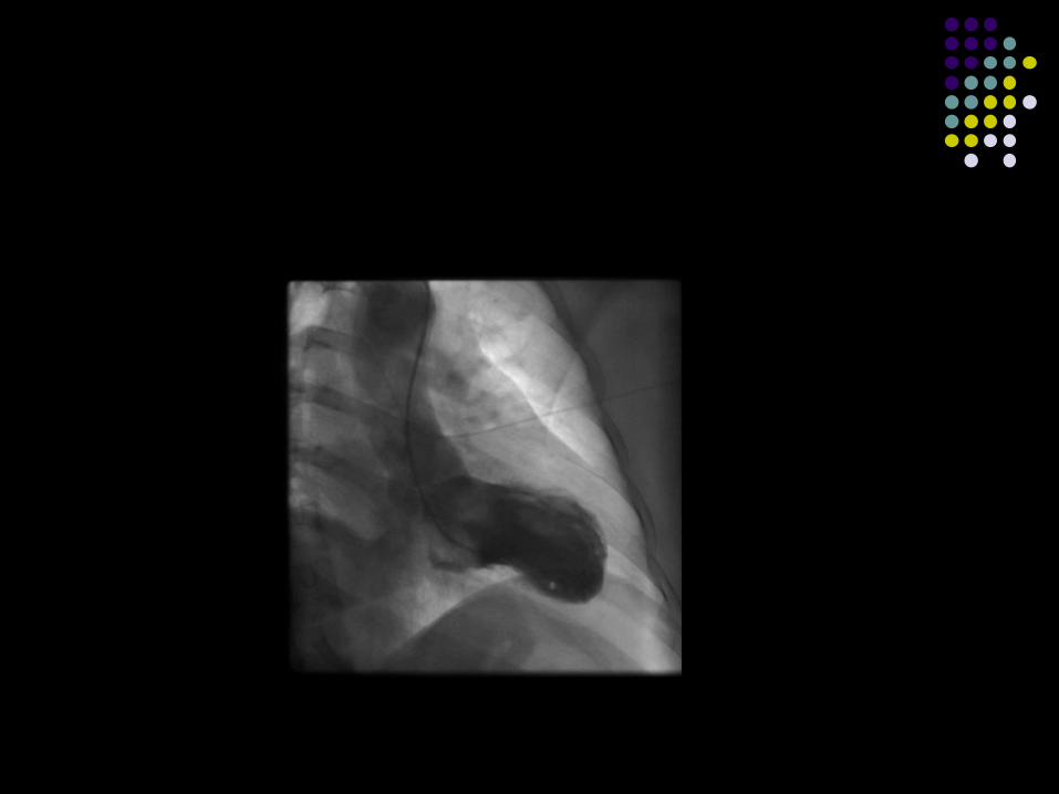


Left, X-ray of the heart during the contraction
phase from a patient with takotsubo.
Right, a ‘tako tsubo’ or
octopus pot (Japan)
Sharkey S W et al. Circulation 2011;124:e460-e462
Copyright © American Heart Association

Takotsubo
Women over 50 years
Male:Female = 1:9
Associated with extreme stress (‘broken heart
syndrome’) or adrenaline surges (e.g.
phaeochromocytoma), 15% idiopathic
Muscle is ‘stunned’ and recovery is usually
complete (complications during event similar
to MI although less frequent )
Further events in only 5% of cases
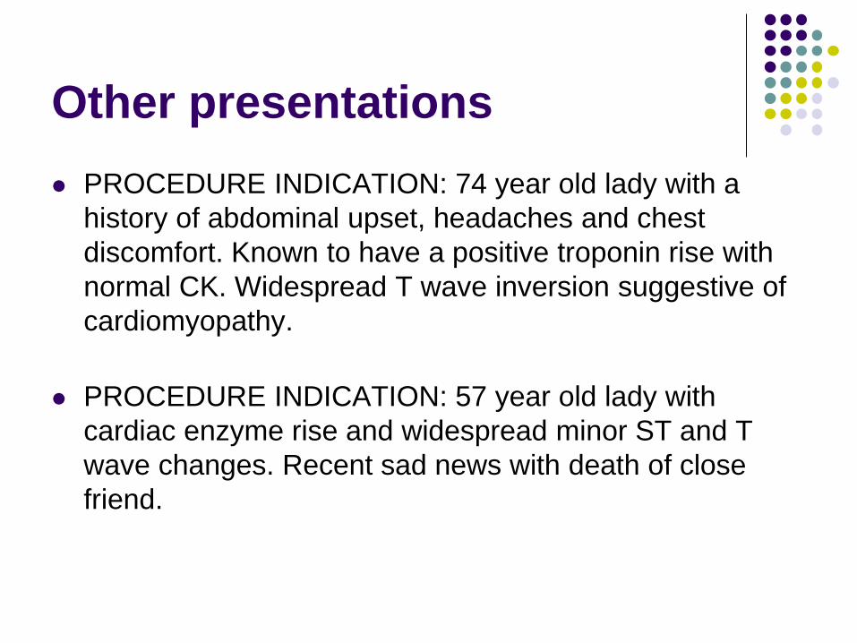
Other presentations
PROCEDURE INDICATION: 74 year old lady with a
history of abdominal upset, headaches and chest
discomfort. Known to have a positive troponin rise with
normal CK. Widespread T wave inversion suggestive of
cardiomyopathy.
PROCEDURE INDICATION: 57 year old lady with
cardiac enzyme rise and widespread minor ST and T
wave changes. Recent sad news with death of close
friend.
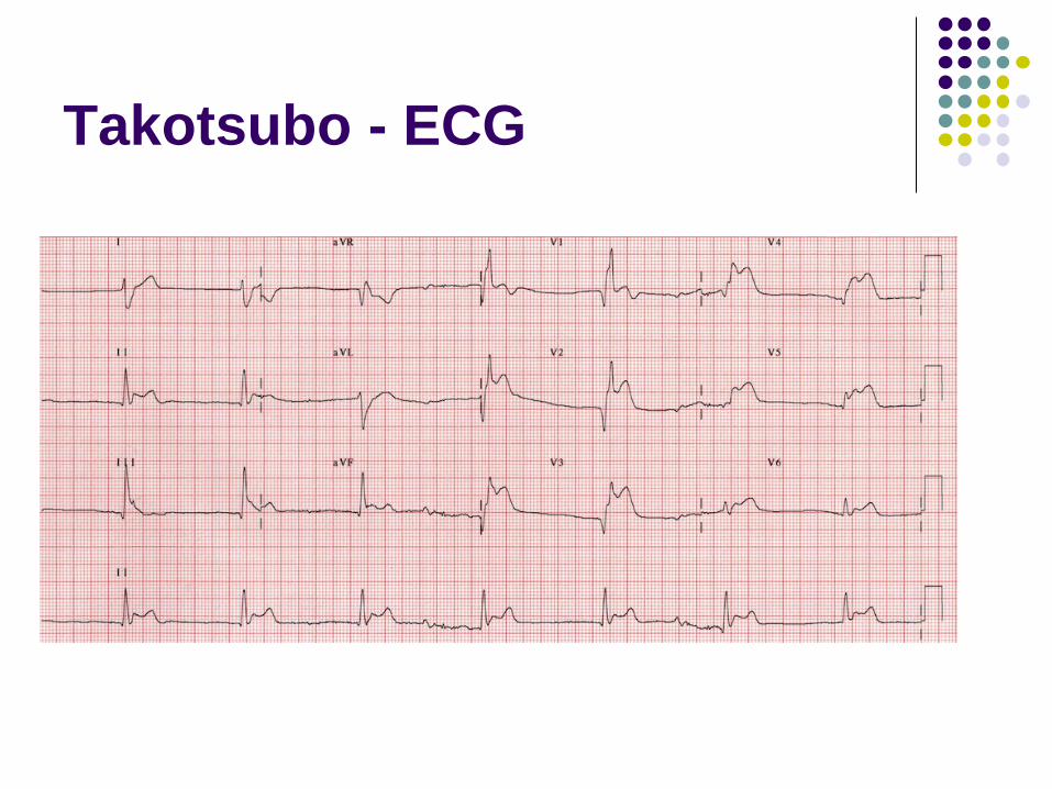
Takotsubo - ECG
Takotsubo
Women over 50 years
Male:Female = 1:9
Associated with extreme stress (‘broken heart
syndrome’) or adrenaline surges (e.g.
phaeochromocytoma), 15% idiopathic
Muscle is ‘stunned’ and recovery is usually
complete (complications during event similar
to MI although less frequent )
Further events in only 5% of cases
Aortic Dissection
Challenging diagnosis, lethal if missed (75%
in 2/52), higher if Rx as ACS
Classic Hx:
Sudden onset ripping / tearing PIC, radiates to
back (interscapular, migratory) & Hx HTN
Not sensitive enough
Best approach is risk factor assessment
Be aware of atypical presentations

Risk factors for Aortic
Dissection
HTN
Aortic valve
Bicuspid, previous surgery (Ao Regurg at
presentation)
Abnormal Aorta – mainly congenital
Coarctation / Marfan’s / Ehlers-Danlos
Arteritis (Giant cell)
Aortic ‘stresses’
COCAINE, TRAUMA, PREGNANCY
Atypical presentations
Atypical history
Non-classical pain, chest or back only, severe &
sharp pain, abdo pain
Syncope (normally present with raised BP)
Acute stroke (& peripheral neuro) Horners Syndrome
Limb weakness
Hemiplegic stroke
Others just get too weird & make you paranoid
Eg. Pulsatile stenoclavicular joint!!
AD - imaging
Widened mediastinum (54-62%) and
abnormal aortic contour (50%) most sensitive
CXR findings
Normal CXR may decrease the likelihood but
if examination, Hx and RF’s raise suspicion
needs further imaging

Aortic dissection CXR findings
1. Mediastinal widening
2. Widening of aortic
contour/knuckle
3. Opacification of aorto-
pulmonary window
4. Pleural effusion (L>R)
5. (Displaced calcification)
6. (Left apical pleural cap)
7. Aortic kinking
8. Tracheal or
oesophageal
displacement
9. (Depressed left main
bronchus)
3
4
6
7
8
AD - Ix
Widened mediastinum (62%) and abnormal
aortic contour (50%) most sensitive CXR
findings
Normal CXR may decrease the likelihood but
if examination, Hx and RF’s raise suspicion
needs further imaging
D-Dimer negative – reduces probability
Troponin or renal function depending on
vessels involved


Classification
Stanford (most commonly used)
Type A — Involves ascending aorta. Can extend distally ad
infinitum. Surgery usually indicated
Type B — Involves aorta beyond left subclavian artery
only. Often managed medically with BP control.
De Bakey
1 – entire aorta affected
2 – confined to the ascending aorta
3 – descending aorta affected distal to subclavian artery
Svensson (defines type of acute aortic syndrome)

Treatment
Analgesia
Blood pressure control
IV labetalol to maintain BP of systolic just above
100mmHg
Appropriate transfer via ARV

Summary
Ischaemic Heart disease is common with significant
mortality
ECG and troponin are mainstay of admission and
discharge protocols
NSTEMI/non-STEMI (e.g. Type 2 MI, Wellens’
Syndrome, Takotsubo) make cautious management
essential
Some new high sensitivity troponin tests may allow
improved discharges in a large minority of low risk
cases.

Questions?

Thankyou

References
http://medicthriller.blogspot.com.au
http://lifeinthefastlane.com
http://emedicine.medscape.com
http://circ.ahajournals.org/content/113/12/162
2.full (Pericarditis)