Embed Size (px)
Citation preview

Chest X-ray Interpretation
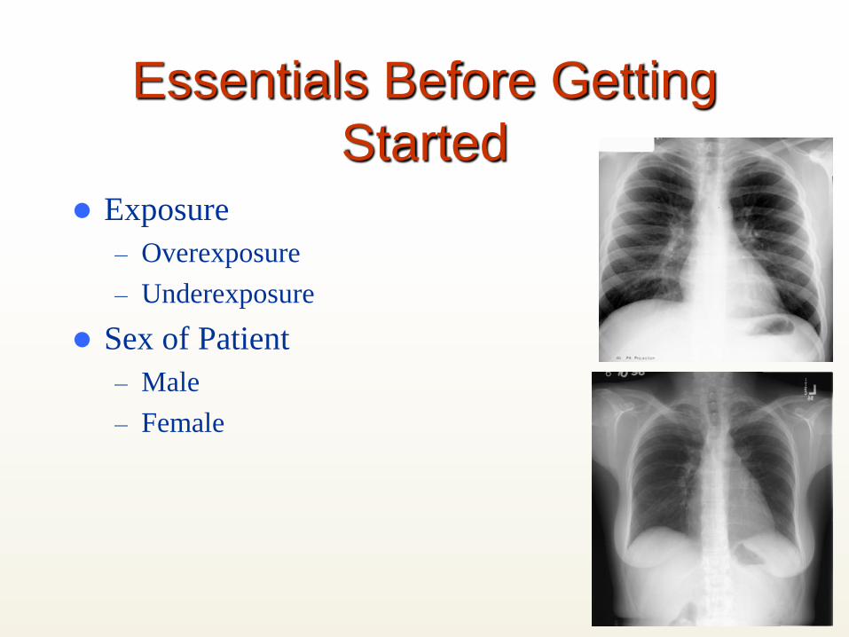
Essentials Before Getting
Started Exposure
– Overexposure
– Underexposure
Sex of Patient
– Male
– Female
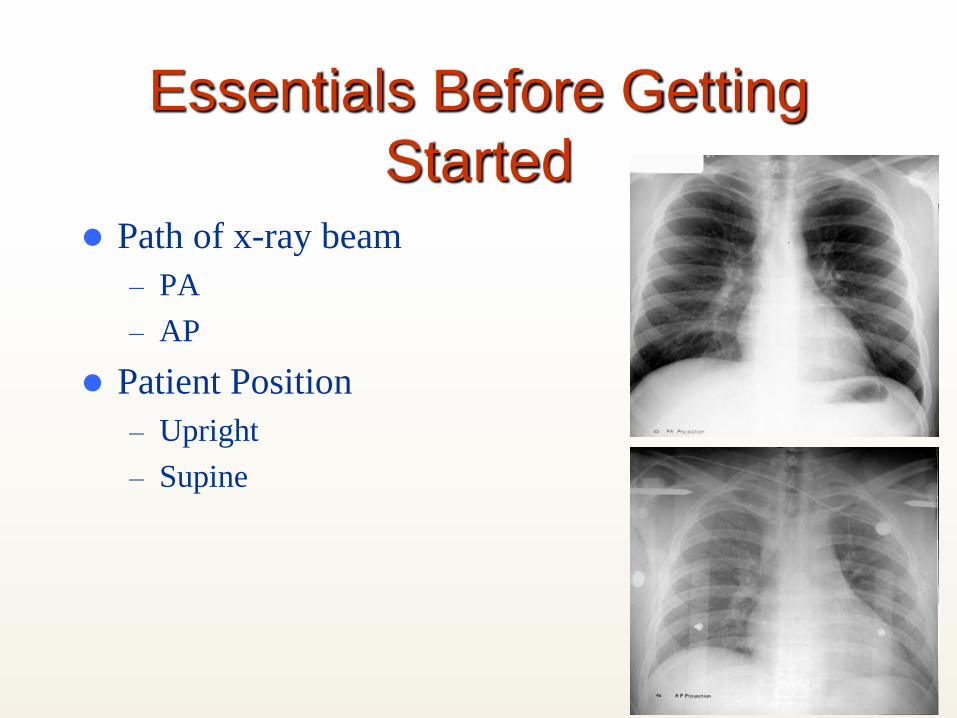
Essentials Before Getting
Started Path of x-ray beam
– PA
– AP
Patient Position
– Upright
– Supine

Male Female PA CHEST
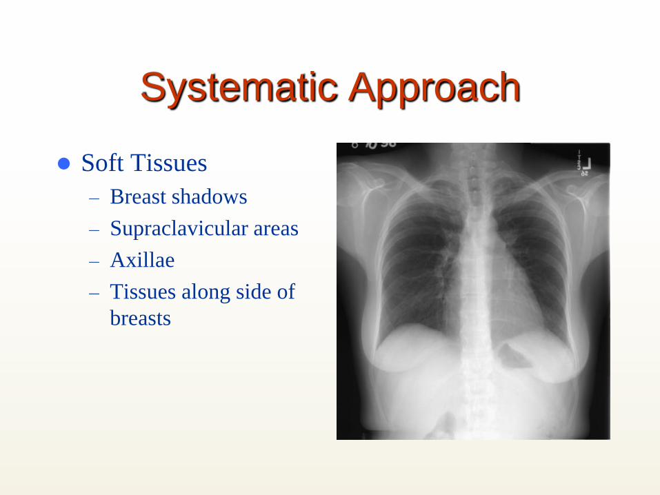
Systematic Approach
Soft Tissues
– Breast shadows
– Supraclavicular areas
– Axillae
– Tissues along side of
breasts

Lung Anatomy
Right Lung
– Superior lobe
– Middle lobe
– Inferior lobe
Left Lung
– Superior lobe
– Inferior lobe

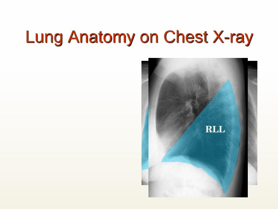
Lung Anatomy on Chest X-ray
PA View:
– Extensive overlap
– Lower lobes extend
high
Lateral View:
– Extent of lower lobes

Lung Anatomy on Chest X-ray

Lung Anatomy on Chest X-ray
The right middle lobe
is typically the
smallest of the three,
and appears triangular
in shape, being
narrowest near the
hilum
Lung Anatomy on Chest X-ray

Lung Anatomy on Chest X-ray
these lobes separated from one
another by two fissures .
The minor fissure separates the
RUL from the RML , at the level
of the fourth vertebral body and
crosses the right sixth rib in the
midaxillary line
The right major fissure separating
the right upper and middle lobes
from the larger right lower lobe ,
The major fissure extend
anteroinferiorly, intersecting the
diaphragm at the anterior
cardiophrenic angle
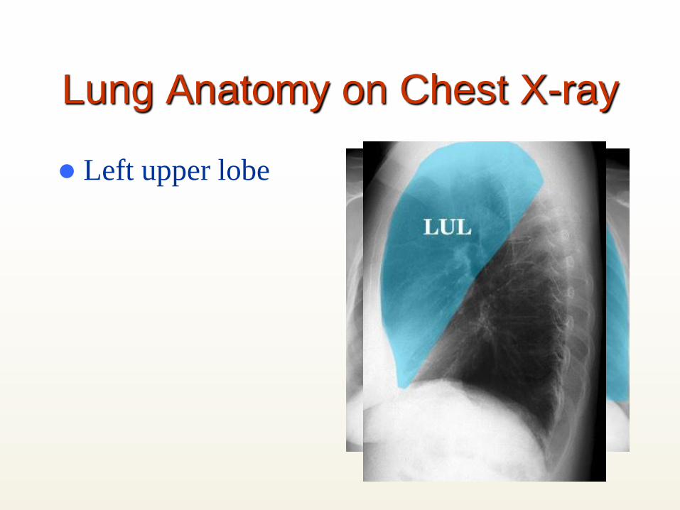
Lung Anatomy on Chest X-ray
Left upper lobe
Lung Anatomy on Chest X-ray
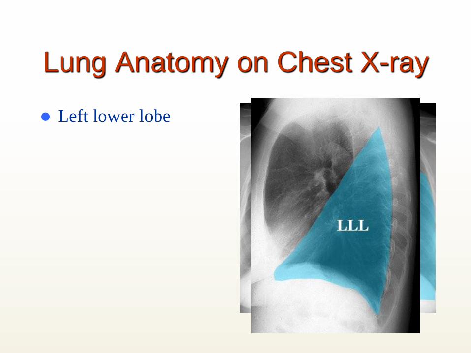
Left lower lobe

Lung Anatomy on Chest X-ray
the left lung is
slightly different
than the right.
Because there is no
defined left minor
fissure, there are
only two lobes .
fissures are not readily
identifiable on plain films
This is because fissures which are
composed of only two layers of visceral
pleura, may not present a significant
radiographic interface and will not produce
a shadow. However, if there is fluid within
the pleural space or if the visceral pleura is
thickened, fissures may be seen in their
entirety.

ATELECTASIS
No ventilation to lobe beyond the obstruction
Trapped air absorbed by pulmonary circulation
Segmental/lobar density
Compensatory hyper-inflation of normal lungs.
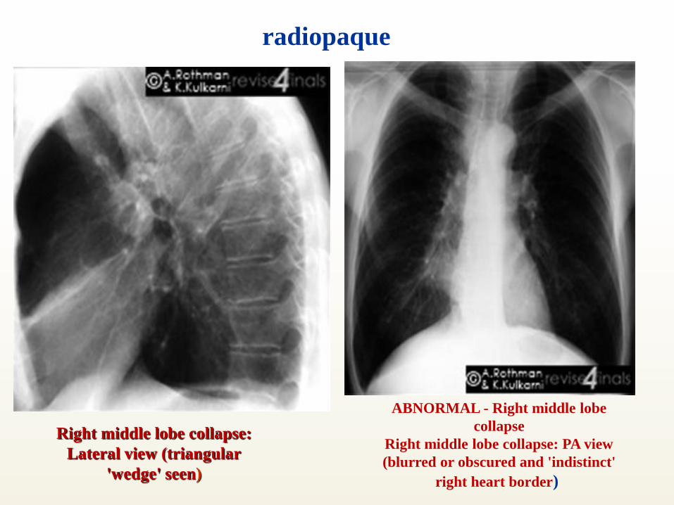
Right middle lobe collapse:
Lateral view (triangular
'wedge' seen)
ABNORMAL - Right middle lobe
collapse
Right middle lobe collapse: PA view
(blurred or obscured and 'indistinct'
right heart border)
radiopaque
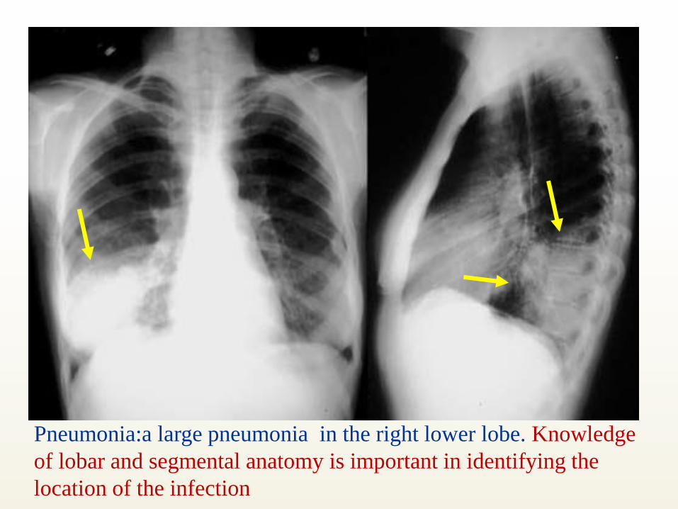
Pneumonia:a large pneumonia in the right lower lobe. Knowledge
of lobar and segmental anatomy is important in identifying the
location of the infection

Right Middle and Left Upper Lobe Pneumonia

Chest wall lesion: arising off the chest wall and not the lung
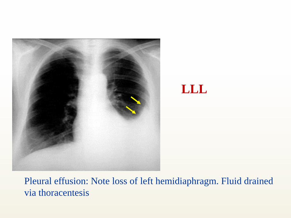
Pleural effusion: Note loss of left hemidiaphragm. Fluid drained
via thoracentesis
LLL

Lung Mass -LUL

Metastatic Lung Cancer: multiple nodules seen

Right Middle Lobe Pneumothorax: complete lobar collapse

AP ABDOMEN Gas in
stomach
Gas in a few
loops of
small bowel
Gas in
rectum or
sigmoid
Normal Gas Pattern

Erect Abdomen
Always
air/fluid level
in stomach
A few
air/fluid
levels in
small bowel
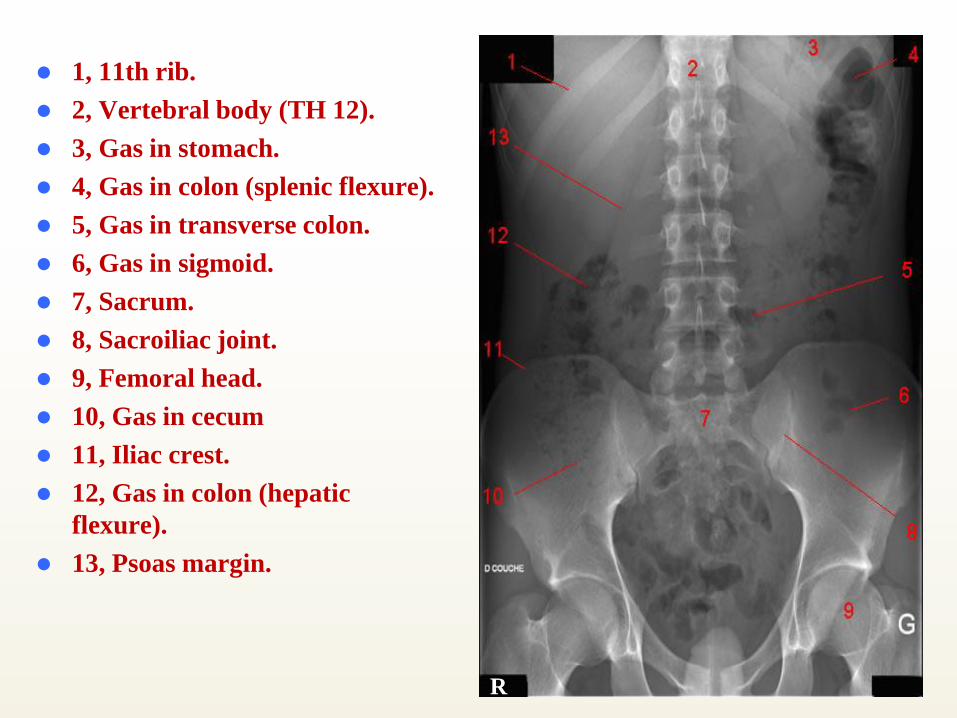
1, 11th rib.
2, Vertebral body (TH 12).
3, Gas in stomach.
4, Gas in colon (splenic flexure).
5, Gas in transverse colon.
6, Gas in sigmoid.
7, Sacrum.
8, Sacroiliac joint.
9, Femoral head.
10, Gas in cecum
11, Iliac crest.
12, Gas in colon (hepatic
flexure).
13, Psoas margin.
R


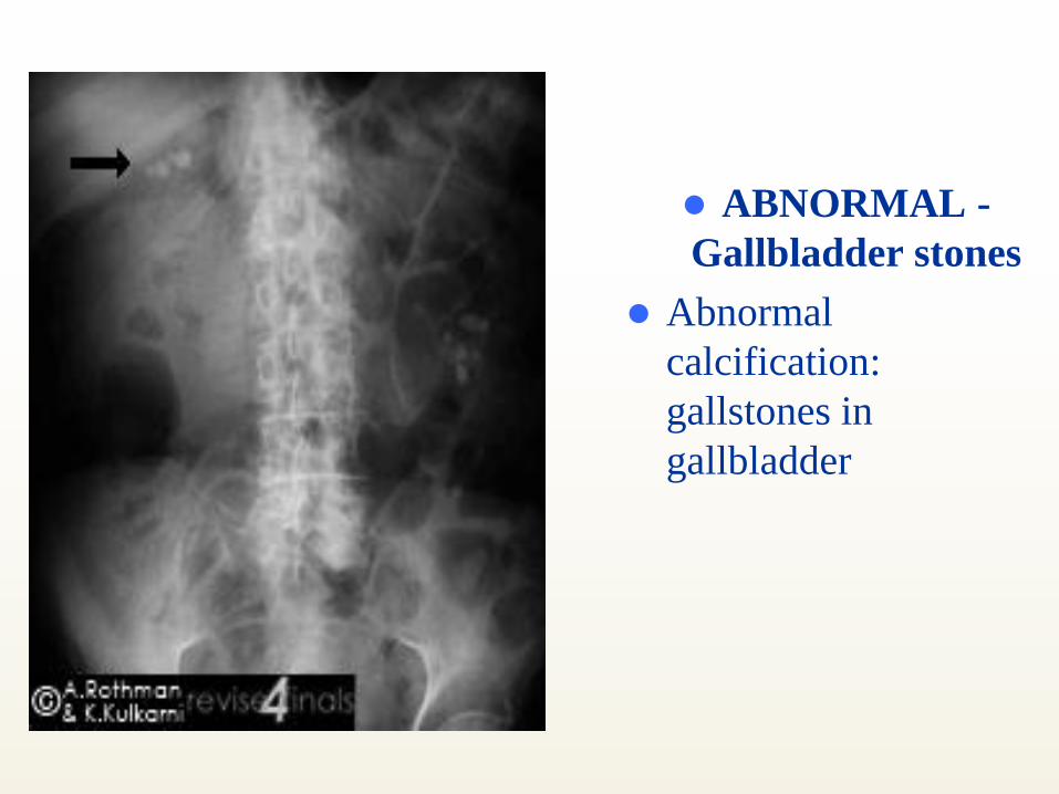
ABNORMAL -
Gallbladder stones
Abnormal
calcification:
gallstones in
gallbladder
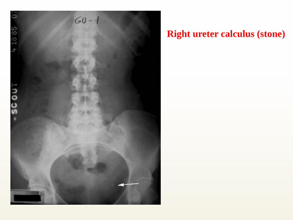
ririr Right ureter calculus (stone)

IVU-INTRAVENOUS UROGRAM
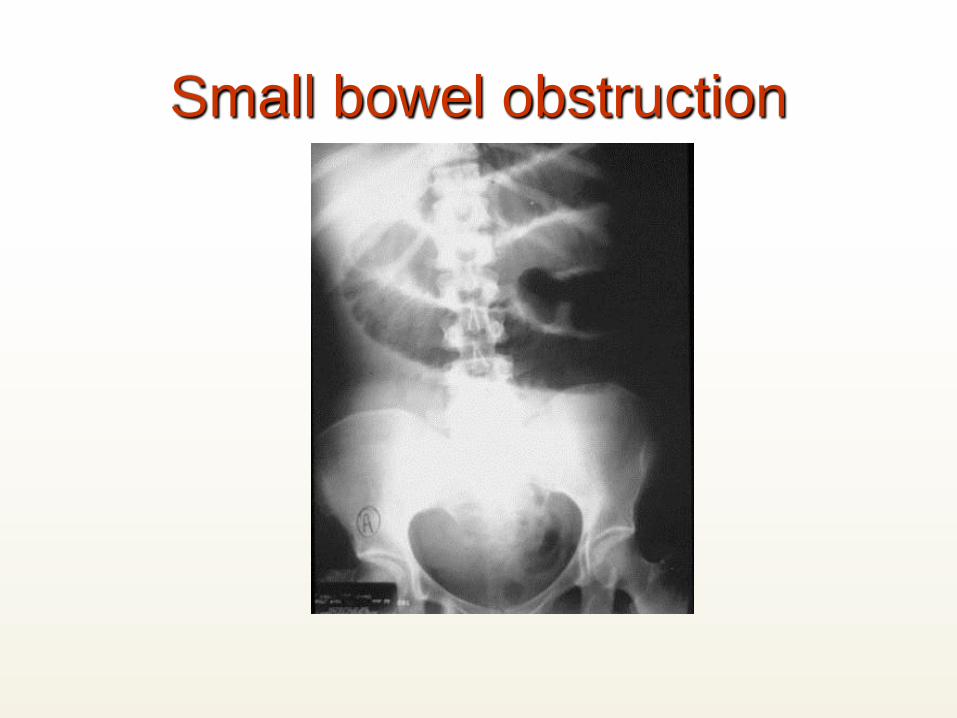
Small bowel obstruction
Mechanical LBO
Causes:-
Tumor
Volvulus
Hernia

LBO
Supine Prone


![Radiology Lecture CXR.ppt [Read-Only] · 2018. 4. 3. · • Right lung lobes – Upper – Middle – Lower • Left lung ... carcinoma Cardiomyopathy. 10/2/2014 25 Pulmonary edema](https://img.pdfslide.net/doc/110x75/60e9c7cc55752749b92c5670/radiology-lecture-cxrppt-read-only-2018-4-3-a-right-lung-lobes-a-upper.jpg)