Embed Size (px)
Citation preview
Genetic Engineering of heavy metal sequestering /
precipitating proteins for bioremediation
By
Chitra Seetharam Misra
LIFE01200904001
Bhabha Atomic Research Centre, Mumbai
A thesis submitted to the
Board of Studies in Life Sciences
In partial fulfilment of requirements
for the Degree of
DOCTOR OF PHILOSOPHY
of
HOMI BHABHA NATIONAL INSTITUTE
December, 2014
STATEMENT BY AUTHOR
This dissertation has been submitted in partial fulfillment of requirements for an
advanced degree at Homi Bhabha National Institute (HBNI) and is deposited in the
Library to be made available to borrowers under rules of the HBNI.
Brief quotations from this dissertation are allowable without special permission,
provided that accurate acknowledgement of source is made. Requests for permission
for extended quotation from or reproduction of this manuscript in whole or in part
may be granted by the Competent Authority of HBNI when in his or her judgment the
proposed use of the material is in the interests of scholarship. In all other instances,
however, permission must be obtained from the author.
Chitra Seetharam Misra

DECLARATION
I, hereby declare that the investigation presented in the thesis has been carried out by
me. The work is original and has not been submitted earlier as a whole or in part for a
degree / diploma at this or any other Institution / University.
Chitra Seetharam Misra
List of Publications arising from the thesis
Journal 1. Lyophilized, non-viable, recombinant E. coli cells for cadmium bioprecipitation and recovery.
Seetharam Chitra, Soundarajan Suvarna, Udas A. C., Rao A. S. and Apte S. K., Process
Biochemistry, 2009, 44, 24-250.
2. PhoN-expressing, lyophilized, recombinant Deinococcus radiodurans cells for uranium
bioprecipitation. Appukuttan Deepti1, Seetharam Chitra
1, N. Padma, Rao A. S. and Apte S. K.
Journal of Biotechnology, 2011, 154(4), 285-290. 1These authors contributed equally to the work.
3. Recombinant D. radiodurans cells for bioremediation of heavy metals from acidic/neutral
aqueous wastes. Misra Chitra Seetharam1, Appukuttan Deepti
1, Kantamreddi VSS, Rao A. S and
Apte S. K., Bioengineered Bugs, 2012, 3, 44-48. 1These authors contributed equally to the work.
Conferences
1. Natural and recombinant bacteria for bioremediation of uranium from acidic/alkaline
aqueous solutions in high radiation environment. Appukuttan Deepti, Nilgiriwala
Kayzad, Seetharam Chitra, and Apte S. K. In Abstracts of the 14th International
Biotechnology Symposium and Exhibition "IBS2010" in September 2010 in Rimini,
Italy.
2. Seetharam Chitra,, Soundarajan Suvarna, Udas A C, Basu Bhakti and Apte SK. The
utility of Hpi, the Surface layer protein of D. radiodurans in metal removal from
solutions. In Abstracts of the Life Sciences Symposium held in 2015 in BARC, Mumbai.
Chitra Seetharam Misra

Dedicated to my family

Acknowledgements
Pursuing Phd is not only an academic challenge; it is also an exercise in recognizing the
importance of scientific and personal interactions with peers and non-peers in putting together a
decent piece of work. In this regard, I have been lucky to be mostly surrounded by people from
whom I received enormous help, constructive advice, bright ideas, technical help and moral
support and I would like to acknowledge them here.
I would like to first thank my Guide, Dr. Shree Kumar Apte for guiding my work in spite of a
very busy schedule, giving me a good and just hearing whenever I discussed my work with him
and for honing my scientific writing skills. There is a lot I have learnt that will stand me in good
stead throughout my life.
I thank my doctoral committee for their encouragement and valuable suggestions. I was fortunate
to be able to discuss my work without any inhibitions with them on account of their friendly and
cordial demeanour. Dr. Hema Rajaram, Dean, Life Sciences to whom I went very often for all
Ph.D related paperwork and who was always obliging.
I sincerely thank Shri. A. S. Rao who was always supportive throughout my initial work and
gave me constant encouragement to do my Ph.D. He provided the basic ideas which started this
work and I will always be thankful to him.
I thank my collaborators, Dr. Ambuja C Udas and Dr. Suvarna Soundarajan, Dr. Padma N, for
un-complainingly running my samples for analysis and giving valuable scientific inputs. I also
thank Dr. Rita Mukhopadhyaya for sequencing a couple of my constructs, Dr. Bhakti Basu, for
peptide mass fingerprinting using MALDI-TOF-MS, Mr. Anand Ballal and Mrs. Alka Gupta for
electron microscopic studies, Dr. Hassan and Mrs. Suman for help with the zetameter. I must
acknowledge Dr. Mary Lidstrom for very promptly sending me a strain which was used in this
study. I also thank Dr. Celin Acharya for letting me use a plasmid she constructed in the lab,
which was a starting point for some of the work reported in this thesis.
This work got a lot of help from my colleague, Mrs. Saraswathi Perumal who in spite of ill
health helped me in carrying out many routine experiments to perfection. I must thank Dr. Deepti
Appukuttan who taught me quite a few things during my initial years as a scientist and whose
work served as a base for this study. I was fortunate to have very amicable colleagues, Mr.
Shyam Sunder, a great sounding board for all my scientific and non-scientific gibberish, Ms.
Sayali Kulkarni, for her unflinching help and for being another sounding board, Mrs. Pallavi
Joshi and Mr. Pawan Nimje for all their help which came timely. Thanks to Nilesh and Divya for
keeping the mood in the lab young and vibrant.
This would never have happened without enormous support from my family, my parents and
husband. But I will surely fall short of words when it comes to thanking them. I therefore,
dedicate this thesis to them.
ABBREVIATIONS
µg microgram
µl microlitre
µM micromolar
AAS Atomic Absorption Spectrophotometer
Ap Ampicillin
bp base pairs
Cm Chloramphenicol
CBB Coomassie Brilliant Blue
D/W Distilled water
EDX Energy Dispersive X-ray Spectroscopy
IPTG Isopropyl-β-D-thiogalactopyranoside
Kan Kanamycin
kb kilo bases
kDa kilo Dalton
LDS Lithium dodecyl sulphate
MG Methyl Green
MT Metallothionein
NBT/BCIP Nitro Blue Tetrazolium / 5’Bromo 4’ Chloro 3-Indolyl Phosphate
ng nanogram
nmoles nano moles
OD Optical Density
ORF Open Reading Frame
PAGE Polyacrylamide gel electrophoresis
PCR Polymerase Chain Reaction
PDP Phenolphthalein Di-Phosphate
pNP para Nitro Phenol
pNPP para Nitro Phenyl Phosphate
SCWP Secondary Cell Wall Polymers
SDS Sodium dodecyl sulphate
SEM Scanning Electron Microscopy
S-layer Surface layer
SLH Surface Layer Homology Domain
TEM Transmission Electron Microscopy
TEMED N,N,N`,N`-Tetramethylenediamine
TGY Tryptone, Glucose, Yeast Extract
Tris Tris (hydroxymethyl)-aminomethane
i
CONTENTS PAGE
Contents page i
Synopsis v
List of figures viii
List of tables xxi
Chapter 1. Introduction
1.1 Metal Pollution 2
1.2. Bioremediation of heavy metals 3
1.2.1 Biosorption 5
1.2.2 Metal binding molecules 6
1.2.3 Biomineralization 7
1.3 Genetic Engineering for heavy metal bioremediation 9
1.3.1 Genetic engineering for heterologous expression of metallothionein encoding
genes
9
1.3.2 Genetic engineering for phosphate mediated bioprecipitation of metals 10
1.3.3 Surface Expression of proteins for Bioremediation 10
1.4 Surface layer proteins 12
1.5 Applications of S layer proteins 14
1.6 Chimeric fusion proteins tagged to S layer for bioremediation 16
1.7 Deinococcus radiodurans, an ideal candidate for bioremediation of
radioactive waste
16
1.8 S layer proteins in D. radiodurans 18
1.9 Stabilization of biomass for bioremediation 21
1.10 This study 22
Chapter 2. Materials and Methods
2.1 Growth media and culture conditions 25
2.2 Histochemical screening of recombinants expressing PhoN 26
2.3 Recombinant DNA techniques 26
ii
2.3.1 Isolation of chromosomal DNA from D. radiodurans 26
2.3.2 Restriction endonuclease digestion and electrophoresis of DNA 27
2.3.3 Amplification of DNA by Polymerase Chain Reaction 27
2.3.4 Ligation and transformation 28
2.3.5 Plasmid Isolation 29
2.4 Isolation of Hpi layer from D. radiodurans cells 31
2.5 Extraction, estimation and electrophoresis of cellular proteins 31
2.6 Matrix Assisted Laser Desorption/ Ionization - Time of Flight -
Mass Spectrometry (MALDI-TOF-MS)
33
2.7 Bioinformatic analysis 34
2.8 Determination of phosphatase activity 34
2.8.1 In vitro acid phosphatase activity by zymogram analysis 34
2.8.2 In vivo cell-based acid phosphatase activity 34
2.9 Western Blotting and Immunodetection 35
2.10 Over-expression of SLH-PhoN 36
2.11 Peptidoglycan isolation from D. radiodurans 36
2.12 Peptidoglycan binding studies and glutaraldehyde stabilization 37
2.13 Determination of surface charge of cells 38
2.14 Metal binding studies using recombinants expressing SmtA 39
2.15 Bioprecipitation of metals 39
2.16 Lyophilisation 40
2.17 Scanning Electron Microscopy 41
2.18 Transmission Electron Microscopy 41
Chapter 3. Construction of deinococcal S layer fusion proteins with metallothionein (SmtA)
and acid phosphatase (PhoN): cloning and expression
3.1 Isolation of Hpi protein from Deinococcus cells and its characterization by Peptide
Mass Fingerprinting 45
3.2 Cloning, over-expression and localization of SmtA, Hpi-SmtA and SLH-SmtA
proteins in recombinant bacteria
54
iii
3.2.1 Cloning of smtA gene 54
3.2.2 Cloning of the hpi-smtA fusion gene 56
3.2.3 Cloning of the SLH-smtA fusion gene 59
3.2.4 Cloning of the hpi gene downstream of PgroESL promoter 62
3.2.5 Localization studies of the Hpi-SmtA and SLH-SmtA fusion proteins in
various recombinant strains
64
3.3 Cloning, over-expression and localization of Hpi-PhoN and SLH-PhoN fusion
proteins in recombinant bacteria
68
3.3.1 Cloning and expression of the hpi-phoN fusion gene 68
3.3.2 Expression of the Hpi-PhoN fusion protein 70
3.3.3 Cloning and expression of the SLH-phoN fusion gene and localization of the
SLH-PhoN protein
75
3.4 Peptidoglycan based immobilization of SLH-PhoN protein in vitro 79
3.4.1 Over-expression of SLH-PhoN protein and confirmation of its identity 80
3.4.2 Isolation of deinococcal peptidoglycan and its interaction with SLH domain 85
3.5 Discussion 86
Chapter 4. Metal bioremediation using engineered proteins and recombinant bacteria
4.1 Metal binding ability of deinococcal S layer protein 93
4.1.1 Metal binding by isolated Hpi protein 93
4.1.2 Effect of Hpi on uranium binding ability and surface charge of D.
radiodurans cells
95
4.2 Metal binding by recombinant S layer-SmtA fusion proteins 97
4.2.1 Metal binding by recombinant bacteria expressing SmtA, Hpi-SmtA and
SLH-SmtA proteins
97
4.2.2 Cadmium binding by Hpi layer isolated from recombinant D. radiodurans 102
4.3 Metal precipitation by recombinant bacteria over-expressing PhoN 103
4.3.1 Cadmium precipitation by recombinant E. coli strain expressing phoN 103
4.3.2 Comparison of uranium and cadmium precipitation by recombinant cells
expressing phoN
105
4.4 Uranium precipitation by recombinant S layer-PhoN fusion proteins 106
4.4.1 Uranium precipitation by D. radiodurans cells expressing Hpi-PhoN and Hpi
iv
layer isolated from this recombinant 106
4.4.2 Uranium precipitation by SLH-PhoN protein immobilized on peptidoglycan 110
4.4.3 Comparative uranium precipitation by different biomass carrying the PhoN
protein
111
4.5 Lyophilisation of PhoN expressing recombinant bacteria for metal precipitation 112
4.5.1 Effect of lyophilisation on cell integrity as observed by Scanning electron
Microscopy
112
4.5.2 Cadmium precipitation by lyophilised recombinant E. coli cells expressing
phoN
114
4.5.3 Lyophilisation of S layer-PhoN fusion protein bearing biomass 116
4.6 Cell Surface localization of metal phosphate precipitates 117
4.6.1 Electron microscopy to visualize surface association of metal precipitate 117
4.7 Recovery of cadmium precipitated by recombinant strains expressing phoN
and possible re-use of biomass
123
4.8 Discussion 123
5 Chapter 5. Summary and Conclusions 132
6 References 138
7 Publications from this work 155
v
1. Name of the Student: Chitra Seetharam Misra
2. Name of the Constituent Institution: Bhabha atomic Research Centre
3. Enrolment No.: LIFE01200904001
4. Title of the Thesis: Genetic Engineering of heavy metal sequestering /
precipitating proteins for bioremediation
5. Board of Studies: Life Sciences
Synopsis
Environmental pollution, including that by metals, is on the rise. Metals such as cadmium
(Cd) and uranium (U) are generally toxic to all life-forms (1). They form non-specific complex
compounds with cellular components thereby exerting toxicity (1). Due to rapid industrialization,
large amount of toxic effluents containing a cocktail of such metals is released into the
environment, contaminating soil and water. Environmental pollution by Cd, arising mainly from
mining, smelting, electronic waste and use of phosphate fertilizers is increasing alarmingly (2). U
mining and re-processing of nuclear fuel has resulted in generation of U containing effluents
which exert both chemical and radiological toxicity (3). While the uranium waste is managed by
the nuclear industry, the ever increasing cadmium containing waste is a cause for concern.
The removal of metals from the environment is difficult and unlike many other
pollutants, they cannot be chemically or biologically degraded. A number of physico-chemical
vi
processes have been used for removing metals from waste solutions (2). However, such methods
are expensive and may themselves contribute to secondary environmental pollution (4). The use
of micro-organisms for decontamination of metals has attracted considerable attention. Owing to
relatively low costs and high efficiency microbial remediation can be used to supplement
conventional methods (2). Microbes have evolved measures to respond to metal stress via
processes such as transport across cell membrane, biosorption to cell walls, extra or intracellular
precipitation, and complexation and oxidation-reduction reactions (5). Among the biological
mechanisms involved in metal remediation, intracellular metal complexation by metal binding
peptides called metallothioneins and extracellular metal precipitation mediated by phosphatases
have been extensively studied and hold promise for development of effective bioremediation
technologies.
Metallothioneins (MT) are low molecular weight (6–7 kDa), cysteine-rich proteins found
in animals, higher plants, eukaryotic microorganisms and some prokaryotes. The large number of
cysteine residues in MTs bind a variety of metals such as cadmium, zinc, copper, mercury etc. by
mercaptide bonds (6). In prokaryotes, ‘MT-like’ proteins have been defined only in
Synechococcus sp. and Pseudomonas putida (7). The smtA gene codes for metallothionein in
Synechococcus elongatus and has been cloned in E. coli for increased metal uptake and removal
earlier (8). In addition to this, MTs from various sources have been expressed intracellularly in
Escherichia coli including monkey MT, yeast MT, human MT-II, mouse MT-I, rainbow trout
MT and plant MT (2). Bioprecipitation of metals as phosphates is mediated by phosphatases
which cleave a phosphomonoester substrate to release the phosphate moiety, which in turn
precipitates metals such as U and Cd from solutions (9). Earlier, Citrobacter strain harbouring an
vii
acid phosphatase was shown to precipitate several metals such as uranium, cadmium, nickel,
americium and plutonium from solution (10).
Genetic engineering has been modestly successful in endowing microbes, which occur
and grow in waste, with ability to remediate metals, and in enhancing their bioremediation
potential (5). For example, E. coli, has been genetically manipulated for such purpose and offers
the advantage of convenient expression of foreign proteins (11-13), but is highly radiosensitive.
Radioactive waste sites pose a unique problem since their remediation requires a radio-resistant
system which must not only remove metals but also survive in such environment. The Gram
positive bacterium, Deinococcus radiodurans can tolerate very high doses of ionizing radiation
and exhibits remarkable resistance to DNA damage caused by ionizing radiation, desiccation and
other stresses (14-15). These properties make D. radiodurans an attractive candidate for
bioremediation of radioactive waste. The organism has been engineered earlier for degradation
of toluene, and detoxification of Hg and Cr in radioactive environments (16-17). It has also been
successfully manipulated to express acid and alkaline phosphatases from other bacteria to
precipitate U from aqueous waste solutions over a wide pH range (9, 18).
In the past, phoN gene of Salmonella enteric serovar Typhi that encodes a non-specific
acid phosphatase (NSAP) was over-expressed in D. radiodurans (9). Such recombinant D.
radiodurans cells exhibited metal precipitation even after being subjected to 6 kGy gamma
radiation, while E. coli cells carrying the same construct failed to do so. However, the
phosphatase activity obtained in D. radiodurans was far less (10%) than that obtained in
recombinant E. coli cells carrying the same construct. It was suggested that the seven layered cell
wall in D. radiodurans might limit substrate accessibility to PhoN. Since, the thick complex cell
wall of this organism could pose a major problem in engineering this organism for any kind of
viii
extracellular metal remediation, the possibility of exploiting the membrane localized proteins on
the cell surface, Surface (S) - layer proteins for display of proteins relevant to bioremediation has
been explored in the present study.
S layer proteins are two dimensional protein layers found in a variety of prokaryotes as
the outermost cell boundary (19). They have been employed for surface display of proteins such
as major birch allergen, fluorescent proteins, core streptavidin etc. for specific applications (19).
Whether the surface display of phosphatases and metallothionein by their fusion to S layer
proteins can enhance their metal bioremediation potential is the question addressed by the
present work. It is equally important to evaluate the biochemical attributes of engineered proteins
in vitro and the biology of bioremediation by the recombinant bacterial strains expressing them.
Converting the biomass into a formulation for easy application and storage was also explored.
The present work addressed the aforesaid issues with the following objectives:
1. Construction of the S layer fusion proteins with bioremediation active phosphatase
(PhoN) and metallothionein (SmtA) proteins for metal removal, using recombinant DNA
technology.
2. Studies on the expression, localization and activity of the fusion proteins.
3. Evaluation of microbes expressing fusion proteins for removal of metals such as U and
Cd from solution.
4. Assessment of the utility of purified S-layer proteins and S-layer fusion proteins in
removing metals from solutions in vitro, and
5. Evaluation of lyophilized recombinant microbes for metal removal from solution.
The work is presented in a thesis comprising of the following chapters:
ix
Chapter 1. General Introduction. This chapter provides a brief overview of the problem of
metal pollution, its sources, nature of toxicity and remediation strategies used. The chapter also
describes microbes capable of removing metals, the underlying mechanism and attempts made at
genetic manipulation of microbes to enhance their ability to remove metals from aqueous waste.
A number of organisms have also been genetically engineered to express phosphatases and
metallothionein to enhance their potential to remove metals from waste solution. Engineering
proteins for cell surface display on microbes endows intact cells with new functionalities and
applications, and with respect to bioremediation, provides the protein with better metal
accessibility. Such approaches using surface layer proteins are discussed.
As a radio-resistant organism, D. radiodurans is highly suited for remediation of
radioactive waste solutions and has been used for over-expression of proteins relevant to
bioremediation of metals likely to be present in such waste solutions. Deinococcus possesses two
S layer proteins, Hpi and SlpA, which can be suitably exploited for fusion for surface display of
relevant proteins like, phosphatase and metallothionein. Hpi is a well characterized layer which
forms the outermost proteinaceous coat in deinococcal cell envelope, while SlpA protein is a
very large protein which is poorly characterized. Deinococcal SlpA protein carries a surface
layer homology (SLH) domain which, in other microbes have been shown to bind Secondary
Cell Wall Polymers (SCWP) covalently attached to the peptidoglycan and has been employed for
cell surface targeting (20). The chapter summarizes the available information on deinococcal S
layer proteins. It also discusses possibilities of engineering them for surface display of PhoN and
SmtA and delineates the specific objectives of this study.
Chapter 2. Materials & Methods.
The methods employed in the present work are detailed in Chapter 2.
x
E. coli JM109/E. coli (DE3) were grown in Luria Bertani broth at 37°C, while D. radiodurans
strain R1 was grown in Tryptone Glucose Yeast extract medium at 32°C, under agitation.
Standard protocols for plasmid isolation, restriction digestion, ligation, transformation etc. used
in all the cloning work are described in this chapter. A binary shuttle vector for E. coli and D.
radiodurans, pRAD1, was used for cloning. The deinococcal PgroESL promoter was employed
for expression of all genes in D. radiodurans. The hpi gene and the sequence coding for the SLH
domain were independently fused to phoN or the smtA ORF. The SLH-phoN fusion gene was
also cloned for over-expression in E. coli. Expression of proteins was assessed using
electrophoresis, Western blotting followed by immunodetection, zymograms and phosphatase
activity assays. Matrix Assisted Laser Desorption Ionization-Time of Flight-Mass Spectrometer
(MALDI-TOF) was also used to confirm identity of proteins. The chapter provides details of
isolation of Hpi and peptidoglycan from D. radiodurans. Cd or U precipitation assays were
performed, along with appropriate controls, to estimate Cd by Atomic Absorption
Spectrophotometer while U was estimated spectrophotometrically using the Arsenazo III
reagent. Protocols used for scanning and transmission electron microscopy are described.
Details of lyophilisation of cells have been specified.
Chapter 3. Construction of deinococcal S layer fusion proteins with metallothionein (SmtA)
and acid phosphatase (PhoN): cloning, expression and cellular localization.
Characterization of the two S layer proteins of D. radiodurans was carried out to gain
insight into their localization in the seven layered deinococcal cell envelope. MALDI-TOF-MS
identified bands obtained on electrophoretic separation of isolated Hpi layer on denaturing gel.
The 123 kDa protein which is a contaminant in all Hpi preparations was found to be DR2577, the
protein from D. radiodurans annotated as an S layer protein homologous to the S layer protein,
xi
SlpA from Thermus thermophilus. The work showed, for the first time, that the two S layer
proteins, Hpi and SlpA in D. radiodurans are intimately associated with each other as part of the
architecture of the deinococcal cell envelope. Since Hpi is the better characterized S layer protein
and the SLH domain in other S layer proteins have been used for surface display of proteins, the
PhoN and SmtA proteins were independently fused to Hpi or the SLH domain of SlpA in D.
radiodurans.
The smtA gene, from the cyanobacterium, Synechococcus elongatus, which encodes a
metallothionein was cloned and expressed from PgroESL promoter in D. radiodurans.
Recombinant strains carrying three different type of constructs were generated, one with smtA
alone (pPS1), one with hpi-smtA fusion gene (pPHS1) and one with SLH-smtA fusion gene
(pPSS1). Independent fusions with the Hpi protein or the SLH domain facilitated surface
localization of the fusion protein, as determined by Western blotting and immunodetection with
Anti-SmtA antibody. When SmtA protein was expressed without such fusions with S-layer
protein, it was found to localize exclusively in the cytoplasm of recombinant D. radiodurans.
The phoN gene was also fused to hpi gene or the nucleotide sequence encoding SLH
domain, to generate the plasmids pGDRF3 and pPSP1 respectively. Recombinant D.
radiodurans strains carrying hpi-phoN fusion displayed whole cell PhoN phosphatase activity, as
determined on histochemical plate and by cell-based enzyme assays, while those carrying SLH-
phoN did not. PhoN activity bands of 127 kDa and 36 kDa, which are the expected sizes for the
Hpi-PhoN and SLH-PhoN fusion protein, could be distinctly visualized in in-gel zymograms of
electrophoretically resolved protein extracts from recombinants bearing hpi-phoN and SLH-phoN
respectively. Localization studies in D. radiodurnas cells clearly showed that the Hpi-PhoN
fusion protein was membrane bound, while the SLH-PhoN protein was present both in the
xii
membrane and cytosolic fractions of recombinant cells. Absence of whole cell phosphatase
activity in SLH-PhoN expressing recombinants may therefore be either due to the protein being
lodged deep into the cell wall or because the protein was not folded appropriately and lodged in
the cytosol.
To circumvent the lack of whole cell phosphatase activity in recombinants expressing
SLH-PhoN, an alternate approach was attempted. SLH domains are known to bind SCWP on
peptidoglycan and therefore, the possibility of using SLH-PhoN protein immobilized on
deinococcal peptidoglycan was evaluated. The SLH-PhoN protein was over-expressed in E. coli
BL21 cells and its identity confirmed by peptide mass fingerprinting using MALDI-TOF-MS.
Peptidoglycan binding assays using SLH-PhoN fusion protein showed that while the SLH-PhoN
protein bound peptidoglycan, PhoN alone did not bind the cell wall. The data demonstrate that
the SLH domain indeed contributes to binding of SLH-PhoN to peptidoglycan. Peptidoglycan
itself was used as an immobilization matrix for the SLH-PhoN protein in U precipitation assays,
reported in Chapter 4.
Thus, except for SLH-PhoN, fusion of PhoN and SmtA to S layer proteins resulted in expression,
membrane localization and expected activity in recombinant D. radiodurans cells. The fusion
proteins and recombinants expressing them were evaluated for metal removal.
Chapter 4. Metal bioremediation using engineered proteins and recombinant bacteria. The
native Hpi layer isolated from D. radiodurans could bind nearly 166 µg / mg protein at near
neutral pH. D. radiodurans cells could bind more U than mutant cells (HMR202) lacking the Hpi
protein. Zetameter measurements with whole cells revealed that the wild type cells carried much
more surface negative charge than mutant cells. The results suggested that the Hpi protein
xiii
contributes to a net negative charge on the cell surface which in turn might have a bearing on
metal interactions.
Metal binding experiments were carried out with D. radiodurans carrying the hpi-smtA
or SLH-smt or smtA or pRAD1 plasmid alone. Recombinants expressing hpi-smtA bound 1.2 mg
Cd/g dry weight biomass, while all other recombinants showed metal binding in the range of 0.5
mg/g dry weight biomass. Similarly, D. radiodurans recombinants carrying hpi-smtA could bind
225 µg Cu and 210 µg Zn/g dry weight biomass compared to around 100-125 µg Cu and Zn/g
dry biomass bound by all other recombinants. Recombinants carrying the SLH-smtA fusion gene
did not show enhanced metal binding compared to those carrying pRAD1 alone. The data
suggested that while surface localization mediated by fusion to Hpi enhanced the metal removal
ability of recombinants cells, fusion to SLH domain did not result in enhanced metal binding.
This may be consequence of SLH domain localization proximal to the peptidoglycan but away
from cell surface.
Among recombinants expressing the PhoN fusion protein, D. radiodurans cells carrying
hpi-phoN fusion gene precipitated 119 µg U/mg cells in 6 h while D. radiodurans (PhoN) could
precipitate 214 µg U/mg cells in the same time. However, when recombinant strains were used at
equivalent PhoN specific activity, the U precipitation kinetics were similar. SLH-PhoN over-
expressed in E. coli BL21 cells and immobilized on peptidoglycan (SPhoNP) could remove 95%
U in 4 h from a 1 mM solution. Isolated Hpi layer from recombinant D. radiodurans cells
expressing Hpi-PhoN could remove 90% uranium in 24 h as compared to that from control cells
carrying pRAD1 which could remove 35% uranium from solution. SPhoNP was tested for its
ability to precipitate U in multiple cycles. The interaction between peptidoglycan and SLH
domain showed reasonable stability in U precipitation assays with only a 10 % drop in amount of
xiv
U precipitated in 4 h for upto three cycles. SLH-PhoN immobilized on peptidoglycan when
stabilized by glutaraldehyde cross linking showed a marginal decrease in phosphatase activity,
but showed excellent re-usability in U precipitation assays for upto five cycles.
In order to compare various biomass bearing PhoN for their metal precipitation ability,
equal dry weights of D. radiodurans expressing Hpi-PhoN or PhoN alone and glutaraldehyde
stabilized SPhoNP were used in a typical metal precipitation assay. While SPhoNP removed 317
µg U/mg biomass in 4h, recombinants carrying phoN alone and hpi-phoN precipitated 230 µg
U/mg cells and 138 µg U/mg cells respectively, showing superiority of SPhoNP over other
biomass in precipitating U.
Lyophilization of E. coli and D. radiodurans cells bearing phoN was carried out to reduce
the bulk volume and convert the biomass into a dry powdered form, thereby increasing the ease
of handling, storage, transport and application. Lyophilized recombinants retained PhoN activity
as well as U precipitation ability, while also retaining cellular integrity and surface precipitation
property. This facilitated easy recovery of precipitated metal with the biomass. Most importantly,
lyophilisation significantly extended the shelf life of the product in terms of metal precipitation
up to six months.
E. coli and D. radiodurans cells expressing PhoN alone could remove Cd much faster
than U. Visualization of recombinant cells which had precipitated the metal showed cell surface
association of the cadmium phosphate and uranyl phosphate precipitates. The presence of U/Cd
and phosphate was confirmed in such cells by EDX. In both cases, the metal precipitate was
firmly lodged onto the cell surface with no free precipitate seen in the fields observed. This
observation is important since cell bound precipitate makes cells heavy and easy to settle down
compared to free precipitate.
xv
The purpose of making S layer fusions with PhoN and SmtA was to construct
recombinant proteins which would display superior bioremediation ability through surface
display. This was achieved best by Hpi-SmtA fusion protein in recombinant cells and by SLH-
PhoN fusion protein immobilized on peptidoglycan, SPhoNP.
Chapter 5. Summary and Conclusions. The salient findings of this study are summarized in
the last chapter. The study showed that the two S layer proteins of D. radiodurans, Hpi and SlpA
are closely associated in the deinococcal cell envelope. The SLH domain of the SlpA protein was
shown to bind peptidoglycan, forming continuity in the interactions between different layers of
the deinococcal cell wall. The Hpi protein layer itself bound U, but not Cd, at near neutral pH.
Further, Hpi was found to confer substantial negative charge to deinococcal cell surface, thereby
enhancing its interaction and binding with metals.
The S-layer fusion proteins, Hpi-SmtA, SLH-SmtA and Hpi-PhoN localized exclusively
to the deinococcal cell membrane, while SLH-PhoN was also present in the cytoplasmic fraction.
Therefore, Hpi emerged as an efficient membrane targeting vehicle. Recombinants expressing
Hpi-SmtA bound higher amounts of Cu, Zn and Cd, compared to those expressing SLH-SmtA or
SmtA alone. The Hpi-PhoN carrying D. radiodurans cells when used at equivalent phosphatase
activity, could remove around 214 µg U/mg cells in 6 h similar to PhoN expressing D.
radiodurans. However, D. radiodurans expressing SLH-PhoN did not display any cell bound
phosphatase activity. Use of peptidoglycan as an immobilization matrix by exploiting its
interaction with the SLH domain is an important finding of this study. SLH-PhoN immobilized
on peptidoglycan could efficiently remove U from solution and showed excellent stability and
re-usability.
xvi
Lyophilization provided a good value addition to phosphatase mediated bioremediation
by increasing the shelf life of recombinants and making their handling and storage easy. Cd
could be precipitated more rapidly than U using phoN expressing E. coli and D. radiodurans
cells. Both the uranyl phosphate and cadmium phosphate precipitates were shown to be cell
surface associated leading to easy separation of metal laden cells and convenient downstream
processing of effluent. Generation of S layers exclusively of fusion proteins, by expressing the
protein at high levels in Hpi mutant cells, and evaluation of metal precipitation utility of SPhoNP
in a flow-through process may hold promise for superior bioremediation in future.
References:
1. Nies DH. 1999. Applied Microbiology and Biotechnology 51: 730-50
2. Mejare M, Bulow L. 2001. Trends Biotechnol 19: 67-73
3. Merroun ML, Selenska-Pobell S. 2008. Journal of Contaminant Hydrology 102: 285-95
4. Malik A. 2004. Environment International 30: 261-78
5. Gadd GM. 2000. Curr Opin Biotechnol 11: 271-9
6. Cobbett C, Goldsbrough P. 2002. Annual Review of Plant Biology 53: 159-82
7. Turner JS, Robinson NJ. 1995. J Ind Microbiol 14: 119-25
8. Shi J, Lindsay WP, Huckle JW, Morby AP, Robinson NJ. 1992. FEBS Lett 303: 159-63
9. Appukuttan D, Rao AS, Apte SK. 2006. Appl Environ Microbiol 72: 7873-8
10. Macaskie LE, Jeong BC, Tolley MR. 1994. FEMS Microbiol Rev 14: 351-67
11. Yuan C, Lu X, Qin J, Rosen BP, Le XC. 2008. Environmental Science & Technology 42: 3201-6
12. Deng X, Yi XE, Liu G. 2007. Journal of Hazardous Materials 139: 340-4
13. Zhao XW, Zhou MH, Li QB, Lu YH, He N, et al. 2005. Process Biochemistry 40: 1611-6
14. Battista JR. 1997. Annu Rev Microbiol 51: 203-24
15. Venkateswaran A, McFarlan SC, Ghosal D, Minton KW, Vasilenko A, et al. 2000. Appl Environ
Microbiol 66: 2620-6
16. Brim H, McFarlan SC, Fredrickson JK, Minton KW, Zhai M, et al. 2000. Nat Biotechnol 18: 85-90
17. Lange CC, Wackett LP, Minton KW, Daly MJ. 1998. Nat Biotechnol 16: 929-33
18. Kulkarni S, Ballal A, Apte SK. 2013. J Hazard Mater 262: 853-61
19. Sleytr UB, Schuster B, Egelseer EM, Pum D. 2014. FEMS Microbiol Rev
20. Cava F, de Pedro MA, Schwarz H, Henne A, Berenguer J. 2004. Mol Microbiol 52: 677-90
xvii
Journal Publications:
1. Seetharam Chitra, Soundarajan Suvarna, Udas A. C., Rao A. S. and Apte S. K. (2009)
Lyophilized, non-viable, recombinant E. coli cells for cadmium bioprecipitation and recovery.
Process Biochemistry; 44: 246-250.
2. Appukuttan Deepti, Seetharam Chitra, N. Padma, Rao A. S. and Apte S. K. (2011) PhoN-
expressing, lyophilized, recombinant Deinococcus radiodurans cells for uranium
bioprecipitation. Journal of Biotechnology. 154(4): 285-290.
3. Misra Chitra Seetharam, Appukuttan Deepti, Kantamreddi VSS, Rao A. S and Apte S. K
(2012). Recombinant D. radiodurans cells for bioremediation of heavy metals from
acidic/neutral aqueous wastes; Bioengineered, 3,44-48
Conferences:
Appukuttan Deepti, Nilgiriwala Kayzad, Seetharam Chitra, and Apte S. K.Natural and
recombinant bacteria for bioremediation of uranium from acidic/alkaline aqueous solutions in
high radiation environment. In Abstracts of the 14th International Biotechnology Symposium
and Exhibition "IBS2010" in September 2010 in Rimini, Italy.
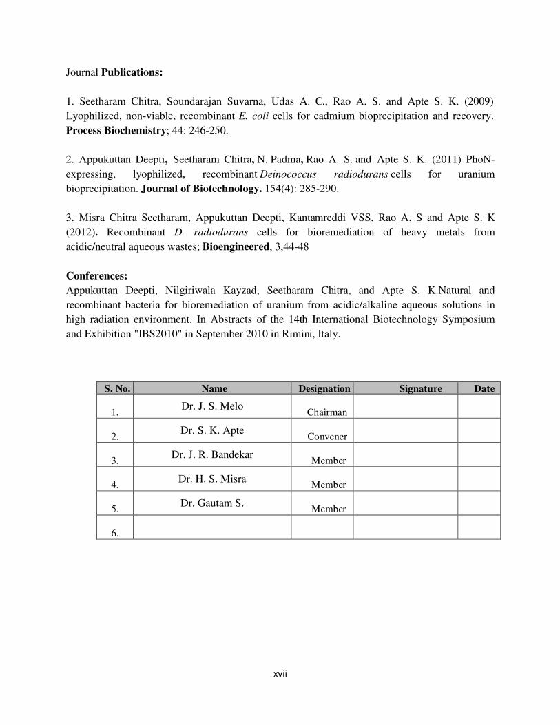
S. No. Name Designation Signature Date
1. Dr. J. S. Melo
Chairman
2.
Dr. S. K. Apte
Convener
3.
Dr. J. R. Bandekar
Member
4.
Dr. H. S. Misra
Member
5.
Dr. Gautam S.
Member
6.
xviii
List of Figures
Chapter 1. Introduction
1.1 How bacteria cope with toxic concentrations of heavy ions 4
1.2 S layer covering bacterial surface 12
1.3 A schematic representation of the deinococcal cell envelope 18
1.4 The Hpi protein of D. radiodurans 19
Chapter 2. Materials and Methods
2.1 Peptidoglycan binding assay 38
Chapter 3. Construction of deinococcal S layer fusion proteins with metallothionein
(SmtA) and acid phosphatase (PhoN): cloning and expression
3.1 TEM of the Hpi layer preparation from D. radiodurans cells 45
3.2 Hpi protein of D. radiodurans 46
3.3 Identification of 123 kDa CS1 band 47
3.4 Identification of 123 kDa CS1 band 49
3.5 MASCOT search results for mass spectra of 91 kDa band CS3 and its peptide
coverage
50
3.6 MASCOT search results for mass spectra of 61 k Da band CS4 and its peptide
coverage
51
3.7 MASCOT search results for mass spectra of 58 kDa band C5 and its peptide
coverage
52
3.8 Detection of the Hpi protein by Western Blotting and Immunodetection 53
3.9 Construction and cloning of pPS1 55
3.10 Cloning and confirmation of pPS1 56
3.11 Construction and cloning of pPHS1 57
3.12 Cloning and confirmation of pPHS1 58
3.13 Identification of the SLH domain in Dr_2577 ORF 59
3.14 Construction and cloning of pPSS1 60
xix
3.15 Cloning and confirmation of pPSS1 61
3.16 Construction and cloning of pPH1 63
3.17 Cloning and confirmation of pPH1 64
3.18 Western Blotting and Immunodetection of SmtA in recombinant
D. radiodurans cells
65
3.19 Localization of the S layer fusion proteins determined by Western Blotting and
Immunodetection in recombinant D. radiodurans cells
66
3.20 Localization of the Hpi-SmtA protein determined by Western Blotting and
Immunodetection in recombinant E.coli cells
67
3.21 Construction and cloning of pGDRF3 69
3.22 Cloning and confirmation of pGDRF3 70
3.23 Zymogram showing in-gel PhoN activities 71
3.24 Western Blot and immunodetection of the Hpi-PhoN fusion protein 72
3.25 Histochemical screening for whole cell phosphatase activity in recombinant D.
radiodurans cells
73
3.26 Localization of the Hpi-PhoN fusion protein in recombinant cells- 74
3.27 Construction and cloning of pPSP1 76
3.28 Cloning and confirmation of pSPS1 77
3.29 Phosphatase activity of SLH-PhoN fusion protein 78
3.30 Localization of the fusion protein in recombinant cells carrying SLH-phoN fusion
gene-
79
3.31 Construction and cloning of pPSP3 81
3.32 Cloning and construction of pPSP3 82
3.33 Overexpression of the SLH-PhoN protein in recombinant E. coli cells 83
3.34 MASCOT search result for the mass spectra generated for the 38 kDa SLH-PhoN
protein over-expressed in E. coli BL21 cells
84
3.35 Peptidoglycan binding by SLH-PhoN fusion protein 86
Chapter 4. Heavy metal bioremediation using recombinant proteins and bacteria
4.1 Uranium biosorption by Hpi layer 95
4.2 Differential uranium biosorption ability and cell surface charge of wild type D.
xx
radiodurans and Hpi mutant cells, HMR202 96
4.3 Cadmium binding using recombinant D. radiodurans cells 99
4.4 Cadmium binding using recombinant E. coli cells 100
4.5 Zinc and copper binding using recombinant D. radiodurans cells 101
4.6 pH dependence of PhoN phosphatase activity and cadmium precipitation ability of
recombinant E. coli cells expressing phoN
104
4.7 Kinetics of bioprecipitation of uranium and cadmium by E. coli (PhoN) and D.
radiodurans (PhoN) cells
106
4.8 Uranium precipitation by D. radiodurans recombinant cells expressing PhoN or
Hpi-PhoN
108
4.9 Uranium precipitation by Hpi isolated from recombinant D. radiodurans cells 109
4.10 SLH-PhoN immobilized on peptidoglycan for uranium precipitation 111
4.11 Comparison of PhoN carrying biomass for uranium precipitation 112
4.12 Effect of lyophilization on cell morphology as observed by scanning electron
microscopy
113
4.13 Cadmium bioprecipitation by lyophilized E. coli cells bearing PhoN- 115
4.14 Effect of lyophilisation on uranium precipitation by SPhoNP 117
4.15 Scanning electron microscopy images of E. coli cells expressing phoN 118
4.16 EDX spectra and scanning electron microscopy of lyophilized E. coli cells bearing
phoN
119
4.17 Localization of uranium phosphate precipitated by D. radiodurans cells expressing
PhoN
120
4.18 Scanning Electron Microscopy and Energy Dispersive X-ray (EDX) spectra of
uranium bioprecipitation by recombinant Deinococcus cells
121
4.19 Surface association of uranium phosphate precipitate as observed by Transmission
electron microscopy
122
xxi
List of Tables
Chapter 2. Materials & Methods
2.1 List of bacterial strains used in this study 25
2.2 List of primers used in this study 28
2.3 List of plasmids used in this study 29
2.4 Composition of polyacrylamide gels 32
Chapter 3. Construction of deinococcal S layer fusion proteins with metallothionein
(SmtA) and acid phosphatase (PhoN): cloning and expression
3.1 Specific activity of various recombinants bearing the phoN gene 73
Chapter 4. Heavy metal bioremediation using recombinant proteins and bacteria
4.1 Metal binding by Hpi protein 94
4.2 Metal binding by isolated Hpi layer from recombinant D. radiodurans cells 102
4.3 Phosphatase activity and uranium precipitation ability of E. coli cells bearing PhoN 105
4.4 Effect of lyophilisation of E. coli cells bearing phoN on phosphatase activity,
cadmium precipitation and cell viability
114
4.5 Effect of lyophilisation on uranium precipitation 117
4.6 Recovery of precipitated metal from recombinant cells and their reuse 128

1 | P a g e
Chapter 1
Introduction

2 | P a g e
Introduction
Man’s increasing mastery of natural law has brought us increased life expectancy and a
higher quality of life but has also resulted in sites contaminated with residuals of our
industrial output, which are a threat to the environment and impair human health [1]. Tens of
thousands of sites stand contaminated and waste management has become an international
problem. Among the components of waste, heavy metals are the most problematic to remove
due to their indestructible nature, unlike organic contaminants which can be degraded [2].
Waste from the nuclear industry, for example, predominantly consists of heavy metals, which
being radioactive in nature, add a complication to nuclear waste management. The need to
manage such waste has led to the development of new technologies. While conventional
methods mostly include physico-chemical approaches, alternative approaches like
bioremediation are gaining importance due to their eco-friendly nature and cost-effectivenes
[3].
1.1 Metal Pollution
Metals are essential for life as trace elements but are toxic at higher concentrations
[2]. Due to rapid industrialization, large amounts of toxic effluents, containing a cocktail of
heavy metals are released into the environment, contaminating soil and water. The main
sources of heavy metal pollution are mining, milling and surface finishing industries,
discharging a variety of toxic metals such as Cd, Cu, Ni, Co, Zn and Pb into the environment
[4]. Eventually, build up of dangerous concentrations in food crops grown in contaminated
soil poses a much more serious hazard [2].
Heavy metals form complexes with cellular components which interfere with normal
metabolism of the cell [5]. Apart from this chemical toxicity, heavy metals which exist as
radioisotopes, also exert radiological toxicity. Mining of uranium and re-processing of
3 | P a g e
nuclear fuel has resulted in generation of large amount of effluents containing radioisotopes
of uranium, plutonium, cesium, strontium etc. The release of radio nuclides, either as
discharge of process effluents produced by industrial activities allied to nuclear power or
through accidental release is a subject of intense public concern [6].
Heavy metals are difficult to remove from the environment and unlike many other
pollutants cannot be chemically or biologically degraded. A number of physico-chemical
processes such as oxidation and reduction, electrochemical treatment, evaporation, ion
exchange and reverse osmosis have been used for removing heavy metals from waste
solutions [2]. High reagent requirement and unpredictable metal ion removal are major
disadvantages associated with such techniques. Further, contaminating reagents are used for
desorption, resulting in toxic sludge and secondary environmental pollution. These
disadvantages can become more pronounced and further aggravate the process cost in case of
contaminated ground waters, mine tailings effluent and other industrial wastewaters due to
voluminous effluents containing complexing organic matter [4]. Biotechnological approaches
can succeed in these areas where physico-chemical methods are inefficient.
In the context of radiological contamination, in a few instances, containment of nuclear
waste has been compromised leading to contamination of trillions of gallons of groundwater.
Aggressive invasive chemical treatments on such large scale can have negative impacts on
biodiversity and can even result in increased dispersion of radioactive materials. Thus,
passive, in situ biological treatment processes are highly desirable [6].
1.2 Bioremediation of heavy metals
Micro-organisms have evolved measures to respond to heavy metal stress via numerous
processes, some of which can be exploited for the remediation of heavy metal containing
waste (Fig. 1.1). Bacterial metal detoxification and removal can be an efficient strategy due
4 | P a g e
to its low cost, high efficiency and eco-friendly nature [7]. Besides this, microbes have
proven capability to take up heavy metals from aqueous solutions, especially when the metal
concentrations in the effluent range from less than 1 to about 20 mg/l. In addition, microbes
also show selectivity in binding specific metals [4].
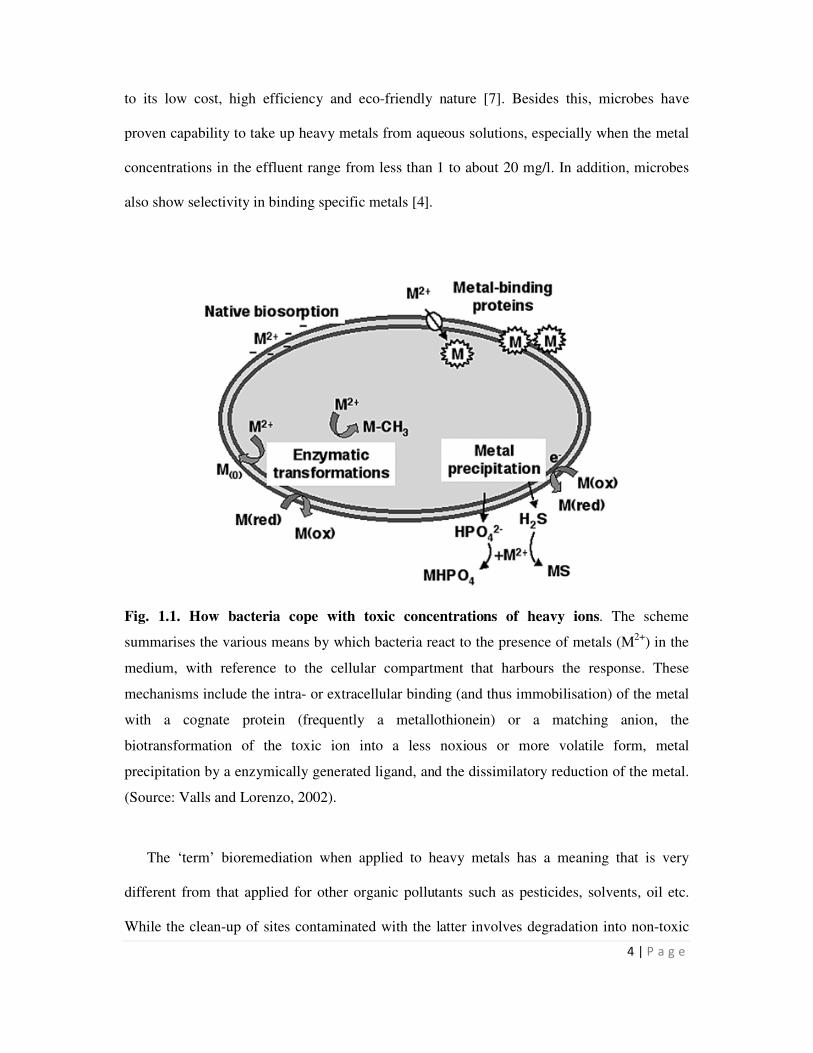
Fig. 1.1. How bacteria cope with toxic concentrations of heavy ions. The scheme
summarises the various means by which bacteria react to the presence of metals (M2+
) in the
medium, with reference to the cellular compartment that harbours the response. These
mechanisms include the intra- or extracellular binding (and thus immobilisation) of the metal
with a cognate protein (frequently a metallothionein) or a matching anion, the
biotransformation of the toxic ion into a less noxious or more volatile form, metal
precipitation by a enzymically generated ligand, and the dissimilatory reduction of the metal.
(Source: Valls and Lorenzo, 2002).
The ‘term’ bioremediation when applied to heavy metals has a meaning that is very
different from that applied for other organic pollutants such as pesticides, solvents, oil etc.
While the clean-up of sites contaminated with the latter involves degradation into non-toxic
5 | P a g e
components, the only way to remediate sites contaminated by heavy metals is to convert them
into relatively less toxic forms which mainly results in immobilization of the metal. Heavy
metal de-contamination using biological means mainly involves the following steps [8]:
• Immobilization of the metal
• Concentration and volume reduction of contaminated matrix
• Compartmentalization of matrix or recovery of metals for re-use
Many microbial-metal interactions have been exploited to facilitate metal immobilization and
these have been shown in Fig. 1.1 and elaborated upon in the text that follows.
1.2.1 Biosorption
Biosorption refers to the passive, metabolically independent adsorption of heavy metals from
aqueous solutions on biomass [9]. The biomass may be living or dead and the sequestration
of metals may involve ion exchange, adsorption, micro-precipitation, and electrostatic and
hydrophobic interactions. Biosorption is affected by molecular size, charge, solubility,
hydrophobicity as well as waste water composition [10]. Several active groups on cell surface
like acetamido group in chitin, the structural polysaccharides of fungi, amine, sulphydryl and
carboxyl groups in protein, phosphodiester (teichoic acid), phosphate, hydroxyl groups in
polysaccharides, participate in biosorption [11]. Ligand preferences of metal ions on
microbial cell surfaces are governed by the hard-soft-acid principle. Hard acids like uranyl
ion preferentially bind to oxygen containing ligands, while soft acids such as Cd2+
bind to –S
and –N containing ligands [10]. Both bacterial as well as fungal biomass have been used
either in batch bioreactors or in immobilized matrices to remove a host of heavy metals from
waste waters [9].
To be able to compare metal uptake capacities of different types of biosorbents, the
adsorption process can be expressed as a batch equilibrium isotherm curve. The biosorption
6 | P a g e
phenomenon can therefore be simulated and used for predicting changes in operational
conditions and for process optimization [7]. It is, at present, the most practical and widely
used approach for the bioremediation of metals and radionuclides due to its simplicity, ease
of operation and availability of biomass and waste bio-products [8,10].
1.2.2 Metal binding molecules
A number of organisms respond to heavy metal stress by producing metal binding
molecules which bind and sequester the heavy metal ion making it unavailable for exerting its
toxicity. For example, Metallothioneins (MTs) are low molecular weight (6–7 kDa), cysteine-
rich proteins found in animals, higher plants, eukaryotic microorganisms and some
prokaryotes. The large number of cysteine residues in MTs bind a variety of metals such as
cadmium, zinc, copper, mercury etc. by mercaptide bonds [12]. MT proteins are classified
based on the arrangement of Cys residues. Class I MTs contain 20 highly conserved Cys
residues based on mammalian MTs and are widespread in vertebrates. MTs without this strict
arrangement of cysteines are referred to as Class II MTs and include all those from plants and
fungi as well as non vertebrate animals. In this MT classification system, Phytochelatins are,
somewhat confusingly, described as Class III MTs [2].
In prokaryotes, ‘MT-like’ proteins have been defined only in Synechococcus sp. and
Pseudomonas putida [13]. MT in Synechococcus sp. was induced following exposure to
elevated concentrations of Cd2+
or Zn2+
, but not Cu2+
. Cyanobacterial (Synechococcus PCC
7942) mutants, smt-, are sensitive (5-fold reduction in tolerance) to Zn2+
, and show some
reduction in tolerance to Cd2+
[14].
While MTs are genetically coded, enzymically synthesized polypeptides which occur in
plants, fungi, nematodes and all groups of algae including cyanobacteria called
phytochelatins (PCs) are also known to bind heavy metals such as Cd, Hg, As and Pb [7].
7 | P a g e
PCs form a family of structures with increasing repetitions of the γ-Glu-Cys dipeptide
followed by a terminal Gly; (γ-GluCys)n-Gly, where n is generally in the range of 2 to 5 [12].
The biosynthesis of PCs is induced by many metals including Cd, Hg, Ag, Cu, Ni, Au, Pb
and Zn; however Cd is by far the strongest inducer. The metal binds to the constitutively
expressed enzyme, PC synthase, thereby activating it to catalyse the conversion of
glutathione (GSH) to phytochelatin [2].
Apart from metallothionein and phytochelatins, randomly generated synthetic
polypeptides, synthetic phytochelatins and polyhistidines have also been engineered for
enhanced accumulation of metals by recombinant microbes [8].
1.2.3 Biomineralization
Biomineralization is the formation of insoluble metal precipitates by interactions with
microbial metabolic products. Metal precipitation as phosphates, sulfides and as a
consequence of metal reduction are known to be mediated by microbes and have potential for
bioremediation [9]. Sulfate-reducing bacteria (SRB) are anaerobic heterotrophs utilizing a
range of organic substrates and SO42–
as a terminal electron acceptor. The sulfide produced
from sulfate reduction not only plays a major role in metal sulfide immobilization in
sediments but has also been applied to bioremediation of metals in waters and leachates [15].
While all metal sulphides are insoluble at neutral pH, several are also insoluble in moderately
acidic anaerobic solutions. Metal sulphides exhibiting these properties include those of
copper, mercury, cadmium, arsenic, selenium and lead. Other metal sulphides, such as cobalt,
zinc, nickel and iron, require a more-alkaline environment to ensure complete precipitation
and stability. It is possible, therefore, to separate some of the highly toxic metals from the
less toxic ones based on sulphide chemistry [11]. However, even low concentrations of free
8 | P a g e
metals are toxic to SRB. A highly cadmium resistant Kebsiella planticola strain could
precipitate 50 times more cadmium than that reported for isolated SRBs [16].
Reductive precipitation involves reducing the metal to a lower redox state which is less
soluble. For example, U(VI) can be reduced to U(IV) by certain Fe(III) dissimilatory
microbes, such as Geobacter metallireducens, in turn leading to precipitation of uranium
metal from solution. Such processes can also accompany other indirect reductive metal
precipitation mechanisms, for example, in sulfate-reducing bacterial systems where reduction
of Cr(VI) can be a result of indirect reduction by Fe2+
and the sulfide produced [9].
A third mode of metal precipitation involves release of a phosphate ligand (Pi) by
biological processes which in turn results in precipitation of heavy metals. One of the first
reports on this involved a Citrobacter strain harboring an acid phosphatase which was
efficiently used for precipitating several heavy metals such as uranium, cadmium, nickel,
americium and plutonium from solution [17-21]. Another mechanism relevant to phosphate
mediated metal precipitation involves degradation of polyphosphates under specific
conditions which results in release of Pi for metal precipitation [9,22-23].
Phosphatase mediated metal precipitation occurs at even low concentrations of metal
due to localized high concentration of the phosphate released, and downstream processing is
easier due to the cell bound nature of the precipitate [24]. While biosorption and metal
binding molecules are typically useful at very low metal concentrations in the micromolar
range, bioprecipitation is more effective at higher metal concentrations [25]. Therefore, the
mechanisms described here, together cover the spectrum of metal concentrations likely to be
present in waste sites.
9 | P a g e
1.3 Genetic Engineering for heavy metal bioremediation
Genetic engineering for bioremediation of heavy metals has been modestly successful in
endowing microbes, which occur and grow in waste, with ability to remediate metals, and in
enhancing their bioremediation potential. E. coli, has been a favourite for genetic
manipulation for such purpose and offers the advantage of convenient expression of foreign
proteins [26-28]. But other microbes such as Ralstonia eutropha, Pseudomonas fluorecens,
Sphingomonas desiccabilis, Bacillus subtilis, Deinococcus radiodurans and Caulobacter
crescentus have also been genetically manipulated for metal bioremediation [7].
1.3.1 Genetic engineering for heterologous expression of metallothionein encoding genes
MTs from various sources, including monkey MT, yeast MT, human MT-II, mouse
MT-I, rainbow trout MT and plant MT [2], have been expressed intracellularly in Escherichia
coli. Recombinants expressing the engineered protein not only displayed superior metal
binding but in many cases also showed higher tolerance to the metal. The smtA from
Synechococcus elongatus was expressed as a Glutathione-S-transferase fusion protein in E.
coli cells. Such recombinant protein showed enhanced accumulation of zinc but E. coli cells
harboring the fusion protein did not show any detectable increase in tolerance towards
cadmium, copper or zinc [14].
In certain cases, the genes involved in metal transport were co-engineered with the
MT gene resulting in increased metal sequestration by recombinant cells compared to
engineering MT alone. This has been done for several metals such as Hg, Cd and Ni [7].
Ralstonia eutropha CH34 strain, which is adapted to thrive in metal contaminated soils, was
engineered to express the mouse metallothionein I protein on the cell surface. Inoculation of
such recombinant cells into Cd contaminated soils significantly decreased the toxic effects of
the metal on the growth of tobacco plants [29].
10 | P a g e
1.3.2 Genetic engineering for phosphate mediated bioprecipitation of metals
Genes encoding enzymes involved in phosphate metabolism have been cloned for
phosphate mediated bioprecipitation. Gene (phoN) coding a non specific-acid phosphatase,
PhoN from Salmonella enterica serovar Typhi was introduced into E. coli for removing
uranium and nickel from solutions [19]. In another variation of exploitation of biologically
generated phosphate for metal precipitation, Pseudomonas aeruginosa was engineered to
over-express its native polyphosphate kinase which resulted in accumulation of large amount
of polyphosphate. Under carbon starvation conditions, polyphosphate was degraded, in turn
resulting in uranium precipitation from solution. Nearly 80% of uranium could be removed
by such cells within 48 h from a 1 mM metal solution [23]. In E. coli, manipulation of
polyphosphate metabolism by overexpression of the native genes for polyphosphate kinase
(ppk) and polyphosphatase (ppx) resulted in lower intracellular polyphosphate, phosphate
secretion and increased metal tolerance [22].
An alkaline phosphatase PhoK encoding gene, phoK, from Sphingomonas was
introduced into E. coli and used for bioprecipitation of uranium from alkaline solutions [30].
Such recombinant cells could remove >90% of input uranium in less than 2 h from alkaline
solutions containing 0.5 to 5 mM of uranyl carbonate. Specifically for uranium, the uranyl
phosphate mineral is extremely stable providing a long-term sink for uranium contaminated
sites. These attributes make this mechanism highly desirable for metal bioremediation.
1.3.3 Surface Expression of proteins for Bioremediation
The expression of recombinant protein of interest inside suitable hosts is fraught with
problems of stability and short half-life of the expressed heterologous proteins. For eg., in the
case of metallothionein, the high cysteine content might interfere with cellular redox
pathways in the cytosol [2]. In, addition to this, the intracellular expression of proteins leads
11 | P a g e
to decreased contact between enzymes and target contaminant and low uptake of substrates
resulting in poor bioremediation potential. Engineering proteins for cell surface display on
microbes endows intact cells with new functionalities that have a vast sphere of new
applications [31].
Among gram negative bacteria, E. coli has been extensively investigated for cell
surface engineering. Metal binding peptides of sequences Gly-His-His-Pro-His-Gly (namely
HP) and Gly-Cys-Gly-Cys-Pro-Cys-Gly-Cys-Gly (namely CP) were genetically engineered
into the native LamB protein and expressed in E. coli. The potential of E. coli expressing CP
to bind cadmium (Cd2+
) from the growth medium was increased fourfold compared to wild-
type cells. Synthetic phytochelatin were fused to Outer membrane protein A and maltose
binding protein for enhanced cadmium accumulation. Similarly genetically modified E. coli
co-expressing a Hg2+
transport system with (Glu-Cys)20Gly(EC20) or by directly expressing
EC20 on the cell surface effectively removed Hg2+
from liquid medium [31].
Mouse metallothionein protein was targeted to the cell surface of Ralstonia eutropha
by fusing it to the auto transporter β-domain of IgA protease of Neisseria gonorrhoeae which
targeted the hybrid protein toward the bacterial outer membrane [32]. The genetically
engineered strain R. eutropha MTB accumulated increased amounts of Cd2+
from liquid
media.
In Gram positive bacteria, there are relatively fewer examples of surface display. The
fungal cellulose-binding domain (CBD), derived from Trichoderma reesei cellulase Cel6A,
has been expressed in its non-engineered form onto the surface of Staphylococcus carnosus.
The cell surface expression of the CBD scaffold could prove helpful for enhanced bio-
accumulation of Ni2+
[31].While all these techniques of surface engineering have been
successful, a more recent tool for surface display of proteins in bacteria is by fusing to
Surface Layer Proteins.
12 | P a g e
1.4 Surface layer proteins
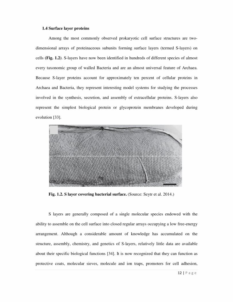
Among the most commonly observed prokaryotic cell surface structures are two-
dimensional arrays of proteinaceous subunits forming surface layers (termed S-layers) on
cells (Fig. 1.2). S-layers have now been identified in hundreds of different species of almost
every taxonomic group of walled Bacteria and are an almost universal feature of Archaea.
Because S-layer proteins account for approximately ten percent of cellular proteins in
Archaea and Bacteria, they represent interesting model systems for studying the processes
involved in the synthesis, secretion, and assembly of extracellular proteins. S-layers also
represent the simplest biological protein or glycoprotein membranes developed during
evolution [33].
Fig. 1.2. S layer covering bacterial surface. (Source: Seytr et al. 2014.)
S layers are generally composed of a single molecular species endowed with the
ability to assemble on the cell surface into closed regular arrays occupying a low free-energy
arrangement. Although a considerable amount of knowledge has accumulated on the
structure, assembly, chemistry, and genetics of S-layers, relatively little data are available
about their specific biological functions [34]. It is now recognized that they can function as
protective coats, molecular sieves, molecule and ion traps, promoters for cell adhesion,
13 | P a g e
immunomodulators, surface recognition, antifouling coatings, and as virulence factors in
pathogenic organisms [35]. In those Archaea that possess S-layers as the exclusive envelope
component external to the cytoplasmic membrane, the lattice is involved in the determination
of cell shape and as a structure aiding in the cell division process [35].
On an ultrastructural level, S-layer proteins form regular crystalline lattices, which
may have an oblique (p1, p2), square (p4), or hexagonal (p3, p6) symmetry. Lattice constants
can vary from 5 to 30 nm and the S-layer thickness from 5 to 20 nm. Detailed atomic
resolution structures of lattices are not available, but several low-resolution three-dimensional
(3D) structures have been obtained by electron microscopy of negatively stained samples. A
striking similarity even among quite unrelated species is the central core forming region,
which is usually oriented toward the cell envelope, giving rise to an overall corrugated inner
surface. By contrast, the outer surface appears smooth despite highly variable and species-
specific ultrastructure. Between 30% and 70% of one unit cell is occupied by the protein,
which leads to the formation of identical and well-defined pores with a diameter of 2–8 nm
[36].
S-layer proteins exhibit mostly two separated morphological regions, responsible for
cell wall binding and for self-assembly, as shown by mutagenesis studies. The position of the
cell wall anchoring region within the protein, as well as its sequence composition, can vary
among the bacterial species, similar to the molecular binding partner within the underlying
cell wall [33]. In several Gram-positive and Gram-negative bacteria, S-layers bind via the N-
terminal region to the underlying peptidoglycan sacculus by recognizing Secondary Cell Wall
Polymers (SCWP). This specific molecular interaction with the polysaccharide is mediated
by a recurring structural motif termed the ‘‘surface layer homology’’ (SLH) motif. S-layer
proteins devoid of SLH motifs are anchored to different types of SCWPs through their N- or
14 | P a g e
C-terminal domains. In Gram-negative bacteria, the S-layer is attached with its N- or C-
terminus to the component of the outer membrane [36].
1.5 Applications of S layer proteins
The wealth of information accumulated on the general principles of S-layers has led
to a broad spectrum of applications. As S-layers are periodic structures, they exhibit
repetitive identical physicochemical properties down to the subnanometer scale and possess
pores identical in size and morphology. Most importantly, properties of S-layer proteins can
be changed by chemical modifications and genetic engineering. It is now evident that S-
layers also represent a unique structural basis and pattering element for generating complex
supramolecular assemblies involving all relevant ‘building blocks’ such as proteins, lipids,
glycans, and nucleic acids [33].
The regularly arranged pores in S layer proteins have been exploited for developing S
layer isoporous ultrafiltration membranes, with sharp molecular weight cut-offs. S layers
have also been used as an immobilization matrix for functional molecules and nano particles.
Because S-layer lattices are composed of identical protein or glycoprotein species, functional
sequences introduced either by chemical modification or genetic engineering must be aligned
in exact positions and orientation down to the subnanometer scale. Enzymes like invertase,
glucose oxidase, β-galactosidase were immobilized on the outer surface of surface layers
[33]. Furthermore, a universal biospecific matrix for immunoassays and dipsticks could be
generated by immobilizing monolayers of either protein A or streptavidin onto S layers. S
layer supported lipid membranes have been developed to study characteristics of archaeal cell
envelopes, as surfaces with new properties such as anti-fouling and as a matrix for re-
constitution of transmembrane proteins [33]. Further, S layer coated liposomes demonstrated
much higher mechanical and thermal stability than plain ones. S layers proteins being surface
15 | P a g e
components are likely to have an important role in virulene of pathogenic bacteria. They are
prime candidates for vaccine development. They have been used as attenuated pathogens, as
antigen/hapten carrier, as adjuvants or as part of vaccination vesicles [33].
Due to the promising results obtained with native S layer protein, as immobilization
matrix, genetic engineering of S layer proteins was envisaged [37]. While earlier, the
functional molecules were adsorbed and/ or cross-linked to the S layer protein, now
construction of chimeric functional S layer protein has taken precedence. S layer are capable
of tolerating fusions with foreign proteins that never participate in lattice formation while
retaining the ability to assemble into geometrically highly defined layers. Fusions with the
major birch allergen, fluorescent proteins, core streptavidin, a C-terminally fused cysteine
residue for patterning of nanoparticles, or enzymes from extremophiles have been re-
crystallized on various supports [33]. Significant advantages for enzyme immobilization by
the S-layer self assembly system over processes based on random immobilization of sole
enzymes include the requirement of only a simple, one-step incubation process for site-
directed immobilization without preceding surface activation of the support. Moreover, the
provision of a cushion for the enzyme through the S-layer moiety of the fusion protein
prevents denaturation and consequently loss of enzyme activity upon immobilization [33].
Another current approach considers the use of bacterial S-layers as a potential
alternative for bioremediation processes of heavy metals in field. The S-layer of Bacillus
sphaericus JG-A12, an isolate from a uranium mining waste pile in Germany, was shown to
bind high amounts of toxic metals such as U, Cu, Pd(II), Pt(II), and Au(III) [38].
Furthermore, Velasquez and coworkers in 2009 determined the tolerance of different
Colombian B. sphaericus native strains to different heavy metals and came to the conclusion
that their S-layer proteins might have the ability to entrap metallic ions, either on living or
dead cells [39].
16 | P a g e
1.6 Chimeric Fusion proteins tagged to S layer for bioremediation
While S layer proteins have been fused to many proteins for various applications, very
few examples for chimeric proteins for bioremediation exist in literature. A Hexa-histidine
peptide was inserted to a permissive site of the surface layer (S-layer) protein RsaA of
Caulobacter crescentus. The recombinant strain JS4022/p723–6H, expressing RsaA-6His
fusion protein was examined for its ability to sequester Cd (II) from the bacterial growth
medium. When mixed with 1 ppm CdCl2, JS4022/p723–6H removed 94.3∼99.9% of the
Cd(II), whereas the control strain removed only 11.4∼37.0% leading to a metal loading of 16
mg/g dry cell weight [40]. In another study, the S layer protein from Bacillus spahericus was
overexpressed in E. coli with a 6X-histidine tag. The purified protein was re-assembled and
tested for nickel binding ability. While the wild type protein could remove 13.8 mg Ni/g
proteins, the recombinant protein could remove 31 mg Ni/g protein respectively [41].
1.7 Deinococcus radiodurans, an ideal candidate for bioremediation of radioactive waste
Deinococcus radiodurans is an ancient Gram positive bacterium that can tolerate
extremely high doses of ionizing radiation. Members of the Deinococcaceae family are
vegetative, non-pathogenic, ubiquitous, and show resistance to DNA damage caused by
ionizing radiation, desiccation, ultraviolet radiation, oxidizing agents, and electrophilic
mutagen [42-46]. The organism has been extensively studied for its highly proficient DNA
damage repair mechanisms that are aided by proteins rapidly induced upon irradiation [47-
48].
Radioactive waste sites typically contain organopollutants such as toluene,
trichloroethylene; radionuclides such as uranium, plutonium, cesium etc. and heavy metals
such as lead, mercury, chromium, arsenic etc. [49]. Numerous organisms have the ability to
degrade, transform, detoxify and immobilize these pollutants as has been already described.
17 | P a g e
However, most of them are sensitive to the damaging effects of radiation. Choice of the
appropriate organism depends on its ability to survive and efficiently express desired genes
under the harsh conditions prevailing in waste sites. This necessitates use of radioresistant
organism like D. radiodurans for bioremediation of radioactive waste.
Genes coding for Toluene dioxygenase were cloned from Pseuodomonas putida F1
into the chromosome of D. radiodurans to produce a recombinant strain which could oxidize
toluene, chlorobenzene, indole etc. Such cells could grow in the presence of chlorobenzene
and at 60 Gy /min irradiation, while degrading the pollutant simultaneously [50]. In addition
to the toluene genes, the mercury resistance gene, merA was also cloned from E. coli strain
BL308 into D. radiodurans. Such cells could grow in the presence of both radiation and the
metal while detoxifying mercury to its volatile form [49].
In another study, phoN, an acid phosphatase gene from Salmonella enteric serovar
Typhi was cloned into D. radiodurans under a strong deinococcal promoter, PgroESL. The
phosphatase activity of the recombinant strain was utilized for precipitation of uranium.
Nearly 90% of 1 mM uranium could be precipitated in 6 h by recombinant D. radiodurans
strain carrying PgroESL-phoN construct on plasmid pPN1. Further, they could bring about
metal precipitation even after 6 kGy gamma radiation under non-growing conditions, while
E. coli cells carrying the same construct failed to do so [51]. This organism has also been
used for engineering an alkaline phosphatase, PhoK for removal of uranyl carbonate from
alkaline waste solutions [52]. Taken together, D. radiodurans has emerged as an organism
which is easy to genetically manipulate and is also radiation resistant making it an ideal
candidate for genetic manipulation for radioactive waste management.
18 | P a g e
1.8 S layer proteins in D. radiodurans.
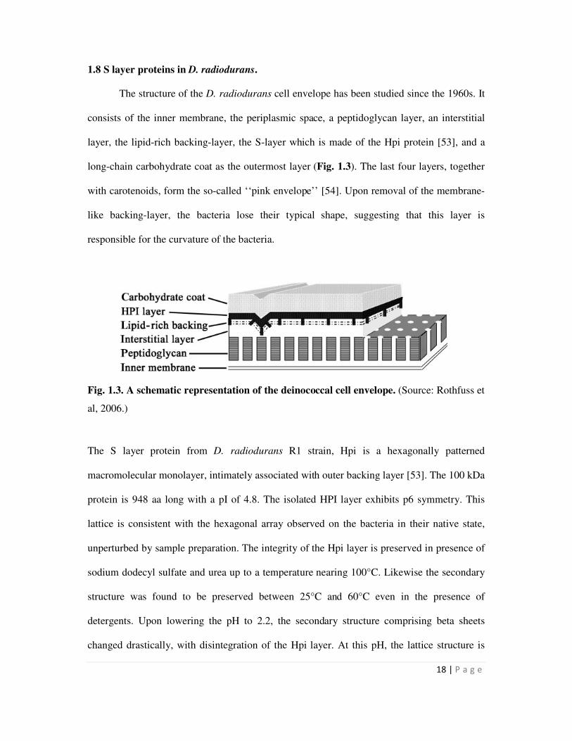
The structure of the D. radiodurans cell envelope has been studied since the 1960s. It
consists of the inner membrane, the periplasmic space, a peptidoglycan layer, an interstitial
layer, the lipid-rich backing-layer, the S-layer which is made of the Hpi protein [53], and a
long-chain carbohydrate coat as the outermost layer (Fig. 1.3). The last four layers, together
with carotenoids, form the so-called ‘‘pink envelope’’ [54]. Upon removal of the membrane-
like backing-layer, the bacteria lose their typical shape, suggesting that this layer is
responsible for the curvature of the bacteria.
Fig. 1.3. A schematic representation of the deinococcal cell envelope. (Source: Rothfuss et
al, 2006.)
The S layer protein from D. radiodurans R1 strain, Hpi is a hexagonally patterned
macromolecular monolayer, intimately associated with outer backing layer [53]. The 100 kDa
protein is 948 aa long with a pI of 4.8. The isolated HPI layer exhibits p6 symmetry. This
lattice is consistent with the hexagonal array observed on the bacteria in their native state,
unperturbed by sample preparation. The integrity of the Hpi layer is preserved in presence of
sodium dodecyl sulfate and urea up to a temperature nearing 100°C. Likewise the secondary
structure was found to be preserved between 25°C and 60°C even in the presence of
detergents. Upon lowering the pH to 2.2, the secondary structure comprising beta sheets
changed drastically, with disintegration of the Hpi layer. At this pH, the lattice structure is
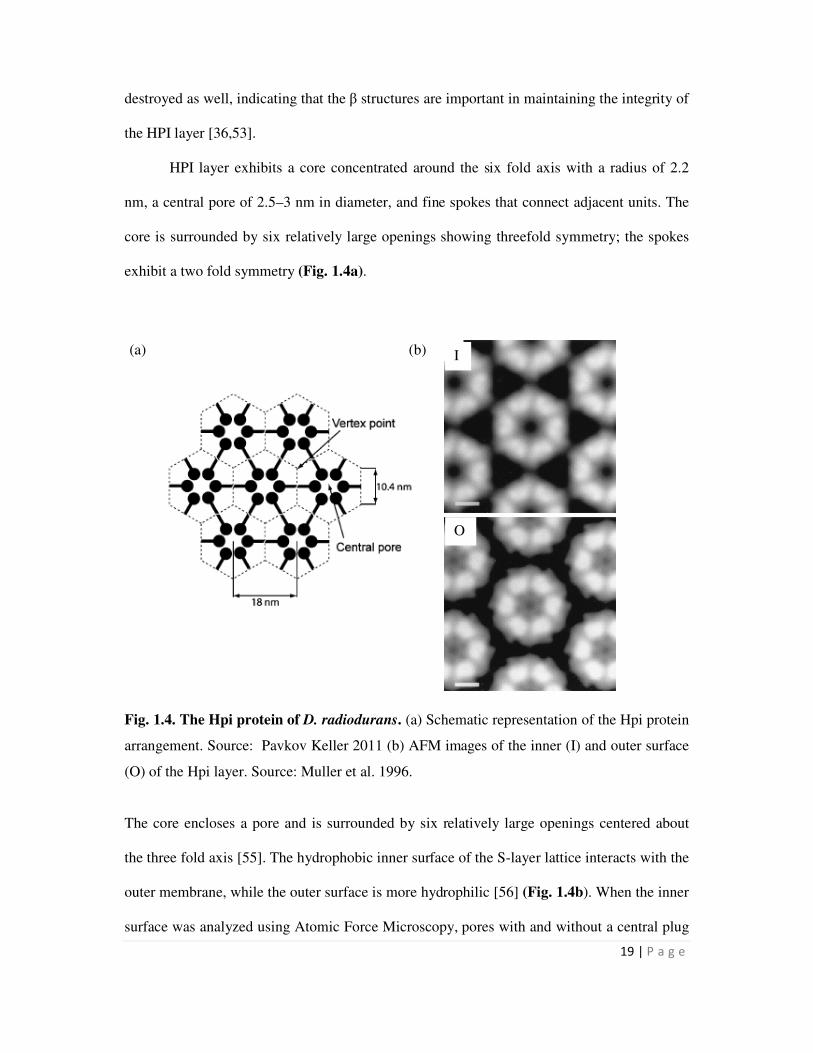
19 | P a g e
destroyed as well, indicating that the β structures are important in maintaining the integrity of
the HPI layer [36,53].
HPI layer exhibits a core concentrated around the six fold axis with a radius of 2.2
nm, a central pore of 2.5–3 nm in diameter, and fine spokes that connect adjacent units. The
core is surrounded by six relatively large openings showing threefold symmetry; the spokes
exhibit a two fold symmetry (Fig. 1.4a).
Fig. 1.4. The Hpi protein of D. radiodurans. (a) Schematic representation of the Hpi protein
arrangement. Source: Pavkov Keller 2011 (b) AFM images of the inner (I) and outer surface
(O) of the Hpi layer. Source: Muller et al. 1996.
The core encloses a pore and is surrounded by six relatively large openings centered about
the three fold axis [55]. The hydrophobic inner surface of the S-layer lattice interacts with the
outer membrane, while the outer surface is more hydrophilic [56] (Fig. 1.4b). When the inner
surface was analyzed using Atomic Force Microscopy, pores with and without a central plug
(a) (b) I
O
20 | P a g e
were found. By repeating the measurement after 5 min, switching from the open to the closed
state was observed for individual pores. It is not known what induces the switching, although
tip-induced conformational changes are ruled out. It was suggested that molecules up to 5
kDa could pass through the central 2.2 nm wide channel [57]. Further, serine and threonine
are clustered in the N-terminal region of the sequence that faces the underlying outer
membrane. Close to the C-terminus which faces the outer surface, there is a stretch that
shows a significant elevated content of tyrosine as well as serine plus threonine, with
hydroxylamine totaling almost 30% [58].
The existence of an additional protein involved in the maintenance and integrity of the
S-layer in D. radiodurans was first reported by Cava et al [59]. SlpA (DR2577) is a
proposed homolog of an S-layer protein from T. thermophilus, since both proteins contain
one SLH motif at the N-terminus. Cava et al. 2004 proposed that most of the cell wall of
Thermus thermophilus was covered by secondary cell wall polymers (SCWP) which was
recognized by an antibody (αSAC) raised against S-layer attached cell wall fragments.
Further, pyruvylation of the SCWP was essential for SLH-domain mediated binding by SlpA.
The pyruvylation function was assigned to the csaB gene. Cell walls from csaB mutant bound
SlpA with much lesser affinity. Immunological cross-reactivity was detected with αSAC on
cell walls of D. radiodurans. Further a csaB homologue was found in D. radiodurans lending
support to occurrence of similar interactions in this organism [59].
SlpA of D. radiodurans comprises 1167 amino acids, of which the first 19 amino
acids most likely constitute a signaling peptide. The mature protein has a theoretical
molecular mass of 121.8 kDa, a theoretical pI of 4.9 and contains two cysteines. Deletion of
the slpA gene in this organism leads to a change in the structure of the cell envelope as well
as in resistance to solvent and shear stress [54]. By comparison deleting the hpi gene caused
little alternations in the structure of the cell envelope or its reaction to shear- or solvent-
21 | P a g e
induced stress. This indicates that the SlpA protein may play an important role in the
preservation of the cell envelope structure. Since the Hpi protein does not contain any SLH
motifs, it was proposed that the SlpA protein could be responsible for anchoring to the inner
cell envelope [54].The structure of the SlpA lattice has not been analyzed so far.
1.9 Stabilization of biomass for bioremediation
While bioremediation shows promise, the application of microbial biomass to waste sites
and its stability are issues which also need to be addressed. Often microbes are pre-treated to
generate a formulation which can be readily used and is likely to retain metal removal
potential for a longer period of time. Physical treatments such as heating, autoclaving, freeze
drying and chemical pre-treatments such as using acids, alkali and organic chemicals showed
enhancement in metal biosorption in a few cases [60]. For example pre-treatment of
Streptomyces rimosus with NaOH resulted in increasing Cd biosorption capacity [61]. Some
of these treatments render the biomass non-viable which offers a few advantages. For
example, constant nutrient supply is not required for systems using dead cells and recovery of
metals and regeneration of biosorbents is less complicated than for living cells [10,60].
Specifically freeze drying/lyophilization of biomass is a technique which preserves cell
structure and functionalities better than other treatments and is therefore well suited for
bioremediation. Lyophilized residual brewery yeast was used for removing uranium from
aqueous solutions [62]. A Pseudomonas strain was lyophilized and used for remediation of
uranium and thorium [63]. Freeze dried marine microalgae could adsorb cadmium more
rapidly than fresh cells [64]. Similarly, use of ‘reagent-like’ freeze dried recombinant bacteria
for measuring bioavailability of arsenic and antimony have been reported [65]. While a
number of examples for lyophilization of biomass exist in literature for biosorption of heavy
metals, there are very few examples for enzyme mediated metal immobilization. The only
22 | P a g e
examples that can be found are reports on chromium reductase and uranium reductase
activity in D. desulfuricans which were stable after freeze drying and storing at room
temperature [66].
1.10 This study
In the past, phoN gene of Salmonella enterica serovar Typhi that encodes a non-
specific acid phosphatase (NSAP) was over-expressed in D. radiodurans [51]. However, the
phosphatase activity obtained in D. radiodurans was far less (10%) than that obtained in
recombinant E. coli cells carrying the same construct. It was suggested that this may be due
to the multi layered complex cell wall in D. radiodurans [54] which might limit substrate
accessibility to PhoN. Since, the thick complex cell wall of this organism could pose a major
problem in engineering this organism for any kind of extracellular metal remediation, the
possibility of exploiting membrane localized proteins on the cell surface for display of
proteins relevant to bioremediation has been explored in the present study. Earlier work has
shown that Surface (S) layer proteins are ideally suited for display of peptides and proteins.
D. radiodurans is known to possess two S-layer proteins, Hpi and SlpA. The possibility of
fusing such proteins to PhoN for enhanced metal precipitation ability was explored along
with another protein relevant to bioremediation, the metallothionein SmtA from
Synechococcus elongatus, which would be useful for sequestering metals such as cadmium,
copper and zinc. The utility of the fusion proteins so generated as well as recombinant strains
expressing them were studied. The specific objectives of the present work were as follows:
1. Construction of the S layer fusion proteins with bioremediation active phosphatase
(PhoN) and metallothionein (SmtA) proteins for metal removal, using recombinant
DNA technology.
2. Studies on the expression, localization and activity of the fusion proteins.
23 | P a g e
3. Evaluation of microbes expressing fusion proteins for removal of metals such as U
and Cd from solution.
4. Assessment of the utility of purified S-layer proteins and S-layer fusion proteins in
removing metals from solutions in vitro, and
5. Evaluation of lyophilized recombinant microbes for metal removal from solution.
The work carried out is presented in the following chapters:
Chapter 2 describes materials, methods and protocols followed in this study. The Chapters 3
and 4 describe the results from this work. Chapter 3 deals with the cloning and expression of
S layer fusion proteins. The hpi gene and the nucleotide sequence encoding SLH domain of
the SlpA protein of D. radiodurans were fused independently to phoN and smtA and their
expression and localization was studied. Chapter 4 describes the potential of recombinants
expressing the fusion protein as well as the purified S-layer proteins and S-layer fusion
proteins for metal binding / precipitation. The utility of lyophilization in microbe based
bioremediation has also been studied and elaborated in this chapter. Chapter 5 summarizes all
the major findings of this study.
24 | P a g e
Chapter 2
Materials and Methods
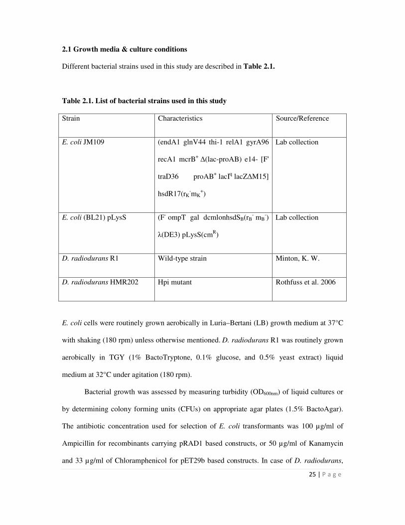
25 | P a g e
2.1 Growth media & culture conditions
Different bacterial strains used in this study are described in Table 2.1.
Table 2.1. List of bacterial strains used in this study
Strain Characteristics Source/Reference
E. coli JM109 (endA1 glnV44 thi-1 relA1 gyrA96
recA1 mcrB+ ∆(lac-proAB) e14- [F'
traD36 proAB+ lacI
q lacZ∆M15]
hsdR17(rK-mK
+)
Lab collection
E. coli (BL21) pLysS (F- ompT gal dcmlonhsdSB(rB
- mB
-)
λ(DE3) pLysS(cmR)
Lab collection
D. radiodurans R1 Wild-type strain Minton, K. W.
D. radiodurans HMR202 Hpi mutant Rothfuss et al. 2006
E. coli cells were routinely grown aerobically in Luria–Bertani (LB) growth medium at 37°C
with shaking (180 rpm) unless otherwise mentioned. D. radiodurans R1 was routinely grown
aerobically in TGY (1% BactoTryptone, 0.1% glucose, and 0.5% yeast extract) liquid
medium at 32°C under agitation (180 rpm).
Bacterial growth was assessed by measuring turbidity (OD600nm) of liquid cultures or
by determining colony forming units (CFUs) on appropriate agar plates (1.5% BactoAgar).
The antibiotic concentration used for selection of E. coli transformants was 100 µg/ml of
Ampicillin for recombinants carrying pRAD1 based constructs, or 50 µg/ml of Kanamycin
and 33 µg/ml of Chloramphenicol for pET29b based constructs. In case of D. radiodurans,
26 | P a g e
recombinants were grown on 3 µg/ml chloramphenicol for pRAD1 based constructs. The D.
radiodurans Hpi mutant, HMR202 was routinely grown in TGY containing 8 µg/ml of
Kanamycin.
2.2 Histochemical screening of recombinants expressing PhoN
Histochemical screening of PhoN expressing E. coli clones was carried out by growing cells
on LB plates containing phenolphthalein diphosphate (PDP) at 1 mg/ml and methyl green
(MG) at 50 µg/ml [67]. For screening D. radiodurans cells, the MG concentration was
reduced to 5 µg /ml due to its toxicity to growth of this organism. Recombinants which
display cell based phosphatase activity give rise to dark green colored colonies on PDP-MG
plates. Phosphoric acid released from PDP due to action of phosphatases causes local
acidification of medium leading to pH indicator dye, MG stain turning dark green.
Precipitation of the dark green MG on phosphatase producing colonies forms the basis for
screening of phosphatase positive strains.
2.3 Recombinant DNA techniques
2.3.1 Isolation of chromosomal DNA from D. radiodurans
D. radiodurans cells from a 25 ml overnight grown culture was re-suspended in 250 µl
ethanol and kept at 4°C for 5 min. The cells were re-suspended in 250 µl Tris-EDTA (T10E1)
buffer (10 mM Tris and 1 mM EDTA), pH 8. Cells were lysed by addition of 50 µl lysozyme
(2 mg/ml) and incubation at 37°C for 30 min, followed by addition of 6 units of Proteinase
K (New England Biolabs) and 50 µl SDS incubated at 37°C for 3 h. To this, equal amount of
phenol equilibrated with 100 mM Tris-HCl, pH 8.0 was added, mixed gently and subjected to
centrifugation at 10,000 rpm for 15 min. The aqueous phase was removed and phenol
extraction was repeated. This was followed by two rounds of chloroform extraction. To the
27 | P a g e
aqueous layer, 8 µl of RNAse (10 mg/ml) was added, followed by incubation at 37°C for 1.5
h. The aqueous layer was extracted twice with phenol:chloroform:isoamyl alchohol (25:24:1).
Chromosomal DNA was spooled after gently mixing the aqueous layer with 1/10th
volume of
150 mM NaCl, pH 7.5 and two volumes of chilled absolute ethanol. The spooled DNA was
washed with 70% ethanol, air dried, dissolved in T10E1 buffer, pH 8.0 and stored at -20°C till
further use.
2.3.2 Restriction endonuclease digestion and electrophoresis of DNA
Restriction endonuclease digestion of DNA was carried out according to the manufacturer’s
protocol (New England Biolabs). The digested DNA (with appropriate amount of 10X
loading dye) was resolved on a 1 % agarose gel containing 0.5 µg/ml ethidium bromide
prepared in 0.5 X TBE (4.45 mM Tris, 4.45 mM boric acid, 0.1 mM EDTA, pH 8.3) buffer.
Electrophoresis was carried out at 8V/cm and DNA fragments were visualized using UV
transilluminator. The 1 kb and 100 bp DNA ladder from New England Biolabs were used as
markers to calculate the molecular mass of the various DNA fragments.
2.3.3 Amplification of DNA by Polymerase Chain Reaction
Primers for PCR amplification were designed on the basis of the published sequence of the
genes for D. radiodurans (http://www.tigr.org) and Salmonella enterica serovar Typhi
(http://www.ncbi.nlm.nih.gov). Primers used in this study are listed in Table 2.2. PCR
amplification was generally carried out using 20 pmoles of each primer, 200 µM dNTPs, 1.5
U Taq DNA polymerase in 1X Taq buffer (Bangalore Genei Ltd.). For PCR of longer DNA
fragments from deinococcal chromosome, the GC Rich PCR amplification kit from Roche
was used. The template for PCR was either recombinant plasmid or chromosomal DNA or a
colony. In case of D. radiodurans, colony PCR was carried out by making a suspension of
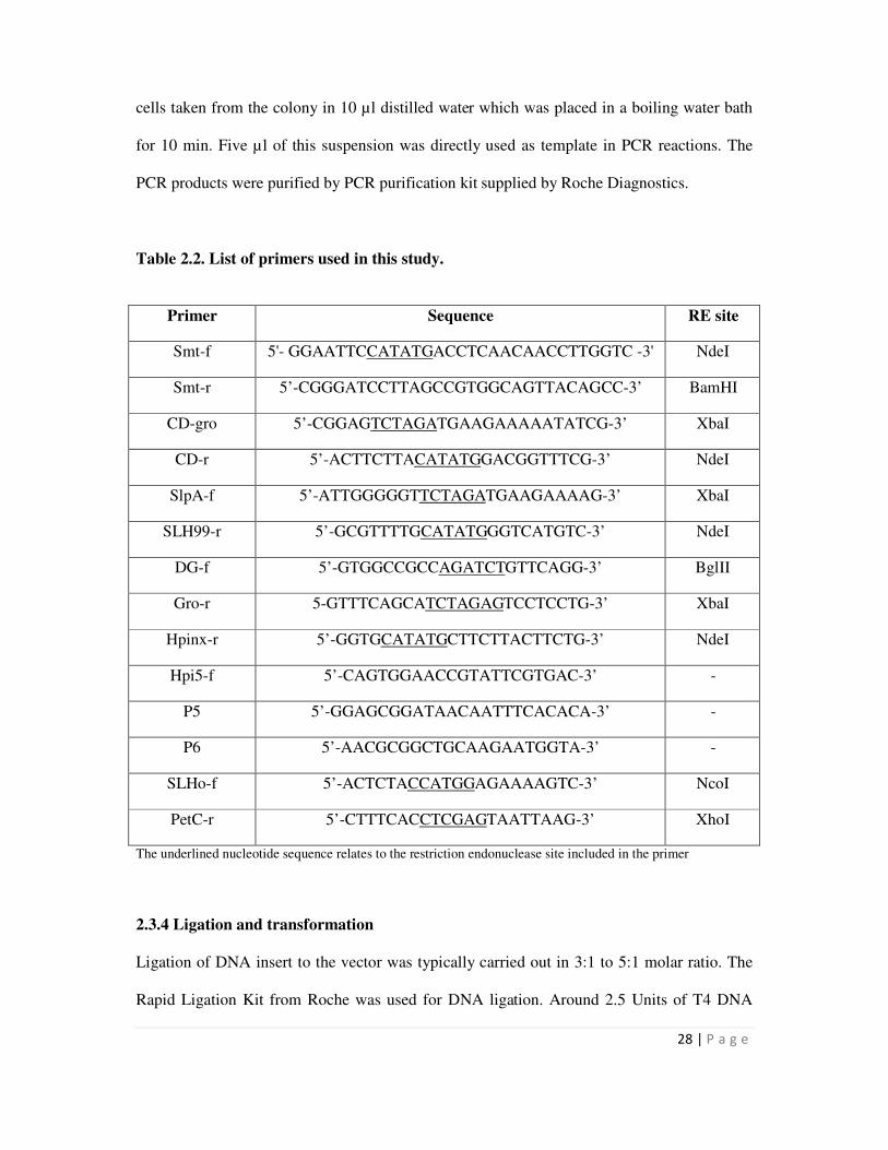
28 | P a g e
cells taken from the colony in 10 µl distilled water which was placed in a boiling water bath
for 10 min. Five µl of this suspension was directly used as template in PCR reactions. The
PCR products were purified by PCR purification kit supplied by Roche Diagnostics.
Table 2.2. List of primers used in this study.
The underlined nucleotide sequence relates to the restriction endonuclease site included in the primer
2.3.4 Ligation and transformation
Ligation of DNA insert to the vector was typically carried out in 3:1 to 5:1 molar ratio. The
Rapid Ligation Kit from Roche was used for DNA ligation. Around 2.5 Units of T4 DNA
Primer Sequence RE site
Smt-f 5'- GGAATTCCATATGACCTCAACAACCTTGGTC -3' NdeI
Smt-r 5’-CGGGATCCTTAGCCGTGGCAGTTACAGCC-3’ BamHI
CD-gro 5’-CGGAGTCTAGATGAAGAAAAATATCG-3’ XbaI
CD-r 5’-ACTTCTTACATATGGACGGTTTCG-3’ NdeI
SlpA-f 5’-ATTGGGGGTTCTAGATGAAGAAAAG-3’ XbaI
SLH99-r 5’-GCGTTTTGCATATGGGTCATGTC-3’ NdeI
DG-f 5’-GTGGCCGCCAGATCTGTTCAGG-3’ BglII
Gro-r 5-GTTTCAGCATCTAGAGTCCTCCTG-3’ XbaI
Hpinx-r 5’-GGTGCATATGCTTCTTACTTCTG-3’ NdeI
Hpi5-f 5’-CAGTGGAACCGTATTCGTGAC-3’ -
P5 5’-GGAGCGGATAACAATTTCACACA-3’ -
P6 5’-AACGCGGCTGCAAGAATGGTA-3’ -
SLHo-f 5’-ACTCTACCATGGAGAAAAGTC-3’ NcoI
PetC-r 5’-CTTTCACCTCGAGTAATTAAG-3’ XhoI
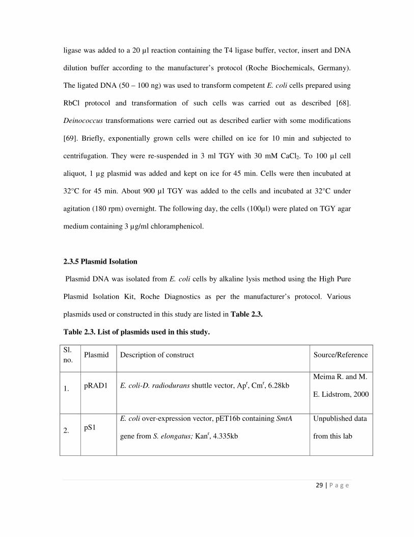
29 | P a g e
ligase was added to a 20 µl reaction containing the T4 ligase buffer, vector, insert and DNA
dilution buffer according to the manufacturer’s protocol (Roche Biochemicals, Germany).
The ligated DNA (50 – 100 ng) was used to transform competent E. coli cells prepared using
RbCl protocol and transformation of such cells was carried out as described [68].
Deinococcus transformations were carried out as described earlier with some modifications
[69]. Briefly, exponentially grown cells were chilled on ice for 10 min and subjected to
centrifugation. They were re-suspended in 3 ml TGY with 30 mM CaCl2. To 100 µl cell
aliquot, 1 µg plasmid was added and kept on ice for 45 min. Cells were then incubated at
32°C for 45 min. About 900 µl TGY was added to the cells and incubated at 32°C under
agitation (180 rpm) overnight. The following day, the cells (100µl) were plated on TGY agar
medium containing 3 µg/ml chloramphenicol.
2.3.5 Plasmid Isolation
Plasmid DNA was isolated from E. coli cells by alkaline lysis method using the High Pure
Plasmid Isolation Kit, Roche Diagnostics as per the manufacturer’s protocol. Various
plasmids used or constructed in this study are listed in Table 2.3.
Table 2.3. List of plasmids used in this study.
Sl.
no. Plasmid Description of construct Source/Reference
1. pRAD1 E. coli-D. radiodurans shuttle vector, Apr, Cm
r, 6.28kb
Meima R. and M.
E. Lidstrom, 2000
2. pS1
E. coli over-expression vector, pET16b containing SmtA
gene from S. elongatus; Kanr, 4.335kb
Unpublished data
from this lab
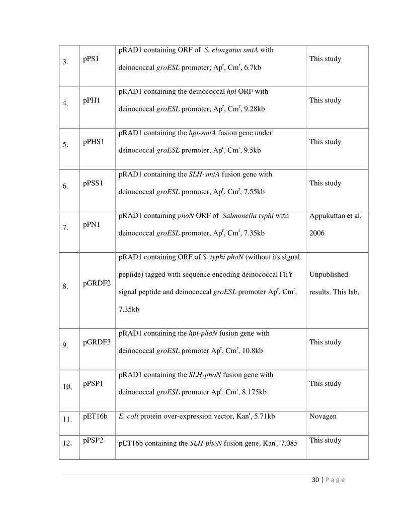
30 | P a g e
3. pPS1
pRAD1 containing ORF of S. elongatus smtA with
deinococcal groESL promoter; Apr, Cm
r, 6.7kb
This study
4. pPH1
pRAD1 containing the deinococcal hpi ORF with
deinococcal groESL promoter; Apr, Cm
r, 9.28kb
This study
5. pPHS1
pRAD1 containing the hpi-smtA fusion gene under
deinococcal groESL promoter, Apr, Cm
r, 9.5kb
This study
6. pPSS1
pRAD1 containing the SLH-smtA fusion gene with
deinococcal groESL promoter, Apr, Cm
r, 7.55kb
This study
7. pPN1
pRAD1 containing phoN ORF of Salmonella typhi with
deinococcal groESL promoter, Apr, Cm
r, 7.35kb
Appukuttan et al.
2006
8. pGRDF2
pRAD1 containing ORF of S. typhi phoN (without its signal
peptide) tagged with sequence encoding deinococcal FliY
signal peptide and deinococcal groESL promoter Apr, Cm
r,
7.35kb
Unpublished
results. This lab.
9. pGRDF3
pRAD1 containing the hpi-phoN fusion gene with
deinococcal groESL promoter Apr, Cm
r, 10.8kb
This study
10. pPSP1
pRAD1 containing the SLH-phoN fusion gene with
deinococcal groESL promoter Apr, Cm
r, 8.175kb
This study
11. pET16b E. coli protein over-expression vector, Kanr, 5.71kb Novagen
12. pPSP2 pET16b containing the SLH-phoN fusion gene, Kanr, 7.085 This study
31 | P a g e
kb
13. pET29b E. coli protein over-expression vector, Kanr,5.37kb Novagen
14. pPSP3 pET29b containing the SLH-phoN fusion gene, Kanr, 6.45kb This study
15. pUC18 E. coli cloning vector, Apr, 2.68kb NEB
16. pASK1
pUC18 containing the S. typhi phoN gene with phoN
promoter, Apr, 3.62kb
Work from this
lab
2.4 Isolation of Hpi layer from D. radiodurans cells
Deinococcus radiodurans was grown in TGY to an OD600nm ∼ 1.5/ml and subsequently
harvested by centrifugation at 5000 g for 5 min at 4°C. Cells were washed twice by
resuspension in distilled water containing protease inhibitor, 1 mM phenylmethanesulfonyl
fluoride (PMSF), followed by centrifugation as above. The cell pellet was resuspended in
distilled water containing 1 mM PMSF and 2 % Lithium dodecyl sulphate in a volume 20
times the initial cell pellet volume. Extraction of Hpi S-layer sheet was carried out at 50 rpm
agitation at 4 °C for 4 h. After extraction, cells were removed by centrifugation at 3500 g for
6 min and the Hpi layer was subsequently pelleted by centrifugation at 20,000 g for 40 min at
4°C. Hpi preparation was washed in distilled water by repeated resuspension-centrifugation
at 20,000 g, as above. The isolated Hpi layer was stored (~2 mg/mL protein in distilled
water) at 4°C, until further use.
2.5 Extraction, estimation and electrophoresis of cellular proteins
Cultures of E. coli or D. radiodurans were harvested by centrifugation, washed in
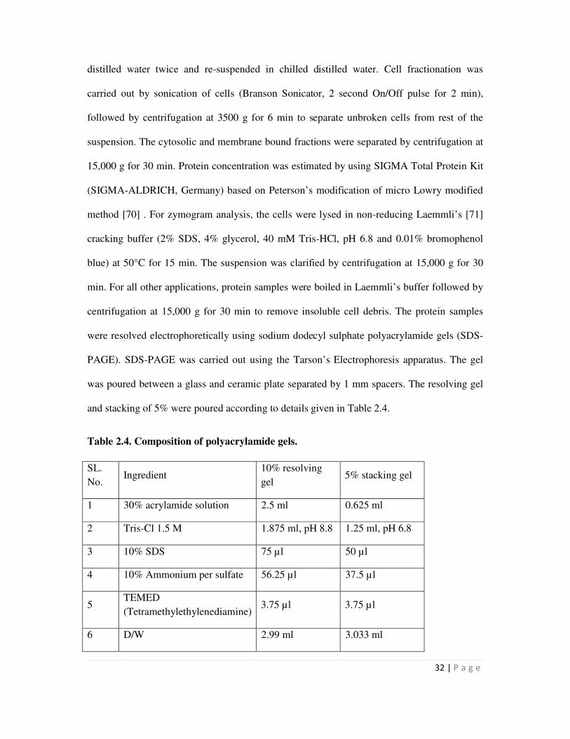
32 | P a g e
distilled water twice and re-suspended in chilled distilled water. Cell fractionation was
carried out by sonication of cells (Branson Sonicator, 2 second On/Off pulse for 2 min),
followed by centrifugation at 3500 g for 6 min to separate unbroken cells from rest of the
suspension. The cytosolic and membrane bound fractions were separated by centrifugation at
15,000 g for 30 min. Protein concentration was estimated by using SIGMA Total Protein Kit
(SIGMA-ALDRICH, Germany) based on Peterson’s modification of micro Lowry modified
method [70] . For zymogram analysis, the cells were lysed in non-reducing Laemmli’s [71]
cracking buffer (2% SDS, 4% glycerol, 40 mM Tris-HCl, pH 6.8 and 0.01% bromophenol
blue) at 50°C for 15 min. The suspension was clarified by centrifugation at 15,000 g for 30
min. For all other applications, protein samples were boiled in Laemmli’s buffer followed by
centrifugation at 15,000 g for 30 min to remove insoluble cell debris. The protein samples
were resolved electrophoretically using sodium dodecyl sulphate polyacrylamide gels (SDS-
PAGE). SDS-PAGE was carried out using the Tarson’s Electrophoresis apparatus. The gel
was poured between a glass and ceramic plate separated by 1 mm spacers. The resolving gel
and stacking of 5% were poured according to details given in Table 2.4.
Table 2.4. Composition of polyacrylamide gels.
SL.
No. Ingredient
10% resolving
gel 5% stacking gel
1 30% acrylamide solution 2.5 ml 0.625 ml
2 Tris-Cl 1.5 M 1.875 ml, pH 8.8 1.25 ml, pH 6.8
3 10% SDS 75 µl 50 µl
4 10% Ammonium per sulfate 56.25 µl 37.5 µl
5 TEMED
(Tetramethylethylenediamine) 3.75 µl 3.75 µl
6 D/W 2.99 ml 3.033 ml
33 | P a g e
After gelation, the protein extracts were loaded into the wells and electrophoresis was carried
at 15 V/cm. Post-run, gels were washed in distilled water followed by processing for Western
blotting or zymogram or staining by a 0.25% Coomassie brilliant blue solution.
2.6 Matrix Assisted Laser Desorption/ Ionization - Time of Flight - Mass Spectrometry
(MALDI-TOF-MS)
Proteins resolved by SDS-PAGE, were excised as 1mm X 1mm gel plugs. The gel protein
plug was subjected to destaining, reduction, alkylation, in-gel trypsin digestion and elution of
oligopeptides as described earlier [72]. The eluted peptides were subjected to mass
spectrometry (UltraFlex III MALDI-TOF/TOF mass spectrometer, Bruker Daltonics,
Germany). The oligopeptides were co-crystallized with α-Cyano-4-hydroxycinnamic acid
(CHCA) (5 mg/ml in 0.1% Trifluoro acetic acid and 30% Acetonitrile) on target plate (384-
well stainless steel plate, Bruker Daltonics, Germany). The machine was externally calibrated
using Peptide calibration mix I (Bruker Daltonics, Germany) or with the trypsin autodigest
peptides. The mass spectra were generated in the mass range of 600-4500 Da using standard
ToF-MS protocol in positive ion reflection mode. Laser was set to fire 150 times per spot.
Peak list was generated using FlexAnalysis software 3.0 (Bruker Daltonics, Germany) and
mass spectra were imported into the database search engine (BioTools v3.1 connected to
Mascot, Version 2.2.04, Matrix Science). Mascot searches were conducted using the NCBI
non redundant database (released in March 2013 or later with minimum of 14344429 entries
actually searched) with the following settings: Number of miscleavages permitted was 1;
fixed modifications such as Carbamidomethyl on cysteine, variable modification of oxidation
on methionine residue; peptide tolerance as 100 ppm; enzyme used as trypsin and a peptide
charge setting as +1. A match with D. radiodurans / Salmonella enterica as appropriate with
34 | P a g e
the best score in each Mascot search was accepted as successful identification. A Mascot
score of >65 with a minimum of 6 peptide matches was considered to be a significant
identification (p < 0.05) when sequence coverage was at least 25%.
2.7 Bioinformatic analysis
The sequence alignment of proteins was carried out using the web-based ClustalW software
(http://www.ebi.ac.uk/Tools/msa/clustalw2/).
2.8 Determination of phosphatase activity
2.8.1 In vitro acid phosphatase activity by zymogram analysis
Equal amount of proteins in non-reducing Laemmli’s buffer were electrophoretically
resolved by 10% SDS-PAGE at 75 V for 30 min followed by 100 V. After electrophoresis,
the gel was rinsed briefly with water to remove surface SDS and renatured using 1% Triton
X-100 in 100 mM acetate buffer pH 5.0 (Two washes of 20 min each) followed by a wash in
100 mM acetate buffer pH 5.0. The gel was developed using 200 µl of nitroblue tetrazolium
chloride/5-bromo-4-chloro-indolyl phosphate (NBT/BCIP) (Roche Diagnostics, 18.75 mg/ml
NBT and 9.4 mg/ml BCIP in 67% Dimethyl sulfoxide) mix in 20 ml of 100 mM acetate
buffer pH 5.0 for 16 h. The assay was terminated by rinsing the gel in distilled water.
2.8.2 In vivo cell-based acid phosphatase activity
Cell bound acid phosphatase activity was estimated by the liberation of p-nitophenol from di-
sodium p-nitrophenyl phosphate as described earlier [73]. Recombinant cells were re-
35 | P a g e
suspended in 100 mM acetate buffer, pH 5.0 containing 8 mM p-nitrophenol and incubated at
37°C for 30 min. The reaction was stopped by addition of 2N NaOH and the amount of p-
nitrophenol released was spectrophotometrically determined by measuring the OD at 405 nm.
The cell-bound PhoN activity was expressed as nmoles of p-nitrophenol (pNP) liberated
min−1
mg−1
bacterial protein.
2.9 Western Blotting and Immunodetection
Equal amount of whole cell protein extracts (50 µg-80 µg) prepared in Laemmli’s cracking
buffer (2% SDS, 2 mM dithiothreitol, 4% glycerol, 40 mM Tris-HCl, pH 6.8 and 0.01%
bromophenol blue) were resolved by 10% SDS-PAGE. The gel was equilibrated in Transfer
buffer (0.125 M Tris base, 0.192M Glycine, 20% methanol) for 20 min. The proteins were
electroblotted on nitrocellulose membrane at 300 V for 2 h. The blot was processed for
immunodetection. Briefly, the blot was placed in an appropriate dilution of the antibody
serum in MaNa (0.1M Maleic acid and 0.15 M NaCl) buffer, containing 1% casein
suspension, for interaction with primary antibody. After 1 h of incubation, the blot was
washed in 100 mM MaNa buffer twice for 20 min each to remove antibody bound non-
specifically to the blot. The blot was then incubated in blocking buffer containing the
secondary antibody, alkaline phosphatase conjugated anti-rabbit IgG (SIGMA-ALDRICH) at
1:10,000 dilution for 1 h. The blot was rinsed in MaNa buffer for 20 min, twice. After
equilibration in Tris-NaCl buffer pH 9.0, the blot was processed for colour development
using NBT-BCIP (Roche Diagnostics) as substrate in the same buffer. The colour
development reaction was stopped by adding distilled water.
36 | P a g e
2.10 Over-expression of SLH-PhoN
Recombinant E. coli BL21 pLysS cells carrying the SLH-phoN construct (pPSP3) were
grown at 37°C in LB broth containing 50 µg/ml kanamycin and 33 µg/ml chloramphenicol
till they attained a OD600nm of 0.6-0.8/ml. SLH-PhoN expression was induced by addition of 1
mM isopropyl-β-D-thiogalactopyranoside (IPTG) followed by incubation at 30°C under
shaking conditions (180 rpm). Cells were harvested after 5 h of growth and lysed for analysis
by 10% SDS-PAGE or used in peptidoglycan binding assays.
2.11 Peptidoglycan isolation from D. radiodurans
Isolation of peptidoglycan from D. radiodurans cells was carried out by boiling cells in
sodium dodecyl sulphate which dissolves all cellular components except the peptidoglycan
which remains insoluble and can be separated by centrifugation. D. radiodurans cells grown
to stationary phase were harvested by centrifugation and washed in distilled water. The pellet
obtained was re-suspended in 10 times the volume of 2% sodium dodecyl sulphate solution.
The suspension was refluxed under constant stirring conditions for 8 h continuously. This
was followed by centrifugation of the suspension at 20,000 g for 40 min. The resultant pellet
was re-suspended in 2% SDS and the entire process repeated two times. The final pellet
obtained was washed in distilled water 6 times to remove SDS and stored at 4°C.
Peptidoglycan concentration was routinely calculated from their diamino acid,
ornithine content as described [59]. Around 10 mg dry weight of peptidoglycan was
resuspended in 2 ml of concentrated HCl and transferred to a round bottom flask. The flask
was placed in an oil bath at 115°C for 6 h. The solution was vacuum dried. The residue was
37 | P a g e
re-suspended in 200 µl distilled water and clarified by centrifugation at 15,000 g for 30 min.
This solution was diluted 1:10 and used for estimation of ornithine.
Ornithine estimation was carried out as described [74]. Appropriate volume of
hydrolyzed peptidoglycan solution was taken in 50 µl distilled water. To this 50 µl acetic acid
and 50 µl Ninhydrin reagent (250 mg in 6 ml acetc acid and 4 ml 0.6 M phosphoric acid)
were added and kept in a boiling water bath for 7 min. The solution was cooled and the
absorbance of the resulting red color was measured spectrophotometrically at 515 nm. A
standard curve constructed by estimation of free ornithine amino acid similarly, was used to
determine concentration of ornithine in peptidoglycan.
2.12 Peptidoglycan binding studies and glutaraldehyde stabilization
Peptidoglycan binding assay was carried out as shown in the schematic below, as described
(Fig. 2.1) [75]. Briefly, 30 µl of peptidoglycan (corresponding to 43 µg of ornithine
equivalent) was added to 60 µl of cell extract from E. coli cells expressing SLH-PhoN in 50
mM Tris-Cl, pH 8.0 and incubated at 4°C for 2 h. This was followed by centrifugation to
obtain supernatant (soluble fraction, S) and a pellet. The pellet was washed and again
fractionated into the supernatant, (W) and the pellet. The pellet was then incubated in non-
reducing cracking buffer [71] at 50oC for 15 min and again subjected to centrifugation to
obtain the supernatant (insoluble fraction, I) and pellet. The insoluble fraction, I, represents
the protein which bound to the peptidoglycan, while the soluble fraction, S represents protein
which did not bind to the peptidoglycan. A zymogram was carried out with the soluble, wash
and the insoluble fractions.
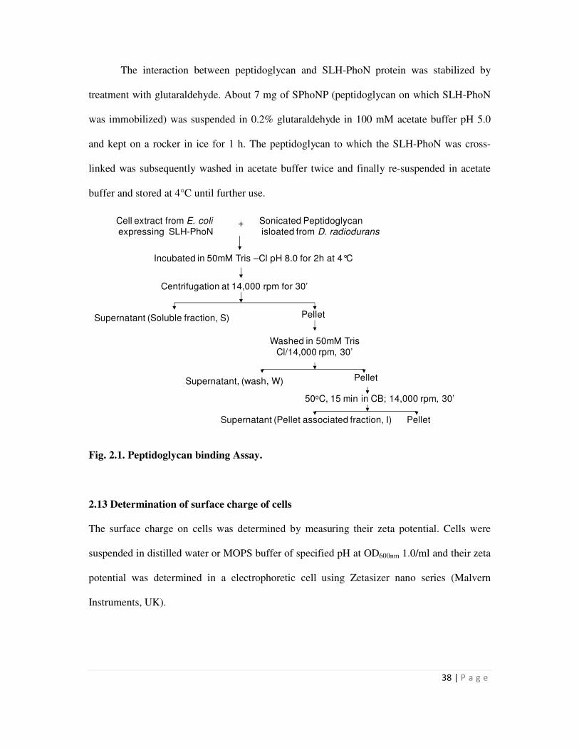
38 | P a g e
The interaction between peptidoglycan and SLH-PhoN protein was stabilized by
treatment with glutaraldehyde. About 7 mg of SPhoNP (peptidoglycan on which SLH-PhoN
was immobilized) was suspended in 0.2% glutaraldehyde in 100 mM acetate buffer pH 5.0
and kept on a rocker in ice for 1 h. The peptidoglycan to which the SLH-PhoN was cross-
linked was subsequently washed in acetate buffer twice and finally re-suspended in acetate
buffer and stored at 4°C until further use.
Fig. 2.1. Peptidoglycan binding Assay.
2.13 Determination of surface charge of cells
The surface charge on cells was determined by measuring their zeta potential. Cells were
suspended in distilled water or MOPS buffer of specified pH at OD600nm 1.0/ml and their zeta
potential was determined in a electrophoretic cell using Zetasizer nano series (Malvern
Instruments, UK).
Cell extract from E. coliexpressing SLH-PhoN
Sonicated Peptidoglycanisloated from D. radiodurans
Incubated in 50mM Tris –Cl pH 8.0 for 2h at 4°C
Centrifugation at 14,000 rpm for 30’
Supernatant (Soluble fraction, S) Pellet
Washed in 50mM TrisCl/14,000 rpm, 30’
+
Supernatant, (wash, W) Pellet
50oC, 15 min in CB; 14,000 rpm, 30’
Supernatant (Pellet associated fraction, I) Pellet
39 | P a g e
2.14 Metal binding studies using recombinants expressing SmtA
Overnight grown cultures of recombinant cells were inoculated into fresh medium at a
starting OD600nmof 0.05/ml. After 5 h of growth under aerobic conditions, when OD600nm
reached 0.6/ml, the metal (Cd, Cu, Zn) was added at the specified concentrations. The culture
was then grown in presence of the metal overnight. Cells were removed by centrifugation,
washed in the growth medium once, and dried at 80°C for 4 h. The samples were weighed
and digested in hot concentrated HNO3. Acid digested samples were diluted and the metal
estimated by Flame/Graphite Atomic Absorption Spectrophotometry (Model 906AA FAAS,
GBC Scientific Equipment Pvt. Ltd., Australia). When the experiment was carried out with
40 µM cadmium for recombinant D. radiodurans cultures, the metal was added after a pre-
culture of 16 h (OD600nm of 3.0/ml), instead of 5 h to allow for more growth, because at this
concentration the metal is known to be toxic to D. radiodurans.
2.15 Bioprecipitation of metals
Metal precipitation assays were performed as described, earlier [18] with certain
modifications. In a typical experiment, overnight grown cultures of D. radiodurans and E.
coli recombinants were harvested and washed in distilled water. Cell suspensions (OD600nm of
0.3/ml of E. coli and 3.0/ml of D. radiodurans or 0.1 mg/ml of E. coli and 0.8 mg/ml of D.
radiodurans lyophilized cells) were incubated in 1 mM uranyl nitrate or 1 mM cadmium
nitrate in 2 mM acetate buffer (pH 5.0), supplemented with 5 mM β-glycerophosphate at 30
°C under shaking conditions (50 rpm). The final pH of this solution was 6.8. The cell density
and concentrations of uranyl nitrate and β-glycerophosphate were varied in different
experiments. Bioprecipitation studies using glutaraldehyde stabilized SPhoNP (peptidoglycan
to which PhoN was immobilized) were carried out using 7 mg/ml of material in a typical
assay as mentioned above. After complete precipitation of metal, SPhoNP was transferred to
40 | P a g e
a fresh solution of similar composition. This was repeated multiple times. Appropriate
controls were included to ascertain (a) spontaneous chemical precipitation of the metal or
sorption to the container surface by excluding cells in the reaction mix and (b) biosorption of
metal on the cell surface by excluding the substrate, β-glycerophosphate.
In all metal precipitation assays, aliquots were removed at specified time intervals,
and residual metal in the supernatant was estimated following removal of cells by
centrifugation. Metal was estimated in the supernatant or in the cell lysate, obtained from acid
(Conc. HNO3) digestion of cell pellet as described earlier (Section 2.13). Uranium was
estimated using Arsenazo-III reagent by a modification of the method by Fritz and Bradford
[76]. A 0.1% solution of Arsenazo-III was prepared by dissolving 0.2 of the reagent in 180
ml of 0.01N HCl and 20 ml of absolute ethanol (30 min, stirring) and filtered through
Whatman No. 1 filter paper. Twenty µl of the supernatant was diluted to a total volume of
600 µl. The samples were acidified (200 µl of 0.01N HCl), followed by addition of 200 µl of
Arsenazo-III. The resultant purple colored metal-Arsenazo-III complex was estimated
spectrophotometrically at 655nm. Cadmium was estimated by Flame/Graphite Atomic
Absorption Spectrophotometry (Model 906AA FAAS, GBC Scientific Equipment Pvt. Ltd.,
Australia).
2.16 Lyophilization
A thick saline-based suspension of recombinant cells, was placed in Petriplates, frozen in
liquid nitrogen for 5 min and lyophilized without any lyo/cryoprotectant overnight in a
Lyophilizer (Lyospeed, Genevac, United Kingdom) at 0.07 mbar for 18 h and stored in vials,
at room temperature without applying vacuum, until further use. Equivalence between the
OD600nm of the suspension submitted for lyophilisation and the dry weight of the lyophilized
powder obtained was determined to calculate the total number of viable cells in the
41 | P a g e
lyophilized powder and to equalize cell numbers used in metal precipitation studies. The
lyophilized cells when re-suspended, rapidly formed a uniform suspension in 100 mM acetate
buffer, pH 5.0 and were allowed to equilibrate for 5 min before the PhoN activity and
uranium precipitation assays were carried out. For metal precipitation studies, ~ 80 mg/mL
suspension of lyophilized cells was prepared in 100 mM MOPS buffer and used immediately.
2.17 Scanning Electron Microscopy
E. coli cell samples were washed in normal saline and fixed in 2.5% glutaraldehyde for 2 h
while Deinococcus cells samples were washed in cacodylate buffer (100 mM, pH 7.4) and
fixed in Karnovskys fixative [77] for 2 h at 4°C. Samples were washed in 100 mM cacodylate
buffer. Fixed E. coli and D. radiodurans cells were dehydrated in a graded ethanol series
ranging from 20%-100%. The samples were spotted on aluminium studs and dried at 37°C
for 1 h. The dried samples were gold coated by thermal evaporation technique and analyzed
by scanning electron microscopy (SEM) and energy dispersive X-ray spectroscopy (EDXS)
using a Tescan VEGA 40 Microscope and INCA energy 250, Oxford Instrument EDXS
system.
2.18 Transmission Electron Microscopy
E. coli or D. radiodurans cells were washed twice with 50 mM cacodylate buffer (pH 7.4)
and fixed in 2.5 % glutaraldehyde and 0.5 % Para-formaldehyde overnight at 4°C. The fixed
samples were washed with cacodylate buffer three times and embedded in 2 % noble agar.
The samples were dehydrated in a graded series of ethanol (30, 60, 75, 90, and 100 %).
Ethanol was removed by treatment with propylene oxide and the agar blocks were
subsequently infiltrated with Spurr reagent on sequential incubation with 1:3, 3:1 and 1:1
(propylene oxide:Spurr reagent v/v) for 2 h each. The samples were finally infiltrated with
42 | P a g e
Spurr resin for 16 h and embedded in it by incubation at 60°C for 72 h. Thin sections of
samples were prepared with a microtome (Leica, Germany), placed on 200 mesh formvar-
coated copper grids and viewed with a Libra 120 plus Transmission electron Microscope
(Carl Zeiss, Germany) without uranyl acetate staining. TEM of the Hpi layer was done by
dropping 10µl of 2 µg/µl suspension of the Hpi protein layer on formvar coated copper grids
followed by staining in 1 % uranyl acetate. The copper grids were air dried and analyzed as
mentioned above.
43 | P a g e
Chapter 3
Construction of deinococcal S layer fusion proteins
with metallothionein (SmtA) and acid phosphatase
(PhoN): cloning and expression
44 | P a g e
Deinococcus radiodurans is known to possess two S layer proteins, Hpi and SlpA
[54]. Of these, the Hpi has been studied extensively while the SlpA has been annotated as an
S layer protein based on its homology with the S layer protein from Thermus thermophilus
[54]. It is not known how Hpi and SlpA proteins are placed with respect to each other in the
deinococcal cell envelope. The SlpA is a large protein whose location in the deinococcal cell
envelope is not very clear. It contains a surface layer homology (SLH) motif which is known
to bind secondary cell wall polymers present on peptidoglycan in a number of bacteria such
as Bacillus anthracis, Clostridium thermocellum, Paenibacillus alvei and others [75,78-79].
Further, the SLH domain has been exploited for anchoring proteins to peptidoglycan for cell
surface display [80]. In Thermus thermophilus also, the SLH domain binds secondary cell
wall polymer which is covalently attached to the peptidoglycan [59]. As a result, instead of
the entire SlpA protein, sequence coding only for the SLH domain have often been used for
generating fusion proteins.
SmtA is a cyanobacterial metallothionein rich in cysteine residues and therefore
relevant to metal bioremediation. SmtA has been over-expressed earlier in E. coli for
enhanced metal binding [14]. The PhoN is a non-specific acid phosphatase from Salmonella
enterica serovar Typhi. The corresponding gene was earlier cloned and over-expressed in D.
radiodurans for bioremediation of heavy metals, especially uranium [51]. phoN was
expressed under the control of the strong deinococcal PgroESL promoter on the binary D.
radiodurans/E. coli shuttle vector, pRAD1 [81].
This chapter describes partial characterization of the S layer proteins of D.
radiodurans and construction of fusion proteins involving Hpi protein and SLH domain with
PhoN on one hand, and SmtA on the other. Further, the expression and localization of the
various fusion proteins in the recombinant strain were also determined.
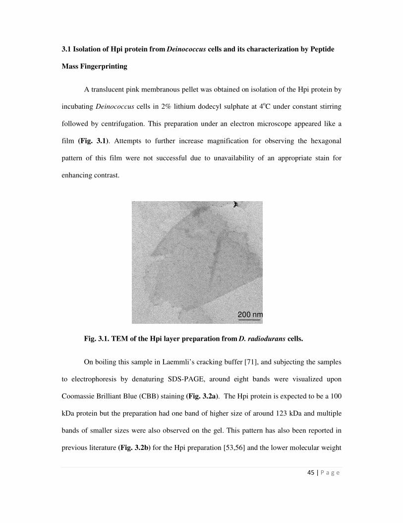
45 | P a g e
200 nm
3.1 Isolation of Hpi protein from Deinococcus cells and its characterization by Peptide
Mass Fingerprinting
A translucent pink membranous pellet was obtained on isolation of the Hpi protein by
incubating Deinococcus cells in 2% lithium dodecyl sulphate at 4oC under constant stirring
followed by centrifugation. This preparation under an electron microscope appeared like a
film (Fig. 3.1). Attempts to further increase magnification for observing the hexagonal
pattern of this film were not successful due to unavailability of an appropriate stain for
enhancing contrast.
Fig. 3.1. TEM of the Hpi layer preparation from D. radiodurans cells.
On boiling this sample in Laemmli’s cracking buffer [71], and subjecting the samples
to electrophoresis by denaturing SDS-PAGE, around eight bands were visualized upon
Coomassie Brilliant Blue (CBB) staining (Fig. 3.2a). The Hpi protein is expected to be a 100
kDa protein but the preparation had one band of higher size of around 123 kDa and multiple
bands of smaller sizes were also observed on the gel. This pattern has also been reported in
previous literature (Fig. 3.2b) for the Hpi preparation [53,56] and the lower molecular weight
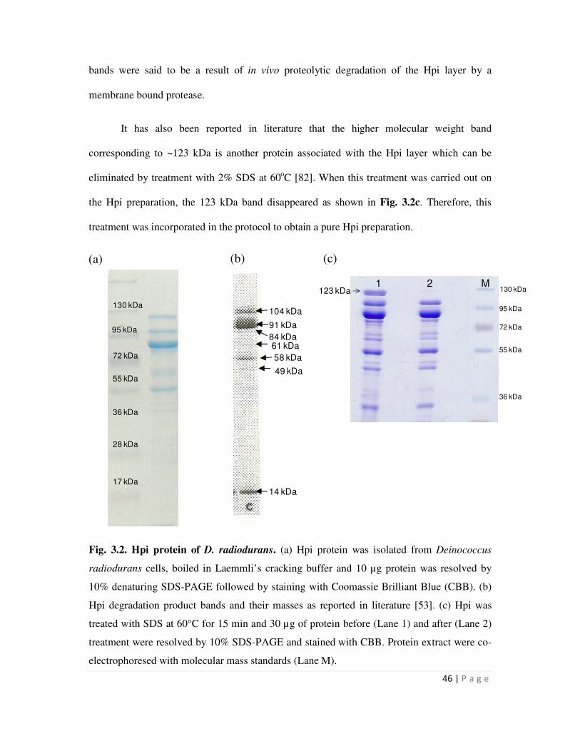
46 | P a g e
104 kDa
91 kDa
14 kDa
84 kDa
58 kDa
49 kDa
61 kDa
130 kDa
95 kDa
72 kDa
55 kDa
17 kDa
28 kDa
36 kDa
1 2 M123 kDa 130 kDa
95 kDa
72 kDa
55 kDa
36 kDa
bands were said to be a result of in vivo proteolytic degradation of the Hpi layer by a
membrane bound protease.
It has also been reported in literature that the higher molecular weight band
corresponding to ~123 kDa is another protein associated with the Hpi layer which can be
eliminated by treatment with 2% SDS at 60oC [82]. When this treatment was carried out on
the Hpi preparation, the 123 kDa band disappeared as shown in Fig. 3.2c. Therefore, this
treatment was incorporated in the protocol to obtain a pure Hpi preparation.
Fig. 3.2. Hpi protein of D. radiodurans. (a) Hpi protein was isolated from Deinococcus
radiodurans cells, boiled in Laemmli’s cracking buffer and 10 µg protein was resolved by
10% denaturing SDS-PAGE followed by staining with Coomassie Brilliant Blue (CBB). (b)
Hpi degradation product bands and their masses as reported in literature [53]. (c) Hpi was
treated with SDS at 60°C for 15 min and 30 µg of protein before (Lane 1) and after (Lane 2)
treatment were resolved by 10% SDS-PAGE and stained with CBB. Protein extract were co-
electrophoresed with molecular mass standards (Lane M).
(a) (b) (c)
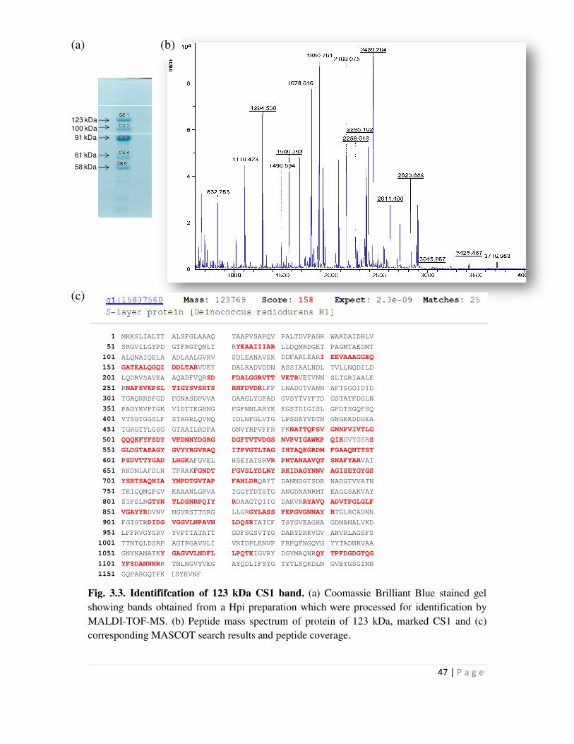
123 kDa
100 kDa
91 kDa
61 kDa
58 kDa
1 MKKSLIALTT ALSFGLAAAQ
51 SRGVILGYPD GTFRGTQNLT
101 ALQNAIQELA ADLAALGVRV
151 GATEALQGQI DDLTARVDEY
201 LQDRVSAVEA AQADFVQRSD
251 RNAFSVKPSL TIGYSVSRTS
301 TGAQRRDFGD FGNASDPVVA
351 PADYKVPTGK VIDTTKGRNG
401 VTSGTGGSLF STAGRLQVNQ
451 TGRGTYLGSG GTAAILRDPA
501 QQQKFYFSDY VFDNNYDGRG
551 GLDGTAEAGY GVYYRGVRAQ
601 PSDVTTYGAD LHGKAFGVEL
651 RKDNLAFDLN TPAAKFGNDT
701 YSRTSAQNIA YNPDTGVTAP
751 TKIGQMGFGV KAAANLGPVA
801 SIFSLRGTYN TLDSNRPQIY
851 VGAYYRDVNV NGVRSTTDRG
901 FGTGTRDIDG VGGVLNPAVN
951 LFFRVGYSRV YVPTTATATT
1001 TTNTQLDSRP AGTRGAVGLI
1051 GNYNANATKY GAGVVLNDFL
1101 YFSDANNNRR TNLNGVYVEG
1151 GQPARGQTFK ISYKVNF
Fig. 3.3. Identififcation of 123 kDa CS1 band.
showing bands obtained from a Hpi preparation which were processed for identification by
MALDI-TOF-MS. (b) Peptide mass spectr
corresponding MASCOT search results
(a) (b)
(c)
TAAPVSAPQV PALTDVPAGH WAKDAIDRLV
RYEAAIIIAR LLDQMRDGET PAGMTAEDMT
SDLEANAVSK DDFARLEARI EEVAAAGGEQ
DALRADVDDN ASSIAALNDL TVLLNQDILD
FDALGGRVTT VETRVETVNN SLTGRIAALE
RNFDVDRLFP LNADGTVANN AFTSGGIDTD
GAAGLYGFAD GVSYTVYFTD GSTATFDGLN
FGFNNLARYK EGSTDIGISL GFDTSGQFSQ
IDLNFGLVTG LPSDAYVDTN GNGKKDDGEA
GNVYRPVFFR FKNATTQFSV GNNPVIVTLG
DGFTVTVDGS NVPVIGAWKP QIKGVYGSRS
ITPVGTLTAG IHYAQEGRDM FGAAQNTTST
HSEYATSRVR PNTANAAVQT SNAFYARVAT
FGVSLYDLNY RKIDAGYNNV AGISEYGYGS
FANLDRQAYT DANNDGTSDR NADGTVVATN
IGGYYDTSTG ANGDNANRMT EAGGSAKVAY
RDAAGTQIIG DAKVRRYAVQ ADVTPGLGLF
LLGRGYLASS FEPGVGNNAY RTGLRCADNN
LDQSRTATCF TSYGVEAGHA GDNANALVKD
GDFSGSVTYG DARYDRKVGV ANVRLAGSFS
VRTDPLENVP FRPQFNGQVG YYTADNRVAA
LPQTKIGVRY DGYMAQNRQY TPFDGDGTQG
AYQDLIFSYG TYTLSQKDLN GVEYGSGINN
Fig. 3.3. Identififcation of 123 kDa CS1 band. (a) Coomassie Brilliant Blue stained gel
showing bands obtained from a Hpi preparation which were processed for identification by
Peptide mass spectrum of protein of 123 kDa, marked CS1 and (c)
corresponding MASCOT search results and peptide coverage.
47 | P a g e
a) Coomassie Brilliant Blue stained gel
showing bands obtained from a Hpi preparation which were processed for identification by
protein of 123 kDa, marked CS1 and (c)
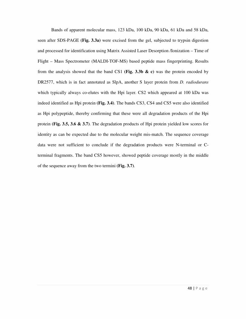
48 | P a g e
Bands of apparent molecular mass, 123 kDa, 100 kDa, 90 kDa, 61 kDa and 58 kDa,
seen after SDS-PAGE (Fig. 3.3a) were excised from the gel, subjected to trypsin digestion
and processed for identification using Matrix Assisted Laser Desorption /Ionization – Time of
Flight – Mass Spectrometer (MALDI-TOF-MS) based peptide mass fingerprinting. Results
from the analysis showed that the band CS1 (Fig. 3.3b & c) was the protein encoded by
DR2577, which is in fact annotated as SlpA, another S layer protein from D. radiodurans
which typically always co-elutes with the Hpi layer. CS2 which appeared at 100 kDa was
indeed identified as Hpi protein (Fig. 3.4). The bands CS3, CS4 and CS5 were also identified
as Hpi polypeptide, thereby confirming that these were all degradation products of the Hpi
protein (Fig. 3.5, 3.6 & 3.7). The degradation products of Hpi protein yielded low scores for
identity as can be expected due to the molecular weight mis-match. The sequence coverage
data were not sufficient to conclude if the degradation products were N-terminal or C-
terminal fragments. The band CS5 however, showed peptide coverage mostly in the middle
of the sequence away from the two termini (Fig. 3.7).
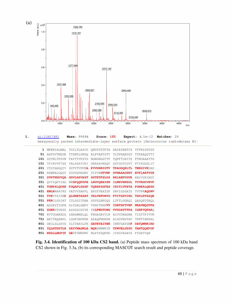
49 | P a g e
1522.765
1475.787
2372.192
1037.488
1869.837
1253.598
2943.408
1377.689
833.495 2550.2232163.055
3549.6732083.088
2716.263
3153.446
0.0
0.2
0.4
0.6
0.8
1.0
5x10
Inte
ns. [a
.u.]
1000 1500 2000 2500 3000 3500 4000 4500m/z
1 MKKNIALMAL TGILTLASCG QNGTGTTPTA DACATANTCS VTVNISGVSS
51 ADFDVTMDGK TTSMTLSNGQ KLPVAKTGTV TLTPKAKDGY TTPAAQSTTI
101 SSTNLTPSVN FAYTTVPSTG NGNGNGGTTP TQPFTLNITS PTNGAAATTG
151 TPIRVVFTSS VALSSATCKI GNSAAVNAQV SSTGGYCDVT PTTAGGGLIT
201 VTGTANGQTV SSTVTVDVKA PVVDNRYGTV TPAGDQELTL TNEGIVKDAD
251 NGWRRLGQGV STPSDPNGNV DIYVKGTVNF SVNAAAGSKV EVFLARTTGS
301 DVPTNDDVQA GDVLRSVAST SGTETFSLDS RRLAEFDGVR KWIVVRINGT
351 QVTYQPVIAD NKGPQQPDPE LNGVQNAYSN ILNNYNNSGL TYVRGDVNVF
401 TGNPSLQDRE FGQAPLGSSF VQRRPSGFES IRYYLVPETA FGNKALQESD
451 EMLRAKAIKS VATVVSAPVL EPGTVKATSF SRVIGSGATS TVTPKAQDNV
501 TYRVYAISRD QLGNETASAT YELVRFDNVG PTITGSVIRD TSDLPFASQE
551 PERCLSDIAT ITLGGITDNA GGVGLNPGQG LTFTLGGRQI QAGQFDTNQL
601 ADGEYTIGFN SLTDALGNPV VSAPTNAKVY IDNTDPTVNF NRAVMQGTFA
651 SGERVSVESD ASDGGCGVYE TRLFWDTDNG VVDDATTTPA IGHPVQFARQ
701 RVTDGAKADS LNAGWNALQL PNGAGAVYLR ALVVDRAGNA TISTTPIVVN
751 AKITNQARPL LGGFDAFKRN ASAQFMSNSN AISGVNGTAV TPNTTANSAL
801 DNILSLDSVG TLTTNAYLPR GATETAITEK IRNVGAYGRF DATQWNRIRD
851 YQLNTDPTLR SAYVNAGNLA NQRGNNWRIR TPWVELGSSD TANTQQKFDF
901 NSDLLNDFYF GRTFGNNDNV NLFSYDQFNG IVSGTAGAYS FYGETVQK
Fig. 3.4. Identification of 100 kDa CS2 band. (a) Peptide mass spectrum of 100 kDa band
CS2 shown in Fig. 3.3a, (b) its corresponding MASCOT search result and peptide coverage.
(a)
50 | P a g e
1
MKKNIALMAL
TGILTLASCG
QNGTGTTPTA
DACATANTCS
VTVNISGVSS
51 ADFDVTMDGK TTSMTLSNGQ KLPVAKTGTV TLTPKAKDGY TTPAAQSTTI
101 SSTNLTPSVN FAYTTVPSTG NGNGNGGTTP TQPFTLNITS PTNGAAATTG
151 TPIRVVFTSS VALSSATCKI GNSAAVNAQV SSTGGYCDVT PTTAGGGLIT
201 VTGTANGQTV SSTVTVDVKA PVVDNRYGTV TPAGDQELTL TNEGIVKDAD
251 NGWRRLGQGV STPSDPNGNV DIYVKGTVNF SVNAAAGSKV EVFLARTTGS
301 DVPTNDDVQA GDVLRSVAST SGTETFSLDS RRLAEFDGVR KWIVVRINGT
351 QVTYQPVIAD NKGPQQPDPE LNGVQNAYSN ILNNYNNSGL TYVRGDVNVF
401 TGNPSLQDRE FGQAPLGSSF VQRRPSGFES IRYYLVPETA FGNKALQESD
451 EMLRAKAIKS VATVVSAPVL EPGTVKATSF SRVIGSGATS TVTPKAQDNV
501 TYRVYAISRD QLGNETASAT YELVRFDNVG PTITGSVIRD TSDLPFASQE
551 PERCLSDIAT ITLGGITDNA GGVGLNPGQG LTFTLGGRQI QAGQFDTNQL
601 ADGEYTIGFN SLTDALGNPV VSAPTNAKVY IDNTDPTVNF NRAVMQGTFA
651 SGERVSVESD ASDGGCGVYE TRLFWDTDNG VVDDATTTPA IGHPVQFARQ
701 RVTDGAKADS LNAGWNALQL PNGAGAVYLR ALVVDRAGNA TISTTPIVVN
751 AKITNQARPL LGGFDAFKRN ASAQFMSNSN AISGVNGTAV TPNTTANSAL
801 DNILSLDSVG TLTTNAYLPR GATETAITEK IRNVGAYGRF DATQWNRIRD
851 YQLNTDPTLR SAYVNAGNLA NQRGNNWRIR TPWVELGSSD TANTQQKFDF
901 NSDLLNDFYF GRTFGNNDNV NLFSYDQFNG IVSGTAGAYS FYGETVQK
Fig. 3.5. MASCOT search results for mass spectra of 91 kDa band, CS3 shown in Fig. 3.3a
and its peptide coverage.
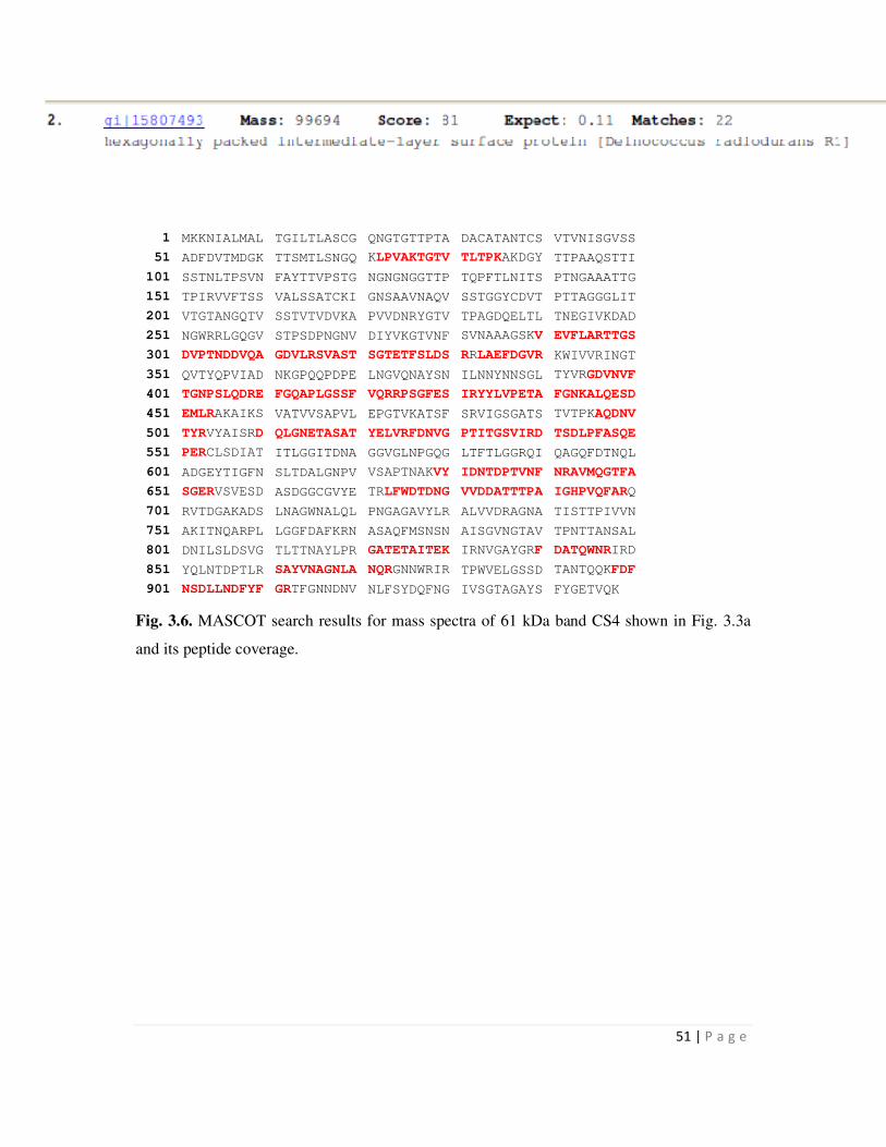
51 | P a g e
1 MKKNIALMAL TGILTLASCG QNGTGTTPTA DACATANTCS VTVNISGVSS
51 ADFDVTMDGK TTSMTLSNGQ KLPVAKTGTV TLTPKAKDGY TTPAAQSTTI
101 SSTNLTPSVN FAYTTVPSTG NGNGNGGTTP TQPFTLNITS PTNGAAATTG
151 TPIRVVFTSS VALSSATCKI GNSAAVNAQV SSTGGYCDVT PTTAGGGLIT
201 VTGTANGQTV SSTVTVDVKA PVVDNRYGTV TPAGDQELTL TNEGIVKDAD
251 NGWRRLGQGV STPSDPNGNV DIYVKGTVNF SVNAAAGSKV EVFLARTTGS
301 DVPTNDDVQA GDVLRSVAST SGTETFSLDS RRLAEFDGVR KWIVVRINGT
351 QVTYQPVIAD NKGPQQPDPE LNGVQNAYSN ILNNYNNSGL TYVRGDVNVF
401 TGNPSLQDRE FGQAPLGSSF VQRRPSGFES IRYYLVPETA FGNKALQESD
451 EMLRAKAIKS VATVVSAPVL EPGTVKATSF SRVIGSGATS TVTPKAQDNV
501 TYRVYAISRD QLGNETASAT YELVRFDNVG PTITGSVIRD TSDLPFASQE
551 PERCLSDIAT ITLGGITDNA GGVGLNPGQG LTFTLGGRQI QAGQFDTNQL
601 ADGEYTIGFN SLTDALGNPV VSAPTNAKVY IDNTDPTVNF NRAVMQGTFA
651 SGERVSVESD ASDGGCGVYE TRLFWDTDNG VVDDATTTPA IGHPVQFARQ
701 RVTDGAKADS LNAGWNALQL PNGAGAVYLR ALVVDRAGNA TISTTPIVVN
751 AKITNQARPL LGGFDAFKRN ASAQFMSNSN AISGVNGTAV TPNTTANSAL
801 DNILSLDSVG TLTTNAYLPR GATETAITEK IRNVGAYGRF DATQWNRIRD
851 YQLNTDPTLR SAYVNAGNLA NQRGNNWRIR TPWVELGSSD TANTQQKFDF
901 NSDLLNDFYF GRTFGNNDNV NLFSYDQFNG IVSGTAGAYS FYGETVQK
Fig. 3.6. MASCOT search results for mass spectra of 61 kDa band CS4 shown in Fig. 3.3a
and its peptide coverage.
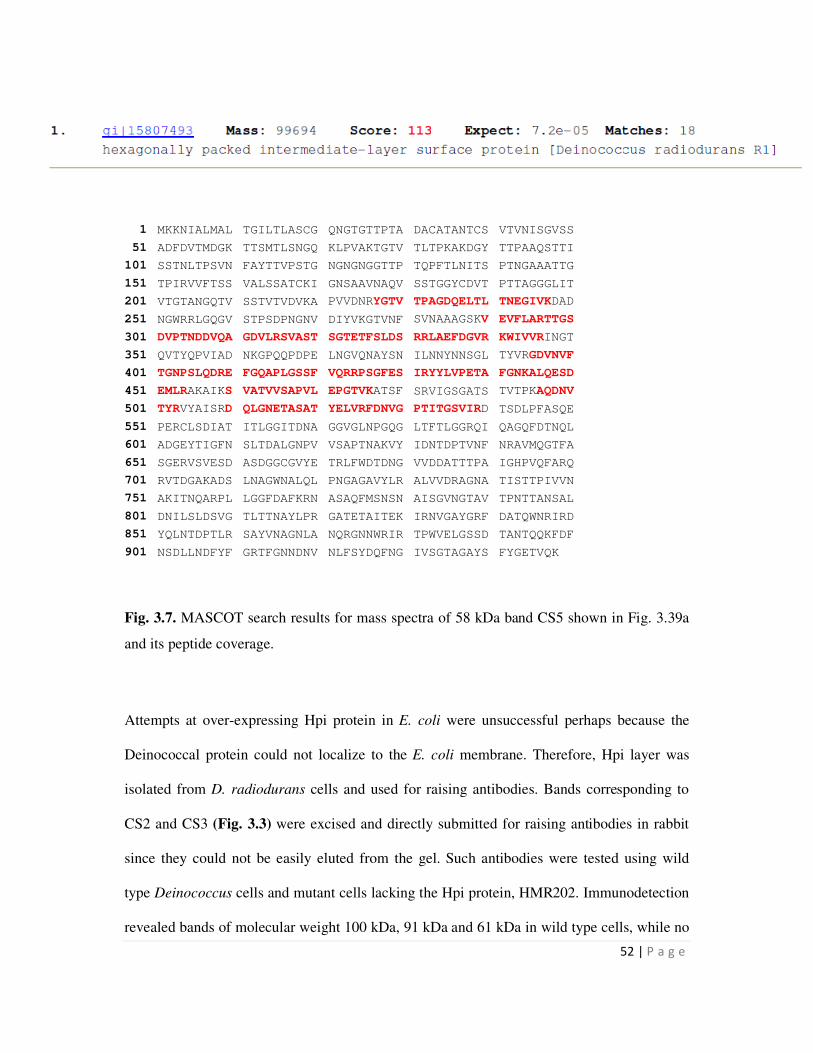
52 | P a g e
1 MKKNIALMAL TGILTLASCG QNGTGTTPTA DACATANTCS VTVNISGVSS
51 ADFDVTMDGK TTSMTLSNGQ KLPVAKTGTV TLTPKAKDGY TTPAAQSTTI
101 SSTNLTPSVN FAYTTVPSTG NGNGNGGTTP TQPFTLNITS PTNGAAATTG
151 TPIRVVFTSS VALSSATCKI GNSAAVNAQV SSTGGYCDVT PTTAGGGLIT
201 VTGTANGQTV SSTVTVDVKA PVVDNRYGTV TPAGDQELTL TNEGIVKDAD
251 NGWRRLGQGV STPSDPNGNV DIYVKGTVNF SVNAAAGSKV EVFLARTTGS
301 DVPTNDDVQA GDVLRSVAST SGTETFSLDS RRLAEFDGVR KWIVVRINGT
351 QVTYQPVIAD NKGPQQPDPE LNGVQNAYSN ILNNYNNSGL TYVRGDVNVF
401 TGNPSLQDRE FGQAPLGSSF VQRRPSGFES IRYYLVPETA FGNKALQESD
451 EMLRAKAIKS VATVVSAPVL EPGTVKATSF SRVIGSGATS TVTPKAQDNV
501 TYRVYAISRD QLGNETASAT YELVRFDNVG PTITGSVIRD TSDLPFASQE
551 PERCLSDIAT ITLGGITDNA GGVGLNPGQG LTFTLGGRQI QAGQFDTNQL
601 ADGEYTIGFN SLTDALGNPV VSAPTNAKVY IDNTDPTVNF NRAVMQGTFA
651 SGERVSVESD ASDGGCGVYE TRLFWDTDNG VVDDATTTPA IGHPVQFARQ
701 RVTDGAKADS LNAGWNALQL PNGAGAVYLR ALVVDRAGNA TISTTPIVVN
751 AKITNQARPL LGGFDAFKRN ASAQFMSNSN AISGVNGTAV TPNTTANSAL
801 DNILSLDSVG TLTTNAYLPR GATETAITEK IRNVGAYGRF DATQWNRIRD
851 YQLNTDPTLR SAYVNAGNLA NQRGNNWRIR TPWVELGSSD TANTQQKFDF
901 NSDLLNDFYF GRTFGNNDNV NLFSYDQFNG IVSGTAGAYS FYGETVQK
Fig. 3.7. MASCOT search results for mass spectra of 58 kDa band CS5 shown in Fig. 3.39a
and its peptide coverage.
Attempts at over-expressing Hpi protein in E. coli were unsuccessful perhaps because the
Deinococcal protein could not localize to the E. coli membrane. Therefore, Hpi layer was
isolated from D. radiodurans cells and used for raising antibodies. Bands corresponding to
CS2 and CS3 (Fig. 3.3) were excised and directly submitted for raising antibodies in rabbit
since they could not be easily eluted from the gel. Such antibodies were tested using wild
type Deinococcus cells and mutant cells lacking the Hpi protein, HMR202. Immunodetection
revealed bands of molecular weight 100 kDa, 91 kDa and 61 kDa in wild type cells, while no
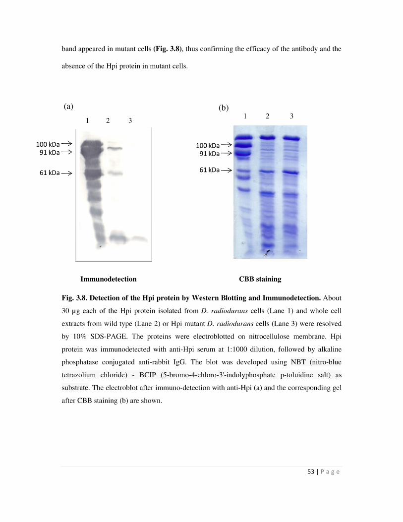
53 | P a g e
100 kDa91 kDa
61 kDa
100 kDa91 kDa
61 kDa
band appeared in mutant cells (Fig. 3.8), thus confirming the efficacy of the antibody and the
absence of the Hpi protein in mutant cells.
Immunodetection CBB staining
Fig. 3.8. Detection of the Hpi protein by Western Blotting and Immunodetection. About
30 µg each of the Hpi protein isolated from D. radiodurans cells (Lane 1) and whole cell
extracts from wild type (Lane 2) or Hpi mutant D. radiodurans cells (Lane 3) were resolved
by 10% SDS-PAGE. The proteins were electroblotted on nitrocellulose membrane. Hpi
protein was immunodetected with anti-Hpi serum at 1:1000 dilution, followed by alkaline
phosphatase conjugated anti-rabbit IgG. The blot was developed using NBT (nitro-blue
tetrazolium chloride) - BCIP (5-bromo-4-chloro-3'-indolyphosphate p-toluidine salt) as
substrate. The electroblot after immuno-detection with anti-Hpi (a) and the corresponding gel
after CBB staining (b) are shown.
(a) (b)
1 2 3 1 2 3
54 | P a g e
3.2 Cloning, over-expression and localization of SmtA, Hpi-SmtA and SLH-SmtA
proteins in recombinant bacteria
The smtA gene from Synechococcus elongatus codes for a 6 kDa metal binding peptide,
metallothionein (MT). The entire gene was cloned as such or as a fusion gene with
Deinococcal hpi ORF, or with the nucleotide sequence coding for the SLH domain of SlpA in
D. radiodurans. All constructs were expressed from the deinococcal PgroESL promoter.
3.2.1 Cloning of smtA gene
In order to clone smtA gene downstream to PgroESL in pRAD1 (Table 2.3), an earlier
construct harbouring the smtA ORF in pET16b, pS1 was utilized. Primers Smt-f and Smt-r
(Table 2.2) were used to PCR amplify the smtA gene from pS1 as a 176 bp DNA fragment as
shown in Fig. 3.9. The plasmid pPN1 (Table 2.3) carrying phoN gene downstream of the
groESL promoter was digested with NdeI and BamHI to release the phoN ORF. The PCR
amplified smtA ORF was restriction digested with NdeI and BamHI and ligated to identical
sites in the cut pPN1 plasmid, downstream to the PgroESL promoter to obtain the plasmid,
pPS1 (Fig. 3.9). E. coli strain JM109 was transformed with the plasmid and Apr
transformants selected.
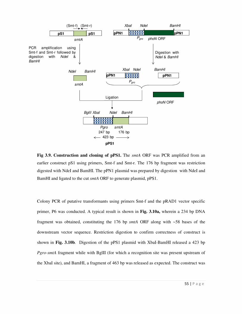
55 | P a g e
PCR amplification using
Smt-f and Smt-r followed bydigestion with NdeI &
BamHI
Digestion with
NdeI & BamHI
Ligation
smtA
NdeI BamHI
smtA
(Smt-f) (Smt-r)
pS1 pPN1
phoN ORF
XbaI NdeI BamHI
Pgro
pPN1pS1
Pgro
pPN1
XbaI NdeI BamHI
pPN1
phoN ORF
BglII XbaI NdeI BamHI
247 bp 176 bp
Pgro smtA
423 bp
pPS1
Fig 3.9. Construction and cloning of pPS1. The smtA ORF was PCR amplified from an
earlier construct pS1 using primers, Smt-f and Smt-r. The 176 bp fragment was restriction
digested with NdeI and BamHI. The pPN1 plasmid was prepared by digestion with NdeI and
BamHI and ligated to the cut smtA ORF to generate plasmid, pPS1.
Colony PCR of putative transformants using primers Smt-f and the pRAD1 vector specific
primer, P6 was conducted. A typical result is shown in Fig. 3.10a, wherein a 234 bp DNA
fragment was obtained, constituting the 176 bp smtA ORF along with ~58 bases of the
downstream vector sequence. Restriction digestion to confirm correctness of construct is
shown in Fig. 3.10b. Digestion of the pPS1 plasmid with XbaI-BamHI released a 423 bp
Pgro-smtA fragment while with BglII (for which a recognition site was present upstream of
the XbaI site), and BamHI, a fragment of 463 bp was released as expected. The construct was
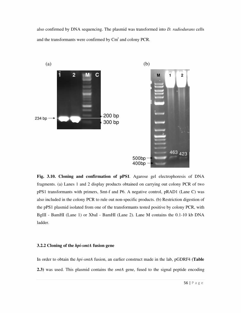
56 | P a g e
1 2 M C
234 bp200 bp
300 bp
500bp423463
M 1 2
400bp
also confirmed by DNA sequencing. The plasmid was transformed into D. radiodurans cells
and the transformants were confirmed by Cmr and colony PCR.
Fig. 3.10. Cloning and confirmation of pPS1. Agarose gel electrophoresis of DNA
fragments. (a) Lanes 1 and 2 display products obtained on carrying out colony PCR of two
pPS1 transformants with primers, Smt-f and P6. A negative control, pRAD1 (Lane C) was
also included in the colony PCR to rule out non-specific products. (b) Restriction digestion of
the pPS1 plasmid isolated from one of the transformants tested positive by colony PCR, with
BglII - BamHI (Lane 1) or XbaI - BamHI (Lane 2). Lane M contains the 0.1-10 kb DNA
ladder.
3.2.2 Cloning of the hpi-smtA fusion gene
In order to obtain the hpi-smtA fusion, an earlier construct made in the lab, pGDRF4 (Table
2.3) was used. This plasmid contains the smtA gene, fused to the signal peptide encoding
(a) (b)
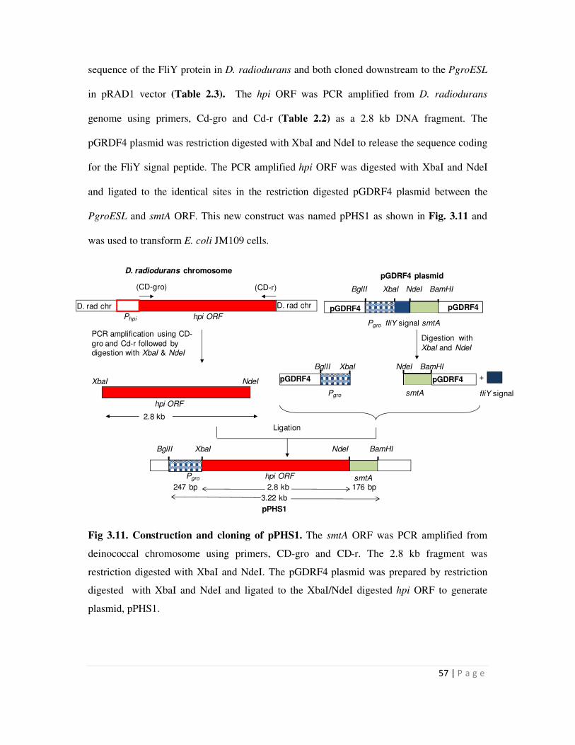
57 | P a g e
Phpi hpi ORF
(CD-gro) (CD-r)
D. rad chrD. rad chr pGDRF4
Pgro fliY signal smtA
pGDRF4
BglII XbaI NdeI BamHI
PCR amplification using CD-
gro and Cd-r followed by digestion with XbaI & NdeI
Digestion with
XbaI and NdeI
Ligation
fliY signal
+
2.8 kb
D. radiodurans chromosomepGDRF4 plasmid
Pgro smtA
pGDRF4
BglII XbaI NdeI BamHI
pGDRF4
hpi ORF
XbaI NdeI
smtA
BglII XbaI NdeI BamHI
Pgro hpi ORF
3.22 kb
247 bp 2.8 kb 176 bp
pPHS1
sequence of the FliY protein in D. radiodurans and both cloned downstream to the PgroESL
in pRAD1 vector (Table 2.3). The hpi ORF was PCR amplified from D. radiodurans
genome using primers, Cd-gro and Cd-r (Table 2.2) as a 2.8 kb DNA fragment. The
pGRDF4 plasmid was restriction digested with XbaI and NdeI to release the sequence coding
for the FliY signal peptide. The PCR amplified hpi ORF was digested with XbaI and NdeI
and ligated to the identical sites in the restriction digested pGDRF4 plasmid between the
PgroESL and smtA ORF. This new construct was named pPHS1 as shown in Fig. 3.11 and
was used to transform E. coli JM109 cells.
Fig 3.11. Construction and cloning of pPHS1. The smtA ORF was PCR amplified from
deinococcal chromosome using primers, CD-gro and CD-r. The 2.8 kb fragment was
restriction digested with XbaI and NdeI. The pGDRF4 plasmid was prepared by restriction
digested with XbaI and NdeI and ligated to the XbaI/NdeI digested hpi ORF to generate
plasmid, pPHS1.
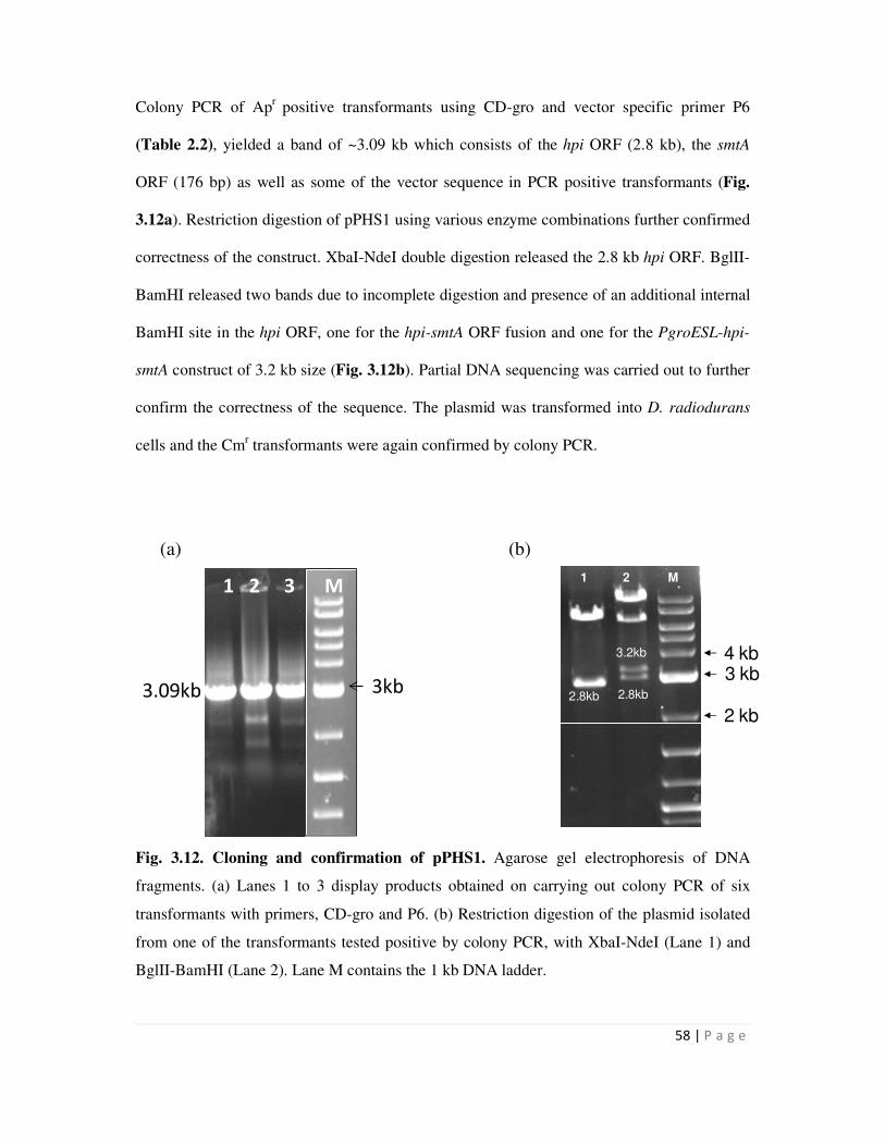
58 | P a g e
1 1 2 M
3 kb
2 kb
4 kb
2.8kb 2.8kb
3.2kb
3kb3.09kb
1 2 3 M
Colony PCR of Apr
positive transformants using CD-gro and vector specific primer P6
(Table 2.2), yielded a band of ~3.09 kb which consists of the hpi ORF (2.8 kb), the smtA
ORF (176 bp) as well as some of the vector sequence in PCR positive transformants (Fig.
3.12a). Restriction digestion of pPHS1 using various enzyme combinations further confirmed
correctness of the construct. XbaI-NdeI double digestion released the 2.8 kb hpi ORF. BglII-
BamHI released two bands due to incomplete digestion and presence of an additional internal
BamHI site in the hpi ORF, one for the hpi-smtA ORF fusion and one for the PgroESL-hpi-
smtA construct of 3.2 kb size (Fig. 3.12b). Partial DNA sequencing was carried out to further
confirm the correctness of the sequence. The plasmid was transformed into D. radiodurans
cells and the Cmr transformants were again confirmed by colony PCR.
(a) (b)
Fig. 3.12. Cloning and confirmation of pPHS1. Agarose gel electrophoresis of DNA
fragments. (a) Lanes 1 to 3 display products obtained on carrying out colony PCR of six
transformants with primers, CD-gro and P6. (b) Restriction digestion of the plasmid isolated
from one of the transformants tested positive by colony PCR, with XbaI-NdeI (Lane 1) and
BglII-BamHI (Lane 2). Lane M contains the 1 kb DNA ladder.
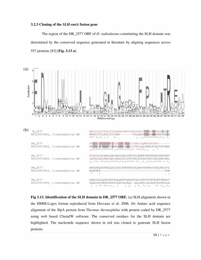
59 | P a g e
DR_2577 MKKSLIALTTALSFGLAAAQTAAPVSAPQVPALTDVPAGHWAKDAIDRLV
NTL03TT1893_ T.thermophilus HB MKKRLVTLLAGLLTVLSMG--------FGLAQFSDVPAGHWAKEAVEALA
*** *::* :.* *: . :. ::*********:*:: *.
DR_2577 SRGVILGYPDGTFRGTQNLTRYEAAIIIARLLDQMRDGETPAG-------
NTL03TT1893_ T.thermophilus HB AKGIILGFPDGTFRGNENLTRYQAALLIYRLLQQIEEELKTQGTSPTMEA
::*:***:*******.:*****:**::* ***:*:.: .. *
DR_2577 MTAEDMTALQNAIQELAADLAALGVRVSDLEANAVSKDDFARLEARIEEV
NTL03TT1893_ T.thermophilus HB LAPEDLEALKNAVQELAAELASLGVRVSALEDSAATKEDIARLEAMIAEL
::.**: **:**:*****:**:****** ** .*.:*:*:***** * *:
DR_2577 AAAGGEQGATEALQGQIDDLTARVDEYDALRADVDDNASSIAALNDLTVL
NTL03TT1893_ T.thermophilus HB KAQPMPE----------------------------------------PGM
* : . :
DR_2577 LNQDILDLQDRVSAVEAAQADFVQRSDFDALGGRVTTVETRVETVNNSLT
NTL03TT1893_ T.thermophilus HB DQAALKDLMDRVEAASIAADTALAQA--QQLAERLDALAQDVEGVKGDLA
: : ** ***.*.. * : :: : *. *: :: ** *:..*:
3.2.3 Cloning of the SLH-smtA fusion gene
The region of the DR_2577 ORF of D. radiodurans constituting the SLH domain was
determined by the conserved sequence generated in literature by aligning sequences across
557 proteins [83] (Fig. 3.13 a).
Fig 3.13. Identification of the SLH domain in DR_2577 ORF. (a) SLH alignment shown in
the HMM-Logos format reproduced from Desvaux et al. 2006. (b) Amino acid sequence
alignment of the SlpA protein from Thermus thermophilus with protein coded by DR_2577
using web based ClustalW software. The conserved residues for the SLH domain are
highlighted. The nucleotide sequence shown in red was cloned to generate SLH fusion
proteins
(b)
(a)
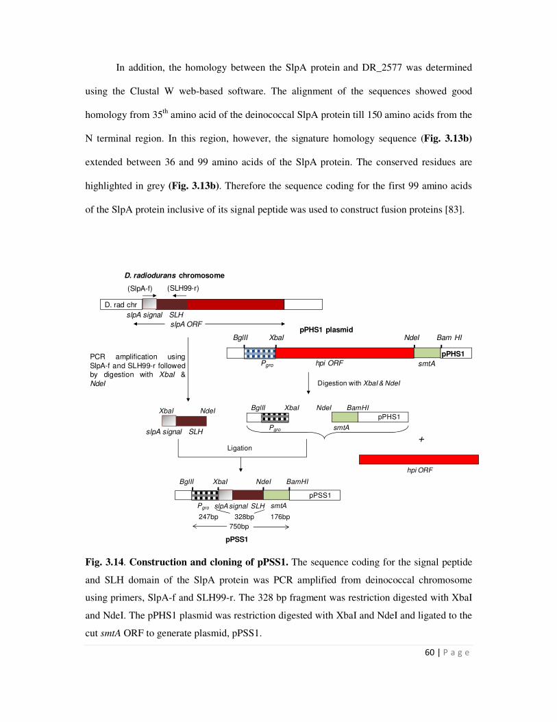
60 | P a g e
In addition, the homology between the SlpA protein and DR_2577 was determined
using the Clustal W web-based software. The alignment of the sequences showed good
homology from 35th
amino acid of the deinococcal SlpA protein till 150 amino acids from the
N terminal region. In this region, however, the signature homology sequence (Fig. 3.13b)
extended between 36 and 99 amino acids of the SlpA protein. The conserved residues are
highlighted in grey (Fig. 3.13b). Therefore the sequence coding for the first 99 amino acids
of the SlpA protein inclusive of its signal peptide was used to construct fusion proteins [83].
Fig. 3.14. Construction and cloning of pPSS1. The sequence coding for the signal peptide
and SLH domain of the SlpA protein was PCR amplified from deinococcal chromosome
using primers, SlpA-f and SLH99-r. The 328 bp fragment was restriction digested with XbaI
and NdeI. The pPHS1 plasmid was restriction digested with XbaI and NdeI and ligated to the
cut smtA ORF to generate plasmid, pPSS1.
BglII XbaI
smtAPgro
hpi ORF
Digestion with XbaI & NdeI
NdeI BamHI
Ligation
Pgro
BglII XbaI
smtA
NdeI BamHI
pPHS1
pPSS1
176bp328bp
slpA signal SLH
247bp
pPHS1
smtA
BglII XbaI NdeI Bam HI
Pgro hpi ORF
slpA signal SLH
(SlpA-f) (SLH99-r)
D. rad chr
slpA ORF
XbaI NdeI
slpA signal SLH
PCR amplification using
SlpA-f and SLH99-r followedby digestion with XbaI &
NdeI
D. radiodurans chromosome
pPHS1 plasmid
750bp
pPSS1
+
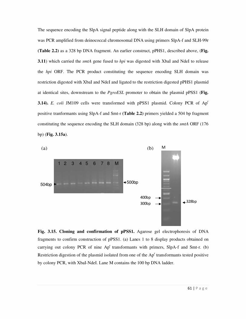
61 | P a g e
M
328bp400bp
300bp
The sequence encoding the SlpA signal peptide along with the SLH domain of SlpA protein
was PCR amplified from deinococcal chromosomal DNA using primers SlpA-f and SLH-99r
(Table 2.2) as a 328 bp DNA fragment. An earlier construct, pPHS1, described above, (Fig.
3.11) which carried the smtA gene fused to hpi was digested with XbaI and NdeI to release
the hpi ORF. The PCR product constituting the sequence encoding SLH domain was
restriction digested with XbaI and NdeI and ligated to the restriction digested pPHS1 plasmid
at identical sites, downstream to the PgroESL promoter to obtain the plasmid pPSS1 (Fig.
3.14). E. coli JM109 cells were transformed with pPSS1 plasmid. Colony PCR of Apr
positive tranformants using SlpA-f and Smt-r (Table 2.2) primers yielded a 504 bp fragment
constituting the sequence encoding the SLH domain (328 bp) along with the smtA ORF (176
bp) (Fig. 3.15a).
Fig. 3.15. Cloning and confirmation of pPSS1. Agarose gel electrophoresis of DNA
fragments to confirm construction of pPSS1. (a) Lanes 1 to 8 display products obtained on
carrying out colony PCR of nine Apr
transformants with primers, SlpA-f and Smt-r. (b)
Restriction digestion of the plasmid isolated from one of the Apr transformants tested positive
by colony PCR, with XbaI-NdeI. Lane M contains the 100 bp DNA ladder.
500bp504bp
1 2 3 4 5 6 7 8 M
(a) (b)
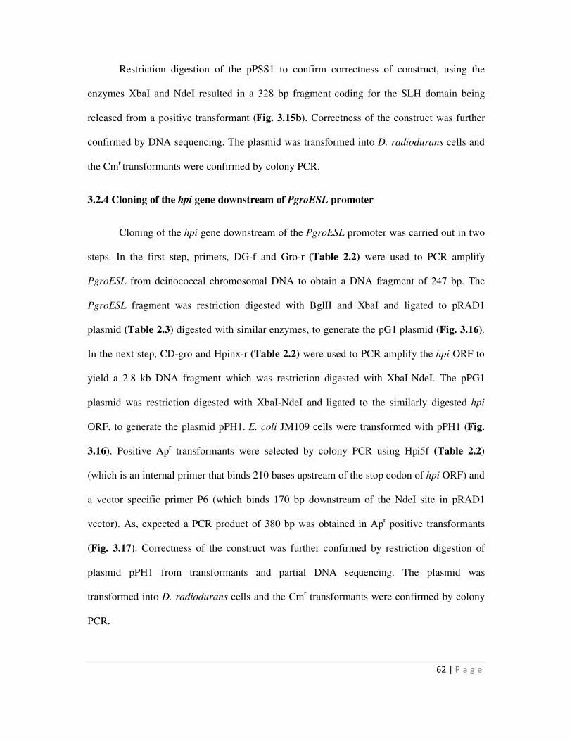
62 | P a g e
Restriction digestion of the pPSS1 to confirm correctness of construct, using the
enzymes XbaI and NdeI resulted in a 328 bp fragment coding for the SLH domain being
released from a positive transformant (Fig. 3.15b). Correctness of the construct was further
confirmed by DNA sequencing. The plasmid was transformed into D. radiodurans cells and
the Cmr transformants were confirmed by colony PCR.
3.2.4 Cloning of the hpi gene downstream of PgroESL promoter
Cloning of the hpi gene downstream of the PgroESL promoter was carried out in two
steps. In the first step, primers, DG-f and Gro-r (Table 2.2) were used to PCR amplify
PgroESL from deinococcal chromosomal DNA to obtain a DNA fragment of 247 bp. The
PgroESL fragment was restriction digested with BglII and XbaI and ligated to pRAD1
plasmid (Table 2.3) digested with similar enzymes, to generate the pG1 plasmid (Fig. 3.16).
In the next step, CD-gro and Hpinx-r (Table 2.2) were used to PCR amplify the hpi ORF to
yield a 2.8 kb DNA fragment which was restriction digested with XbaI-NdeI. The pPG1
plasmid was restriction digested with XbaI-NdeI and ligated to the similarly digested hpi
ORF, to generate the plasmid pPH1. E. coli JM109 cells were transformed with pPH1 (Fig.
3.16). Positive Apr transformants were selected by colony PCR using Hpi5f (Table 2.2)
(which is an internal primer that binds 210 bases upstream of the stop codon of hpi ORF) and
a vector specific primer P6 (which binds 170 bp downstream of the NdeI site in pRAD1
vector). As, expected a PCR product of 380 bp was obtained in Apr positive transformants
(Fig. 3.17). Correctness of the construct was further confirmed by restriction digestion of
plasmid pPH1 from transformants and partial DNA sequencing. The plasmid was
transformed into D. radiodurans cells and the Cmr transformants were confirmed by colony
PCR.
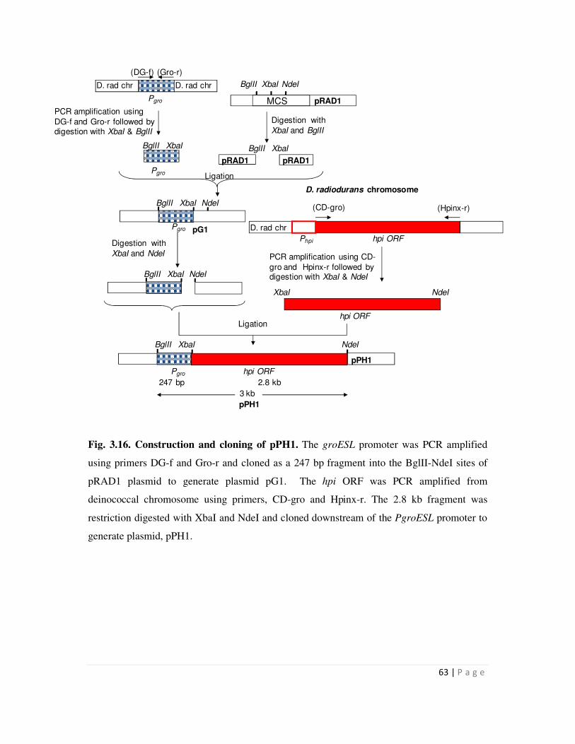
63 | P a g e
D. rad chr D. rad chr
Pgro MCS
BglII XbaI NdeI
pRAD1
BglII XbaI
pRAD1pRAD1
BglII XbaI
Pgro
Phpi hpi ORF
(CD-gro) (Hpinx-r)
D. rad chr
PCR amplification using CD-
gro and Hpinx-r followed by digestion with XbaI & NdeI
D. radiodurans chromosome
hpi ORF
XbaI NdeI
Digestion with
XbaI and NdeI
BglII XbaI NdeI
BglII XbaI NdeI
BglII XbaI NdeI
PCR amplification using
DG-f and Gro-r followed by digestion with XbaI & BglII
Digestion with
XbaI and BglII
Ligation
Ligation
pPH1
Pgro hpi ORF
247 bp 2.8 kb
3 kb
pPH1
(Gro-r)(DG-f)
pG1Pgro
Fig. 3.16. Construction and cloning of pPH1. The groESL promoter was PCR amplified
using primers DG-f and Gro-r and cloned as a 247 bp fragment into the BglII-NdeI sites of
pRAD1 plasmid to generate plasmid pG1. The hpi ORF was PCR amplified from
deinococcal chromosome using primers, CD-gro and Hpinx-r. The 2.8 kb fragment was
restriction digested with XbaI and NdeI and cloned downstream of the PgroESL promoter to
generate plasmid, pPH1.
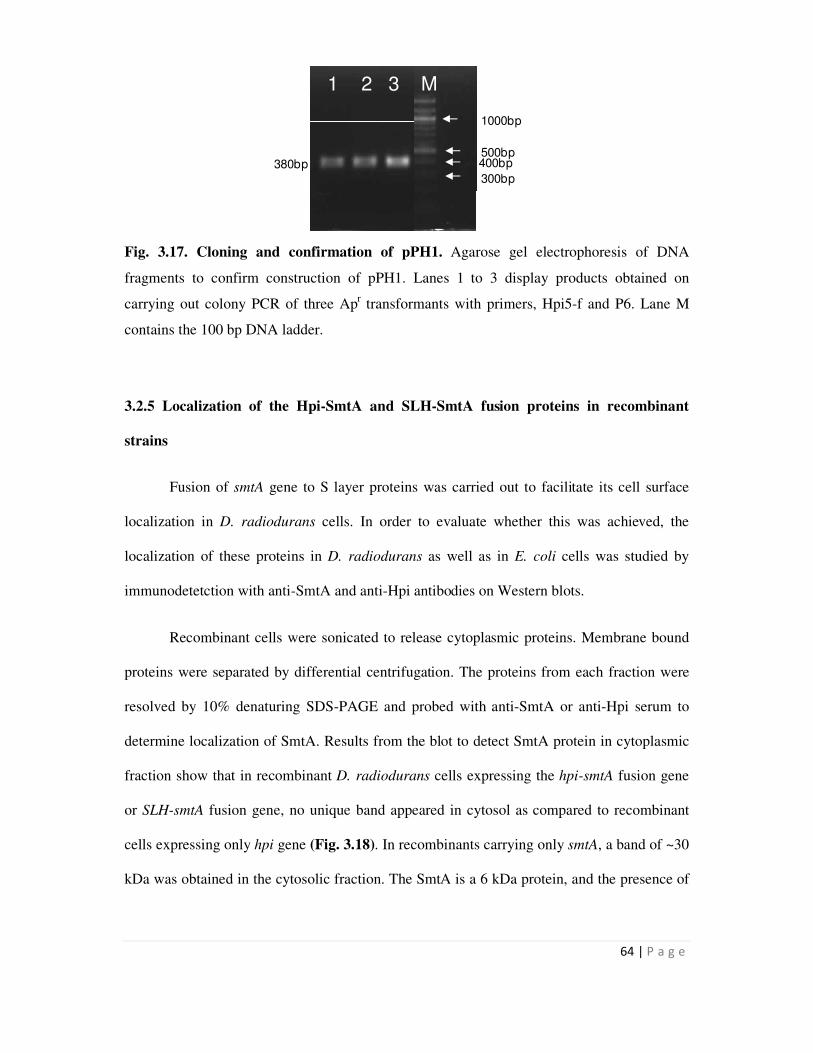
64 | P a g e
1 1 2 3 M
380bp
M
500bp400bp
300bp
1000bp
Fig. 3.17. Cloning and confirmation of pPH1. Agarose gel electrophoresis of DNA
fragments to confirm construction of pPH1. Lanes 1 to 3 display products obtained on
carrying out colony PCR of three Apr transformants with primers, Hpi5-f and P6. Lane M
contains the 100 bp DNA ladder.
3.2.5 Localization of the Hpi-SmtA and SLH-SmtA fusion proteins in recombinant
strains
Fusion of smtA gene to S layer proteins was carried out to facilitate its cell surface
localization in D. radiodurans cells. In order to evaluate whether this was achieved, the
localization of these proteins in D. radiodurans as well as in E. coli cells was studied by
immunodetetction with anti-SmtA and anti-Hpi antibodies on Western blots.
Recombinant cells were sonicated to release cytoplasmic proteins. Membrane bound
proteins were separated by differential centrifugation. The proteins from each fraction were
resolved by 10% denaturing SDS-PAGE and probed with anti-SmtA or anti-Hpi serum to
determine localization of SmtA. Results from the blot to detect SmtA protein in cytoplasmic
fraction show that in recombinant D. radiodurans cells expressing the hpi-smtA fusion gene
or SLH-smtA fusion gene, no unique band appeared in cytosol as compared to recombinant
cells expressing only hpi gene (Fig. 3.18). In recombinants carrying only smtA, a band of ~30
kDa was obtained in the cytosolic fraction. The SmtA is a 6 kDa protein, and the presence of
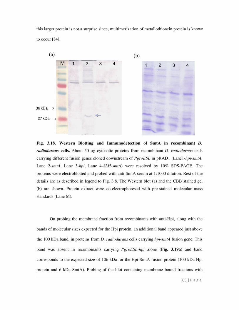
65 | P a g e
1 2 3 4 1 2 3 4
27 kDa
36 kDa
1 2 3 4 M
this larger protein is not a surprise since, multimerization of metallothionein protein is known
to occur [84].
Fig. 3.18. Western Blotting and Immunodetection of SmtA in recombinant D.
radiodurans cells. About 50 µg cytosolic proteins from recombinant D. radiodurnas cells
carrying different fusion genes cloned downstream of PgroESL in pRAD1 (Lane1-hpi-smtA,
Lane 2-smtA, Lane 3-hpi, Lane 4-SLH-smtA) were resolved by 10% SDS-PAGE. The
proteins were electroblotted and probed with anti-SmtA serum at 1:1000 dilution. Rest of the
details are as described in legend to Fig. 3.8. The Western blot (a) and the CBB stained gel
(b) are shown. Protein extract were co-electrophoresed with pre-stained molecular mass
standards (Lane M).
On probing the membrane fraction from recombinants with anti-Hpi, along with the
bands of molecular sizes expected for the Hpi protein, an additional band appeared just above
the 100 kDa band, in proteins from D. radiodurans cells carrying hpi-smtA fusion gene. This
band was absent in recombinants carrying PgroESL-hpi alone (Fig. 3.19a) and band
corresponds to the expected size of 106 kDa for the Hpi-SmtA fusion protein (100 kDa Hpi
protein and 6 kDa SmtA). Probing of the blot containing membrane bound fractions with
(b) (a)
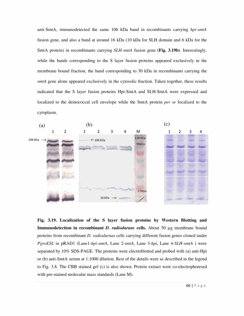
66 | P a g e
1 2 3 4
106 kDa1 2 3 4 130 kDa
95kDa
17kDa
1 2 3 4 M
16 kDa
1 2
106 kDa
anti-SmtA, immunodetected the same 106 kDa band in recombinants carrying hpi-smtA
fusion gene, and also a band at around 16 kDa (10 kDa for SLH domain and 6 kDa for the
SmtA protein) in recombinants carrying SLH-smtA fusion gene (Fig. 3.19b). Interestingly,
while the bands corresponding to the S layer fusion proteins appeared exclusively in the
membrane bound fraction, the band corresponding to 30 kDa in recombinants carrying the
smtA gene alone appeared exclusively in the cytosolic fraction. Taken together, these results
indicated that the S layer fusion proteins Hpi-SmtA and SLH-SmtA were expressed and
localized to the deinococcal cell envelope while the SmtA protein per se localized to the
cytoplasm.
Fig. 3.19. Localization of the S layer fusion proteins by Western Blotting and
Immunodetection in recombinant D. radiodurans cells. About 50 µg membrane bound
proteins from recombinant D. radiodurnas cells carrying different fusion genes cloned under
PgroESL in pRAD1 (Lane1-hpi-smtA, Lane 2-smtA, Lane 3-hpi, Lane 4-SLH-smtA ) were
separated by 10% SDS-PAGE. The proteins were electroblotted and probed with (a) anti-Hpi
or (b) anti-SmtA serum at 1:1000 dilution. Rest of the details were as described in the legend
to Fig. 3.8. The CBB stained gel (c) is also shown. Protein extract were co-electrophoresed
with pre-stained molecular mass standards (Lane M).
(a) (b) (c)
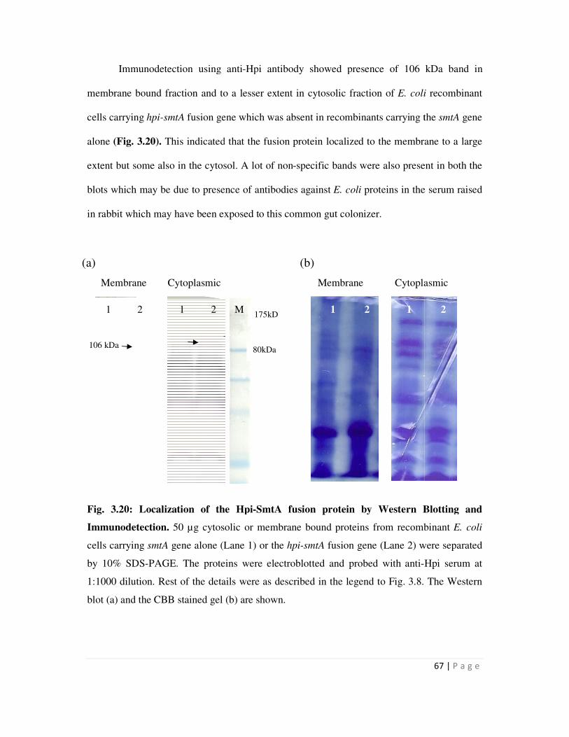
Immunodetection using anti
membrane bound fraction and to a lesser ex
cells carrying hpi-smtA fusion gene which was absent in recombinants carrying the
alone (Fig. 3.20). This indicated that the fusion protein localize
extent but some also in the cytosol. A lot of non
blots which may be due to presence of antibodies against
in rabbit which may have been exposed t
Fig. 3.20: Localization of the Hpi
Immunodetection. 50 µg cytosolic or membrane bound
cells carrying smtA gene alone (Lane 1) or the
by 10% SDS-PAGE. The proteins were electroblotted and probed with anti
1:1000 dilution. Rest of the details were
blot (a) and the CBB stained gel (b) are shown.
Membrane Cytoplasmic Membrane Cytoplasmic
1 2 1 2 M
(a)
106 kDa
Immunodetection using anti-Hpi antibody showed presence of 106 kDa band in
membrane bound fraction and to a lesser extent in cytosolic fraction of E. coli
fusion gene which was absent in recombinants carrying the
This indicated that the fusion protein localized to the membrane to a large
extent but some also in the cytosol. A lot of non-specific bands were also present in both
be due to presence of antibodies against E. coli proteins in the serum raised
in rabbit which may have been exposed to this common gut colonizer.
tion of the Hpi-SmtA fusion protein by Western Blotting and
50 µg cytosolic or membrane bound proteins from recombinant
(Lane 1) or the hpi-smtA fusion gene (Lane 2) were separated
. The proteins were electroblotted and probed with anti-Hpi serum at
ion. Rest of the details were as described in the legend to Fig. 3.8.
blot (a) and the CBB stained gel (b) are shown.
Membrane Cytoplasmic Membrane Cytoplasmic
1 2 1 2 M 1 2 1 2
80kDa
175kD
a
(b)
67 | P a g e
Hpi antibody showed presence of 106 kDa band in
E. coli recombinant
fusion gene which was absent in recombinants carrying the smtA gene
to the membrane to a large
were also present in both the
proteins in the serum raised
y Western Blotting and
proteins from recombinant E. coli
were separated
Hpi serum at
3.8. The Western
Membrane Cytoplasmic Membrane Cytoplasmic
1 2 1 2
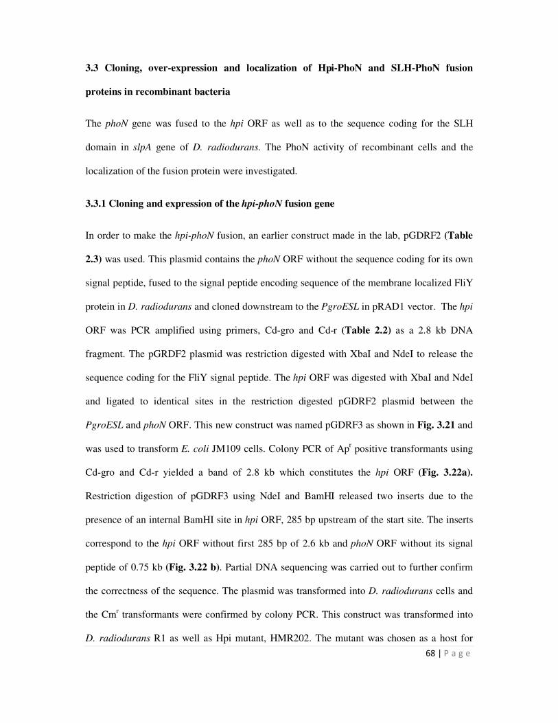
68 | P a g e
3.3 Cloning, over-expression and localization of Hpi-PhoN and SLH-PhoN fusion
proteins in recombinant bacteria
The phoN gene was fused to the hpi ORF as well as to the sequence coding for the SLH
domain in slpA gene of D. radiodurans. The PhoN activity of recombinant cells and the
localization of the fusion protein were investigated.
3.3.1 Cloning and expression of the hpi-phoN fusion gene
In order to make the hpi-phoN fusion, an earlier construct made in the lab, pGDRF2 (Table
2.3) was used. This plasmid contains the phoN ORF without the sequence coding for its own
signal peptide, fused to the signal peptide encoding sequence of the membrane localized FliY
protein in D. radiodurans and cloned downstream to the PgroESL in pRAD1 vector. The hpi
ORF was PCR amplified using primers, Cd-gro and Cd-r (Table 2.2) as a 2.8 kb DNA
fragment. The pGRDF2 plasmid was restriction digested with XbaI and NdeI to release the
sequence coding for the FliY signal peptide. The hpi ORF was digested with XbaI and NdeI
and ligated to identical sites in the restriction digested pGDRF2 plasmid between the
PgroESL and phoN ORF. This new construct was named pGDRF3 as shown in Fig. 3.21 and
was used to transform E. coli JM109 cells. Colony PCR of Apr positive transformants using
Cd-gro and Cd-r yielded a band of 2.8 kb which constitutes the hpi ORF (Fig. 3.22a).
Restriction digestion of pGDRF3 using NdeI and BamHI released two inserts due to the
presence of an internal BamHI site in hpi ORF, 285 bp upstream of the start site. The inserts
correspond to the hpi ORF without first 285 bp of 2.6 kb and phoN ORF without its signal
peptide of 0.75 kb (Fig. 3.22 b). Partial DNA sequencing was carried out to further confirm
the correctness of the sequence. The plasmid was transformed into D. radiodurans cells and
the Cmr transformants were confirmed by colony PCR. This construct was transformed into
D. radiodurans R1 as well as Hpi mutant, HMR202. The mutant was chosen as a host for
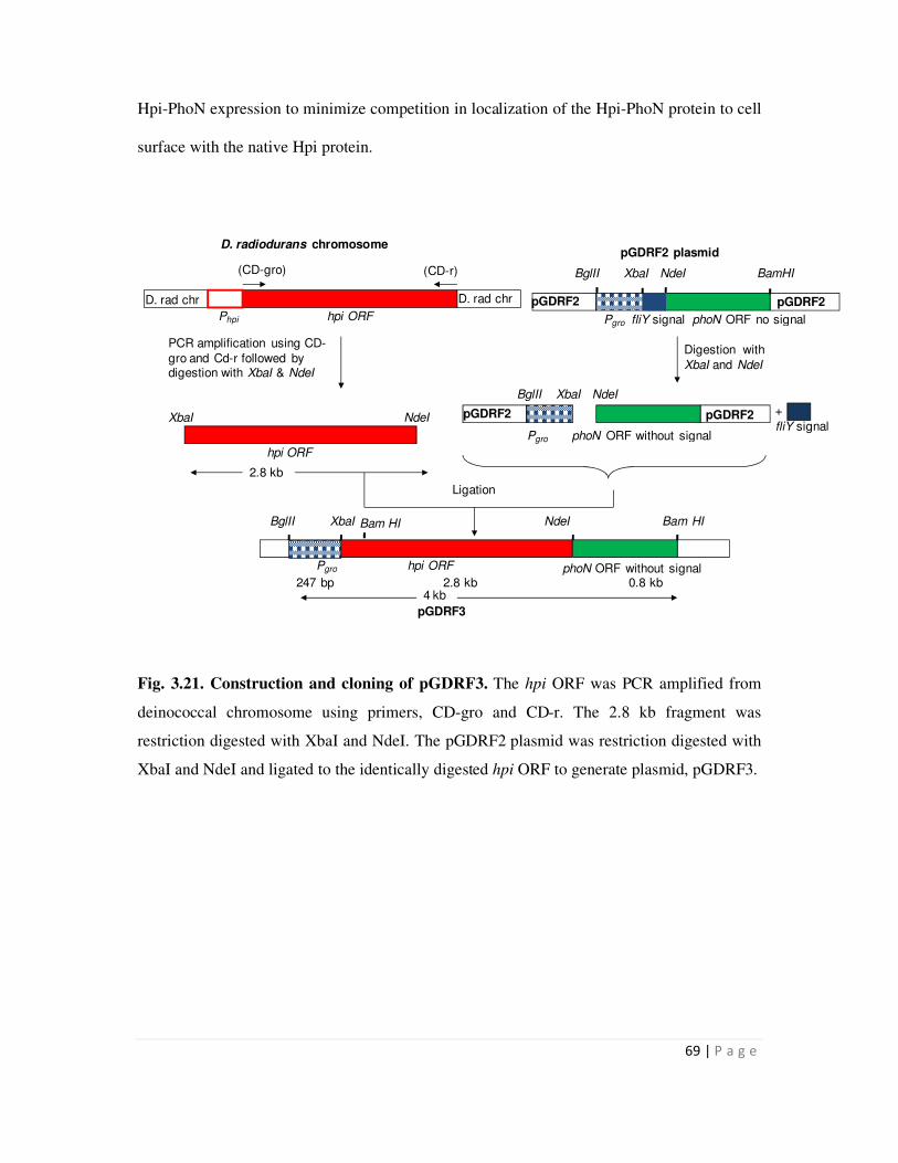
69 | P a g e
Hpi-PhoN expression to minimize competition in localization of the Hpi-PhoN protein to cell
surface with the native Hpi protein.
Fig. 3.21. Construction and cloning of pGDRF3. The hpi ORF was PCR amplified from
deinococcal chromosome using primers, CD-gro and CD-r. The 2.8 kb fragment was
restriction digested with XbaI and NdeI. The pGDRF2 plasmid was restriction digested with
XbaI and NdeI and ligated to the identically digested hpi ORF to generate plasmid, pGDRF3.
phoN ORF without signal
BglII XbaI NdeI Bam HI
D. rad chr pGDRF2
Pgro fliY signal phoN ORF no signal
pGDRF2
BglII XbaI NdeI BamHI
Digestion with
XbaI and NdeI
Pgro hpi ORF
Ligation
fliY signal
+
2.8 kb
pGDRF2 plasmid
4 kb
Pgro phoN ORF without signal
pGDRF2
BglII XbaI NdeI
pGDRF2
Phpi hpi ORF
(CD-gro) (CD-r)
D. rad chr
PCR amplification using CD-
gro and Cd-r followed by digestion with XbaI & NdeI
D. radiodurans chromosome
hpi ORF
XbaI NdeI
247 bp 2.8 kb 0.8 kb
pGDRF3
Bam HI
70 | P a g e
2.8 kb
0.8 kb
3 kb
2 kb
1 kb
0.5 kb
1 2 1 2 3 4 5 6 M
3 kb2.8 kb
Fig. 3.22. Cloning and confirmation of pGDRF3. Agarose gel electrophoresis of DNA
fragments. (a) Lanes 1 to 6 display products obtained on carrying out colony PCR of eight
transformants with primers, CD-gro and CD-r. (b) Restriction digestion of the plasmid
isolated from one of the Apr transformants tested positive by colony PCR, with XbaI-NdeI.
Lane M contains the 1 kb DNA ladder.
3.3.2 Expression of the Hpi-PhoN fusion protein
` To ascertain expression of the Hpi-PhoN fusion protein, total cell extracts were made
using non-reducing cracking buffer. The proteins were then separated by 10% denaturing gel
and developed for phosphatase activity using NBT-BCIP substrate on a zymogram. In both,
E. coli as well as D. radiodurans recombinants bearing the pGRDF3 plasmid, an activity
band was obtained at ~127 kDa, as expected for the fusion protein as against 27 kDa band
which is normally obtained with phoN expressing cells (Fig. 3.23). In addition, several
discreet activity bands of smaller size were obtained which may be due to degradation of the
fusion protein.
(a) (b) M
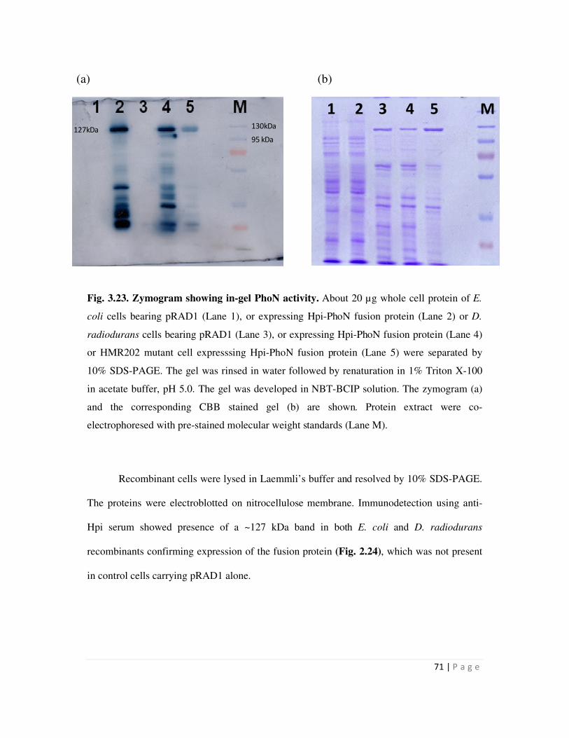
71 | P a g e
130kDa
95 kDa
127kDa
1 2 3 4 5 M
Fig. 3.23. Zymogram showing in-gel PhoN activity. About 20 µg whole cell protein of E.
coli cells bearing pRAD1 (Lane 1), or expressing Hpi-PhoN fusion protein (Lane 2) or D.
radiodurans cells bearing pRAD1 (Lane 3), or expressing Hpi-PhoN fusion protein (Lane 4)
or HMR202 mutant cell expresssing Hpi-PhoN fusion protein (Lane 5) were separated by
10% SDS-PAGE. The gel was rinsed in water followed by renaturation in 1% Triton X-100
in acetate buffer, pH 5.0. The gel was developed in NBT-BCIP solution. The zymogram (a)
and the corresponding CBB stained gel (b) are shown. Protein extract were co-
electrophoresed with pre-stained molecular weight standards (Lane M).
Recombinant cells were lysed in Laemmli’s buffer and resolved by 10% SDS-PAGE.
The proteins were electroblotted on nitrocellulose membrane. Immunodetection using anti-
Hpi serum showed presence of a ~127 kDa band in both E. coli and D. radiodurans
recombinants confirming expression of the fusion protein (Fig. 2.24), which was not present
in control cells carrying pRAD1 alone.
(a) (b)
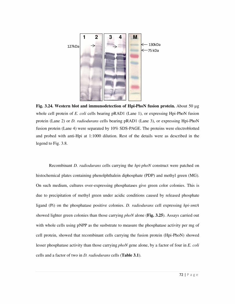
72 | P a g e
1 2 3 4 M
130kDa
75 kDa127kDa
Fig. 3.24. Western blot and immunodetection of Hpi-PhoN fusion protein. About 50 µg
whole cell protein of E. coli cells bearing pRAD1 (Lane 1), or expressing Hpi-PhoN fusion
protein (Lane 2) or D. radiodurans cells bearing pRAD1 (Lane 3), or expressing Hpi-PhoN
fusion protein (Lane 4) were separated by 10% SDS-PAGE. The proteins were electroblotted
and probed with anti-Hpi at 1:1000 dilution. Rest of the details were as described in the
legend to Fig. 3.8.
Recombinant D. radiodurans cells carrying the hpi-phoN construct were patched on
histochemical plates containing phenolphthalein diphosphate (PDP) and methyl green (MG).
On such medium, cultures over-expressing phosphatases give green color colonies. This is
due to precipitation of methyl green under acidic conditions caused by released phosphate
ligand (Pi) on the phosphatase positive colonies. D. radiodurans cell expressing hpi-smtA
showed lighter green colonies than those carrying phoN alone (Fig. 3.25). Assays carried out
with whole cells using pNPP as the susbstrate to measure the phosphatase activity per mg of
cell protein, showed that recombinant cells carrying the fusion protein (Hpi-PhoN) showed
lesser phosphatase activity than those carrying phoN gene alone, by a factor of four in E. coli
cells and a factor of two in D. radiodurans cells (Table 3.1).
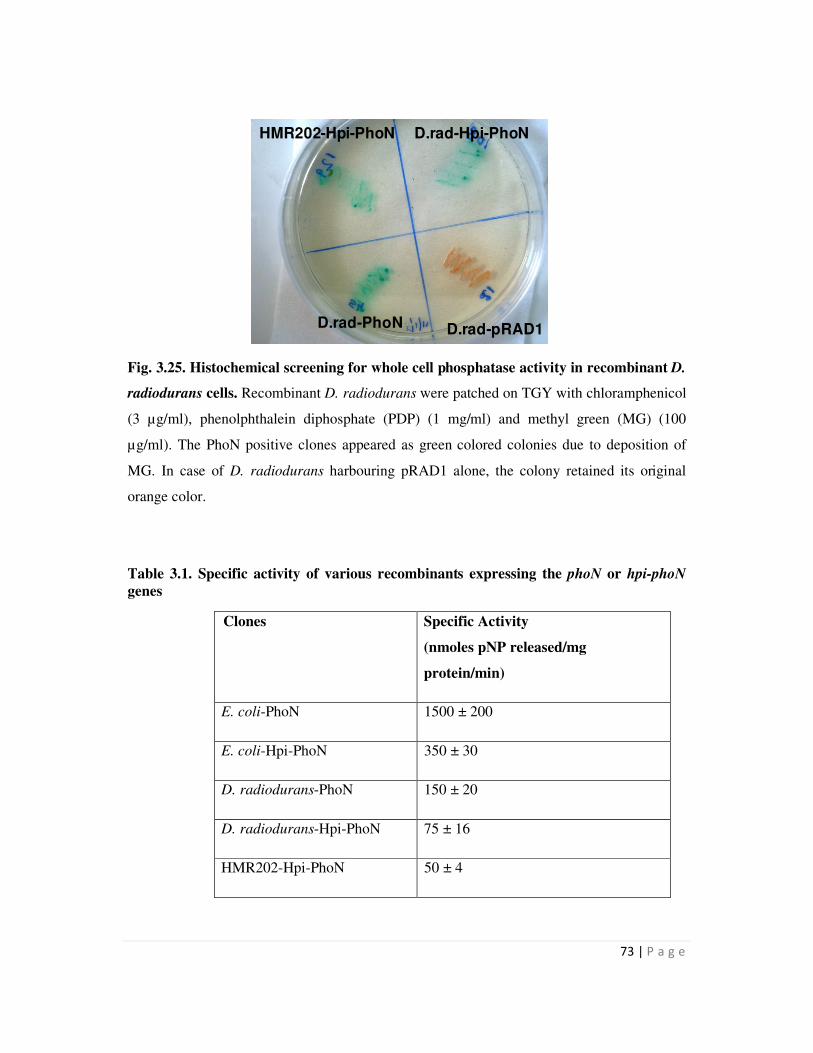
73 | P a g e
D.rad-Hpi-PhoN
D.rad-PhoN
HMR202-Hpi-PhoN
D.rad-pRAD1
D.rad-Hpi-PhoN
Fig. 3.25. Histochemical screening for whole cell phosphatase activity in recombinant D.
radiodurans cells. Recombinant D. radiodurans were patched on TGY with chloramphenicol
(3 µg/ml), phenolphthalein diphosphate (PDP) (1 mg/ml) and methyl green (MG) (100
µg/ml). The PhoN positive clones appeared as green colored colonies due to deposition of
MG. In case of D. radiodurans harbouring pRAD1 alone, the colony retained its original
orange color.
Table 3.1. Specific activity of various recombinants expressing the phoN or hpi-phoN
genes
Clones Specific Activity
(nmoles pNP released/mg
protein/min)
E. coli-PhoN 1500 ± 200
E. coli-Hpi-PhoN 350 ± 30
D. radiodurans-PhoN 150 ± 20
D. radiodurans-Hpi-PhoN 75 ± 16
HMR202-Hpi-PhoN 50 ± 4
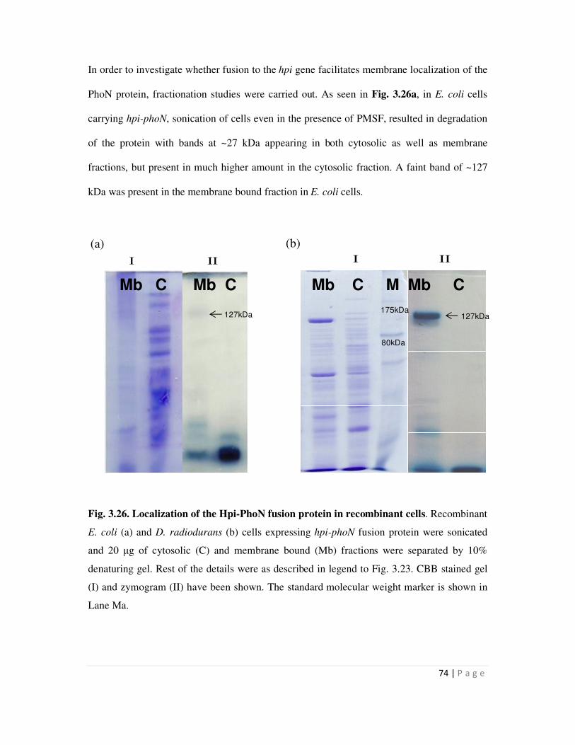
74 | P a g e
Mb C Mb C Mb C M Mb C
127kDa127kDa175kDa
80kDa
I II I II
In order to investigate whether fusion to the hpi gene facilitates membrane localization of the
PhoN protein, fractionation studies were carried out. As seen in Fig. 3.26a, in E. coli cells
carrying hpi-phoN, sonication of cells even in the presence of PMSF, resulted in degradation
of the protein with bands at ~27 kDa appearing in both cytosolic as well as membrane
fractions, but present in much higher amount in the cytosolic fraction. A faint band of ~127
kDa was present in the membrane bound fraction in E. coli cells.
Fig. 3.26. Localization of the Hpi-PhoN fusion protein in recombinant cells. Recombinant
E. coli (a) and D. radiodurans (b) cells expressing hpi-phoN fusion protein were sonicated
and 20 µg of cytosolic (C) and membrane bound (Mb) fractions were separated by 10%
denaturing gel. Rest of the details were as described in legend to Fig. 3.23. CBB stained gel
(I) and zymogram (II) have been shown. The standard molecular weight marker is shown in
Lane Ma.
(a) (b)
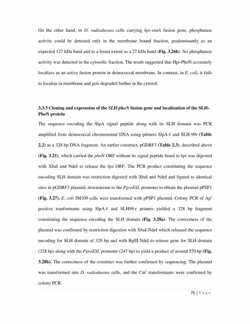
75 | P a g e
On the other hand, in D. radiodurans cells carrying hpi-smtA fusion gene, phosphatase
activity could be detected only in the membrane bound fraction, predominantly as an
expected 127 kDa band and to a lesser extent as a 27 kDa band (Fig. 3.26b). No phosphatase
activity was detected in the cytosolic fraction. The result suggested that Hpi-PhoN accurately
localizes as an active fusion protein in deinococcal membrane. In contrast, in E. coli, it fails
to localize in membrane and gets degraded further in the cytosol.
3.3.3 Cloning and expression of the SLH-phoN fusion gene and localization of the SLH-
PhoN protein
The sequence encoding the SlpA signal peptide along with its SLH domain was PCR
amplified from deinococcal chromosomal DNA using primers SlpA-f and SLH-99r (Table
2.2) as a 328 bp DNA fragment. An earlier construct, pGDRF3 (Table 2.3), described above
(Fig. 3.21), which carried the phoN ORF without its signal peptide fused to hpi was digested
with XbaI and NdeI to release the hpi ORF. The PCR product constituting the sequence
encoding SLH domain was restriction digested with XbaI and NdeI and ligated to identical
sites in pGDRF3 plasmid, downstream to the PgroESL promoter to obtain the plasmid pPSP1
(Fig. 3.27). E. coli JM109 cells were transformed with pPSP1 plasmid. Colony PCR of Apr
positive tranformants using SlpA-f and SLH99-r primers yielded a 328 bp fragment
constituting the sequence encoding the SLH domain (Fig. 3.28a). The correctness of the
plasmid was confirmed by restriction digestion with XbaI-NdeI which released the sequence
encoding for SLH domain of 328 bp and with BglII-NdeI to release gene for SLH domain
(328 bp) along with the PgroESL promoter (247 bp) to yield a product of around 570 bp (Fig.
3.28b). The correctness of the construct was further confirmed by sequencing. The plasmid
was transformed into D. radiodurans cells, and the Cmr transformants were confirmed by
colony PCR.
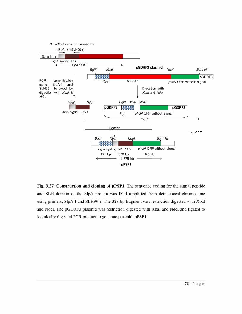
76 | P a g e
Fig. 3.27. Construction and cloning of pPSP1. The sequence coding for the signal peptide
and SLH domain of the SlpA protein was PCR amplified from deinococcal chromosome
using primers, SlpA-f and SLH99-r. The 328 bp fragment was restriction digested with XbaI
and NdeI. The pGDRF3 plasmid was restriction digested with XbaI and NdeI and ligated to
identically digested PCR product to generate plasmid, pPSP1.
phoN ORF without signal
BglII XbaI NdeI Bam HI
PCR amplification
using SlpA-f andSLH99-r followed by
digestion with XbaI &
NdeI
Digestion with
XbaI and NdeI
Ligation
D. radiodurans chromosome
1.375 kb
Pgro phoN ORF without signal
pGDRF3
BglII XbaI NdeI
pGDRF3
247 bp 328 bp 0.8 kb
pPSP1
phoN ORF without signal
BglII XbaI NdeI Bam HI
Pgro hpi ORF
pGDRF3
slpA ORF
Pgro slpA signal SLH
slpA signal SLH
(SlpA-f) (SLH99-r)
D. rad chr
XbaI NdeI
slpA signal SLH
+
pGDRF3 plasmid
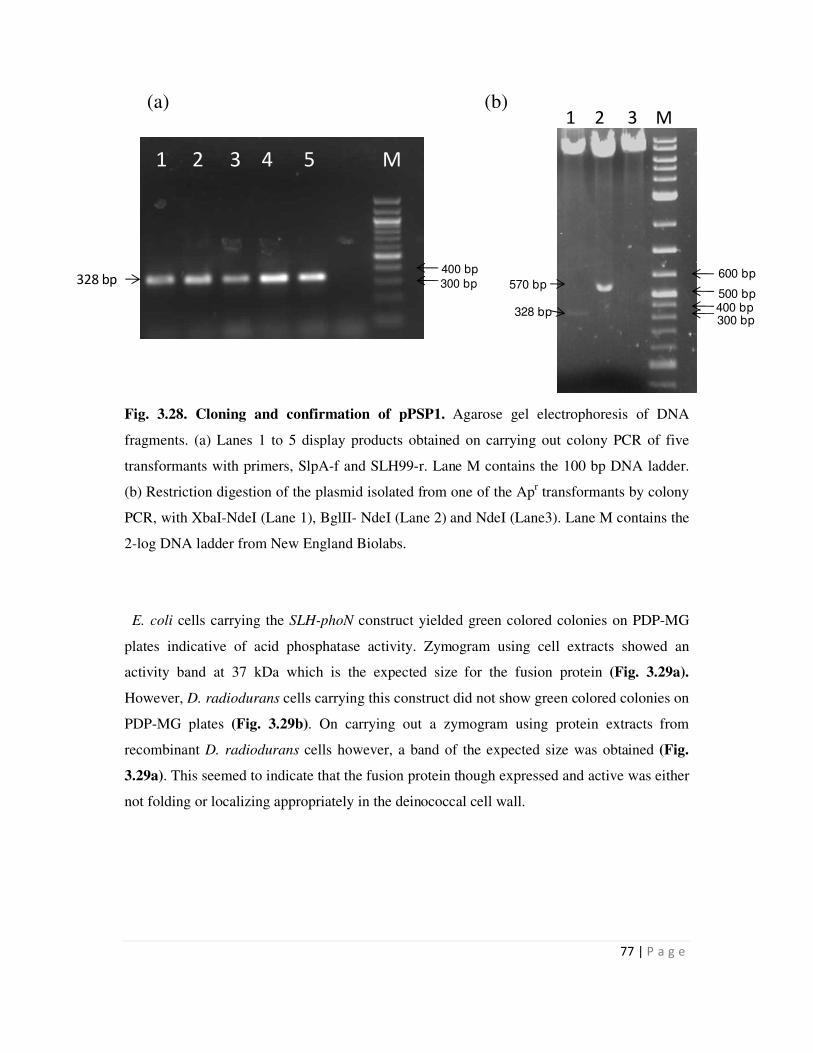
77 | P a g e
400 bp
600 bp
300 bp
500 bp
328 bp
570 bp
1 2 3 M
400 bp
300 bp
1 1 2 3 4 5 M
328 bp
(a) (b)
Fig. 3.28. Cloning and confirmation of pPSP1. Agarose gel electrophoresis of DNA
fragments. (a) Lanes 1 to 5 display products obtained on carrying out colony PCR of five
transformants with primers, SlpA-f and SLH99-r. Lane M contains the 100 bp DNA ladder.
(b) Restriction digestion of the plasmid isolated from one of the Apr transformants by colony
PCR, with XbaI-NdeI (Lane 1), BglII- NdeI (Lane 2) and NdeI (Lane3). Lane M contains the
2-log DNA ladder from New England Biolabs.
E. coli cells carrying the SLH-phoN construct yielded green colored colonies on PDP-MG
plates indicative of acid phosphatase activity. Zymogram using cell extracts showed an
activity band at 37 kDa which is the expected size for the fusion protein (Fig. 3.29a).
However, D. radiodurans cells carrying this construct did not show green colored colonies on
PDP-MG plates (Fig. 3.29b). On carrying out a zymogram using protein extracts from
recombinant D. radiodurans cells however, a band of the expected size was obtained (Fig.
3.29a). This seemed to indicate that the fusion protein though expressed and active was either
not folding or localizing appropriately in the deinococcal cell wall.
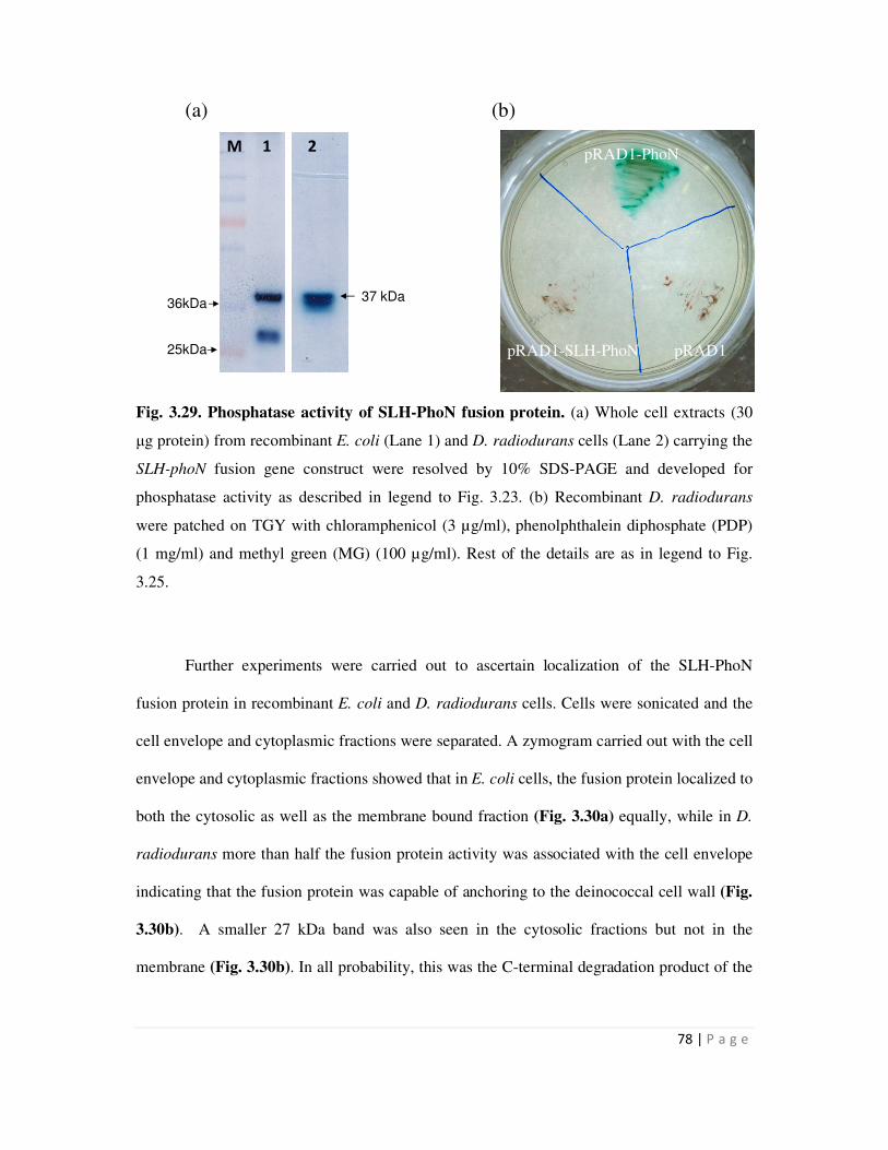
78 | P a g e
1 2 3 4 5 6 7 M
36kDa37 kDa
25kDa
M 1 2 pRAD1-PhoN
pRAD1-SLH-PhoN pRAD1
(a) (b)
Fig. 3.29. Phosphatase activity of SLH-PhoN fusion protein. (a) Whole cell extracts (30
µg protein) from recombinant E. coli (Lane 1) and D. radiodurans cells (Lane 2) carrying the
SLH-phoN fusion gene construct were resolved by 10% SDS-PAGE and developed for
phosphatase activity as described in legend to Fig. 3.23. (b) Recombinant D. radiodurans
were patched on TGY with chloramphenicol (3 µg/ml), phenolphthalein diphosphate (PDP)
(1 mg/ml) and methyl green (MG) (100 µg/ml). Rest of the details are as in legend to Fig.
3.25.
Further experiments were carried out to ascertain localization of the SLH-PhoN
fusion protein in recombinant E. coli and D. radiodurans cells. Cells were sonicated and the
cell envelope and cytoplasmic fractions were separated. A zymogram carried out with the cell
envelope and cytoplasmic fractions showed that in E. coli cells, the fusion protein localized to
both the cytosolic as well as the membrane bound fraction (Fig. 3.30a) equally, while in D.
radiodurans more than half the fusion protein activity was associated with the cell envelope
indicating that the fusion protein was capable of anchoring to the deinococcal cell wall (Fig.
3.30b). A smaller 27 kDa band was also seen in the cytosolic fractions but not in the
membrane (Fig. 3.30b). In all probability, this was the C-terminal degradation product of the
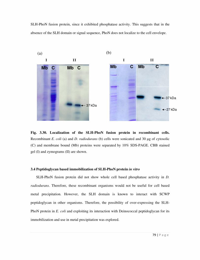
79 | P a g e
Mb C Mb C
37 kDa
I II I II
Mb C Mb C
37 kDa
27 kDa
SLH-PhoN fusion protein, since it exhibited phosphatase activity. This suggests that in the
absence of the SLH domain or signal sequence, PhoN does not localize to the cell envelope.
Fig. 3.30. Localization of the SLH-PhoN fusion protein in recombinant cells.
Recombinant E. coli (a) and D. radiodurans (b) cells were sonicated and 30 µg of cytosolic
(C) and membrane bound (Mb) proteins were separated by 10% SDS-PAGE. CBB stained
gel (I) and zymograms (II) are shown.
3.4 Peptidoglycan based immobilization of SLH-PhoN protein in vitro
SLH-PhoN fusion protein did not show whole cell based phosphatase activity in D.
radiodurans. Therefore, these recombinant organisms would not be useful for cell based
metal precipitation. However, the SLH domain is known to interact with SCWP
peptidoglycan in other organisms. Therefore, the possibility of over-expressing the SLH-
PhoN protein in E. coli and exploiting its interaction with Deinococcal peptidoglycan for its
immobilization and use in metal precipitation was explored.
(a) (b)
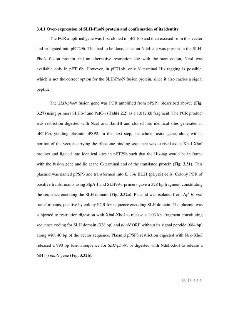
80 | P a g e
3.4.1 Over-expression of SLH-PhoN protein and confirmation of its identity
The PCR amplified gene was first cloned in pET16b and then excised from this vector
and re-ligated into pET29b. This had to be done, since an NdeI site was present in the SLH-
PhoN fusion protein and an alternative restriction site with the start codon, NcoI was
available only in pET16b. However, in pET16b, only N terminal His tagging is possible,
which is not the correct option for the SLH-PhoN fusion protein, since it also carries a signal
peptide.
The SLH-phoN fusion gene was PCR amplified from pPSP1 (described above) (Fig.
3.27) using primers SLHo-f and PetC-r (Table 2.2) as a 1.012 kb fragment. The PCR product
was restriction digested with NcoI and BamHI and cloned into identical sites generated in
pET16b, yielding plasmid pPSP2. In the next step, the whole fusion gene, along with a
portion of the vector carrying the ribosome binding sequence was excised as an XbaI-XhoI
product and ligated into identical sites in pET29b such that the His-tag would be in frame
with the fusion gene and lie at the C-terminal end of the translated protein (Fig. 3.31). This
plasmid was named pPSP3 and transformed into E. coli BL21 (pLysS) cells. Colony PCR of
positive tranformants using SlpA-f and SLH99-r primers gave a 328 bp fragment constituting
the sequence encoding the SLH domain (Fig. 3.32a). Plasmid was isolated from Apr E. coli
transformants, positive by colony PCR for sequence encoding SLH domain. The plasmid was
subjected to restriction digestion with XbaI-XhoI to release a 1.03 kb fragment constituting
sequence coding for SLH domain (328 bp) and phoN ORF without its signal peptide (684 bp)
along with 40 bp of the vector sequence. Plasmid pPSP3 restriction digested with Nco-XhoI
released a 990 bp fusion sequence for SLH-phoN, or digested with NdeI-XhoI to release a
684 bp phoN gene (Fig. 3.32b).
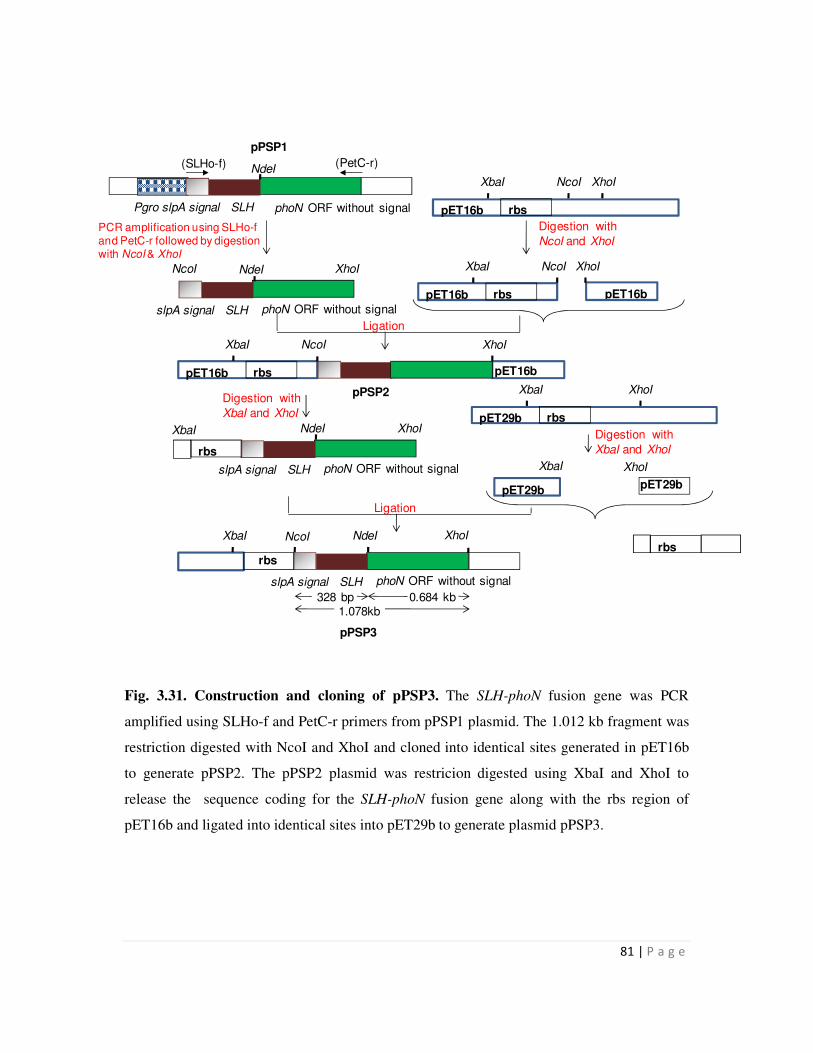
81 | P a g e
phoN ORF without signal Pgro slpA signal SLH
(SLHo-f) (PetC-r)
pET16b rbs
XbaI NcoI XhoI
PCR amplification using SLHo-f and PetC-r followed by digestion with NcoI & XhoI
Digestion with
NcoI and XhoI
pET16b rbs
XbaI NcoI
pET16b
XhoI
pET16b rbs
XbaI NcoI
pET16b
XhoI
pET29b rbs
XbaI XhoI
Digestion with
XbaI and XhoI
rbs
Digestion with
XbaI and XhoI
Ligation
phoN ORF without signal slpA signal SLH
XbaI XhoI
rbs
NdeI
phoN ORF without signal slpA signal SLH
NcoI XhoINdeI
pET29b
XbaI XhoI
pET29b
XbaI
phoN ORF without signal slpA signal SLH
XhoI
rbs
1.078kb 328 bp 0.684 kb
pPSP3
NcoI NdeI
Ligation
pPSP1
NdeI
pPSP2
Fig. 3.31. Construction and cloning of pPSP3. The SLH-phoN fusion gene was PCR
amplified using SLHo-f and PetC-r primers from pPSP1 plasmid. The 1.012 kb fragment was
restriction digested with NcoI and XhoI and cloned into identical sites generated in pET16b
to generate pPSP2. The pPSP2 plasmid was restricion digested using XbaI and XhoI to
release the sequence coding for the SLH-phoN fusion gene along with the rbs region of
pET16b and ligated into identical sites into pET29b to generate plasmid pPSP3.
82 | P a g e
1 2 3 M
1.03kb
990bp
684bp
1kb900bp
700bp
600bp300bp400bp
328bp
1 2 M
Fig. 3.32. Cloning and confirmation of pPSP3. Agarose gel electrophoresis of DNA
fragments. (a) Lanes 1 and 2 display products obtained on carrying out colony PCR of two
transformants with primers, SlpA-f and SLH99-r. (b) Restriction digestion of the plasmid
isolated from one of the Apr transformants positive by colony PCR, with XbaI-Xho (Lane 1),
NcoI-XhoI (Lane 2) and NdeI-XhoI (Lane 3). Lane M contains the DNA molecular weight
marker.
Over-expression of the SLH-PhoN fusion protein was standardized by varying the time and
temperature of incubation of cultures after induction by addition of 1 mM Isopropyl β-D-1-
thiogalactopyranoside (IPTG). Maximum induction of the SLH-PhoN fusion protein,
visualized as a 38 kDa CBB stained band was obtained when the culture was incubated with
IPTG at 30°C for 5 h (Fig. 3.33a). Zymogram of protein extracted from such cells showed
that the fusion protein displayed an active phosphatase visible as a 38 kDa band (Fig. 3.33b).
(a) (b)
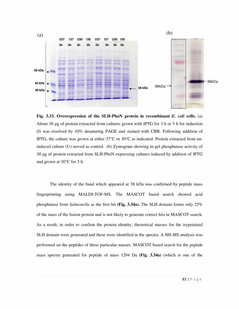
83 | P a g e
38kDa
36kDa
U37 I37 U30 I30 U37 I37 U30 I30
3h 3h 3h 3h 5h 5h 5h 5h
66 kDa
45 kDa
36 kDa38 kDa
Fig. 3.33. Overexpression of the SLH-PhoN protein in recombinant E. coli cells. (a)
About 30 µg of protein extracted from cultures grown with IPTG for 3 h or 5 h for induction
(I) was resolved by 10% denaturing PAGE and stained with CBB. Following addition of
IPTG, the culture was grown at either 37°C or 30°C as indicated. Protein extracted from un-
induced culture (U) served as control. (b) Zymogram showing in-gel phosphatase activity of
30 µg of protein extracted from SLH-PhoN expressing cultures induced by addition of IPTG
and grown at 30°C for 5 h.
The identity of the band which appeared at 38 kDa was confirmed by peptide mass
fingerprinting using MALDI-TOF-MS. The MASCOT based search showed acid
phosphatase from Salmonella as the first hit (Fig. 3.34a). The SLH domain forms only 25%
of the mass of the fusion protein and is not likely to generate correct hits in MASCOT search.
As a result, in order to confirm the protein identity, theoretical masses for the trypsinized
SLH domain were generated and these were identified in the spectra. A MS-MS analysis was
performed on the peptides of these particular masses. MASCOT based search for the peptide
mass spectra generated for peptide of mass 1294 Da (Fig. 3.34a) (which is one of the
(a) (b)

84 | P a g e
1294.665
1011.519
966.435
1670.766
857.431
832.283
1296.684
2228.071
1964.951 2797.236
1496.733
2603.248
1822.764
3299.415
0.00
0.25
0.50
0.75
1.00
1.25
1.50
5x10
Inte
ns. [a
.u.]
1000 1500 2000 2500 3000 3500 4000 4500m/z
peptides that is predicted to be generated due to action of trypsin on the SLH domain), gave
SlpA protein as the first hit (Fig. 3.34b).
Fig. 3.34. MASCOT search result for the mass spectra generated for the 38 kDa protein
over-expressed in E. coli BL21 cells shown in Fig. 3.32. (a) Mass spectrum and the
MASCOT search result for mass spectra of the 38 kDa protein, (b) MASCOT search result
for mass spectra generated by MS-MS for peptide of mass 1294 Da.
(a)
(b)
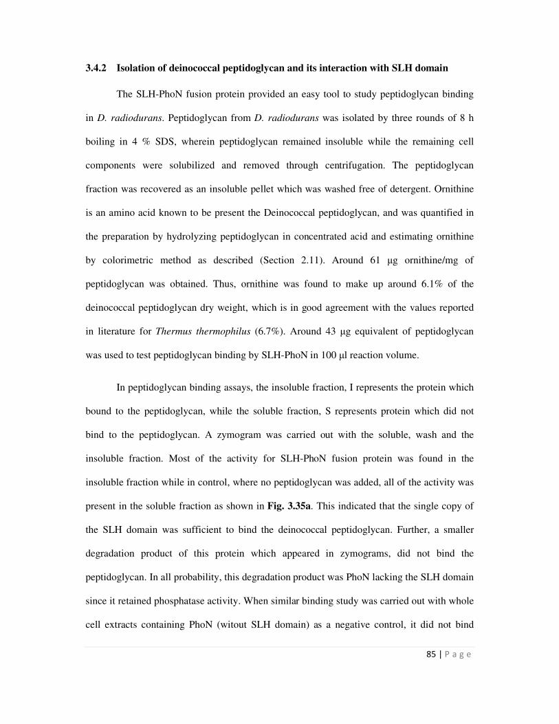
85 | P a g e
3.4.2 Isolation of deinococcal peptidoglycan and its interaction with SLH domain
The SLH-PhoN fusion protein provided an easy tool to study peptidoglycan binding
in D. radiodurans. Peptidoglycan from D. radiodurans was isolated by three rounds of 8 h
boiling in 4 % SDS, wherein peptidoglycan remained insoluble while the remaining cell
components were solubilized and removed through centrifugation. The peptidoglycan
fraction was recovered as an insoluble pellet which was washed free of detergent. Ornithine
is an amino acid known to be present the Deinococcal peptidoglycan, and was quantified in
the preparation by hydrolyzing peptidoglycan in concentrated acid and estimating ornithine
by colorimetric method as described (Section 2.11). Around 61 µg ornithine/mg of
peptidoglycan was obtained. Thus, ornithine was found to make up around 6.1% of the
deinococcal peptidoglycan dry weight, which is in good agreement with the values reported
in literature for Thermus thermophilus (6.7%). Around 43 µg equivalent of peptidoglycan
was used to test peptidoglycan binding by SLH-PhoN in 100 µl reaction volume.
In peptidoglycan binding assays, the insoluble fraction, I represents the protein which
bound to the peptidoglycan, while the soluble fraction, S represents protein which did not
bind to the peptidoglycan. A zymogram was carried out with the soluble, wash and the
insoluble fraction. Most of the activity for SLH-PhoN fusion protein was found in the
insoluble fraction while in control, where no peptidoglycan was added, all of the activity was
present in the soluble fraction as shown in Fig. 3.35a. This indicated that the single copy of
the SLH domain was sufficient to bind the deinococcal peptidoglycan. Further, a smaller
degradation product of this protein which appeared in zymograms, did not bind the
peptidoglycan. In all probability, this degradation product was PhoN lacking the SLH domain
since it retained phosphatase activity. When similar binding study was carried out with whole
cell extracts containing PhoN (witout SLH domain) as a negative control, it did not bind
86 | P a g e
S W I S W I
Control Test Control
38kDa
27kDa
38kDa
27kDa
S I S I
PhoN SLH-PhoN
38kDa
27kDa
peptidoglycan, showing that peptidoglycan binding results exclusively from the SLH domain
(Fig. 3.35b).
Fig. 3.35. Peptidoglycan binding by SLH-PhoN fusion protein. (a) Cell extract from
recombinant E. coli cells expressing SLH-PhoN fusion protein was incubated in the absence
(Control) or presence (Test) of deinococcal peptidoglycan at 4°C following which soluble
fraction (S) was separated by centrifugation. A wash (W) was given and the insoluble
fraction (I) was obtained by a 50°C treatment for 15 min in non-reducing Laemmli’s buffer.
All the fractions were resolved on 10% denaturing gel and developed for phosphatase
activity. (b) Peptidoglycan binding by PhoN and SLH-PhoN protein.
3.5 Discussion
Several strategies are in vogue for surface display of proteins for a variety of
biotechnological and industrial applications. In Gram negative bacteria, proteins for surface
display may be generated by N-terminal, C-terminal, or sandwich fusions to cell wall
anchoring proteins [85]. E. coli has been a preferred organism for most of the studies owing
(a) (b)
87 | P a g e
to its status as a model organism. Peptidoglycan associated lipoprotein (PAL), E. coli
adhesion protein AIDA-1, Shigella Vir-G protein etc. have been used for display of proteins
through N-terminal fusions for vaccine and biosensor development. A popular example for
C-terminal fusion is that of Lpp-OmpA hybrid which contains the first nine N-terminal
residues of mature E. coli lipoprotein and a few residues of the outer membrane porin A
protein. Similar system has also been used for display of SmtA protein and the
organophosphorous hydrolase for bioremediation of metals and pesticides respectively [86-
87].
S layer proteins and outer membrane proteins have been exploited in making
sandwich fusions for surface display of proteins [83,85]. The N terminal domain which
bound to outer membrane and C terminal secretion signal of S layer protein of Caulobacter
crescentus were used to display a 12 amino acid peptide from Pseudomonas aeruoginosa
[85]. In Gram positive bacteria, the anchoring domain of Staphylococcal protein A (SpA)
constituting the LPXTG box has been used for anchoring proteins to cell surface [83].
Besides this, the SLH domain of S layer proteins of Bacillus anthracis and Bacillus
sphaericus have also been used to display tetanus toxins fragments and allergens for
diagnostics [75,80,85].
The status of D. radiodurans as a Gram positive organism is somewhat misleading
because while it stains violet in Gram reaction, its complex cell wall resembles that of Gram
negative organisms [54]. In designing a suitable strategy for surface display in this organism,
the Hpi protein seems to be the protein of choice due to its location as a penultimate layer in
the multi layered cell wall [45]. Since the crystal structure of the protein is not available, the
entire protein was used for generating fusions. Further, since the C terminal of the protein is
known to be exposed to the outer surface [58], the PhoN and SmtA protein were fused to C
88 | P a g e
terminus of the Hpi protein in the present work The other S layer protein, SlpA is a rather
large protein (123 kDa) which is poorly characterized. Therefore, the utility of the small SLH
domain of SlpA protein in membrane anchoring was studied as has been done in some Gram
positive organisms mentioned above. To date, surface display in D. radiodurans has not been
attempted.
As a prelude to this work, some preliminary work to characterize the S layer proteins
of D. radiodurans was carried out. The Hpi protein layer has been under study for several
years. In D. radiodurans Sark strain, isolation of the Hpi layer followed by its electrophoresis
on a denaturing gel yields a single band of ~108 kDa, while in D. radiodurans R1, a 100 kDa
band along with several bands of lower molecular sizes are usually obtained [82]. It has been
speculated that these bands appear due to in vivo proteolysis of the Hpi layer and is
unavoidable even when protease inhibitors are used during its isolation [82]. Two
dimensional peptide fingerprinting was used to determine that all the bands indeed belonged
to Hpi protein. It was also reported that an exonuclease activity was always found to be
present in Hpi preparations [88], and since on SDS gels, a 123 kDa protein was found to co-
purify, the exonuclease activity was assigned to this protein. Further, it has been elaborated
that the Hpi layer forms a 1:1 complex with this exonuclease, and that the six fold symmetry
of Hpi layer subunits is reflected in the exonuclease layer [58], though evidence for the same
has not been provided. No further elaboration on the same exists in literature.
The present study provides further confirmation that the various peptide bands
obtained on resolving the Hpi layer after total denaturation on a denaturing gel are all indeed
discreet degradation products of the Hpi protein as determined by peptide mass fingerprinting
using MALDI-TOF-MS. Further, the 123 kDa protein which came up as a contaminant in all
Hpi preparations was identified to be the deinococcal S layer protein, SlpA (DR2577) based
89 | P a g e
on its homology with the S layer protein from Thermus thermophilus. This study showed, for
the first time that the two S layer proteins, Hpi and SlpA in D. radiodurans seem to be
intimately associated with each other as part of the architecture of the deinococcal cell
envelope. This further supports the popular theory that three-dimensional extension of
symmetry into the pink envelope or at least into its outer membrane may be needed in order
to satisfy the physiological requirements of cell trafficking [89]. Further, it can be concluded
that the nature of the interaction between the Hpi layer and the SlpA protein is non-covalent
in nature since it can be disrupted by SDS treatment at high temperature.
All the fusion genes constructed in this study, hpi-smtA, SLH-smtA, hpi-phoN and
SLH-phoN were actively expressed in D. radiodurans as determined by Western Blot and
zymograms. Interestingly, both the Hpi constructs made, Hpi-SmtA and Hpi-PhoN fusion
proteins were found to be present exclusively in the membrane bound fraction in D.
radiodurans (Fig. 3.19 & 3.26), making this protein an efficient membrane targeting vehicle
for any future work requiring cell surface display in D. radiodurans. The Hpi protein has
been used for patterning of nanoparticles such as gold and CdS/ZnS quantum dots [90], [91].
The results from this work show that the Hpi protein may also be used for surface display of
proteins in D. radiodurans. In contrast, the SLH-PhoN protein was found both in the cytosol
as well as membrane bound fraction of D. radiodurans cells, while SLH-SmtA was found
exclusively in the membrane bound fraction. Therefore, utility of SLH domain in surface
display in this organism is not clear. In many other Gram positive organisms such as
Paenibacillus alvei [78] Bacillus anthracis and Bacillus spahericus, with peptidoglycan as
the major cell wall component, the SLH domain has been used for surface display of proteins
[75]. Broadly, in this study, all the fusion proteins achieved membrane localization as a result
of their fusion to S layer proteins.
90 | P a g e
Attempts at making S layer-PhoN fusions proteins showed that only the Hpi-PhoN
fusion protein showed cell based phosphatase activity in D. radiodurans, while SLH-PhoN
did not display cell-based activity in D. radiodurans cells. The whole cell PhoN activity
obtained in D. radiodurans expressing Hpi-PhoN was only half of that obtained in the earlier
recombinants expressing PhoN alone (Table 3.1). This implied that cell surface display did
not enhance cell-based phosphatase activity of recombinant D. radiodurans ruling out
substrate accessibility as a limiting factor for phosphatase activity. Low expression levels of
Hpi-PhoN protein compared to PhoN in D. radiodurans cells may be a possible reason for
lower activity, but is unlikely since both the proteins were expressed from PgroESL promoter.
Hpi may have interfered with the ability of the enzyme to dimerize effectively or fold
appropriately due to the large size of Hpi protein and therefore, result in lower activity. D.
radiodurans recombinant cells bearing SLH-phoN fusion construct, however failed to show
cell based PhoN activity on histochemical plates or in pNPP assay in solution. But cell
extracts showed activity in zymograms. Perhaps the SLH-PhoN fusion protein is lodged deep
into the cell wall or does not fold appropriately in the cell wall, leading to loss of activity.
The SLH domain present in S layer proteins of many organisms bind to the
underlying peptidoglycan sacculus by interacting with secondary cell wall polymers (SCWP)
attached to the peptidoglycan. Earlier studies carried out in Thermus thermophilus had shown
that single SLH domain found at the N terminus of the SlpA protein bound a cell wall
polymer covalently attached to the peptidoglycan [92]. This polymer was thought to be
present as an envelope of thick layer of unstructured material between the peptidoglycan and
the S-layer – outer membrane complex. Such an uncharacterized layer has also been
visualized in D. radiodurans cells by TEM. An antibody raised against S layer-attached cell
wall fragments of T. thermophilus, αSAC showed cross reactivity with cell walls of D.
91 | P a g e
radiodurans [59]. It was further shown that pyruvylation of the SCWP was an absolute
requirement for SLH domain to bind. The function of adding a pyruvyl group to SCWP was
assigned to a putative pyruvltransferase, CsaB. Further, all organisms whose genome encoded
a SLH domain also harbour csaB gene, including D. radiodurans. Earlier work showed that
SlpA mutants of T. thermophilus resulted in the outer membrane peeling off from the
peptidoglycan layer suggesting that the SLH domain anchors the outer membrane to the
bacterial cell wall [59]. Similar results were reported in SlpA mutant in D. radiodurans [54]
The presence of the SLH domain in SlpA protein in D. radiodurans was strongly
suggestive of a similar mechanism of S layer-cell wall interaction operating in this organism
also. S layer proteins from Gram-positive bacteria typically possess three SLH motifs with
10-15 conserved amino acids [93]. The SLH domain occurs in variable numbers in many
types of proteins of phylogenetically unrelated bacteria [36]. In D. radiodurans, this study
showed that the single SLH domain present in a single copy at the N terminus of SlpA is
sufficient for binding peptidoglycan. The deinococcal cell wall architecture has been an
enigma from the 1980’s. The present study has provided the following two insights into the
cell wall architecture: (1) Hpi layer interacts non-covalently with SlpA, and (2) in turn the
SlpA protein interacts non-covalently with the peptidoglycan, forming continuity in the
interactions between different layers of the deinococcal cell wall.
The possibility of using the peptidoglycan as an immobilization matrix for the PhoN
enzyme demonstrated here is exciting, especially since peptidoglycan itself is known to
adsorb uranium on its functional groups [94]. Further, peptidoglycan is a sturdy biopolymer,
resistant to heat and detergents and is hydrolyzed only with concentrated mineral acids at
high temperature. These virtues make SLH-peptidoglycan interaction a promising tool for
enzyme/protein immobilization.
92 | P a g e
Chapter 4
Heavy metal bioremediation using
recombinant proteins and bacteria
93 | P a g e
The previous chapter described the characterization of the surface layer proteins, Hpi
and SlpA of D. radiodurans and the construction of fusion proteins Hpi-PhoN, SLH-PhoN,
Hpi-SmtA and SLH-SmtA. The expression of fusion proteins and their localization in the
recombinant cells was also assessed to predict their likely utility for metal bioremediation.
This chapter describes the evaluation of the S layer protein, Hpi and recombinant
strains expressing various S layer fusion proteins, for their ability to remove metals from
solution. Since the SmtA protein localized cytoplasmically while the SLH-SmtA and Hpi-
SmtA proteins localized into membrane in recombinant D. radiodurans cells, the effect of
such differential localization of metallothionein on metal binding ability of recombinant cells
has been evaluated. While Hpi-PhoN displayed whole cell phosphatase activity, the SLH-
PhoN fusion protein did not show whole cell phosphatase activity. Therefore, as an
alternative, its utility in metal precipitation after immobilization on peptidoglycan was
assessed. Both the recombinant organisms as well as the fusion proteins isolated from them
were tested for their ability to bind/precipitate heavy metals. In addition, biomass bearing the
PhoN enzyme was subjected to lyophilisation to see if its shelf life could be enhanced. The
cellular localization of precipitated metals was determined by electron microscopy to explore
the possibility of recovery of the metal and re-use of biomass. These results are detailed in
the following sections:
4.1 Metal binding ability of deinococcal S layer protein
4.1.1 Metal binding by isolated Hpi protein
Hpi layer isolated from D. radiodurans was evaluated for its ability to bind uranium
and cadmium. The Hpi protein could bind around 37% of 1 mM input uranium, resulting in a
loading of around 166 µg uranium/mg Hpi protein, while in a similar set up without any
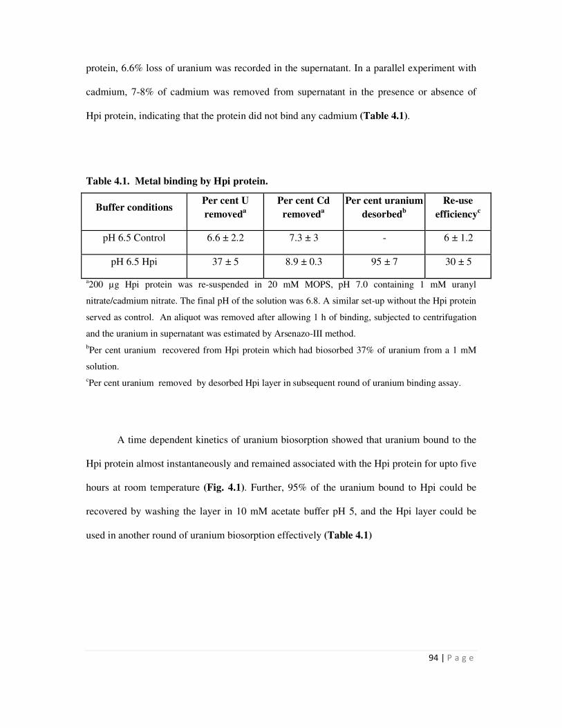
94 | P a g e
protein, 6.6% loss of uranium was recorded in the supernatant. In a parallel experiment with
cadmium, 7-8% of cadmium was removed from supernatant in the presence or absence of
Hpi protein, indicating that the protein did not bind any cadmium (Table 4.1).
Table 4.1. Metal binding by Hpi protein.
Buffer conditions Per cent U
removeda
Per cent Cd
removeda
Per cent uranium
desorbedb
Re-use
efficiencyc
pH 6.5 Control 6.6 ± 2.2 7.3 ± 3 - 6 ± 1.2
pH 6.5 Hpi 37 ± 5 8.9 ± 0.3 95 ± 7 30 ± 5
a200 µg Hpi protein was re-suspended in 20 mM MOPS, pH 7.0 containing 1 mM uranyl
nitrate/cadmium nitrate. The final pH of the solution was 6.8. A similar set-up without the Hpi protein
served as control. An aliquot was removed after allowing 1 h of binding, subjected to centrifugation
and the uranium in supernatant was estimated by Arsenazo-III method.
bPer cent uranium recovered from Hpi protein which had biosorbed 37% of uranium from a 1 mM
solution.
cPer cent uranium removed by desorbed Hpi layer in subsequent round of uranium binding assay.
A time dependent kinetics of uranium biosorption showed that uranium bound to the
Hpi protein almost instantaneously and remained associated with the Hpi protein for upto five
hours at room temperature (Fig. 4.1). Further, 95% of the uranium bound to Hpi could be
recovered by washing the layer in 10 mM acetate buffer pH 5, and the Hpi layer could be
used in another round of uranium biosorption effectively (Table 4.1)
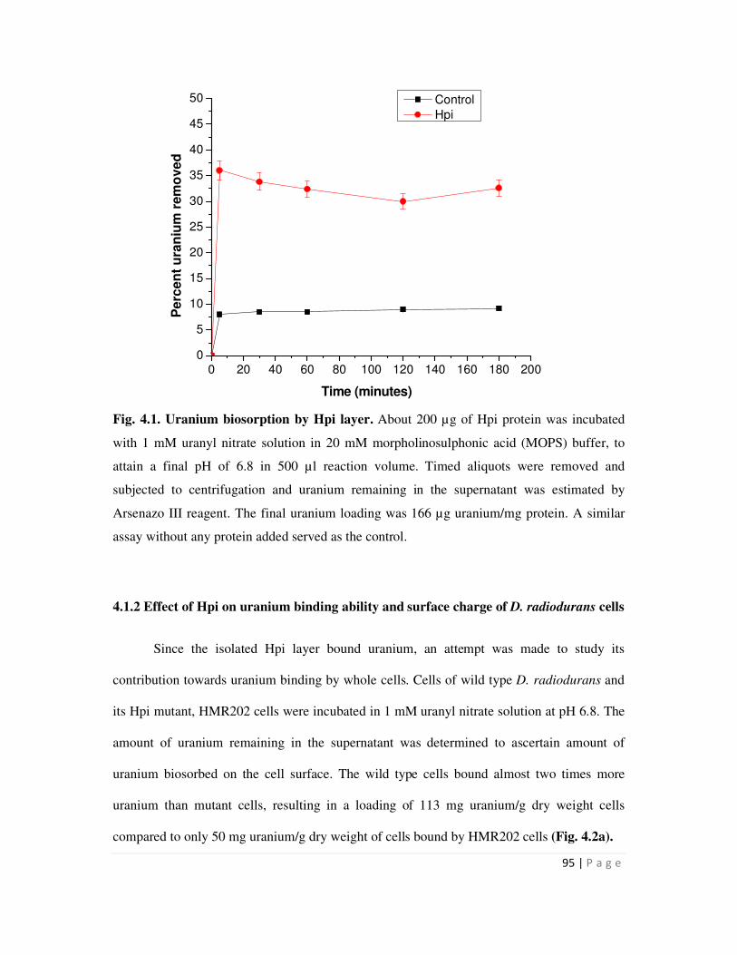
95 | P a g e
0 20 40 60 80 100 120 140 160 180 2000
5
10
15
20
25
30
35
40
45
50
Pe
rce
nt
ura
niu
m r
em
ov
ed
Time (minutes)
Control
Hpi
Fig. 4.1. Uranium biosorption by Hpi layer. About 200 µg of Hpi protein was incubated
with 1 mM uranyl nitrate solution in 20 mM morpholinosulphonic acid (MOPS) buffer, to
attain a final pH of 6.8 in 500 µl reaction volume. Timed aliquots were removed and
subjected to centrifugation and uranium remaining in the supernatant was estimated by
Arsenazo III reagent. The final uranium loading was 166 µg uranium/mg protein. A similar
assay without any protein added served as the control.
4.1.2 Effect of Hpi on uranium binding ability and surface charge of D. radiodurans cells
Since the isolated Hpi layer bound uranium, an attempt was made to study its
contribution towards uranium binding by whole cells. Cells of wild type D. radiodurans and
its Hpi mutant, HMR202 cells were incubated in 1 mM uranyl nitrate solution at pH 6.8. The
amount of uranium remaining in the supernatant was determined to ascertain amount of
uranium biosorbed on the cell surface. The wild type cells bound almost two times more
uranium than mutant cells, resulting in a loading of 113 mg uranium/g dry weight cells
compared to only 50 mg uranium/g dry weight of cells bound by HMR202 cells (Fig. 4.2a).
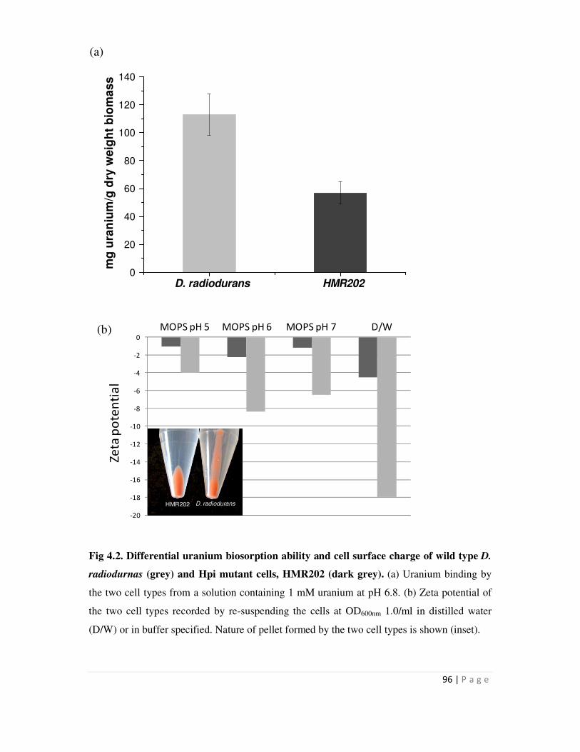
96 | P a g e
D. radiodurans HMR202
0
20
40
60
80
100
120
140
mg
ura
niu
m/g
dry
weig
ht
bio
ma
ss
-20
-18
-16
-14
-12
-10
-8
-6
-4
-2
0
Ze
ta p
ote
nti
al
MOPS pH 5 MOPS pH 6 MOPS pH 7 D/W
HMR202 D. radiodurans
Fig 4.2. Differential uranium biosorption ability and cell surface charge of wild type D.
radiodurnas (grey) and Hpi mutant cells, HMR202 (dark grey). (a) Uranium binding by
the two cell types from a solution containing 1 mM uranium at pH 6.8. (b) Zeta potential of
the two cell types recorded by re-suspending the cells at OD600nm 1.0/ml in distilled water
(D/W) or in buffer specified. Nature of pellet formed by the two cell types is shown (inset).
(a)
(b)
97 | P a g e
Since metal binding is usually a result of electrostatic interactions between functional
groups present on cell surface and the metal, the effect of Hpi on cell surface charge was
investigated by determining the zeta potential on the cell surface of wild type and HMR202
cells using a Zetameter. In distilled water, the zeta potential values of the two cell types
differed widely (wild type cells at -18 mV and HMR202 cells at -4.4 mV). In buffer solutions
also, zeta potential of HMR202 cells was always found to be less negative than that of wild
type cells (Fig. 4.2b). Visually, pellets of HMR202 cells were found to be more compact than
those of wild type cells, perhaps due to lesser cell to cell repulsion on account of their
reduced surface charge (Fig. 4.2b inset). These results indicated that Hpi contributes a net
negative cell surface charge which may have a bearing on metal binding by whole cells.
4.2 Metal binding by recombinant S layer-SmtA fusion proteins
4.2.1 Metal binding by recombinant bacteria expressing SmtA, Hpi-SmtA and SLH-
SmtA proteins
Metal binding studies were carried out using recombinant strains, expressing various
fusion proteins with different metals. The SmtA protein has earlier been shown to bind Cd,
Zn and Cu [14]. Typically, concentrations of the metal, at which binding studies are carried
out, are at or below the Minimum Inhibitory Concentration (MIC) of that metal for the
organism. The MICs of Cd, Zn and Cu for D. radiodurans are 1.8 µM, 150 µM and 470 µM
respectively [95]. Since, the MIC for cadmium is far too low for studying cadmium
biosorption, a concentration of 5 µM was employed for studying Cd biosorption along with
150 µM for Zn and 250 µM for Cu. In subsequent studies, concentration of Cd was raised to
40 µM, though it was known to far exceed the MIC, to test whether even when cells which
98 | P a g e
are unable to grow at the inhibitory concentrations of metals used, they can still continue to
show enhanced metal binding ability.
Cadmium binding studies showed that at 5 µM Cd2+
concentration, recombinant D.
radiodurans cells expressing Hpi-SmtA fusion protein showed marginally higher cadmium
binding than other recombinants (Fig. 4.3a). Further, neither of the recombinant D.
radiodurans cells expressing SLH-SmtA or SmtA showed improved metal binding compared
to D. radiodurans cells expressing only the native Hpi gene or carrying empty vector
pRAD1. At higher cadmium concentration, D. radiodurans expressing Hpi-SmtA showed a
metal loading of 1.2 mg/g dry weight biomass, while all other recombinants showed metal
binding in the range of 0.5 mg/g dry weight biomass (Fig. 4.3b). On the other hand, when
recombinant E. coli cells were used for cadmium biosorption, recombinants expressing SmtA
gene alone showed superior metal binding (1.2 mg/g dry weight biomass) ability compared to
other recombinant strains (0.5 mg/g dry weight biomass). Cadmium bound by recombinant E.
coli cells expressing Hpi-SmtA or SLH-SmtA was in the same range as control cells carrying
pRAD1 alone (Fig. 4.4).
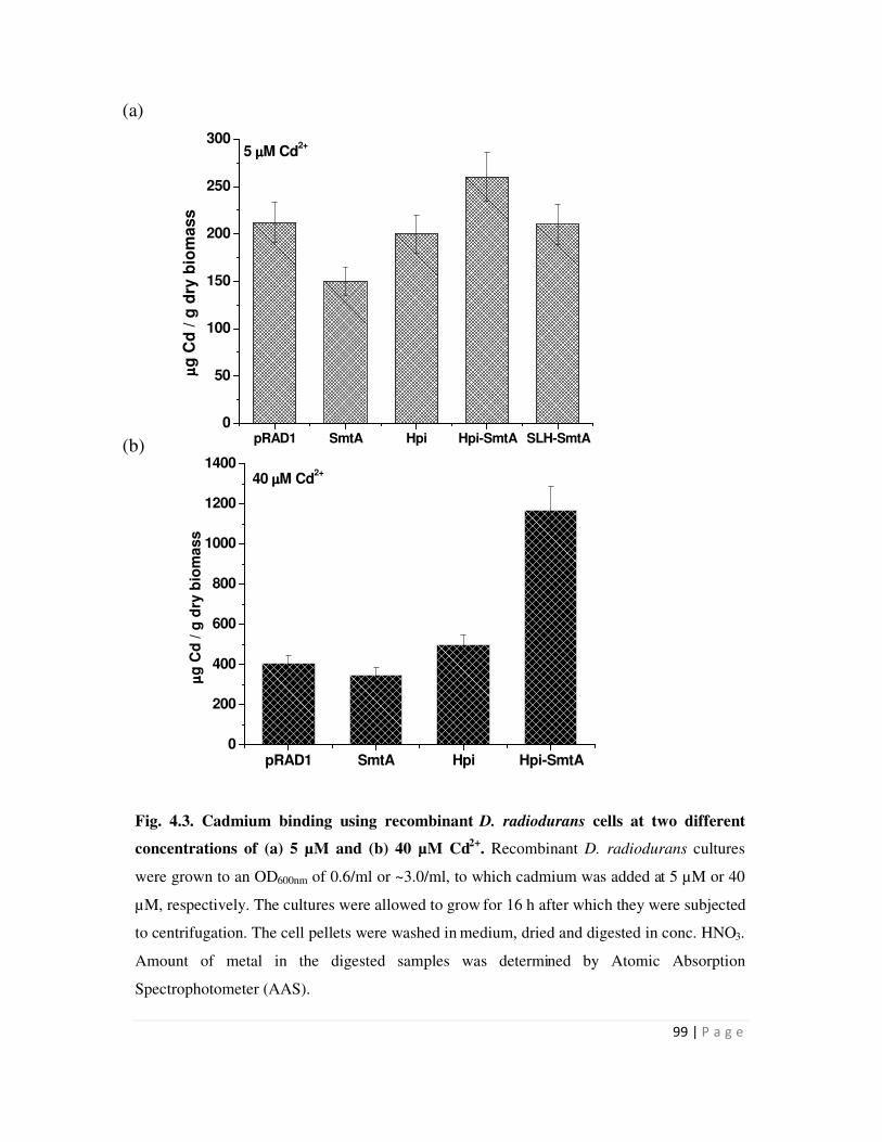
99 | P a g e
pRAD1 SmtA Hpi Hpi-SmtA SLH-SmtA0
50
100
150
200
250
3005 µµµµM Cd
2+
µµ µµg
Cd
/ g
dry
bio
mass
pRAD1 SmtA Hpi Hpi-SmtA0
200
400
600
800
1000
1200
140040 µµµµM Cd
2+
µµ µµg
Cd
/ g
dry
bio
ma
ss
Fig. 4.3. Cadmium binding using recombinant D. radiodurans cells at two different
concentrations of (a) 5 µM and (b) 40 µM Cd2+
. Recombinant D. radiodurans cultures
were grown to an OD600nm of 0.6/ml or ~3.0/ml, to which cadmium was added at 5 µM or 40
µM, respectively. The cultures were allowed to grow for 16 h after which they were subjected
to centrifugation. The cell pellets were washed in medium, dried and digested in conc. HNO3.
Amount of metal in the digested samples was determined by Atomic Absorption
Spectrophotometer (AAS).
(a)
(b)
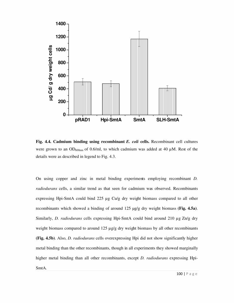
100 | P a g e
pRAD1 Hpi-SmtA SmtA SLH-SmtA0
200
400
600
800
1000
1200
1400
µµ µµg
Cd
/ g
dry
we
igh
t cell
s
Fig. 4.4. Cadmium binding using recombinant E. coli cells. Recombinant cell cultures
were grown to an OD600nm of 0.6/ml, to which cadmium was added at 40 µM. Rest of the
details were as described in legend to Fig. 4.3.
On using copper and zinc in metal binding experiments employing recombinant D.
radiodurans cells, a similar trend as that seen for cadmium was observed. Recombinants
expressing Hpi-SmtA could bind 225 µg Cu/g dry weight biomass compared to all other
recombinants which showed a binding of around 125 µg/g dry weight biomass (Fig. 4.5a).
Similarly, D. radiodurans cells expressing Hpi-SmtA could bind around 210 µg Zn/g dry
weight biomass compared to around 125 µg/g dry weight biomass by all other recombinants
(Fig. 4.5b). Also, D. radiodurans cells overexpressing Hpi did not show significantly higher
metal binding than the other recombinants, though in all experiments they showed marginally
higher metal binding than all other recombinants, except D. radiodurans expressing Hpi-
SmtA.
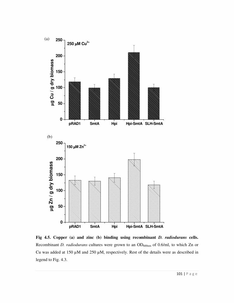
101 | P a g e
Fig 4.5. Copper (a) and zinc (b) binding using recombinant D. radiodurans cells.
Recombinant D. radiodurans cultures were grown to an OD600nm of 0.6/ml, to which Zn or
Cu was added at 150 µM and 250 µM, respectively. Rest of the details were as described in
legend to Fig. 4.3.
(a)
(b)
pRAD1 SmtA Hpi Hpi-SmtA SLH-SmtA0
50
100
150
200
250150 µµµµM Zn
2+
µµ µµg
Zn
/ g
dry
bio
mas
s
pRAD1 SmtA Hpi Hpi-SmtA SLH-SmtA0
50
100
150
200
250 250 µµµµM Cu
2+
µµ µµg
Cu
/ g
dry
bio
ma
ss
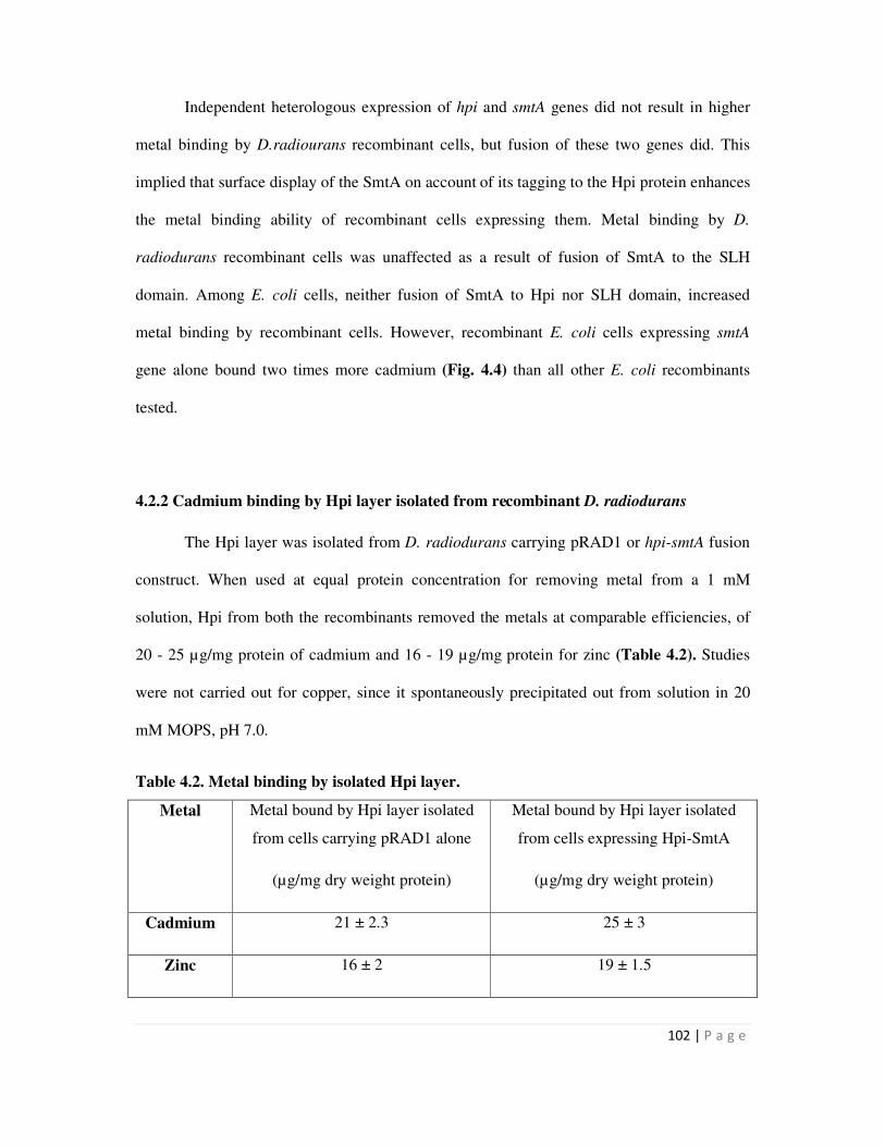
102 | P a g e
Independent heterologous expression of hpi and smtA genes did not result in higher
metal binding by D.radiourans recombinant cells, but fusion of these two genes did. This
implied that surface display of the SmtA on account of its tagging to the Hpi protein enhances
the metal binding ability of recombinant cells expressing them. Metal binding by D.
radiodurans recombinant cells was unaffected as a result of fusion of SmtA to the SLH
domain. Among E. coli cells, neither fusion of SmtA to Hpi nor SLH domain, increased
metal binding by recombinant cells. However, recombinant E. coli cells expressing smtA
gene alone bound two times more cadmium (Fig. 4.4) than all other E. coli recombinants
tested.
4.2.2 Cadmium binding by Hpi layer isolated from recombinant D. radiodurans
The Hpi layer was isolated from D. radiodurans carrying pRAD1 or hpi-smtA fusion
construct. When used at equal protein concentration for removing metal from a 1 mM
solution, Hpi from both the recombinants removed the metals at comparable efficiencies, of
20 - 25 µg/mg protein of cadmium and 16 - 19 µg/mg protein for zinc (Table 4.2). Studies
were not carried out for copper, since it spontaneously precipitated out from solution in 20
mM MOPS, pH 7.0.
Table 4.2. Metal binding by isolated Hpi layer.
Metal Metal bound by Hpi layer isolated
from cells carrying pRAD1 alone
(µg/mg dry weight protein)
Metal bound by Hpi layer isolated
from cells expressing Hpi-SmtA
(µg/mg dry weight protein)
Cadmium 21 ± 2.3 25 ± 3
Zinc 16 ± 2 19 ± 1.5
103 | P a g e
4.3 Metal precipitation by recombinant bacteria over-expressing PhoN
In general, an input metal concentration of 1 mM was used for metal precipitation by
recombinants cells or proteins constructed in this study. The main purpose of this study was
to investigate if metals can be removed at such low concentrations. Assays at this
concentration were used to compare metal precipitation efficiencies of recombinants carrying
these constructs with earlier constructs and among themselves. Values for metal removal
have been given in per cent for ease of comparison between different recombinants and the
loading values have been included in the text.
4.3.1 Cadmium precipitation by recombinant E. coli strain expressing phoN
All earlier studies involving recombinants expressing phoN gene were carried out to
precipitate uranium from solution [51]. Since phosphatase based metal precipitation is
essentially non-specific in nature, any metal can be precipitated using this approach. As
cadmium is a highly toxic metal, the PhoN based approach was evaluated for its precipitation.
The optimum pH for whole cell phosphatase activity as well as for cadmium
precipitation was determined in E. coli cells bearing phoN. While the optimum pH for
phosphatase activity was 5.0, optimum pH for cadmium precipitation was 7.0 (Fig. 4.6). In
addition, the curve for phosphatase activity over various pH was quite broad; in the range of
4-7, with the activity dipping at pH 8 and 9. Cadmium precipitation on the other hand was
optimum in the range of 6.5-7.5. This implied that metal precipitation mediated by this
phosphatase occurred best in the near-neutral range.
At 1 mM concentration, at pH 7, 83% of the cadmium was precipitated in 3 h using 5
mM β-glycerophosphate as substrate and 0.3 OD600nm/ml cells in 5 ml assay (Table 4.3).
Nearly all the cadmium removed from the supernatant was recovered in the cell pellet.
Spontaneous precipitation of cadmium in assay solution and sorption to the container walls, if
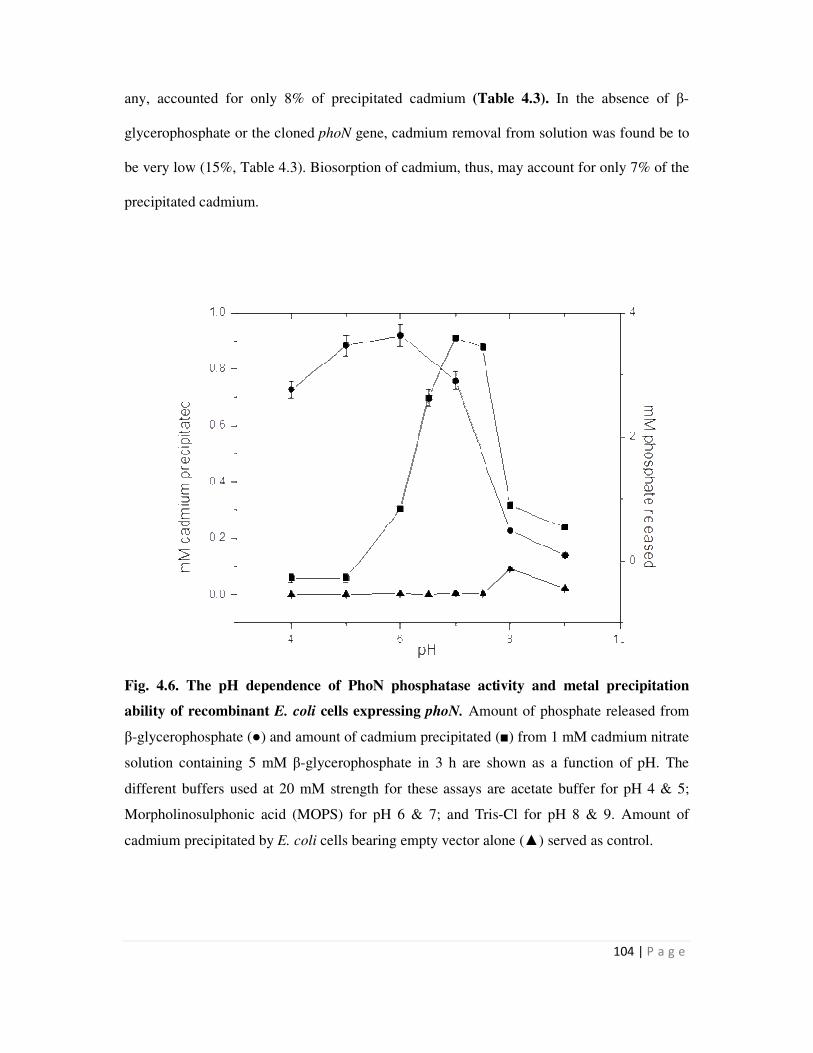
104 | P a g e
any, accounted for only 8% of precipitated cadmium (Table 4.3). In the absence of β-
glycerophosphate or the cloned phoN gene, cadmium removal from solution was found be to
be very low (15%, Table 4.3). Biosorption of cadmium, thus, may account for only 7% of the
precipitated cadmium.
Fig. 4.6. The pH dependence of PhoN phosphatase activity and metal precipitation
ability of recombinant E. coli cells expressing phoN. Amount of phosphate released from
β-glycerophosphate (●) and amount of cadmium precipitated (■) from 1 mM cadmium nitrate
solution containing 5 mM β-glycerophosphate in 3 h are shown as a function of pH. The
different buffers used at 20 mM strength for these assays are acetate buffer for pH 4 & 5;
Morpholinosulphonic acid (MOPS) for pH 6 & 7; and Tris-Cl for pH 8 & 9. Amount of
cadmium precipitated by E. coli cells bearing empty vector alone (▲) served as control.
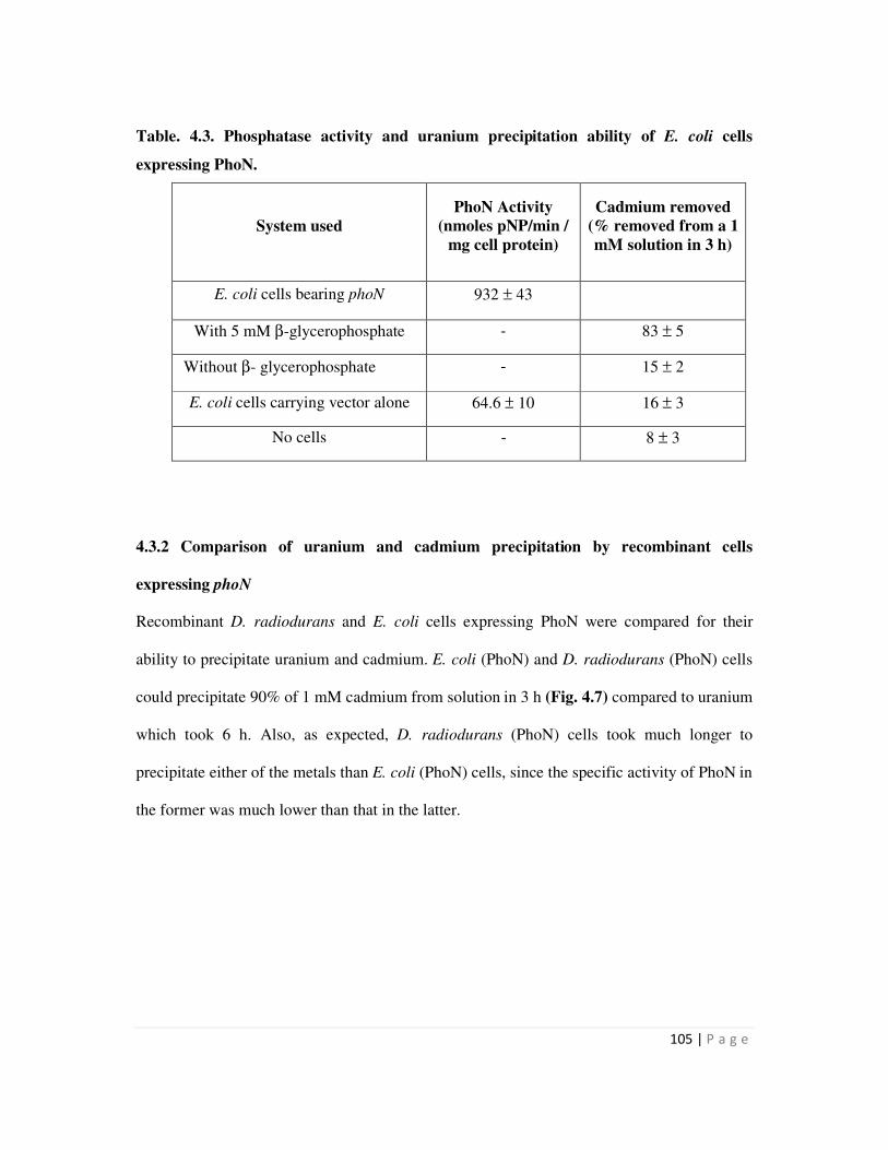
105 | P a g e
Table. 4.3. Phosphatase activity and uranium precipitation ability of E. coli cells
expressing PhoN.
System used
PhoN Activity
(nmoles pNP/min /
mg cell protein)
Cadmium removed
(% removed from a 1
mM solution in 3 h)
E. coli cells bearing phoN 932 ± 43
With 5 mM β-glycerophosphate - 83 ± 5
Without β- glycerophosphate - 15 ± 2
E. coli cells carrying vector alone 64.6 ± 10 16 ± 3
No cells - 8 ± 3
4.3.2 Comparison of uranium and cadmium precipitation by recombinant cells
expressing phoN
Recombinant D. radiodurans and E. coli cells expressing PhoN were compared for their
ability to precipitate uranium and cadmium. E. coli (PhoN) and D. radiodurans (PhoN) cells
could precipitate 90% of 1 mM cadmium from solution in 3 h (Fig. 4.7) compared to uranium
which took 6 h. Also, as expected, D. radiodurans (PhoN) cells took much longer to
precipitate either of the metals than E. coli (PhoN) cells, since the specific activity of PhoN in
the former was much lower than that in the latter.
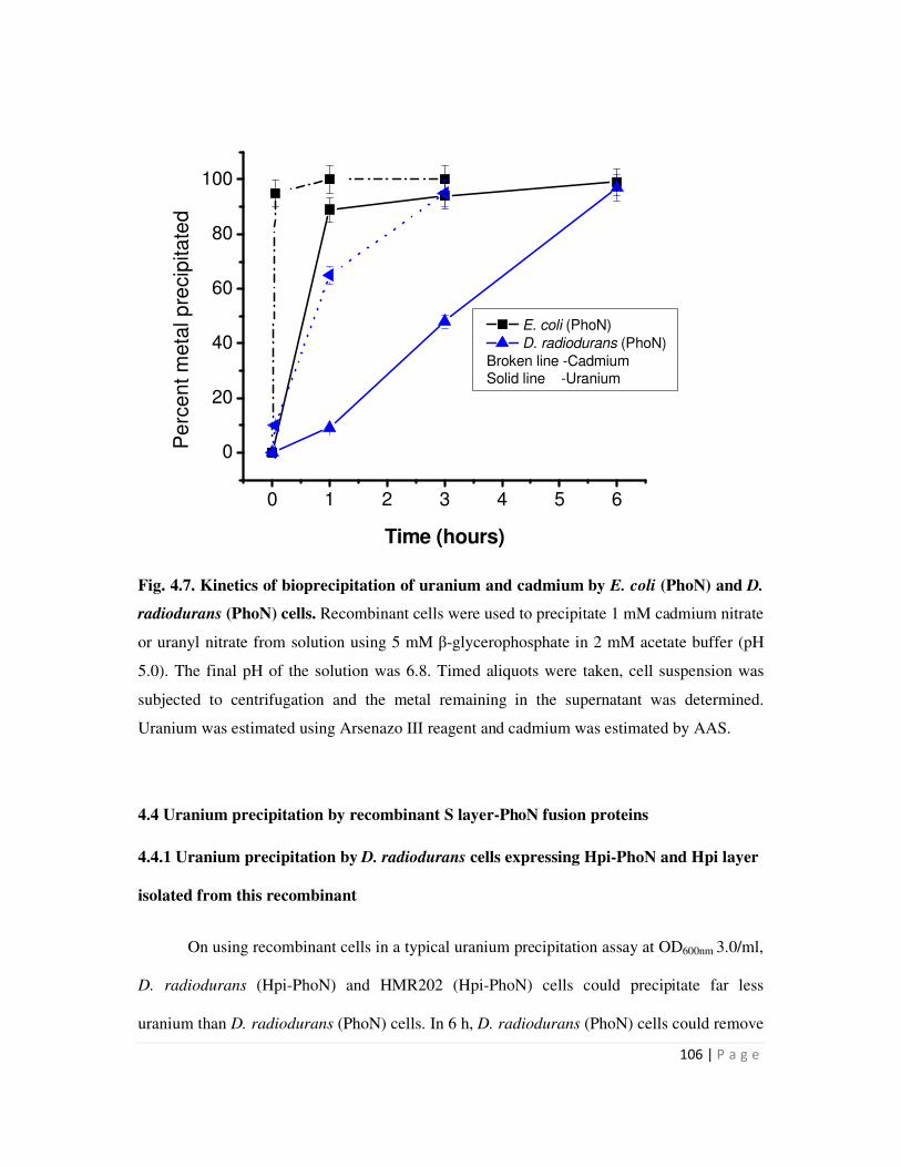
106 | P a g e
Fig. 4.7. Kinetics of bioprecipitation of uranium and cadmium by E. coli (PhoN) and D.
radiodurans (PhoN) cells. Recombinant cells were used to precipitate 1 mM cadmium nitrate
or uranyl nitrate from solution using 5 mM β-glycerophosphate in 2 mM acetate buffer (pH
5.0). The final pH of the solution was 6.8. Timed aliquots were taken, cell suspension was
subjected to centrifugation and the metal remaining in the supernatant was determined.
Uranium was estimated using Arsenazo III reagent and cadmium was estimated by AAS.
4.4 Uranium precipitation by recombinant S layer-PhoN fusion proteins
4.4.1 Uranium precipitation by D. radiodurans cells expressing Hpi-PhoN and Hpi layer
isolated from this recombinant
On using recombinant cells in a typical uranium precipitation assay at OD600nm 3.0/ml,
D. radiodurans (Hpi-PhoN) and HMR202 (Hpi-PhoN) cells could precipitate far less
uranium than D. radiodurans (PhoN) cells. In 6 h, D. radiodurans (PhoN) cells could remove
0 1 2 3 4 5 6
0
20
40
60
80
100P
erc
en
t m
eta
l p
recip
ita
ted
Time (hours)
E. coli (PhoN)
D. radiodurans (PhoN)
Broken line -CadmiumSolid line -Uranium
107 | P a g e
as much as 83% uranium leading to a loading 310 µg U/mg dry weight cells, while Hpi-PhoN
bearing recombinants could remove only 40-50% of input uranium leading to a loading of
136-185 µg U/mg dry weight cells (Fig. 4.8a). However, when the cell densities were
adjusted to equivalent PhoN specific activities in uranium precipitation assays, the uranium
precipitation kinetics were similar. In order to equalize phosphatase activity, higher cell
density of OD600nm 6-6.5/ml were used for recombinants expressing Hpi-PhoN. An
appropriate control of similar cell density of D. radiodurans cells carrying pRAD1 alone was
also included. Such control cells at the higher cell density employed, could remove 25% (94
µg U/mg dry weight cells) uranium from solution which appears to be a result of biosorption
and presence of native phosphatases which contribute to metal precipitation (Fig. 4.8b).
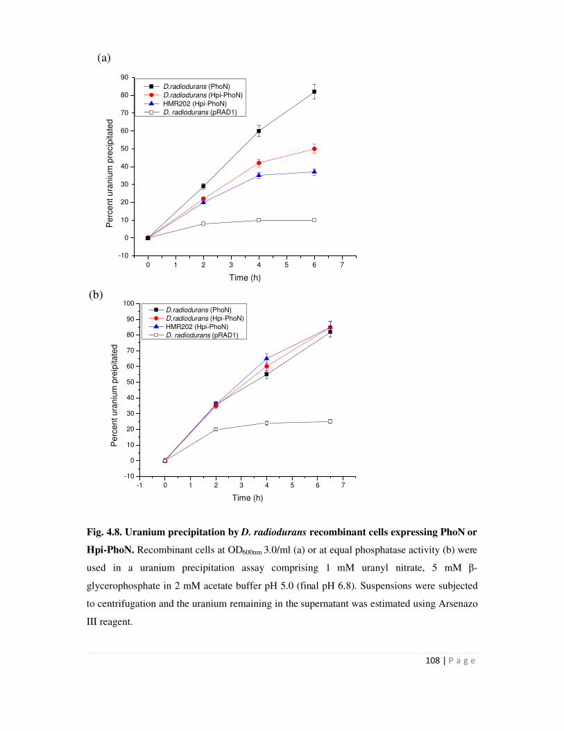
108 | P a g e
0 1 2 3 4 5 6 7
-10
0
10
20
30
40
50
60
70
80
90
Pe
rce
nt u
ran
ium
pre
cip
ita
ted
D.radiodurans (PhoN)
D.radiodurans (Hpi-PhoN)
HMR202 (Hpi-PhoN)
D. radiodurans (pRAD1)
Time (h)
-1 0 1 2 3 4 5 6 7
-10
0
10
20
30
40
50
60
70
80
90
100 D.radiodurans (PhoN)
D.radiodurans (Hpi-PhoN)
HMR202 (Hpi-PhoN)
D. radiodurans (pRAD1)
Pe
rce
nt u
ran
ium
pre
ipita
ted
Time (h)
Fig. 4.8. Uranium precipitation by D. radiodurans recombinant cells expressing PhoN or
Hpi-PhoN. Recombinant cells at OD600nm 3.0/ml (a) or at equal phosphatase activity (b) were
used in a uranium precipitation assay comprising 1 mM uranyl nitrate, 5 mM β-
glycerophosphate in 2 mM acetate buffer pH 5.0 (final pH 6.8). Suspensions were subjected
to centrifugation and the uranium remaining in the supernatant was estimated using Arsenazo
III reagent.
(b)
(a)

109 | P a g e
7 h 24 h0
10
20
30
40
50
60
70
80 pRAD1
pRAD1-Hpi-PhoN
Per
ce
nt
ura
niu
m r
em
oved
To evaluate whether the Hpi layer isolated from recombinants expressing Hpi-PhoN
fusion protein could also precipitate uranium from solutions, equal amount of isolated Hpi
protein (400 ug/ml) from D. radiodurans (pRAD1) and D. radiodurans (Hpi-PhoN) cells
were used in a typical uranium precipitation assay. As seen, Hpi layer itself from pRAD1
cells could remove around 39% of 1 mM uranium (208 µg U/mg protein), while that from
Hpi-PhoN expressing cells could remove 75% of 1 mM uranium in 24 h (446 µg U/mg
protein) (Fig. 4.9). However, it was observed that when the Hpi layer isolated from Hpi-
PhoN expressing cells was extensively washed in water during preparation, it lost
phosphatase activity and showed uranium removal comparable to that from cells bearing
pRAD1 alone. This seemed to indicate that the Hpi-PhoN was not firmly anchored in the Hpi
layer.
Fig. 4.9. Uranium precipitation by Hpi isolated from recombinant D. radiodurans cells.
Hpi protein isolated from recombinant D. radiodurans cells were re-suspended at 400 µg/ml
concentration in a solution containing 1 mM uranyl nitrate, 5 mM β-gycerophosphate and 2
mM acetate buffer, pH 5.0 (final pH 6.8). Suspensions were subjected to centrifugation and
the uranium remaining in the supernatant was estimated using Arsenazo III reagent.

110 | P a g e
4.4.2 Uranium precipitation by SLH-PhoN protein immobilized on peptidoglycan
The utility of SLH-PhoN recombinant protein immobilized on peptidoglycan, for
precipitation of uranium was evaluated. Since the interaction between the protein and
peptidoglycan is predominantly non-covalent in nature, an attempt to stabilize this interaction
was made by treatment with glutaraldehyde.
SLH-PhoN immobilized on peptidoglycan (SPhoNP) was treated with 0.2%
glutaraldehyde at 4°C in 100 mM acetate buffer for one hour and washed in acetate buffer
twice. Studies on uranium precipitation by glutaraldehyde treated and un-treated SPhoNP
were done by re-suspending it in a solution containing 1 mM uranyl nitrate and 5 mM β-
glycerophosphate in 2 mM acetate buffer. At the end of 6 h, the peptidoglycan was re-
suspended in fresh metal solution and this was repeated for around 6 cycles to evaluate the
stability of SPhoNP
SPhoNP could remove 95% uranium from a 1 mM uranyl nitrate solution in ~4 h.
SPhoNP which was not treated with glutaraldehyde retained uranium precipitation ability for
three cycles, after which the amount of uranium precipitated in 4 h fell to 60% in the fourth
cycle and 50% in the fifth cycle. SPhoNP treated with glutaraldehyde, however retained
uranium precipitation ability for five cycles to display a total loading of around 160 µg U/mg
dry biomass weight (Fig. 4.10). However, after the 5th
cycle even glutaraldehyde treated
SPhoNP showed decreased uranium removal. Peptidoglycan alone, on the other hand, could
remove around 20% uranium in the first cycle of metal precipitation assay, after which it did
not bind any uranium in subsequent cycles.
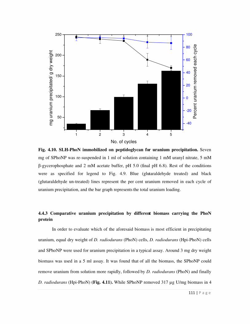
111 | P a g e
1 2 3 4 5
50
100
150
200
250
No. of cycles
mg
ura
niu
m p
recip
ita
ted
/ g d
ry w
eig
ht
-40
-20
0
20
40
60
80
100
Perc
en
t ura
niu
m r
em
oved
ea
ch c
ycle
Fig. 4.10. SLH-PhoN immobilized on peptidoglycan for uranium precipitation. Seven
mg of SPhoNP was re-suspended in 1 ml of solution containing 1 mM uranyl nitrate, 5 mM
β-gycerophosphate and 2 mM acetate buffer, pH 5.0 (final pH 6.8). Rest of the conditions
were as specified for legend to Fig. 4.9. Blue (glutaraldehyde treated) and black
(glutaraldehyde un-treated) lines represent the per cent uranium removed in each cycle of
uranium precipitation, and the bar graph represents the total uranium loading.
4.4.3 Comparative uranium precipitation by different biomass carrying the PhoN
protein
In order to evaluate which of the aforesaid biomass is most efficient in precipitating
uranium, equal dry weight of D. radiodurans (PhoN) cells, D. radiodurans (Hpi-PhoN) cells
and SPhoNP were used for uranium precipitation in a typical assay. Around 3 mg dry weight
biomass was used in a 5 ml assay. It was found that of all the biomass, the SPhoNP could
remove uranium from solution more rapidly, followed by D. radiodurans (PhoN) and finally
D. radiodurans (Hpi-PhoN) (Fig. 4.11). While SPhoNP removed 317 µg U/mg biomass in 4
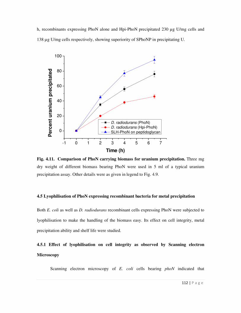
112 | P a g e
h, recombinants expressing PhoN alone and Hpi-PhoN precipitated 230 µg U/mg cells and
138 µg U/mg cells respectively, showing superiority of SPhoNP in precipitating U.
Fig. 4.11. Comparison of PhoN carrying biomass for uranium precipitation. Three mg
dry weight of different biomass bearing PhoN were used in 5 ml of a typical uranium
precipitation assay. Other details were as given in legend to Fig. 4.9.
4.5 Lyophilisation of PhoN expressing recombinant bacteria for metal precipitation
Both E. coli as well as D. radiodurans recombinant cells expressing PhoN were subjected to
lyophilisation to make the handling of the biomass easy. Its effect on cell integrity, metal
precipitation ability and shelf life were studied.
4.5.1 Effect of lyophilisation on cell integrity as observed by Scanning electron
Microscopy
Scanning electron microscopy of E. coli cells bearing phoN indicated that
-1 0 1 2 3 4 5 6 7
0
20
40
60
80
100
Pe
rcen
t u
ran
ium
pre
cip
itate
d
Time (h)
D. radiodurans (PhoN)
D. radiodurans (Hpi-PhoN)
SLH-PhoN on peptidoglycan
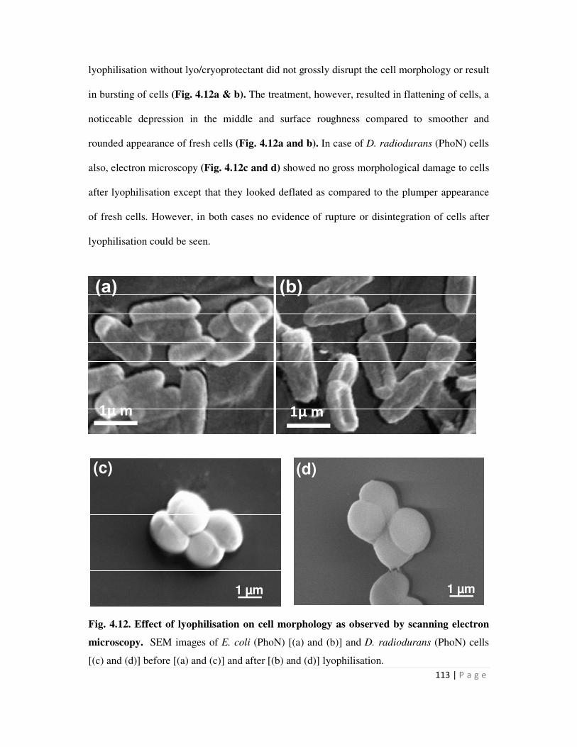
113 | P a g e
lyophilisation without lyo/cryoprotectant did not grossly disrupt the cell morphology or result
in bursting of cells (Fig. 4.12a & b). The treatment, however, resulted in flattening of cells, a
noticeable depression in the middle and surface roughness compared to smoother and
rounded appearance of fresh cells (Fig. 4.12a and b). In case of D. radiodurans (PhoN) cells
also, electron microscopy (Fig. 4.12c and d) showed no gross morphological damage to cells
after lyophilisation except that they looked deflated as compared to the plumper appearance
of fresh cells. However, in both cases no evidence of rupture or disintegration of cells after
lyophilisation could be seen.
Fig. 4.12. Effect of lyophilisation on cell morphology as observed by scanning electron
microscopy. SEM images of E. coli (PhoN) [(a) and (b)] and D. radiodurans (PhoN) cells
[(c) and (d)] before [(a) and (c)] and after [(b) and (d)] lyophilisation.
1µ m 1µ m
1 µm 1 µm
(c) (d)
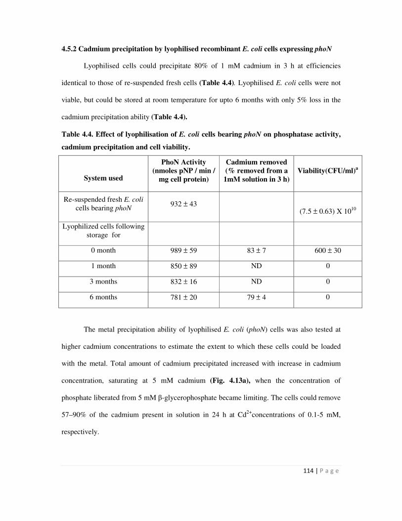
114 | P a g e
4.5.2 Cadmium precipitation by lyophilised recombinant E. coli cells expressing phoN
Lyophilised cells could precipitate 80% of 1 mM cadmium in 3 h at efficiencies
identical to those of re-suspended fresh cells (Table 4.4). Lyophilised E. coli cells were not
viable, but could be stored at room temperature for upto 6 months with only 5% loss in the
cadmium precipitation ability (Table 4.4).
Table 4.4. Effect of lyophilisation of E. coli cells bearing phoN on phosphatase activity,
cadmium precipitation and cell viability.
System used
PhoN Activity
(nmoles pNP / min /
mg cell protein)
Cadmium removed
(% removed from a
1mM solution in 3 h)
Viability(CFU/ml)a
Re-suspended fresh E. coli
cells bearing phoN 932 ± 43
(7.5 ± 0.63) X 1010
Lyophilized cells following
storage for
0 month 989 ± 59 83 ± 7 600 ± 30
1 month 850 ± 89 ND 0
3 months 832 ± 16 ND 0
6 months 781 ± 20 79 ± 4 0
The metal precipitation ability of lyophilised E. coli (phoN) cells was also tested at
higher cadmium concentrations to estimate the extent to which these cells could be loaded
with the metal. Total amount of cadmium precipitated increased with increase in cadmium
concentration, saturating at 5 mM cadmium (Fig. 4.13a), when the concentration of
phosphate liberated from 5 mM β-glycerophosphate became limiting. The cells could remove
57–90% of the cadmium present in solution in 24 h at Cd2+
concentrations of 0.1-5 mM,
respectively.
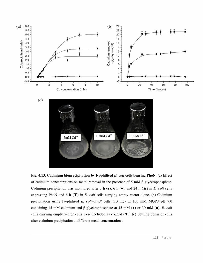
115 | P a g e
Fig. 4.13. Cadmium bioprecipitation by lyophilised E. coli cells bearing PhoN. (a) Effect
of cadmium concentrations on metal removal in the presence of 5 mM β-glycerophosphate.
Cadmium precipitation was monitored after 3 h (■), 6 h (●), and 24 h (▲) in E. coli cells
expressing PhoN and 6 h (▼) in E. coli cells carrying empty vector alone. (b) Cadmium
precipitation using lyophilised E. coli–phoN cells (10 mg) in 100 mM MOPS pH 7.0
containing 15 mM cadmium and β-glycerophosphate at 15 mM (●) or 30 mM (■). E. coli
cells carrying empty vector cells were included as control (▼). (c) Settling down of cells
after cadmium precipitation at different metal concentrations.
5mM Cd2+ 10mM Cd
2+ 15mMCd
2+
(c)
(b) (a)
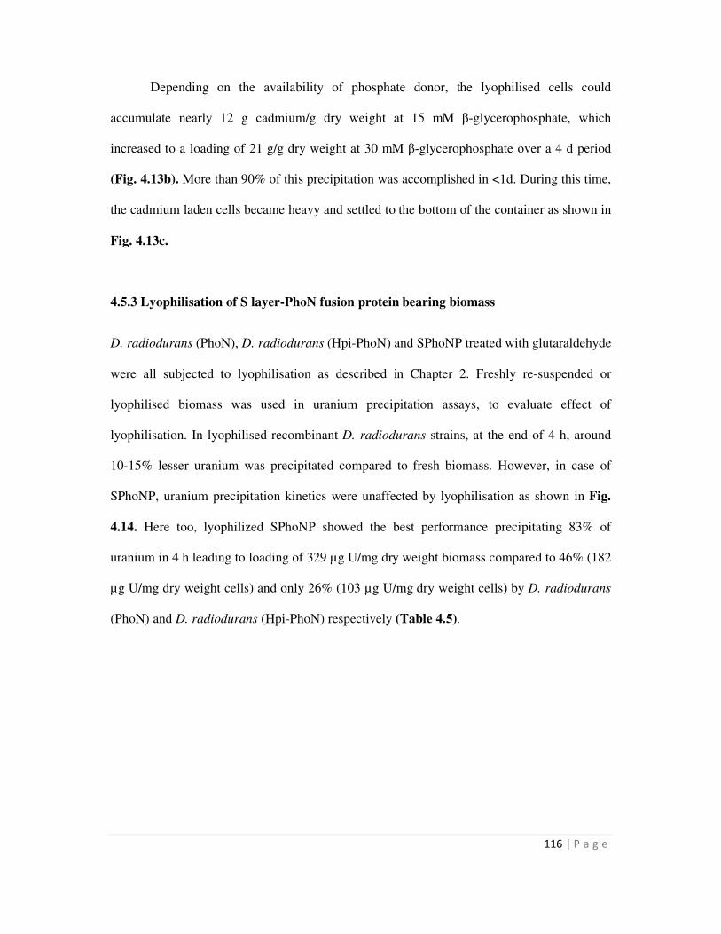
116 | P a g e
Depending on the availability of phosphate donor, the lyophilised cells could
accumulate nearly 12 g cadmium/g dry weight at 15 mM β-glycerophosphate, which
increased to a loading of 21 g/g dry weight at 30 mM β-glycerophosphate over a 4 d period
(Fig. 4.13b). More than 90% of this precipitation was accomplished in <1d. During this time,
the cadmium laden cells became heavy and settled to the bottom of the container as shown in
Fig. 4.13c.
4.5.3 Lyophilisation of S layer-PhoN fusion protein bearing biomass
D. radiodurans (PhoN), D. radiodurans (Hpi-PhoN) and SPhoNP treated with glutaraldehyde
were all subjected to lyophilisation as described in Chapter 2. Freshly re-suspended or
lyophilised biomass was used in uranium precipitation assays, to evaluate effect of
lyophilisation. In lyophilised recombinant D. radiodurans strains, at the end of 4 h, around
10-15% lesser uranium was precipitated compared to fresh biomass. However, in case of
SPhoNP, uranium precipitation kinetics were unaffected by lyophilisation as shown in Fig.
4.14. Here too, lyophilized SPhoNP showed the best performance precipitating 83% of
uranium in 4 h leading to loading of 329 µg U/mg dry weight biomass compared to 46% (182
µg U/mg dry weight cells) and only 26% (103 µg U/mg dry weight cells) by D. radiodurans
(PhoN) and D. radiodurans (Hpi-PhoN) respectively (Table 4.5).

117 | P a g e
Fig. 4.14. Effect of lyophilisation on uranium precipitation by SPhoNP. Around 7 mg dry
weight/ml of SPhoNP was used in a typical uranium precipitation assay containing 1 mM
uranyl nitrate and 5 mM β-glycerophosphate in 2 mM acetate buffer before and after
lyophilisation. Rest of the details were as described in legend to Fig. 4.9.
Table 4.5. Effect of lyophilisation on uranium precipitation
D. radiodurans (PhoN) D. radiodurans (Hpi-PhoN) SPhoNP U precipitated by fresh
biomass 55 ± 4 38 ± 6 85 ± 8
U precipitated by
lyophilised biomass 46 ± 2 26 ± 3 83 ± 6
All values refer to the amount of uranium precipitated in 4 h by 3 mg of biomass in a 5 ml
typical metal precipitation assay.
4.6 Cell Surface localization of metal phosphate precipitates
4.6.1 Electron microscopy to visualize surface association of metal precipitate
Lyophilized cells, which had precipitated all of 1 mM cadmium did not look markedly
different from cells which had not precipitated any cadmium (Fig. 4.15 a & b), but had a
0 1 2 3 4 5 6
0
20
40
60
80
100
Pe
rcen
t u
ran
ium
pre
cip
itati
on
Time (h)
SLH-PhoN
SLH-PhoN lyophilized
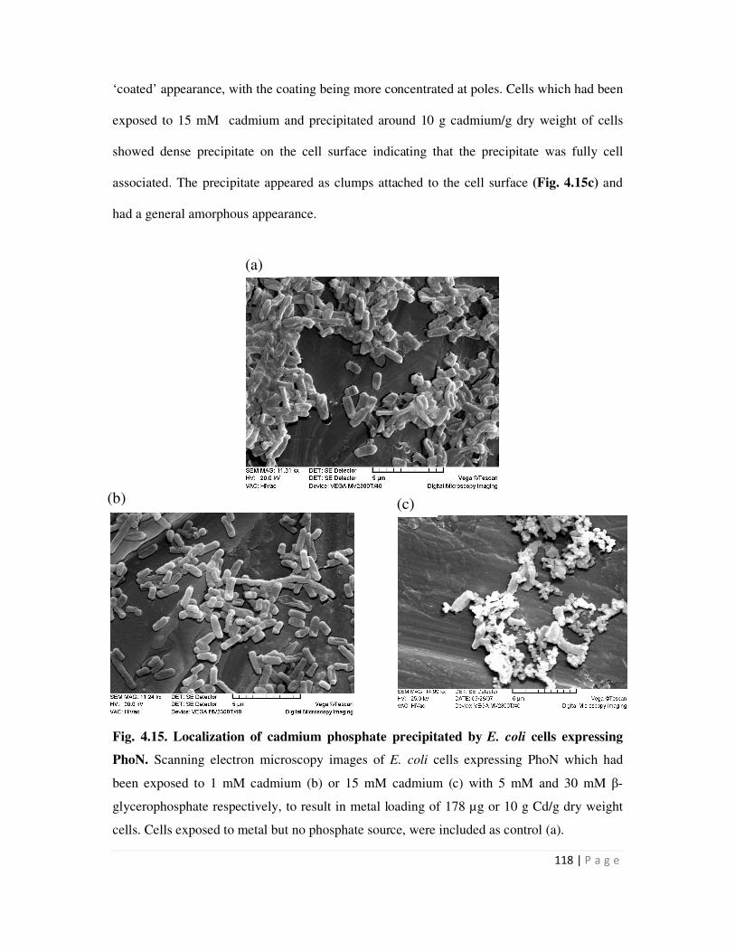
118 | P a g e
‘coated’ appearance, with the coating being more concentrated at poles. Cells which had been
exposed to 15 mM cadmium and precipitated around 10 g cadmium/g dry weight of cells
showed dense precipitate on the cell surface indicating that the precipitate was fully cell
associated. The precipitate appeared as clumps attached to the cell surface (Fig. 4.15c) and
had a general amorphous appearance.
Fig. 4.15. Localization of cadmium phosphate precipitated by E. coli cells expressing
PhoN. Scanning electron microscopy images of E. coli cells expressing PhoN which had
been exposed to 1 mM cadmium (b) or 15 mM cadmium (c) with 5 mM and 30 mM β-
glycerophosphate respectively, to result in metal loading of 178 µg or 10 g Cd/g dry weight
cells. Cells exposed to metal but no phosphate source, were included as control (a).
(a)
(b) (c)
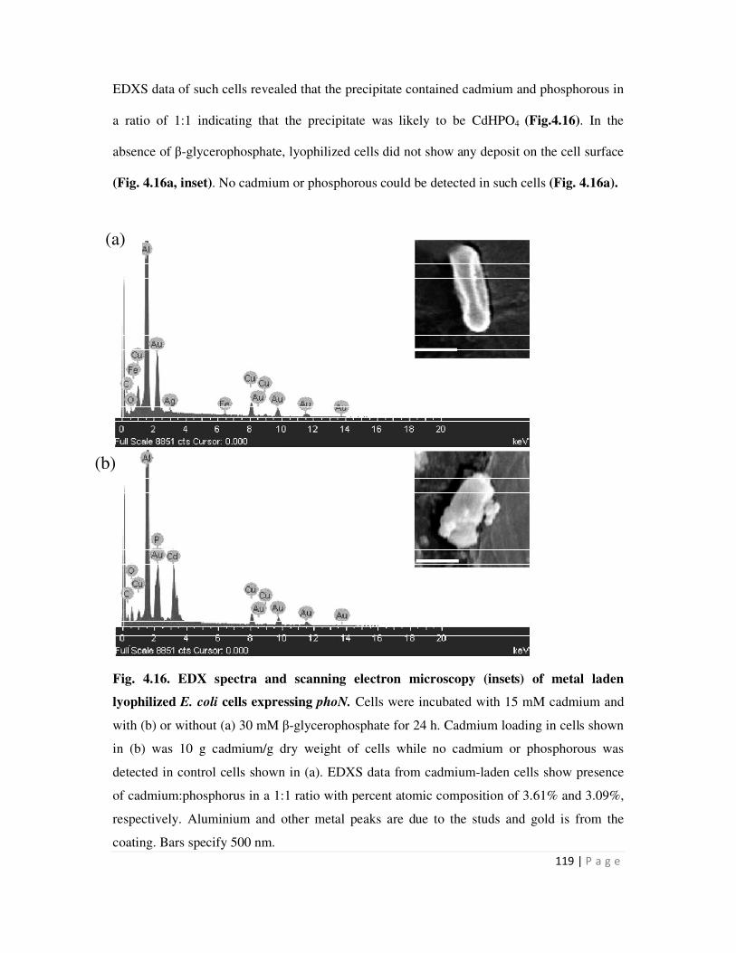
119 | P a g e
EDXS data of such cells revealed that the precipitate contained cadmium and phosphorous in
a ratio of 1:1 indicating that the precipitate was likely to be CdHPO4 (Fig.4.16). In the
absence of β-glycerophosphate, lyophilized cells did not show any deposit on the cell surface
(Fig. 4.16a, inset). No cadmium or phosphorous could be detected in such cells (Fig. 4.16a).
Fig. 4.16. EDX spectra and scanning electron microscopy (insets) of metal laden
lyophilized E. coli cells expressing phoN. Cells were incubated with 15 mM cadmium and
with (b) or without (a) 30 mM β-glycerophosphate for 24 h. Cadmium loading in cells shown
in (b) was 10 g cadmium/g dry weight of cells while no cadmium or phosphorous was
detected in control cells shown in (a). EDXS data from cadmium-laden cells show presence
of cadmium:phosphorus in a 1:1 ratio with percent atomic composition of 3.61% and 3.09%,
respectively. Aluminium and other metal peaks are due to the studs and gold is from the
coating. Bars specify 500 nm.
(a)
(b)
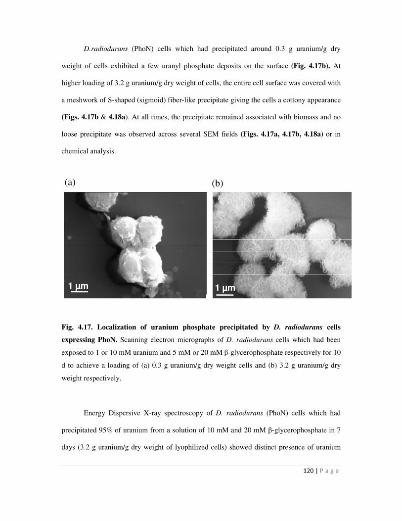
120 | P a g e
D.radiodurans (PhoN) cells which had precipitated around 0.3 g uranium/g dry
weight of cells exhibited a few uranyl phosphate deposits on the surface (Fig. 4.17b). At
higher loading of 3.2 g uranium/g dry weight of cells, the entire cell surface was covered with
a meshwork of S-shaped (sigmoid) fiber-like precipitate giving the cells a cottony appearance
(Figs. 4.17b & 4.18a). At all times, the precipitate remained associated with biomass and no
loose precipitate was observed across several SEM fields (Figs. 4.17a, 4.17b, 4.18a) or in
chemical analysis.
Fig. 4.17. Localization of uranium phosphate precipitated by D. radiodurans cells
expressing PhoN. Scanning electron micrographs of D. radiodurans cells which had been
exposed to 1 or 10 mM uranium and 5 mM or 20 mM β-glycerophosphate respectively for 10
d to achieve a loading of (a) 0.3 g uranium/g dry weight cells and (b) 3.2 g uranium/g dry
weight respectively.
Energy Dispersive X-ray spectroscopy of D. radiodurans (PhoN) cells which had
precipitated 95% of uranium from a solution of 10 mM and 20 mM β-glycerophosphate in 7
days (3.2 g uranium/g dry weight of lyophilized cells) showed distinct presence of uranium
1 µm1 µm
(a) (b)
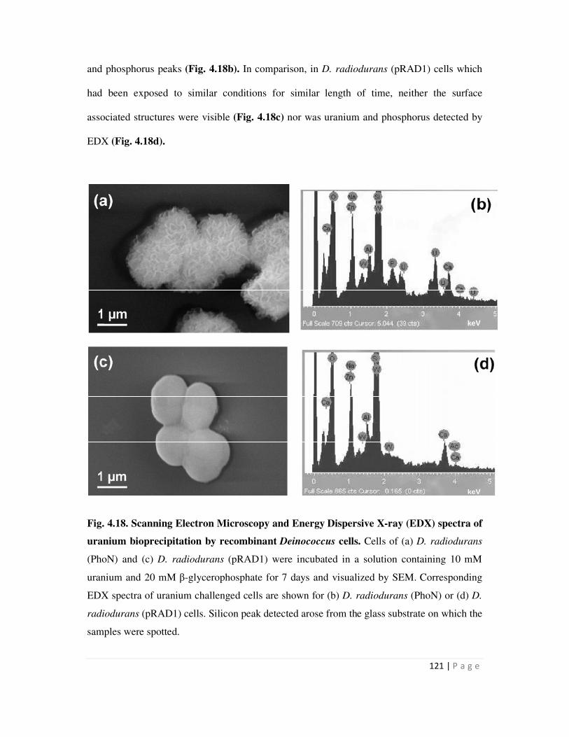
121 | P a g e
and phosphorus peaks (Fig. 4.18b). In comparison, in D. radiodurans (pRAD1) cells which
had been exposed to similar conditions for similar length of time, neither the surface
associated structures were visible (Fig. 4.18c) nor was uranium and phosphorus detected by
EDX (Fig. 4.18d).
Fig. 4.18. Scanning Electron Microscopy and Energy Dispersive X-ray (EDX) spectra of
uranium bioprecipitation by recombinant Deinococcus cells. Cells of (a) D. radiodurans
(PhoN) and (c) D. radiodurans (pRAD1) were incubated in a solution containing 10 mM
uranium and 20 mM β-glycerophosphate for 7 days and visualized by SEM. Corresponding
EDX spectra of uranium challenged cells are shown for (b) D. radiodurans (PhoN) or (d) D.
radiodurans (pRAD1) cells. Silicon peak detected arose from the glass substrate on which the
samples were spotted.
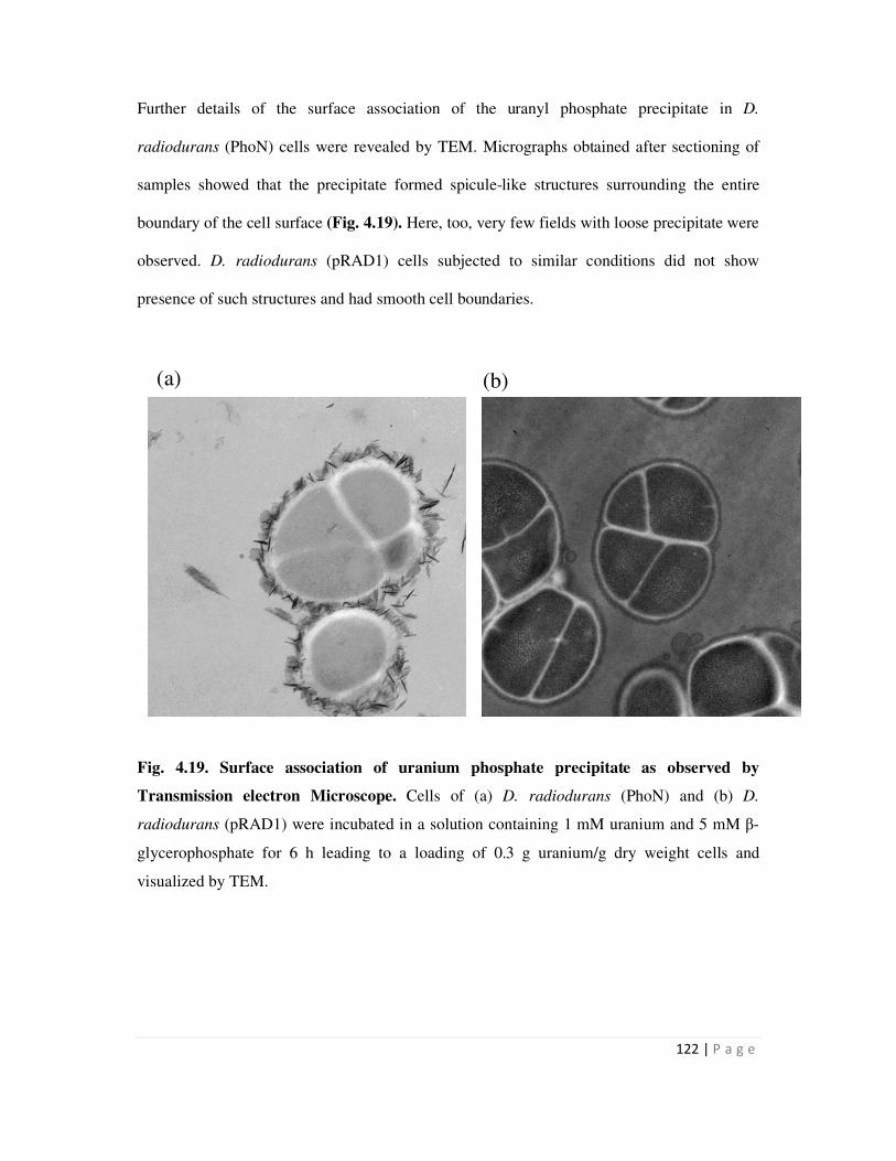
122 | P a g e
Further details of the surface association of the uranyl phosphate precipitate in D.
radiodurans (PhoN) cells were revealed by TEM. Micrographs obtained after sectioning of
samples showed that the precipitate formed spicule-like structures surrounding the entire
boundary of the cell surface (Fig. 4.19). Here, too, very few fields with loose precipitate were
observed. D. radiodurans (pRAD1) cells subjected to similar conditions did not show
presence of such structures and had smooth cell boundaries.
Fig. 4.19. Surface association of uranium phosphate precipitate as observed by
Transmission electron Microscope. Cells of (a) D. radiodurans (PhoN) and (b) D.
radiodurans (pRAD1) were incubated in a solution containing 1 mM uranium and 5 mM β-
glycerophosphate for 6 h leading to a loading of 0.3 g uranium/g dry weight cells and
visualized by TEM.
(a) (b)
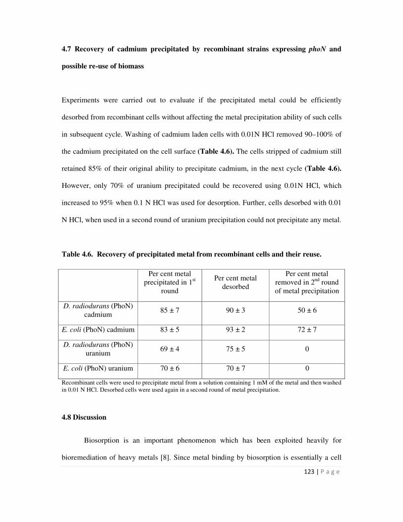
123 | P a g e
4.7 Recovery of cadmium precipitated by recombinant strains expressing phoN and
possible re-use of biomass
Experiments were carried out to evaluate if the precipitated metal could be efficiently
desorbed from recombinant cells without affecting the metal precipitation ability of such cells
in subsequent cycle. Washing of cadmium laden cells with 0.01N HCl removed 90–100% of
the cadmium precipitated on the cell surface (Table 4.6). The cells stripped of cadmium still
retained 85% of their original ability to precipitate cadmium, in the next cycle (Table 4.6).
However, only 70% of uranium precipitated could be recovered using 0.01N HCl, which
increased to 95% when 0.1 N HCl was used for desorption. Further, cells desorbed with 0.01
N HCl, when used in a second round of uranium precipitation could not precipitate any metal.
Table 4.6. Recovery of precipitated metal from recombinant cells and their reuse.
Recombinant cells were used to precipitate metal from a solution containing 1 mM of the metal and then washed
in 0.01 N HCl. Desorbed cells were used again in a second round of metal precipitation.
4.8 Discussion
Biosorption is an important phenomenon which has been exploited heavily for
bioremediation of heavy metals [8]. Since metal binding by biosorption is essentially a cell
Per cent metal
precipitated in 1st
round
Per cent metal
desorbed
Per cent metal
removed in 2nd
round
of metal precipitation
D. radiodurans (PhoN)
cadmium 85 ± 7 90 ± 3 50 ± 6
E. coli (PhoN) cadmium 83 ± 5 93 ± 2 72 ± 7
D. radiodurans (PhoN)
uranium 69 ± 4 75 ± 5 0
E. coli (PhoN) uranium 70 ± 6 70 ± 7 0
124 | P a g e
surface phenomenon, the role of S layer proteins in this process assumes considerable
importance. Earlier, a surface layer protein from Bacillus sphaericus JG-A12 was reported to
bind uranium and such cells reversibly and selectively bound U, Al, Cu and Pb [96-97]. The
nature of interaction between uranium and S layer protein was thoroughly investigated and
found to result from protein phosphorylation. Further sol-gel ceramics were prepared by
dispersing B. sphaericus JG-A12 vegetative cells, spores, and stabilized surface layer proteins
(S-layer) in aqueous silica nanosols and such “biocers” were used for removal of uranium and
copper [38,97]. While the S layer protein could remove 20 mg uranium/g proteins, the
biocers could remove around 25 mg uranium/g dry weight [98]. In another study, the S layer
protein from Bacillus spahericus was overexpressed in E. coli with a poly-histidine tag. The
purified protein was re-assembled and tested for nickel binding ability. While the wild type
protein could remove 13.8 mg Ni/g proteins, the recombinant protein removed 31 mg Ni/g
protein respectively [41].
In this study, the Hpi protein from D. radiodurans was found to possess uranium
binding ability. The Hpi protein bound 166 mg uranium/g protein, at pH 6.8. At this pH,
uranium is predicted to exist as uranyl-hydroxy species with a net positive charge [99]. The
Hpi protein is an acidic protein with a pI of 4.5 [36]. At near neutral pH, the protein is
expected to be negatively charged. In addition, the protein has many glutamic and aspartic
acid residues, both of which contribute carboxyl groups. A number of studies have reported
that the de-protonated carboxyl group of various biomolecules bind uranium with high
efficiency [96,100]. It remains to be proven whether these functional groups are the ones
involved in interaction of uranium with Hpi. The Hpi protein is one of the most sturdy S layer
proteins studied so far and retains its integrity even at 60°C in detergent and urea and at pH
as low as 2.0. Its subunits are covalently linked making it a very robust layer [53]. The
Deinococcal Hpi layer therefore forms an excellent material for uranium biosorption.
125 | P a g e
In a number of Bacillus species, the S layer protein was found to mask the negative
charge of peptidoglycan resulting in cell surface charge neutralization. This was thought to
prevent non-specific macromolecule adsorption to prevent pore plugging [101-102].
However, absence of the deinococcal Hpi protein made the overall charge on the cell surface
less negative, indicating that the Hpi layer actually enhances the net negative charge on
deinococcal cell surface. It should be noted that in the cell wall architecture of the D.
radiodurans strain R1, Hpi does not form the outermost layer but is overlayed by a
carbohydrate layer [54]. The less negative zeta potential of Hpi mutant cells indicates that in
spite of an outer carbohydrate coat, the Hpi protein still manages to gain enough surface
access to influence cell boundary charge. Wild type cells could bind more uranium than the
Hpi mutant, HMR202. Considering that electrostatic interactions play an important role in
metal binding, a more negative cell surface charge would be predicted to result in higher
binding of uranyl hydroxy complexes.
Since both whole cells, as well as the S layer of D. radiodurans show good uranium
binding ability, an attempt to extend this ability to other metal ions was made by cloning the
metal binding protein, metallothionein in this organism. D. radiodurans expressing SmtA
alone in cytosol did not show increased metal binding. Similarly, fusion of the SmtA protein
with the SLH domain also did not enhance the bioremediation capability of the recombinant
cells, in spite of membrane bound localization of the fusion protein. Only fusion with Hpi,
increased the metal biosorption capability of recombinant cells (Fig. 4.3 and 4.5). Further, all
the Hpi-SmtA fusion protein could be detected in the membrane fraction. The E. coli
recombinants expressing the Hpi-SmtA fusion protein did not show enhanced metal binding
compared to control cells but those carrying the smtA gene alone could remove twice as much
metal as control cells. In E. coli cells deinococcal Hpi protein does not have a designated
niche in the cell envelope which appears to impair its surface display, resulting cytosolic
126 | P a g e
localization and possible degradation and reduced metal binding. However, intracellular
metallothionein expression of the recombinant cells did enhance the metal binding capacity
of E. coli cells as has also been observed with intracellular expression of metallothioneins in
this organism earlier [103].
Different approaches have earlier been used for surface display of metal binding
peptides/proteins. A similar report of surface display of the SmtA protein by fusion to Lpp-
OmpA system in E. coli (elaborated in Chapter 3), resulted in a loading of 16 nmoles Cd/mg
dry weight cells (1.792 mg Cd/g dry weight cells) [87]. When a eukaryotic metallothionien
was fused to Lam B in E. coli, recombinant cells could remove 30 nmoles Cd/mg dry cell
weight ( 3.360 mg Cd/g dry weight cells) [104]. In an example of surface display of
metallothioniens in organisms other than E. coli, when mouse metallothionein (MT) was
fused to the β domain of IgA protease of Neisseria gonorrhoeae for localization of the MT in
outer membrane of Pseudomonas putida, it resulted in three fold higher metal binding
compared to intracellular expression of the MT, leading to a loading of 27 nmoles Cd/mg dry
weight cells (3.024 mg/g dry weight cells) [105]. In Ralstonia eutropha, which was
engineered using the same approach as Pseudomonas to display mouse MT, recombinants
could remove a maximum of 42 nmoles Cd/mg dry weight (4.7 mg/g dry weight cells) cells
at 300 µM and 16 nmoles Cd/mg dry weight cells (1.7 mg/g dry weight cells) at 30 µM input
Cd concentration [32]. A Hexa-histidine peptide was inserted to a permissive site of the
surface layer (S-layer) protein RsaA of Caulobacter crescentus. The recombinant strain
JS4022/p723–6H removed 94.3∼99.9% of the Cd(II), leading to a metal loading of 16 mg/g
dry cell weight whereas the control strain removed only 11.4∼37.0% [40]. The result
described in the thesis is the only report of an S-layer-metallothonein fusion protein (Hpi-
127 | P a g e
SmtA) and resulted in a cadmium loading of 1.2 mg/g dry biomass in recombinant D.
radiodurans cells exposed to 40 µM Cd2+
ions..
While biosorption and complexation of metals are useful mechanisms at nanomolar or
lower metal concentrations, they are ineffective when the, metal concentrations are in range
of micro or millimolar [25]. At such concentrations, bioremediation by bioprecipitation and
bioreduction are much more useful [25]. In earlier studies, the efficiency of PhoN mediated
uranium precipitation in the millimolar range has been reported in D. radiodurans [51].
Phosphatase mediated metal precipitation is essentially non-specific in nature i.e. any heavy
metal present in solution can be precipitated using this approach [17-18]. Therefore, the
potential of phoN bearing cells in cadmium precipitation was explored since it is a hazardous
metal, and widely used in paints and electronic industry.
The rate of cadmium bioprecipitation was found to be significantly higher than
uranium bioprecipitation. An important observation was that the toxicity of the metal to the
organisms per se, did not affect the corresponding precipitation efficiency. D. radiodurans is
a metal sensitive bacterium with a MIC of 0.018 mM for cadmium compared with 0.5–1 mM
for E. coli DH5α (Ruggiero et al., 2005). However, both organisms could precipitate out the
metal at comparable efficiencies, commensurate with their phosphatase activity. Perhaps
extracellular precipitation shields the bacterium from metabolic ill effects of metals.
Efforts to increase uranium precipitation in recombinant cells by fusion of PhoN to
surface layer proteins did not per se increase metal precipitation ability. As was observed in
zymograms, the Hpi-PhoN fusion protein was expressed and displayed phosphatase activity
in recombinant D. radiodurans cells (Fig. 3.23 and 3.25). It was also exclusively cell
membrane localized. Since both the PhoN and Hpi-PhoN proteins were expressed from the
same promoter, the likelihood of differential expression are low but possible. While, this
128 | P a g e
could be one of the factors, the low whole cell activity in hpi-phoN bearing cells may also be
due to low phosphatase activity of the Hpi-PhoN fusion protein per se. Hpi being a much
larger protein may interfere with correct folding of the fusion protein lowering the
phosphatase activity of the protein per se. PhoN is an enzyme which is known to function as
a dimer. In creating fusion proteins, there is a possibility that the dimerization of the enzyme
may be affected and may result in lower activity of the recombinant protein. In contrast,
fusion of SmtA to Hpi enhanced metal binding activity because in metallothioneins, metal
binding is facilitated by Cys-X-Cys conserved sequence, and only sufficient exposure of this
repeating group is required for metal binding.
Both the SLH fusion to SmtA as well as to PhoN failed to enhance metal removal by
recombinant cells expressing them. The SLH domain is likely to be present more proximal to
peptidoglycan layer than Hpi, and away from the cell surface in D. radiodurans. Such cell
envelope localization of SLH may not be conducive both to the activity of PhoN enzyme due
to low substrate accessibility as well as for the metal binding ability of SmtA. The results
show that while surface display of SmtA by fusion to Hpi resulted in superior metal binding
by recombinant cells, fusion of PhoN to either of the S layer proteins did not provide value
addition.
Isolated Hpi protein from recombinant cells expressing Hpi-PhoN could precipitate
uranium more efficiently than that isolated from control pRAD1 cells (Fig. 4.9). However, on
washing the Hpi pellet to remove cytoplasmic contamination, this enhanced metal
precipitation ability was also lost. Further, the Hpi layer isolated from hpi-smtA bearing cells
did not show better cadmium/zinc binding compared to the layer isolated from pRAD1
bearing cells. The two results put together seem to indicate that while the fusion to Hpi
protein might have conferred membrane localization to the proteins tagged to it, the fusion
129 | P a g e
protein may not have been properly incorporated covalently into the Hpi layer in the face of
competition from native Hpi protein.
Attempt to create S layer-PhoN fusion proteins, however, gave rise to a new possible
mode of PhoN exploitation i.e. by immobilization on peptidoglycan using the SLH domain.
This was especially relevant considering that peptidoglycan itself is known to adsorb uranium
on its functional groups [94]. Further, peptidoglycan is a sturdy biopolymer, resistant to heat
and detergents and is hydrolyzed only with concentrated mineral acids at high temperature.
SLH-PhoN immobilized on peptidoglycan when stabilized by glutaraldehyde cross linking,
showed efficient metal precipitation and excellent re-usability in uranium precipitation assays
for upto five cycles (Fig. 4.10). PhoN itself proved to be a stable and sturdy enzyme which
could withstand glutaraldehyde treatment, prolonged uranium exposure and room
temperature incubations.
The usage of interaction between the SLH domain and peptidoglycan for
immobilization of enzymes is reported here for the first time. This allows one-step
purification of SLH-PhoN from cell extracts and immobilization on peptidoglycan making
this a convenient system to implement. Further, of all the systems that were developed in this
study using D. radiodurans for uranium precipitation, SLH-PhoN immobilized on
peptidoglycan (SPhoNP) was found to be the most efficient in precipitating uranium. While
SPhoNP removed 376 µg U/mg dry weight in 6 h, recombinants expressing PhoN or Hpi-
PhoN alone precipitated only 297 µg U/mg cells and 178 µg U/mg cells respectively,
showing superiority of SPhoNP over other biomass in precipitating U. Proteins immobilized
on an inert support are far more attractive than using recombinant organisms in the
environment, which are associated with risks such as instability, horizontal gene transfer to
non-target organisms etc.
130 | P a g e
While it is important to design organisms with superior bioremediation abilities, it is
equally important to make the system appealing by ensuring that using such biomass is non-
cumbersome. With this in mind, lyophilisation of recombinant organisms was attempted to
achieve volume reduction and ease of handling, storage and transport of such cells. Not only
did lyophilisation of PhoN bearing cells reduce volume and convert cell suspensions into a
dry easy-to-use-and-disperse-powder, it also achieved the following: (1) preserved the PhoN
activity and uranium precipitation ability of cells, (2) retained cellular integrity and surface
precipitation property, thereby facilitating easy recovery of precipitated metal with the
biomass, (3) significantly extended the shelf life of the product in terms of uranium
precipitation capability for up to six months at room temperature.
E. coli is not considered a safe organism for environmental use due to instability, and
occurrence of pathogenic strains. Attempts have been made earlier to employ liposomes
carrying PhoN for metal removal in vitro to avoid direct use of genetically manipulated live
bacteria in filtration systems [106]. The present study managed this by using lyophilisation
without cryopreservatives to render the recombinant cells non-viable, while preserving their
PhoN and metal precipitation activities. Lyophilisation is used as a cell-preservation
technique, but is known to cause irreversible damage to E. coli cells resulting in death [107].
A number of factors including use of protective excipients during freeze-drying [108], [109]
and storage in vacuo after lyophilisation [110] are known to maintain viability of microbes.
The present study deliberately excluded these from the lyophilisation process to render the
cells non-viable. Though lyophilisation is known to cause protein unfolding and denaturation
[111], PhoN activity was well preserved after lyophilisation perhaps due to the protection
afforded by its periplasmic location in the cell.
131 | P a g e
D. radiodurans scores over E. coli based systems in its utility for bioremediation of
radioactive waste solution [49,112]. In the case of D. radiodurans, lyophilized cells retained
viability which allowed use of such cell powder as inoculum to build-up biomass for in situ
bioremediation experiments. In addition, no pathogenic strains of this species are known and
it is therefore considered safer for environmental use. Lyophilisation was also applied to
SPhoNP stabilized by glutaraldehyde, which fully retained its uranium precipitation ability.
Uranium precipitation ability of lyophilized SPhoNP was also found to be superior to
recombinant D. radiodurans cells expressing PhoN or Hpi-PhoN.
Scanning Electron Microscopy revealed that the precipitated metal was firmly lodged
on to the surface of recombinant cells. Even at very high loadings, no free precipitate was
seen in the fields observed. This has important implications for settling down of precipitated
sludge and downstream processing of the effluent as emphasized earlier [18]. Surface
precipitation of metal also allowed easy recovery of metal by dilute acid wash.
The two potential candidates for bioremediation of heavy metals from radioactive
wastes that emerged from this study as a result of expression of surface layer fusion proteins
are as follows: (1) Recombinant D. radiodurans expressing Hpi-SmtA for sequestration of
metals at micromolar concentrations, and (2) SPhoNP for precipitation of metals from
solutions carrying millimolar concentration of metals. In the light of isolated Hpi layer from
recombinants not displaying superior metal remediation ability, overexpression of this protein
at much higher levels in HMR202 mutant cells, may enable formation of a homogenous Hpi
layer comprising of the fusion protein alone, thus increasing metal bioremediation ability of
recombinant cells as well as protein. Another important lead from this work is the evaluation
of metal precipitation utility of SPhoNP in a flow-through system for superior bioremediation
in future.

132 | P a g e
Chapter 5
Summary and Conclusions
133 | P a g e
Summary and Conclusions
This study was conceived to develop genetically engineered bacteria expressing S-layer
proteins fused either to a non-specific acid phosphatase (PhoN) or a metallothionein (SmtA),
aimed at metal bioremediation. The radioresistant organism, D. radiodurans, was chosen as a
host for expression of constructed fusion proteins, since it is well suited for remediation of
radioactive waste. The salient findings of the study are summarized below:
� The two S layer proteins of D. radiodurans, Hpi and SlpA, were partially
characterized and found to be associated with one another in the deinococcal cell
envelope.
� The Hpi layer was isolated and found to undergo in vivo degradation, as ascertained
by peptide mass fingerprinting of multiple protein bands obtained on resolving the
isolated Hpi protein layer on denaturing gels.
� The Hpi protein layer bound (166 µg U/mg protein) U at near neutral pH. Hpi protein
was found to confer substantial negative charge to deinococcal cell surface, thereby
possibly enhancing its interaction and binding with heavy metals.
� Bioinformatic analysis of the SlpA protein showed presence of a single surface layer
homology domain (SLH) at the N terminus, which is known to bind the secondary
cell wall polymers (SCWP) attached to peptidoglycan in other organisms, making it a
suitable membrane anchoring vehicle.
� The smtA gene from the unicellular cyanobacterium, Synechococcus elongatus, which
codes for a metal binding metallothionein (MT) peptide, was cloned for expression
from the strong deinococcal PgroESL promoter, or as a fusion gene with hpi ORF, or
134 | P a g e
fused to the nucleotide sequence encoding the SLH domain of SlpA protein in D.
radiodurans.
� The expressed Hpi-SmtA and SLH-SmtA fusion proteins localized to the deinococcal
cell surface as desired, while the SmtA protein per se localized to the cytoplasm. Thus
tagging SmtA to Hpi/SLH domain facilitated localization of the SmtA protein in the
deinococcal cell envelope.
� Recombinant D. radiodurans cells expressing the Hpi-SmtA fusion protein removed
1.5-2 times more metals (Cd, Zn and Cu) than those expressing only SmtA or SLH-
SmtA fusion protein. Surface display of SmtA by fusion to Hpi enhanced the metal
binding ability of recombinants (1.2 mg Cd, 225 µg Cu and 210 µg Zn/g dry weight
cells) but its fusion to SLH domain did not. This indicated a more inward localization
of SLH closer to peptidoglycan and away from cell surface. Further, intracellular
expression of SmtA also did not enhance metal binding ability of recombinant cells
(0.35 mg Cd, 100 µg Cu and 125 µg Zn/g dry weight cells).
� The phoN gene was fused to hpi ORF or sequence encoding the SLH domain of the
SlpA protein. Recombinant D. radiodurans expressing the Hpi-PhoN and SLH-PhoN
proteins showed phosphatase activity bands of 127 kDa and 37 kDa respectively in
zymograms, as was expected for the fusion proteins.
� The Hpi-PhoN protein localized exclusively to the cell membrane and showed cell-
based phosphatase activity. The SLH-PhoN protein however, localized to both
cytoplasm as well as membrane in D. radiodurans cells and did not show any cell-
based activity.

135 | P a g e
� Hpi-PhoN fusion protein expression did not enhance cell-based phosphatase activity
in D. radiodurans. In 6 h, D. radiodurans (PhoN) cells could remove as much as 83%
uranium leading to a loading 310 µg U/mg dry weight cells, while Hpi-PhoN bearing
recombinants could remove only 40-50% of input uranium leading to a loading of
136-185 µg U/mg dry weight cells. But when cells were used at equivalent
phosphatase activity, recombinants carrying Hpi-PhoN or PhoN per se removed
around 90% U in 6 h.
� The Hpi-PhoN protein layer, isolated from recombinant D. radiodurans cells could
precipitate uranium more efficiently (446 µg U/mg protein) than Hpi layer isolated
from D. radiodurans (pRAD1) cells (208 µg U/mg protein), but this feature was lost
upon extensive washing of the Hpi pellet. The Hpi layer isolated from Hpi-SmtA
expressing cells also did not exhibit enhanced cadmium/zinc binding, compared to the
Hpi layer isolated from D. radiodurans (pRAD1). Thus, while Hpi-SmtA and Hpi-
PhoN proteins did localize to membrane, their incorporation into Hpi layer appears to
have been hampered in the face of competition from native resident Hpi layer.
� The SLH domain of SlpA protein was shown to bind peptidoglycan in vitro. This
indicated that the association of SlpA with secondary cell wall polymers of
peptidoglycan at one end and Hpi at the other, formed a continuous link in the
interactions between different layers of the deinococcal cell wall.
� Utility of peptidoglycan as an immobilization matrix by exploiting its interaction with
the SLH domain is an important finding of this study. SLH-PhoN immobilized on
peptidoglycan could efficiently remove U from solution (95% in 4 h) and
glutaraldehyde stabilized SLH-PhoN-peptidoglycan complex showed excellent
136 | P a g e
stability and re-usability for upto five cycles of uranium precipitation, leading to a
final loading of 160 µg U/mg dry biomass.
� SLH-PhoN immobilized on peptidoglycan (SPhoNP) showed most efficient uranium
precipitation (376 µg U/mg dry weight biomass) compared to D. radiodurans bearing
Hpi-PhoN (178 µg U/mg dry weight cells) or only PhoN (297 µg U/mg dry weight
cells).
� Recombinant Deinococcus cells expressing PhoN could precipitate cadmium more
efficiently (1.59 µmoles/mg dry weight cells) than uranium (0.79 µmoles/mg dry
weight cells) from solution.
� Lyophilization of PhoN expressing E. coli and D. radiodurans reduced the bulk
volume and converted the biomass into a dry powdered form. Lyophilized
recombinants retained PhoN activity as well as U precipitation ability, while also
retaining cellular integrity and surface precipitation property.
� Lyophilisation significantly extended the shelf life of the product in terms of metal
precipitation up to six months at ambient temperature.
� Both the uranyl phosphate and cadmium phosphate precipitates were shown to be cell
surface associated, leading to easy separation of metal laden cells thereby aiding
metal recovery from liquid waste.
To conclude, this study generated the following important results:
1. Hpi emerged as an efficient membrane targeting vehicle in D. radiodurans. When
used for membrane localization of SmtA, it resulted in enhanced metal binding
ability of recombinants expressing the fusion protein.
137 | P a g e
2. The property of SLH domain to bind SCWP of peptidoglycan could be exploited
for one step immobilization of enzymes. SLH-PhoN immobilized on
peptidoglycan provided an excellent and efficient biomaterial for metal
precipitation.
3. Lyophilization proved to be a good value addition to phosphatase mediated
bioremediation by increasing the shelf life of recombinants and making their
handling and storage easy.
Generation of S layers, exclusively of fusion proteins, by expressing the protein at
high levels in Hpi mutant cells, and evaluation of metal precipitation utility of
SPhoNP in a flow-through process may hold promise for superior bioremediation in
future.

138 | P a g e
References
139 | P a g e
[1] LaGrega, M.D., Buckingham, P.L. and Evans, J.C. (2010) Hazardous waste
management, Waveland Press
[2] Mejare, M. and Bulow, L. (2001). Metal-binding proteins and peptides in
bioremediation and phytoremediation of heavy metals. Trends Biotechnol 19, 67-73.
[3] Boopathy, R. (2000). Factors limiting bioremediation technologies. Bioresource
Technology 74, 63-67.
[4] Malik, A. (2004). Metal bioremediation through growing cells. Environment
International 30, 261-278.
[5] Nies, D.H. (1999). Microbial heavy-metal resistance. Applied Microbiology and
Biotechnology 51, 730-750.
[6] Lloyd, J.R. and Renshaw, J.C. (2005). Bioremediation of radioactive waste:
radionuclide-microbe interactions in laboratory and field-scale studies. Curr Opin
Biotechnol 16, 254-60.
[7] Singh, J.S., Abhilash, P.C., Singh, H.B., Singh, R.P. and Singh, D.P. (2011).
Genetically engineered bacteria: An emerging tool for environmental remediation and
future research perspectives. Gene 480, 1-9.
[8] Barkay, T. and Schaefer, J. (2001). Metal and radionuclide bioremediation: issues,
considerations and potentials. Curr Opin Microbiol 4, 318-23.
[9] Gadd, G.M. (2000). Bioremedial potential of microbial mechanisms of metal
mobilization and immobilization. Curr Opin Biotechnol 11, 271-9.
[10] Fomina, M. and Gadd, G.M. (2014). Biosorption: current perspectives on concept,
definition and application. Bioresource Technology 160, 3-14.
[11] Eccles, H. (1999). Treatment of metal-contaminated wastes: why select a biological
process? Trends Biotechnol 17, 462-5.
[12] Cobbett, C. and Goldsbrough, P. (2002). PHYTOCHELATINS AND
METALLOTHIONEINS: Roles in Heavy Metal Detoxification and Homeostasis.
Annual Review of Plant Biology 53, 159-182.
[13] Turner, J.S. and Robinson, N.J. (1995). Cyanobacterial metallothioneins:
biochemistry and molecular genetics. J Ind Microbiol 14, 119-25.
[14] Shi, J., Lindsay, W.P., Huckle, J.W., Morby, A.P. and Robinson, N.J. (1992).
Cyanobacterial metallothionein gene expressed in Escherichia coli. Metal-binding
properties of the expressed protein. FEBS Lett 303, 159-63.
140 | P a g e
[15] Webb, McGinness and Lappin, S. (1998). Metal removal by sulphate-reducing
bacteria from natural and constructed wetlands. Journal of Applied Microbiology 84,
240-248.
[16] Sharma, P.K., Balkwill, D.L., Frenkel, A. and Vairavamurthy, M.A. (2000). A New
Klebsiella planticola Strain (Cd-1) Grows Anaerobically at High Cadmium
Concentrations and Precipitates Cadmium Sulfide. Applied and Environmental
Microbiology 66, 3083-3087.
[17] Macaskie, L.E., Wates, J.M. and Dean, A.C. (1987). Cadmium accumulation by a
Citrobacter sp. immobilized on gel and solid supports: Applicability to the treatment
of liquid wastes containing heavy metal cations. Biotechnol Bioeng 30, 66-73.
[18] Macaskie, L.E., Jeong, B.C. and Tolley, M.R. (1994). Enzymically accelerated
biomineralization of heavy metals: application to the removal of americium and
plutonium from aqueous flows. FEMS Microbiol Rev 14, 351-67.
[19] Basnakova, G., Stephens, E.R., Thaller, M.C., Rossolini, G.M. and Macaskie, L.E.
(1998). The use of Escherichia coli bearing a phoN gene for the removal of uranium
and nickel from aqueous flows. Appl Microbiol Biotechnol 50, 266-72.
[20] Basnakova, G. and Macaskie, L.E. (1997). Microbially enhanced chemisorption of
nickel into biologically synthesized hydrogen uranyl phosphate: a novel system for
the removal and recovery of metals from aqueous solutions. Biotechnol Bioeng 54,
319-28.
[21] Bonthrone, K.M., Basnakova, G., Lin, F. and Macaskie, L.E. (1996).
Bioaccumulation of nickel by intercalation into polycrystalline hydrogen uranyl
phosphate deposited via an enzymatic mechanism. Nat Biotechnol 14, 635-8.
[22] Keasling, J.D., Van Dien, S.J. and Pramanik, J. (1998). Engineering polyphosphate
metabolism in Escherichia coli: implications for bioremediation of inorganic
contaminants. Biotechnol Bioeng 58, 231-9.
[23] Renninger, N., Knopp, R., Nitsche, H., Clark, D.S. and Keasling, J.D. (2004). Uranyl
precipitation by Pseudomonas aeruginosa via controlled polyphosphate metabolism.
Appl Environ Microbiol 70, 7404-12.
[24] Macaskie, L.E., Yong, P., Doyle, T.C., Roig, M.G., Diaz, M. and Manzano, T. (1997).
Bioremediation of uranium-bearing wastewater: Biochemical and chemical factors
influencing bioprocess application. Biotechnology and Bioengineering 53, 100-109.
141 | P a g e
[25] Valls, M. and Lorenzo, V. (2002). Exploiting the genetic and biochemical capacities
of bacteria for the remediation of heavy metal pollution. FEMS Microbiology
Reviews 26, 327-338.
[26] Deng, X., Yi, X.E. and Liu, G. (2007). Cadmium removal from aqueous solution by
gene-modified Escherichia coli JM109. Journal of Hazardous Materials 139, 340-344.
[27] Yuan, C., Lu, X., Qin, J., Rosen, B.P. and Le, X.C. (2008). Volatile Arsenic Species
Released from Escherichia coli Expressing the AsIII S-adenosylmethionine
Methyltransferase Gene. Environmental Science & Technology 42, 3201-3206.
[28] Zhao, X.W., Zhou, M.H., Li, Q.B., Lu, Y.H., He, N., Sun, D.H. and Deng, X. (2005).
Simultaneous mercury bioaccumulation and cell propagation by genetically
engineered Escherichia coli. Process Biochemistry 40, 1611-1616.
[29] Valls, M., de Lorenzo, V., Gonzalez-Duarte, R. and Atrian, S. (2000). Engineering
outer-membrane proteins in Pseudomonas putida for enhanced heavy-metal
bioadsorption. J Inorg Biochem 79, 219-23.
[30] Nilgiriwala, K.S., Alahari, A., Rao, A.S. and Apte, S.K. (2008). Cloning and
overexpression of alkaline phosphatase PhoK from Sphingomonas sp. strain BSAR-1
for bioprecipitation of uranium from alkaline solutions. Appl Environ Microbiol 74,
5516-23.
[31] Saleem, M., Brim, H., Hussain, S., Arshad, M., Leigh, M.B. and Zia ul, H. (2008).
Perspectives on microbial cell surface display in bioremediation. Biotechnol Adv 26,
151-61.
[32] Valls, M., Atrian, S., de Lorenzo, V. and Fernandez, L.A. (2000). Engineering a
mouse metallothionein on the cell surface of Ralstonia eutropha CH34 for
immobilization of heavy metals in soil. Nat Biotechnol 18, 661-5.
[33] Sleytr, U.B., Schuster, B., Egelseer, E.M. and Pum, D. (2014). S-layers: principles
and applications. FEMS Microbiol Rev
[34] Engelhardt, H. (2007). Are S-layers exoskeletons? The basic function of protein
surface layers revisited. J Struct Biol 160, 115-24.
[35] Fagan, R.P. and Fairweather, N.F. (2014). Biogenesis and functions of bacterial S-
layers. Nat Rev Micro 12, 211-222.
[36] Pavkov-Keller, T., Howorka, S. and Keller, W. (2011) Chapter 3 - The Structure of
Bacterial S-Layer Proteins. In Progress in Molecular Biology and Translational
Science (Stefan, H., ed.^eds), pp. 73-130. Academic Press
142 | P a g e
[37] Ilk, N., Egelseer, E.M. and Sleytr, U.B. (2011). S-layer fusion proteins--construction
principles and applications. Curr Opin Biotechnol 22, 824-31.
[38] Pollmann, K., Raff, J., Merroun, M., Fahmy, K. and Selenska-Pobell, S. (2006). Metal
binding by bacteria from uranium mining waste piles and its technological
applications. Biotechnol Adv 24, 58-68.
[39] Velasquez, L. and Dussan, J. (2009). Biosorption and bioaccumulation of heavy
metals on dead and living biomass of Bacillus sphaericus. J Hazard Mater 167, 713-6.
[40] Patel, J., Zhang, Q., McKay, R.M., Vincent, R. and Xu, Z. (2010). Genetic
engineering of Caulobacter crescentus for removal of cadmium from water. Appl
Biochem Biotechnol 160, 232-43.
[41] Pollmann, K. and Matys, S. (2007). Construction of an S-layer protein exhibiting
modified self-assembling properties and enhanced metal binding capacities. Appl
Microbiol Biotechnol 75, 1079-85.
[42] Battista, J.R. (1997). Against all odds: the survival strategies of Deinococcus
radiodurans. Annu Rev Microbiol 51, 203-24.
[43] Minton, K.W. (1994). DNA repair in the extremely radioresistant bacterium
Deinococcus radiodurans. Mol Microbiol 13, 9-15.
[44] Minton, K.W. (1996). Repair of ionizing-radiation damage in the radiation resistant
bacterium Deinococcus radiodurans. Mutat Res 363, 1-7.
[45] Slade, D. and Radman, M. (2011). Oxidative stress resistance in Deinococcus
radiodurans. Microbiol Mol Biol Rev 75, 133-91.
[46] Venkateswaran, A., McFarlan, S.C., Ghosal, D., Minton, K.W., Vasilenko, A.,
Makarova, K., Wackett, L.P. and Daly, M.J. (2000). Physiologic determinants of
radiation resistance in Deinococcus radiodurans. Appl Environ Microbiol 66, 2620-6.
[47] Zahradka, K., Slade, D., Bailone, A., Sommer, S., Averbeck, D., Petranovic, M.,
Lindner, A.B. and Radman, M. (2006). Reassembly of shattered chromosomes in
Deinococcus radiodurans. Nature 443, 569-73.
[48] Misra, H.S., Khairnar, N.P., Kota, S., Shrivastava, S., Joshi, V.P. and Apte, S.K.
(2006). An exonuclease I-sensitive DNA repair pathway in Deinococcus radiodurans:
a major determinant of radiation resistance. Molecular Microbiology 59, 1308-1316.
[49] Brim, H., McFarlan, S.C., Fredrickson, J.K., Minton, K.W., Zhai, M., Wackett, L.P.
and Daly, M.J. (2000). Engineering Deinococcus radiodurans for metal remediation
in radioactive mixed waste environments. Nat Biotechnol 18, 85-90.

143 | P a g e
[50] Lange, C.C., Wackett, L.P., Minton, K.W. and Daly, M.J. (1998). Engineering a
recombinant Deinococcus radiodurans for organopollutant degradation in radioactive
mixed waste environments. Nat Biotechnol 16, 929-33.
[51] Appukuttan, D., Rao, A.S. and Apte, S.K. (2006). Engineering of Deinococcus
radiodurans R1 for bioprecipitation of uranium from dilute nuclear waste. Appl
Environ Microbiol 72, 7873-8.
[52] Kulkarni, S., Ballal, A. and Apte, S.K. (2013). Bioprecipitation of uranium from
alkaline waste solutions using recombinant Deinococcus radiodurans. J Hazard Mater
262, 853-61.
[53] Baumeister, W., Karrenberg, F., Rachel, R., Engel, A., Ten Heggeler, B. and Saxton,
W.O. (1982). The Major Cell Envelope Protein of Micrococcus radiodurans (R1).
European Journal of Biochemistry 125, 535-544.
[54] Rothfuss, H., Lara, J.C., Schmid, A.K. and Lidstrom, M.E. (2006). Involvement of the
S-layer proteins Hpi and SlpA in the maintenance of cell envelope integrity in
Deinococcus radiodurans R1. Microbiology 152, 2779-87.
[55] Mark, S.S., Bergkvist, M., Yang, X., Teixeira, L.M., Bhatnagar, P., Angert, E.R. and
Batt, C.A. (2006). Bionanofabrication of metallic and semiconductor nanoparticle
arrays using S-layer protein lattices with different lateral spacings and geometries.
Langmuir 22, 3763-74.
[56] Thompson, B.G., Murray, R.G.E. and Boyce, J.F. (1982). The association of the
surface array and the outer membrane of Deinococcus radiodurans. Canadian Journal
of Microbiology 28, 1081-1088.
[57] Müller, D.J., Baumeister, W. and Engel, A. (1996). Conformational change of the
hexagonally packed intermediate layer of Deinococcus radiodurans monitored by
atomic force microscopy. Journal of Bacteriology 178, 3025-30.
[58] Engelhardt, H. and Peters, J. (1998). Structural Research on Surface Layers: A Focus
on Stability, Surface Layer Homology Domains, and Surface Layer–Cell Wall
Interactions. Journal of Structural Biology 124, 276-302.
[59] Cava, F., de Pedro, M.A., Schwarz, H., Henne, A. and Berenguer, J. (2004). Binding
to pyruvylated compounds as an ancestral mechanism to anchor the outer envelope in
primitive bacteria. Mol Microbiol 52, 677-90.
144 | P a g e
[60] Kapoor, A. and Viraraghavan, T. (1995). Fungal biosorption — an alternative
treatment option for heavy metal bearing wastewaters: a review. Bioresource
Technology 53, 195-206.
[61] Selatnia, A., Bakhti, M.Z., Madani, A., Kertous, L. and Mansouri, Y. (2004).
Biosorption of Cd2+ from aqueous solution by a NaOH-treated bacterial dead
Streptomyces rimosus biomass. Hydrometallurgy 75, 11-24.
[62] Riordan, C., Bustard, M., Putt, R. and McHale, A.P. (1997). Removal of uranium
from solution using residual brewery yeast: combined biosorption and precipitation.
Biotechnology Letters 19, 385-388.
[63] Sar, P., K. Kazy, S. and D'Souza, S.F. (2004). Radionuclide remediation using a
bacterial biosorbent. International Biodeterioration & Biodegradation 54, 193-202.
[64] Matsunaga, T., Takeyama, H., Nakao, T. and Yamazawa, A. (1999). Screening of
marine microalgae for bioremediation of cadmium-polluted seawater. Journal of
Biotechnology 70, 33-38.
[65] Tauriainen, S.M., Virta, M.P.J. and Karp, M.T. (2000). Detecting bioavailable toxic
metals and metalloids from natural water samples using luminescent sensor bacteria.
Water Research 34, 2661-2666.
[66] Lovley, D.R. and Phillips, E.J.P. (1994). Reduction of Chromate by Desulfovibrio
vulgaris and Its c3 Cytochrome. Applied and Environmental Microbiology 60, 726-
728.
[67] Riccio, M., Rossolini, G., Lombardi, G., Chiesurin, A. and Satta, G. (1997).
Expression cloning of different bacterial phosphataseencoding genes by histochemical
screening of genomic libraries onto an indicator medium containing phenolphthalein
diphosphate and methyl green. Journal of Applied Microbiology 82, 177-185.
[68] Sambrook, J., Fritsch, E.F. and Maniatis, T. (1989) Molecular cloning, Cold spring
harbor laboratory press New York
[69] Lennon, E. and Minton, K.W. (1990). Gene fusions with lacZ by duplication insertion
in the radioresistant bacterium Deinococcus radiodurans. Journal of Bacteriology 172,
2955-2961.
[70] Peterson, G.L. (1977). A simplification of the protein assay method of Lowry< i> et
al.</i> which is more generally applicable. Analytical biochemistry 83, 346-356.
[71] Laemmli, U.K. (1970). Cleavage of structural proteins during the assembly of the
head of bacteriophage T4. Nature 227, 680-685.
145 | P a g e
[72] Panda, B., Basu, B., Rajaram, H. and Kumar Apte, S. (2014). Methyl viologen
responsive proteome dynamics of Anabaena sp. strain PCC7120. Proteomics 14,
1895-1904.
[73] Bolton, P. and Dean, A. (1972). Phosphatase synthesis in< italic>
Klebsiella</italic>(< italic> Aerobacter</italic>)< italic> aerogenes</italic> growing
in continuous culture. Biochem. J 127, 87-96.
[74] Work, E. (1957). Reaction of ninhydrin in acid solution with straight-chain amino
acids containing two amino groups and its application to the estimation of α∈-
diaminopimelic acid. Biochemical Journal 67, 416.
[75] Mesnage, S., Tosi-Couture, E. and Fouet, A. (1999). Production and cell surface
anchoring of functional fusions between the SLH motifs of the Bacillus anthracis S-
layer proteins and the Bacillus subtilis levansucrase. Molecular Microbiology 31, 927-
936.
[76] Fritz, J. and Bradford, E. (1958). Detection of thorium and uranium. Analytical
Chemistry 30, 1021-1022.
[77] Karnovsky, M.J. (1965). A formaldehyde-glutaraldehyde fixative of high osmolarity
for use in electron microscopy. J. cell Biol 27
[78] Janesch, B., Messner, P. and Schäffer, C. (2013). Are the Surface Layer Homology
Domains Essential for Cell Surface Display and Glycosylation of the S-Layer Protein
from Paenibacillus alvei CCM 2051T? Journal of Bacteriology 195, 565-575.
[79] Lemaire, M., Ohayon, H., Gounon, P., Fujino, T. and Béguin, P. (1995). OlpB, a new
outer layer protein of Clostridium thermocellum, and binding of its S-layer-like
domains to components of the cell envelope. Journal of Bacteriology 177, 2451-9.
[80] Mesnage, S., Tosi-Couture, E., Mock, M. and Fouet, A. (1999). The S-layer
homology domain as a means for anchoring heterologous proteins on the cell surface
of Bacillus anthracis. Journal of Applied Microbiology 87, 256-260.
[81] Meima, R. and Lidstrom, M.E. (2000). Characterization of the minimal replicon of a
cryptic Deinococcus radiodurans SARK plasmid and development of versatile
Escherichia coli-D. radiodurans shuttle vectors. Appl Environ Microbiol 66, 3856-67.
[82] Rachel, R., Engel, A. and Baumeister, W. (1983). Proteolysis of the major cell
envelope protein of Deinococcus radiodurans remains morphologically latent. FEMS
Microbiology Letters 17, 115-119.

146 | P a g e
[83] Desvaux, M., Dumas, E., Chafsey, I. and Hébraud, M. (2006). Protein cell surface
display in Gram-positive bacteria: from single protein to macromolecular protein
structure. FEMS Microbiology Letters 256, 1-15.
[84] Vergani, L., Grattarola, M., Dondero, F. and Viarengo, A. (2003). Expression,
purification, and characterization of metallothionein-A from rainbow trout. Protein
Expression and Purification 27, 338-345.
[85] Lee, S.Y., Choi, J.H. and Xu, Z. (2003). Microbial cell-surface display. TRENDS in
Biotechnology 21, 45-52.
[86] Richins, R.D., Kaneva, I., Mulchandani, A. and Chen, W. (1997). Biodegradation of
organophosphorus pesticides by surface-expressed organophosphorus hydrolase.
Nature biotechnology 15, 984-987.
[87] Tafakori, V., Ahmadian, G. and Amoozegar, M.A. (2012). Surface display of
bacterial metallothioneins and a chitin binding domain on Escherichia coli increase
cadmium adsorption and cell immobilization. Applied biochemistry and
biotechnology 167, 462-473.
[88] Mitchel, R.E. (1980). Micrococcus radiodurans surface exonuclease. Dimer to
monomer conversion by ionizing radiation-generated aqueous free radicals. Biochim
Biophys Acta 621, 138-46.
[89] Farci, D., Bowler, M.W., Kirkpatrick, J., McSweeney, S., Tramontano, E. and Piano,
D. (2014). New features of the cell wall of the radio-resistant bacterium Deinococcus
radiodurans. Biochimica et Biophysica Acta (BBA) - Biomembranes 1838, 1978-
1984.
[90] Bergkvist, M., Mark, S.S., Yang, X., Angert, E.R. and Batt, C.A. (2004).
Bionanofabrication of Ordered Nanoparticle Arrays:� Effect of Particle Properties
and Adsorption Conditions. The Journal of Physical Chemistry B 108, 8241-8248.
[91] Mark, S.S., Bergkvist, M., Yang, X., Teixeira, L.M., Bhatnagar, P., Angert, E.R. and
Batt, C.A. (2006). Bionanofabrication of Metallic and Semiconductor Nanoparticle
Arrays Using S-Layer Protein Lattices with Different Lateral Spacings and
Geometries. Langmuir 22, 3763-3774.
[92] Olabarria, G., Carrascosa, J.L., de Pedro, M.A. and Berenguer, J. (1996). A conserved
motif in S-layer proteins is involved in peptidoglycan binding in Thermus
thermophilus. J Bacteriol 178, 4765-72.
147 | P a g e
[93] Sára, M. (2001). Conserved anchoring mechanisms between crystalline cell surface S-
layer proteins and secondary cell wall polymers in Gram-positive bacteria? Trends in
Microbiology 9, 47-49.
[94] Barkleit, A., Moll, H. and Bernhard, G. (2009). Complexation of uranium (VI) with
peptidoglycan. Dalton Transactions, 5379-5385.
[95] Ruggiero, C.E., Boukhalfa, H., Forsythe, J.H., Lack, J.G., Hersman, L.E. and Neu,
M.P. (2005). Actinide and metal toxicity to prospective bioremediation bacteria.
Environmental Microbiology 7, 88-97.
[96] Merroun, M.L., Raff, J., Rossberg, A., Hennig, C., Reich, T. and Selenska-Pobell, S.
(2005). Complexation of uranium by cells and S-layer sheets of Bacillus sphaericus
JG-A12. Appl Environ Microbiol 71, 5532-43.
[97] Raff, J., Soltmann, U., Matys, S., Selenska-Pobell, S., Böttcher, H. and Pompe, W.
(2002). Biosorption of Uranium and Copper by Biocers. Chemistry of Materials 15,
240-244.
[98] Raff, J., Soltmann, U., Matys, S., Selenska-Pobell, S., Böttcher, H. and Pompe, W.
(2003). Biosorption of uranium and copper by biocers. Chemistry of Materials 15,
240-244.
[99] Prikryl, J.D., Jain, A., Turner, D.R. and Pabalan, R.T. (2001). UraniumVI sorption
behavior on silicate mineral mixtures. Journal of Contaminant Hydrology 47, 241-
253.
[100] Merroun, M.L. and Selenska-Pobell, S. (2008). Bacterial interactions with uranium:
An environmental perspective. Journal of Contaminant Hydrology 102, 285-295.
[101] Sára, M., Pum, D. and Sleytr, U.B. (1992). Permeability and charge-dependent
adsorption properties of the S-layer lattice from Bacillus coagulans E38-66. Journal of
Bacteriology 174, 3487-3493.
[102] Sára, M. and Sleytr, U.B. (1987). Charge distribution on the S layer of Bacillus
stearothermophilus NRS 1536/3c and importance of charged groups for
morphogenesis and function. Journal of Bacteriology 169, 2804-2809.
[103] Odawara, F., Kurasaki, M., Suzuki-Kurasaki, M., Oikawa, S., Emoto, T., Yamasaki,
F., Arias, A.R.L. and Kojima, Y. (1995). Expression of Human Metallothionein-2 in
Escherichia coli: Cadmium Tolerance of Transformed Cells. Journal of Biochemistry
118, 1131-1137.
148 | P a g e
[104] Valls, M., González-Duarte, R., Atrian, S. and De Lorenzo, V. (1998).
Bioaccumulation of heavy metals with protein fusions of metallothionein to bacteriol
OMPs. Biochimie 80, 855-861.
[105] Valls, M., de Lorenzo, V.c., Gonzàlez-Duarte, R. and Atrian, S.l. (2000). Engineering
outer-membrane proteins in< i> Pseudomonas putida</i> for enhanced heavy-metal
bioadsorption. Journal of inorganic biochemistry 79, 219-223.
[106] Jeong, B.C., Hawes, C., Bonthrone, K.M. and Macaskie, L.E. (1997). Localization of
enzymically enhanced heavy metal accumulation by Citrobacter sp. and metal
accumulation in vitro by liposomes containing entrapped enzyme. Microbiology 143 (
Pt 7), 2497-507.
[107] Israeli, E., Kohn, A. and Gitelman, J. (1975). The molecular nature of damage by
oxygen to freeze-dried Escherichia coli. Cryobiology 12, 15-25.
[108] Tessema, D.A., Rosen, R., Pedazur, R., Belkin, S., Gun, J., Ekeltchik, I. and Lev, O.
(2006). Freeze-drying of sol–gel encapsulated recombinant bioluminescent E. coli by
using lyo-protectants. Sensors and Actuators B: Chemical 113, 768-773.
[109] Zárate, G. and Nader-Macias, M.E. (2006). Viability and biological properties of
probiotic vaginal lactobacilli after lyophilization and refrigerated storage into gelatin
capsules. Process Biochemistry 41, 1779-1785.
[110] Morgan, C.A., Herman, N., White, P.A. and Vesey, G. (2006). Preservation of micro-
organisms by drying; A review. Journal of Microbiological Methods 66, 183-193.
[111] Arakawa, T., Prestrelski, S.J., Kenney, W.C. and Carpenter, J.F. (2001). Factors
affecting short-term and long-term stabilities of proteins. Advanced Drug Delivery
Reviews 46, 307-326.
[112] Appukuttan, D., Seetharam, C., Padma, N., Rao, A.S. and Apte, S.K. (2011). PhoN-
expressing, lyophilized, recombinant Deinococcus radiodurans cells for uranium
bioprecipitation. J Biotechnol 154, 285-90.

149 | P a g e
Publications from this work
Short communication
Lyophilized, non-viable, recombinant E. coli cells for cadmium bioprecipitationand recovery
Chitra Seetharam a, Suvarna Soundarajan b, Ambuja C. Udas b, Amara Sambasiva Rao a,Shree Kumar Apte a,*a Molecular Biology Division, Bhabha Atomic Research Centre, Trombay, Mumbai 400085, Indiab Analytical Chemistry Division, Bhabha Atomic Research Centre, Trombay, Mumbai 400085, India
Process Biochemistry 44 (2009) 246–250
A R T I C L E I N F O
Article history:
Received 7 March 2008
Received in revised form 13 October 2008
Accepted 22 October 2008
Keywords:
Lyophilization
Non-viable
Recombinant
E. coli
phoN
Cadmium
A B S T R A C T
The phoN gene, encoding a non-specific acid phosphatase from Salmonella enterica sv. Typhi, was cloned
and overexpressed into Escherichia coli. The E. coli cells bearing phoN showed high acid phosphatase
activity and removed 83% of cadmium from a 1 mM solution in 3 h, when provided with 5 mM b-
glycerophosphate as a source of phosphate. Such cells, when lyophilized without any lyo/cryoprotectant,
were rendered non-viable but fully retained cadmium precipitation ability. Lyophilized recombinant
cells could be stored at room temperature, without significant loss of activity for up to 6 months, and
removed upto 21 g cadmium/g dry weight. The precipitated cadmium could be easily recovered from the
cells by dilute acid wash, following which the cells retained their cadmium precipitation ability,
facilitating their reuse. The use of genetically engineered, non-viable E. coli cells offers an
environmentally safe biotechnology for bioremediation of cadmium from contaminated sites.
� 2008 Elsevier Ltd. All rights reserved.
Contents lists available at ScienceDirect
Process Biochemistry
journa l homepage: www.e lsev ier .com/ locate /procbio
1. Introduction
Microbe-mediated removal of heavy metals [1] holds promisesince it spans a wide range of concentrations and is eco-friendlyand less expensive compared to physico-chemical methods such asion exchange, adsorption, precipitation, solvent extraction, etc. [2].Citrobacter cells harboring a periplasmic non-specific acid phos-phatase (PhoN) have been earlier shown to precipitate cadmiumand uranium [3]. PhoN hydrolyzes organo-phosphates like b-glycerophosphate to release inorganic phosphate, which interactswith the metal to form insoluble metal phosphate, CdHPO4 whichis deposited on the cell surface [4].
Microbes have been genetically modified to enhance theirinherent bioremediation capacity or to introduce new abilities.However, undesirable mutagenesis, uncontrolled survival ordispersal of genetically engineered microbes (GEM) into theenvironment and the possibility of lateral or horizontal genetransfer to non-target organisms in the vicinity are major concernsassociated with the use of GEM in the field [5]. We have exploredthe possibilities of (a) engineering E. coli to overexpress the PhoNnon-specific acid phosphatase, from S. enterica sv. Typhi MB2 for
* Corresponding author. Tel.: +91 22 25595342; fax: +91 22 25505189.
E-mail addresses: [email protected], [email protected] (S.K. Apte).
1359-5113/$ – see front matter � 2008 Elsevier Ltd. All rights reserved.
doi:10.1016/j.procbio.2008.10.015
improved bioprecipitation of cadmium, followed by (b) lyophiliza-tion under conditions, which did not affect the high acidphosphatase activity of PhoN enzyme, but rendered the cellsnon-viable making their usage environmentally safe. Our datashow that lyophilized, non-viable cells of the genetically modifiedE. coli strain overexpressing phoN could bioprecipitate up to 21 gcadmium/g biomass dry weight from aqueous solutions, exhibiteda 6-month long shelf life and could be reused after desorption ofprecipitated cadmium.
2. Materials and methods
2.1. Bacterial strains and growth conditions
The E. coli strain DH5a (w80 d lacZD M15, recA1, gyrA96, thi-1 hsdR17 (rk�mk
+),
supE44, relA1, deoR, D(lacZYA-argF) U169) and a local isolate of S. enterica sv. Typhi
MB2 were grown aerobically in Luria–Bertani (LB) growth medium at 37 8C with
shaking (180 rpm). For PhoN activity assay and cadmium precipitation experi-
ments, recombinant E. coli cells were grown to OD600 nm of 1.0–1.5, re-inoculated in
fresh growth medium at a OD600 nm of 0.05 and grown overnight under normal
growth conditions. The culture was then harvested, washed twice with saline and
stored at 4 8C until further use.
2.2. Cloning of PhoN gene
The phoN gene was PCR amplified from S. enterica sv. Typhi MB2 chromosome,
using the following gene specific primers: 50-CCG GTA TGG ACA GAC GAT TT-30
(forward) and 50-CCT ACG CAG TTG CAC TTC CT-30 (reverse) (GenBank accession No:
X59036) [6]. The amplified 0.94 kb product, which included the phoN gene along
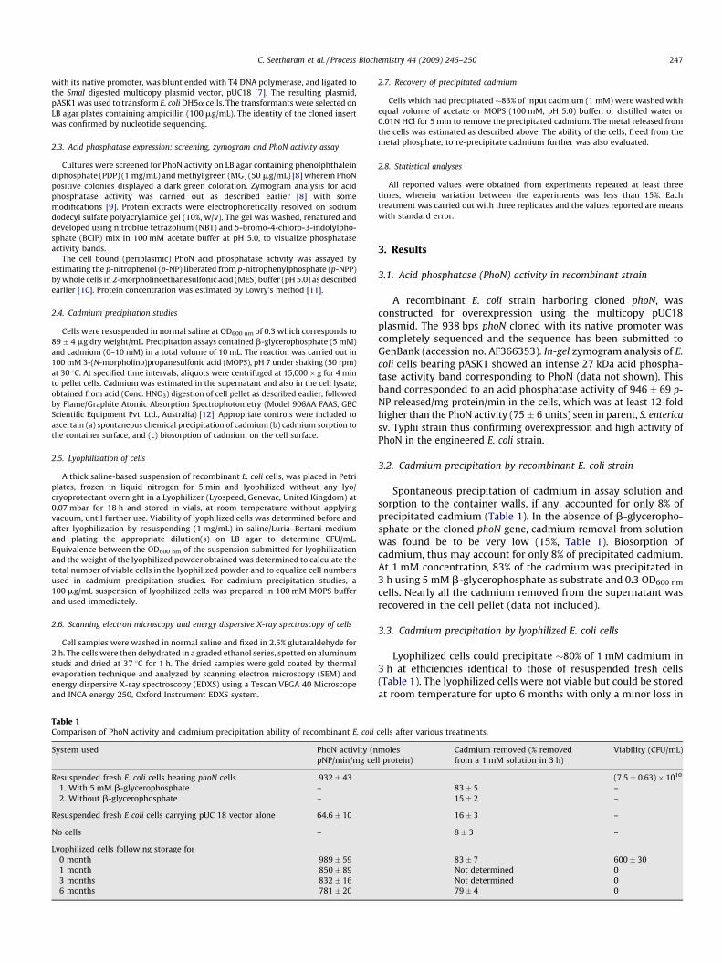
C. Seetharam et al. / Process Biochemistry 44 (2009) 246–250 247
with its native promoter, was blunt ended with T4 DNA polymerase, and ligated to
the SmaI digested multicopy plasmid vector, pUC18 [7]. The resulting plasmid,
pASK1 was used to transform E. coli DH5a cells. The transformants were selected on
LB agar plates containing ampicillin (100 mg/mL). The identity of the cloned insert
was confirmed by nucleotide sequencing.
2.3. Acid phosphatase expression: screening, zymogram and PhoN activity assay
Cultures were screened for PhoN activity on LB agar containing phenolphthalein
diphosphate (PDP) (1 mg/mL) and methyl green (MG) (50 mg/mL) [8] wherein PhoN
positive colonies displayed a dark green coloration. Zymogram analysis for acid
phosphatase activity was carried out as described earlier [8] with some
modifications [9]. Protein extracts were electrophoretically resolved on sodium
dodecyl sulfate polyacrylamide gel (10%, w/v). The gel was washed, renatured and
developed using nitroblue tetrazolium (NBT) and 5-bromo-4-chloro-3-indolylpho-
sphate (BCIP) mix in 100 mM acetate buffer at pH 5.0, to visualize phosphatase
activity bands.
The cell bound (periplasmic) PhoN acid phosphatase activity was assayed by
estimating the p-nitrophenol (p-NP) liberated from p-nitrophenylphosphate (p-NPP)
by whole cells in 2-morpholinoethanesulfonic acid (MES) buffer (pH 5.0) as described
earlier [10]. Protein concentration was estimated by Lowry’s method [11].
2.4. Cadmium precipitation studies
Cells were resuspended in normal saline at OD600 nm of 0.3 which corresponds to
89 � 4 mg dry weight/mL. Precipitation assays contained b-glycerophosphate (5 mM)
and cadmium (0–10 mM) in a total volume of 10 mL. The reaction was carried out in
100 mM 3-(N-morpholino)propanesulfonic acid (MOPS), pH 7 under shaking (50 rpm)
at 30 8C. At specified time intervals, aliquots were centrifuged at 15,000 � g for 4 min
to pellet cells. Cadmium was estimated in the supernatant and also in the cell lysate,
obtained from acid (Conc. HNO3) digestion of cell pellet as described earlier, followed
by Flame/Graphite Atomic Absorption Spectrophotometry (Model 906AA FAAS, GBC
Scientific Equipment Pvt. Ltd., Australia) [12]. Appropriate controls were included to
ascertain (a) spontaneous chemical precipitation of cadmium (b) cadmium sorption to
the container surface, and (c) biosorption of cadmium on the cell surface.
2.5. Lyophilization of cells
A thick saline-based suspension of recombinant E. coli cells, was placed in Petri
plates, frozen in liquid nitrogen for 5 min and lyophilized without any lyo/
cryoprotectant overnight in a Lyophilizer (Lyospeed, Genevac, United Kingdom) at
0.07 mbar for 18 h and stored in vials, at room temperature without applying
vacuum, until further use. Viability of lyophilized cells was determined before and
after lyophilization by resuspending (1 mg/mL) in saline/Luria–Bertani medium
and plating the appropriate dilution(s) on LB agar to determine CFU/mL.
Equivalence between the OD600 nm of the suspension submitted for lyophilization
and the weight of the lyophilized powder obtained was determined to calculate the
total number of viable cells in the lyophilized powder and to equalize cell numbers
used in cadmium precipitation studies. For cadmium precipitation studies, a
100 mg/mL suspension of lyophilized cells was prepared in 100 mM MOPS buffer
and used immediately.
2.6. Scanning electron microscopy and energy dispersive X-ray spectroscopy of cells
Cell samples were washed in normal saline and fixed in 2.5% glutaraldehyde for
2 h. The cells were then dehydrated in a graded ethanol series, spotted on aluminum
studs and dried at 37 8C for 1 h. The dried samples were gold coated by thermal
evaporation technique and analyzed by scanning electron microscopy (SEM) and
energy dispersive X-ray spectroscopy (EDXS) using a Tescan VEGA 40 Microscope
and INCA energy 250, Oxford Instrument EDXS system.
Table 1Comparison of PhoN activity and cadmium precipitation ability of recombinant E. coli
System used PhoN activity (n
pNP/min/mg ce
Resuspended fresh E. coli cells bearing phoN cells 932 � 43
1. With 5 mM b-glycerophosphate –
2. Without b-glycerophosphate –
Resuspended fresh E coli cells carrying pUC 18 vector alone 64.6 � 10
No cells –
Lyophilized cells following storage for
0 month 989 � 59
1 month 850 � 89
3 months 832 � 16
6 months 781 � 20
2.7. Recovery of precipitated cadmium
Cells which had precipitated �83% of input cadmium (1 mM) were washed with
equal volume of acetate or MOPS (100 mM, pH 5.0) buffer, or distilled water or
0.01N HCl for 5 min to remove the precipitated cadmium. The metal released from
the cells was estimated as described above. The ability of the cells, freed from the
metal phosphate, to re-precipitate cadmium further was also evaluated.
2.8. Statistical analyses
All reported values were obtained from experiments repeated at least three
times, wherein variation between the experiments was less than 15%. Each
treatment was carried out with three replicates and the values reported are means
with standard error.
3. Results
3.1. Acid phosphatase (PhoN) activity in recombinant strain
A recombinant E. coli strain harboring cloned phoN, wasconstructed for overexpression using the multicopy pUC18plasmid. The 938 bps phoN cloned with its native promoter wascompletely sequenced and the sequence has been submitted toGenBank (accession no. AF366353). In-gel zymogram analysis of E.
coli cells bearing pASK1 showed an intense 27 kDa acid phospha-tase activity band corresponding to PhoN (data not shown). Thisband corresponded to an acid phosphatase activity of 946 � 69 p-NP released/mg protein/min in the cells, which was at least 12-foldhigher than the PhoN activity (75 � 6 units) seen in parent, S. enterica
sv. Typhi strain thus confirming overexpression and high activity ofPhoN in the engineered E. coli strain.
3.2. Cadmium precipitation by recombinant E. coli strain
Spontaneous precipitation of cadmium in assay solution andsorption to the container walls, if any, accounted for only 8% ofprecipitated cadmium (Table 1). In the absence of b-glyceropho-sphate or the cloned phoN gene, cadmium removal from solutionwas found be to be very low (15%, Table 1). Biosorption ofcadmium, thus may account for only 8% of precipitated cadmium.At 1 mM concentration, 83% of the cadmium was precipitated in3 h using 5 mM b-glycerophosphate as substrate and 0.3 OD600 nm
cells. Nearly all the cadmium removed from the supernatant wasrecovered in the cell pellet (data not included).
3.3. Cadmium precipitation by lyophilized E. coli cells
Lyophilized cells could precipitate �80% of 1 mM cadmium in3 h at efficiencies identical to those of resuspended fresh cells(Table 1). The lyophilized cells were not viable but could be storedat room temperature for upto 6 months with only a minor loss in
cells after various treatments.
moles
ll protein)
Cadmium removed (% removed
from a 1 mM solution in 3 h)
Viability (CFU/mL)
(7.5 � 0.63) � 1010
83 � 5 –
15 � 2 –
16 � 3 –
8 � 3 –
83 � 7 600 � 30
Not determined 0
Not determined 0
79 � 4 0
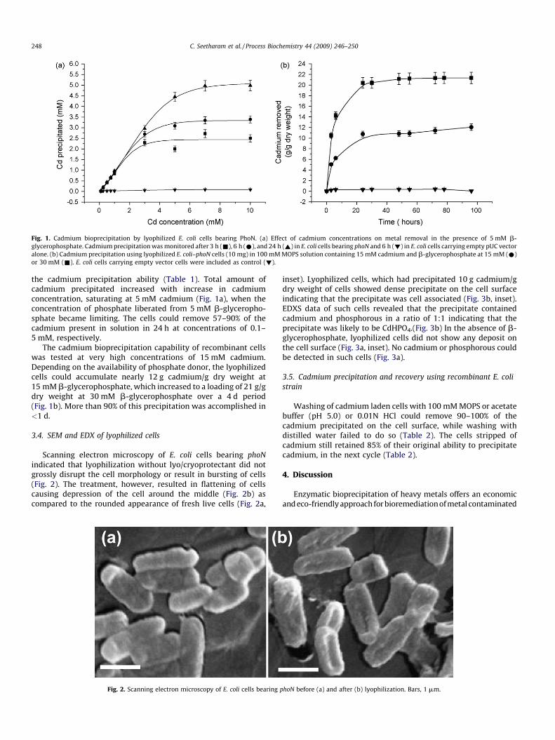
Fig. 1. Cadmium bioprecipitation by lyophilized E. coli cells bearing PhoN. (a) Effect of cadmium concentrations on metal removal in the presence of 5 mM b-
glycerophosphate. Cadmium precipitation was monitored after 3 h (&), 6 h (*), and 24 h (~) in E. coli cells bearing phoN and 6 h (!) in E. coli cells carrying empty pUC vector
alone. (b) Cadmium precipitation using lyophilized E. coli–phoN cells (10 mg) in 100 mM MOPS solution containing 15 mM cadmium and b-glycerophosphate at 15 mM (*)
or 30 mM (&). E. coli cells carrying empty vector cells were included as control (!).
C. Seetharam et al. / Process Biochemistry 44 (2009) 246–250248
the cadmium precipitation ability (Table 1). Total amount ofcadmium precipitated increased with increase in cadmiumconcentration, saturating at 5 mM cadmium (Fig. 1a), when theconcentration of phosphate liberated from 5 mM b-glyceropho-sphate became limiting. The cells could remove 57–90% of thecadmium present in solution in 24 h at concentrations of 0.1–5 mM, respectively.
The cadmium bioprecipitation capability of recombinant cellswas tested at very high concentrations of 15 mM cadmium.Depending on the availability of phosphate donor, the lyophilizedcells could accumulate nearly 12 g cadmium/g dry weight at15 mM b-glycerophosphate, which increased to a loading of 21 g/gdry weight at 30 mM b-glycerophosphate over a 4 d period(Fig. 1b). More than 90% of this precipitation was accomplished in<1 d.
3.4. SEM and EDX of lyophilized cells
Scanning electron microscopy of E. coli cells bearing phoN
indicated that lyophilization without lyo/cryoprotectant did notgrossly disrupt the cell morphology or result in bursting of cells(Fig. 2). The treatment, however, resulted in flattening of cellscausing depression of the cell around the middle (Fig. 2b) ascompared to the rounded appearance of fresh live cells (Fig. 2a,
Fig. 2. Scanning electron microscopy of E. coli cells bearing p
inset). Lyophilized cells, which had precipitated 10 g cadmium/gdry weight of cells showed dense precipitate on the cell surfaceindicating that the precipitate was cell associated (Fig. 3b, inset).EDXS data of such cells revealed that the precipitate containedcadmium and phosphorous in a ratio of 1:1 indicating that theprecipitate was likely to be CdHPO4.(Fig. 3b) In the absence of b-glycerophosphate, lyophilized cells did not show any deposit onthe cell surface (Fig. 3a, inset). No cadmium or phosphorous couldbe detected in such cells (Fig. 3a).
3.5. Cadmium precipitation and recovery using recombinant E. coli
strain
Washing of cadmium laden cells with 100 mM MOPS or acetatebuffer (pH 5.0) or 0.01N HCl could remove 90–100% of thecadmium precipitated on the cell surface, while washing withdistilled water failed to do so (Table 2). The cells stripped ofcadmium still retained 85% of their original ability to precipitatecadmium, in the next cycle (Table 2).
4. Discussion
Enzymatic bioprecipitation of heavy metals offers an economicand eco-friendly approach for bioremediation of metal contaminated
hoN before (a) and after (b) lyophilization. Bars, 1 mm.
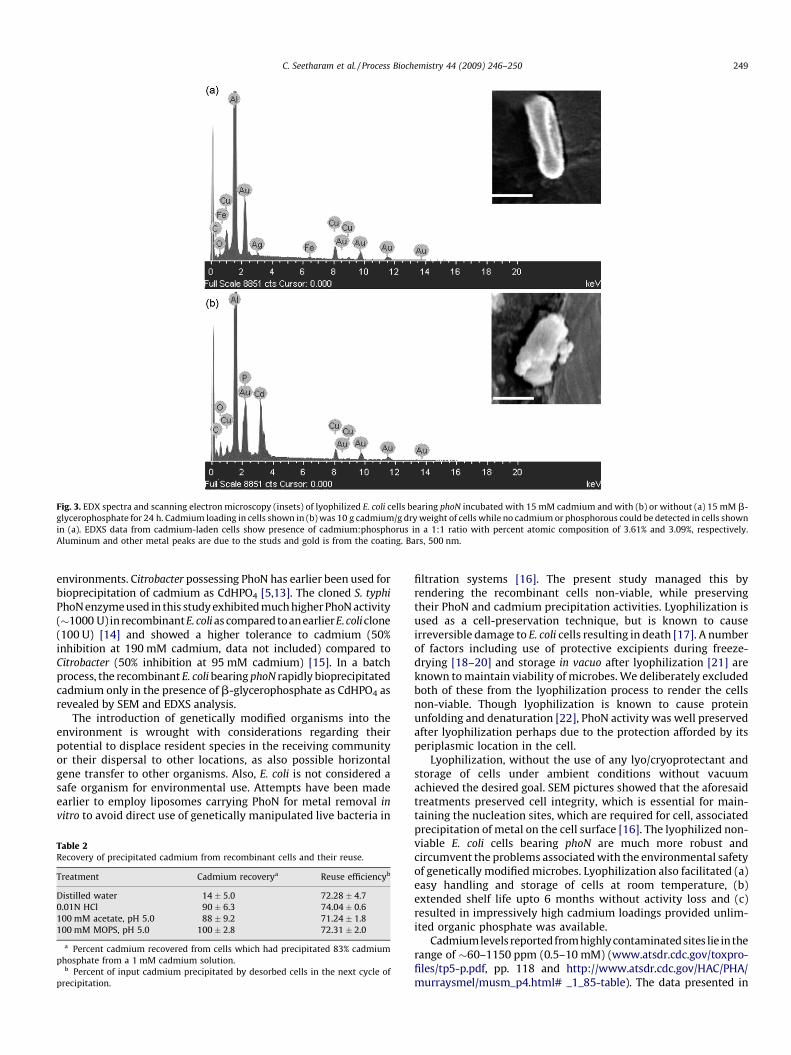
Fig. 3. EDX spectra and scanning electron microscopy (insets) of lyophilized E. coli cells bearing phoN incubated with 15 mM cadmium and with (b) or without (a) 15 mM b-
glycerophosphate for 24 h. Cadmium loading in cells shown in (b) was 10 g cadmium/g dry weight of cells while no cadmium or phosphorous could be detected in cells shown
in (a). EDXS data from cadmium-laden cells show presence of cadmium:phosphorus in a 1:1 ratio with percent atomic composition of 3.61% and 3.09%, respectively.
Aluminum and other metal peaks are due to the studs and gold is from the coating. Bars, 500 nm.
C. Seetharam et al. / Process Biochemistry 44 (2009) 246–250 249
environments. Citrobacter possessing PhoN has earlier been used forbioprecipitation of cadmium as CdHPO4 [5,13]. The cloned S. typhi
PhoN enzyme used in this study exhibited much higher PhoN activity(�1000 U) inrecombinantE. coli as compared toanearlier E.coliclone(100 U) [14] and showed a higher tolerance to cadmium (50%inhibition at 190 mM cadmium, data not included) compared toCitrobacter (50% inhibition at 95 mM cadmium) [15]. In a batchprocess, the recombinant E. coli bearing phoN rapidly bioprecipitatedcadmium only in the presence of b-glycerophosphate as CdHPO4 asrevealed by SEM and EDXS analysis.
The introduction of genetically modified organisms into theenvironment is wrought with considerations regarding theirpotential to displace resident species in the receiving communityor their dispersal to other locations, as also possible horizontalgene transfer to other organisms. Also, E. coli is not considered asafe organism for environmental use. Attempts have been madeearlier to employ liposomes carrying PhoN for metal removal in
vitro to avoid direct use of genetically manipulated live bacteria in
Table 2Recovery of precipitated cadmium from recombinant cells and their reuse.
Treatment Cadmium recoverya Reuse efficiencyb
Distilled water 14 � 5.0 72.28 � 4.7
0.01N HCl 90 � 6.3 74.04 � 0.6
100 mM acetate, pH 5.0 88 � 9.2 71.24 � 1.8
100 mM MOPS, pH 5.0 100 � 2.8 72.31 � 2.0
a Percent cadmium recovered from cells which had precipitated 83% cadmium
phosphate from a 1 mM cadmium solution.b Percent of input cadmium precipitated by desorbed cells in the next cycle of
precipitation.
filtration systems [16]. The present study managed this byrendering the recombinant cells non-viable, while preservingtheir PhoN and cadmium precipitation activities. Lyophilization isused as a cell-preservation technique, but is known to causeirreversible damage to E. coli cells resulting in death [17]. A numberof factors including use of protective excipients during freeze-drying [18–20] and storage in vacuo after lyophilization [21] areknown to maintain viability of microbes. We deliberately excludedboth of these from the lyophilization process to render the cellsnon-viable. Though lyophilization is known to cause proteinunfolding and denaturation [22], PhoN activity was well preservedafter lyophilization perhaps due to the protection afforded by itsperiplasmic location in the cell.
Lyophilization, without the use of any lyo/cryoprotectant andstorage of cells under ambient conditions without vacuumachieved the desired goal. SEM pictures showed that the aforesaidtreatments preserved cell integrity, which is essential for main-taining the nucleation sites, which are required for cell, associatedprecipitation of metal on the cell surface [16]. The lyophilized non-viable E. coli cells bearing phoN are much more robust andcircumvent the problems associated with the environmental safetyof genetically modified microbes. Lyophilization also facilitated (a)easy handling and storage of cells at room temperature, (b)extended shelf life upto 6 months without activity loss and (c)resulted in impressively high cadmium loadings provided unlim-ited organic phosphate was available.
Cadmiumlevels reported fromhighlycontaminated sites lie in therange of �60–1150 ppm (0.5–10 mM) (www.atsdr.cdc.gov/toxpro-files/tp5-p.pdf, pp. 118 and http://www.atsdr.cdc.gov/HAC/PHA/murraysmel/musm_p4.html# _1_85-table). The data presented in
C. Seetharam et al. / Process Biochemistry 44 (2009) 246–250250
this study show that PhoN bearing E. coli can effectively removecadmium from this concentration range. In addition, the ease ofrecovery of precipitated cadmium and the possibility of reuse of therecombinant cells makes this system very attractive, since the metal-laden cells settle down as a compact mass and facilitate easyseparation from the clear solution. This is in contrast to chemicalprecipitation by direct addition of phosphate, which may result in acolloidal suspension requiring extensive settling out [23]. Efforts areon to employ alternate substrates cheaper than b-glycerophosphate,to make the process more economical.
Acknowledgement
The authors wish to thank Ms. N. Padma for SEM and EDXanalyses.
References
[1] Valls M, de Lorenzo V. Exploiting the genetic and biochemical capabilities ofbacteria for the remediation of heavy metal pollution. FEMS Microbiol Rev2002;26:327–38.
[2] Eccles H. Treatment of metal-contaminated wastes: why select a biologicalprocess? Biotopic 1999;17:462–5.
[3] Macaskie LE. An immobilized cell bioprocess for the removal of heavy metalsfrom aqueous flows. J Chem Technol Biotechnol 1990;49:357–79.
[4] Macaskie LE, Dean ACR, Cheetham AK, Jakeman RJB, Skarnulis AJ. Cadmiumaccumulation by a Citrobacter sp., the chemical nature of the accumulatedmetal precipitate and its location on the bacterial cells. J Gen Microbiol1987;133:539–44.
[5] Pandey G, Paul D, Jain RK. Conceptualizing suicidal engineered microorgan-isms for bioremediation applications. Biochem Biophys Res Commun 2005;327:637–9.
[6] Kasahara M, Nakata A, Shingagawa H. Molecular analysis of the Salmonellatyphimurium phoN gene, which encodes nonspecific acid phosphatase. J Bac-teriol 1991;173:6760–5.
[7] Yanisch-Perron C, Vieira J, Messing J. Improved M13 phage cloning vectors andhost strains: nucleotide sequences of the M13mp18 and pUC19 vectors. Gene1985;33:103–19.
[8] Riccio ML, Rossolini GM, Lombardi G, Chiesurin A, Satta G. Expression cloningof different bacterial phosphatase-encoding genes by histochemical screeningof genomic libraries onto an indicator medium containing phenolphthaleindiphosphate and methyl green. J Appl Microbiol 1997;82:177–85.
[9] Appukuttan D, Rao AS, Apte SK. Engineering of Deinococcus radiodurans R1 forbioremediation of uranium from dilute nuclear waste. Appl Environ Microbiol2006;72:7873–8.
[10] Bolton PG, Dean ACR. Phosphatase synthesis in Klebsiella (Aerobacter) aero-genes growing in continuous cultures. J Biochem 1972;127:87–96.
[11] Lowry OH, Rosebrough NJ, Farr AL, Randall RJ. Protein measurement with theFolin phenol reagent. J Biol Chem 1951;193:265–75.
[12] Cordero B, Lodiero P, Herrero R, de Vicente MES. Biosorption of cadmium byFucus spiralis. Environ Chem 2004;1:180–7.
[13] Michel LJ, Macaskie LE, Dean ACR. Cadmium accumulation by immobilizedcells of a Citrobacter sp. using various phosphate donors. Biotechnol Bioeng1986;28:1358–65.
[14] Basnakova G, Stephens ER, Thaller MC, Rossolini GM, Macaskie LE. The use ofEscherichia coli bearing a phoN gene for the removal of uranium and nickelfrom aqueous flows. Appl Microbiol Biotechnol 1998;50:266–72.
[15] Hambling SG, Macaskie LE, Dean ACR. Phosphatase synthesis in a Citrobactersp. growing in continuous culture. J Gen Microbiol 1987;133:2743–9.
[16] Jeong BC, Hawes C, Bonthrone KM, Macaskie LE. Localization of enzymicallyenhanced heavy metal accumulation by Citrobacter sp. and metal accumula-tion in vitro by liposomes containing entrapped enzyme. Microbiology1997;143:2497–507.
[17] Israeli E, Kohn A, Gitelman J. The molecular nature of damage by oxygen tofreeze-dried Escherichia coli. Cryobiology 1975;12(1):15–25.
[18] Dejene AT, Rosen R, Pedazur R, Belkin S, Gunb J, Ekeltchik I, et al. Freeze-dryingof sol–gel encapsulated recombinant bioluminescent E. coli by using lyo-protectants. Sens Actuator B 2006;113:768–73.
[19] Zarate G, Nader-Macias ME. Viability and biological properties of probioticvaginal Lactobacilli after lyophilization and refrigerated storage into gelatincapsules. Process Biochem 2006;41:1779–85.
[20] Oterio ML, Espeche MC, Nader-Macias ME. Optimization of the freeze-dryingmedia and survival throughout storage of freeze-dried Lactobacillus gasseri andLactobacillus delbrueckii subsp. dellbrueckii for veterinarian probiotic applica-tions. Process Biochem 2007;42:1406–11.
[21] Morgan CA, Herman N, White PA, Vesey G. Preservation of micro-organisms bydrying; a review. J Microbiol Methods 2006;66:183–93.
[22] Tsutomu A, Steven JP, William CK, John FC. Factors affecting short-term andlong-term stabilities of proteins. Adv Drug Deliv Rev 2001;46:307–26.
[23] Macaskie LE, Jeong BC, Tolley MR. Enzymically accelerated biomineralizationof heavy metals: application to the removal of americium and plutonium fromaqueous flows. FEMS Microbiol Rev 1994;14:351–68.
Pu
Da
b
a
ARRAA
KLRDU
1
occtsatopaPtp1r1(
0d
Journal of Biotechnology 154 (2011) 285– 290
Contents lists available at ScienceDirect
Journal of Biotechnology
jou rn al hom epage: www.elsev ier .com/ locate / jb io tec
hoN-expressing, lyophilized, recombinant Deinococcus radiodurans cells forranium bioprecipitation
eepti Appukuttana,1, Chitra Seetharama,1, N. Padmab, Amara Sambasiva Raoa, Shree Kumar Aptea,∗
Molecular Biology Division, Bhabha Atomic Research Centre, Trombay, Mumbai 400085, IndiaTechnical Physics Division, Bhabha Atomic Research Centre, Trombay, Mumbai 400085, India
r t i c l e i n f o
rticle history:eceived 18 November 2010eceived in revised form 1 April 2011ccepted 9 May 2011vailable online 14 May 2011
a b s t r a c t
Employment of genetically engineered radiation resistant organisms to recover radionuclides/heavy met-als from radioactive wastes is an attractive proposition. Cells of recombinant Deinococcus radioduransstrain expressing, a non-specific acid phosphatase encoding phoN gene, were lyophilized. Lyophilizedrecombinant Deinococcus cells retained viability and PhoN activity and could efficiently precipitate ura-nium from aqueous solutions for up to six months of storage at room temperature. Batch process for
eywords:yophilizedecombinanteinococcus radioduransranium
uranium removal using lyophilized cells was more efficient compared to a flow through system, in termsof percent uranium removed, substrate conservation and time taken. Lyophilized recombinant Deinococ-cus cells exhibited high loading of up to 5.7 g uranium/g dry weight of cells in a batch process at 20 mMinput uranium concentration. Lyophilization deflated the cells but did not alter gross cell morphologyor surface nucleation capability of cells for uranium precipitation. The precipitated uranyl phosphateremained tightly associated with the cell surface, thus facilitating easy recovery.
© 2011 Elsevier B.V. All rights reserved.
. Introduction
Nuclear waste management has been a contentious aspectf nuclear power programs around the world. Radionuclide-ontaining wastes are produced at all steps in the nuclear fuelycle from milling and mining of uranium ores to fuel fabrica-ion, reactor operation and fuel reprocessing. Biological treatmenttrategies of waste have evoked considerable interest (Gadd, 2000)s viable and eco-friendly alternatives, especially at low concentra-ions of the radionuclide and for in situ bioremediation. Naturallyccurring organisms such as Citrobacter harboring a non-specificeriplasmic acid phosphatase PhoN, have been considered suit-ble for bioremediation of heavy metals (Macaskie et al., 1994).hoN hydrolyses organic phosphates and the inorganic phosphate,hus released, brings about metal precipitation as insoluble metalhosphate on the cell surface (Kier et al., 1977; Macaskie et al.,994). In the past, efficient bioprecipitation of uranium with Cit-
obacter strains (Macaskie, 1990; Jeong et al., 1997; Macaskie et al.,994) and recombinant Escherichia coli strains expressing PhoNBasnakova et al., 1998) have been reported. However, radioac-∗ Corresponding author. Tel.: +91 22 25595342; fax: +91 22 25505189.E-mail address: [email protected] (S.K. Apte).
1 These authors contributed equally to this work.
168-1656/$ – see front matter © 2011 Elsevier B.V. All rights reserved.oi:10.1016/j.jbiotec.2011.05.002
tivity restricts the survival, cellular integrity and functionality ofsuch microbes in nuclear wastes (Daly, 2000; Appukuttan et al.,2006).
The bacteria belonging to Deinococcaceae family have theextraordinary ability to withstand radiation doses up to 10–15 kGy(Battista, 1997; Daly, 2000). Recently our laboratory geneti-cally engineered Deinococcus radiodurans R1 to express phoN(Appukuttan et al., 2006). The recombinant strain, DrPhoN sur-vived extreme radiation stress, unlike recombinant E. coli-PhoNclones and precipitated uranium from dilute aqueous solutionsin high radiation environment. In order to extend the use of therecombinant Deinococcus strain for application in nuclear waste,the following factors need to be suitably addressed: (a) the ease ofhandling and transportation of biomass, (b) shelf-life and storageof cells, (c) mode of application, and (d) substrate conservation.Biological materials often must be dried to reduce bulk volumeand to stabilize them for storage or distribution. Drying causessignificant loss of activity and viability. But, freeze-drying, signif-icantly reduces such damage (Snowman, 1988; Seetharam et al.,2009).
The present study investigated the utility of lyophilization to
achieve aforesaid desirable features for PhoN expressing D. radio-durans strains. Our results show that lyophilized recombinantDeinococcus cells retained viability, PhoN activity as well as ura-nium precipitation ability up to 6 months of storage at room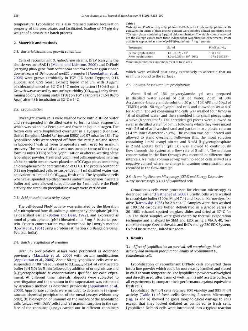
2 f Biotechnology 154 (2011) 285– 290
tpw
2
2
s(d2goGmA
2
awfUlirfloc0ewba
2
oant(P
2
p(sb�icb2tccf
Table 1Viability and PhoN activity of lyophilized DrPhoN cells. Fresh and lyophilized cellsequivalent in terms of their protein content were suitably diluted and plated ontoTGY agar plates containing 3 �g/ml chloramphenicol. The viable counts reportedare the average values from three independent lyophilization experiments. PhoNactivity is expressed as nmol of p-NP liberated min−1 mg−1 protein.
Treatment cfu/ml PhoN activity
Before lyophilization (1.1 ± 0.07) × 108 190 ± 10
86 D. Appukuttan et al. / Journal o
emperature. Lyophilized cells also retained surface localizationroperty of the precipitate, and facilitated, loading of 5.7 g/g dryeight of biomass in a batch process.
. Materials and methods
.1. Bacterial strains and growth conditions
Cells of recombinant D. radiodurans strains, DrEV (carrying thehuttle vector pRAD1) (Meima and Lidstrom, 2000) and DrPhoNcarrying phoN gene from Salmonella enterica serovar Typhi clonedownstream of Deinococcal groESL promoter) (Appukuttan et al.,006) were grown aerobically in TGY (1% Bacto Tryptone, 0.1%lucose, and 0.5% yeast extract) liquid medium with 3 �g/mlf chloramphenicol at 32 ◦C ± 1 ◦C under agitation (180 ± 5 rpm).rowth was assessed by measuring turbidity (OD600nm) or by deter-ining colony forming units (CFUs) on TGY agar plates (1.5% Bactogar) after 48 h incubation at 32 ◦C ± 1 ◦C.
.2. Lyophilization
Overnight grown cells were washed twice with distilled waternd re-suspended in distilled water to form a thick suspensionhich was taken in a Petri plate and frozen in liquid nitrogen. The
rozen cells were lyophilized overnight in a Lyospeed (Genevac,nited Kingdom, Model Refrigerant R502) at 0.07 mbar for 18 h. The
yophilized cells were scraped off from the Petri plate and storedn Eppendorf vials at room temperature until used for uraniumecovery. The survival of cells was measured in terms of the colonyorming units (CFUs) before lyophilization or after re-wetting of theyophilized powder. Fresh and lyophilized cells, equivalent in termsf their protein content were plated onto TGY agar plates containinghloramphenicol for determination of CFUs. The protein content of.33 mg lyophilized cells re-suspended in 1 ml distilled water wasquivalent to 1 ml of 1.0 OD600nm fresh cells. The lyophilized cellshen re-suspended rapidly formed a uniform suspension in acetate
uffer and were allowed to equilibrate for 5 min before the PhoNctivity and uranium precipitation assays were carried out.
.3. Acid phosphatase activity assays
The cell-bound PhoN activity was estimated by the liberationf p-nitrophenol from di-sodium p-nitrophenyl phosphate (pNPP),s described earlier (Bolton and Dean, 1972), and expressed asmol of p-nitrophenol (pNP) liberated min−1 mg−1 bacterial pro-ein. Protein concentration was determined by Lowry’s methodLowry et al., 1951) using a protein estimation kit (Bangalore Geneivt. Ltd., India).
.4. Batch precipitation of uranium
Uranium precipitation assays were performed as describedreviously (Macaskie et al., 2000) with certain modificationsAppukuttan et al., 2006). About 80 mg lyophilized cells were re-uspended in 100 ml (equivalent to O.D600nm of 2.4) of 2 mM acetateuffer (pH 5.0) for 5 min followed by addition of uranyl nitrate and-glycerophosphate at concentrations specified for each exper-
ment. At different time intervals, aliquots were subjected toentrifugation and the uranium in the supernatant was estimatedy Arsenazo method as described previously (Appukuttan et al.,006). Appropriate controls were included to determine (a) spon-
aneous chemical precipitation of the metal (assays without anyells), (b) biosorption of uranium on the surface of the lyophilizedells (assays with DrEV cells) and (c) uranium sorption to the sur-ace of the container (assays carried out in different containersAfter lyophilization (1.0 ± 0.056) × 108 (90%) 167 ± 5 (87.8%)
Values in parentheses indicate percent of fresh cells.
which were washed post assay extensively to ascertain that nouranium bound to the surface).
2.5. Column-based uranium precipitation
About 5 ml of 15% polyacrylamide gel was preparedin distilled water (2.4 ml of distilled water, 2.5 ml of 30%Acrylamide–bisacrylamide solution, 50 �l of 10% APS and 50 �l ofTEMED) with 150 mg of lyophilized cells and allowed to set at 4 ◦Cfor 45 min. The gel containing the cells was washed four times in10 ml distilled water and then shredded into small pieces usinga sieve (8 pores cm−1). The shredded gel pieces were allowed toswell in distilled water for 5 min, following which they were mixedwith 2.5 ml of acid washed sand and packed into a plastic column(1.4 cm inner diameter × 9 cm). The column was equilibrated andallowed to stand overnight. Following this, the input solutioncontaining 1 mM uranyl nitrate and 5 mM �-glycerophosphatein 2 mM acetate buffer (pH 5.0) was allowed to continuouslyflow through the system at a flow rate of 5 ml h−1. The uraniumconcentration in the flow through was recorded at different timeintervals. A similar column set-up with no added cells served as anegative control where no change in uranium concentration wasrecorded in the flow-through.
2.6. Scanning Electron Microscopy (SEM) and Energy DispersiveX-ray spectroscopy (EDX) of lyophilized cells
Deinococcus cells were processed for electron microscopy asdescribed earlier (Heather et al., 2006). Briefly, cells were washedin cacodylate buffer (100 mM, pH 7.4) and fixed in Karnovskys fix-ative (Karnovsky, 1965) for 2 h at 4 ◦C. Samples were then washedin 100 mM cacodylate buffer, dehydrated in a graded series of20–100% ethanol, spotted on glass slides and dried at 37 ◦C for1 h. The dried samples were gold coated by thermal evaporationtechnique and analyzed by SEM and EDX using a VEGA 40, Tes-can Microscope, Czechoslovakia and INCA energy 250 EDX System,Oxford Instrument, United Kingdom.
3. Results
3.1. Effect of lyophilization on survival, cell morphology, PhoNactivity and uranium precipitation ability of recombinant D.radiodurans cells
Lyophilization of recombinant DrPhoN cells converted theminto a fine powder which could be more easily handled and storedin vials at room temperature. The lyophilized powder was weighedand directly used, after 5 min of wetting in 2 mM acetate buffer, inall experiments to compare their performance against equivalentfresh cells.
Lyophilized DrPhoN cells retained 90% viability and 88% PhoN
activity (Table 1) of fresh cells. Scanning Electron Microscopy(Fig. 1a and b) showed no gross morphological damage to cellsexcept that they looked deflated as compared to fresh cells.Lyophilized DrPhoN cells were introduced into a typical reaction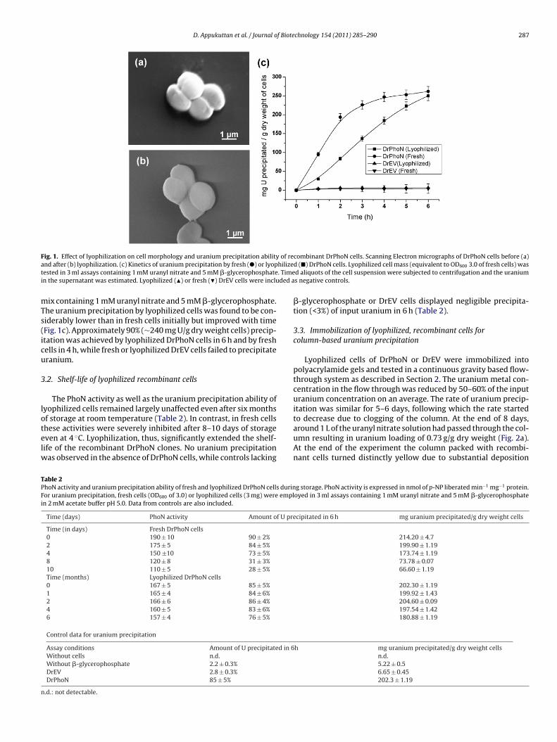
D. Appukuttan et al. / Journal of Biotechnology 154 (2011) 285– 290 287
Fig. 1. Effect of lyophilization on cell morphology and uranium precipitation ability of recombinant DrPhoN cells. Scanning Electron micrographs of DrPhoN cells before (a)a hilizet . Timei ded a
mTs(icu
3
lotelw
TPFi
n
nd after (b) lyophilization. (c) Kinetics of uranium precipitation by fresh (�) or lyopested in 3 ml assays containing 1 mM uranyl nitrate and 5 mM �-glycerophosphaten the supernatant was estimated. Lyophilized (�) or fresh (�) DrEV cells were inclu
ix containing 1 mM uranyl nitrate and 5 mM �-glycerophosphate.he uranium precipitation by lyophilized cells was found to be con-iderably lower than in fresh cells initially but improved with timeFig. 1c). Approximately 90% (∼240 mg U/g dry weight cells) precip-tation was achieved by lyophilized DrPhoN cells in 6 h and by freshells in 4 h, while fresh or lyophilized DrEV cells failed to precipitateranium.
.2. Shelf-life of lyophilized recombinant cells
The PhoN activity as well as the uranium precipitation ability ofyophilized cells remained largely unaffected even after six monthsf storage at room temperature (Table 2). In contrast, in fresh cells
hese activities were severely inhibited after 8–10 days of storageven at 4 ◦C. Lyophilization, thus, significantly extended the shelf-ife of the recombinant DrPhoN clones. No uranium precipitationas observed in the absence of DrPhoN cells, while controls lacking
able 2hoN activity and uranium precipitation ability of fresh and lyophilized DrPhoN cells durinor uranium precipitation, fresh cells (OD600 of 3.0) or lyophilized cells (3 mg) were empln 2 mM acetate buffer pH 5.0. Data from controls are also included.
Time (days) PhoN activity Amount of U pre
Time (in days) Fresh DrPhoN cells0 190 ± 10 90 ± 2%
2 175 ± 5 84 ± 5%
4 150 ±10 73 ± 5%
8 120 ± 8 31 ± 3%
10 110 ± 5 28 ± 5%
Time (months) Lyophilized DrPhoN cells0 167 ± 5 85 ± 5%
1 165 ± 4 84 ± 6%
2 166 ± 6 86 ± 4%
4 160 ± 5 83 ± 6%
6 157 ± 4 76 ± 5%
Control data for uranium precipitation
Assay conditions Amount of U precipitated in 6Without cells n.d.
Without �-glycerophosphate 2.2 ± 0.3%
DrEV 2.8 ± 0.3%
DrPhoN 85 ± 5%
.d.: not detectable.
d (�) DrPhoN cells. Lyophilized cell mass (equivalent to OD600 3.0 of fresh cells) wasd aliquots of the cell suspension were subjected to centrifugation and the uraniums negative controls.
�-glycerophosphate or DrEV cells displayed negligible precipita-tion (<3%) of input uranium in 6 h (Table 2).
3.3. Immobilization of lyophilized, recombinant cells forcolumn-based uranium precipitation
Lyophilized cells of DrPhoN or DrEV were immobilized intopolyacrylamide gels and tested in a continuous gravity based flow-through system as described in Section 2. The uranium metal con-centration in the flow through was reduced by 50–60% of the inputuranium concentration on an average. The rate of uranium precip-itation was similar for 5–6 days, following which the rate startedto decrease due to clogging of the column. At the end of 8 days,
around 1 L of the uranyl nitrate solution had passed through the col-umn resulting in uranium loading of 0.73 g/g dry weight (Fig. 2a).At the end of the experiment the column packed with recombi-nant cells turned distinctly yellow due to substantial depositiong storage. PhoN activity is expressed in nmol of p-NP liberated min−1 mg−1 protein.oyed in 3 ml assays containing 1 mM uranyl nitrate and 5 mM �-glycerophosphate
cipitated in 6 h mg uranium precipitated/g dry weight cells
214.20 ± 4.7199.90 ± 1.19173.74 ± 1.1973.78 ± 0.0766.60 ± 1.19
202.30 ± 1.19199.92 ± 1.43204.60 ± 0.09197.54 ± 1.42180.88 ± 1.19
h mg uranium precipitated/g dry weight cellsn.d.5.22 ± 0.56.65 ± 0.45202.3 ± 1.19
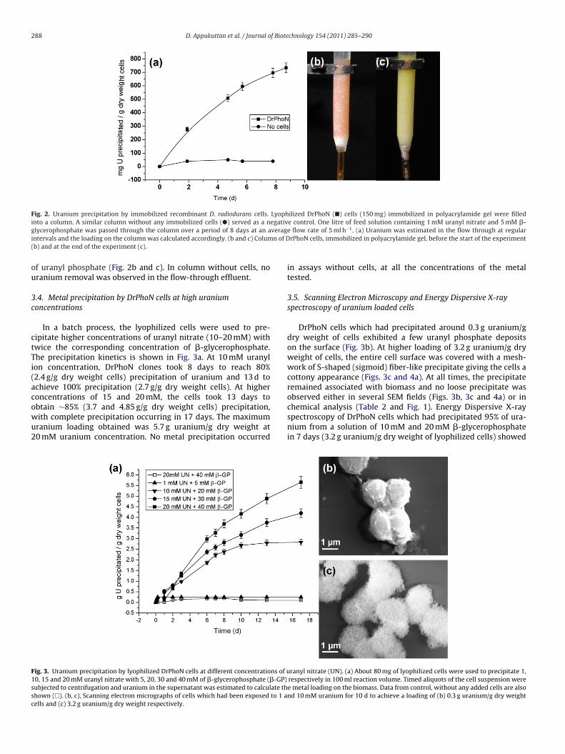
288 D. Appukuttan et al. / Journal of Biotechnology 154 (2011) 285– 290
Fig. 2. Uranium precipitation by immobilized recombinant D. radiodurans cells. Lyophilized DrPhoN (�) cells (150 mg) immobilized in polyacrylamide gel were filledinto a column. A similar column without any immobilized cells (�) served as a negative control. One litre of feed solution containing 1 mM uranyl nitrate and 5 mM �-g veragi n of D(
ou
3c
ctTi(acowu2
F1ssc
lycerophosphate was passed through the column over a period of 8 days at an antervals and the loading on the column was calculated accordingly. (b and c) Columb) and at the end of the experiment (c).
f uranyl phosphate (Fig. 2b and c). In column without cells, noranium removal was observed in the flow-through effluent.
.4. Metal precipitation by DrPhoN cells at high uraniumoncentrations
In a batch process, the lyophilized cells were used to pre-ipitate higher concentrations of uranyl nitrate (10–20 mM) withwice the corresponding concentration of �-glycerophosphate.he precipitation kinetics is shown in Fig. 3a. At 10 mM uranylon concentration, DrPhoN clones took 8 days to reach 80%2.4 g/g dry weight cells) precipitation of uranium and 13 d tochieve 100% precipitation (2.7 g/g dry weight cells). At higheroncentrations of 15 and 20 mM, the cells took 13 days to
btain ∼85% (3.7 and 4.85 g/g dry weight cells) precipitation,ith complete precipitation occurring in 17 days. The maximumranium loading obtained was 5.7 g uranium/g dry weight at0 mM uranium concentration. No metal precipitation occurredig. 3. Uranium precipitation by lyophilized DrPhoN cells at different concentrations of u0, 15 and 20 mM uranyl nitrate with 5, 20, 30 and 40 mM of �-glycerophosphate (�-GP)
ubjected to centrifugation and uranium in the supernatant was estimated to calculate thhown (�). (b, c), Scanning electron micrographs of cells which had been exposed to 1 aells and (c) 3.2 g uranium/g dry weight respectively.
e flow rate of 5 ml h−1. (a) Uranium was estimated in the flow through at regularrPhoN cells, immobilized in polyacrylamide gel, before the start of the experiment
in assays without cells, at all the concentrations of the metaltested.
3.5. Scanning Electron Microscopy and Energy Dispersive X-rayspectroscopy of uranium loaded cells
DrPhoN cells which had precipitated around 0.3 g uranium/gdry weight of cells exhibited a few uranyl phosphate depositson the surface (Fig. 3b). At higher loading of 3.2 g uranium/g dryweight of cells, the entire cell surface was covered with a mesh-work of S-shaped (sigmoid) fiber-like precipitate giving the cells acottony appearance (Figs. 3c and 4a). At all times, the precipitateremained associated with biomass and no loose precipitate wasobserved either in several SEM fields (Figs. 3b, 3c and 4a) or in
chemical analysis (Table 2 and Fig. 1). Energy Dispersive X-rayspectroscopy of DrPhoN cells which had precipitated 95% of ura-nium from a solution of 10 mM and 20 mM �-glycerophosphatein 7 days (3.2 g uranium/g dry weight of lyophilized cells) showedranyl nitrate (UN). (a) About 80 mg of lyophilized cells were used to precipitate 1,respectively in 100 ml reaction volume. Timed aliquots of the cell suspension weree metal loading on the biomass. Data from control, without any added cells are alsond 10 mM uranium for 10 d to achieve a loading of (b) 0.3 g uranium/g dry weight
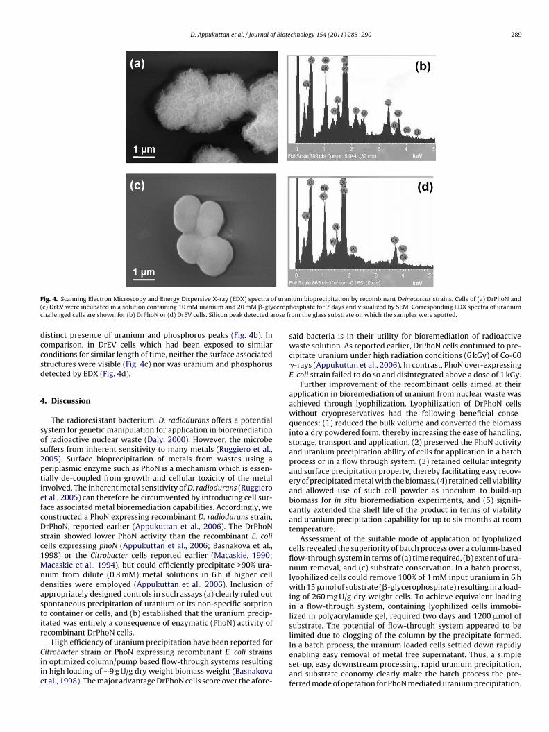
D. Appukuttan et al. / Journal of Biotechnology 154 (2011) 285– 290 289
F f uran( ceropc rose fr
dccsd
4
sos2ptiefcDsc1Mndastir
Ciie
ig. 4. Scanning Electron Microscopy and Energy Dispersive X-ray (EDX) spectra oc) DrEV were incubated in a solution containing 10 mM uranium and 20 mM �-glyhallenged cells are shown for (b) DrPhoN or (d) DrEV cells. Silicon peak detected a
istinct presence of uranium and phosphorus peaks (Fig. 4b). Inomparison, in DrEV cells which had been exposed to similaronditions for similar length of time, neither the surface associatedtructures were visible (Fig. 4c) nor was uranium and phosphorusetected by EDX (Fig. 4d).
. Discussion
The radioresistant bacterium, D. radiodurans offers a potentialystem for genetic manipulation for application in bioremediationf radioactive nuclear waste (Daly, 2000). However, the microbeuffers from inherent sensitivity to many metals (Ruggiero et al.,005). Surface bioprecipitation of metals from wastes using aeriplasmic enzyme such as PhoN is a mechanism which is essen-ially de-coupled from growth and cellular toxicity of the metalnvolved. The inherent metal sensitivity of D. radiodurans (Ruggierot al., 2005) can therefore be circumvented by introducing cell sur-ace associated metal bioremediation capabilities. Accordingly, weonstructed a PhoN expressing recombinant D. radiodurans strain,rPhoN, reported earlier (Appukuttan et al., 2006). The DrPhoN
train showed lower PhoN activity than the recombinant E. coliells expressing phoN (Appukuttan et al., 2006; Basnakova et al.,998) or the Citrobacter cells reported earlier (Macaskie, 1990;acaskie et al., 1994), but could efficiently precipitate >90% ura-
ium from dilute (0.8 mM) metal solutions in 6 h if higher cellensities were employed (Appukuttan et al., 2006). Inclusion ofppropriately designed controls in such assays (a) clearly ruled outpontaneous precipitation of uranium or its non-specific sorptiono container or cells, and (b) established that the uranium precip-tated was entirely a consequence of enzymatic (PhoN) activity ofecombinant DrPhoN cells.
High efficiency of uranium precipitation have been reported for
itrobacter strain or PhoN expressing recombinant E. coli strainsn optimized column/pump based flow-through systems resultingn high loading of ∼9 g U/g dry weight biomass weight (Basnakovat al., 1998). The major advantage DrPhoN cells score over the afore-
ium bioprecipitation by recombinant Deinococcus strains. Cells of (a) DrPhoN andhosphate for 7 days and visualized by SEM. Corresponding EDX spectra of uraniumom the glass substrate on which the samples were spotted.
said bacteria is in their utility for bioremediation of radioactivewaste solution. As reported earlier, DrPhoN cells continued to pre-cipitate uranium under high radiation conditions (6 kGy) of Co-60�-rays (Appukuttan et al., 2006). In contrast, PhoN over-expressingE. coli strain failed to do so and disintegrated above a dose of 1 kGy.
Further improvement of the recombinant cells aimed at theirapplication in bioremediation of uranium from nuclear waste wasachieved through lyophilization. Lyophilization of DrPhoN cellswithout cryopreservatives had the following beneficial conse-quences: (1) reduced the bulk volume and converted the biomassinto a dry powdered form, thereby increasing the ease of handling,storage, transport and application, (2) preserved the PhoN activityand uranium precipitation ability of cells for application in a batchprocess or in a flow through system, (3) retained cellular integrityand surface precipitation property, thereby facilitating easy recov-ery of precipitated metal with the biomass, (4) retained cell viabilityand allowed use of such cell powder as inoculum to build-upbiomass for in situ bioremediation experiments, and (5) signifi-cantly extended the shelf life of the product in terms of viabilityand uranium precipitation capability for up to six months at roomtemperature.
Assessment of the suitable mode of application of lyophilizedcells revealed the superiority of batch process over a column-basedflow-through system in terms of (a) time required, (b) extent of ura-nium removal, and (c) substrate conservation. In a batch process,lyophilized cells could remove 100% of 1 mM input uranium in 6 hwith 15 �mol of substrate (�-glycerophosphate) resulting in a load-ing of 260 mg U/g dry weight cells. To achieve equivalent loadingin a flow-through system, containing lyophilized cells immobi-lized in polyacrylamide gel, required two days and 1200 �mol ofsubstrate. The potential of flow-through system appeared to belimited due to clogging of the column by the precipitate formed.In a batch process, the uranium loaded cells settled down rapidlyenabling easy removal of metal free supernatant. Thus, a simple
set-up, easy downstream processing, rapid uranium precipitation,and substrate economy clearly make the batch process the pre-ferred mode of operation for PhoN mediated uranium precipitation.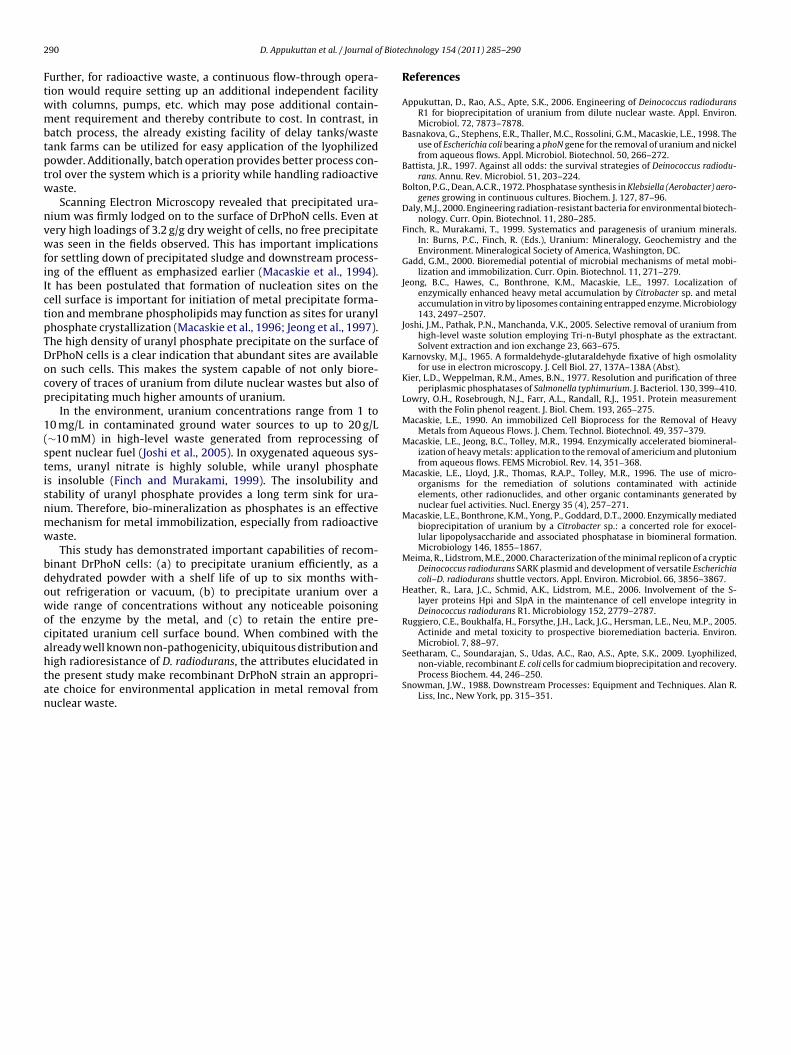
2 f Biote
Ftwmbtptw
nvwfiIctpTDocp
1(stisnmw
bdowocahtan
Seetharam, C., Soundarajan, S., Udas, A.C., Rao, A.S., Apte, S.K., 2009. Lyophilized,
90 D. Appukuttan et al. / Journal o
urther, for radioactive waste, a continuous flow-through opera-ion would require setting up an additional independent facilityith columns, pumps, etc. which may pose additional contain-ent requirement and thereby contribute to cost. In contrast, in
atch process, the already existing facility of delay tanks/wasteank farms can be utilized for easy application of the lyophilizedowder. Additionally, batch operation provides better process con-rol over the system which is a priority while handling radioactiveaste.
Scanning Electron Microscopy revealed that precipitated ura-ium was firmly lodged on to the surface of DrPhoN cells. Even atery high loadings of 3.2 g/g dry weight of cells, no free precipitateas seen in the fields observed. This has important implications
or settling down of precipitated sludge and downstream process-ng of the effluent as emphasized earlier (Macaskie et al., 1994).t has been postulated that formation of nucleation sites on theell surface is important for initiation of metal precipitate forma-ion and membrane phospholipids may function as sites for uranylhosphate crystallization (Macaskie et al., 1996; Jeong et al., 1997).he high density of uranyl phosphate precipitate on the surface ofrPhoN cells is a clear indication that abundant sites are availablen such cells. This makes the system capable of not only biore-overy of traces of uranium from dilute nuclear wastes but also ofrecipitating much higher amounts of uranium.
In the environment, uranium concentrations range from 1 to0 mg/L in contaminated ground water sources to up to 20 g/L∼10 mM) in high-level waste generated from reprocessing ofpent nuclear fuel (Joshi et al., 2005). In oxygenated aqueous sys-ems, uranyl nitrate is highly soluble, while uranyl phosphates insoluble (Finch and Murakami, 1999). The insolubility andtability of uranyl phosphate provides a long term sink for ura-ium. Therefore, bio-mineralization as phosphates is an effectiveechanism for metal immobilization, especially from radioactiveaste.
This study has demonstrated important capabilities of recom-inant DrPhoN cells: (a) to precipitate uranium efficiently, as aehydrated powder with a shelf life of up to six months with-ut refrigeration or vacuum, (b) to precipitate uranium over aide range of concentrations without any noticeable poisoning
f the enzyme by the metal, and (c) to retain the entire pre-ipitated uranium cell surface bound. When combined with thelready well known non-pathogenicity, ubiquitous distribution and
igh radioresistance of D. radiodurans, the attributes elucidated inhe present study make recombinant DrPhoN strain an appropri-te choice for environmental application in metal removal fromuclear waste.chnology 154 (2011) 285– 290
References
Appukuttan, D., Rao, A.S., Apte, S.K., 2006. Engineering of Deinococcus radioduransR1 for bioprecipitation of uranium from dilute nuclear waste. Appl. Environ.Microbiol. 72, 7873–7878.
Basnakova, G., Stephens, E.R., Thaller, M.C., Rossolini, G.M., Macaskie, L.E., 1998. Theuse of Escherichia coli bearing a phoN gene for the removal of uranium and nickelfrom aqueous flows. Appl. Microbiol. Biotechnol. 50, 266–272.
Battista, J.R., 1997. Against all odds: the survival strategies of Deinococcus radiodu-rans. Annu. Rev. Microbiol. 51, 203–224.
Bolton, P.G., Dean, A.C.R., 1972. Phosphatase synthesis in Klebsiella (Aerobacter) aero-genes growing in continuous cultures. Biochem. J. 127, 87–96.
Daly, M.J., 2000. Engineering radiation-resistant bacteria for environmental biotech-nology. Curr. Opin. Biotechnol. 11, 280–285.
Finch, R., Murakami, T., 1999. Systematics and paragenesis of uranium minerals.In: Burns, P.C., Finch, R. (Eds.), Uranium: Mineralogy, Geochemistry and theEnvironment. Mineralogical Society of America, Washington, DC.
Gadd, G.M., 2000. Bioremedial potential of microbial mechanisms of metal mobi-lization and immobilization. Curr. Opin. Biotechnol. 11, 271–279.
Jeong, B.C., Hawes, C., Bonthrone, K.M., Macaskie, L.E., 1997. Localization ofenzymically enhanced heavy metal accumulation by Citrobacter sp. and metalaccumulation in vitro by liposomes containing entrapped enzyme. Microbiology143, 2497–2507.
Joshi, J.M., Pathak, P.N., Manchanda, V.K., 2005. Selective removal of uranium fromhigh-level waste solution employing Tri-n-Butyl phosphate as the extractant.Solvent extraction and ion exchange 23, 663–675.
Karnovsky, M.J., 1965. A formaldehyde-glutaraldehyde fixative of high osmolalityfor use in electron microscopy. J. Cell Biol. 27, 137A–138A (Abst).
Kier, L.D., Weppelman, R.M., Ames, B.N., 1977. Resolution and purification of threeperiplasmic phosphatases of Salmonella typhimurium. J. Bacteriol. 130, 399–410.
Lowry, O.H., Rosebrough, N.J., Farr, A.L., Randall, R.J., 1951. Protein measurementwith the Folin phenol reagent. J. Biol. Chem. 193, 265–275.
Macaskie, L.E., 1990. An immobilized Cell Bioprocess for the Removal of HeavyMetals from Aqueous Flows. J. Chem. Technol. Biotechnol. 49, 357–379.
Macaskie, L.E., Jeong, B.C., Tolley, M.R., 1994. Enzymically accelerated biomineral-ization of heavy metals: application to the removal of americium and plutoniumfrom aqueous flows. FEMS Microbiol. Rev. 14, 351–368.
Macaskie, L.E., Lloyd, J.R., Thomas, R.A.P., Tolley, M.R., 1996. The use of micro-organisms for the remediation of solutions contaminated with actinideelements, other radionuclides, and other organic contaminants generated bynuclear fuel activities. Nucl. Energy 35 (4), 257–271.
Macaskie, L.E., Bonthrone, K.M., Yong, P., Goddard, D.T., 2000. Enzymically mediatedbioprecipitation of uranium by a Citrobacter sp.: a concerted role for exocel-lular lipopolysaccharide and associated phosphatase in biomineral formation.Microbiology 146, 1855–1867.
Meima, R., Lidstrom, M.E., 2000. Characterization of the minimal replicon of a crypticDeinococcus radiodurans SARK plasmid and development of versatile Escherichiacoli–D. radiodurans shuttle vectors. Appl. Environ. Microbiol. 66, 3856–3867.
Heather, R., Lara, J.C., Schmid, A.K., Lidstrom, M.E., 2006. Involvement of the S-layer proteins Hpi and SlpA in the maintenance of cell envelope integrity inDeinococcus radiodurans R1. Microbiology 152, 2779–2787.
Ruggiero, C.E., Boukhalfa, H., Forsythe, J.H., Lack, J.G., Hersman, L.E., Neu, M.P., 2005.Actinide and metal toxicity to prospective bioremediation bacteria. Environ.Microbiol. 7, 88–97.
non-viable, recombinant E. coli cells for cadmium bioprecipitation and recovery.Process Biochem. 44, 246–250.
Snowman, J.W., 1988. Downstream Processes: Equipment and Techniques. Alan R.Liss, Inc., New York, pp. 315–351.
Recombinant D. radiodurans cells for bioremediation of heavy metalsfrom acidic/neutral aqueous wastes
Chitra Seetharam Misra,1,† Deepti Appukuttan,1,† Venkata Siva Satyanarayana Kantamreddi,2 Amara S. Rao1 and Shree Kumar Apte1,*1Molecular Biology Division, Bhabha Atomic Research Centre; Mumbai, India; 2Department of Chemistry, GITAM University; Hyderabad, India
†These authors contributed equally to the work.
Keywords: D. radiodurans, heavy metals,bioremediation, radioactive waste,lyophilized, PhoN
Submitted: 08/31/11
Revised: 11/26/11
Accepted: 11/28/11
http://dx.doi.org/10.4161/bbug.3.1.18878*Correspondence to: Shree Kumar Apte;Email: [email protected]
The stability and superior metal bio-remediation ability of genetically
engineered Deinococcus radiodurans cells,expressing a non-specific acid phospha-tase, PhoN in high radiation environ-ment has already been established. Thelyophilized recombinant DrPhoN cellsretained PhoN activity and uraniumprecipitation ability. Such cells also dis-played an extended shelf life of 6 monthsduring storage at room temperature andshowed surface associated precipitation ofuranium as well as other metals likecadmium. Lyophilized cells, immobilizedin polyacrylamide gels could be used foruranium bioprecipitation in a flowthrough system resulting in 70% removalfrom 1mM input uranium solution and aloading of 1 g uranium/g dry weight cells.Compared with a batch process whichachieved a loading of 5.7 g uranium/gbiomass, the efficiency of the columnprocess was low due to clogging of thecolumn by the precipitate.
Introduction
Bacteria like Citrobacter and Pseudomonassp. have the ability to transform, detoxifyor immobilize a variety of metallic andorganic pollutants.1,2 However, like mostorganisms, these bacteria are very sensitiveto ionizing radiation, and their use forbioremediation in high radiation environ-ments is very limited. Aqueous liquidnuclear waste generated during the repro-cessing of spent fuel rods contains highlevels of radioactivity.3 Actinides presentin this waste along with other fission
products emit highly damaging beta andgamma radiation. Use of radio-resistantmicrobes is an essential prerequisite forbioremediation in such an environment.
In recent years, the extremely radio-resistant Gram positive bacterium, Deinoco-ccus radiodurans, has become an organismof choice for engineering a number ofstrategies for bioremediation of radioactivewaste.4-6 The genus, Deinococcus comprisesof a large number of radioresistant species,many of which also display dessication,temperature and metal tolerance (D. deserti,D. geothermalis and D. indicus).7-9 Themost studied, D. radiodurans strain R1has a small 3.28 Mb genome comprisedof two chromosomes (2.64 Mb and0.41 Mb), a megaplasmid (0.18 Mb) andplasmid (0.045 Mb) and can withstandexposure to 5–6 kGy of ionizing radiationwithout significant loss of viability.10
Such extreme radioresistance of the strainstems from its phenomenal DNA repairproficiency. Multiplicity of genome orga-nized in a torroid ring-like structure,possession of novel genes for repair ofdamaged DNA (Extended Synthesis-Dependent Strand Annealing, PprAetc.), Mn(II) complexes which protectcellular enzymes from oxidative stressdamage and efficient damage cleaningsystems that restore homeostasis post-irradiation, all contribute to radioresis-tance.11-15 Comparative genomes of atleast three deinococcal strains has revealedcommonality of genes, molecular mechan-isms underlying radioresistance across thespecies.16
Attempts to introduce novel bioreme-diation capabilities into D. radiodurans
Bioengineered Bugs 3:1, 44–48; January/February 2012; G 2012 Landes Bioscience
44 Bioengineered Bugs Volume 3 Issue 1
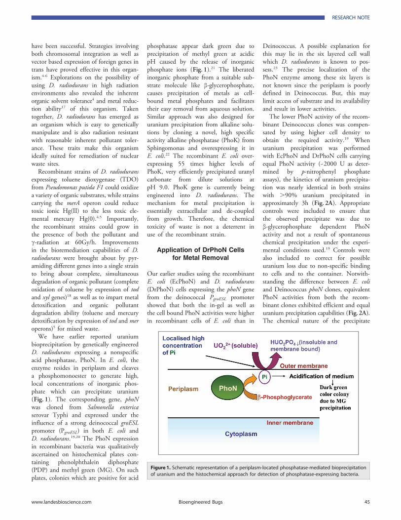
have been successful. Strategies involvingboth chromosomal integration as well asvector based expression of foreign genes intrans have proved effective in this organ-ism.4-6 Explorations on the possibility ofusing D. radiodurans in high radiationenvironments also revealed the inherentorganic solvent tolerance4 and metal reduc-tion ability17 of this organism. Takentogether, D. radiodurans has emerged asan organism which is easy to geneticallymanipulate and is also radiation resistantwith reasonable inherent pollutant toler-ance. These traits make this organismideally suited for remediation of nuclearwaste sites.
Recombinant strains of D. radioduransexpressing toluene dioxygenase (TDO)from Pseudomonas putida F1 could oxidizea variety of organic substrates, while strainscarrying the merA operon could reducetoxic ionic Hg(II) to the less toxic ele-mental mercury Hg(0).4,5 Importantly,the recombinant strains could grow inthe presence of both the pollutant andc-radiation at 60Gy/h. Improvementsin the bioremediation capabilities of D.radiodurans were brought about by pyr-amiding different genes into a single strainto bring about complete, simultaneousdegradation of organic pollutant (completeoxidation of toluene by expression of todand xyl genes)18 as well as to impart metaldetoxification and organic pollutantdegradation ability (toluene and mercurydetoxification by expression of tod and meroperons)5 for mixed waste.
We have earlier reported uraniumbioprecipitation by genetically engineeredD. radiodurans expressing a nonspecificacid phosphatase, PhoN. In E. coli, theenzyme resides in periplasm and cleavesa phosphomonoester to generate high,local concentrations of inorganic phos-phate which can precipitate uranium(Fig. 1). The corresponding gene, phoNwas cloned from Salmonella entericaserovar Typhi and expressed under theinfluence of a strong deinococcal groESLpromoter (PgroESL) in both E. coli andD. radiodurans.19,20 The PhoN expressionin recombinant bacteria was qualitativelyascertained on histochemical plates con-taining phenolphthalein diphosphate(PDP) and methyl green (MG). On suchplates, colonies which are positive for acid
phosphatase appear dark green due toprecipitation of methyl green at acidicpH caused by the release of inorganicphosphate ions (Fig. 1).21 The liberatedinorganic phosphate from a suitable sub-strate molecule like β-glycerophosphate,causes precipitation of metals as cell-bound metal phosphates and facilitatestheir easy removal from aqueous solution.Similar approach was also designed foruranium precipitation from alkaline solu-tions by cloning a novel, high specificactivity alkaline phosphatase (PhoK) fromSphingomonas and overexpressing it inE. coli.22 The recombinant E. coli over-expressing 55 times higher levels ofPhoK, very efficiently precipitated uranylcarbonate from dilute solutions atpH 9.0. PhoK gene is currently beingengineered into D. radiodurans. Themechanism for metal precipitation isessentially extracellular and de-coupledfrom growth. Therefore, the chemicaltoxicity of waste is not a deterrent inuse of the recombinant strain.
Application of DrPhoN Cellsfor Metal Removal
Our earlier studies using the recombinantE. coli (EcPhoN) and D. radiodurans(DrPhoN) cells expressing the phoN genefrom the deinococcal PgroESL promotershowed that both the in-gel as well asthe cell bound PhoN activities were higherin recombinant cells of E. coli than in
Deinococcus. A possible explanation forthis may lie in the six layered cell wallwhich D. radiodurans is known to pos-sess.23 The precise localization of thePhoN enzyme among these six layers isnot known since the periplasm is poorlydefined in Deinococcus. But, this maylimit access of substrate and its availabilityand result in lower activities.
The lower PhoN activity of the recom-binant Deinococcus clones was compen-sated by using higher cell density toobtain the required activity.19 Whenuranium precipitation was performedwith EcPhoN and DrPhoN cells carryingequal PhoN activity (~2000 U as deter-mined by p-nitrophenyl phosphateassays), the kinetics of uranium precipita-tion was nearly identical in both strainswith .90% uranium precipitated inapproximately 3h (Fig. 2A). Appropriatecontrols were included to ensure thatthe observed precipitate was due toβ-glycerophosphate dependent PhoNactivity and not a result of spontaneouschemical precipitation under the experi-mental conditions used.19 Controls werealso included to correct for possibleuranium loss due to non-specific bindingto cells and to the container. Notwith-standing the difference between E. coliand Deinococcus phoN clones, equivalentPhoN activities from both the recom-binant clones exhibited efficient and equaluranium precipitation capabilities (Fig. 2A).The chemical nature of the precipitate
Figure 1. Schematic representation of a periplasm-located phosphatase-mediated bioprecipitationof uranium and the histochemical approach for detection of phosphatase-expressing bacteria.
RESEARCH NOTE
www.landesbioscience.com Bioengineered Bugs 45
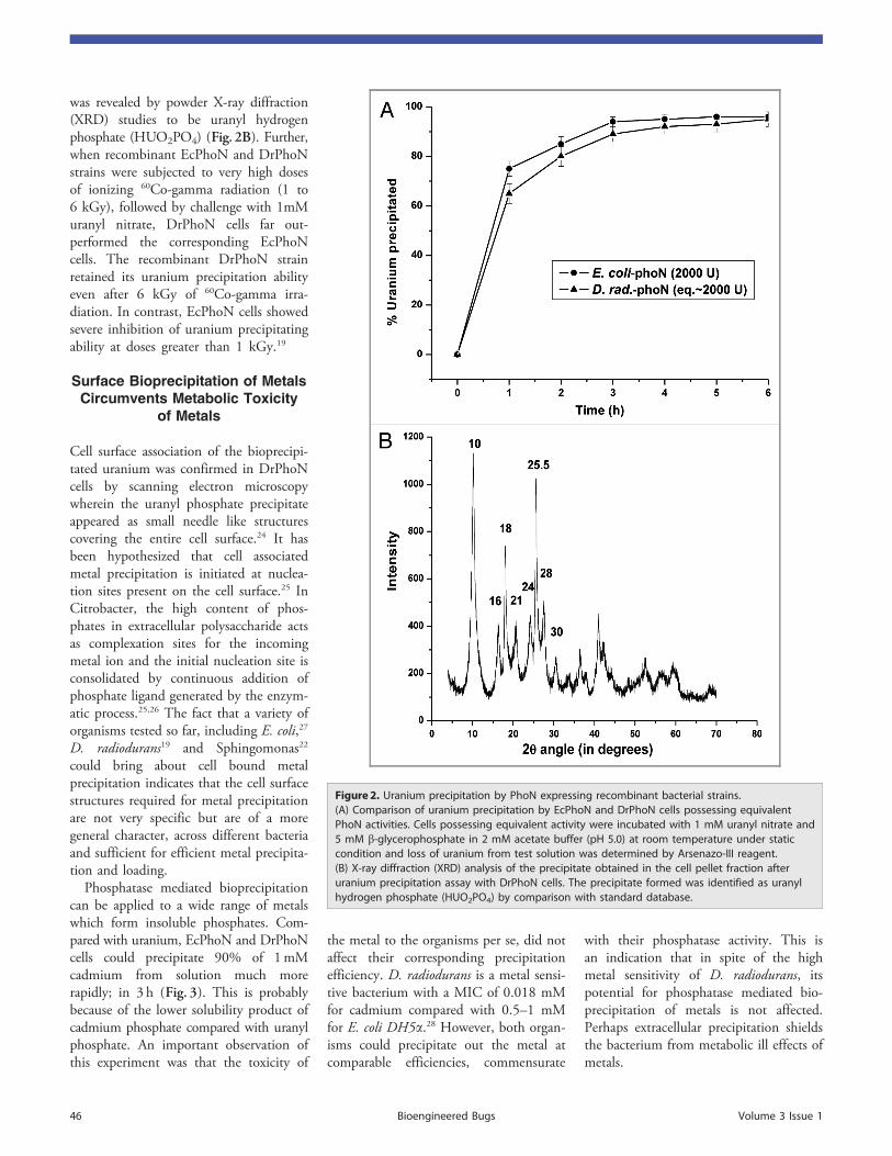
was revealed by powder X-ray diffraction(XRD) studies to be uranyl hydrogenphosphate (HUO2PO4) (Fig. 2B). Further,when recombinant EcPhoN and DrPhoNstrains were subjected to very high dosesof ionizing 60Co-gamma radiation (1 to6 kGy), followed by challenge with 1mMuranyl nitrate, DrPhoN cells far out-performed the corresponding EcPhoNcells. The recombinant DrPhoN strainretained its uranium precipitation abilityeven after 6 kGy of 60Co-gamma irra-diation. In contrast, EcPhoN cells showedsevere inhibition of uranium precipitatingability at doses greater than 1 kGy.19
Surface Bioprecipitation of MetalsCircumvents Metabolic Toxicity
of Metals
Cell surface association of the bioprecipi-tated uranium was confirmed in DrPhoNcells by scanning electron microscopywherein the uranyl phosphate precipitateappeared as small needle like structurescovering the entire cell surface.24 It hasbeen hypothesized that cell associatedmetal precipitation is initiated at nuclea-tion sites present on the cell surface.25 InCitrobacter, the high content of phos-phates in extracellular polysaccharide actsas complexation sites for the incomingmetal ion and the initial nucleation site isconsolidated by continuous addition ofphosphate ligand generated by the enzym-atic process.25,26 The fact that a variety oforganisms tested so far, including E. coli,27
D. radiodurans19 and Sphingomonas22
could bring about cell bound metalprecipitation indicates that the cell surfacestructures required for metal precipitationare not very specific but are of a moregeneral character, across different bacteriaand sufficient for efficient metal precipita-tion and loading.
Phosphatase mediated bioprecipitationcan be applied to a wide range of metalswhich form insoluble phosphates. Com-pared with uranium, EcPhoN and DrPhoNcells could precipitate 90% of 1mMcadmium from solution much morerapidly; in 3 h (Fig. 3). This is probablybecause of the lower solubility product ofcadmium phosphate compared with uranylphosphate. An important observation ofthis experiment was that the toxicity of
the metal to the organisms per se, did notaffect their corresponding precipitationefficiency. D. radiodurans is a metal sensi-tive bacterium with a MIC of 0.018 mMfor cadmium compared with 0.5–1 mMfor E. coli DH5a.28 However, both organ-isms could precipitate out the metal atcomparable efficiencies, commensurate
with their phosphatase activity. This isan indication that in spite of the highmetal sensitivity of D. radiodurans, itspotential for phosphatase mediated bio-precipitation of metals is not affected.Perhaps extracellular precipitation shieldsthe bacterium from metabolic ill effects ofmetals.
Figure 2. Uranium precipitation by PhoN expressing recombinant bacterial strains.(A) Comparison of uranium precipitation by EcPhoN and DrPhoN cells possessing equivalentPhoN activities. Cells possessing equivalent activity were incubated with 1 mM uranyl nitrate and5 mM b-glycerophosphate in 2 mM acetate buffer (pH 5.0) at room temperature under staticcondition and loss of uranium from test solution was determined by Arsenazo-III reagent.(B) X-ray diffraction (XRD) analysis of the precipitate obtained in the cell pellet fraction afteruranium precipitation assay with DrPhoN cells. The precipitate formed was identified as uranylhydrogen phosphate (HUO2PO4) by comparison with standard database.
46 Bioengineered Bugs Volume 3 Issue 1
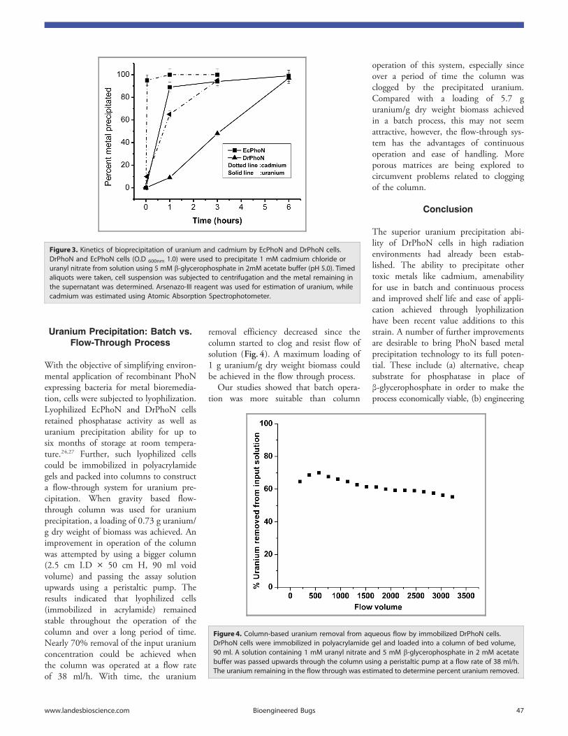
Uranium Precipitation: Batch vs.Flow-Through Process
With the objective of simplifying environ-mental application of recombinant PhoNexpressing bacteria for metal bioremedia-tion, cells were subjected to lyophilization.Lyophilized EcPhoN and DrPhoN cellsretained phosphatase activity as well asuranium precipitation ability for up tosix months of storage at room tempera-ture.24,27 Further, such lyophilized cellscould be immobilized in polyacrylamidegels and packed into columns to constructa flow-through system for uranium pre-cipitation. When gravity based flow-through column was used for uraniumprecipitation, a loading of 0.73 g uranium/g dry weight of biomass was achieved. Animprovement in operation of the columnwas attempted by using a bigger column(2.5 cm I.D � 50 cm H, 90 ml voidvolume) and passing the assay solutionupwards using a peristaltic pump. Theresults indicated that lyophilized cells(immobilized in acrylamide) remainedstable throughout the operation of thecolumn and over a long period of time.Nearly 70% removal of the input uraniumconcentration could be achieved whenthe column was operated at a flow rateof 38 ml/h. With time, the uranium
removal efficiency decreased since thecolumn started to clog and resist flow ofsolution (Fig. 4). A maximum loading of1 g uranium/g dry weight biomass couldbe achieved in the flow through process.
Our studies showed that batch opera-tion was more suitable than column
operation of this system, especially sinceover a period of time the column wasclogged by the precipitated uranium.Compared with a loading of 5.7 guranium/g dry weight biomass achievedin a batch process, this may not seemattractive, however, the flow-through sys-tem has the advantages of continuousoperation and ease of handling. Moreporous matrices are being explored tocircumvent problems related to cloggingof the column.
Conclusion
The superior uranium precipitation abi-lity of DrPhoN cells in high radiationenvironments had already been estab-lished. The ability to precipitate othertoxic metals like cadmium, amenabilityfor use in batch and continuous processand improved shelf life and ease of appli-cation achieved through lyophilizationhave been recent value additions to thisstrain. A number of further improvementsare desirable to bring PhoN based metalprecipitation technology to its full poten-tial. These include (a) alternative, cheapsubstrate for phosphatase in place ofβ-glycerophosphate in order to make theprocess economically viable, (b) engineering
Figure 3. Kinetics of bioprecipitation of uranium and cadmium by EcPhoN and DrPhoN cells.DrPhoN and EcPhoN cells (O.D 600nm 1.0) were used to precipitate 1 mM cadmium chloride oruranyl nitrate from solution using 5 mM b-glycerophosphate in 2mM acetate buffer (pH 5.0). Timedaliquots were taken, cell suspension was subjected to centrifugation and the metal remaining inthe supernatant was determined. Arsenazo-III reagent was used for estimation of uranium, whilecadmium was estimated using Atomic Absorption Spectrophotometer.
Figure 4. Column-based uranium removal from aqueous flow by immobilized DrPhoN cells.DrPhoN cells were immobilized in polyacrylamide gel and loaded into a column of bed volume,90 ml. A solution containing 1 mM uranyl nitrate and 5 mM b-glycerophosphate in 2 mM acetatebuffer was passed upwards through the column using a peristaltic pump at a flow rate of 38 ml/h.The uranium remaining in the flow through was estimated to determine percent uranium removed.
www.landesbioscience.com Bioengineered Bugs 47
the phosphatase to localize closer to thecell surface especially in an organism likeD. radiodurans where multi-layered cellwalls may seriously limit the substrateavailability and metal access to theenzyme and (c) recombinant Deinococcuscells also need to be tested for a widearray of metals for even non-nuclearapplications, such as in nickel cadmium
battery waste clean-up. These possibilitiesare currently being investigated.
Application of metal bio-precipitatingDeinococcus strains for in situ bioreme-diation would require investigation on (a)survival and stability of such strains inactual waste sites, (b) better insight intothe precise metal-microbe interactionin cells, (c) scaling up of such cells for
large scale use and (d) improvementsin their bioprecipitation efficiency.Although D. radiodurans is not a humanpathogen, the concerns related to useof genetically engineered microbe arerelevant and will need to be adequ-ately addressed by ensuring its environ-mental biosafety and ecological safety forapplication.
References1. Lovley DR, Coates JD. Bioremediation of metal
contamination. Curr Opin Biotechnol 1997; 8:285-9;PMID:9206008; http://dx.doi.org/10.1016/S0958-1669(97)80005-5
2. Macaskie LE, Empson RM, Cheetham AK, Grey CP,Skarnulis AJ. Uranium bioaccumulation by a Citro-bacter sp. as a result of enzymically mediated growthof polycrystalline HUO2PO4. Science 1992; 257:782-4; PMID:1496397; http://dx.doi.org/10.1126/science.1496397
3. Yeotikar RG. High level liquid radioactive waste,their characterization and quality assurance. IndianAssociation of Nuclear Chemists and Allied Scientists(IANCAS).Bulletin. 2007; 6:204-17.
4. Lange CC, Wackett LP, Minton KW, Daly MJ.Engineering a recombinant Deinococcus radiodurans fororganopollutant degradation in radioactive mixed wasteenvironments. Nat Biotechnol 1998; 16:929-33;PMID:9788348; http://dx.doi.org/10.1038/nbt1098-929
5. Brim H, McFarlan SC, Fredrickson JK, Minton KW,Zhai M, Wackett LP, et al. Engineering Deinococcusradiodurans for metal remediation in radioactive mixedwaste environments. Nat Biotechnol 2000; 18:85-90;PMID:10625398; http://dx.doi.org/10.1038/71986
6. Brim H, Venkateshwaran A, Kostandarithes HM,Fredrickson JK, Daly MJ. Engineering Deinococcusgeothermalis for bioremediation of high-temperatureradioactive waste environments. Appl Environ Micro-biol 2003; 69:4575-82; PMID:12902245; http://dx.doi.org/10.1128/AEM.69.8.4575-4582.2003
7. de Groot AD, Chapon V, Servant P, Christin R, SauxMF, Sommer S, et al. Deinococcus deserti sp. nov. agamma-radiation tolerant bacterium isolated from theSahara desert. Int J Syst Evol Microbiol 2005; 55:2441-6; PMID:16280508; http://dx.doi.org/10.1099/ijs.0.63717-0
8. Ferreira AC, Nobre MF, Rainey FA, Silva MT, Wait R,Burghardt J, et al. Deinococcus geothermalis sp. nov. andDeinococcus murrayi sp. nov., two extremely radiation-resistant and slightly thermophilic species from hotsprings. Int J Syst Bacteriol 1997; 47:939-47; PMID:9336890; http://dx.doi.org/10.1099/00207713-47-4-939
9. Suresh K, Reddy GSN, Sengupta S, Shivaji S. Deino-coccus indicus sp. nov., an arsenic-resistant bacteriumfrom an aquifer in West Bengal, India. Int J Syst EvolMicrobiol 2004; 54:457-61; PMID:15023960; http://dx.doi.org/10.1099/ijs.0.02758-0
10. White O, Eisen JA, Heidelberg JF, Hickey EK,Peterson JD, Dodson RJ, et al. Genome sequence ofthe radioresistant bacterium Deinococcus radioduransR1. Science 1999; 286:1571-7; PMID:10567266;http://dx.doi.org/10.1126/science.286.5444.1571
11. Levin-Zaidman S, Englander J, Shimoni E, SharmaAK, Minton KW, Minsky A. Ringlike structure of theD. radiodurans genome: a key to radioresistance?.Science 2003; 299:254-6; PMID:12522252; http://dx.doi.org/10.1126/science.1077865
12. Zahradka K, Slade D, Bailone A, Sommer S, AverbeckD, Petranovic M, et al. Reassembly of shattered chro-mosomes in D. radiodurans. Nature 2006; 443:569-73;PMID:17006450
13. Narumi I, Satoh K, Cui S, Funayama T, Kitayama S,Watanabe H. PprA: a novel protein from Deinococcusradiodurans that stimulates DNA ligation. Mol Micro-biol 2004; 54:278-85; PMID:15458422; http://dx.doi.org/10.1111/j.1365-2958.2004.04272.x
14. Daly MJ, Gaidamakova EK, Matrosova VY, VasilenkoA, Zhai M, Venkateswaran A, et al. Accumulation ofMn(II) in Deinococcus radiodurans facilitates gamma-radiation resistance. Science 2004; 306:1025-8; PMID:15459345; http://dx.doi.org/10.1126/science.1103185
15. Basu B, Apte SK. Gamma radiation induced proteomeof D. radiodurans primarily targets DNA repair andoxidative stress alleviation. Mol Cell Prot 2011doi:10.1074/mcp.M111.011734.
16. de Groot A, Dulermo R, Ortet P, Blanchard L, GuérinP, Fernandez B, et al. Alliance of Proteomics andGenomics to Unravel the Specificities of SaharaBacterium Deinococcus deserti.. PLoS Genet 2009; 5:e1000434; PMID:19370165; http://dx.doi.org/10.1371/journal.pgen.1000434
17. Fredrickson JK, Kostandarithes HM, Li SW, PlymaleAE, Daly MJ. Reduction of Fe(III), Cr(VI), U(VI) andTc(VII) by D. radiodurans R1. Appl Environ Microbiol2000; 66:2006-11; PMID:10788374; http://dx.doi.org/10.1128/AEM.66.5.2006-2011.2000
18. Brim H, Osborne JP, Kostandarithes M, FredricksonJK, Wackett LP, Daly MJ. D. radiodurans engineeredfor complete toluene degradation facilitates Cr(VI)reduction. Microbiology 2006; 152:2469-77; PMID:16849809; http://dx.doi.org/10.1099/mic.0.29009-0
19. Appukuttan D, Rao AS, Apte SK. Engineering ofDeinococcus radiodurans R1 for bioprecipitation ofuranium from dilute nuclear waste. Appl EnvironMicrobiol 2006; 72:7873-8; PMID:17056698; http://dx.doi.org/10.1128/AEM.01362-06
20. Meima R, Lidstrom ME. Characterization of theMinimal Replicon of a Cryptic Deinococcus radioduransSARK Plasmid and Development of Versatile Escheri-chia coli-D. radiodurans Shuttle Vectors. Appl EnvironMicrobiol 2000; 66:3856-67; PMID:10966401;http://dx.doi.org/10.1128/AEM.66.9.3856-3867.2000
21. Riccio ML, Rossolini GM, Lombardi G, Chiesurin A,Satta G. Expression cloning of different bacterialphosphatase-encoding genes by histochemical screen-ing of genomic libraries onto an indicator mediumcontaining phenolphthalein diphosphate and methylgreen. J Appl Microbiol 1997; 82:177-85; PMID:12452591
22. Nilgiriwala KS, Alahari A, Rao AS, Apte SK. Cloningand overexpression of alkaline phosphatase PhoK fromSphingomonas sp. strain BSAR-1 for bioprecipitation ofuranium from alkaline solutions. Appl Environ Micro-biol 2008; 74:5516-23; PMID:18641147; http://dx.doi.org/10.1128/AEM.00107-08
23. Makarova KS, Aravind L, Wolf YI, Tatusov RL,Minton KW, Koonin EV, et al. Genome of theextremely radiation-resistant bacterium Deinococcusradiodurans viewed from the perspective of comparativegenomics. Microbiol Mol Biol Rev 2001; 65:44-79;PMID:11238985; http://dx.doi.org/10.1128/MMBR.65.1.44-79.2001
24. Appukuttan D, Seetharam C, Padma N, Rao AS, ApteSK. PhoN-expressing, lyophilized, recombinant Deino-coccus radiodurans cells for uranium bioprecipitation.[REMOVED HYPERLINK FIELD]. J Biotechnol2011; 154:285-90; PMID:21616102; http://dx.doi.org/10.1016/j.jbiotec.2011.05.002
25. Jeong BC, Hawes C, Bonthrone KM, Macaskie LE.Localization of enzymically heavy metal accumulationby Citrobacter sp. and metal accumulation in vitro byliposomes containing entrapped enzyme. Microbiology1997; 143:2497-507; PMID:9245830; http://dx.doi.org/10.1099/00221287-143-7-2497
26. Macaskie LE, Lloyd JR, Thomas RAP, Tolley MR.The use of micro-organisms for the remediation ofsolutions contaminated with actinide elements, otherradionuclides, and other organic contaminants gener-ated by nuclear fuel activities. Nuclear energy 1996;35:257-271.
27. Seetharam C, Soundarajan S, Udas AC, Rao AS, ApteSK. Lyophilized, non-viable, recombinant E. coli cellsfor cadmium bioprecipitation and recovery. ProcessBiochem 2009; 44:246-50; http://dx.doi.org/10.1016/j.procbio.2008.10.015
28. Ruggiero CE, Boukhalfa H, Forsythe JH, Lack JG,Hersman LE, Neu MP. Actinide and metal toxicity toprospective bioremediation bacteria. Environ Microbiol2005; 7:88-97; PMID:15643939; http://dx.doi.org/10.1111/j.1462-2920.2004.00666.x
48 Bioengineered Bugs Volume 3 Issue 1


![[Amar Chitra Katha Pvt] Amar Chitra Katha 266 Man(BookFi.org)](https://img.pdfslide.net/doc/110x75/563dbb98550346aa9aae8a9e/amar-chitra-katha-pvt-amar-chitra-katha-266-manbookfiorg.jpg)