Embed Size (px)
Citation preview
HEALTH CENTER INTERNATIONAL RESEARCH
http://healthcenterinternationalresearches.webs.com/
Dr. Maddalena Frau
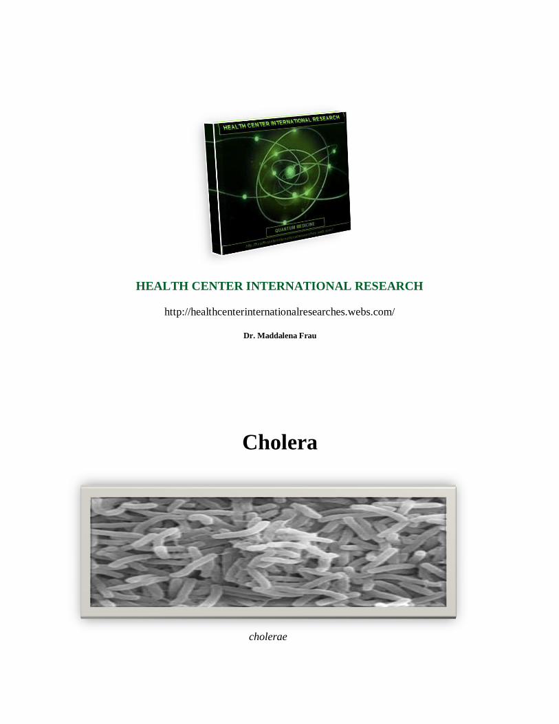
Cholera
cholerae
Cholera is an infection of the small intestine caused by the bacterium Vibrio cholerae. The main
symptoms are profuse, watery diarrhea and vomiting. Transmission occurs primarily by drinking
water or eating food that has been contaminated by the feces of an infected person (even an
asymptomatic one). The severity of the diarrhea and vomiting can lead to rapid dehydration and
electrolyte imbalance, and death in some cases. The primary treatment is with oral rehydration
solution (ORS) to replace water and electrolytes; if this is not tolerated or does not provide quick
enough treatment, intravenous fluids can also be used. Antibiotics are beneficial in those with
severe disease to shorten its duration and severity. Worldwide, it affects 3–5 million people and
causes 100,000–130,000 deaths a year as of 2010. Cholera was one of the earliest infections to
be studied by epidemiological methods.
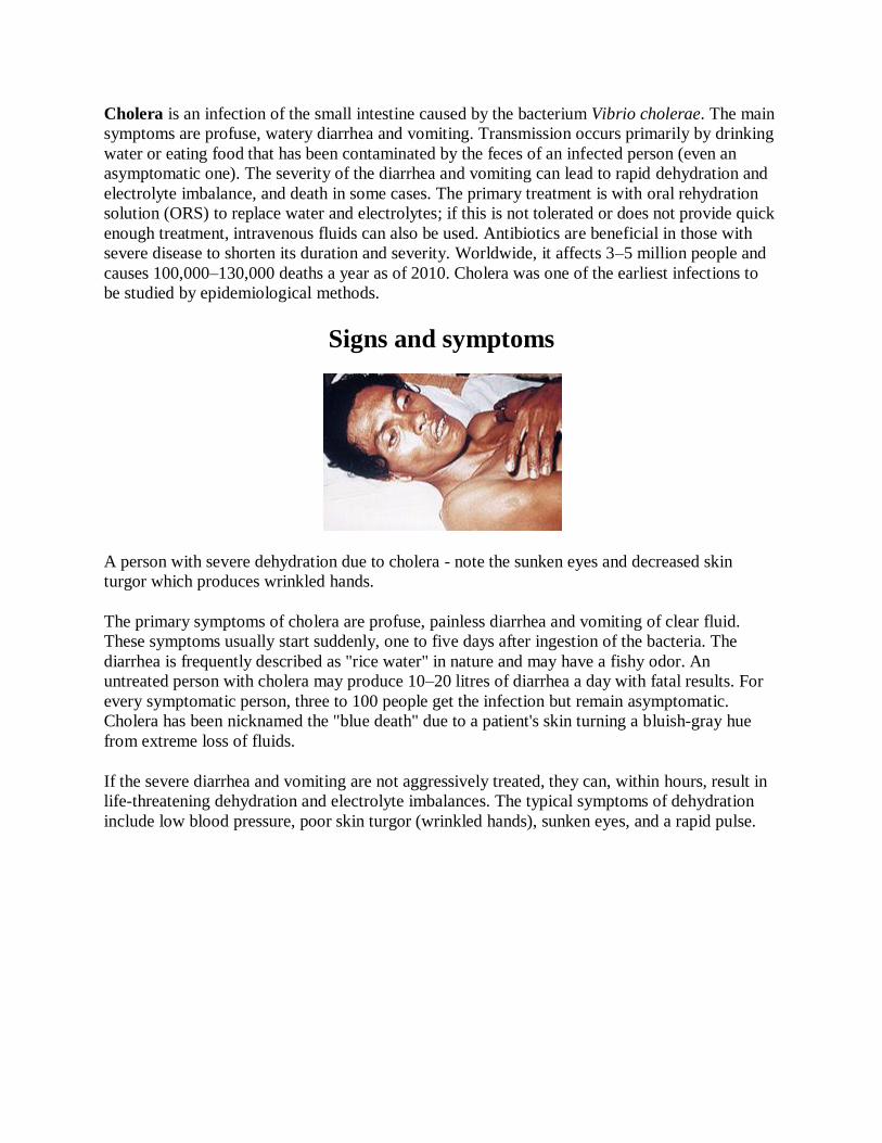
Signs and symptoms
A person with severe dehydration due to cholera - note the sunken eyes and decreased skin
turgor which produces wrinkled hands.
The primary symptoms of cholera are profuse, painless diarrhea and vomiting of clear fluid.
These symptoms usually start suddenly, one to five days after ingestion of the bacteria. The
diarrhea is frequently described as "rice water" in nature and may have a fishy odor. An
untreated person with cholera may produce 10–20 litres of diarrhea a day with fatal results. For
every symptomatic person, three to 100 people get the infection but remain asymptomatic.
Cholera has been nicknamed the "blue death" due to a patient's skin turning a bluish-gray hue
from extreme loss of fluids.
If the severe diarrhea and vomiting are not aggressively treated, they can, within hours, result in
life-threatening dehydration and electrolyte imbalances. The typical symptoms of dehydration
include low blood pressure, poor skin turgor (wrinkled hands), sunken eyes, and a rapid pulse.
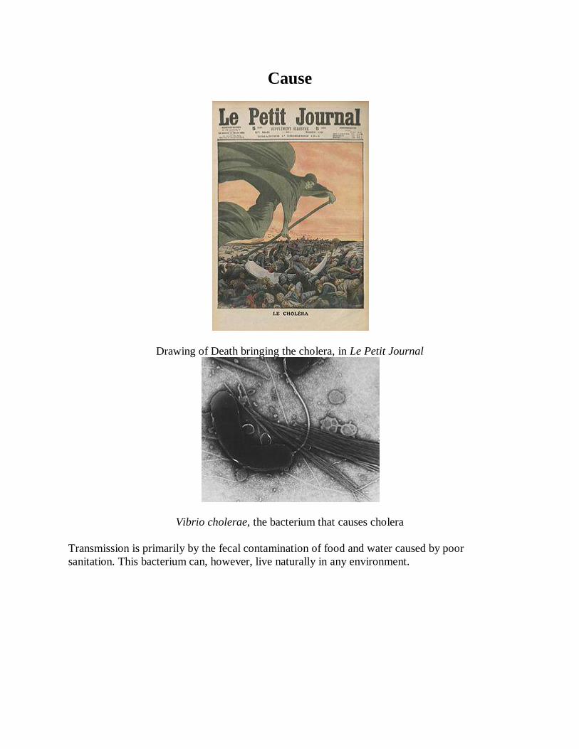
Cause
Drawing of Death bringing the cholera, in Le Petit Journal
Vibrio cholerae, the bacterium that causes cholera
Transmission is primarily by the fecal contamination of food and water caused by poor
sanitation. This bacterium can, however, live naturally in any environment.
About one hundred million bacteria must typically be ingested to cause cholera in a normal
healthy adult. This dose, however, is less in those with lower gastric acidity (for instance those
using proton pump inhibitors). Children are also more susceptible, with two- to four-year-olds
having the highest rates of infection. Individuals' susceptibility to cholera is also affected by their
blood type, with those with type O blood being the most susceptible. Persons with lower
immunity, such as persons with AIDS or children who are malnourished, are more likely to
experience a severe case if they become infected. However, it should be noted that any
individual, even a healthy adult in middle age, can experience a severe case, and each person's
case should be measured by the loss of fluids, preferably in consultation with a doctor or other
health worker.
The cystic fibrosis genetic mutation in humans has been said to maintain a selective advantage:
heterozygous carriers of the mutation (who are thus not affected by cystic fibrosis) are more
resistant to V. cholerae infections. In this model, the genetic deficiency in the cystic fibrosis
transmembrane conductance regulator channel proteins interferes with bacteria binding to the
gastrointestinal epithelium, thus reducing the effects of an infection.
Transmission
Cholera is typically transmitted by either contaminated food or water. In the developed world,
seafood is the usual cause, while in the developing world it is more often water. Cholera has
been found in only two other animal populations: shellfish and plankton.
People infected with cholera often have diarrhea, and if this highly liquid stool, colloquially
referred to as "rice-water" or "faucet butt", contaminates water used by others, disease
transmission may occur. The source of the contamination is typically other cholera sufferers
when their untreated diarrheal discharge is allowed to get into waterways, groundwater or
drinking water supplies. Drinking any infected water and eating any foods washed in the water,
as well as shellfish living in the affected waterway, can cause a person to contract an infection.
Cholera is rarely spread directly from person to person. Both toxic and nontoxic strains exist.
Nontoxic strains can acquire toxicity through a temperate bacteriophage. Coastal cholera
outbreaks typically follow zooplankton blooms, thus making cholera a zoonotic disease.
Most bacteria, when consumed, do not survive the acidic conditions of the human stomach. The
few surviving bacteria conserve their energy and stored nutrients during the passage through the
stomach by shutting down much protein production. When the surviving bacteria exit the
stomach and reach the small intestine, they need to propel themselves through the thick mucus
that lines the small intestine to get to the intestinal walls, where they can thrive. V. cholerae
bacteria start up production of the hollow cylindrical protein flagellin to make flagella, the cork-
screw helical fibers they rotate to propel themselves through the mucus of the small intestine.
Once the cholera bacteria reach the intestinal wall, they no longer need the flagella to move. The
bacteria stop producing the protein flagellin, thus again conserving energy and nutrients by
changing the mix of proteins which they manufacture in response to the changed chemical
surroundings. On reaching the intestinal wall, V. cholerae start producing the toxic proteins that
give the infected person a watery diarrhea. This carries the multiplying new generations of V.
cholerae bacteria out into the drinking water of the next host if proper sanitation measures are
not in place.
The cholera toxin (CTX or CT) is an oligomeric complex made up of six protein subunits: a
single copy of the A subunit (part A), and five copies of the B subunit (part B), connected by a
disulfide bond. The five B subunits form a five-membered ring that binds to GM1 gangliosides
on the surface of the intestinal epithelium cells. The A1 portion of the A subunit is an enzyme
that ADP-ribosylates G proteins, while the A2 chain fits into the central pore of the B subunit
ring. Upon binding, the complex is taken into the cell via receptor-mediated endocytosis. Once
inside the cell, the disulfide bond is reduced, and the A1 subunit is freed to bind with a human
partner protein called ADP-ribosylation factor 6 (Arf6). Binding exposes its active site, allowing
it to permanently ribosylate the Gs alpha subunit of the heterotrimeric G protein. This results in
constitutive cAMP production, which in turn leads to secretion of H2O, Na+, K
+, Cl
−, and HCO3
−
into the lumen of the small intestine and rapid dehydration. The gene encoding the cholera toxin
is introduced into V. cholerae by horizontal gene transfer. Virulent strains of V. cholerae carry a
variant of temperate bacteriophage called CTXf or CTXφ.
Microbiologists have studied the genetic mechanisms by which the V. cholerae bacteria turn off
the production of some proteins and turn on the production of other proteins as they respond to
the series of chemical environments they encounter, passing through the stomach, through the
mucous layer of the small intestine, and on to the intestinal wall. Of particular interest have been
the genetic mechanisms by which cholera bacteria turn on the protein production of the toxins
that interact with host cell mechanisms to pump chloride ions into the small intestine, creating an
ionic pressure which prevents sodium ions from entering the cell. The chloride and sodium ions
create a salt-water environment in the small intestines, which through osmosis can pull up to six
litres of water per day through the intestinal cells, creating the massive amounts of diarrhea. The
host can become rapidly dehydrated if an appropriate mixture of dilute salt water and sugar is not
taken to replace the blood's water and salts lost in the diarrhea.
By inserting separate, successive sections of V. cholerae DNA into the DNA of other bacteria,
such as E. coli that would not naturally produce the protein toxins, researchers have investigated
the mechanisms by which V. cholerae responds to the changing chemical environments of the
stomach, mucous layers, and intestinal wall. Researchers have discovered a complex cascade of
regulatory proteins controls expression of V. cholerae virulence determinants. In responding to
the chemical environment at the intestinal wall, the V. cholerae bacteria produce the TcpP/TcpH
proteins, which, together with the ToxR/ToxS proteins, activate the expression of the ToxT
regulatory protein. ToxT then directly activates expression of virulence genes that produce the
toxins, causing diarrhea in the infected person and allowing the bacteria to colonize the intestine.
Current research aims at discovering "the signal that makes the cholera bacteria stop swimming
and start to colonize (that is, adhere to the cells of) the small intestine."
Genetic structure
Amplified fragment length polymorphism fingerprinting of the pandemic isolates of V. cholerae
has revealed variation in the genetic structure. Two clusters have been identified: Cluster I and
Cluster II. For the most part, Cluster I consists of strains from the 1960s and 1970s, while Cluster
II largely contains strains from the 1980s and 1990s, based on the change in the clone structure.
This grouping of strains is best seen in the strains from the African continent.
Diagnosis
A rapid dip-stick test is available to determine the presence of V. cholerae. In those samples that
test positive, further testing should be done to determine antibiotic resistance. In epidemic
situations, a clinical diagnosis may be made by taking a patient history and doing a brief
examination. Treatment is usually started without or before confirmation by laboratory analysis.
Stool and swab samples collected in the acute stage of the disease, before antibiotics have been
administered, are the most useful specimens for laboratory diagnosis. If an epidemic of cholera is
suspected, the most common causative agent is V. cholerae O1. If V. cholerae serogroup O1 is
not isolated, the laboratory should test for V. cholerae O139. However, if neither of these
organisms is isolated, it is necessary to send stool specimens to a reference laboratory. Infection
with V. cholerae O139 should be reported and handled in the same manner as that caused by V.
cholerae O1. The associated diarrheal illness should be referred to as cholera and must be
reported in the United States.
A number of special media have been employed for the cultivation for cholera vibrios. They are
classified as follows:
Enrichment media
1. Alkaline peptone water at pH 8.6
2. Monsur's taurocholate tellurite peptone water at pH 9.2
Plating media
1. Alkaline bile salt agar (BSA): The colonies are very similar to those on nutrient agar.
2. Monsur's gelatin Tauro cholate trypticase tellurite agar (GTTA) medium: Cholera vibrios
produce small, translucent colonies with a greyish-black center.
3. TCBS medium: This is the mostly widely used medium; it contains thiosulphate, citrate,
bile salts and sucrose. Cholera vibrios produce flat, 2–3 mm in diameter, yellow-
nucleated colonies.
Direct microscopy of stool is not recommended, as it is unreliable. Microscopy is preferred only
after enrichment, as this process reveals the characteristic motility of Vibrio and its inhibition by
appropriate antisera. Diagnosis can be confirmed, as well, as serotyping done by agglutination
with specific sera.
Prevention
Although cholera may be life-threatening, prevention of the disease is normally straightforward
if proper sanitation practices are followed. In developed countries, due to nearly universal
advanced water treatment and sanitation practices, cholera is no longer a major health threat. The
last major outbreak of cholera in the United States occurred in 1910–1911. Effective sanitation
practices, if instituted and adhered to in time, are usually sufficient to stop an epidemic. There
are several points along the cholera transmission path at which its spread may be halted:
Sterilization: Proper disposal and treatment of infected fecal waste water produced by
cholera victims and all contaminated materials (e.g. clothing, bedding, etc.) are essential.
All materials that come in contact with cholera patients should be sanitized by washing in
hot water, using chlorine bleach if possible. Hands that touch cholera patients or their
clothing, bedding, etc., should be thoroughly cleaned and disinfected with chlorinated
water or other effective antimicrobial agents.
Sewage: antibacterial treatment of general sewage by chlorine, ozone, ultraviolet light or
other effective treatment before it enters the waterways or underground water supplies
helps prevent undiagnosed patients from inadvertently spreading the disease.
Sources: Warnings about possible cholera contamination should be posted around
contaminated water sources with directions on how to decontaminate the water (boiling,
chlorination etc.) for possible use.
Water purification: All water used for drinking, washing, or cooking should be sterilized
by either boiling, chlorination, ozone water treatment, ultraviolet light sterilization (e.g.
by solar water disinfection), or antimicrobial filtration in any area where cholera may be
present. Chlorination and boiling are often the least expensive and most effective means
of halting transmission. Cloth filters, though very basic, have significantly reduced the
occurrence of cholera when used in poor villages in Bangladesh that rely on untreated
surface water. Better antimicrobial filters, like those present in advanced individual water
treatment hiking kits, are most effective. Public health education and adherence to
appropriate sanitation practices are of primary importance to help prevent and control
transmission of cholera and other diseases.
Surveillance and prompt reporting allow for containing cholera epidemics rapidly. Cholera exists
as a seasonal disease in many endemic countries, occurring annually mostly during rainy
seasons. Surveillance systems can provide early alerts to outbreaks, therefore leading to
coordinated response and assist in preparation of preparedness plans. Efficient surveillance
systems can also improve the risk assessment for potential cholera outbreaks. Understanding the
seasonality and location of outbreaks provide guidance for improving cholera control activities
for the most vulnerable. For prevention to be effective, it is important that cases are reported to
national health authorities[
Vaccine
A number of safe and effective oral vaccines for cholera are available. Dukoral, an orally
administered, inactivated whole cell vaccine, has an overall efficacy of about 52% during the
first year after being given and 62% in the second year, with minimal side effects. It is available
in over 60 countries. However, it is not currently recommended by the Centers for Disease
Control and Prevention (CDC) for most people traveling from the United States to endemic
countries. One injectable vaccine was found to be effective for two to three years. The protective
efficacy was 28% lower in children less than 5 years old. However, as of 2010, it has limited
availability. Work is under way to investigate the role of mass vaccination. The World Health
Organization (WHO) recommends immunization of high risk groups, such as children and
people with HIV, in countries where this disease is endemic. If people are immunized broadly,
herd immunity results, with a decrease in the amount of contamination in the environment.
Treatment
Continued eating speeds the recovery of normal intestinal function. The World Health
Organization recommends this generally for cases of diarrhea from whatever cause. A CDC
training manual specifically for cholera states: “Continue to breastfeed your baby if the baby has
watery diarrhea, even when traveling to get treatment. Adults and older children should continue
to eat frequently.”
In most cases, cholera can be successfully treated with oral rehydration therapy (ORT), which is
highly effective, safe, and simple to administer. Rice-based solutions are preferred to glucose-
based ones due to greater efficiency. In severe cases with significant dehydration, intravenous
rehydration may be necessary. Ringer's lactate is the preferred solution, often with added
potassium. Large volumes and continued replacement until diarrhea has subsided may be needed.
Ten percent of a person's body weight in fluid may need to be given in the first two to four hours.
This method was first tried on a mass scale during the Bangladesh Liberation War, and was
found to have much success.
If commercially produced oral rehydration solutions are too expensive or difficult to obtain,
solutions can be made. One such recipe calls for 1 litre of boiled water, 1/2 teaspoon of salt, 6
teaspoons of sugar, and added mashed banana for potassium and to improve taste.[26]
As there frequently is initially acidosis, the potassium level may be normal, even though large
losses have occurred. As the dehydration is corrected, potassium levels may decrease rapidly,
and thus need to be replaced.[
Antibiotic treatments for one to three days shorten the course of the disease and reduce the
severity of the symptoms. People will recover without them, however, if sufficient hydration is
maintained. Doxycycline is typically used first line, although some strains of V. cholerae have
shown resistance. Testing for resistance during an outbreak can help determine appropriate
future choices. Other antibiotics proven to be effective include cotrimoxazole, erythromycin,
tetracycline, chloramphenicol, and furazolidone. Fluoroquinolones, such as norfloxacin, also
may be used, but resistance has been reported.
In many areas of the world, antibiotic resistance is increasing. In Bangladesh, for example, most
cases are resistant to tetracycline, trimethoprim-sulfamethoxazole, and erythromycin. Rapid
diagnostic assay methods are available for the identification of multiple drug-resistant cases.
New generation antimicrobials have been discovered which are effective against in in vitro
studies.
Sari filtration
An effective and relatively cheap method to prevent transmission of V. cholera is the practice of
folding a sari multiple times to create a simple filter for drinking water. Folding saris four to
eight times may create a simple filter to reduce the amount of active V. cholera in the filtered
water. The education of proper sari filter use is imperative, as there is a positive correlation
between sari misuse and the incidence of childhood diarrhea; soiled saris worn by women are
vectors of transmission of enteric pathogens to young children. Educating at-risk populations
about the proper use of the sari filter method may decrease V. cholera-associated disease.
If people with cholera are treated quickly and properly, the mortality rate is less than 1%;
however, with untreated cholera, the mortality rate rises to 50–60%. For certain genetic strains of
cholera, such as the one present during the 2010 epidemic in Haiti and the 2004 outbreak in
India, death can occur within two hours of the first sign of symptoms.
References
1. Sack DA, Sack RB, Nair GB, Siddique AK (January 2004). "Cholera". Lancet 363
(9404): 223–33. doi:10.1016/S0140-6736(03)15328-7. PMID 14738797.
2. ^ King AA, Ionides EL, J.Luckhurst, Bouma MJ (August 2008). "Inapparent infections
and cholera dynamics". Nature 454 (7206): 877–80. doi:10.1038/nature07084.
PMID 18704085.
3. ^ McElroy, Ann and Patricia K. Townsend. Medical Anthropology in Ecological
Perspective. Boulder, CO: Westview, 2009, 375.
4. "Cholera vaccines. A brief summary of the March 2010 position paper" (PDF). World
Health Organization.
5. Sack DA, Sack RB, Chaignat CL (August 2006). "Getting serious about cholera". N.
Engl. J. Med. 355 (7): 649–51. doi:10.1056/NEJMp068144. PMID 16914700.
6. ^ Harris JB, Khan AI, LaRocque RC, et al. (November 2005). "Blood Group, Immunity,
and Risk of Infection with Vibrio cholerae in an Area of Endemicity". Infect. Immun. 73
(11): 7422–7. doi:10.1128/IAI.73.11.7422-7427.2005. PMC 1273892. PMID 16239542.
7. ^ Prevention and control of cholera outbreaks: WHO policy and recommendations,
World Health Organization, Regional Office for the Eastern Mediterranean, undated but
citing sources from ’07, ’04, ’03, ’04, and ’05.
8. ^ Bertranpetit J, Calafell F (1996). "Genetic and geographical variability in cystic
fibrosis: evolutionary considerations". Ciba Found. Symp. 197: 97–114; discussion 114–
8. PMID 8827370.
9. ^ Ryan KJ, Ray CG (editors) (2004). Sherris Medical Microbiology (4th ed.). McGraw
Hill. pp. 376–7. ISBN 0838585299.
10. ^ Archivist (1997). "Cholera phage discovery". Arch Dis Child 76 (3): 274.
doi:10.1136/adc.76.3.274.
11. ^ Hartwell LH, Hood L, Goldberg ML, Reynolds AE, Silver LM, and Veres RC (2004).
Genetics: From genes to genomes. Boston: Mc-Graw Hill. pp. 551–552, 572–574. (using
the turning off and turning on of gene expression to make toxin proteins in cholera
bacteria as a "comprehensive example" of what is known about the mechanisms by which
bacteria change the mix of proteins they manufacture to respond to the changing
opportunities for surviving and thriving in different chemical environments).
12. ^ O'Neal C, Jobling M, Holmes R, Hol W (2005). "Structural basis for the activation of
cholera toxin by human ARF6-GTP". Science 309 (5737): 1093–6.
doi:10.1126/science.1113398. PMID 16099990.
13. ^ a b c DiRita VJ, Parsot C, Jander G, Mekalanos JJ (June 1991). "Regulatory cascade
controls virulence in Vibrio cholerae". Proc. Natl. Acad. Sci. U.S.A. 88 (12): 5403–7.
doi:10.1073/pnas.88.12.5403. PMC 51881. PMID 2052618.
14. ^ Lan R, Reeves PR (Jan 2002). "Pandemic Spread of Cholera: Genetic Diversity and
Relationships within the Seventh Pandemic Clone of Vibrio cholerae Determined by
Amplified Fragment Length Polymorphism". Journal of Clinical Microbiology 40 (1):
172–181. doi:10.1128/JCM.40.1.172-181.2002. ISSN 0095-1137. PMC 120103.
PMID 11773113.
15. ^ "Laboratory Methods for the Diagnosis of Epidemic Dysentery and Cholera" (PDF).
Atlanta, GA: CDC. 1999. Retrieved 2010-02-01.
16. ^ "Cholera Kills Boy. All Other Suspected Cases Now in Quarantine and Show No
Alarming Symptoms." (PDF). New York Times. July 18, 1911. Retrieved 2008-07-28.
"The sixth death from cholera since the arrival in this port from Naples of the steamship
Moltke, thirteen days ago, occurred yesterday at Swineburne Island. The victim was
Francesco Farando, 14 years old."
17. ^ "More Cholera in Port". Washington Post. October 10, 1910. Retrieved 2008-12-11. "A
case of cholera developed today in the steerage of the Hamburg-American liner Moltke,
which has been detained at quarantine as a possible cholera carrier since Monday last. Dr.
A.H. Doty, health officer of the port, reported the case tonight with the additional
information that another cholera patient from the Moltke is under treatment at Swinburne
Island."
18. ^ "Cholera: prevention and control". Health topics. WHO. 2008. Retrieved 2008-12-08.
19. ^ a b Sinclair D, Abba K, Zaman K, Qadri F, Graves PM (2011). "Oral vaccines for
preventing cholera". Cochrane Database Syst Rev (3): CD008603.
doi:10.1002/14651858.CD008603.pub2. PMID 21412922.
20. ^ "Is a vaccine available to prevent cholera?". CDC disease info: Cholera. 2010-10-22.
Retrieved 2010-10-24.
21. ^ Graves PM, Deeks JJ, Demicheli V, Jefferson T (2010). Graves, Patricia M. ed.
"Vaccines for preventing cholera: killed whole cell or other subunit vaccines (injected)".
Cochrane Database Syst Rev (8): CD000974. doi:10.1002/14651858.CD000974.pub2.
PMID 20687062.
22. ^ "Cholera vaccines". Health topics. WHO. 2008. Retrieved 2010-02-01.
23. ^ a b THE TREATMENT OF DIARRHOEA, A manual for physicians and other senior
health workers, World Health Organization, 2005. See page 10 (14 in PDF) and esp
chapter “5. MANAGEMENT OF SUSPECTED CHOLERA,” pages 16-17 (20-21 in
PDF).
24. ^ Community Health Worker Training Materials for Cholera Prevention and Control,
CDC, slides at back are dated 11/17/2010. See esp pages 7-8.
25. ^ The Civil War That Killed Cholera, foreignpolicy.com.
26. ^ "Oral Rehydration Solutions: Made at Home". The Mother and Child Health and
Education Trust. 2010. Retrieved 2010-10-29.
27. ^ "Cholera treatment". Molson Medical Informatics. 2007. Retrieved 2008-01-03.
28. ^ Krishna BV, Patil AB, Chandrasekhar MR (March 2006). "Fluoroquinolone-resistant
Vibrio cholerae isolated during a cholera outbreak in India". Trans. R. Soc. Trop. Med.
Hyg. 100 (3): 224–6. doi:10.1016/j.trstmh.2005.07.007. PMID 16246383.
29. ^ Mackay IM (editor) (2007). Real-Time PCR in microbiology: From diagnosis to
characterization. Caister Academic Press. ISBN 978-1-904455-18-9.
30. ^ Ramamurthy T (2008). "Antibiotic resistance in Vibrio cholerae". Vibrio cholerae:
Genomics and molecular biology. Caister Academic Press. ISBN 978-1-904455-33-2.
31. ^ Ali M, Emch M, Yunus M, Sack D, Lopez AL, Holmgren J, Clemens J (Jan 2008).
"Vaccine Protection of Bangladeshi infants and young children against cholera:
implications for vaccine deployment and person-to-person transmission". Pediatr Infect
Dis J 27 (1): 33–7. doi:10.1097/INF.0b013e318149dffd. PMID 18162935.
32. ^ Z. Bhutta. Background Paper on the Integration of Oral Cholera Vaccines into Global
Cholera Control Programmes. To be presented to the WHO SAGE in October 2009
33. ^ Stanton BF, Clemens JD, Clements JD (1986). "Soiled saris: a vector of disease
transmission?". Trans R Soc Trop Med Hyg 80 (3): 485–8. PMID 3798547.
34. ^ Todar, Kenneth. "Vibrio cholerae and Asiatic Cholera". Todar's Online Textbook of
Bacteriology. Retrieved 2010-12-20.
35. ^ NPR News. Presenter: Richard Knox. NPR. 2010-12-10.
36. ^ Reidl J, Klose KE (June 2002). "Vibrio cholerae and cholera: out of the water and into
the host". FEMS Microbiol. Rev. 26 (2): 125–39. doi:10.1111/j.1574-
6976.2002.tb00605.x. PMID 12069878.
37. ^ Blake, PA (1993). "Epidemiology of cholera in the Americas". Gastroenterology
clinics of North America 22 (3): 639–60. PMID 7691740.
38. ^ "Cholera's seven pandemics". CBC News. October 22, 2010.
39. ^ Aberth, John. Plagues in world History. Lanham, MD: Rowman & Littlefield, 2011,
102.
40. ^ .Kelley Lee (2003) "Health impacts of globalization: towards global governance".
Palgrave Macmillan. p.131. ISBN 0333802543
41. ^ Geoffrey A. Hosking (2001). "Russia and the Russians: a history". Harvard University
Press. p.9. ISBN 0674004736
42. ^ Byrne, Joseph Patrick (2008). Encyclopedia of Pestilence, Pandemics, and Plagues: A-
M. ABC-CLIO. p. 99. ISBN 0313341028.
43. ^ J. N. Hays (2005). "Epidemics and pandemics: their impacts on human history". p.347.
ISBN 1851096582
44. ^ Sehdev PS (November 2002). "The origin of quarantine". Clin. Infect. Dis. 35 (9):
1071–2. doi:10.1086/344062. PMID 12398064.
45. ^ "Archaic medical terms". Antiquus Morbus. 2007. Retrieved 2010-02-01.
46. ^ Rosenberg, Charles E. (1987). The cholera years: the United States in 1832, 1849 and
1866. Chicago: University of Chicago Press. ISBN 0-226-72677-0.
47. ^ Dr John Snow, The mode of communication of cholera, London 1855
48. ^ Aberth,John. Plagues in World History. Lanham, MD: Rowman & Littlefield, 2011,
101.
49. ^ Merrell DS, Butler SM, Qadri F, et al. (June 2002). "Host-induced epidemic spread of
the cholera bacterium". Nature 417 (6889): 642–5. doi:10.1038/nature00778.
PMC 2776822. PMID 12050664.
50. ^ Brown, Man and Music, 430–32; Holden, 371; Warrack, Tchaikovsky, 269–270.
51. ^ Meumayr A (1997). Music and medicine: Chopin, Smetana, Tchaikovsky, Mahler:
Notes on their lives, works, and medical histories. Med-Ed Press. pp. 282–3.
(summarizing various theories on what killed the composer Tchaikovsky, including his
brother Modest's idea that Tchaikovsky drank cholera-infested water the day before he
became ill).
52. ^ David Brown, Early Years, 46.
53. ^ Holden, 23.
54. ^ Brown, Man and Music, 431–35; Holden, 373–400.
55. ^ Susan Nagel, Marie Thérèse: Child of Terror, p. 349-350.
56. ^ Haynes, Sam W. (1997). James K. Polk and the Expansionist Impulse. New York:
Longman. p. 191. ISBN 978-0-673-99001-3.
57. ^ Smith, Rupert, The Utility of Force, Penguin Books, 2006, page 57
58. ^ Burnshaw S (2000). "Robert Frost". American National Biography Online. Archived
from the original on 2001-03-18.
[edit] Further reading
Colwell RR (December 1996). "Global climate and infectious disease: the cholera
paradigm". Science 274 (5295): 2025–31. doi:10.1126/science.274.5295.2025.
PMID 8953025.
Drasar, B. S.; Forrest, Bruce D., eds. (1996). Cholera and the ecology of Vibrio cholerae.
Springer. p. 355. ISBN 0412612208
Myron Echenberg: Africa in the Time of Cholera. A History of Pandemics from 1817 to
the Present, Cambridge University Press, New York 2011 (Paperback) ISBN 978-0-521-
18820-3
Furuque, Shah M.; Nair, G. Balakrish, eds. (2008). Vibrio Cholerae: Genomics and
Molecular Biology. Horizon Scientific Press. p. 218. ISBN 1904455336
Gilbert, Pamela K. (2008). Cholera and Nation: Doctoring the Social Body in Victorian
England. SUNY Press. p. 231. ISBN 0791473430
Jermyn, William S.; O'Shea, Yvonne A.; Quirke, Anne Marie; Boyd, E. Fidelma (2006).
"Genomics and the Evolution of Pathogenic Vibrio Cholerae". In Chan, Voon L.;
Sherman, Philip M.; Bourke, Billy. Bacterial genomes and infectious diseases. Humana
Press. p. 270. ISBN 158829496X
Johnson, Steven (2006). The Ghost Map: The Story of London's Most Terrifying
Epidemic--and How It Changed Science, Cities, and the Modern World (1854 epidemic).
Riverhead Hardcover. ISBN 1594489254.
Mintz ED, Guerrant RL (March 2009). "A lion in our village--the unconscionable tragedy
of cholera in Africa". N. Engl. J. Med. 360 (11): 1060–3. doi:10.1056/NEJMp0810559.
PMID 19279337.
Pardio Sedas, Violeta T. (2008). "Impact of Climate and Environmental Factors on the
Epidemiology of Vibrio choerae in Aquatic Ecosystems". In Hofer, Tobias N.. Marine
Pollution: New Research. Nova Science publishers. p. 448. pp. 221–254.
ISBN 1604562420
Ryan, Kenneth J.; Ray, C. George, eds. (2003). Sherris medical microbiology: an
introduction to infectious diseases (4th ed.). ISBN 0838585299
Wachsmuth, Kaye; Blake, Paul A.; Olsvik, Ørjan, eds. (1994). Vibrio cholerae and
cholera: molecular to global perspectives. ASM Press. p. 465. ISBN 1555810675