Embed Size (px)
Citation preview
J Int Adv Otol 2017 • DOI: 10.5152/iao.2017.3411
Review
INTRODUCTIONThe management of cholesteatoma continues to be a challenge for otolaryngologists around the world. Even in countries with advanced healthcare facilities, undertaking routine physical examinations, with good access to specialists, and where efforts are taken for the prevention, early detection, and treatment of cholesteatoma, there is a considerable prevalence of cholesteatoma and its complications in children and adults.
Diagnosis of cholesteatoma is performed by otolaryngologists using different methods, including obtaining the history that is char-acteristic for cholesteatoma suspicion, searching for or evidence of cholesteatoma during the physical examination using otoscopy and/or otomicroscopy, and interpretation of imaging (computed tomography and/or magnetic resonance) [1].
Despite the fact that cholesteatoma is diagnosed throughout the world, there are differences in the definition, classification, and management of cholesteatoma. These differences make it difficult to compare the reports in the literature, and limit the ability to derive further conclusions from individual or regional outcomes. Therefore, it is essential to create a common scientific language, with the definitions of an issue as a principle. Furthermore, utilizing a comparable classification system will allow investigators to share their experience across the world, leading to better assessment and management of cholesteatoma.
To achieve this, a recent initiative aimed to explore opinions among the members of European Academy of Otology & Neuro-otolo-gy (EAONO) regarding the definition and classification of cholesteatoma. Although consensus was achieved on the cholesteatoma definitions, it could not be achieved on its classification [2]. The process of development of the questionnaires, the responses ob-tained from the EAONO members through three cycles of questionnaires, and the final set of statements were reported in detail. Here we report the literature review that led to the development of the questionnaire on the definitions and classification to pro-vide a basis for the outcome. In addition, we present various classifications of cholesteatoma in the literature and emphasize the strengths and weaknesses of each of these classifications to stimulate an effort to develop a consensus on the classification as well.
METHODSEAONO steering group decided to undertake the task of developing guidelines in the field of otology and neurotology. Guidelines regarding the assessment and management of cholesteatoma were established as a priority. Among the committees established, the task of developing the guidelines for the definition and classification of cholesteatoma was assigned to Ewa Olszewska in June 2011. Statements on the definition and classification of cholesteatoma were developed by authors based on the literature review. Prepared questionnaires concerning cholesteatoma definition and classification were sent to EAONO members, inviting them to state their opinion for achieving consensus among them. Throughout the process, several committee meetings were conducted in Athens (2011) immediately preceding the 28th Politzer Society Meeting of the International Society for Otologic Surgery and Sci-
Cholesteatoma Definition and Classification: A Literature Review
Cholesteatoma is a serious otolaryngologic condition that to date remains an important problem and poses a challenge to otolaryngologists around the world. To improve the approach pertaining to the diagnosis and management of middle ear cholesteatoma, clear, clinically applicable, and useful definition and classification of cholesteatoma are required. This review aimed to evaluate the current and most accepted descriptions and opinions concerning cholesteatoma. A review of the literature concerning different definitions and classifications of cholesteatoma was used in the preparation of the Cholesteatoma Guidelines, a project implemented by the European Academy of Otology & Neuro-otology.
KEYWORDS: Cholesteatoma, middle ear cholesteatoma, cholesteatoma definition, cholesteatoma classification
Justyna Rutkowska, Nuri Özgirgin, Ewa OlszewskaDepartment of Otolaryngology, Medical University of Bialystok, Bialystok, Poland (JR, EO)Department of Otolaryngology, Bayındır Hospital, Ankara, Turkey (NO)
Corresponding Address: Ewa Olszewska E-mail: [email protected]
Submitted: 19.12.2016 Accepted: 31.12.2016 Available Online Date: 09.03.2017©Copyright 2017 by The European Academy of Otology and Neurotology and The Politzer Society - Available online at www.advancedotology.org
ence, in Nagasaki (2012) at the 9th International Conference on Cho-lesteatoma and Ear Surgery, in Nice (2013) at the 2nd Meeting of the European Academy of ORL-HNS and CEORL-HNS, in Antalya (2013) at the 29th Politzer Society Meeting, in Siena (2014) at the EAONO Meet-ing, in İstanbul (2015) at the Steering Cholesteatoma Group Meeting, in Niigata (2015) at the 30th Politzer Society Meeting, and in Edin-burgh (2016) at the 10th International Conference on Cholesteatoma and Ear Surgery. Presentations on cholesteatoma definition and clas-sification have been given by Ewa Olszewska in Athens (2011), Naga-saki (2012), Nice (2013), Siena (2014), İstanbul (2015), and at the 10th International Conference on Cholesteatoma and Ear Surgery Chole 2016 in Edinburgh during June 4–8, 2016. Throughout the process, there has been an intense interaction among EAONO members on guideline development. The results of this process and the end-prod-uct of consensus statements were published [2]. After this publication, to develop a broader consensus among the otologists and neurotol-ogists worldwide, the EAONO committee on consensus reached out to the Japanese Otology Society (JOS). An intense interaction and discussion among the members of EAONO and JOS led to the draft-ing of a consensus document that was presented and discussed at the panel in Edinburgh during Chole 2016. At the panel session, feed-back regarding the joint EAONO-JOS consensus document draft and input from participants representing many countries and otologic societies were discussed. A separate manuscript was prepared as an outcome of this work.
Literature ReviewTo capture the published material on the definitions, descriptions, and classification of cholesteatoma, a literature review was con-ducted. The following terms were used in the literature review: cho-lesteatoma definition, cholesteatoma classification, cholesteatoma symptoms, cholesteatoma risk factors, and cholesteatoma diagno-sis.
Medical Subject Headings (MeSH) is a comprehensive vocabulary used for indexing journal articles and books that has been created in the National Library of Medicine, USA, and is used by the National Center for Biotechnology Information (NCBI). The subject headings are hierarchically arranged. An electronic search of the English and non-English literature indexed in the Ovid–Medline database, Em-base database, and Cochrane Library was performed.
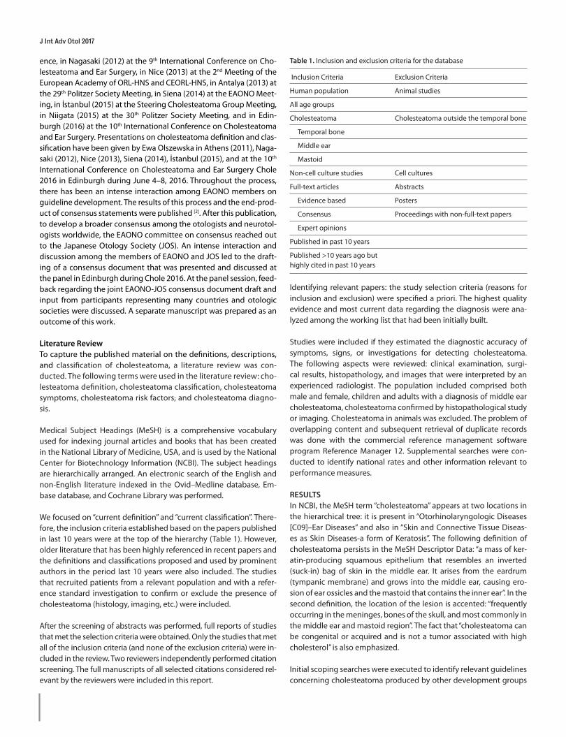
We focused on “current definition” and “current classification”. There-fore, the inclusion criteria established based on the papers published in last 10 years were at the top of the hierarchy (Table 1). However, older literature that has been highly referenced in recent papers and the definitions and classifications proposed and used by prominent authors in the period last 10 years were also included. The studies that recruited patients from a relevant population and with a refer-ence standard investigation to confirm or exclude the presence of cholesteatoma (histology, imaging, etc.) were included.
After the screening of abstracts was performed, full reports of studies that met the selection criteria were obtained. Only the studies that met all of the inclusion criteria (and none of the exclusion criteria) were in-cluded in the review. Two reviewers independently performed citation screening. The full manuscripts of all selected citations considered rel-evant by the reviewers were included in this report.
Identifying relevant papers: the study selection criteria (reasons for inclusion and exclusion) were specified a priori. The highest quality evidence and most current data regarding the diagnosis were ana-lyzed among the working list that had been initially built.
Studies were included if they estimated the diagnostic accuracy of symptoms, signs, or investigations for detecting cholesteatoma. The following aspects were reviewed: clinical examination, surgi-cal results, histopathology, and images that were interpreted by an experienced radiologist. The population included comprised both male and female, children and adults with a diagnosis of middle ear cholesteatoma, cholesteatoma confirmed by histopathological study or imaging. Cholesteatoma in animals was excluded. The problem of overlapping content and subsequent retrieval of duplicate records was done with the commercial reference management software program Reference Manager 12. Supplemental searches were con-ducted to identify national rates and other information relevant to performance measures.
RESULTSIn NCBI, the MeSH term “cholesteatoma” appears at two locations in the hierarchical tree: it is present in “Otorhinolaryngologic Diseases [C09]–Ear Diseases” and also in “Skin and Connective Tissue Diseas-es as Skin Diseases-a form of Keratosis”. The following definition of cholesteatoma persists in the MeSH Descriptor Data: “a mass of ker-atin-producing squamous epithelium that resembles an inverted (suck-in) bag of skin in the middle ear. It arises from the eardrum (tympanic membrane) and grows into the middle ear, causing ero-sion of ear ossicles and the mastoid that contains the inner ear”. In the second definition, the location of the lesion is accented: “frequently occurring in the meninges, bones of the skull, and most commonly in the middle ear and mastoid region”. The fact that “cholesteatoma can be congenital or acquired and is not a tumor associated with high cholesterol” is also emphasized.
Initial scoping searches were executed to identify relevant guidelines concerning cholesteatoma produced by other development groups
J Int Adv Otol 2017
Table 1. Inclusion and exclusion criteria for the database
Inclusion Criteria Exclusion Criteria
Human population Animal studies
All age groups
Cholesteatoma Cholesteatoma outside the temporal bone
Temporal bone
Middle ear
Mastoid
Non-cell culture studies Cell cultures
Full-text articles Abstracts
Evidence based Posters
Consensus Proceedings with non-full-text papers
Expert opinions
Published in past 10 years
Published >10 years ago but highly cited in past 10 years
(local, national, and international) and establish relevant definitions. However, there were no guidelines concerning cholesteatoma itself in popular databases. “Imaging of non-operated cholesteatoma: clinical practice guidelines” prepared for the annual congress of the French Society of Otolaryngology Head and Neck Surgery in 2010 by a panel of experts from the society was the only published guideline accessible [3]. The search strategy developed based on MeSH terms identified in the scoping search, cholesteatoma, congenital cho-lesteatoma, acquired cholesteatoma, definition, and classification, yielded 6061 references in Medline. Articles published in last 10 years were selected (from 2002 to 2013), and based on a previous selection study, 1544 articles were included.
DefinitionsCholesteatoma concerning the shape and form of the lesion has been termed as a growth of abnormal keratinizing squamous epithe-lium with a collection of keratin debris [4, 5], cystic lesion [5], three-di-mensional structure [6], cystic mass with a surrounding inflammatory reaction [7, 8], middle ear tumor [4], form of chronic otitis media [3, 9, 10], and “epidermoid cyst” [11]. It was called “skin in the wrong place” [12]. Cholesteatoma comprises layers of epithelial cells with the accumu-lation of differentiated keratin debris,” similar to the epidermis of the skin [13]. The significant component of the formation is subepithelial connective tissue, perimatrix [8]. Cholesteatoma was also determined as a “chronic wound healing process” [6] that replaces middle ear mucosa and resorbs underlying bone [14]. It was also defined as an “aggressive form of chronic otitis media requiring surgical therapy” and as a “subcategory of chronic otitis media” [3, 15, 16]. Many clinical, biochemical, and imaging abnormalities are typical for chronic otitis media, cholesteatoma. Chronic otitis media usually coexist in most individuals, especially in those with acquired one [10]. Therefore, with cholesteatoma is present concurrent with chronic otitis media and manifests itself with purulent otorrhea, tympanic membrane perfo-ration, and/or hearing loss.
Preciado suggested that cholesteatoma formation was related to both internal molecular dysregulation and external stimuli in the form of pro-inflammatory cytokines, growth factors, and/or bacte-rial toxins [7]. Florid inflammation and angiogenesis are distinctive features of the condition in most cases. It is also believed that the inflammation associated within the perimatrix of cholesteatoma in-duces bone resorption [17]. It is suspected that an early treatment of the inflammatory process of the ear may probably prevent the devel-opment of hyperplastic destructive epidermis [7]. Authors of the Co-chrane Library article present the following definition: “a destructive formation of layers of keratinizing epithelium, accumulating in the middle ear and mastoid” (referencing Bluestone, 1996) [15]. Authors also describe cholesteatoma as an “active squamous (epithelial) chronic otitis media” (referencing Browning, 1997) [15].
Surgical revision is important and often required for the definitive di-agnosis and to differentiate chronic suppurative otitis media (CSOM) with or without cholesteatoma. There are often no specific clinical indicators preoperatively to distinquish CSOM with cholesteatoma from CSOM without cholesteatoma. Cholesteatoma was not evident until surgical exploration was conducted in around 24% of cases in the study conducted by Khan et al. [18]. Therefore, surgical exploration appears mandatory for the final diagnosis of cholesteatoma.
It is universally accepted that cholesteatoma is non-neoplastic, noncancerous, and a “histopathologically benign” lesion that is “de-structive” and “locally invasive” [4, 19]. Potential complications may be life-threatening, “causing damage by passive growth and active de-struction of adjacent structures” [20]. Alterations of specific molecule expression levels, as for example detected altered level of p27 in ke-ratinocytes of cholesteatoma may influence the proliferative state of cells and suggest a molecular pathology in cholesteatoma [21]. There is an imperative need for cellular and molecular research to develop new therapeutic strategies [19, 21].
With respect to its histopathology, cholesteatoma was defined as “containing layers of keratin in a cavity lined by squamous epithe-lium and subepithelial connective tissue” [19], “development of a Malpighian epithelium” [3], lesion “formed from keratinizing strati-fied squamous epithelium, the matrix of which comprises epitheli-um that rests on a stroma with varying thickness and is called the perimatrix” [8]. Symptoms such as otorrhea, deafness, or conductive hearing loss were also suggested to be included in the definition of cholesteatoma [22].
The early form of advanced retraction pocket in the absence of bony destruction and expansion may be defined as “precholesteatoma” [23]. Black and Gutteridge called pre-cholesteatoma as the “final phase of collapsing tympanic membrane process before perforation with hyperkeratosis” [24]. The term “precholesteatoma” was used for “the condition with disturbed migration of the surface epithelial cells and self-cleaning property in the retraction pocket leading to an accu-mulation of keratin within the retraction pocket” [25]. Clarós suggest-ed the precholesteatoma be defined as “the retraction of Sharpnell membrane with disturbed epithelium migration, accumulation of debris, crust formation, infection behind the crust, and proliferation of epithelium keratinization” [26]. A clear and short definition was the “retraction pockets that accumulate keratin debris” used by Rosen-feld and Bluestone [27]. Belal et al. [28] in their new staging of tympa-nomastoid cholesteatoma used the term precholesteatoma as a synonym for retraction pocket. Precholesteatoma was also defined as the development of an epitympanic retraction pocket or that of a facial recess retraction pocket after the surgical opening of the facial recess [24]. It may be controversial and not easy to predict the role of precholesteatoma in the pathogenesis of cholesteatoma. However, it seems acceptable to consider precholesteatoma as a “stage of retrac-tion pocket with/without invisible depth or partially visible depth, with/without bony erosion, with early signs of loss of self-cleaning ability without apparent accumulation of keratin debris” [2].
Differences between residual and recurrent cholesteatoma have also been noted and discussed in the literature. Residual cholesteatoma is considered as the incomplete local resection of pathological squa-mous epithelium at the time of surgery [10, 29]. However, “recurrence” is defined as the development of cholesteatoma after complete sur-gical removal and when a newly formed lesion arises from the re-traction pocket [10]. The “tendency to recurrence” is also a designation often mentioned during cholesteatoma description [29]. Cholesteato-ma recurrence after surgical treatment is still a highly debated issue. In children, the factors associated with an increased risk for residual or recurrent cholesteatoma are the location of cholesteatoma in the sinus tympani and the presence of incus destruction [30].
Rutkowska et al. Literature Review of Cholesteatoma Definition and Classification
ClassificationCholesteatoma is primarily classified as congenital and acquired [20]. Acquired cholesteatoma is subdivided into primary and secondary [8, 20] based on the presence or absence of a perforation and the mi-gration of the epithelium into the middle ear through the perfora-tion. Middle ear acquired primary cholesteatoma was described as a sequel of the tympanic membrane retraction that would accumu-late the desquamated epithelium and lose its self-cleaning ability, whereas acquired secondary cholesteatoma was considered as the result of migration of the epithelium through a marginal perforation in the tympanic membrane [20]. Congenital cholesteatoma is defined as a developmental defect wherein an epithelial rest is entrapped in the middle ear cleft during embryogenesis. This is the most plausible explanation because of the persistence of fetal epidermoid forma-tion and the presence of rests of keratinizing squamous epithelium before birth that grow over time [5, 19]. It is described as “a whitish mass lesion in the middle ear cleft behind an intact tympanic membrane early in life”, a “keratinous mass located behind an intact tympanic membrane”, an “epidermal inclusion cyst”, and a “cystic epidermoid growth” [5, 31]. The criteria proposed by Levenson et al. [32] and accept-ed by many clinicians are as follows: “(1) a white mass medial to nor-mal tympanic membrane, (2) a normal pars flaccida and pars tensa, (3) no prior history of otorrhea or perforation, (4) no prior otologic procedures, (5) exclusion of canal atresia and intramembranous and giant cholesteatomas, and (6) prior bouts of otitis media were not grounds for exclusion”. Congenital cholesteatoma is most commonly described as being located in the middle ear cavity, i.e., a colloqui-al keratin pearl in the anterosuperior quadrant of the mesotympa-num juxtaposed to the malleolar manubrium or in the second-most common location of posterosuperior quadrant behind an otherwise healthy appearing eardrum [32, 33]. Clinical presentation is determined by the location and extent of the lesion [34]. It may be characterized by abnormal otoscopic examination, white mass medial to normal tympanic membrane, and more rarely pain (either neck or ear) and conductive hearing loss [34, 35].
The clinical classification of cholesteatoma is very important in plan-ning the surgical treatment method, assessing the results of a spe-cific treatment method for a specific classification, reporting to the scientific community, and comparing the outcomes of different sur-geons and institutions. Classification is often described based on the location of the cholesteatoma. Cholesteatoma may spread beyond the temporal bone, and this spread may be extradural or intradural. Extradural extension of cholesteatoma most commonly originates from the middle ear cleft and mastoid but may originate from all por-tions of the temporal bone, including the petrous apex and external ear canal [5, 8].
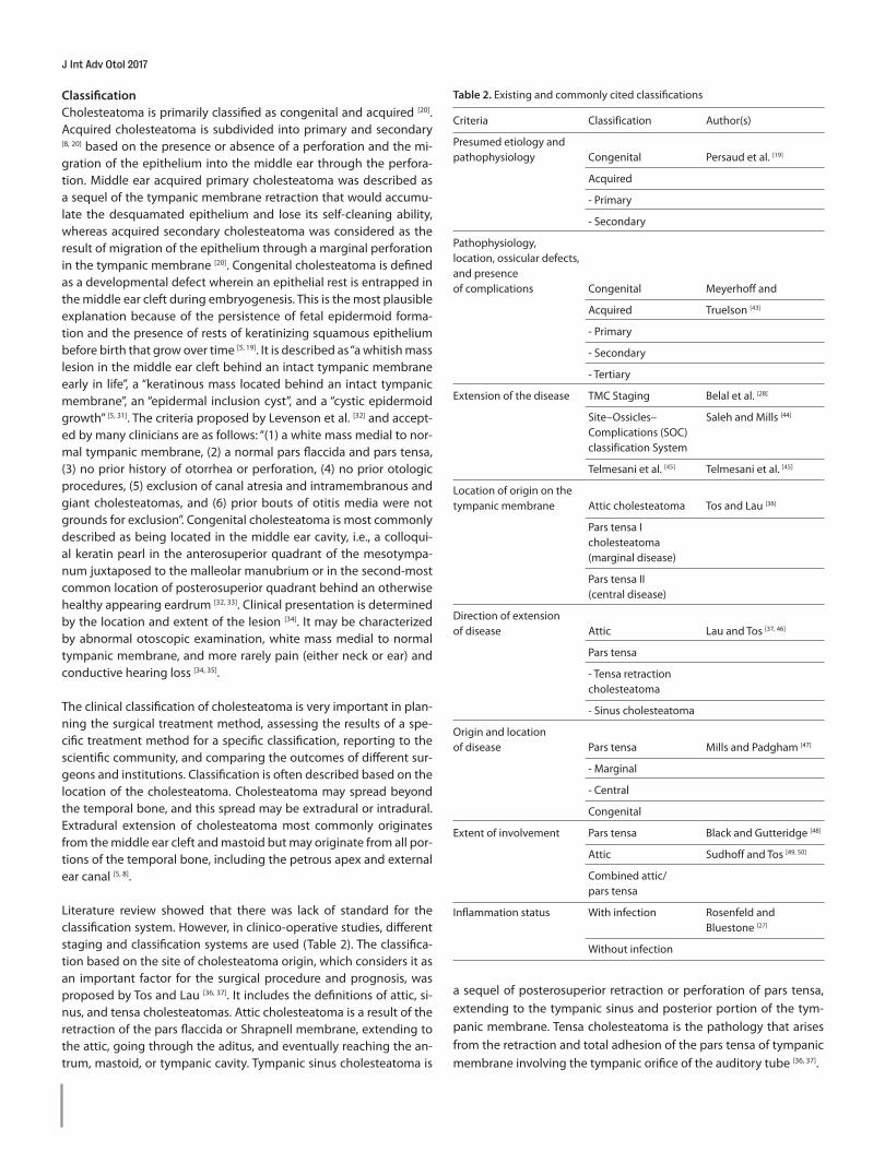
Literature review showed that there was lack of standard for the classification system. However, in clinico-operative studies, different staging and classification systems are used (Table 2). The classifica-tion based on the site of cholesteatoma origin, which considers it as an important factor for the surgical procedure and prognosis, was proposed by Tos and Lau [36, 37]. It includes the definitions of attic, si-nus, and tensa cholesteatomas. Attic cholesteatoma is a result of the retraction of the pars flaccida or Shrapnell membrane, extending to the attic, going through the aditus, and eventually reaching the an-trum, mastoid, or tympanic cavity. Tympanic sinus cholesteatoma is
a sequel of posterosuperior retraction or perforation of pars tensa, extending to the tympanic sinus and posterior portion of the tym-panic membrane. Tensa cholesteatoma is the pathology that arises from the retraction and total adhesion of the pars tensa of tympanic membrane involving the tympanic orifice of the auditory tube [36, 37].
J Int Adv Otol 2017
Table 2. Existing and commonly cited classifications
Criteria Classification Author(s)
Presumed etiology and pathophysiology Congenital Persaud et al. [19]
Acquired
- Primary
- Secondary
Pathophysiology, location, ossicular defects, and presence of complications Congenital Meyerhoff and
Acquired Truelson [43]
- Primary
- Secondary
- Tertiary
Extension of the disease TMC Staging Belal et al. [28]
Site–Ossicles– Saleh and Mills [44] Complications (SOC) classification System
Telmesani et al. [45] Telmesani et al. [45]
Location of origin on the tympanic membrane Attic cholesteatoma Tos and Lau [36]
Pars tensa I cholesteatoma (marginal disease)
Pars tensa II (central disease)
Direction of extension of disease Attic Lau and Tos [37, 46]
Pars tensa
- Tensa retraction cholesteatoma
- Sinus cholesteatoma
Origin and location of disease Pars tensa Mills and Padgham [47]
- Marginal
- Central
Congenital
Extent of involvement Pars tensa Black and Gutteridge [48]
Attic Sudhoff and Tos [49, 50]
Combined attic/ pars tensa
Inflammation status With infection Rosenfeld and Bluestone [27]
Without infection
The exact recommendations for the histopathological examination and imaging are not formalized. The question concerning the clini-cal utility of routine cholesteatoma histopathologic evaluation was explored by Kircher et al. [38]. Good correlation between the surgeon’s intraoperative findings and pathologist’s histopathologic diagnosis of cholesteatoma was proved. The current data confirms that the histopathological analysis is not mandatory required for the diagno-sis of cholesteatoma in the absence of concerns pertaining to other pathologies [38]. Non-contrast-enhanced temporal bone computed tomography has its limitations with a soft tissue lesion and does not show any pathognomonic bony erosion pattern for cholesteatoma. The “gold standard” method adopted by clinicians for the accurate diagnosis of cholesteatoma is still combining a “detailed otologic history, physical examination of ears, and the temporal bone com-puted tomography findings interpretation” [39, 40]. Echo-planar diffu-sion-weighted magnetic resonance imaging is also a valuable tech-nique for cholesteatoma imaging [41].
DISCUSSIONA systematic review of the literature for the therapeutic options of a disease or illness has established principles. Systematic reviews and meta-analyses are key elements of evidence-based healthcare. The adjective “systematic” implies conducting the review based on a clearly formulated question, identifying relevant studies, appraising their quality, and summarizing the evidence using explicit method-ologies. The term “systematic review” implies obeying the predefined rules [42].
The literature review for definitions and classifications does not meet all criteria required for systematic review. Definitions and classifica-tions that are published are not comparable based on the level of evidence. There are no randomized blinded clinical trials to test one definition or classification or the other. Instead, definitions are key initial elements of any scientific study or correspondence that are as-sumed to be agreed upon upfront. Although they may not be subject to clinical trials, they are essential elements of the scientific language.
In this review, we inquired about a set of terms related to the choles-teatoma hierarchy of evidence for the terms relevant to its definitions and classification. Even if the methodological quality and the name “systematic review” may be disputable, the use of this study for defi-nitions and classification is an essential step in exploring a consensus on staging, treatment methods, and reporting of outcomes.
This literature review on the definitions and classification of cho-lesteatoma was performed to create the online survey for EAONO members [2]. Constructing sentences for the survey were based on the valuable considerations of existing researchers of this issue. Al-though a consensus was achieved among EAONO members on the definition, this effort failed regarding its classification [2]. Leaders in the field should make an effort to develop a classification of choles-teatoma, utilizing the useful aspects of various existing classifications [43-50].
There are a number of limitations in attaining a consensus among the members of the society on the definition and classification of choles-teatoma. These include but are not limited to having inadequate or outdated knowledge and an understanding of the underlying patho-
physiology, having been trained under a specific school of otology, having not had ongoing postgraduate training, not being up-to-date on the current discussions and publications. The lack of convincing evidence for some definitions and classifications over others may be highly significant. The task of overcoming these limitations continues to primarily lie with the academic and scientific institutions and lead-ing professional societies.
Peer-review: Externally peer-reviewed.
Author Contributions: Concept - E.O.; Design - E.O.; Supervision - N.Ö.; Data Collection and/or Processing - J.R.; Analysis and/or Interpretation - E.O.; Litera-ture Search - J.R.; Writing Manuscript - J.R., E.O.; Critical Review - N.Ö.
Acknowledgements: Acknowledgements for EAONO members for complet-ing questionnaires and for EAONO Guideline Group for comprehensive co-operation.
Conflict of Interest: No conflict of interest was declared by the authors.
Financial Disclosure: The authors declared that this study has received no financial support.
REFERENCES1. Choi HG, Park KH, Park SN, Jun BC, Lee DH, Park YS, et al. Clinical experi-
ence of 71 cases of congenital middle ear cholesteatoma. Acta Otolaryn-gol 2010; 130: 62-7. [CrossRef ]
2. Olszewska E, Rutkowska J, Özgirgin N. Consensus-Based Recommenda-tions on the Definition and Classification of Cholesteatoma. J Int Adv Otol 2015; 11: 81-7. [CrossRef ]
3. Ayache D, Darrouzet V, Dubrulle F, Vincent C, Bobin S, Williams M, et al. French Society of Otolaryngology Head and Neck Surgery (SFORL). Im-aging of non-operated cholesteatoma: clinical practice guidelines. Eur Ann Otorhinolaryngol Head Neck Dis 2012; 129: 148-52. [CrossRef ]
4. Huisman MA, De Heer E, Grote JJ. Cholesteatoma epithelium is char-acterized by increased expression of Ki-67, p53 and p21, with minimal apoptosis. Acta Otolaryngol 2003; 123: 377-82. [CrossRef ]
5. Isaacson G. Diagnosis of pediatric cholesteatoma. Pediatrics 2007; 120: 603-8. [CrossRef ]
6. Huisman MA, de Heer E, Ten Dijke P, Grote JJ. Transforming growth factor beta and wound healing in human cholesteatoma. Laryngoscope 2008; 118: 94-8. [CrossRef ]
7. Preciado DA. Biology of cholesteatoma: Special considerations in pediat-ric patients. Int J Pediatr Otorhinolaryngol 2012; 76: 319-21. [CrossRef ]
8. Semaan MT, Megerian CA. The pathophysiology of cholesteatoma. Oto-laryngol Clin North Am 2006; 39: 1143-59. [CrossRef ]
9. Sudhoff H, Bujía J, Holly A, Kim C, Fisseler-Eckhoff A. Functional charac-terization of middle ear mucosa residues in cholesteatoma samples. Am J Otol 1994; 15: 217-21.
10. Arsovic N, Djeric D, Petrovic Z, Djordjevic V, Krejovic-Trivic S, Djukic V. Etio-pathogenetic aspects of recurrent cholesteatoma development. Interna-tional Congress Series Volume: 1240, October, 2003, pp. 37-42. [CrossRef]
11. Ferlito A, Devaney KO, Rinaldo A, Milroy CM, Wenig BM, Iurato S, et al. Clinicopathological consultation. Ear cholesteatoma versus cholesterol granuloma. Ann Otol Rhinol Laryngol 1997; 106: 79-85. [CrossRef ]
12. Robinson JM. Cholesteatoma: skin in the wrong place. J Roy Soc Med 1997; 90: 93-6.
13. Suzuki R, Kojima H, Moriyama H, Manome Y. Utilization of Caspase-14 Promoter for Selective Transgene Expression in Squamous Layers of Cholesteatoma in the Middle Ear. J Int Adv Otol 2012; 8: 21-9.
14. Topaloğlu I, Uğuz MZ, Ardiç FN. Giant cholesteatoma presenting as a postauricular mass. Otolaryngol Head Neck Surg 1997; 116: 678-9. [CrossRef ]
Rutkowska et al. Literature Review of Cholesteatoma Definition and Classification
15. Macfadyen CA, Acuin JM, Gamble C. Systemic antibiotics versus topical treatments for chronically discharging ears with underlying eardrum perforations. Cochrane Database Syst Rev 2006; 1: CD005608. [CrossRef ]
16. Austin DF. Reporting results in tympanoplasty. Am J Otol 1985; 6: 85-8.17. Hamzei M, Ventriglia G, Hagnia M, Antonopolous A, Bernal-Sprekelsen
M, Dazert S, et al. Osteoclast stimulating and differentiating factors in human cholesteatoma. Laryngoscope 2003; 113: 436-42. [CrossRef ]
18. Khan AU, Khan Q, Ahmed N, Ullah I, Khan MF. Clinical findings and diag-nosis of cholesteatoma. Pak J Med Health Sci 2013; 7: 1184-9.
19. Persaud R, Hajioff D, Trinidade A, Khemani S, Bhattacharyya MN, N Papadimi-triou, et al. Evidence-based review of aetiopathogenic theories of congenital and acquired cholesteatoma. J Laryngol Otol 2007; 121: 1013-9. [CrossRef]
20. Dornelles C, Costa SS, Meurer L, Schweiger C. Some considerations about acquired adult and pediatric cholesteatomas. Braz J Otorhinolaryngol 2005; 71: 536-45. [CrossRef ]
21. Bayazít YA, Karakök M, Uçak R, Kanlíkama M. Cycline-dependent kinase inhibitor, p27 (KIP1), is associated with cholesteatoma. Laryngoscope 2001; 111: 1037-41. [CrossRef ]
22. Diom ES, Cisse Z, Tall A, Ndiaye M, Pegbessou E, Ndiaye IC, et al. Management of acquired cholesteatoma in children: a 15 year review in ENT service of CHNU de FANN Dakar. Int J Pediatr Otorhinolaryngol 2013; 77: 1998-2003. [CrossRef]
23. Sudhoff H, Tos M. Pathogenesis of attic cholesteatoma: clinical and im-munohistochemical support for combination of retraction theory and proliferation theory. Am J Otol 2000; 21: 786-92.
24. Black B, Gutteridge I. Acquired cholesteatoma: classification and out-comes. Otol Neurotol 2011; 32: 992-5. [CrossRef ]
25. Tos M. Cartilage tympanoplasty methods: proposal of a classification. Otolaryngol Head Neck Surg 2008; 139: 747-58. [CrossRef ]
26. Clarós P. Retraction pockets. In: Alper CM, Bluestone CD, Casselbrant ML, Dohar JE, Mandel EM, editors. Advanced therapy of otitis media. Hamil-ton, London: BC Decker; 2004. p.402.
27. Rosenfeld RM, Bluestone CD. Clinical Pathway for Otitis Media with Ef-fusion. In: Rosenfeld RM, Bluestone CD, editors. Evidence-Based Otitis Media, Hamilton, London: BC Decker; 2003. p.313-24.
28. Belal A, Reda M, Mehana A, Belal Y. A New Staging System for Tympa-no-mastoid Cholesteatoma. J Int Adv Otol 2012; 8: 63-8.
29. Gaillardin L, Lescanne E, Morinière S, Cottier JP, Robier A. Residual choles-teatoma: prevalence and location. Follow-up strategy in adults. Eur Ann Otorhinolaryngol Head Neck Dis 2012; 129: 136-40. [CrossRef ]
30. McRackan TR, Abdellatif WM, Wanna GB, Rivas A, Gupta N, Dietrich MS, et al. Evaluation of second look procedures for pediatric cholesteatomas. Otolaryngol Head Neck Surg 2011; 145: 154-60. [CrossRef ]
31. Lim HW, Yoon TH, Kang WS. Congenital cholesteatoma: clinical features and growth patterns. Am J Otolaryngol 2012; 33: 538-42. [CrossRef ]
32. Levenson M, Michaels L, Parisier SC. Congenital cholesteatomas of the middle ear in children: Origin and management. Otolaryngol Clin North Am 1989; 22: 941-54.
33. Richter GT, Lee KH. Contemporary assessment and management of con-genital cholesteatoma. Curr Opin Otolaryngol Head Neck Surg 2009; 17: 339-45. [CrossRef ]
34. Yeo SW, Kim SW, Chang KH, Suh BD. The clinical evaluations of patho-physiology for congenital middle ear cholesteatoma. Am J Otolaryngol 2001; 22: 184-9. [CrossRef ]
35. Warren FM, Bennett ML, Wiggins RH , Saltzman KL, Blevins KS, Shelton C, et al. Congenital cholesteatoma of the mastoid temporal bone. Laryngo-scope 2007; 117: 1389-94. [CrossRef ]
36. Tos M, Lau T. Late results of surgery in different cholesteatoma types. ORL J Otorhinolaryngol Relat Spec 1989; 51: 33-49. [CrossRef ]
37. Lau T, Tos M. Treatment of sinus cholesteatoma. Long-term results and recurrence rate. Arch Otolaryngol Head Neck Surg 1988; 114: 1428-34. [CrossRef ]
38. Kircher ML, Thottam PJ, Bojrab DI, Babu SC. Utility and cost analysis of cholesteatoma histopathologic evaluation. Laryngoscope 2014; 124: 538-40. [CrossRef ]
39. Tatlipinar A, Tuncel A, Öğredik EA, Gökçeer T, Uslu C. The role of comput-ed tomography scanning in chronic otitis media. Eur Arch Otorhinolar-yngol 2012; 269: 33-8. [CrossRef ]
40. Lee DH, Kim CS, Park CW, Chung DY. Is preoperative computed tomo-graphic density measurement of soft tissues helpful in the diagnosis of cholesteatoma? Ann Otol Rhinol Laryngol 2012; 121: 792-7. [CrossRef ]
41. Evlice A, Tarkan Ö, Kiroğlu M, Biçakci K, Özdemir S, Tuncer Ü, et al. Detec-tion of recurrent and primary acquired cholesteatoma with echo-planar diffusion-weighted magnetic resonance imaging. J Laryngol Otol 2012; 126: 670-6. [CrossRef ]
42. Khan KS, Kunz R, Kleijnen J, Antes G. Five steps to conducting a system-atic review. J R Soc Med 2003; 96: 118-21. [CrossRef ]
43. Meyerhoff WL, Truelson J. Cholesteatoma staging. Laryngoscope 1986; 96: 935-9. [CrossRef ]
44. Saleh HA, Mills RP. Classification and staging of cholesteatoma. Clin Oto-laryngol Allied Sci 1999; 24: 355-9. [CrossRef ]
45. Telmesani L, Sayed H, Bahrani N. Proposed clinical classification of cho-lesteatoma. Egyptian J Ear Nose Throat Allied Sciens 2009; 10: 50-3.
46. Lau T, Tos M. Tensa retraction cholesteatoma: treatment and long-term results. J Laryngol Otol 1989; 103: 149-57. [CrossRef ]
47. Mills RP, Padgham ND. Management of childhood cholesteatoma. J Lar-yngol Otol 1991; 105: 343-5. [CrossRef ]
48. Black B, Gutteridge I. Acquired cholesteatoma: classification and out-comes. Otol Neurotol 2011; 32: 992-5. [CrossRef ]
49. Sudhoff H, Tos M. Pathogenesis of attic cholesteatoma: clinical and im-munohistochemical support for combination of retraction theory and proliferation theory. Am J Otol 2000; 21: 786-92.
50. Sudhoff H, Tos M. Pathogenesis of sinus cholesteatoma. Eur Arch Otorhi-nolaryngol 2007; 264: 1137-43.[CrossRef ]
J Int Adv Otol 2017