Embed Size (px)
Citation preview
REVIEW
Chondriokinesis during microsporogenesis in plants
Dorota Tchorzewska1
Received: 14 January 2017 / Accepted: 29 April 2017 / Published online: 8 May 2017
� The Author(s) 2017. This article is an open access publication
Abstract
Main conclusion Chondriokinesis represents a highly
orchestrated process of organelle rearrangement in all
dividing plant and animal cells, ensuring a proper
course of karyokinesis and cytokinesis. This process
plays a key role in male gametophyte formation.
Chondriokinesis is a regular rearrangement of cell orga-
nelles, assuring their regular inheritance, during both
mitotic and meiotic divisions in plant and animal cells. The
universal occurrence of the process implies its high con-
servatism and its probable origin at an early stage of plant
evolution. The role of chondriokinesis is not only limited to
segregation of cell organelles into daughter cells, but also
prevention of fusion of karyokinetic spindles and delin-
eation of the cell division plane. Thus, chondriokinesis
plays an indispensable role in mitosis and meiosis as one of
the various factors in harmonised cell division, being a key
process in the formation of viable cells. Therefore, distur-
bances in this process often result in development of
abnormal daughter cells. This has far-reaching conse-
quences for the meiotic division, as emergence of abnormal
generative cells impedes sexual reproduction in plants.
This review is focused on microsporogenesis, because
various plants exhibit a problem with sexual reproduction
caused by male sterility. In this paper for the first time in
almost 100 years, it is presented a compilation of data on
chondriokinesis proceeding during microsporogenesis in
plants, and providing view of the role, mechanism, and
classification of this process in male gametophyte
formation.
Keywords Meiosis � Microsporogenesis � Cell organelles �Chondriokinesis
Introduction
In plants, a haploid generation of the male generative line,
which is directly involved in sexual reproduction, emerges
through a process called microsporogenesis in spermato-
phytes—gymnosperms and angiosperms. A crucial role in
this complex multi-step process is played by meiotic
division comprising karyokinesis (nuclear division) and
cytokinesis (cytoplasm division). Karyokinesis consists of
two stages; the first stage involves the reduction division of
chromosomes and is characterised by recombination lead-
ing to exchange of genetic material between homologous
chromosomes. This extremely important process results in
increased genotypic diversity and adaptation to environ-
mental fluctuations (Harrison et al. 2010; Wijnker and
Schnitter 2013). The second stage of karyokinesis has a
conservative, mitotic nature, finally producing four inde-
pendent nuclei. The second step, cytokinesis, in a majority
of monocotyledonous angiosperms, and some gym-
nosperms exhibits successive cytokinesis taking place
during meiosis (Sheffield and Bell 1987; Brown and
Lemmon 1988b; Furness and Rudall 1999). In this process,
a callose wall is formed between two nuclei in the first
karyokinesis stage, and the second stage proceeds within
the dyad. In Bryophyta, Pteridophyta, and dicotyledonous
angiosperms, simultaneous cytokinesis predominates—the
cell wall is formed already at the end of meiosis after both
stages of karyokinesis (Davis 1966; Kapil and Bhatnagar
& Dorota Tchorzewska
1 Department of Plant Anatomy and Cytology, Maria Curie-
Skłodowska University, Akademicka 19 Street,
20-033 Lublin, Poland
123
Planta (2017) 246:1–18
DOI 10.1007/s00425-017-2706-8
1991; Shimamura et al. 2003; Brown et al. 2010; Brown
and Lemmon 2013). Besides these two main types, inter-
mediate cytokinesis types are distinguished (Murty 1964;
Bhandari 1984; Blackmore and Crane 1998). Finally, four
haploid cells with half the chromosome number of the
mother cell are formed through meiosis.
As early as at the turn of the 19th and 20th centuries, it
was observed that karyokinesis and cytokinesis were
accompanied by characteristic rearrangements of cell
organelles, a process called chondriokinesis, during cell
division (Fullmer 1899). It was found later that cell orga-
nelles (chondrion) did not migrate in a random way during
the cell division stage, but exhibited a specific pattern of
cellular distribution (Marquette 1907, 1908; Michaelis
1955). In subsequent studies, the authors demonstrated,
using a mathematical approach, that organelle partitioning
is not precisely uniform, but is much more nearly uniform
(Birky 1983; Birky and Skavaril 1984). Recent investiga-
tions have shown that movement and distribution of
organelles proceed in a highly organised manner with the
involvement of the cytoskeleton, which ensures high pre-
cision of the distribution (Sheahan et al. 2004; Tchor-
zewska et al. 2008; Tchorzewska and Bednara 2011).
Already in 1924, Guilliermond was the first to discover that
‘‘changes in the chondriosome’’, i.e. rearrangements of cell
organelles, were as important as chromosome segregation
and later, based on several observations, the first classifi-
cation of chondriokinesis was systematised in 1938
(Bakowski 1938). The proposed classification comprised
four main types of chondriokinesis: neutral, capsular, polar,
and equatorial. Additionally, intermediate types, e.g. cap-
sular-polar chondriokinesis, and more complex types, e.g.
neutral chondriokinesis equatorial during telophase have
also been included. The key criterion for classification of
the chondriokinesis types was the arrangement of cell
organelles during two meiosis phases: metaphase I and
telophase I. The first comprehensive description of
numerous variants of chondriokinesis described by
Bakowski indicates a large variety of rearrangements of
cell organelles during cell division, characterising different
plant species and in some animals.
Chondriokinesis later on was recognized as a very
important process, as it involves migration of semi-au-
tonomous organelles, such as plastids and mitochondria.
These organelles with their own DNA are involved in the
so-called cytoplasmic inheritance; therefore, their precise
distribution to daughter cells determines formation of
identical, viable microspores (Chase 2006). Furthermore,
disturbances in the distribution of these organelles often
cause cytoplasmic male sterility (Holford et al. 1991;
Majewska-Sawka and Sadoch 2003). However, it is cur-
rently thought that grouping and migration of cell orga-
nelles is vital not only for precise distribution thereof into
daughter cells, but rearrangements of cell organelles ensure
an efficient course of cell division. For instance, in meiosis
with simultaneous cytokinesis, in which the cell wall is
formed only at the end of the process, i.e. during telophase
II, there are two rounds of chromosome separation within
one cell and cell organelle groups as equatorial plates,
spatially limiting karyokinesis sites. It is postulated that the
presence of an equatorial organelle plate prevents fusion of
separating chromosomes or emerging karyokinetic spindles
(Kudlicka and Rodkiewicz 1990; Rodkiewicz et al. 1992;
Bednara et al. 1986, 1995; Tchorzewska et al. 1996, 2008;
Brownfield et al. 2015). Additionally, the course of meiosis
also depends on formation of the successive configurations
of the microtubular cytoskeleton and plastids play a crucial
role in this process, which has been described in numerous
analyses of the meiosis process in monoplastid plant spe-
cies (Brown and Lemmon 1982a, 1985, 1987a, b,
1988a, 1990, 1991a, 2004). Moreover, the phenomenon of
cell polarity, which is extremely important for cell and
tissue differentiation, depends on various external and
internal factors (Noher de Halac and Harte 1985). In terms
of the internal factors, irrespective of tissue interactions
and additional metabolic factors during meiosis, cell
polarity is influenced by formation of vacuoles, migration
of the nucleus, dispersion of starch, formation of callose,
and, particularly relevant, distribution of organelles (Ekici
and Dane 2004). It should also be mentioned that the sig-
nificant role of cell organelles is not limited to meiocytes,
as it has been reported that plastids, which are located in
the different cell layers of the microsporangium, serve
various very important functions contributing to formation
of the functional male gametophyte (Nepi et al. 1996;
Clement and Pacini 2001). Thus, it can be pointed out that
plastids and mitochondria, apart from their canonical
indispensable role in energy metabolism, were adopted to
perform additional equally important functions facilitating
cell division and differentiation.
This paper, for the first time in almost 100 years, pro-
vides a comprehensive overview of information on the
chondriokinesis process exclusively during microsporoge-
nesis in plant species. The first survey along with classifi-
cation developed in 1938 was based on single studies
performed at the end of the nineteenth and the beginning of
the twentieth centuries. Those investigations were con-
ducted with limited methods and research tools, which
substantially reduced the insight into the chondriokinesis
process. The significant technological progress achieved
later not only allowed validation of the previous observa-
tions, but also, what is more important substantially
expanded our knowledge about meiotic division in plant
cells. Thus, it provides the first complete review on chon-
driokinesis in microsporogenesis, taking into account
numerous plant species. Although the study is focused on
2 Planta (2017) 246:1–18
123
microsporogenesis, given the abundant literature on the
sporogenesis, in the manuscript I took into account chon-
driokinesis process in monoplastid species, described as a
new type of chondriokinesis, which has never been clas-
sified before. Additionally, this report provides a current
view on the function and mechanism of chondriokinesis,
thereby significantly extending the knowledge of this very
important process in male gametophyte formation.
Types of chondriokinesis
Chondriokinesis in early prophase meiocytes
As described for various plants, the sexual life cycle, i.e.
the transformation of a diploid sporophyte generation into a
haploid gametophyte, is a result of meiotic division, con-
sisting of karyokinesis and cytokinesis as well as the no
less important process of chondriokinesis. In the male
generative line, meiotic division occurs in pollen mother
cells (PMC). Rearrangement of cell organelles, sometimes
extremely dynamic, takes place in PMC as early as during
early prophase I. The process was observed at the begin-
ning of the twentieth century and described as ‘‘cytoplas-
mic granularities’’ grouping during prophase I (Marquette
1907; Jungers 1934; Lenoir 1934); however, mitochondria
were not distinguished from plastids at that time, hence the
descriptions were not precise. Currently, meiosis with both
successive and simultaneous cytokinesis is characterised by
the presence of various arrangements of plastids and
mitochondria in prophase cells. There can be one or two
groups of plastids and mitochondria (Albertsten and Palmer
1979; Bednara et al. 1986; Rodkiewicz and Duda 1988;
Rodkiewicz et al. 1988a, b; Giełwanowska et al. 2003); a
group of plastids and some mitochondria, with other
mitochondria surrounding the cell nucleus (Rodkiewicz
et al. 1988b, c, d; Brown and Lemmon 2001a); a separate
group of plastids and a separate group of mitochondria
(Geneves 1967, 1971; Audran 1964); and one group of
plastids and mitochondria and another group of numerous
endoplasmic reticulum cisternae located on the opposite
side of the nucleus (Bednara and Rodkiewicz 1988). These
organelle groupings are transient; as described below, they
disperse in the cytoplasm and migrate, usually at the end of
prophase I, in a way characteristic for each chondriokinesis
type.
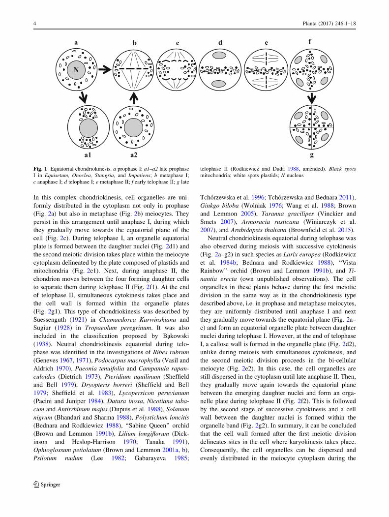
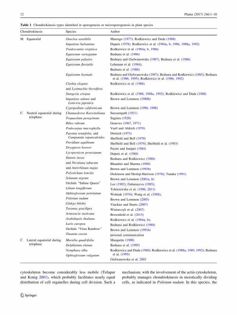
Equatorial chondriokinesis
Equatorial chondriokinesis is one of the four main types of
the process (Fig. 1). Although plastids and mitochondria in
this type are dispersed in the cytoplasm during prophase I
(Fig. 1a), during metaphase I they group in the equatorial
plane on both sides of the metaphase chromosome plate
(Fig. 1b). Such an arrangement of organelles in the meta-
phase meiocyte classifies chondriokinesis to the equatorial
type. Next, during anaphase I, the chondrion gradually dis-
perses in the cytoplasm (Fig. 1c) and forms an equatorial
plate between the daughter nuclei during telophase I
(Fig. 1d). Cell organelles remain in this position during the
second meiotic division (Fig. 1e) until telophase II and, after
karyokinesis, they form other plate separating successive
daughter nuclei (Fig. 1f). Within such an equatorial
arrangement, the cell wall is formed at the end of telophase II
(Fig. 1g). Equatorial chondriokinesis was described by
Bakowski (1938) only in meiosis occurring in animal sperm
cells. The researcher claimed that this type of chondrioki-
nesis did not take place in the world of plants. Subsequent
investigations revealed that equatorial chondriokinesis was
characteristic for many plant species. It was described in
Tradescantia virginica (Rodkiewicz et al. 1984b, 1986),
Clarkia elegans and Lysimachia thyrsiflora (Rodkiewicz
et al. 1986), Impatiens sultani andLonicera japonica (Brown
and Lemmon 1988b), and Cypripedium californicum
(Brown and Lemmon 1996, 1998). Furthermore, at the end of
the twentieth century, numerous analyses focused on the
distribution of plastids and mitochondria in plant cells pro-
vided a detailed description of the organisation of the
chondrion in early prophase meiocytes. Cytological analyses
of meiosis in Equisetum hyemale (Bednara and
Giełwanowska (1987); Bednara and Rodkiewicz 1985;
Bednara et al. 1986, 1995; Rodkiewicz et al. 1986, 1992), E.
fluviatile (Lehmann et al. 1984; Bednara et al. 1986), E.
palustre (Bednara and Giełwanowska (1987); Bednara et al.
1986), E. variegatum (Bednara et al. 1986), Onoclea sensi-
bilis (Marengo 1977; Rodkiewicz and Duda 1988), Stange-
ria eriopus (Rodkiewicz et al. 1986, 1988a, 1992;
Rodkiewicz and Duda 1988), and Impatiens balsamina
(Dupuis 1978; Rodkiewicz et al. 1984b, 1986, 1988a, 1992)
showed that plastids and mitochondria of these species
formed two groups visible at the two cell poles during late
prophase I (Fig. 1a1). Such groupings are visible transiently
during prophase I, and the organelles disperse in the cyto-
plasm at the end of this phase (Fig. 1a2). In the successive
stages, the organelle rearrangements correspond to equato-
rial chondriokinesis; therefore, although this process in
Equisetum, Onoclea, Stangria, and Impatiens initially has a
polar character, it can be regarded as equatorial chon-
driokinesis as the organelles are grouped in the equatorial
plane during metaphase I.
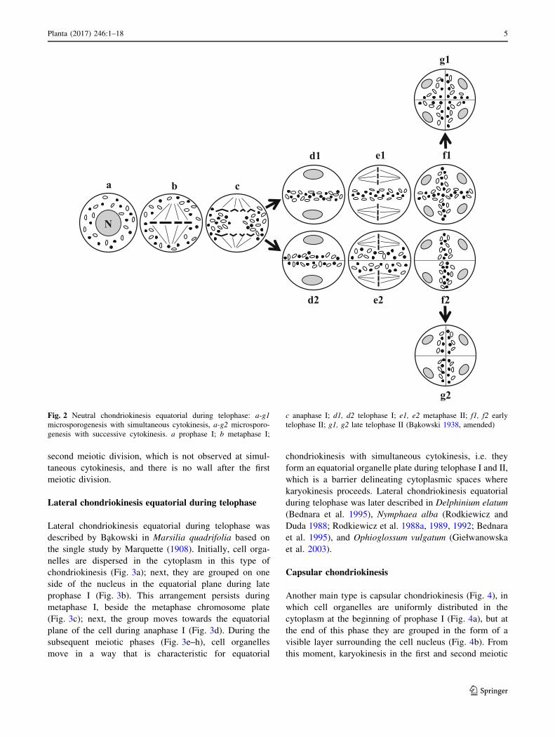
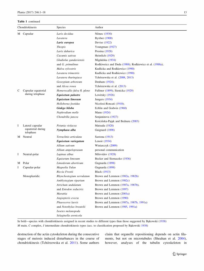
Neutral chondriokinesis equatorial during telophase
The most frequently described type is neutral chondrioki-
nesis equatorial during telophase, which occurs in the
meiosis stage with simultaneous cytokinesis (Fig. 2a–g1).
Planta (2017) 246:1–18 3
123
In this complex chondriokinesis, cell organelles are uni-
formly distributed in the cytoplasm not only in prophase
(Fig. 2a) but also in metaphase (Fig. 2b) meiocytes. They
persist in this arrangement until anaphase I, during which
they gradually move towards the equatorial plane of the
cell (Fig. 2c). During telophase I, an organelle equatorial
plate is formed between the daughter nuclei (Fig. 2d1) and
the second meiotic division takes place within the meiocyte
cytoplasm delineated by the plate composed of plastids and
mitochondria (Fig. 2e1). Next, during anaphase II, the
chondrion moves between the four forming daughter cells
to separate them during telophase II (Fig. 2f1). At the end
of telophase II, simultaneous cytokinesis takes place and
the cell wall is formed within the organelle plates
(Fig. 2g1). This type of chondriokinesis was described by
Suessenguth (1921) in Chamaedorea Karwinskiana and
Sugiur (1928) in Tropaeolum peregrinum. It was also
included in the classification proposed by Bakowski
(1938). Neutral chondriokinesis equatorial during telo-
phase was identified in the investigations of Ribes rubrum
(Geneves 1967, 1971), Podocarpus macrophylla (Vasil and
Aldrich 1970), Paeonia tenuifolia and Campanula rapan-
culoides (Dietrich 1973), Pteridium aquilinum (Sheffield
and Bell 1979), Dryopteris borreri (Sheffield and Bell
1979; Sheffield et al. 1983), Lycopersicon peruvianum
(Pacini and Juniper 1984), Datura inoxa, Nicotiana taba-
cum and Antirrhinum majus (Dupuis et al. 1988), Solanum
nigrum (Bhandari and Sharma 1988), Polystichum loncitis
(Bednara and Rodkiewicz 1988), ‘‘Sabine Queen’’ orchid
(Brown and Lemmon 1991b), Lilium longiflorum (Dick-
inson and Heslop-Harrison 1970; Tanaka 1991),
Ophioglossum petiolatum (Brown and Lemmon 2001a, b),
Psilotum nudum (Lee 1982; Gabarayeva 1985;
Tchorzewska et al. 1996; Tchorzewska and Bednara 2011),
Ginkgo biloba (Wolniak 1976; Wang et al. 1988; Brown
and Lemmon 2005), Taranna gracilipes (Vinckier and
Smets 2007), Armoracia rusticana (Winiarczyk et al.
2007), and Arabidopsis thaliana (Brownfield et al. 2015).
Neutral chondriokinesis equatorial during telophase was
also observed during meiosis with successive cytokinesis
(Fig. 2a–g2) in such species as Larix europea (Rodkiewicz
et al. 1984b; Bednara and Rodkiewicz 1988), ‘‘Vista
Rainbow’’ orchid (Brown and Lemmon 1991b), and Ti-
nantia erecta (own unpublished observations). The cell
organelles in these plants behave during the first meiotic
division in the same way as in the chondriokinesis type
described above, i.e. in prophase and metaphase meiocytes,
they are uniformly distributed until anaphase I and next
they gradually move towards the equatorial plane (Fig. 2a–
c) and form an equatorial organelle plate between daughter
nuclei during telophase I. However, at the end of telophase
I, a callose wall is formed in the organelle plate (Fig. 2d2),
unlike during meiosis with simultaneous cytokinesis, and
the second meiotic division proceeds in the bi-cellular
meiocyte (Fig. 2e2). In this case, the cell organelles are
still dispersed in the cytoplasm until late anaphase II. Then,
they gradually move again towards the equatorial plane
between the emerging daughter nuclei and form an orga-
nelle plate during telophase II (Fig. 2f2). This is followed
by the second stage of successive cytokinesis and a cell
wall between the daughter nuclei is formed within the
organelle band (Fig. 2g2). In summary, it can be concluded
that the cell wall formed after the first meiotic division
delineates sites in the cell where karyokinesis takes place.
Consequently, the cell organelles can be dispersed and
evenly distributed in the meiocyte cytoplasm during the
a b c d e f
a2a1 g
N
Fig. 1 Equatorial chondriokinesis. a prophase I; a1–a2 late prophase
I in Equisetum, Onoclea, Stangria, and Impatiens; b metaphase I;
c anaphase I; d telophase I; e metaphase II; f early telophase II; g late
telophase II (Rodkiewicz and Duda 1988, amended). Black spots
mitochondria; white spots plastids; N nucleus
4 Planta (2017) 246:1–18
123
second meiotic division, which is not observed at simul-
taneous cytokinesis, and there is no wall after the first
meiotic division.
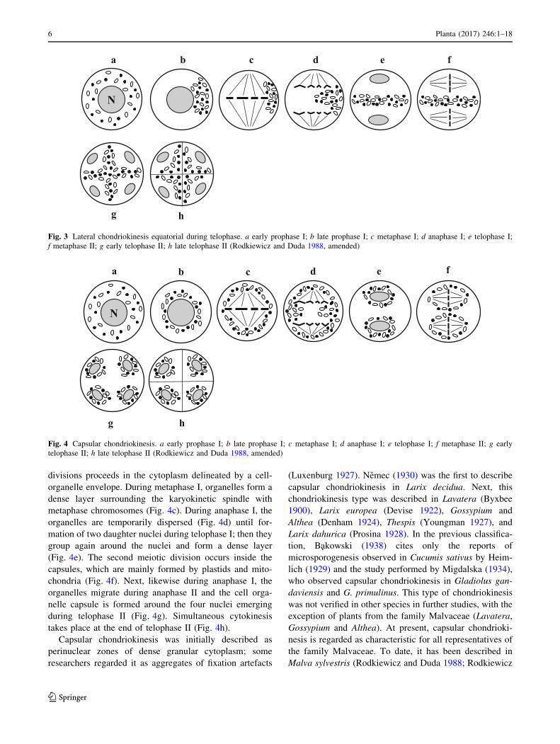
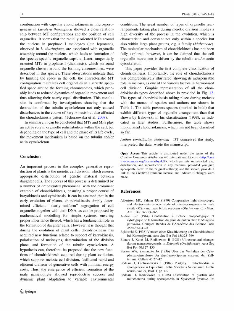
Lateral chondriokinesis equatorial during telophase
Lateral chondriokinesis equatorial during telophase was
described by Bakowski in Marsilia quadrifolia based on
the single study by Marquette (1908). Initially, cell orga-
nelles are dispersed in the cytoplasm in this type of
chondriokinesis (Fig. 3a); next, they are grouped on one
side of the nucleus in the equatorial plane during late
prophase I (Fig. 3b). This arrangement persists during
metaphase I, beside the metaphase chromosome plate
(Fig. 3c); next, the group moves towards the equatorial
plane of the cell during anaphase I (Fig. 3d). During the
subsequent meiotic phases (Fig. 3e–h), cell organelles
move in a way that is characteristic for equatorial
chondriokinesis with simultaneous cytokinesis, i.e. they
form an equatorial organelle plate during telophase I and II,
which is a barrier delineating cytoplasmic spaces where
karyokinesis proceeds. Lateral chondriokinesis equatorial
during telophase was later described in Delphinium elatum
(Bednara et al. 1995), Nymphaea alba (Rodkiewicz and
Duda 1988; Rodkiewicz et al. 1988a, 1989, 1992; Bednara
et al. 1995), and Ophioglossum vulgatum (Giełwanowska
et al. 2003).
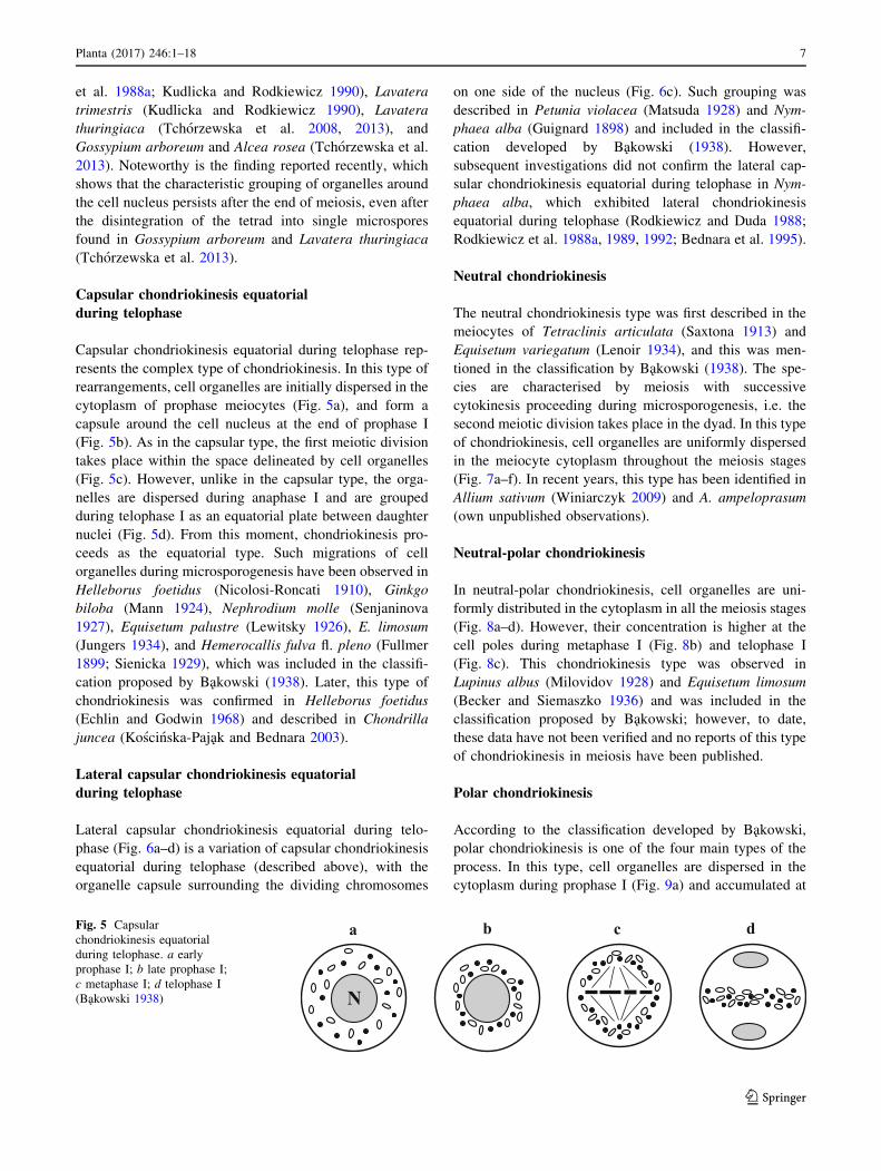
Capsular chondriokinesis
Another main type is capsular chondriokinesis (Fig. 4), in
which cell organelles are uniformly distributed in the
cytoplasm at the beginning of prophase I (Fig. 4a), but at
the end of this phase they are grouped in the form of a
visible layer surrounding the cell nucleus (Fig. 4b). From
this moment, karyokinesis in the first and second meiotic
a b
e1 f1
c
d2 e2 f2
d1
g1
g2
N
Fig. 2 Neutral chondriokinesis equatorial during telophase: a-g1
microsporogenesis with simultaneous cytokinesis, a-g2 microsporo-
genesis with successive cytokinesis. a prophase I; b metaphase I;
c anaphase I; d1, d2 telophase I; e1, e2 metaphase II; f1, f2 early
telophase II; g1, g2 late telophase II (Bakowski 1938, amended)
Planta (2017) 246:1–18 5
123
divisions proceeds in the cytoplasm delineated by a cell-
organelle envelope. During metaphase I, organelles form a
dense layer surrounding the karyokinetic spindle with
metaphase chromosomes (Fig. 4c). During anaphase I, the
organelles are temporarily dispersed (Fig. 4d) until for-
mation of two daughter nuclei during telophase I; then they
group again around the nuclei and form a dense layer
(Fig. 4e). The second meiotic division occurs inside the
capsules, which are mainly formed by plastids and mito-
chondria (Fig. 4f). Next, likewise during anaphase I, the
organelles migrate during anaphase II and the cell orga-
nelle capsule is formed around the four nuclei emerging
during telophase II (Fig. 4g). Simultaneous cytokinesis
takes place at the end of telophase II (Fig. 4h).
Capsular chondriokinesis was initially described as
perinuclear zones of dense granular cytoplasm; some
researchers regarded it as aggregates of fixation artefacts
(Luxenburg 1927). Nemec (1930) was the first to describe
capsular chondriokinesis in Larix decidua. Next, this
chondriokinesis type was described in Lavatera (Byxbee
1900), Larix europea (Devise 1922), Gossypium and
Althea (Denham 1924), Thespis (Youngman 1927), and
Larix dahurica (Prosina 1928). In the previous classifica-
tion, Bakowski (1938) cites only the reports of
microsporogenesis observed in Cucumis sativus by Heim-
lich (1929) and the study performed by Migdalska (1934),
who observed capsular chondriokinesis in Gladiolus gan-
daviensis and G. primulinus. This type of chondriokinesis
was not verified in other species in further studies, with the
exception of plants from the family Malvaceae (Lavatera,
Gossypium and Althea). At present, capsular chondrioki-
nesis is regarded as characteristic for all representatives of
the family Malvaceae. To date, it has been described in
Malva sylvestris (Rodkiewicz and Duda 1988; Rodkiewicz
ca fedb
g h
N
Fig. 3 Lateral chondriokinesis equatorial during telophase. a early prophase I; b late prophase I; c metaphase I; d anaphase I; e telophase I;
f metaphase II; g early telophase II; h late telophase II (Rodkiewicz and Duda 1988, amended)
a b c d e f
g h
N
Fig. 4 Capsular chondriokinesis. a early prophase I; b late prophase I; c metaphase I; d anaphase I; e telophase I; f metaphase II; g early
telophase II; h late telophase II (Rodkiewicz and Duda 1988, amended)
6 Planta (2017) 246:1–18
123
et al. 1988a; Kudlicka and Rodkiewicz 1990), Lavatera
trimestris (Kudlicka and Rodkiewicz 1990), Lavatera
thuringiaca (Tchorzewska et al. 2008, 2013), and
Gossypium arboreum and Alcea rosea (Tchorzewska et al.
2013). Noteworthy is the finding reported recently, which
shows that the characteristic grouping of organelles around
the cell nucleus persists after the end of meiosis, even after
the disintegration of the tetrad into single microspores
found in Gossypium arboreum and Lavatera thuringiaca
(Tchorzewska et al. 2013).
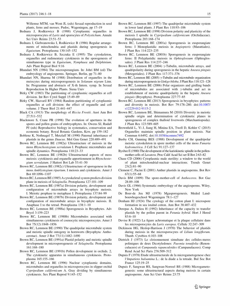
Capsular chondriokinesis equatorial
during telophase
Capsular chondriokinesis equatorial during telophase rep-
resents the complex type of chondriokinesis. In this type of
rearrangements, cell organelles are initially dispersed in the
cytoplasm of prophase meiocytes (Fig. 5a), and form a
capsule around the cell nucleus at the end of prophase I
(Fig. 5b). As in the capsular type, the first meiotic division
takes place within the space delineated by cell organelles
(Fig. 5c). However, unlike in the capsular type, the orga-
nelles are dispersed during anaphase I and are grouped
during telophase I as an equatorial plate between daughter
nuclei (Fig. 5d). From this moment, chondriokinesis pro-
ceeds as the equatorial type. Such migrations of cell
organelles during microsporogenesis have been observed in
Helleborus foetidus (Nicolosi-Roncati 1910), Ginkgo
biloba (Mann 1924), Nephrodium molle (Senjaninova
1927), Equisetum palustre (Lewitsky 1926), E. limosum
(Jungers 1934), and Hemerocallis fulva fl. pleno (Fullmer
1899; Sienicka 1929), which was included in the classifi-
cation proposed by Bakowski (1938). Later, this type of
chondriokinesis was confirmed in Helleborus foetidus
(Echlin and Godwin 1968) and described in Chondrilla
juncea (Koscinska-Pajak and Bednara 2003).
Lateral capsular chondriokinesis equatorial
during telophase
Lateral capsular chondriokinesis equatorial during telo-
phase (Fig. 6a–d) is a variation of capsular chondriokinesis
equatorial during telophase (described above), with the
organelle capsule surrounding the dividing chromosomes
on one side of the nucleus (Fig. 6c). Such grouping was
described in Petunia violacea (Matsuda 1928) and Nym-
phaea alba (Guignard 1898) and included in the classifi-
cation developed by Bakowski (1938). However,
subsequent investigations did not confirm the lateral cap-
sular chondriokinesis equatorial during telophase in Nym-
phaea alba, which exhibited lateral chondriokinesis
equatorial during telophase (Rodkiewicz and Duda 1988;
Rodkiewicz et al. 1988a, 1989, 1992; Bednara et al. 1995).
Neutral chondriokinesis
The neutral chondriokinesis type was first described in the
meiocytes of Tetraclinis articulata (Saxtona 1913) and
Equisetum variegatum (Lenoir 1934), and this was men-
tioned in the classification by Bakowski (1938). The spe-
cies are characterised by meiosis with successive
cytokinesis proceeding during microsporogenesis, i.e. the
second meiotic division takes place in the dyad. In this type
of chondriokinesis, cell organelles are uniformly dispersed
in the meiocyte cytoplasm throughout the meiosis stages
(Fig. 7a–f). In recent years, this type has been identified in
Allium sativum (Winiarczyk 2009) and A. ampeloprasum
(own unpublished observations).
Neutral-polar chondriokinesis
In neutral-polar chondriokinesis, cell organelles are uni-
formly distributed in the cytoplasm in all the meiosis stages
(Fig. 8a–d). However, their concentration is higher at the
cell poles during metaphase I (Fig. 8b) and telophase I
(Fig. 8c). This chondriokinesis type was observed in
Lupinus albus (Milovidov 1928) and Equisetum limosum
(Becker and Siemaszko 1936) and was included in the
classification proposed by Bakowski; however, to date,
these data have not been verified and no reports of this type
of chondriokinesis in meiosis have been published.
Polar chondriokinesis
According to the classification developed by Bakowski,
polar chondriokinesis is one of the four main types of the
process. In this type, cell organelles are dispersed in the
cytoplasm during prophase I (Fig. 9a) and accumulated at
a b c d
N
Fig. 5 Capsular
chondriokinesis equatorial
during telophase. a early
prophase I; b late prophase I;
c metaphase I; d telophase I
(Bakowski 1938)
Planta (2017) 246:1–18 7
123
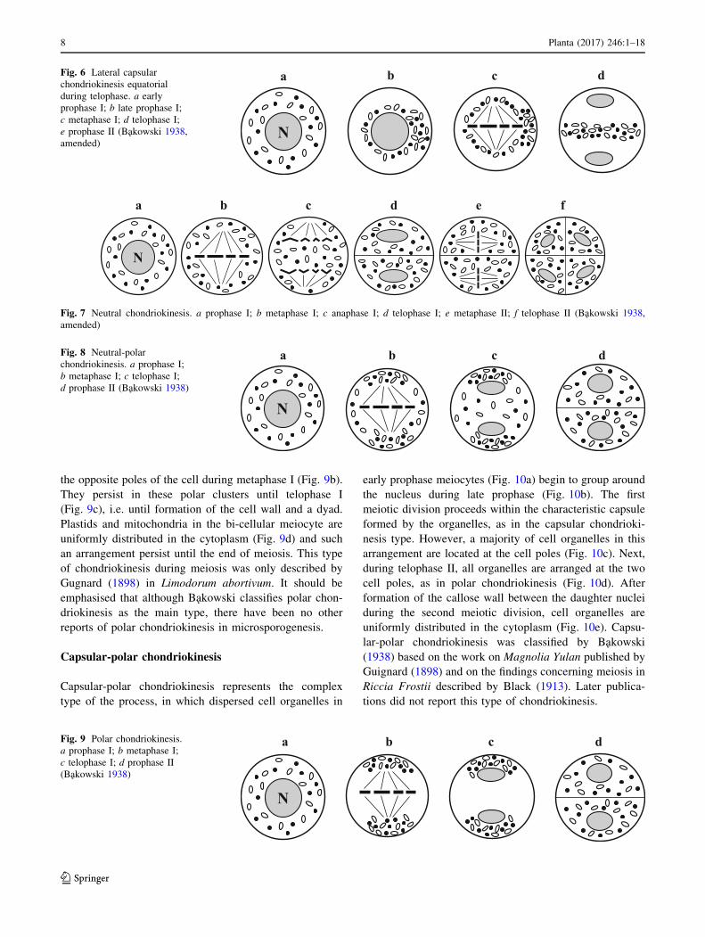
the opposite poles of the cell during metaphase I (Fig. 9b).
They persist in these polar clusters until telophase I
(Fig. 9c), i.e. until formation of the cell wall and a dyad.
Plastids and mitochondria in the bi-cellular meiocyte are
uniformly distributed in the cytoplasm (Fig. 9d) and such
an arrangement persist until the end of meiosis. This type
of chondriokinesis during meiosis was only described by
Gugnard (1898) in Limodorum abortivum. It should be
emphasised that although Bakowski classifies polar chon-
driokinesis as the main type, there have been no other
reports of polar chondriokinesis in microsporogenesis.
Capsular-polar chondriokinesis
Capsular-polar chondriokinesis represents the complex
type of the process, in which dispersed cell organelles in
early prophase meiocytes (Fig. 10a) begin to group around
the nucleus during late prophase (Fig. 10b). The first
meiotic division proceeds within the characteristic capsule
formed by the organelles, as in the capsular chondrioki-
nesis type. However, a majority of cell organelles in this
arrangement are located at the cell poles (Fig. 10c). Next,
during telophase II, all organelles are arranged at the two
cell poles, as in polar chondriokinesis (Fig. 10d). After
formation of the callose wall between the daughter nuclei
during the second meiotic division, cell organelles are
uniformly distributed in the cytoplasm (Fig. 10e). Capsu-
lar-polar chondriokinesis was classified by Bakowski
(1938) based on the work on Magnolia Yulan published by
Guignard (1898) and on the findings concerning meiosis in
Riccia Frostii described by Black (1913). Later publica-
tions did not report this type of chondriokinesis.
a b c d
N
Fig. 6 Lateral capsular
chondriokinesis equatorial
during telophase. a early
prophase I; b late prophase I;
c metaphase I; d telophase I;
e prophase II (Bakowski 1938,
amended)
b fedca
N
Fig. 7 Neutral chondriokinesis. a prophase I; b metaphase I; c anaphase I; d telophase I; e metaphase II; f telophase II (Bakowski 1938,
amended)
c dba
N
Fig. 8 Neutral-polar
chondriokinesis. a prophase I;
b metaphase I; c telophase I;
d prophase II (Bakowski 1938)
c da b
N
Fig. 9 Polar chondriokinesis.
a prophase I; b metaphase I;
c telophase I; d prophase II
(Bakowski 1938)
8 Planta (2017) 246:1–18
123
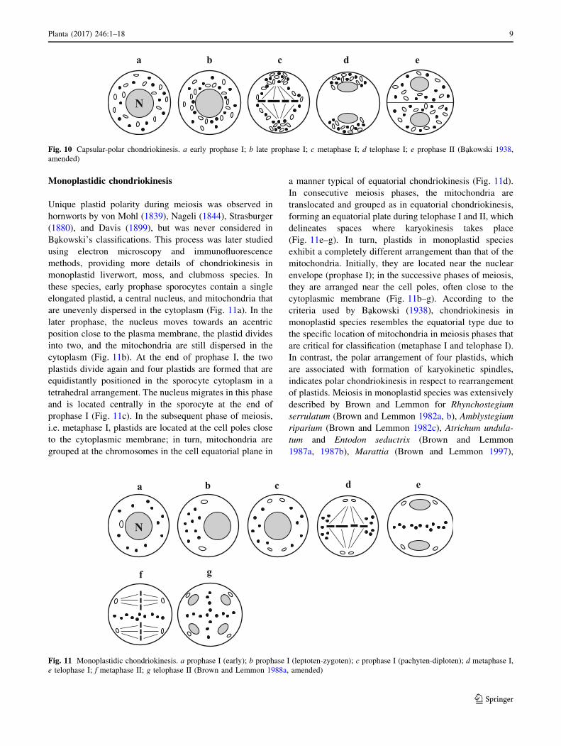
Monoplastidic chondriokinesis
Unique plastid polarity during meiosis was observed in
hornworts by von Mohl (1839), Nageli (1844), Strasburger
(1880), and Davis (1899), but was never considered in
Bakowski’s classifications. This process was later studied
using electron microscopy and immunofluorescence
methods, providing more details of chondriokinesis in
monoplastid liverwort, moss, and clubmoss species. In
these species, early prophase sporocytes contain a single
elongated plastid, a central nucleus, and mitochondria that
are unevenly dispersed in the cytoplasm (Fig. 11a). In the
later prophase, the nucleus moves towards an acentric
position close to the plasma membrane, the plastid divides
into two, and the mitochondria are still dispersed in the
cytoplasm (Fig. 11b). At the end of prophase I, the two
plastids divide again and four plastids are formed that are
equidistantly positioned in the sporocyte cytoplasm in a
tetrahedral arrangement. The nucleus migrates in this phase
and is located centrally in the sporocyte at the end of
prophase I (Fig. 11c). In the subsequent phase of meiosis,
i.e. metaphase I, plastids are located at the cell poles close
to the cytoplasmic membrane; in turn, mitochondria are
grouped at the chromosomes in the cell equatorial plane in
a manner typical of equatorial chondriokinesis (Fig. 11d).
In consecutive meiosis phases, the mitochondria are
translocated and grouped as in equatorial chondriokinesis,
forming an equatorial plate during telophase I and II, which
delineates spaces where karyokinesis takes place
(Fig. 11e–g). In turn, plastids in monoplastid species
exhibit a completely different arrangement than that of the
mitochondria. Initially, they are located near the nuclear
envelope (prophase I); in the successive phases of meiosis,
they are arranged near the cell poles, often close to the
cytoplasmic membrane (Fig. 11b–g). According to the
criteria used by Bakowski (1938), chondriokinesis in
monoplastid species resembles the equatorial type due to
the specific location of mitochondria in meiosis phases that
are critical for classification (metaphase I and telophase I).
In contrast, the polar arrangement of four plastids, which
are associated with formation of karyokinetic spindles,
indicates polar chondriokinesis in respect to rearrangement
of plastids. Meiosis in monoplastid species was extensively
described by Brown and Lemmon for Rhynchostegium
serrulatum (Brown and Lemmon 1982a, b), Amblystegium
riparium (Brown and Lemmon 1982c), Atrichum undula-
tum and Entodon seductrix (Brown and Lemmon
1987a, 1987b), Marattia (Brown and Lemmon 1997),
a b c d e
N
Fig. 10 Capsular-polar chondriokinesis. a early prophase I; b late prophase I; c metaphase I; d telophase I; e prophase II (Bakowski 1938,
amended)
a b c d e
gf
N
Fig. 11 Monoplastidic chondriokinesis. a prophase I (early); b prophase I (leptoten-zygoten); c prophase I (pachyten-diploten); d metaphase I,
e telophase I; f metaphase II; g telophase II (Brown and Lemmon 1988a, amended)
Planta (2017) 246:1–18 9
123
Angiopteris evecta (Brown and Lemmon 2001a), Phaeo-
ceros laevis and Notothylas breutelii (Brown and Lemmon
1990), Isoetes melanopoda (Brown and Lemmon
1987a, b, 1991a), and Selaginella arenicola (Brown and
Lemmon 1985, 1991a).
Role and mechanism of chondriokinesis
Chondriokinesis during meiosis is a widespread phe-
nomenon in all analysed plant species, even from system-
atically distant groups (horsetails, mosses, ferns,
Gymnosperms, and Angiosperms). The universal occur-
rence of the process implies its high conservatism and its
probable origin at an early stage of plant evolution; how-
ever, the course of the process varies between plant spe-
cies. It should be emphasised, that the role of
chondriokinesis has not been synthetically presented as yet,
and currently there are many data indicating varied rele-
vance of the process in the meiotic division of the plant
cell.
It is thought that the most fundamental role of the
specific grouping of organelles throughout the phases of
meiosis is to ensure equal distribution of organelles
between the tetrad cells (Senjaninova 1927; Geneves 1967;
Wolniak 1976; Dupuis 1978; Brown and Lemmon 1982b),
which guarantees formation of identical, metabolically
active microspores. This is confirmed by the fact that
organelles, which are initially dispersed in the meiocyte
cytoplasm, are grouped (e.g. neutral chondriokinesis
equatorial during telophase) during the key meiosis phases
(telophase I and telophase II), which ensures appropriate
segregation into daughter cells. With time, when many
biological phenomena were better explored at the molec-
ular level, i.e. the importance of cytoplasmic inheritance
and the role of semi-autonomous cell organelles in the
formation of fertile pollen grains, chondriokinesis was
assumed to ensure proper cytoplasmic inheritance of
genetic material in the plant cell, wherein both the plastid
and the mitochondrial genomes are equally responsible for
cytoplasmic inheritance (Sears 1980; Hagemann and
Schroder 1989; Kuroiwa 1991; Mogensen 1996; Nagata
2010). The impact of plastid or mitochondrial DNA on
offspring traits has been described, especially restriction
fragment length polymorphisms (RFLPs) were used to
follow the organellar DNA inheritance (Boblenz et al.
1990; Derepas and Dulieu 1992; Hu et al. 1996; Trusty
et al. 2007; Hansen et al. 2007; Matsushima et al. 2008b).
In many plant species, the presence of plastid or mito-
chondrial DNA in male reproductive cells determining the
potential for cytoplasmic inheritance has been shown
(Zhang et al. 2003). Yet, this question seems debatable, as
some authors claim that the presence of plastids in sperm
cells does not indicate their inclusion in the zygote
(Lombardo and Gerola 1968; Reboud and Zeyl 1994). It is,
however, indisputable that cytoplasmic male sterility
(CMS) is determined by a lack of the mitochondrial gen-
ome (Holford et al. 1991; Chase 2006; Wang et al. 2006).
Hence, proper segregation of cell organelles is a key pro-
cess in the formation of a fertile male gametophyte.
Besides the basic function of chondriokinesis (i.e. equal
distribution of organelles and involvement in cytoplasmic
inheritance), it was proposed that cell organelles constitute
a barrier limiting the sites in the meiocyte cytoplasm in
which karyokinesis takes place (Kudlicka and Rodkiewicz
1990; Rodkiewicz et al. 1992; Bednara et al. 1986, 1995;
Tchorzewska et al. 1996, 2008; Brownfield et al. 2015).
Organelles forming a capsule or an equatorial plate prevent
fusion of karyokinetic spindles or separating chromosomes
during the second meiotic division, serving as a ‘‘substitute
of the cell plate’’ (Bednara et al. 1986). This idea was
supported by the fact, that cell organelles were dispersed in
the meiocyte cytoplasm even if they were grouped as an
equatorial plate at the end of telophase I. This phenomenon
has been described in many species characterised by
meiosis with successive cytokinesis, in which neutral
chondriokinesis equatorial during telophase occurs (Rod-
kiewicz et al. 1984b; Bednara and Rodkiewicz 1988;
Brown and Lemmon 1991b). Additionally, in meiosis with
successive cytokinesis, during neutral chondriokinesis
equatorial during telophase, organelles are arranged in an
equatorial plate during telophase II when another cell plate
is formed. In this case, another function of chondriokinesis
can be inferred, i.e. cell organelles are involved in cell plate
formation (Rodkiewicz et al. 1986, 1988a, 1989).
Another function that can be assigned to chondriokinesis
is related to the role of cell organelles in determination of
meiocyte polarisation. This idea was formulated based on
the observation of pollen development in gymnosperms,
where plastids in the microspore tetrad remain close to the
proximal wall of the tetrad after meiosis. The polarisation
is important, as the prothallus cells develop at the proximal
wall of pollen cells in gymnosperms and the pollen tube
grows from the distal wall (Rodkiewicz et al. 1984a). The
relevance of organelle grouping for polarisation of
megasporocytes during megasporogenesis is particularly
evident. Polarisation of meiocytes is extremely important
in the process of female gametophyte development, given
the competition between megaspores for formation of a
functional megaspore, which will develop into the embryo
sac (Steward and Gifford 1967; De Boer-de Jeu 1978;
Willemse and Bednara 1979; Willemse and De Boer-de Jeu
1981; Bednara et al. 1981; Ekici and Dane 2004).
An additional function of chondriokinesis is the rela-
tionship between plastids and the organisation of tubulin
cytoskeleton in the plant cell. This phenomenon has been
10 Planta (2017) 246:1–18
123
described in numerous reports on meiosis in monoplastid
plant species (Brown and Lemmon 1982a, 1985, 1987a, b,
1988a, 1991a, b, 2004, 2006). The acentrosomal spindle
formed in plants begins polymerisation with c-tubulin,
which is the major component of microtubule organizing
centres (MTOCs). Observations of meiosis in monoplastid
plants revealed that c-tubulin was located at the plastid
envelope (Shimamura et al. 2004); therefore, the spindle
apparatus is organised in association with plastid migration
and division (Shimamura et al. 2003). Furthermore, divi-
sion polarity has been described in Bryophyte meiosis,
indicating that the prophase system of axially aligned
microtubules determines the site of cytokinesis. Since
microtubules are associated with plastids, the location of
plastids determines the cell division plane (Brown and
Lemmon 1987a).
The molecular mechanism of cell organelle transport
and movement during cell division remains obscure. One
of the first reports of the mechanism of organelle move-
ment during meiosis suggests that this process is func-
tionally linked to the cytoskeleton (Wolniak 1976). It
should be emphasised that the phenomenon of migration of
cellular organelles associated with the cytoskeleton has
been repeatedly shown in animal cells (Stebbings 1990),
lower plant cells (Menzel 1985; Busby and Gunning 1988),
and higher plant cells (Tanaka 1991; Brownfield et al.
2015). Involvement of both the tubulin (MT) and actin
(MF) cytoskeleton in migration of organelles was reported.
Although most reports are primarily focused on somatic
cells (Williamson 1993; Ligrone and Duckett 1998; Oly-
slaegers and Verbelen 1998; Kandasamy and Meagher
1999), it can be assumed that, due to the prevalence of this
phenomenon, the mechanism involved in cytoskeleton-as-
sisted organelle movement is universal and can be referred
to meiotically dividing generative cells. As shown in Ni-
cotiana tabacum protoplast cells, during initial steps of cell
division, organelles are surrounded by ‘‘actin baskets’’ and
these baskets facilitate their preparatory organisation dur-
ing cell division; subsequently the ‘‘actin baskets’’ lose
their integrity and individual organelles are tethered to
acting filaments, which form a dense cytoplasmic network
(Kandasamy and Meagher 1999; Sheahan et al. 2004). This
leads to enmeshment of organelles and dependence of their
location on dynamically changing cytoskeleton configura-
tions (Sheahan et al. 2004). Consequently, organelles
embedded at an appropriate place and time within
Polar Equatorial Neutral
Neutralequatorial
during telophase
Lateralequatorial
during telophase
Capsular
Capsularequatorial
during telophase
Neutral-polar
Monoplastidic
Lateral capsularequatorial
during telophase
a
b
Capsular-polar
Fig. 12 Graphic summary of
all chondriokinesis types
identified in sporogenesis or
microsporogenesis.
A classification developed by
Bakowski (1938). B unclassified
chondriokinesis in monoplastid
species
Planta (2017) 246:1–18 11
123
cytoskeleton become considerably less mobile (Tirlapur
and Konig 2001), which probably facilitates nearly equal
distribution of cell organelles during cell division. Such a
mechanism, with the involvement of the actin cytoskeleton,
probably manages chondriokinesis in meiotically dividing
cells, as indicated in Psilotum nudum. In this species, the
Table 1 Chondriokinesis types identified in sporogenesis or microsporogenesis in plant species
Chondriokinesis Species Author
M Equatorial Onoclea sensibilis Marengo (1977); Rodkiewicz and Duda (1988)
Impatiens balsamina Dupuis (1978); Rodkiewicz et al. (1984a, b, 1986, 1988a, 1992)
Tradescantia virginica Rodkiewicz et al. (1984a, b, 1986)
Equisetum variegatum Bednara et al. (1986)
Equisetum palustre Bednara and Giełwanowska (1987), Bednara et al. (1986)
Equisetum fluviatile Lehmann et al. (1984);
Bednara et al. (1986)
Equisetum hyemale Bednara and Giełwanowska (1987); Bednara and Rodkiewicz (1985); Bednara
et al. (1986, 1995); Rodkiewicz et al. (1986, 1992)
Clarkia elegans
and Lysimachia thyrsiflora
Rodkiewicz et al. (1986)
Stangeria eriopus Rodkiewicz et al. (1986, 1988a, 1992); Rodkiewicz and Duda (1988)
Impatiens sultani and
Lonicera japonica
Brown and Lemmon (1988b)
Cypripedium californicum Brown and Lemmon (1996, 1998)
C Neutral equatorial during
telophase
Chamaedorea Karwinskiana
Tropaeolum peregrinum
Ribes rubrum
Podocarpus macrophylla
Paeonia tenuifolia, and
Campanula rapanculoides
Pteridium aquilinum
Dryopteris borreri
Lycopersicon peruvianum
Datura inoxa
and Nicotiana tabacum
and Antirrhinum majus
Polystichum loncitis
Solanum nigrum
Orchids ‘‘Sabine Queen’’
Lilium longiflorum
Ophioglossum petiolatum
Psilotum nudum
Ginkgo biloba
Taranna gracilipes
Armoracia rusticana
Arabidopsis thaliana
Larix europea
Orchids ‘‘Vista Rainbow’’
Tinantia erecta
Suessenguth (1921)
Sugiura (1928)
Geneves (1967, 1971)
Vasil and Aldrich (1970)
Dietrich (1973)
Sheffield and Bell (1979)
Sheffield and Bell (1979); Sheffield et al. (1983)
Pacini and Juniper (1984)
Dupuis et al. (1988)
Bednara and Rodkiewicz (1988)
Bhandari and Sharma (1988)
Brown and Lemmon (1991b)
Dickinson and Heslop-Harrison (1970); Tanaka (1991)
Brown and Lemmon (2001a, b)
Lee (1982); Gabarayeva (1985);
Tchorzewska et al. (1996, 2011)
Wolniak (1976); Wang et al. (1988);
Brown and Lemmon (2005)
Vinckier and Smets (2007)
Winiarczyk et al. (2007)
Brownfield et al. (2015)
Rodkiewicz et al. (1984a, b);
Bednara and Rodkiewicz (1988)
Brown and Lemmon (1991b)
personal communication
C Lateral equatorial during
telophase
Marsilia quadrifolia
Delphinium elatum
Nymphaea alba
Ophioglossum vulgatum
Marquette (1908)
Bednara et al. (1995)
Rodkiewicz and Duda (1988); Rodkiewicz et al. (1988a, 1989, 1992); Bednara
et al. (1995)
Giełwanowska et al. 2003
12 Planta (2017) 246:1–18
123
destruction of the actin cytoskeleton during the consecutive
stages of meiosis induced disturbances in the course of
chondriokinesis (Tchorzewska et al. 2011). Some authors
claim that organelle repositioning depends on actin fila-
ments, but not on microtubules (Sheahan et al. 2004),
however, analyses of the tubulin cytoskeleton in
Table 1 continued
Chondriokinesis Species Author
M Capsular Larix decidua
Lavatera
Larix europea
Thespis
Larix dahurica
Cucumis sativus
Gladiolus gandaviensis
and G. primulinus
Malva sylvestris
Lavatera trimestris
Lavatera thuringiaca
Gossypium arboreum
and Alcea rosea
Nemec (1930)
Byxbee (1900)
Devise (1922)
Youngman (1927)
Prosina (1928)
Heimlich (1929)
Migdalska (1934)
Rodkiweicz and Duda (1988); Rodkiewicz et al. (1988a);
Kudlicka and Rodkiewicz (1990)
Kudlicka and Rodkiewicz (1990)
Tchorzewska et al. (2008, 2013)
Denham (1924);
Tchorzewska et al. (2013)
C Capsular equatorial
during telophase
Hemerocallis fulva fl. pleno
Equisetum palustre
Equisetum limosum
Helleborus foetidus
Ginkgo biloba
Nephrodium molle
Chondrilla juncea
Fullmer (1899); Sienicka (1929)
Lewitsky (1926)
Jungers (1934)
Nicolosi-Roncati (1910);
Echlin and Godwin (1968)
Mann (1924)
Senjaninova (1927)
Koscinska-Pajak and Bednara (2003)
I Lateral capsular
equatorial during
telophase
Petunia violacea
Nymphaea alba
Matsuda (1928)
Guignard (1898)
M Neutral Tetraclinis articulata
Equisetum variegatum
Allium sativum
Allium ampeloprasum
Saxtona (1913)
Lenoir (1934)
Winiarczyk (2009)
personal communication
I Neutral-polar Lupinus albus
Equisetum limosum
Milovidov (1928)
Becker and Siemaszko (1936)
M Polar Limodorum abortivum Gugnarda (1898)
I Capsular-polar Magnolia Yulan
Riccia Frostii
Gugnarda (1898)
Black (1913)
Monoplastidic Rhynchostegium serrulatum
Amblystegium riparium
Atrichum undulatum
and Entodon seductrix
Marattia
Angiopteris evecta
Phaeoceros laevis
and Notothylas breutelii
Isoetes melanopoda
Selaginella arenicola
Brown and Lemmon (1982a, 1982b)
Brown and Lemmon (1982c)
Brown and Lemmon (1987a, 1987b),
Brown and Lemmon (1997)
Brown and Lemmon (2001a)
Brown and Lemmon (1990)
Brown and Lemmon (1987a, 1987b, 1991a)
Brown and Lemmon (1985, 1991a)
In bold—species with chondriokinesis assigned in recent studies to different types than those suggested by Bakowski (1938)
M main, C complex, I intermediate chondriokinesis types (acc. to classification proposed by Bakowski 1938)
Planta (2017) 246:1–18 13
123
combination with capsular chondriokinesis in microsporo-
genesis in Lavatera thuringiaca showed a close relation-
ship between MT configurations and the position of cell
organelles. It seems that the radially oriented MTs around
the nucleus in prophase I meiocytes (late leptotene),
observed in L. thuringiaca, are associated with organelle
assembly around the nucleus, which leads to formation of
the species-specific organelle capsule. Later, tangentially
oriented MTs in prophase I (diakinesis), which surround
organelle clusters around the forming chromosomes, were
described in this species. These observations indicate that,
by limiting the space in the cell, the characteristic MT
configuration maintains cell organelles in a strictly speci-
fied space around the forming chromosomes, which prob-
ably leads to reduced dynamics of organelle movement and
thus allowing their nearly equal distribution. This conclu-
sion is confirmed by investigations showing that the
destruction of the tubulin cytoskeleton not only caused
disturbances in the course of karyokinesis but also affected
the chondriokinesis pattern (Tchorzewska et al. 2008).
In summary, it can be concluded that MTs and MFs play
an active role in organelle redistribution within the cell, but
depending on the type of cell and the phase of its life cycle,
the movement mechanism is based on the tubulin and/or
actin cytoskeleton.
Conclusion
An important process in the complex generative repro-
duction of plants is the meiotic cell division, which ensures
appropriate distribution of genetic material between
daughter cells. The success of this process is determined by
a number of orchestrated phenomena, with the prominent
example of chondriokinesis, ensuring a proper course of
karyokinesis and cytokinesis. It can be assumed that in the
early evolution of plants, chondriokinesis simply deter-
mined efficient ‘‘nearly uniform’’ segregation of cell
organelles together with their DNA, as can be proposed by
mathematical modelling for simple systems, ensuring
proper inheritance thereof, which has a fundamental role in
the formation of daughter cells. However, it is thought that
during the evolution of plant cells, chondriokinesis has
acquired new functions related to support of karyokinesis,
polarisation of meiocytes, determination of the division
plane, and formation of the tubulin cytoskeleton. A
hypothesis can, therefore, be proposed that the new func-
tions of chondriokinesis acquired during plant evolution,
which supports meiotic cell division, facilitated rapid and
efficient division of generative cells with minimal energy
costs. Thus, the emergence of efficient formation of the
male gametophyte allowed reproductive success and
dynamic plant adaptation to variable environmental
conditions. The great number of types of organelle rear-
rangements taking place during meiotic division implies a
high diversity of the process in the evolution, which is
characteristic and constant not only within a species but
also within large plant groups, e.g. a family (Malvaceae).
The molecular mechanism of chondriokinesis has not been
fully explored; however, it can be claimed that the cell
organelle movement is driven by the tubulin and/or actin
cytoskeleton.
This paper provides the first complete classification of
chondriokinesis. Importantly, the role of chondriokinesis
was comprehensively illustrated, showing its indispensable
role in meiosis, as one of the various factors in harmonised
cell division. Graphic representation of all the chon-
driokinesis types described above is provided in Fig. 12.
The types of chondriokinesis taking place during meiosis
with the names of species and authors are shown in
Table 1. The table presents species (marked in bold) that
exhibit different types of organelle arrangement than that
shown by Bakowski in his classification (1938), as indi-
cated in later studies. Furthermore, the table shows
monoplastid chondriokinesis, which has not been classified
so far.
Author contribution statement DT–conceived the study,
interpreted the data, wrote the manuscript.
Open Access This article is distributed under the terms of the
Creative Commons Attribution 4.0 International License (http://crea
tivecommons.org/licenses/by/4.0/), which permits unrestricted use,
distribution, and reproduction in any medium, provided you give
appropriate credit to the original author(s) and the source, provide a
link to the Creative Commons license, and indicate if changes were
made.
References
Albertsten MC, Palmer RG (1979) Comparative light-microscopic
and electron-microscopic study of microsporogenesis in male
sterile (MS1) and male fertile soybeans (Glycine max (L.) Merr.
Am J Bot 66:253–265
Audran J-C (1964) Contribution a l’etude morphologique et
cytologique de la formation du grain de pollen chez le Stangeria
paradoxa. Comptes Rendus de l’Academie des Science Paris
258:4322–4325
Bakowski Z (1938) Versuch einer Klassifizierung der Chondriokinese
bei Kormophyten. Acta Soc Bot Pol 15:323–369
Bdnara J, Kuras M, Rodkiewicz B (1981) Ultrastructural changes
during megasporogenesis in Epipactis (Orchidaceae). Acta Soc
Bot Pol 50:127–130
Becker WA, Siemaszko JA (1936) Uber das Verhalten der Cyto-
plasma-einschlusse der Equisetum-Sporen wahrend der Zell-
teilung. Cellule 45:27–42
Bednara J, Giełwanowska I (1987) Plastydy i mitochondria w
sporogenezie u Equisetum. Folia Societatis Scientiarum Lubli-
nensis, vol 29, Biol. I, pp 3–9
Bednara, J, Rodkiewicz B (1985) Distribution of plastids and
mitochondria during sporogenesis in Equisetum hyemale. In:
14 Planta (2017) 246:1–18
123
Willemse MTM, van Went JL (eds) Sexual reproduction in seed
plants, ferns and mosses. Pudoc, Wageningen, pp 17–19
Bednara J, Rodkiewicz B (1988) Cytoplasmic organelles in
microsporocytes of Larix and sporocytes of Polystichum. Annale
Sci Univ Reims 23:51–53
Bednara J, Giełwanowska I, Rodkiewicz B (1986) Regular arrange-
ments of mitochondria and plastids during sporogenesis in
Equisetum. Protoplasma 130:145–152
Bednara J, Rodkiewicz B, Szczuka E (1995) The cytoskeleton,
organelles and rudimentary cytokinesis in the sporogenesis of
simultaneous type in Equisetum, Nymphaea and Delphinium.
Adv Plant Reprod Biol 9:16
Bhandari NN (1984) The microsporangium. In: Johri BM (ed) The
embryology of angiosperms. Springer, Berlin, pp 71–80
Bhandari NN, Sharma M (1988) Distribution of organelles in the
meiocytes during microsporogenesis in Solanum nigrum Linn.
In: Programme and abstracts of X-th Intern. symp. In Sexual
Reproduction In Higher Plants. Siena Univ
Birky CW (1983) The partitioning of cytoplasmic organelles at cell
division. Int Rev Cytol Suppl 15:49–89
Birky CW, Skavaril RV (1984) Random partitioning of cytoplasmic
organelles at cell division: the effect of organelle and cell
volume. J Theor Biol 21:441–447
Black CA (1913) The morphology of Riccia Frostii. Aust Ann Bot
27:511–532
Blackmore S, Crane PR (1998) The evolution of apertures in the
spores and pollen grains of embryophytes. In: Owens SJ, Rudall
PJ (eds) Reproductive biology in systematics, conservation and
economic botany. Royal Botanic Gardens, Kew, pp 159–182
Boblenz K, Nothnagel T, Metzlaff M (1990) Paternal inheritance of
plastids in the genus Daucus. Mol Gen Genet 220:489–491
Brown RC, Lemmon BE (1982a) Ultrastructure of meiosis in the
moss Rhynchostegium serrulatum I. Prophasic microtubules and
spindle dynamics. Protoplasma 110:23–33
Brown RC, Lemmon BE (1982b) Ultrastructural aspects of moss
meiosis: cytokinesis and organelle apportionment in Rhynchoste-
gium serrulatum. J Hattori Bot Lab 53:41–50
Brown RC, Lemmon BE (1982c) Ultrastructure of sporogenese in the
moss Amblystegium riparium. I meiosis and cytokinesis. Amer J
Bot 69:1096–1107
Brown RC, Lemmon BE (1985) A cytoskeletal system predicts division
plane in meiosis of Selaginella. Protoplasma 127:101–109
Brown RC, Lemmon BE (1987a) Division polarity, development and
configuration of microtubule arrays in bryophyte meiosis.
I. Meiotic prophase to metaphase I. Protoplasma 137:84–99
Brown RC, Lemmon BE (1987b) Division polarity, development and
configuration of microtubule arrays in bryophyte meiosis. II.
Anaphase I to the tetrad. Protoplasma 138:1–10
Brown RC, Lemmon BE (1988a) Sporogenesis in Bryophytes. Adv
Bryol 3:159–223
Brown RC, Lemmon BE (1988b) Microtubules associated with
simultaneous cytokinesis of coenocytic microsporocytes. Amer J
Bot 75(12):1848–1856
Brown RC, Lemmon BE (1990) The quadripolar microtubule system
and meiotic spindle ontogeny in hornworts (Bryophyta: Antho-
cerotae). Amer J Bot 77(11):1482–1490
Brown RC, Lemmon BE (1991a) Plastid polarity and meiotic spindle
development in microsporogenesis of Selaginella. Protoplasma
161:168–180
Brown RC, Lemmon BE (1991b) Pollen development in orchids. 2,
The cytokinetic apparatus in simultaneous cytokinesis. Proto-
plasma 165:155–166
Brown RC, Lemmon BE (1996) Nuclear cytoplasmic domains,
microtubules and organelles in microsporocytes in slipper orchid
Cypripedium californicum A. Gray dividing by simultaneous
cytokinesis. Sex Plant Reprod 9:145–152
Brown RC, Lemmon BE (1997) The quadripolar microtubule system
in lower land plants. J Plant Res 110:93–106
Brown RC, Lemmon BE (1998) Division polarity and plasticity of the
meiosis I spindle in Cypripedium californicum (Orchidaceae).
Protoplasma 203:168–174
Brown RC, Lemmon BE (2001a) Sporogenesis in eusporangiate
ferns: I Monoplastidic meiosis in Angiopteris (Marattiales).
J Plant Res 114:223–235
Brown RC, Lemmon BE (2001b) Sporogenesis in eusporangiate
ferns: II Polyplastidic meiosis in Ophioglossum (Ophioglos-
sales). J Plant Res 114:237–246
Brown RC, Lemmon BE (2004) c-Tubulin, microtubule arrays, and
quadripolarity during sporogenesis in the hepatic Aneura pinguis
(Metzgeriales). J Plant Res 117:371–376
Brown RC, Lemmon BE (2005) c-Tubulin and microtubule organization
during microsporogenesis inGinkgo biloba. J Plant Res 118:121–128
Brown RC, Lemmon BE (2006) Polar organizers and girdling bands
of microtubules are associated with c-tubulin and act in
establishment of meiotic quadripolarity in the hepatic Aneura
pinguis (Bryophyta). Protoplasma 227:77–85
Brown RC, Lemmon BE (2013) Sporogenesis in bryophytes: patterns
and diversity in meiosis. Bot Rev 79:178–280. doi:10.1007/
s12229-012-9115-2
Brown RC, Lemmon BE, Shimamura M (2010) Diversity in meiotic
spindle origin and determination of cytokinetic planes in
sporogenesis of complex thalloid liverworts (Marchantiopsida).
J Plant Res 123:589–605
Brownfield L, Yi J, Jiang H, Minina EA, Twell D, Kohler C (2015)
Organelles maintain spindle position in plant meiosis. Nat
Commun 6:6492. doi:10.1038/ncomms7492
Busby CH, Gunning BES (1988) Development of the quadripolar
meiotic cytoskeleton in spore mother cells of the moss Funaria
hydrometrica. J Cell Sci 91:127–137
Byxbee E (1900) The development of the kariokinetic spindle in the pollen-
mother-cells of Lavatera. Proc Calif Acad Sci ser II Bot 2:63–82
Chase CD (2006) Cytoplasmic male sterility: a window to the world
of plant mitochondrial-nuclear interactions. Trends Genet
23(2):81–90
Clement C, Pacini E (2001) Anther plastids in angiosperms. Bot Rev
67(1):55–68
Davis BM (1899) The spore-mother-cell of Anthoceros. Bot Gaz
28:89–108
Davis GL (1966) Systematic embryology of the angiosperms. Wiley,
New York
De Boer-de Jeu MJ (1978) Mgasporogenesis. Meded Land-
bouwhogeschool Wageningen 78:1–127
Denham HJ (1924) The cytology of the cotton plant I: microspore
formation in sea istalnd cotton. Ann Bot 38:407–432
Derepas A, Dulieu H (1992) Inheritance of the capacity to transfer
plastids by the pollen parent in Petunia hybrid. Hort J Hered
83:6–10
Devise R (1922) La figure achromatique et la plaque cellulaire dans
les microsporocytes du Larix europea. Cellule 32:247–309
Dickinson HG, Heslop-Harrison J (1970) The behavior of plastids
during meiosis in the microsporocytes of Lilium longiflorum.
Thunb. Cytobios 6:103–108
Dietrich J (1973) Le cloisonnement simultane des cellules-meres
poliniques de deux Dicotyledones: Paeonia tenuifolia (Renon-
culacees) et Campanula rapanculoides (Campanulacees). Comp
Rend Acad Sci Paris 276:509–512
Dupuis F (1978) Etude ultrastructurale de la microgametogenese chez
l‘Impatiens balsamina L.: de la diade a la tetrade. Bul Soc Bot
France 125:19–25
Dupuis F, Sangwan RS, Sangwan-Norreel BS (1988) Microgameto-
genesis: some ultrastructural aspects during meiosis in certain
angiosperms. Ann Sci Univ Reims 23:75
Planta (2017) 246:1–18 15
123
Echlin P, Godwin H (1968) The ultrastructure and ontogeny of pollen
in Helleborus foetidus L. II. Pollen grain development through
the callose special wall stage. J Cell Sci 3:175–186
Ekici N, Dane F (2004) Polarity during sporogenesis and gametoge-
nesis in plants. Biol, Bratisl 59:678–696
Fullmer EL (1899) The development of the microsporangia and
microspores of Hemerocallis fulva. Bot Gaz 28:8–18
Furness CA, Rudall PJ (1999) Microsporogenesis in monocotyledons.
Ann Bot 84:475–499
Gabarayeva NI (1985) The development of spores in Psilotum nudum
(Psilotaceae): changes in cytoplasm and organelles of spore
mother cells in metaphase and telophase I of meiosis. Bot Zhur
70:441–450
Geneves L (1967) Sur la repartition et les mouvements des organites
cytoplasmiques au cours de la meiose staminale et principale-
ment pendant le telophase heterotypique et homeotypique, dans
le Ribes rubrum. C R Acad Sci Paris ser D 265:1913–1916
Geneves L (1971) Phenomenes ultrastructuraux au cours de la
meiosestaminale chez Ribes rubrum (Grossulariacees). Bull Soc
Bot Fr 118:481–524
Giełwanowska I, Szczuka E, Tchorzewska D, Bednara J (2003)
Microtubular cytoskeleton and organelles during sporogenesis of
homosporous fern Ophioglossum vulgatum. Biol Bratislava
58(4):851–860
Guignard L (1898) Les centres cinetiques chez les vegetaux. Ann Sc
Nat Botan 8:177–220
Hagemann R, Schroder MB (1989) The cytological basis of the
plastid inheritance in angiosperms. Protoplasma 152:57–64
Hansen AK, Escobar LK, Gilbert LE, Jansen RK (2007) Paternal,
maternal, and biparental inheritance of the chloroplast genome in
Passiflora (Passifloraceae): implications for phylogenetic stud-
ies. Am J Bot 94:42–46
Harrison CJ, Alvey E, Henderson IR (2010) Meiosis in flowering
plants and other green organisms. J Exp Bot 6:2863–2875
Heimlich LW (1929) Microsporogenesis in Cucumis sativus. Cellule
39:5–24
Holford P, Croft J, Newbury HJ (1991) Structural studies of
microsporogenesis in fertile and male-sterile onions (Allium
cepa L.) containing the cms-S cytoplasm. Theor Appl Genet
82:745–755
Hu ZM, Hu SY, Zhang JZ (1996) Paternal inheritance of plastid DNA
in genus Pharbitis. Acta Bot Sin 38:253–256
Jungers V (1934) Mitochondries, chromosomes et fuseau dans les
sporocytes de l‘Equisetum limosum. Cellule 43:321–340
Kandasamy MK, Meagher RB (1999) Actin-organelle interaction:
association with chloroplast in Arabidopsis leaf mesophyll cells.
Cell Motil Cytoskeleton 44:110–118
Kapil RN, Bhatnagar AK (1991) Embryological evidence in
angiosperm classification and phylogeny. Bot Jahrb Syst
113:309–538
Koscinska-Pajak M, Bednara J (2003) Microtubule patterns and
organelles during microsporogenesis in apomictic Chondrilla
juncea L. Acta Biol Crac 45(2):175–182
Kudlicka K, Rodkiewicz B (1990) Organelle coatings of meiotic
nuclei during microsporogenesis in Malvaceae. Phytomorphol-
ogy 40(1–2):33–41
Kuroiwa T (1991) The replication, differentiation, and inheritance of
plastids with emphasis on the concept of organelle nuclei. Int
Rev Cytol 128:1–62
Lee KW (1982) Ultrastructural study of sporogenesis in Psilotum. Bot
Soc Amer Misc Publ 162:17–18
Lehmann H, Neidhart KM, Schlenkermann G (1984) Ultrastructural
investigations on sporogenesis in Equisetum fluviatile. Proto-
plasma 123:38–47
Lenoir M (1934) Etude vitale de la sporogenese et des phenomenes
d’apparence electro-magn etiques concomitants chez l’Equise-
tum variegatum. La Cellule 42:355–408
Lewitsky G (1926) Die chondriosomen in der Gonogenese bei
Equisetum palustre L. Planta 1:301–316
Ligrone R, Duckett JG (1998) The leafy stems of Sphagnum
(Bryophyta) contain highly differentiated polarized cells with
axial arrays of endoplasmic microtubules. New Phytol
140:567–579
Lombardo G, Gerola FM (1968) Cytoplasmic inheritance and
ultrastructure of the male generative cell of higher plants. Planta
82:105–110
Luxenburg A (1927) Recharches cytologiques sur les grains de pollen
chez les Malvacees. Bull Int Acad Pol Sci Lett B 4(5):363–394
Majewska-Sawka A, Sadoch Z (2003) Cytoplazmatyczna meska
sterylnosc roslin mechanizmy biologiczne i molekularne. Kos-
mos. Problemy Nauk Biologicznych 52(4):413–423
Mann MC (1924) Microsporogenesis of Ginkgo biloba L. with
especial reference to the distribution of the plastids and to cell
wall formation. Univ of Calif Publ in Agric Sc 2(8):243–248
Marengo NP (1977) Ultrastructural features of the dividing meiocyte
of Onoclea sensibilis. Amer J Bot 64:600–601
Marquette W (1907) Manifestations of polarity in plant cells which
apparently are without centrosomes. Beih Bot Centralbl Abt I
21:281–303
Marquette W (1908) Concerning the organization of the spore
mother-cells of Marsilia quadrifolia. Trans Wisconsin Acad Sci
Arts Let 16:81–106
Matsuda H (1928) On the origin of big pollen grains with an abnormal
number of chromosomes. Cellule 38:213–242
Matsushima R, Hu Y, Koyoda T, Sodmergen Sakamoto W (2008)
The model plant Medicago truncatula exhibits biparental plastid
inheritance. Plant Cell Physiol 49:81–91
Menzel D (1985) Fine structure study on the association of the
caulerpalean plastid with microtubule bundles in the siphonalean
green alga Chlorodesmis fastigiata Ducker (Udoteaceae). Pro-
toplasma 125:103–110
Michaelis P (1955) Cytologia 20:315
Migdalska B (1934) Developpement du pollen et les nombres des
chromosomes chez quelques varieties des Gladiolus. Acta Soc
Bot Polon 11:463–484
Milovidov PF (1928) Sur les methodes de double coloration du
chondriome et de grains d‘amidon. Arch. d‘Ant Microsc 24:9–18
Mogensen HL (1996) The hows and whys of cytoplasmic inheritance
in seed plants. Am J Bot 83:383–404
Mohl H von (1839) Ueber die Entwicklung der Sporen von
Anthoceros laevis. Linnaea 13: 273–290 ?1 plate. (Reprinted
1845. In: Vermischte Schriften Botanischen)
Murty YS (1964) Cytokinesis and microspore tetrad. In: Nair PKK
(ed) Advances in palynology. Lucknow, National Botanic
Gardens, pp 176–202
Nagata N (2010) Mechanisms for independent cytoplasmic inheri-
tance of mitochondria and plastids in angiosperms. J Plant Res
123:193–199
Nageli C (1844) Zellkerne, Zellenbildung, und Zellenwachstum bei
den Pflanzen. Zeitschrift fur Wissenschaftliche Botanik
1:34–118
Nemec B (1930) Nauka o bunce. Anatomie rostlin—Rostlinopis sv II,
Praha Aventinum
Nepi M, Ciampolini F, Pacini E (1996) Plastid differentiation during
Cucurbita pepo (Cucurbitaceae) pollen grain development. Sex
Plant Reprod 9:17–24
Nicolosi-Roncati F (1910) Formazioni mitocondriali negli elementi
sessuali maschili dell’Helleborus foetidus L. Rend Acad Sci Fis
e Mat 3a 16(49):109–119
16 Planta (2017) 246:1–18
123
Noher de Halac I, Harte C (1985) Cell differentiation during
megasporogenesis and megagametogenesis. Phytomorhology
35:189–200
Olyslaegers G, Verbelen JP (1998) Improved staining of F-actin and
co-localization of mitochondria in plant cels. J Mcroscopy
192:73–77
Pacini E, Juniper B (1984) The ultrastructure of pollen grain
development in Lycopersicon peruvianum. Caryologia 37:21–50
Prosina MN (1928) Verhalten der Chondriosomen bei der Pollenen-
twicklung von Larix dahurica Turcz Ztschr Zellforsch u Mikr
Anat 7:114–134
Reboud X, Zey C (1994) Organelle inheritance in plants. Heredity
72:132–140
Rodkiewicz B, Duda E (1988) Aggregations of organelles in meiotic
cells of higher plants. Acta Soc Bot Pol 57(4):637–654
Rodkiewicz B, Bednara J, Kuras M (1984a) Przejawy biegunowosci
w komorkach mejotycznych i pomejotycznych. III Ogolnopolska
Konferencja ,,Mechanizmy regulacji morfogenezy układow
roslinnych’’. Rogow pp. 65–86
Rodkiewicz B, Kudlicka K, Stobiecka H (1984b) Patterns of
amyloplast distribution during microsporogenesis in Tradescant-
ia, Impatiens and Larix. Acta Soc Bot Pol 53(4):437–441
Rodkiewicz B, Bednara J, Mostowska A, Duda E, Stobiecka H (1986)
The change in disposition of plastids and mitochondria during
microsporogenesis and sporogenesis in some higher plants. Acta
Bot Neerl 35(5):209–215
Rodkiewicz B, Duda E, Kudlicka K (1988a) Organelle aggregations
during microsporogenesis in Stangeria, Nymphaea and Malva.
In: Cresti M, Pacini E, Gori P (eds) Sexual reproduction of
higher plants. Springer, Wien, pp 175–180
Rodkiewicz B, Duda E, Bednara J (1988b) Organelle agregations
during microsporogenesis in Nymphae. Flora 183:397–404
Rodkiewicz B, Bednara J, Duda E, Mostowska A (1988c) Cytoplas-
mic organelles during meiosis I in microsporocytes of Stangeria.
Ann Sci Univ Reims 23:48–50
Rodkiewicz B, Bednara J, Kuras M, Mostowska A (1988d)
Organelles and cell walls of microsporocytes in a cycad
Stangeria during meiosis I. Phytomorphology 38:99–110
Rodkiewicz B, Duda E, Bednara J (1989) Organelle aggregations
during microsporogenesis in Nymphaea. Flora 183:397–404
Rodkiewicz B, Bednara J, Szczuka E (1992) The organelle aggrega-
tion, plastid division and incipient cytokinesis in simultaneous
sporo- and microsporogenesis. Ukr Bot Zhurn 49(4):75–80
Saxtona WT (1913) Contributions to the life-history of Tetraclinis
articulata Masters, with some notes on the phylogenyof the
Cupressoideae and Callitroideae. Ann Bot 27:577–605
Sears BB (1980) Elimination of plastids during spermatogenesis and
fertilization in the plant kingdom. Plasmid 4:233–255
Senjaninova M (1927) Chondriokinese bei Nephrodium molle.
Zeitschrift Zellforsch Mikr Anat 6:493–508
Sheahan MB, Rose RJ, McCurdy DW (2004) Organelle inheritance in
plant cell division: the actin cytoskeleton is required for unbiased
inheritance of chloroplasts, mitochondria and endoplasmic
reticulum in dividing protoplasts. Plant J 37(3):379–390.
doi:10.1046/j.1365-313X.2003.01967.x
Sheffield E, Bell PR (1979) Ultrastructural aspects of sporogenesis in
a fern Pteridium aquilinum (L) Kuhn. Ann Bot 44:392–405
Sheffield E, Bell PR (1987) Current studies of the Pteridophyte life
cycle. Bot Rev 53:242–290
Sheffield E, Laird S, Bell PR (1983) Ultrastructural aspects of
sporogenesis in the apogamous fern Dryopteris borreri. J Cell
Sci 63:125–134
Shimamura M, Deguchi H, Mineyuki Y (2003) A review of the
occurrence of monoplastidic meiosis in liverworts. J Hattori Bot
Lab 94:179–186
Shimamura M, Brown RB, Lemmon BE, Akashi T, Mizuno K,
Nishihara N, Tomizawa K-I, Yoshimoto K, Deguchi H, Hosoya
H, Horio T, Mineyuki Y (2004) c-Tubulin in basal land plants:
characterization, localization, and implication in the evolution of
acentriolar microtubule organizing centers. Plant Cell 16:45–59
Sienicka A (1929) O budowie kwiatow i procesach tworzenia sie
pyłku u Hemerocallis fulva L. fl. pleno. Acta Soc Bot Pol
VI:296–334
Stebbings H (1990) How is microtubule-based organelle translocation
regulated? J Cell Sci 95:5–7
Steward KD, Gifford EM Jr (1967) Ultrastructure of the developing
megaspore mother cell of Ginkgo biloba. Amer J Bot
54:375–383
Strasburger E (1880) Zellbildung und Zelllteilung. Jena. Gustav
Fisher, pp 364–365
Suessenguth K (1921) Bemerkungen zur meiotischen und somatis-
chen Kernteilung bei einigen Monokotylen. Flora 114:313–328
Sugiura T (1928) Cytological studies on Tropaeolum. II Tropaeolum
peregrinum. Bot Mag Tokio 42:553–556
Tanaka I (1991) Microtubule-determined plastid distribution during
microsporogenesis in Lilium longiflorum. J Cell Sci 99:21–31
Tchorzewska D, Bednara J (2011) The dynamics of the actin
cytoskeleton during sporogenesis in Psilotum nudum L. Proto-
plasma 248:289–298
Tchorzewska D, Brukhin VB, Bednara J (1996) Plastids and
mitochondria comportment in dividing meiocytes of Psilotum
nudum. Acta Soc Bot Pol 65(1–2):91–96
Tchorzewska D, Winiarczyk K, Pietrusiewicz J, Bednara J (2008) A
new type of microtubular cytoskeleton in microsporogenesis of
Lavatera thuringiaca L. Protoplasma 232:223–231
Tchorzewska D, Pietrusiewicz J, Winiarczyk K (2013) Organelle
aggregations during microsporogenesis with simultaneous
cytokinesis in species from the family Malvaceae (Gossypium
arboreum, Alcea rosea, Lavatera thuringiaca). Ann UMCS
Lublin-Polonia Sectio C LXVIII 2:35–44. doi:10.2478/v10067-
012-0032-x
Tirlapur UK, Konig K (2001) Femtosecond near-infrared lasers as a
novel tool for non-invasive real-time high-resolution time-lapse
imaging of chloroplast division in living bundle sheath cells of
Arabidopsis. Planta 214:1–10
Trusty JL, Johnson KJ, Lockaby BG, Goertzen LR (2007) Bi-parental
cytoplasmic DNA inheritance in Wisteria (Fabaceae): evidence
from a natural experiment. Plant Cell Physiol 48:662–665
Vasil IK, Aldrich HC (1970) A histochemical and ultrastructural
study of the ontogeny and differentiation of pollen in Podocar-
pus macrophyllus D. Don. Protoplasma 71:1–37
Vinckier S, Smets E (2007) A histological study of microsporogenesis
in Taranna gracilipes (Rubiaceae). Grana 44:30–44
Wang FH, Li Y, Shao W (1988) Some observations on the
ultrastructure of male gametophyte in Ginkgo biloba. Ann Sci
Univ Reims 23:45–47
Wang Z, Zou Y, Li X, Zhang Q, Chen L, Wu H, Su D, Chen Y, Guo J,
Luo D, Long Y, Zhong Y, Liu Y-G (2006) Cytoplasmic male
sterility of rice with Boro II cytoplasm is caused by a cytotoxic
peptide and is restored by two related PPR motif genes via
distinct modes of mRNA silencing. Plant Cell 18:676–687
Wijnker E, Schnitter A (2013) Control of the meiotic cell division
program in plants. Plant Reprod 26:143–158
Willemse MTM, Bednara J (1979) Polarity during megasporogenesis
in Gasteria verrucosa. Phytomorphology 29:156–165
Willemse MTM, De Boer-de Jeu MJ (1981) Megasporogenesis and
early megagametogenesis. Acta Soc Bot Pol 50:111–120
Williamson RE (1993) Organele movement. Annu Rev Plant Physiol
Plant Mol Biol 44:181–202
Planta (2017) 246:1–18 17
123
Winiarczyk K (2009) Badania embriologiczne bezpłodnych ekotypow
Allium sativum L. Habilitation thesis. UMCS publishing, Lublin,
Poland, pp 113 (ISSN 1895-7048)Winiarczyk K, Tchorzewska D, Bednara J (2007) Development of the
male gametophyte of an infertile plant Armoracia rusticana
Gaertn. Plant Breeding 126:433–439
Wolniak SM (1976) Organelle distribution and apportionment during
meiosis in the microsporocyte of Ginkgo biloba. Acta Soc Bot
Pol 63:251–258
Youngman W (1927) Studies in the cytology of the Hibisceae. Ann
Bot 41:755–777
Zhang Q, Liu Y, Sodmergen (2003) Examination of the cytoplasmic
DNA in male reproductive cells to determine the potential for
cytoplasmic inheritance in 295 angiosperm species. Plant Cell
Physiol 44:941–951
18 Planta (2017) 246:1–18
123