Embed Size (px)
Citation preview

CHONDROTOXICITY OF LOCAL
ANESTHETIC
Sport Med 2017
Jas Chahal MD FRCSC MSc MBA
University of Toronto

NO DISCLOSURES

Objectives
To understand the clinical presentation and pathogenesis of chondrolysis
Differentiate between the use of IA pain pumps and single-dose local anesthetics
Develop a treatment algorithm for your own practice based on the best available evidence

History
• Suture anchors for labral repair and thermal capsulorraphy were introduced in the early 1990s
• Use of pain pumps for the infusion of LA was introduced in the early 2000s
• The first published case of chondrolysis in a shoulder receiving an infusion of LA via a pain pump was in 2004


Media and the Law

Structure
OrthoBullets
•% by weight
•Water (65-80%%) > collagen (10-20%) > proteoglycan (10-15%)> noncollagenous protein > cells
•PG’s
•function to provide compressive strength and attract water
•aggrecan is most responsible for hydrophilic behavior
•produced by chondrocytes
•composed of GAG subunits
•chondroitin sulfate
•keratin sulfate

• Hyaline cartilage
• Bearing Surface
• Frictionless
• distributes loads
• exhibits stress-shielding of the
solid matrix components due
to its high water content, the
incompressibility of water, and
the structural organization of
the proteoglycan and collagen
molecules
Function

Glenohumeral Chondrolysis
• The irreversible destruction of previously healthy articular cartilage resulting from a loss of the chondrocytes that maintain the intercellular matrix
• Once initiated, it usually progresses to the complete loss of articular cartilage

The At-Risk Post-Operative Joint
• IA LA’s must diffuse through the intercellular matrix of the cartilage to the chondrocytes before they can exert their toxic effects, the superficial layer and an intact intercellular matrix offer protection to the embedded chondrocytes
• When the surface layer is intact, chondrocytes primarily in the superficial layer are affected
• When superficial layer is damaged, LA can more easily reach the chondrocytes within the matrix
• Because lidocaine has a smaller MW, it is more prone to easily diffuse through the matrix, even with intact superficial layers
• Suture anchors breach the integrity of all layers of articular cartilage

Is the shoulder more prone than the
knee?
• Thinner cartilage
• Smaller joint cavity and volume
• Smaller hematoma and less dilution
• Nonweightbearing joint – continuous
exposure of chondrocytes to anesthetic
without extrusion

Clinical Presentation
• Normal appearing cartilage at the index procedure and a period of benign recovery
• Followed in a few months by the onset of pain and stiffness associated with the global loss of cartilage from humeral and glenoid surfaces
• No prominent osteophytes
• No associated infection

Radiographs
• Radiographs of a 24
y/o male, posterior
Bankart repair
– Pre-op and 6 months
post-op

Fellowship Case
Index 6 months


Specimen Retrieval
• Intra-operative
appearance (at
hemi-arthroplasty)

Why does chondrolysis occur months
later?
• Extracellular matrix of cartilage is not
directly affected by local anesthetic
• The delay in the onset of symptoms is
most likely due to a combination of two
factors – Lack of cartilage maintenance by chondrocytes affected by LA will have a
delayed effect
– In addition to immediate necrosis, alteration in mitochondrial DNA leading to
delayed cell death through apoptosis is seen

IAPP
Placed
IAPP
Removed
0.5% Bupivacaine
w/wo Epi infused
PG UptakePG Staining
Cell Viability
PG Uptake
PG Staining
Cell Viability
Arthroscopy, 2006
AJSM, 2008

• 30 rabbits divided into 3 groups (control, bupivacaine, bupivacaine + epi) treated with continuous infusions over 48 hours
• Decreased sulfate uptake (>50%), decreased cell viability (30%) with LA groups
• In this study, bupivacaine showed profound cytotoxic effects – histopathologic and metabolic changes

• 36 rabbits randomized to control, B, B+E groups
• No permanent impairment of cartilage function was detected after 3 months
• Cartilage metabolism was increased suggested a possible reparative response
• Articular cartilage may have the ability to recover from the chondrotoxic effects of bupivacaine in this rabbit model

• Reports of 213 cases of chondrolysis have been identified
• Mean age 30 years
• 79% of all cases had an IA pain pump
• Other reports seen in ankle and knee
• Both suture anchors and pain pumps were used in 119 cases (HR 2.6, p<0.01)
• Suture anchors alone were used in 6 cases

• Risk of glenohumeral chondrolysis in
shoulders with an IA pain pump was
highest with: – Greater doses (esp 0.5% Marcaine, 2% lidocaine)
– Higher flow rates (4-5 mL/hr for 48+ hours)
– These high does increase the amount of agent that diffuses through intact
cartilage matrix to the chondrocytes embedded in the matrix

Postulated Mechanism of Action
• Disruption of the cell membrane causing acute necrosis
• Slow down mitochondrial respiration by disrupting the mitochondrial transmembrane potential
• Delayed cell death through alteration in mitochondrial DNA leading to apoptosis

• Rho-kinase (ROCK) activation is required for bleb formation of the cell membrane
• Lidocaine induces ROCK-dependent membrane blebbing and therefore is cytotoxic at clinically relevant concentrations
• Cartilage in healthy young patients may be more susceptible due to neutral pH that increases the amount of non-ionic lidocaine
• Pre-treatment with ROCK inhibitors may have a protective effect

• Evaluated administration of
single dose 1% lidocaine, 0.25%
bupivacaine, 0.5% ropivicaine
• In vitro culture/bioreactor study

• Human chondrocytes were incubated with different concentrations of bupivacaine, s-ketamine, morphine, dexamethasone for 1 hour
• Morphine and dexamethasone did not induce chondrotoxicity
• Dose dependent chondrotoxicity was observed for bupivacaine
• Significant apoptosis observed with s-ketamine

• 56 patients with frozen shoulder treated with 1.5 injections LA (1% lidocaine or 0.5% Marcaine) + 80mg depomedrol
• Mean followup 54 months
• Good clinical outcomes
• No radiographic evidence of chondrolysis
• These findings do not support the cessation of corticosteroid-analgesic injections for frozen shoulder

• 48 Sprague-dawley rats
• 3 groups: NS, 0.5% bupivacaine, 0.6 mg/mL monoiodoacetate
• No difference with B on gross and histological examination
• No difference in superficial chondrocyte viability
• However, quantitative histological analysis of the bupivacaine-treated knees at six months revealed up to 50% reduction in chondrocyte density compared with that of control
• In the MIA group, despite severe histological damage early, no difference on gross inspection until 6 months
• In vivo effects of a single injection of IA B on articular cartilage are subtle
• Difficult to detect clinically


How will this affect my practice
• Do not use intra-articular pain pumps
• Post-operative local anesthesia only used for soft tissues (e.g. portals)
• Local anesthetic used with cortisone for frozen shoulder
• For IA cortisone injections, small volume of 0.25% bupivacaine used along with 40mg depomedrol.
• No repeat injections

So how do we manage patients with joint
pain?
• Cortisone + local anesthetic
• Cortisone + normal saline
• PRP
• Other biologics?

• Synovium and cartilage harvested from TKA patients and co-cultured with either PRP or HA media
• ELISA – TNF-a, IL-6, and IL-1b in the media
• RT-PCR – Synoviocytes: Hyaluronan synthase-2 (HAS2),
MMP 1, MMP 13, and TNF-a gene expression
– Cartilage: Collagen type 1, collagen type 2a1, aggrecan, MMP-13 expression
ACP HA B-meth
Ctx


B
A
B
1,4
1,2
1
0,8
0,6
0,4
0,2
0
Control ACP HA
log
HA
S-2
Fo
ld C
han
ge o
ver
Co
ntr
ol
HA in synoviocytes
A
B
A
-1,2
-1
-0,8
-0,6
-0,4
-0,2
0
Control ACP HA
log
MM
P-1
3 F
old
Ch
ange
ove
r C
on
tro
l
MMP 13-in synoviocytes
A
B B
12
10
8
6
4
2
0 Control ACP HA
TNF-
α (
pg/
ml)
TNF-α in “joint fluid”

• Media: – TNF-a: decreased in PRP and
HA vs controls
– IL-6: decreased in HA vs PRP and control
• Synoviocytes – MMP-13 expression decreased
in PRP vs HA and control
– HAS-2 expression increased in PRP vs HA and control
• No effect from platelet or WBC [ ] in PRP

• Conclusions – Both HA and PRP decrease catabolism
• Effect on TNFa decreases inflammation and is anti-nociceptive
– PRP also has the ability to increase endogeneous HA production and decrease
MMP-13 gene expression
• The latter suggested decreased cartilage matrix breakdown possible
– Hence, PRP has both anti-nociceptive and anti-inflammatory activities in
osteoarthritis

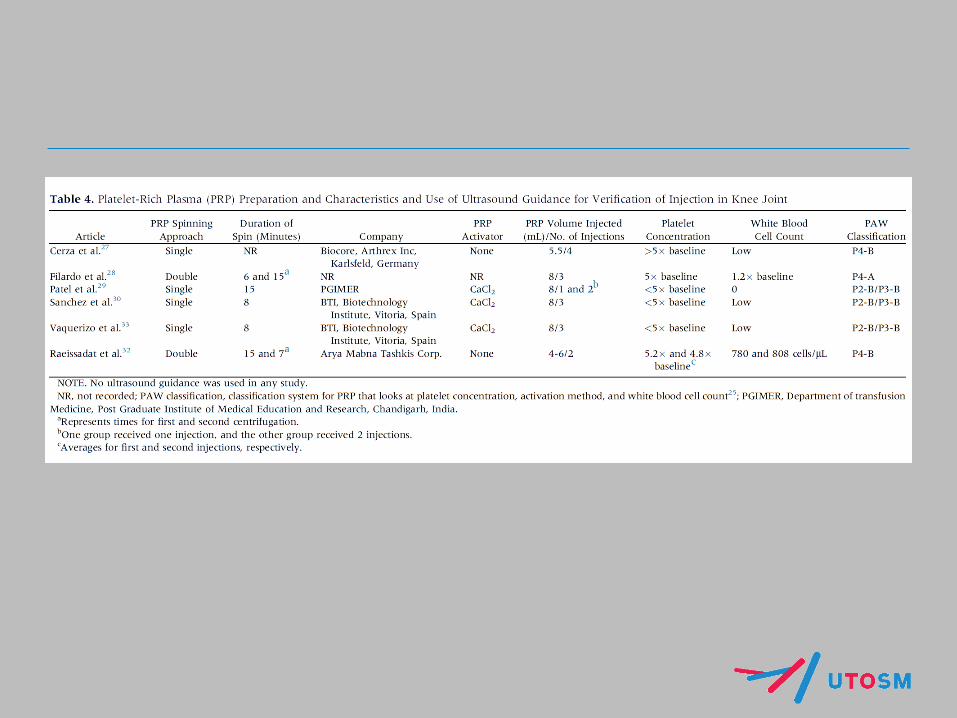
• Arthroscopy 2012
• Six level I and II studies
• N=577, mean age 56, followup 6 months


• Arthroscopy, 2016
• Level 1 evidence only
• Six studies, n=817 knees
• Mean age 60, ave f/u 38 weeks
• Improved change scores in PRP vs HA groups at
three, six and twelve months

• 6 randomized trials
• 3 prospective cohort studies
• N=1055 patients, mean age 55
• LP-PRP resulted in improved WOMAC scores vs HA
• No difference between LR-PRP and HA
• Higher incidence of adverse reactions in PRP vs HA
AJSM 2015
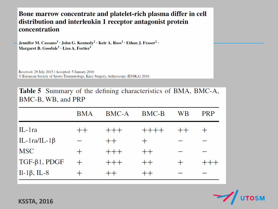
KSSTA, 2016

Does BMC make sense in OA?
IL-1Ra/IRAP/Orthokine

Summary
• There is clear evidence that a dose-dependent relationship between the use of the LA pain pumps and chondrotoxicity exists
• The effect of single dose anesthetics on articular cartilage is less clear – effects are more likely subtle and difficult to diagnose
• Use IA local anesthetics with sparingly and with caution.
• Strong case for alternative carriers or the use of biologics that demonstrate emerging positive clinical evidence