Embed Size (px)
Citation preview
J. exp. Biol, 117, 415-431 (1985) 4 1 5Printed in Great Britain © The Company ofBiobgists Limited 1985
CHROMATOPHORE MOTOR UNITS IN ELEDONECIRRHOSA (CEPHALOPODA: OCTOPODA)
BY F. DUBAS* AND P. R. BOYLE
Department of Zoology, University of Aberdeen, Aberdeen AB9 2TN,Great Britain
Accepted 14 January 1985
SUMMARY
Innervation of chromatophore muscles of the octopus Eledone cirrhosawas investigated by stimulating nerve bundles in the skin with a suctionelectrode and monitoring chromatophore movements with a photo-cell or avideo camera. Attention was focused on the organization of the chromato-phore muscle fibres into motor units. Individual muscle fibres respond tosingle electrical impulses with twitch-like contractions that do not facilitatewith repetition, but summate to a smooth tetanus at about 10-15 Hz. Attetanic frequency, the degree of expansion of single chromatophores isalways maximal. However, the number of expanded chromatophores can begraded by variations of either the stimulus voltage or frequency. Individualchromatophores and probably individual muscle fibres are part of severalmotor units. Chromatophores forming a given motor unit are found amongchromatophores served by other motor axons. The motor units apparentlyform precise parts of natural patterning.
INTRODUCTION
The chromatic patterns of cephalopods are unique within the animal kingdom: theyrely on chromatophores which are true organs made of a central pigment-containingcell (pigment cell) surrounded by ten to twenty radially arranged muscle fibres (Hof-mann, 1907a; Cloney & Florey, 1968; Froesch, 1973; Brocco, 1977). Contraction ofradial muscle fibres produces expansion of the chromatophore while their relaxationleads to its retraction. The activity of the chromatophore muscle fibres is under thedirect control of motor axons running from the chromatophore lobes in the brain(Boycott, 1953, 1961; Young, 1971). This system allows cephalopods to changechromatic patterns with a speed, variety and subtlety impossible in hormonallycontrolled chromatophore systems (Packard & Sanders, 1971; Packard & Hochberg,1977).
Since the chromatic patterns reflect the activity of a true neuromuscular system,they can be used to study neuromuscular relationships. The particular radial arrange-ment of the muscle fibres around the pigment cell allows single fibres to be preciselylocated and their activity monitored individually. Furthermore, the mechanical
• Present address: The Marine Biomedical Institute, The University of Texas Medical Branch, 200 Univer-sity Boulevard, Galveston, Texas 77550. U.S.A.
Key words: Molluscan muscles, chromatophores, nervous control, octopus, Eledone cirrhosa.
416 F. DUBAS AND P. R. BOYLE
behaviour of several muscle fibres can be recorded simultaneously and individually bysimple optical devices monitoring the effect of the muscle contraction on the expan-sion of the pigment cell. Thus, the chromatophore system is suited for studies atintermediate levels of organization (e.g. groups of muscles or motor units), anapproach that is more difficult in muscles where individual fibre activity can bedetected only with intracellular electrodes.
Chromatophore muscles show the following characteristics, shared with othermolluscan muscles (see Hoyle, 1983): mechanical activity mediated by graded post-synaptic potentials, multiple innervation of individual fibres, electrical coupling be-tween at least some of the muscle fibres, spontaneous activity, phasic and toniccontractions (Hofmann, 19076, 1910; Bozler, 1928, 1931; Florey, 1966, 1969; Florey&Kriebel, 1969).
Attempts have been made to relate the chromatic patterns to the underlying or-ganization of the nervous system. In cuttlefish mantle, stimulation of nerve brancheshas shown that chromatophores innervated by a single axon are randomly scatteredamong chromatophores innervated by other axons (innervation type B, Maynard,1967) whereas, in the dorsal skin, motor units consist of the chromatophores involvedin the same pattern or part of a pattern. Stimulation of the skin of Octopus vulgaris(Packard, 1973) has been found to elicit chromatophore expansion in whole parts ofthe normal patterns (components).
In this paper, we attempt to define the distribution of single motor units, usingsuction electrodes to stimulate precise nerve bundles to avoid the problems presentedby stimulation of the skin surface directly. The results obtained in acute preparationsare compared to the patterning of freely behaving animals.
MATERIALS AND METHODS
Animals
Live Eledone cirrhosa (Lamark) were obtained from commercial fishermen ofAberdeen harbour and maintained for several weeks in recycled sea water aquaria(10-18°C) where they were daily fed on live crabs (Boyle, 1981).
Preparation of skin samples
For physiological experiments, the animals were decerebrated without anaesthesia.Pieces of ventral or dorsal skin about 100 cm2 were immediately excised and pinned,slightly stretched, on a piece of dental wax, epidermal side down. Most of the connec-tive tissue normally lying between the chromatophores and the mantle or arm muscleswas then removed. After rinsing in fresh, aerated sea water, the skin preparation waspinned epidermal side up on a cork ring. The ring was placed skin down in a circularPerspex tissue bath where aerated sea water was flowing beneath and above the skinto afford good oxygenation of both surfaces (see Fig. 1). The temperature of the seawater was maintained at 15 °C.
Denervated preparations
The left pallial nerve was sectioned proximal to the stellate ganglion in six animalswhich were subsequently kept in isolation for 7—21 days. Since the somata of the axons
Octopus chromatophores 417
innervating the chromatophore muscles are situated in the brain (Young, 1971), thisoperation produces degeneration of the chromatophore motor axons, in the stellarnerves and more peripherally (Sanders & Young, 1974). Degeneration of theperipheral end of the axons occurred within 3 days after the operation as shown by theabsence of chromatophore response following electrical stimulation of the distal endof the nerve. The absence of functional continuity between the cut ends of the nervewas verified post mortem and the non-denervated half of the mantle provided acontrol.
Recording devicesThe tissue bath containing the skin preparation was placed under an inverted
microscope, fitted with a fibre optic light source. This allowed a good view of thechromatophores, now lying against the lower wall of the bath (2 mm thick), while thenerve bundles travelling in the connective layers were readily accessible from abovefor stimulation. With this inverted microscope (Reichert Me F), the chromatophoreimage (enlarged up to 120 X) could be projected either onto a ground glass screen orto a cin6 or video camera (Fig. 1).
To record the movements of single chromatophores projected on the ground glassscreen, a photo-cell was used (silicon photo-diode with integrated amplifier, R. S.Components, Ltd, rise time 30/is, responsivity oOmV/zW"1 cm2). It was fitted in ablack container which could be closed by a selection of lids. Each lid was pierced bya slot of different dimensions and covered by a light diffuser. During the experiments,the cartridge containing the photo-cell was fixed on the ground glass screen, with theslot positioned across the moving edge of a chromatophore. Expansion of thechromatophore across the slot decreased the amount of light received by the photo-cell. The photo-cell signal was amplified and displayed on a pen recorder (Washington400 MD 2C) together with the stimulus delivered to the nerves.
Alternatively, a black and white video camera monitored the movements of entirechromatophores or groups of chromatophores. The results, recorded on video tape,were analysed from sequential photographs of selected portions of the tape imagesdisplayed on a video monitor.
Strictly speaking, both methods used to observe chromatophore behaviour actuallyrecord the movements of the pigment cell rather than the length changes in themuscles themselves. However, pigment cell expansion depends directly and uniquelyon the length changes in the muscles so that the video recording techniques adequatelyreflect the number and location of contracting muscles while the photo-cell recordsthe time course of the muscle contraction.
Contractions of the skin muscles (either spontaneous or due to the stimulation)causing movements of the whole preparation often produced irregularities in thebaseline. They were minimized by removing most of the dermal layers in which theskin muscles lie and slightly stretching the skin on the cork ring.
StimulationElectrical stimulation was delivered to the chromatophores by one of three methods.
(1) Stimulation of the small nerve bundles {dermal nerves) which run in the dermisseparating the mantle muscle mass from the skin and contain axons innervating the
418 F. DUBAS AND P. R. BOYLE
Stimulator
Pen recorder ,
Fibre opticlight source
Electrodes
Water container(15 °C)
Cork ring
Amplifier
Photo-cell
Videocamera
Monitor
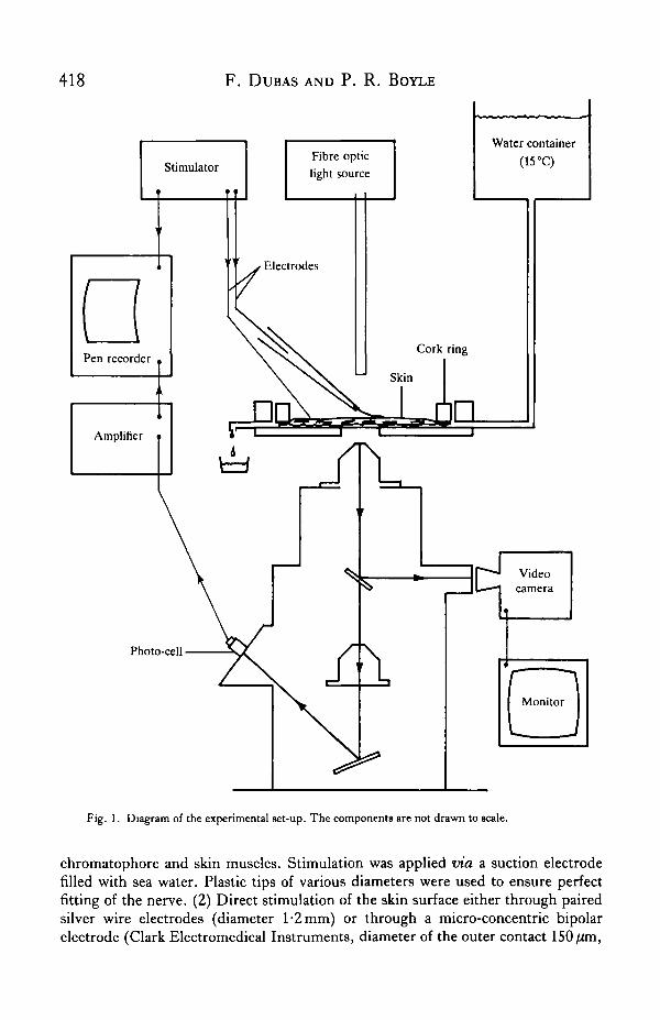
Fig. 1. Diagram of the experimental set-up. The components are not drawn to scale.
chromatophore and skin muscles. Stimulation was applied via a suction electrodefilled with sea water. Plastic tips of various diameters were used to ensure perfectfitting of the nerve. (2) Direct stimulation of the skin surface either through pairedsilver wire electrodes (diameter 1-2 mm) or through a micro-concentric bipolarelectrode (Clark Electromedical Instruments, diameter of the outer contact 150/im,
Octopus chromatophores 419
inner contact 25/im). (3) Stimulation of the chromatophore muscles directly, in'denervated' skin via a glass microelectrode (diameter 10-20/im).
Square wave pulses were produced by a Devices Isolated Stimulator (Type 2533)driven from a Devices Pulse Generator (Type 2521). When the experiment was videorecorded, the stimulus was audible on the sound tract of the video tape and thestimulation parameters were described verbally.
RESULTS
Stimulation pathways
When a dermal nerve bundle in a normal piece of skin was stimulated via a suctionelectrode or when the stimulation was applied to the skin surface directly via a bipolarelectrode with a supra-threshold stimulus, both the chromatophore and skin musclecontracted. Contraction of the chromatophore muscles caused chromatophore expan-sion while skin muscle contraction caused skin movements and formation of papillae.By contrast, in 'denervated' skin in which the chromatophore motor axons had hadtime to degenerate, both stimulation methods failed to trigger chromatophore expan-sion, although the skin muscle response remained unaffected. This suggests that, withboth stimulation methods, the imposed stimulus reached the chromatophore musclesvia their motor axons. Since the skin muscle response persisted, the axons innervatingthese muscles presumably have their soma situated more peripherally than the sec-tion, probably in the stellate ganglion (Dubas, 1982).
Direct stimulation of the chromatophore muscles in 'denervated' skin was possibleonly when a glass microelectrode was positioned very close to the chromatophoremuscles; even then, considerably higher voltage (up to 50 V) was necessary than innormal skin where 2 or 3 V was sufficient.
Single chromatophore responses
Stimulation of dermal nerves with single supra-threshold square pulses (0-3-0-5 msduration) resulted in twitch-like contraction of the chromatophore muscle fibres.Within 2-3 h after excision, the rise-time of the chromatophore response was300-500 ms, depending on the amplitude of the muscle fibre contraction. Theexpansion-retraction cycle lasted 1000-1500 ms. There was no tonic component andretraction immediately followed expansion (Fig. 2A, fresh preparation).
As the preparations aged, the rise-time often became longer and the amplitude ofthe expansion smaller. Chromatophores remained expanded for periods of a fewseconds to a few hours in the absence of further stimulation. Retraction occurredapparently spontaneously and usually extremely slowly (Fig. 2A, aged preparation).
Facilitation of the mechanical response of the chromatophore muscles was rare.Generally, a single electrical pulse was sufficient to trigger muscle contraction and itproduced an expansion of larger amplitude than a train of low frequency stimuli at thesame voltage (Fig. 2B). Also, the degree of chromatophore expansion caused by thefirst pulses of a low frequency train (F < 5 Hz) was larger than for subsequent ones.The amplitude of the initial chromatophore expansion was larger because (1) theamplitude of contraction of individual muscle fibres was larger and (2) more muscle
420 F. DUBAS AND P. R. BOYLEA
Fresh preparation
Aged preparation —'
Is
10s
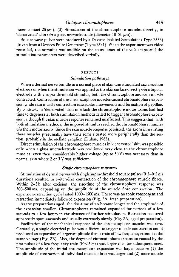
Fig. 2. Photo-cell recordings of mechanical responses of a single chromatophore to electrical stimula-tion of the radial muscles via their motor axons. Each section was recorded from a differentchromatophore. (A) Stimulation of fresh and ageing preparations by single pulses (arrows). In theageing preparation, the chromatophore remains tonically expanded and retraction occurs spon-taneously. (B) Low frequency and single pulse stimulation. The upper trace represents the photo-cellresponse, the lower one the stimulus (duration 5 ms, supra-threshold voltage). The response to thefirst pulse of a train is always larger than the following ones. Single pulses trigger individual responseslarger than those in response to trains of pulses after the first few pulses. The irregularities in thebaseline are due to movements of the skin muscles and do not reflect chromatophore muscle activity.
fibres contracted. A change in the quality of the electrode contact was not responsiblefor this effect since subsequent trains always produced an initially larger expansion.
In fresh preparations, twitches began to fuse at frequencies as low as 2 Hz andsummation of the mechanical response occurred. Smooth tetanus was reached atfrequencies slightly above 10 Hz. Maximal expansion occurred between IS and 20 Hz(Fig. 3A). In older preparations, fusion of the twitches appeared at frequencies belowlHz.
The twitch/tetanus ratio was fairly low: 1/2 to 1/3 for single muscle fibres (photo-cell recordings) and 1/3-5 to 1/5 for entire chromatophores (video recordings) (Fig.3A). The resting/tetanus surface ratio indicated that chromatophores increased theirsurface by a factor of 15-25 times.
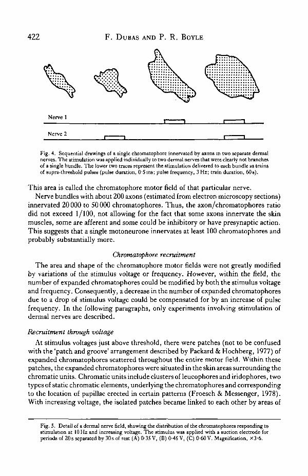
The shape and size of individual expanded chromatophores was modified byvariations of the stimulus voltage (delivered to dermal nerves through the suctionelectrode). At frequencies below 5 Hz, different muscle fibres were brought intocontraction at different voltages, presumably when their axon was recruited and theset of muscles of a particular chromatophore was innervated by several axons, notnecessarily in the same nerve bundle. Indeed, some chromatophores expanded inresponse to stimulation of either of two nerve bundles (Fig. 4). Usually, differentmuscle fibres were recruited by each nerve but sometimes a single muscle fibreresponded to stimulation of each nerve. Variations of the stimulus voltage alsoproduced gradation of the amplitude of shortening of individual muscle fibres. This
Octopus chromatophores 421
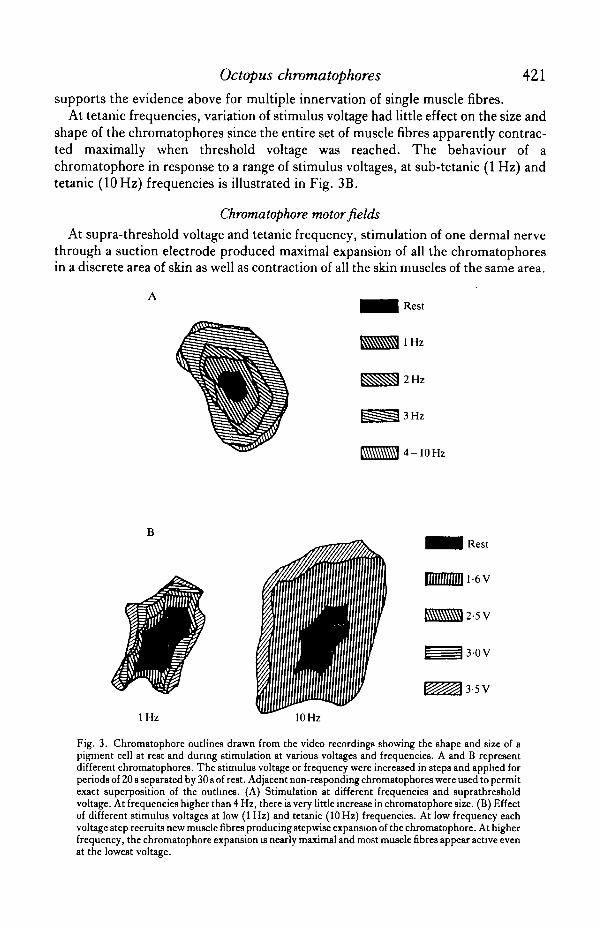
supports the evidence above for multiple innervation of single muscle fibres.At tetanic frequencies, variation of stimulus voltage had little effect on the size and
shape of the chromatophores since the entire set of muscle fibres apparently contrac-ted maximally when threshold voltage was reached. The behaviour of achromatophore in response to a range of stimulus voltages, at sub-tetanic (1 Hz) andtetanic (10 Hz) frequencies is illustrated in Fig. 3B.
Chromatophore motor fields
At supra-threshold voltage and tetanic frequency, stimulation of one dermal nervethrough a suction electrode produced maximal expansion of all the chromatophoresin a discrete area of skin as well as contraction of all the skin muscles of the same area.
Rest
l H z
2 Hz
3 Hz
4-10Hz
l H z
3-OV
3-5V
10 Hz
Fig. 3. Chromatophore outlines drawn from the video recordings showing the shape and size of apigment cell at rest and during stimulation at various voltages and frequencies. A and B representdifferent chromatophores. The stimulus voltage or frequency were increased in steps and applied forperiods of 20 8 separated by 30 s of rest. Adjacent non-responding chromatophores were used to permitexact superposition of the outlines. (A) Stimulation at different frequencies and suprathresholdvoltage. At frequencies higher than 4 Hz, there is very little increase in chromatophore size. (B) Effectof different stimulus voltages at low (1 Hz) and tetanic (10 Hz) frequencies. At low frequency eachvoltage step recruits new muscle fibres producing stepwise expansion of the chromatophore. At higherfrequency, the chromatophore expansion is nearly maximal and most muscle fibres appear active evenat the lowest voltage.
422 F. DUBAS AND P. R. BOYLE
Nerve 1
Nerve 2
Fig. 4. Sequential drawings of a single chromatophore innervated by axons in two separate dermalnerves. The stimulation was applied individually to two dermal nerves that were clearly not branchesof a single bundle. The lower two traces represent the stimulation delivered to each bundle as trainsof supra-threshold pulses (pulse duration, OS ms; pulse frequency, 3 Hz; train duration, 60s).
This area is called the chromatophore motor field of that particular nerve.Nerve bundles with about 200 axons (estimated from electron microscopy sections)
innervated 20 000 to 50 000 chromatophores. Thus, the axon/chromatophores ratiodid not exceed 1/100, not allowing for the fact that some axons innervate the skinmuscles, some are afferent and some could be inhibitory or have presynaptic action.This suggests that a single motoneurone innervates at least 100 chromatophores andprobably substantially more.
Chromatophore recruitment
The area and shape of the chromatophore motor fields were not greatly modifiedby variations of the stimulus voltage or frequency. However, within the field, thenumber of expanded chromatophores could be modified by both the stimulus voltageand frequency. Consequently, a decrease in the number of expanded chromatophoresdue to a drop of stimulus voltage could be compensated for by an increase of pulsefrequency. In the following paragraphs, only experiments involving stimulation ofdermal nerves are described.
Recruitment through voltage
At stimulus voltages just above threshold, there were patches (not to be confusedwith the 'patch and groove' arrangement described by Packard & Hochberg, 1977) ofexpanded chromatophores scattered throughout the entire motor field. Within thesepatches, the expanded chromatophores were situated in the skin areas surrounding thechromatic units. Chromatic units include clusters of leucophores and iridophores, twotypes of static chromatic elements, underlying the chromatophores and correspondingto the location of papillae erected in certain patterns (Froesch & Messenger, 1978).With increasing voltage, the isolated patches became linked to each other by areas of
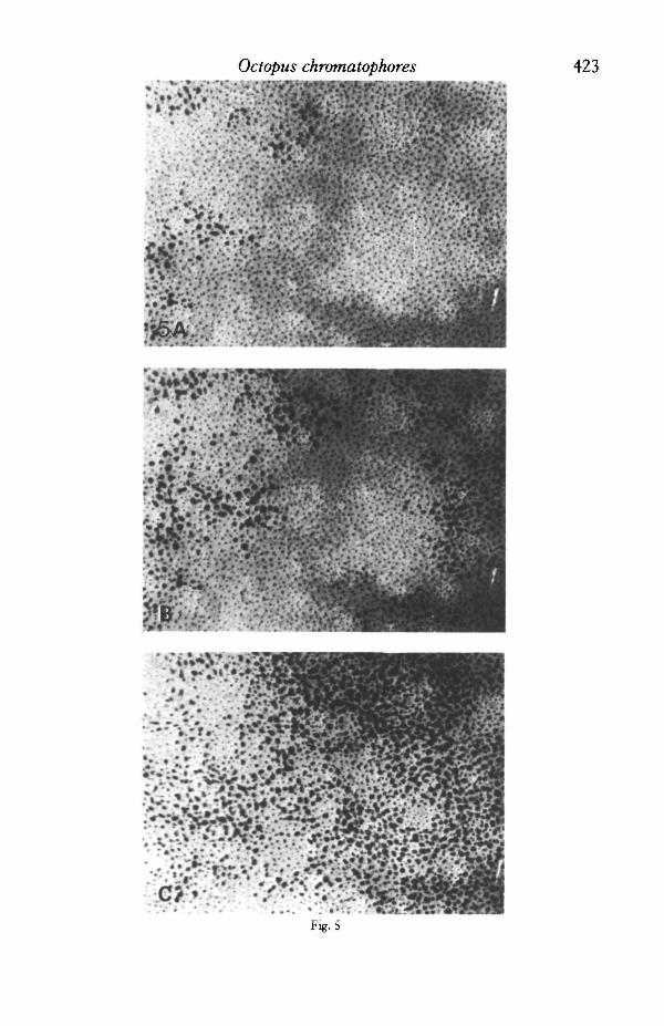
Fig. 5. Detail of a dermal nerve field, showing the distribution of the chromatophores responding tostimulation at 10 Hz and increasing voltage. The stimulus was applied with a suction electrode forperiods of 20s separated by 30s of rest (A) 0-35 V, (B) 0-45 V, (C) 0-60 V. Magnification, X3-6.
Octopus chromatophores
"'••r^.>;ijsj^
424 F. DUBAS AND P. R. BOYLE
newly responding chromatophores and, within the patches, the chromatophores lyingcloser to the centre of each chromatic units were recruited progressively. Only at thehighest effective voltage was the centre of the chromatic units finally covered byexpanded chromatophores (Fig. 5). Chromatophores recruited at the same voltagerarely formed a compact group (contiguous chromatophores) of more than 20chromatophores. These effects were characteristic of all dermal nerves or branches ofdermal nerves investigated.
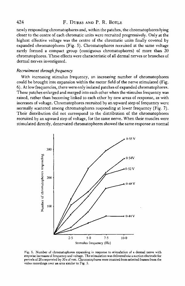
Recruitment through frequencyWith increasing stimulus frequency, an increasing number of chromatophores
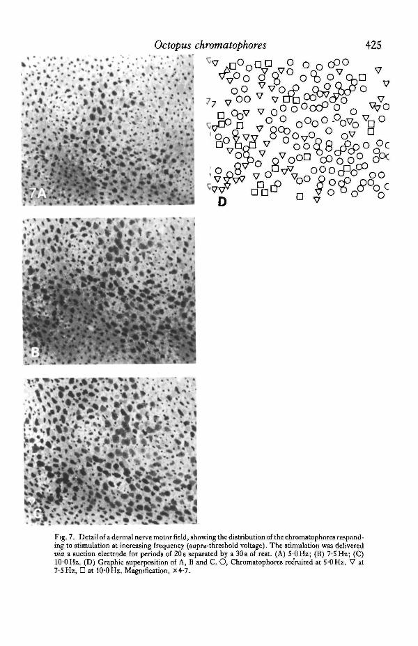
could be brought into expansion within the motor field of the nerve stimulated (Fig.6). At low frequencies, there were only isolated patches of expanded chromatophores.These patches enlarged and merged into each other when the stimulus frequency wasraised, rather than becoming linked to each other by new areas of response, as withincreases of voltage. Chromatophores recruited by an upward step of frequency werenormally scattered among chromatophores responding at lower frequency (Fig. 7).Their distribution did not correspond to the distribution of the chromatophoresrecruited by an upward step of voltage, for the same nerve. When their muscles werestimulated directly, denervated chromatophores showed the same response as normal
300
-acoD.
o.o
3
200
100
0-55 V
0-44V
2-5 5-0 7-5
Stimulus frequency (Hz)
10-0
Fig. 6. Number of chromatophores expanding in response to stimulation of a dermal nerve withstepwise increases of frequency and voltage. The stimulation was delivered via a suction electrode forperiods of 20 s separated by 30 8 of re9t. Chromatophores were counted from selected frames from thevideo recordings over an area similar to Fig. S.
Octopus chromatophores 425
Fig. 7. Detail of a dermal nerve motor field, showing the distribution of the chromatophores respond-ing to stimulation at increasing frequency (supra-threshold voltage). The stimulation was deliveredvia a suction electrode for periods of 20 s separated by a 30 s of rest. (A) 5 0 Hz; (B) 7-5 Hz; (C)100 Hz. (D) Graphic superposition of A, B and C. O, Chromatophores recruited at 5 0 Hz, V at7-5 Hz, • at 10-0 Hz. Magnification, X4-7.
426 F. DUBAS AND P. R. BOYLE
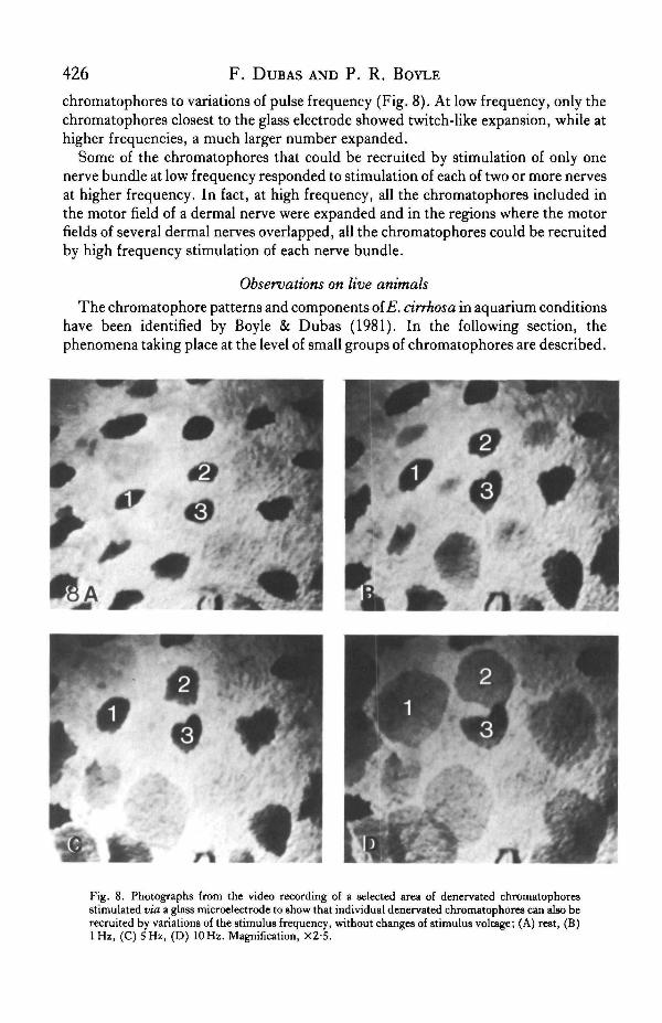
chromatophores to variations of pulse frequency (Fig. 8). At low frequency, only thechromatophores closest to the glass electrode showed twitch-like expansion, while athigher frequencies, a much larger number expanded.
Some of the chromatophores that could be recruited by stimulation of only onenerve bundle at low frequency responded to stimulation of each of two or more nervesat higher frequency. In fact, at high frequency, all the chromatophores included inthe motor field of a dermal nerve were expanded and in the regions where the motorfields of several dermal nerves overlapped, all the chromatophores could be recruitedby high frequency stimulation of each nerve bundle.
Observations on live animals
The chromatophore patterns and components of E. cirrhosa in aquarium conditionshave been identified by Boyle & Dubas (1981). In the following section, thephenomena taking place at the level of small groups of chromatophores are described.
Fig. 8. Photographs from the video recording of a selected area of denervated chromatophoresstimulated via a glass microelectrode to show that individual denervated chromatophores can also berecruited by variations of the stimulus frequency, without changes of stimulus voltage; (A) rest, (B)lHz , (C) 5 Hz, (D) 10 Hz. Magnification, X2-5.
Octopus chromatophores All
In animals at rest and undisturbed, the chromatophores were generally expanded(dressing gown pattern; Boyle & Dubas, 1981). Expanded chromatophores were notvisibly twitching but seemed tetanicaUy or tonically expanded. This suggests that themotoneurones fire at relatively high frequency or that a catch mechanism exists.
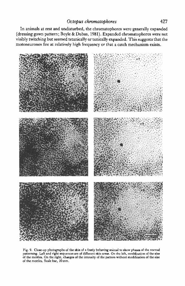
Fig. 9. Close-up photographs of the skin of a freely behaving animal to show phases of the normalpatterning. Left and right sequences are of different skin areas. On the left, modification of the sizeof the mottles. On the right, changes of the intensity of the pattern without modification of the sizeof the mottles. Scale bar, 10 mm.
428 F. DUBAS AND P. R. BOYLE
The variety of chromatophore patterns that could be observed on the dorsal side ofE. cirrhosa consisted mainly of a range of mottled patterns made of light, oftencircular, patches of retracted chromatophores (mottles: this term is used to mean agroup of retracted chromatophores, appearing as a single light patch contrasting witha darker background of expanded chromatophores) surrounded by darker areas whereat least some of the chromatophores were expanded.
On one hand, the range of mottled patterns was based on variations of the size ofthe mottles, in other words, on variations of the surface ratio of light to dark areas.This produced a range of patterns of different coarseness, from 'all dark' with no lightmottles to 'small mottles' where small patches of retracted chromatophores weresurrounded by large dark areas of expanded ones, to 'almost light' where the lightmottles were very large and had almost merged into each other, leaving very few darkareas of expanded chromatophores. The range of patterns finally ended with 'all light'where the light mottles had completely merged into each other and left no moreexpanded chromatophores. Serial close-up photographs showed that changes in thesize of the light mottles occurred by expansion or retraction of small, scattered groupsof chromatophores at the periphery of the mottles (Fig. 9A). The mottles alwayscorresponded to the location of the chromatic units. Thus, a decrease in the mottlesize was very similar to recruitment of motor units obtained experimentally by in-crease of stimulus voltage (compare Figs 9A and 5).
On the other hand, for a given mottle size, the intensity of the patterns was variedby changing the number of expanded chromatophores in the dark areas and changingtheir state of expansion. This produced patterns with a more or less strong degree ofexpression. Close-up photographs of freely behaving animals showed that graduationof the intensity of the dark areas was achieved mainly by changes of the number ofexpanded chromatophores and the degree of expansion of individual chromatophoreswas rarely modified. A given dark area, involved in a low intensity pattern, containedfew expanded chromatophores but most of those which were expanded seemed to havereached their maximal size. The same dark area, in a pattern of higher intensity,showed a larger proportion of expanded chromatophores and there was little changein the state of expansion of the chromatophores which were expanded during thepattern of lower intensity (Fig. 9B). This is similar to the effects of increased stimulusfrequency obtained during experimental nerve stimulation (compare Figs 9B and 7).
DISCUSSION
Muscle response
In the chromatophores of E. cirrhosa, facilitation of the muscle response withstimulus repetition is generally minimal or non-existent, although summation of thecontraction occurs. These results are consistent with those of Florey (1966) andFlorey & Kriebel (1969) on the chromatophore muscles of Loligo opalescens.
Using intracellular electrodes, Florey & Kriebel (1969) have demonstrated thatnerve stimulation results only in excitatory postsynaptic potentials in thechromatophore muscles of L. opalescens. Local responses might also occur in themantle muscle of both octopuses and squids (Wilson, 1960). In the chromatophores

Octopus chromatophores 429
of E. cirrhosa, the contraction of the muscle fibres is clearly not an 'all or none' event.The observations that the amplitude of the muscle responses diminishes withrepetition of the stimulus and can be graded by variations of the voltage both suggestthat the contraction is mediated by graded potentials, rather than 'all or none' spikes.
With stimulation of the dermal nerves there is no evidence for inhibitory innerva-tion but this does not exclude presynaptic inhibition. Indeed, such presynapticcontrol would not have a 'retracting' effect but would presumably prevent the excita-tion reaching the muscle fibres and thus could easily pass unnoticed during theexperiments. Direct inhibitory innervation has been ruled out by Florey & Kriebel(1969), who recorded intracellular changes of potentials, but their method does notexclude presynaptic effects.
Motor unitsSince electrical stimulation was applied to the dermal nerves which contain several
chromatophore motor axons, it was difficult to determine how many motor units werestimulated at any one time and to determine the distribution of single motor units. Onthe other hand, since the dermal nerves were stimulated before they branch (Dubas,1982), it was certain that entire motor units were stimulated and not parts of them.
Thus, the chromatophore motor units of E. cirrhosa seem to have the followingcharacteristics: (1) a single chromatophore and individual muscle fibres can be partof several motor units; (2) a single axon innervates a large number of chromatophores(and within a single chromatophore, several muscle fibres), not all clustered togetherbut scattered over a large part of the chromatophore motor field of the dermal nerveconsidered; (3) the distribution of a single motor unit is not coincident with a singlechromatic unit but chromatophores innervated by a single axon appear to be locatedat similar distances from the centre of the chromatic units they lie in. Points (1) and(2) are in agreement with the results obtained by Florey (1969) although thechromatophores innervated by a single axon are more clustered in squids.
Since the distribution of the motor units coincides with the distribution of themottles shown during normal patterning, it is clear that the motor units of the dorsalskin of E. cirrhosa are not random but correspond to parts of the normal patterns(innervation type C, Maynard, 1967). Thus, the chromatophore muscle fibres whichare innervated by a single axon are involved in a particular part of a pattern. Assumingthat a single axon needs to be active to trigger muscle contraction, the number of axonsinnervating a single muscle fibre is perhaps representative of the number of patternsin which that particular fibre plays a role.
Pattern controlSince the distribution of the motor units is non-random, the repertoire of patterns
is determined to a large extent in the periphery, by the morphological layout of themotor axon branches among the chromatophores. Thus, as far as the mottle patternsare concerned, the role of the brain seems to be to coordinate the activity of thepatterned motor units to produce gradation of the coarseness or intensity of thepatterns.
Comparing the present experimental results with patterning in intact animals, itappears that the size of the light mottles is regulated centrally by recruitment or
430 F. DUBAS AND P. R. BOYLE
inhibition of motor units, possibly by lateral inhibition mechanisms. On the otherhand, the experimental results suggest that changes of the intensity of the patternsmay be brought about simply by variation of the frequency of the nerve dischargesince this produces recruitment of chromatophores in the areas where somechromatophores are already expanded. The colour patterns are thus comparable toother neuromuscular systems where the intensity of the contraction is graded both bythe frequency of the nervous discharge and the number of motor units recruited.
Recruiting effect of frequency
Our evidence suggests that the recruiting effect of frequency is due to characteris-tics of the muscle fibres rather than the nervous system. So far two hypotheses can bebrought forward. (A) Two types of neuromuscular systems may coexist; a non-facilitating and a facilitating system. The non-facilitating system, demonstrated bythe present results, responds with maximal contraction even to a single stimulus. Afacilitating system, for which direct evidence has not been obtained yet, consisting ofseparate axons, may produce visible contraction of the muscle fibres only above athreshold frequency. Since multiple innervation is the case, these muscle fibres maybe the same as those of the non-facilitating system, being able to show two types ofresponses, depending on the type of axon firing. (B) Alternatively, the presence ofelectrical links between the muscles of adjacent chromatophores may allow thedepolarization evoked by nervous activity to invade the coupled muscles. Florey &Kriebel (1969) have presented intracellular evidence that the muscle fibres of a singlechromatophore are linked by low resistance pathways. Froesch-Gaetzi & Froesch(1977) have argued, on the basis of morphological evidence, that such links also existbetween the muscle fibres of neighbouring chromatophores. Further investigationswith intracellular electrodes are required to determine whether one or bothpossibilities occur.
We wish to thank Mr A. Lucas for his excellent assistance with the photographicand video equipment, Mr T. Craig for the care of the animals and Dr R. Ralph forlending the video equipment. Drs J. B. Messenger and A. Packard provided muchdiscussion and encouragement and, together with Drs H. Pinsker, R. T. Hanlon andProfessor E. Florey, many constructive comments on earlier drafts of the manuscript.This work was supported by a postgraduate studentship from the University ofAberdeen to FD and formed part of a Ph.D. thesis submitted to the University ofAberdeen.
R E F E R E N C E S
BOYCOTT, B. B. (1953). The chromatophore system of cephalopoda. Pmc. Linn. Soc. Land. 164, 235-240.BOYCOTT, B. B. (1961). The functional organization of the brain of the cuttlefish Sepia officinalis. Pmc. R. Soc.
B 153, 503-534.BOYLE, P. R. (1981). Methods for the aquarium maintenance of the common octopus of British waters, Eledone
cinhosa. Lab. Animals 15, 327-331.BOYLE, P. R. & DUBAS, F. (1981). Components of body pattern displays in the octopus Eledone cirrhosa. Mar.
Behav. Physiol. 8, 135-148.BOZLEH, E. (1928). Uber die Tatigkeit der einzelnen glatten Muskelfasern bei der Kontraktion. II. Die
chromatophorenmuskeln der Cephalopoden. Z. vergl. Physiol. 7, 379—406.
Octopus chrotnatophores 431BOZLER, E. (1931). Uber die TStigkeit der einzelnen glatten Muskelfasern bei der Kontraktion. III .
Regiatrierung der Kontraktionen der Chromatophorenmuakelzellen von Cephalopoden. Z. vergl. Physiol. 13,762-772.
BROCCO, S. L. (1977). The infrastructure of the epidermis, dermis, iridophores, leucophores andchromatophores of Octopus dofleini martini (Cephalopoda Octopoda). Ph.D. thesis, University ofWashington, Seattle.
CLONEY, R. A. & FLOREY, E. (1968). Ultrastructure of cephalopod chromatophore organs. Z. Zellfonch.mikmsk. Anat. 89, 250-280.
DUBAS, F. (1982). Skin patterning in the octopusEledone cirrhosa: a morphological and functional approach.Ph.D. thesis, University of Aberdeen, Scotland.
FLOREY, E. (1966). Nervous control and spontaneous activity of the chromatophores of a cephalopod, Loligoopalescens. Comp. Biochem. Physiol. 18, 305-324.
FLOREY, E. (1969). Ultrastructure and function of cephalopod chromatophores, An. Zool. 9, 429—442.FLOREY, E. & KRIEBEL, M. D. (1969). Electrical and mechanical responses of chromatophore muscle fibers of
the squid Loligo opalescens, to nerve stimulation and drugs. Z. vergl. Physiol. 65, 98—130.FROESCH, D. (1973). On the fine structure of the octopus iris. Z. Zellforsch. mikmsk. Anat. 145, 119-129.FROESCH, D. & MESSENGER, J. B. (1978). On leucophores and the chromatic units of Octopus vulgaris. J.Zooi,
Land. 186, 163-173.FROESCH-GAETZI, V. & FROESCH, D. (1977). Evidence that chromatophores of cephalopods are linked by their
muscles. Experientia 33, 1448-1449.HOFMANN, F. B. (1907a). Histologische Untersuchungen uber die Innervation der glatten und der ihrverwand-
ten Muskulatur der Wirbeltiere und Mollusken. Arch. mikv. Anat. 70, 361-413.HOFMANN, F. B. (19076). Gibt es in der Muskulatur der Mollusken periphere, kontinuierlich leitende Nerven-
netze bei Abwesenheit von Ganglienzellen? I. Untersuchungen an Cephalopoden. Pflug. Arch. get. Physiol.118, 375-412.
HOFMANN, F. B. (1910). Gibt es in der Muskulator der Mollusken periphere kontinuierlich leitende Nerven-netze bei Abwesenheit von Ganglienzellen? II. Weitere Untersuchungen an den Chromatophoren derKephalopoden. Pflug. Arch. ges. Physiol. 132, 43-81.
HOYLE, G. (1983). Muscles and Their Neural Control. New York: Wiley-Interscience.MAYNARD, D. M. (1967). Organization of central ganglia. In Invertebrate Nervous Systems, (ed. C. A. G.
Wiersma). London: University of Chicago Press.PACKARD, A. (1973). Chromatophore fields in the skin of the octopus. J . Physiol., Land. 238, 38-40.PACKARD, A. (1982). Morphogenesis of chromatophore patterns in cephalopods: are morphological and
physiological 'units' the same? Malacologia 23, 193-201.PACKARD, A. & HOCHBERG, E. G. (1977). Skin patterning in Octopus and other genera. Symp. zool. Soc. Land.
38, 191-231.PACKARD, A. & SANDERS, G. D. (1971). Patterns of Octopus vulgaris and maturation of the responses to
disturbance. Anim. Behav. 19, 780-790.SANDERS, G. D. & YOUNG, J. Z. (1974). Reappearance of specific colour patterns after nerve regeneration in
Octopus. Pmc. R. Soc. B. 186, 1-11.WILSON, D. M. (1960). Nervous control of movement in cephalopods. J. exp. Biol. 37, 57-72.YOUNG, J. Z. (1971). The anatomy of the Nervous System of Octopus vulgaris. Oxford: Clarendon Press.