Embed Size (px)
Citation preview
Proc. Natl. Acad. Sci. USAVol. 85, pp. 2662-2666, April 1988Genetics
Chromosomal organization of the cytochrome P450-2C gene familyin the mouse: A locus associated with constitutive arylhydrocarbon hydroxylase
(cytochrome P450/multigene family/synteny with human/xenobiotic metabolism/carcinogenesis)
R. R. MEEHAN*t, R. M. SPEED*, J. R. GOSDEN*, D. ROUT*, J. J. HUTTONf, B. A. TAYLOR§, J. HILKENS¶,V. KROEZEN¶, J. HILGERS¶, M. ADESNIKII, T. FRIEDBERG**, N. D. HASTIE*, AND C. R. WOLFt'tt*Molecular Genetics Section, Medical Research Council Clinical and Population Cytogenetics Unit, Western General Hospital, Crewe Road, Edinburgh, EH42XU, Scotland; tImperial Cancer Research Fund, Laboratory of Molecular Pharmacology and Drug Metabolism, Department of Biochemistry, Hugh RobsonBuilding, George Square, Edinburgh, EH8 9JY, Scotland; tChildren's Hospital Medical Center, Children's Hospital Research Foundation, Department ofPediatrics, College of Medicine, University of Cincinnati, Cincinnati, OH 45229; §The Jackson Laboratory, Bar Harbor, ME 04609; IDepartment ofTumor Biology, The Netherlands Cancer Institute, Antoni van Lenwenhoek Huis, Plesmanlaan 121, 0166CX, Amsterdam, The Netherlands;IlDepartment of Cell Biology and the Kaplan Center, New York University of Medicine, New York, NY 10016; and **Institute ofToxicology, University of Mainz, ObereZahlbacherstrer 67, D6500, Mainz, Federal Republic of Germany
Communicated by Walter F. Bodmer, November 2, 1987 (received for review September 17, 1987)
ABSTRACT Cytochromes P-450 represent a superfamilyof enzymes with a central role in the metabolism of drugs,chemical toxins, and carcinogens. We have used genetic anal-ysis to establish the complexity and catalytic function of arecently identified constitutively expressed murine hepaticcytochrome P-450 encoded by P450-2C. Southern blottinganalysis shows that there are at least seven or eight geneswithin this family in the mouse and rat and that DNArestriction fragment length variants between different mouseinbred strains are observed. Analysis of recombinant inbredstrains derived from these parent strains shows (i) these genesare clustered within 1 centimorgan, (it) this gene family doesnot correspond to any of the known cytochrome P-450 loci ormap near any well-characterized genomic markers, and (iii)this gene family segregates to within 1-2 centimorgans of alocus controlling constitutive aryl hydrocarbon hydroxylaseactivity in mice. With use of Chinese hamster/mouse somaticcell hybrids, the P450-2C locus was assigned to a region ofmouse chromosome 19 that appears to be syntenic with thepreviously mapped human P450C2C locus on human chromo-some 10. By in situ hybridization to mitotic mouse chromo-somes, we have localized this region to the tip of chromosome19. These results are discussed in relation to the physiologicalroles of this P-450 family in foreign compound metabolism andsteroid oxidations.
The mammalian P-450-dependent monooxygenases have avariety of roles ranging from the biosynthesis of steroidhormones, bile acids, and 1,25-dihydroxyvitamin D3 to themetabolism of foreign compounds (1). Those enzymes in-volved in the synthesis of hormones and bile acids arespecific in their function (2). The enzymes involved inforeign compound metabolism have broad substrate speci-ficities and will also catalyze the metabolism of hormonesand fatty acids. Several gene families belonging to the lattergroup of proteins have been identified (3, 4), and many ofthese contain a large number of genes. The duplication andexpansion of this enzyme system appears to have providedan important selective advantage based on an increasedability to metabolize and excrete potentially harmful chem-icals within our environment. The cytochrome P-450 systemis complex both because of its multiplicity and also becauseof the mechanism of regulation of forms within a particulartissue (5).
This system is polymorphic in humans, and a variety ofgenetically determined differences in cytochrome P-450-mediated metabolism have been implicated in a number ofpharmacogenetic responses (6-8). By inference these meta-bolic differences may also serve as markers for alteredsusceptibility to environmental toxins and carcinogens(9-12). In epidemiology studies polymorphic P-450 expres-sion has been related to chemical-induced lung and livercancer (9, 13) and steroid hormone-related cancer(s), whichinclude breast, cervix, and endometrium (14).One of the outstanding questions is to establish the roles of
constitutively expressed cytochromes P-450 relative to thosethat are expressed only after exposure to chemical-inducingagents in these diseases. Cytochrome P-450 PB-1 is consti-tutively expressed at high levels in rodent liver and ismarginally induced by the xenobiotic phenobarbital (15, 16).An equivalent P-450 that is polymorphic in humans (17) isinvolved in the metabolism of mephenytoin, an anticonvul-sant (18). PB-1 is encoded by a member of the P450-2C genefamily (4). In this report we have used a genetic approach toestablish some of the functions and the chromosomal orga-nization of the P450-2C gene family in mice.
MATERIALS AND METHODSAnimals and Cell Lines. Outbred Wistar and DA rats were
used in this study. 020, STS, GRS, and AKR/Fu strainswere from The Netherlands Cancer Institute (Amsterdam).Except for the CBA/Ca strain from the Western GeneralAnimal Unit (Edinburgh), the remaining strains were fromboth the Western General Animal Unit and The JacksonLaboratory. No subline differences were observed. CD micewere a gift of S. D. M. Brown (St. Mary's Medical School,London). Chinese hamster/mouse somatic cell hybrids wereisolated and characterized as described by Hilkens et al.(19).
Isolation of DNA and Restriction Endonuclease Analysis.DNA was isolated from progenitor strains as described byHill et al. (20). Chinese hamster/mouse somatic cell hybridDNA was isolated as described by Hilkens et al. (19).Recombinant inbred (RI) mice DNAs from the BXD,AKXL, and BXH series were purchased from The JacksonLaboratory, RI mouse DNAs from the OXA and BXC serieswere obtained from The Netherlands Cancer Institute. Re-
Abbreviations: AHOHase, aryl hydrocarbon hydroxylase; RI,recombinant inbred; cM, centimorgan.tfTo whom reprint requests should be addressed.
2662
The publication costs of this article were defrayed in part by page chargepayment. This article must therefore be hereby marked "advertisement"in accordance with 18 U.S.C. §1734 solely to indicate this fact.
Proc. Natl. Acad. Sci. USA 85 (1988) 2663
striction digests were carried out as recommended by thecommercial supplier. Digested DNA was fractionated elec-trophoretically on 0.8-1.0% agarose gels, and the DNA wastransferred to nitrocellulose or Hybond-N (Amersham) filteras described by Southern (21).
Preparation of Probes and Hybridization Analysis. TheP-450f rat liver cDNA clone pTF-1, coding for a member ofthe PB-1 family, has been described previously (22). Themouse cDNA clones were isolated by screening a DBA/2Jmale liver cDNA library (Clontech, Palo Alto, CA) with32P-labeled pTF-1 insert (23). Three clones were used in thepresent analysis: pM8-1 encoding the 3' end of the mousePB-1 cDNA, as demonstrated by restriction mapping and byprobing a set of reference plasmids with the pM8-1 insert,pPB5-21, and pPB3-15. Clones pPB5-21 and pPB3-15 arenearly full-length PB-1 cDNAs. Probes were labeled byrandom priming (24) and hybridized to Southern blots asdescribed by Hill et al. (20). Blots were washed at 68°C in0.30 M NaCl/0.03 M sodium citrate. In the case of somaticcell hybrid DNA blots, these were washed at 68°C in 0.015 MNaCI/0.0015 M sodium citrate.Chromosome Preparation. Somatic chromosome prepara-
tions from the bone marrow of male and female CD micewere made by a modification of the procedure of Ford andHamerton (25). G-banded karyotypes were constructed afterthe trypsin-banding of Gallimore and Richardson (26).
In Situ Hybridization. The preparation and analysis oftritiated pPB3-15 cDNA clones hybridized to mouse meta-phase spreads was as described (27).
Aryl Hydrocarbon Hydroxylase (AHOHase) Assays. AHO-Hase activity was determined fluorimetrically on mouseliver homogenates using benzo[a]pyrene as a substrate (28).Six to 10 animals were used per determination.
RESULTS
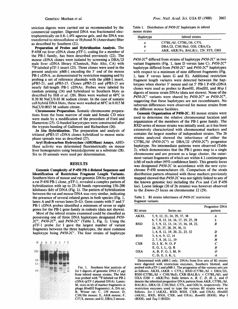
Genomic Complexity of P-450 PB-i-Related Sequences andIdentification of Restriction Fragment Length Variants.Southern blots of mouse and rat genomic DNAs probed witha rat P-450 PB-1 clone, pTF-1, revealed a complex pattern ofhybridization with up to 25-30 bands representing 150-200kilobases (kb) ofDNA (Fig. 1). The pattern of hybridizationbetween the rat and mouse DNA was very similar, indicatingthe presence of several related genes in both species (Fig. 1,lanes A and B versus lanes D-G). Gene counts with 5' and 3'PB-1 cDNA probes identified a minimum of seven or eightgenes for the PB-1 gene family in rodents (data not shown).Most of the inbred strains examined could be classified as
possessing one of three DNA haplotypes designated P450-2Ca, P450-2Cb, and P450-2CC (Table 1, Fig. 1). Using thepTF-1 probe Sst I gave the largest number of variantfragments between the three haplotypes, the most commonhaplotype being P450-2C. The four strains of haplotype
M A B C D E f G
I23.0- .l- 1. -
4-1*
4w_ ,
to O a ...4.4.._a.
FIG. 1. Southern blot analysis ofSst I digests of genomic DNA (5 ,g)from inbred mouse strains. The blotwas probed with 32P-labeled rat PB-1cDNA (pTF-1 plasmid DNA). Lanes:M, sizes in kb of marker fragments (Aphage HindIII fragments); A, DA rat;B, Wistar rat; C, 129 mouse; D,C3H/He mouse; E, AKR mouse; F,C57/L mouse; and G, DBA/2 mouse.
Table 1. Distribution of P450-2C haplotypes in inbredmouse strains
Haplotype Inbred strains
a C57BL/6J, C57BL/10, C57Lb DBA/2J, C3H/HeJ, 020, CBA/Cac AKR, AKR/Fu, BALB/c, 129, STS, GRS
P450-2Cb differed from strains of haplotype P450-2CC in twovariant fragments (Fig. 1, lane D versus lane C). P450-2Cahaplotype differed from P450-2CC and P450-2Cb haplotypeswith respect to a minimum of nine different fragments (Fig.1, lane F versus lanes G and E). Additional restrictionfragment length variants were detected between the hap-lotypes when shorter 3' mouse and rat 5' PB-i P-450 cDNAclones were used as probes to BamHI, HindIII, and Msp Idigests of mouse strain DNAs (data not shown). None of theP450-2CC variants were shared by P450-2Ca or P450-2Cb,suggesting that these haplotypes are not recombinants. Nosubstrain differences were observed for mouse strains fromthe different mouse facilities.Genomic Organization of P450-2C. RI mouse strains were
used to determine the relative chromosomal location andorganization of the members of the PB-1 gene family. TheBXD series of mouse strains was initially used, as it has beenextensively characterized with chromosomal markers andcontains the largest number of independent strains. The 26strains analyzed showed the inheritance of either theC57BL/6J (P450-2Ca) or DBA/2J (P450-2Cb) progenitorhaplotype. No intermediate patterns were observed (Table2), which demonstrates that the PB-1 genes map to a singlechromosome and are present as a large cluster, the outer-most variant fragments of which are within 4.1 centimorgans(cM) of each other (95% confidence limit). This genetic locuswas designated P450-2C in accordance with the new cyto-chrome P-450 nomenclature (4). Comparison of the straindistribution pattern obtained with those markers previouslymapped showed that P450-2C was not tightly linked to any ofthe known genomic loci including the Pcn and Coh P-450loci. Loose linkage (20 of 26 strains) was however observedto the Xmmv-25 locus on chromosome 12 (29).
Table 2. RI strains inheritance of P450-2C restrictionfragment variants
Progenitor DNARI strain Series no. pattern
AKXL 5, 9, 12, 21, 24, 29, 37, 38 A6, 7, 8, 13, 14, 16, 17, 19, 25, 28 L
BXD 2, 5, 9, 12, 13, 14, 15, 16, 18, 22, B24, 25, 27, 28, 29, 30, 311, 6, 8, 11, 19, 20, 21, 23, 32 D
BXH 3, 4, 6, 9,12, 14 B2, 7, 8, 10, 11, 19 H
CXB D, J, K, N, 0, P CE,G,I,L,Q,R B
OXA A, B, F, G, I, M, N 0C, D, E, J, K, L A
Determined with pM8-1 only. DNAs from five sets of RI strainswere digested with restriction enzymes, Southern blotted, andprobed with pTF-1 and pM8-1. The progenitor of the various sets areas follows. AKXL (AKR x C57L), BXD (C57BL/6J x DBA/2J),BXH (C57BL/6J x C3H/HeJ), CXB (BALB/c x C57BL/6J), andOXA (020 x AKR/Fu). Italic letters A, B, C, D, H, L, and 0denote the inherited progenitor DNA pattern from AKR, C57BL/6J,BALB/c, DBA/21, C3H/HeJ, C57L, and 020/A, respectively. Therestriction enzymes used to type the various RI strains were asfollows. Sst I (AKXL, BXD, BXH, CXB, and OXA), HindIII(AKXL, BXD, BXH, CXB, and OXA), BamHI (BXH), Msp I(BXH), and Taq I (BXD).
Genetics: Meehan et al.

Proc. Natl. Acad. Sci. USA 85 (1988)
Possible linkage for the P450-2C locus was further inves-tigated by using the RI series of strains AKXL, BXH, CXB,and OXA. In the AKXL RI strains (Fig. 2), as well as theBXH, OXA, and CXB series, no intermediate haplotypepattern was observed. This provides further evidence thatP450-2C exists as a clustered array. Again no tight linkagewas found with any known genomic markers including theP450-1 locus on chromosome 9 (30).
Linkage Between P450-2C and Constitutive AHOHase Ac-tivity. The AKR/J progenitor strain has a higher constitutivehepatic AHOHase activity than those of the C57L andC57BL/6 strains (28). This difference in AHOHase activityis heritable, and a strain distribution pattern was obtained forthe AKXL series (Table 3). This segregation pattern showedtight linkage (17 of 18) with the P450-2C locus, the onlydiscordance being in the AKXL38 strain. In spite of the useof a number of restriction enzymes as well as different PB-1cDNA clones, no recombination within the P450-2C locus inthe AKXL38 strain was observed. The genetic distance be-tween the P450-2C and the constitutive AHOHase activity is1.5 cM, and the 95% confidence limits are 0.04 to 11.6 cM(31).Nonlinkage Between P450-2C and Candidate Functions.
The oxidative metabolism of estradiol is mediated by cyto-chrome P-450 (1). A correlation between high circulating16a-hydroxyestradiol levels in vivo and the incidence ofbreast tumors has been reported in humans (14) and mice(10). In C3H/HeJ mice the extent of estrogen 16a-hydroxy-lation is increased relative to C57BL/6J mice. This differ-ence has been shown to be heritable, and a segregationdifference pattern was established for the BXH series of RIstrains (10). No linkage was observed between the P450-2Clocus and the reported segregation difference pattern for thisactivity. The P450-2C locus is not linked with either theepoxidation of the hepatocarcinogen aflatoxin B1 (32) or theinduction of liver tumors by N-ethyl-N-nitrosourea (33).Chromosomal Localization of P450-2C Locus. Somatic cell
hybrid DNAs from Chinese hamster mouse fusions wereused to determine the chromosomal location of P450-2C. Tofacilitate these studies, two mouse PB-1 cDNA clones wereisolated from a male DBA/2J liver library to improve detec-tion of mouse sequences on a Chinese hamster background.Partial sequencing and mapping to a set of PB-1 cDNAplasmids showed that the first probe, pM8-1, contains 650base pairs corresponding to the 3' end of the PB-1 mRNA.This clone was shown to be part of the P450-2C locus by
-n -, AK x L RI strainM 38 37 2928252421 19171614 13 12 9 8 7 6
23.1-If%..231
- --
7 .*.44- w ft
6.7- - is..w,* t *
_ o..
4.4-
*,0- _
V
2.3 -
20
FIG. 2. Southern blot analysis of Sst I digests of genomic DNA(5 ,ug) from AKXL mouse strains and their progenitors. The blotwas probed as in Fig. 1.
Table 3. Inbred strains distribution of constitutive liverAHOHase activities and P450-2C in AKXL RI seriesAKXLRI
seriesno.
56789
12131416171921242528293738
AHOHase activity,*nmol/g of tissue
per min
42.5 ± 3.921.1 ± 1.824.4 ± 1.621.8 ± 2.030.4 ± 1.932.0 ± 2.924.4 ± 1.522.7 ± 0.919.0 ± 1.123.0 ± 1.321.6 ± 1.028.0 ± 2.634.8 ± 4.422.2 ± 1.719.9 ± 1.629.8 ± 2.434.4 ± 2.130.4 ± 1.4
GenotypestALLLAALLLLLAALLAAA
Progenitor P450-2CDNA pattern
ALLLAALLLLLAALLAA
X L*Mean + SEM.tDetermined by concentration of AHOHase. A and L refer to thepattern observed for the parental AKR/J and C57L/J strains,respectively. The "X" represents an apparent recombination eventthat occurred between the two loci.
mapping it in the RI strains. This analysis gave 100%6concordance with P450-2C. A comparison of the segregationof P450-2C in the mouse-Chinese hamster hybrids (n = 24)with a set of chromosomal markers shows concordance withGot-i on chromosome 19 (Table 4). Further studies with the
Table 4. Segregation of P450-2C with Got-i inmouse-hamster hybrids
Hybrid
EGR2/1EGR7F/1EGR13/1EGR17BL/1EGR23F/1EGR25K/1EGR30K/1EMT3C/2EMT3F/1EMT6Cl/1EMT12C/0EMT13Dl/0EMT18BlA/1EMT31F/1EMT32C2/1EMT33D3A/1EMT36F/1EMT37Al/0EMT37A4/0EMT40F/1EMT4OF2/1EMT25ElB/1EMT6E4C/1EMT6E6B/1
P450-2C Got-i
++
+
+++
++++
++
+
++
+
+++
++++
++
+
Chromosomal markers were available for all of the mouse auto-somes; only Got-i on mouse chromosome 19 showed 100%6 concor-dance. Backcross analysis of Mus spretus and Mus musculusdomesticus ruled out X and Y chromosomes for the location ofP450-2C.
2664 Genetics: Meehan et al.
Proc. Natl. Acad. Sci. USA 85 (1988) 2665
more complex P450-2C probe pPB5-21 mapped extra frag-ments that also showed concordance with Got-i (Fig. 3).
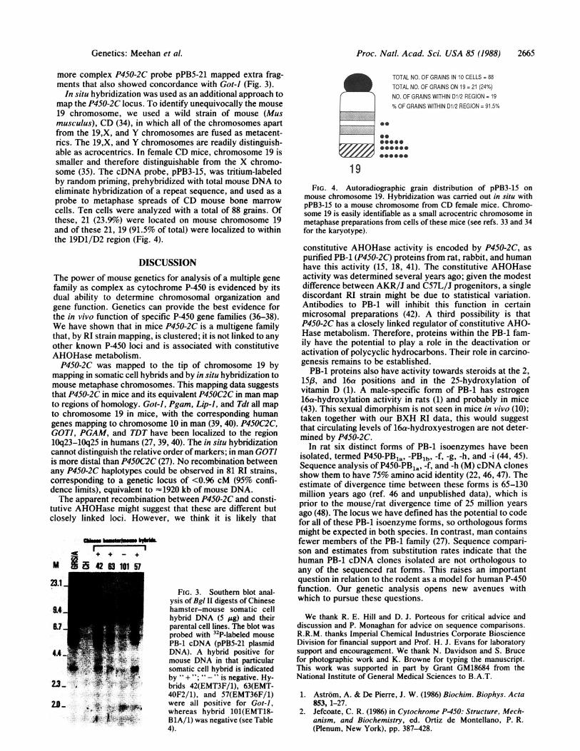
In situ hybridization was used as an additional approach tomap the P450-2C locus. To identify unequivocally the mouse19 chromosome, we used a wild strain of mouse (Musmusculus), CD (34), in which all of the chromosomes apartfrom the 19,X, and Y chromosomes are fused as metacent-rics. The 19,X, and Y chromosomes are readily distinguish-able as acrocentrics. In female CD mice, chromosome 19 issmaller and therefore distinguishable from the X chromo-some (35). The cDNA probe, pPB3-15, was tritium-labeledby random priming, prehybridized with total mouse DNA toeliminate hybridization of a repeat sequence, and used as aprobe to metaphase spreads of CD mouse bone marrowcells. Ten cells were analyzed with a total of 88 grains. Ofthese, 21 (23.9%) were located on mouse chromosome 19and of these 21, 19 (91.5% of total) were localized to withinthe 19D1/D2 region (Fig. 4).
DISCUSSIONThe power of mouse genetics for analysis of a multiple genefamily as complex as cytochrome P-450 is evidenced by itsdual ability to determine chromosomal organization andgene function. Genetics can provide the best evidence forthe in vivo function of specific P-450 gene families (36-38).We have shown that in mice P450-2C is a multigene familythat, by RI strain mapping, is clustered; it is not linked to anyother known P-450 loci and is associated with constitutiveAHOHase metabolism.P450-2C was mapped to the tip of chromosome 19 by
mapping in somatic cell hybrids and by in situ hybridization tomouse metaphase chromosomes. This mapping data suggeststhat P450-2C in mice and its equivalent P45OC2C in man mapto regions of homology. Got-i, Pgam, Lip-i, and Tdt all mapto chromosome 19 in mice, with the corresponding humangenes mapping to chromosome 10 in man (39, 40). P45OC2C,GOTi, PGAM, and TDT have been localized to the region10q23-10q25 in humans (27, 39, 40). The in situ hybridizationcannot distinguish the relative order of markers; in man GOT]is more distal than P45OC2C (27). No recombination betweenany P450-2C haplotypes could be observed in 81 RI strains,corresponding to a genetic locus of <0.96 cM (95% confi-dence limits), equivalent to "1920 kb of mouse DNA.The apparent recombination between P450-2C and consti-
tutive AHOHase might suggest that these are different butclosely linked loci. However, we think it is likely that
Mt8 42 63 101 57
FIG. 3. Southern blot anal-ysis of Bgl II digests of Chinese
&4+ 9 ,hamster-mouse somatic cellhybrid DNA (5 ,ug) and theirparental cell lines. The blot wasprobed with 32P-labeled mousePB-1 cDNA (pPB5-21 plasmid
4 -I DNA). A hybrid positive formouse DNA in that particular
.4I.-somatic cell hybrid is indicatedby "+"0 "-"is negative. Hy-
2... brids 42(EMT3F/1), 63(EMT-40F2/1), and 57(EMT36F/1)
2.9 were all positive for Got-i,whereas hybrid 101(EMT18-B1A/1) was negative (see Table4).
TOTAL NO. OF GRAINS IN 10 CELLS = 88TOTAL NO. OF GRAINS ON 19 = 21 (24%)NO. OF GRAINS WITHIN D1/2 REGION = 19% OF GRAINS WITHIN D1/2 REGION = 91.5%
......... .....
.................................
0@1zBOO66-0
19FIG. 4. Autoradiographic grain distribution of pPB3-15 on
mouse chromosome 19. Hybridization was carried out in situ withpPB3-15 to a mouse chromosome from CD female mice. Chromo-some 19 is easily identifiable as a small acrocentric chromosome inmetaphase preparations from cells of these mice (see refs. 33 and 34for the karyotype).
constitutive AHOHase activity is encoded by P450-2C, aspurified PB-1 (P450-2C) proteins from rat, rabbit, and humanhave this activity (15, 18, 41). The constitutive AHOHaseactivity was determined several years ago; given the modestdifference between AKR/J and C57L/J progenitors, a singlediscordant RI strain might be due to statistical variation.Antibodies to PB-1 will inhibit this function in certainmicrosomal preparations (42). A third possibility is thatP450-2C has a closely linked regulator of constitutive AHO-Hase metabolism. Therefore, proteins within the PB-1 fam-ily have the potential to play a role in the deactivation oractivation of polycyclic hydrocarbons. Their role in carcino-genesis remains to be established.PB-1 proteins also have activity towards steroids at the 2,
15,, and 16a positions and in the 25-hydroxylation ofvitamin D (1). A male-specific form of PB-1 has estrogen16a-hydroxylation activity in rats (1) and probably in mice(43). This sexual dimorphism is not seen in mice in vivo (10);taken together with our BXH RI data, this would suggestthat circulating levels of 16a-hydroxyestrogen are not deter-mined by P450-2C.
In rat six distinct forms of PB-1 isoenzymes have beenisolated, termed P450-PBla, -PBlb, -f, -g, -h, and -i (44, 45).Sequence analysis of P450-PBla, -f, and -h (M) cDNA clonesshow them to have 75% amino acid identity (22, 46, 47). Theestimate of divergence time between these forms is 65-130million years ago (ref. 46 and unpublished data), which isprior to the mouse/rat divergence time of 25 million yearsago (48). The locus we have defined has the potential to codefor all of these PB-1 isoenzyme forms, so orthologous formsmight be expected in both species. In contrast, man containsfewer members of the PB-1 family (27). Sequence compari-son and estimates from substitution rates indicate that thehuman PB-1 cDNA clones isolated are not orthologous toany of the sequenced rat forms. This raises an importantquestion in relation to the rodent as a model for human P-450function. Our genetic analysis opens new avenues withwhich to pursue these questions.
We thank R. E. Hill and D. J. Porteous for critical advice anddiscussion and P. Monaghan for advice on sequence comparisons.R.R.M. thanks Imperial Chemical Industries Corporate BioscienceDivision for financial support and Prof. H. J. Evans for laboratorysupport and encouragement. We thank N. Davidson and S. Brucefor photographic work and K. Browne for typing the manuscript.This work was supported in part by Grant GM18684 from theNational Institute of General Medical Sciences to B.A.T.
1. Astrom, A. & De Pierre, J. W. (1986) Biochim. Biophys. Acta853, 1-27.
2. Jefcoate, C. R. (1986) in Cytochrome P450: Structure, Mech-anism, and Biochemistry, ed. Ortiz de Montellano, P. R.(Plenum, New York), pp. 387-428.
Genetics: Meehan et al.

Proc. Natl. Acad. Sci. USA 85 (1988)
3. Wolf, C. R. (1986) Trends Genet. 8, 209-214.4. Nebert, D. W., Adesnik, M., Coon, M. J., Estabrook, R. W.,
Gonzalez, F. J., Guengerich, F. P., Gunsalus, I. C., Johnson,E. F., Kemper, B., Levin, W., Phillips, I. R., Sato, R. &Waterman, M. R. (1987) DNA 6, 1-11.
5. Adesnik, M. & Atchison, M. (1986) CRC Crit. Rev. Biochem.19, 247-309.
6. Mahgoub, A., Idle, J. R., Dring, L. G., Lancaster, R. &Smith, R. L. (1977) Lancet i, 584-586.
7. Kupfer, A. & Preisig, R. (1984) Eur. J. Clin. Pharmacol. 26,753-759.
8. Lush, I. E. & Andrews, K. M. (1978) Genet. Res. 31, 177-186.9. Ayesh, R., Idle, J. R., Ritchie, J. C., Crothers, M. J. &
Hetzel, M. R. (1985) Nature (London) 311, 169-170.10. Bradlow, H. L., Hershcopf, R. E., Martucci, C. P. & Fish-
man, J. (1985) Proc. Natl. Acad. Sci. USA 82, 6295-6299.11. Beaune, P., Dansette, P. M., Mansuy, D., Keffel, L., Finck,
M., Amar, C., Lenoux, J. P. & Homberg, J. (1987) Proc. Nati.Acad. Sci. USA 84, 551-555.
12. Stout, D. L. & Becker, F. F. (1986) Cancer Res. 46,2693-26%.
13. Idle, J. R. & Ritchie, J. C. (1983) in Human Carcinogenesis,eds. Harris, C. C. & Autrup, H. N. (Academic, New York),pp. 857-881.
14. Schneider, J., Kinne, D., Fracchia, A., Pierce, V., Anderson,K. E., Bradlow, H. L. & Fishman, J. (1982) Proc. Natl. Acad.Sci. USA 79, 3047-3051.
15. Waxman, D. J. & Walsh, C. (1983) Biochemistry 22,4846-4855.
16. Wolf, C. R., Moll, E., Friedberg, T., Oesch, F., Buchmann,A., Kuhlmann, W. D. & Kunz, H. W. (1984) Carcinogenesis5, 993-1001.
17. Inaba, T., Jurima, H. & Kalow, W. (1986) Am. J. Hum. Genet.38, 768-772.
18. Shimada, T., Misono, K. S. & Guengerich, F. P. (1986) J.Biol. Chem. 261, 909-921.
19. Hilkens, J., Cuypers, H. T., Selten, G., Kroezen, V., Hilgers,J. & Berns, A. (1986) Somatic Cell Mol. Genet. 12, 81-88.
20. Hill, R. E., Shaw, P. H., Barth, R. K. & Hastie, N. D. (1985)Mol. Cell. Biol. 5, 2114-2122.
21. Southern, E. M. (1975) J. Mol. Biol. 98, 503-517.22. Friedberg, T., Waxman, D. J., Atchison, M., Kuman, A.,
Haaparanta, T., Raphael, C. & Adesnik, M. (1986) Biochem-istry 25, 7975-7983.
23. Grunstein, M. & Hogness, D. S. (1975) Proc. Natl. Acad. Sci.USA 72, 3961-3965.
24. Feinberg, D. P. & Vogelstein, B. (1983) Anal. Biochem. 136,6-13.
25. Ford, C. E. & Hamerton, J. L. (1956) Stain Technol. 31, 247-251.
26. Gallimore, P. H. & Richardson, C. R. (1973) Chromosoma 41,259-263.
27. Meehan, R. R., Gosden, J. R., Rout, D., Hastie, N. D., Fried-berg, T., Adesnik, M., Buckland, R., van Heyningen, V.,Fletcher, J., Spurr, N. K., Sweeney, J. & Wolf, C. R. (1988)Am. J. Hum. Genet. 42, 26-37.
28. Hutton, J. J., Meier, J. & Hackney, C. (1979) Mutat. Res. 66,75-94.
29. Blatt, C., Mileham, K., Haas, M., Nesbitt, M. N., Harper, M.E. & Simon, M. (1983) Proc. Natl. Acad. Sci. USA 80,6298-6302.
30. Hildebrand, C. E., Gonzalez, F. J., Kozak, C. A. & Nebert,D. W. (1985) Biochem. Biophys. Res. Commun. 130, 396-406.
31. Silver, J. (1985) J. Hered. 76, 436-448.32. Gurtoo, H. L., Dahms, R. P., Kanter, P. & Vaught, J. B.
(1978) J. Biol. Chem. 253, 3952-3961.33. Drinkwater, N. R. & Ginsler, J. J. (1986) Carcinogenesis 7,
1701-1707.34. Capanna, E., Cristaldi, M., Perticone, P. & Rizzoni, M. (1975)
Experimentia 31, 294-297.35. Fisher, E. M. C., Cavanna, J. S. & Brown, S. D. M. (1985)
Proc. Natl. Acad. Sci. USA 82, 5846-5849.36. Simmons, D. L. & Kasper, C. B. (1983) J. Biol. Chem. 258,
9585-9588.37. Nebert, D. W., Eisen, H. J., Negishi, M., Lang, M. A. &
Hjelmeland, L. M. (1981) Annu. Rev. Pharmacol. Toxicol. 21,431-462.
38. Taylor, B. A. (1984) Banbury Rep. 18, 97-108.39. Grzeschik, K. H. & Kazazian, H. H. (1985) Cytogenet. Cell
Genet. 40, 179-205.40. Yang-Feng, T. L., Landan, N. R., Baltimore, D. & Franke, U.
(1986) Cytogenet. Cell Genet. 43, 121-126.41. Raucy, J. L. & Johnson, E. F. (1985) Mol. Pharmacol. 27,
296-301.42. Adams, D. J., Seilman, S., Amelizad, S., Oesch, F. & Wolf,
C. R. (1985) Biochem. J. 232, 869-876.43. Harada, H. & Negishi, M. (1984) J. Biol. Chem. 259,
12285-12290.44. Haniu, M., Ryan, D. E., lida, S., Lieber, C. S., Levin, W. &
Shively, J. E. (1984) Arch. Biochem. Biophys. 235, 304-311.45. Wolf, C. R., Seilman, S., Oesch, F., Mayer, R. T. & Burke,
M. D. (1986) Biochem. J. 240, 27-33.46. Gonzalez, F. J., Kimura, S., Song, B.-J., Pastewka, J., Gel-
boin, H. V. & Hardwick, J. P. (1986) J. Biol. Chem. 251,10667-10672.
47. Yoshioka, H., Morohashi, K.-I., Sugawa, K., Miyata, T.,Kawajiri, K., Hirose, T., Inayama, S., Fuji-Kuriyama, Y. &Omura, T. (1987) J. Biol. Chem. 262, 1706-1711.
48. Sarich, N. M. (1985) in Evolutionary Relationships AmongRodents, eds. Pluckett, N. & Hartenberger, J.-L. (Plenum,New York), pp. 423-452.
2666 Genetics: Meehan et al.