Embed Size (px)
Citation preview
Chromosomes
What Exactly is a chromosome?
Chromosomes are the rod-shaped, filamentous bodies present in the nucleus, which become visible during cell division.
They are the carriers of the gene or unit of heredity.
Chromosome are not visible in active nucleus due to their high water content, but are clearly seen during cell division.

Chromosomes were first described by Strausberger in 1875.
The term “Chromosome”, however was first used by Waldeyer in 1888.
They were given the name chromosome (Chromo = colour; Soma = body) due to their marked affinity for basic dyes.
Their number can be counted easily only during mitotic metaphase.

Chromosomes are composed of thin chromatin threads called Chromatin fibers.
These fibers undergo folding, coiling and supercoiling during prophase so that the chromosomes become progressively thicker and smaller.
Therefore, chromosomes become readily observable under light microscope.
At the end of cell division, on the other hand, the fibers uncoil and extend as fine chromatin threads, which are not visible at light microscope

Number of chromosomes
Normally, all the individuals of a species have the same number of chromosomes.
Closely related species usually have similar chromosome numbers.
Presence of a whole sets of chromosomes is called euploidy.
It includes haploids, diploids, triploids, tetraploids etc.
Gametes normally contain only one set of chromosome – this number is called Haploid
Somatic cells usually contain two sets of chromosome - 2n : Diploid

3n – triploid4n – tetraploidThe condition in which the chromosomes sets
are present in a multiples of “n” is PolyploidyWhen a change in the chromosome number does
not involve entire sets of chromosomes, but only a few of the chromosomes - is Aneuploidy.
Monosomics (2n-1) Trisomics (2n+1) Nullisomics (2n-2) Tetrasomics (2n+2)

Organism No. chromosomes
Human 46 Chimpanzee 48 Dog 78 Horse 64 Chicken 78 Goldfish 94 Fruit fly 8 Mosquito 6 Nematode 11(m), 12(f) Horsetail 216 Sequoia 22 Round worm 2

Organism No. chromosomes
Onion 16 Mold 16 Carrot 20 Tomato 24 Tobacco 48 Rice 24 Maize 20 Haploppus gracilis 4 Crepis capillaris 6

On the extreme, round worm shows only two chromosomes, while the other extreme is represented by Protozoa having 300 or more chromosomes.
However, most organisms have numbers between 12 to 50.
3-8 in fungi
From 8 – 16 in Angiosperms (Most common number being 12).

Chromosome Size
In contrast to other cell organelles, the size of chromosomes shows a remarkable variation depending upon the stages of cell division.
Interphase: chromosome are longest & thinnest
Prophase: there is a progressive decrease in their length accompanied with an increase in thickness
Anaphase: chromosomes are smallest.
Metaphase: Chromosomes are the most easily observed and studied during metaphase when they are very thick, quite short and well spread in the cell.
Therefore, chromosomes measurements are generally taken during mitotic metaphase.

The size of the chromosomes in mitotic phase of animal and plants sp generally varies between 0.5 µ and 32 µ in length, and between 0.2 µ and 3.0 µ in diameter.
The longest metaphase chromosomes found in Trillium -32 µ.
The giant chromosomes found in diptera and they may be as long as 300 µ and up to 10 µ in diameter.
In general, plants have longer chromosomes than animal and species having lower chromosome numbers have long chromosomes than those having higher chromosome numbers
Among plants, dicots in general, have a higher number of chromosome than monocots.
Chromosomes are longer in monocot than dicots.

In order to understand chromosomes and their function, we need to be able to discriminate among different chromosomes.
First, chromosomes differ greatly in size
Between organisms the size difference can be over 100-fold, while within a sp, some chromosomes are often 10 times as large as others.
In a species Karyotype, a pictorial or photographic representation of all the different chromosomes in a cell of an individual, chromosomes are usually ordered by size and numbered from largest to smallest.
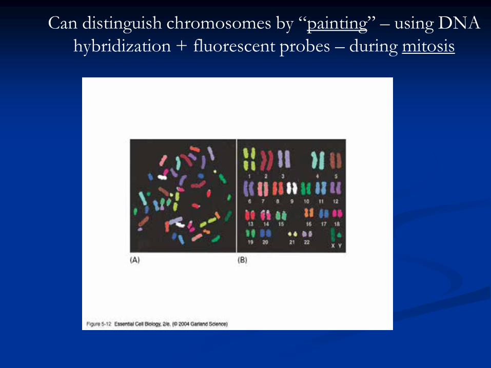
Can distinguish chromosomes by “painting” – using DNA
hybridization + fluorescent probes – during mitosis

Karyotype: is the general morphology of the somatic chromosome. Generally, karyotypes represent by arranging in the descending order of size keeping their centromeres in a straight line.
Idiotype: the karyotype of a species may be represented diagrammatically, showing all the morphological features of the chromosome; such a diagram is known as Idiotype.
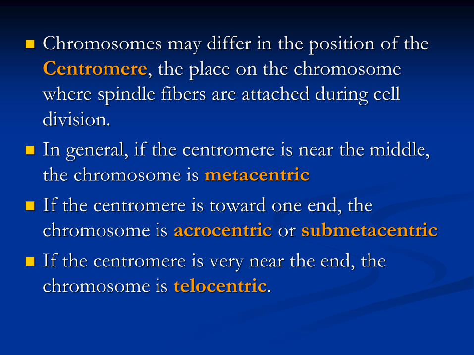
Chromosomes may differ in the position of the
Centromere, the place on the chromosome
where spindle fibers are attached during cell
division.
In general, if the centromere is near the middle,
the chromosome is metacentric
If the centromere is toward one end, the
chromosome is acrocentric or submetacentric
If the centromere is very near the end, the
chromosome is telocentric.

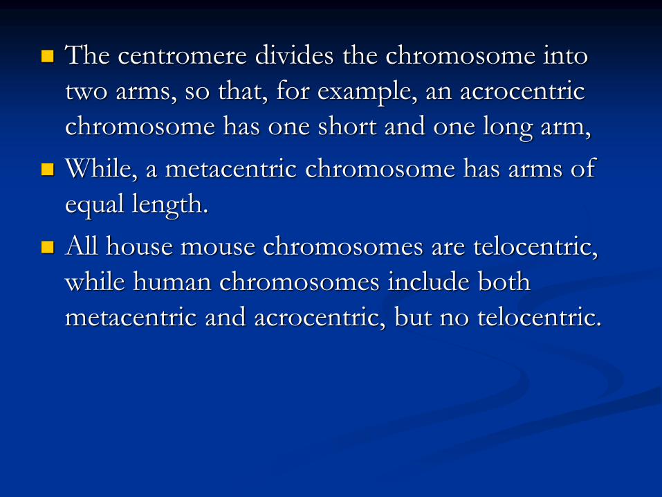
The centromere divides the chromosome into
two arms, so that, for example, an acrocentric
chromosome has one short and one long arm,
While, a metacentric chromosome has arms of
equal length.
All house mouse chromosomes are telocentric,
while human chromosomes include both
metacentric and acrocentric, but no telocentric.

Autosomal pair Sex chromosome
Diploid No. of No. of X Y
(2n) metacentrics acrocentric or telocentric
Cat 38 16 2 M M
Dog 78 0 38 M A
Pig 38 12 6 M M
Goat 60 0 29 A M
Sheep 54 3 23 A M
Cow 60 0 29 M M
Horse 64 13 18 M A
M – Metacentric; A – Acrocentric

Euchromatin and Heterochromatin
Chromosomes may be identified by regions that stain in a
particular manner when treated with various chemicals.
Several different chemical techniques are used to identify
certain chromosomal regions by staining then so that they
form chromosomal bands.
For example, darker bands are generally found near the
centromeres or on the ends (telomeres) of the chromosome,
while other regions do not stain as strongly.
The position of the dark-staining are heterochromatic
region or heterochromatin.
Light staining are euchromatic region or euchromatin.

Heterochromatin is classified into two groups:
(i) Constitutive and (ii) Facultative.
Constitutive heterochromatin remains
permanently in the heterochromatic stage, i.e., it
does not revert to the euchromatic stage.
In contrast, facultative heterochromatin consists
of euchromatin that takes on the staining and
compactness characteristics of heterochromatin
during some phase of development.
Satellite DNAs
When the DNA of a prokaryote, such as E.coli, is isolated, fragmented and centrifuged to equilibrium in a Cesium chloride (CsCl) density gradient, the DNA usually forms a single band in the gradient.
On the other hand, CsCl density-gradient analysis of DNA from eukaryotes usually reveals the presence of one large band of DNA (usually called “Mainband” DNA) and one to several small bands.
These small bands are referred to as “Satellite DNAs”.
For e.g., in Drosophila virilis, contain three distinct satellite DNAs.

Prokaryotic and Eukaryotic
Chromosomes

Not only the genomes of eukaryotes are more
complex than prokaryotes, but the DNA of
eukaryotic cell is also organized differently
from that of prokaryotic cells.
The genomes of prokaryotes are contained in
single chromosomes, which are usually
circular DNA molecules.
In contrast, the genomes of eukaryotes are
composed of multiple chromosomes, each
containing a linear molecular of DNA.

Although the numbers and sizes of chromosomes varyconsiderably between different species, their basicstructure is the same in all eukaryotes
Organism Genome ChromosomeSize (Mb)a numbera
Arabidopsis thaliana 70 5Corn 5000 10Onion 15,000 8Lily 50,000 12Fruit fly 165 4Chicken 50,000 39 Mouse 1,200 20Cow 3000 30Human 3000 23
a – both genome size and chromosome numbers are for haploid cells

The DNA of eukaryotic cell is tightly bound to small
basic proteins (histones) that package the DNA in an
orderly way in the cell nucleus.
This task is substantial (necessary), given the DNA
content of most eukaryotes
For e.g., the total extended length of DNA in a human
cell is nearly 2 m, but this must be fit into a nucleus
with a diameter of only 5 to 10µm.
Although DNA packaging is also a problem in bacteria,
the mechanism by which prokaryotic DNA are
packaged in the cell appears distinct from that
eukaryotes and is not well understood.

Prokaryotic chromosome
The prokaryotes usually have only one chromosome, and it bears little morphological resemblance to eukaryotic chromosomes.
Among prokaryotes there is considerable variation in genome length bearing genes.
The genome length is smallest in RNA viruses
In this case, the organism is provided with only a few genes in its chromosome.
The number of gene may be as high as 150 in some larger bacteriophage genome.

In E.coli, about 3000 to 4000 genes are organized into its one circular chromosome.
The chromosome exists as a highly folded and coiled structure dispersed throughout the cell.
The folded nature of chromosome is due to the incorporation of RNA with DNA.
There are about 50 loops in the chromosome of E.coli.
These loops are highly twisted or supercoiled structure with about four million nucleotide pairs.
Its molecular weight is about 2.8 X10⁹
During replication of DNA, the coiling must be relaxed.
DNA gyrase is necessary for the unwinding the coils.
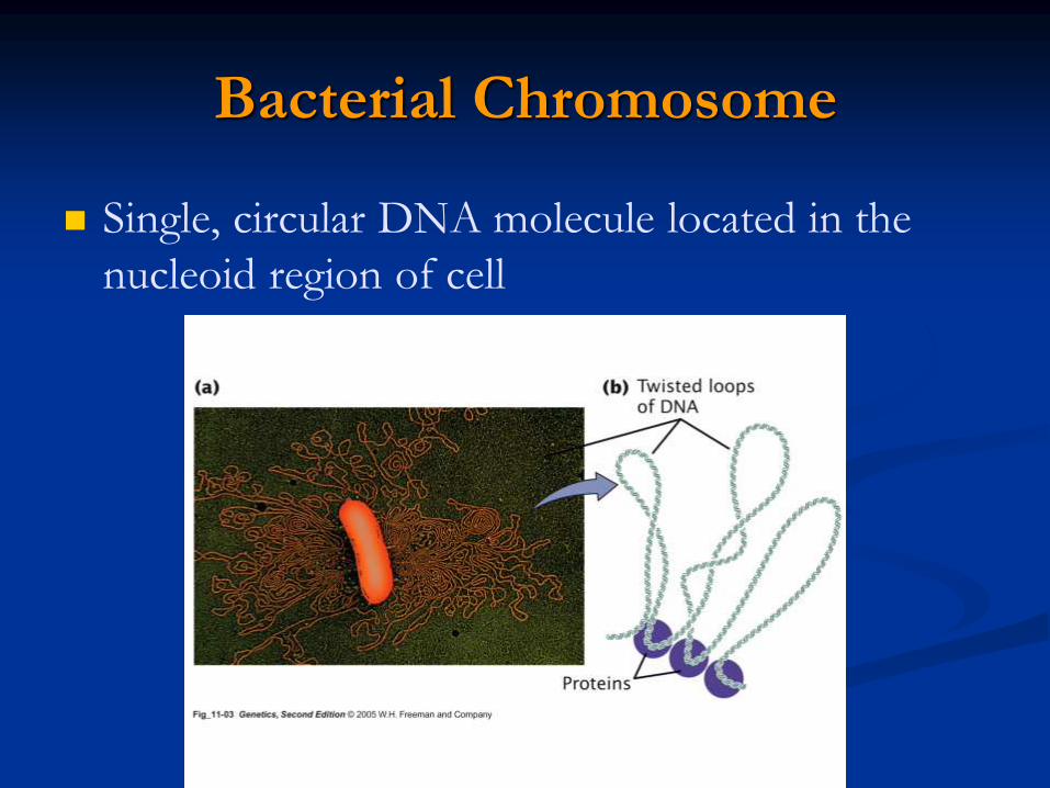
Bacterial Chromosome
Single, circular DNA molecule located in the
nucleoid region of cell

Supercoiling

Supercoiling
Helix twists on
itself in the opposite
direction; twists to
the left
Most common type
of supercoiling

Mechanism of folding of a bacterial
chromosome
There are many supercoiled loops (~100 in E. coli) attached to acentral core. Each loop can be independently relaxed or condensed.
Topoisomerase enzyme – (Type I and II) that introduce or removesupercoiling.

Chromatin
The complexes between eukaryotic DNA and proteins are
called Chromatin, which typically contains about twice as
much protein as DNA.
The major proteins of chromatin are the histones – small
proteins containing a high proportion of basic aminoacids
(arginine and lysine) that facilitate binding negatively
charged DNA molecule .
There are 5 major types of histones: H1, H2A, H2B, H3,
and H4 – which are very similar among different sp of
eukaryotes.
The histones are extremely abundant proteins in eukaryotic
cells.
Their mass is approximately equal to that of the cell’s DNA

The major histone proteins:
Histone Mol. Wt No. of Percentage
Amino acid Lys + Arg
H1 22,500 244 30.8
H2A 13,960 129 20.2
H2B 13,774 125 22.4
H3 15,273 135 22.9
H4 11,236 102 24.5
The DNA double helix is bound to proteins called histones. The
histones have positively charged (basic) amino acids to bind the
negatively charged (acidic) DNA. Here is an SDS gel of histone
proteins, separated by size

In addition, chromatin contains an approximately equal
mass of a wide variety of non-histone chromosomal
proteins.
There are more than a thousand different types of these
proteins, which are involved in a range of activities,
including DNA replication and gene expression.
The DNA of prokaryotes is similarly associated with
proteins, some of which presumably function as histones
do, packing the DNA within the bacterial cell.
Histones, however are unique feature of eukaryotic cells
and are responsible for distinct structural organization of
eukaryotic chromatin

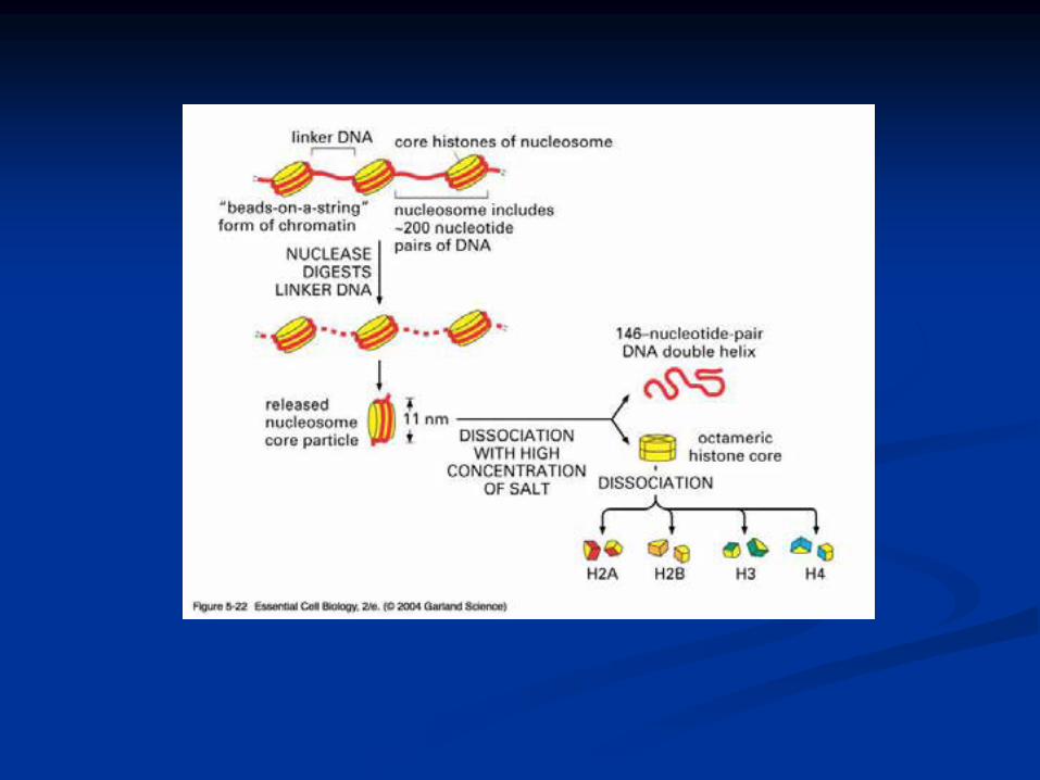
The basic structural unit of chromatin, the nucleosome, was
described by Roger Kornberg in 1974.
Two types of experiments led to Kornberg’s proposal of the
nucleosome model.
First, partial digestion of chromatin with micrococcal nuclease (an
enzyme that degrades DNA) was found to yield DNA fragments
approximately 200 base pairs long.
In contrast, a similar digestion of naked DNA (not associated with
protein) yielded a continuous smear randomly sized fragments.
These results suggest that the binding of proteins to DNA in
chromatin protects the regions of DNA from
nuclease digestion, so that enzyme can
attack DNA only at sites separated by
approximately 200 base pairs.

Electron microscopy revealed that chromatin
fibers have a beaded appearance, with the beads
spaced at intervals of approximately 200 base
pairs.
Thus, both nuclease digestion and the electron
microscopic studies suggest that chromatin is
composed of repeating 200 base pair unit, which
were called nucleosome.

individual nucleosomes = “beads on a string”
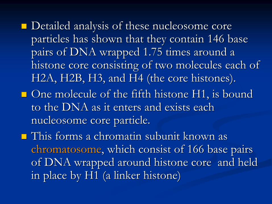
Detailed analysis of these nucleosome core particles has shown that they contain 146 base pairs of DNA wrapped 1.75 times around a histone core consisting of two molecules each of H2A, H2B, H3, and H4 (the core histones).
One molecule of the fifth histone H1, is bound to the DNA as it enters and exists each nucleosome core particle.
This forms a chromatin subunit known as chromatosome, which consist of 166 base pairs of DNA wrapped around histone core and held in place by H1 (a linker histone)


Centromeres and Telomeres
Centromeres and telomeres are two essential
features of all eukaryotic chromosomes.
Each provide a unique function i.e., absolutely
necessary for the stability of the chromosome.
Centromeres are required for the segregation of
the centromere during meiosis and mitosis.
Teleomeres provide terminal stability to the
chromosome and ensure its survival

Centromere The region where two sister chromatids of a chromosome
appear to be joined or “held together” during mitatic metaphase is called Centromere
When chromosomes are stained they typically show a dark-stained region that is the centromere.
Also termed as Primary constriction
During mitosis, the centromere that is shared by the sister chromatids must divide so that the chromatids can migrate to opposite poles of the cell.
On the other hand, during the first meiotic division the centromere of sister chromatids must remain intact
whereas during meiosis II they must act as they do during mitosis.
Therefore the centromere is an important component of chromosome structure and segregation.
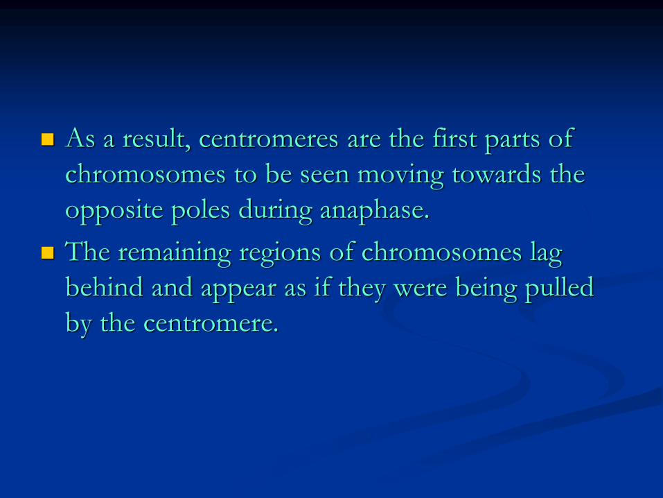
As a result, centromeres are the first parts of
chromosomes to be seen moving towards the
opposite poles during anaphase.
The remaining regions of chromosomes lag
behind and appear as if they were being pulled
by the centromere.

Kinetochore
Within the centromere region, most species have
several locations where spindle fibers attach, and
these sites consist of DNA as well as protein.
The actual location where the attachment occurs
is called the kinetochore and is composed of
both DNA and protein.
The DNA sequence within these regions is
called CEN DNA.
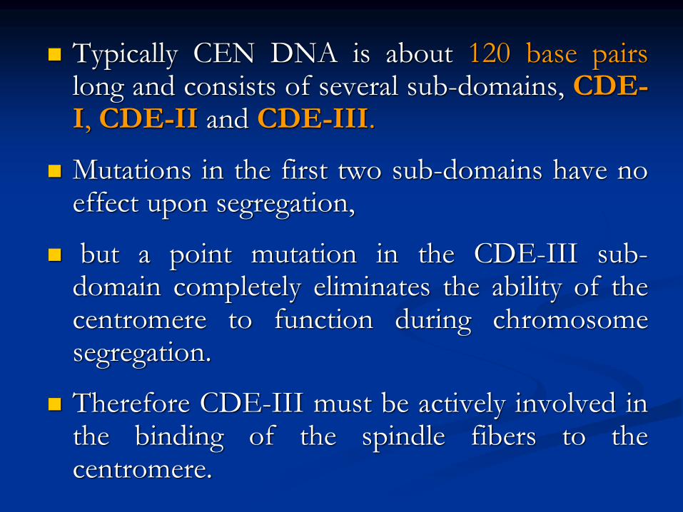
Typically CEN DNA is about 120 base pairslong and consists of several sub-domains, CDE-I, CDE-II and CDE-III.
Mutations in the first two sub-domains have noeffect upon segregation,
but a point mutation in the CDE-III sub-domain completely eliminates the ability of thecentromere to function during chromosomesegregation.
Therefore CDE-III must be actively involved inthe binding of the spindle fibers to thecentromere.

The protein component of the kinetochore is
only now being characterized.
A complex of three proteins called Cbf-III
binds to normal CDE-III regions but can not
bind to a CDE-III region with a point mutation
that prevents mitotic segregation.

Telomere
The two ends of a chromosome are known as telomeres.
It required for the replication and stability of the chromosome.
When telomeres are damaged or removed due to chromosome breakage, the damaged chromosome ends can readily fuse or unite with broken ends of other chromosome.
Thus it is generally accepted that structural integrity and individuality of chromosomes is maintained due to telomeres.

McClintock noticed that if two chromosomes were
broken in a cell, the end of one could attach to the
other and vice versa.
What she never observed was the attachment of the
broken end to the end of an unbroken
chromosome.
Thus the ends of broken chromosomes are sticky,
whereas the normal end is not sticky, suggesting
the ends of chromosomes have unique features.
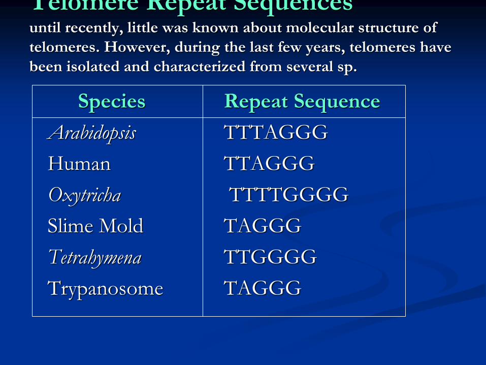
Telomere Repeat Sequences until recently, little was known about molecular structure of
telomeres. However, during the last few years, telomeres have
been isolated and characterized from several sp.
Species Repeat Sequence
Arabidopsis TTTAGGG
Human TTAGGG
Oxytricha TTTTGGGG
Slime Mold TAGGG
Tetrahymena TTGGGG
Trypanosome TAGGG

The telomeres of this organism end in the sequence 5'-TTGGGG-3'.
The telomerase adds a series of 5'-TTGGGG-3' repeats to the ends of the lagging strand.
A hairpin occurs when unusual base pairs between guanine residues in the repeat form.
Finally, the hairpin is removed at the 5'-TTGGGG-3' repeat.
Thus the end of the chromosome is faithfully replicated.
Tetrahymena - protozoa
organism.
RNA Primer - Short stretches of
ribonucleotides (RNA substrates) found on the
lagging strand during DNA replication. Helps
initiate lagging strand replication
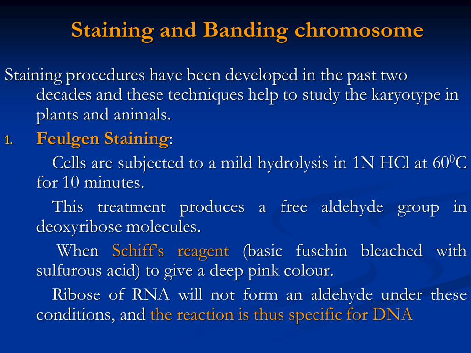
Staining and Banding chromosome
Staining procedures have been developed in the past two decades and these techniques help to study the karyotype in plants and animals.
1. Feulgen Staining:
Cells are subjected to a mild hydrolysis in 1N HCl at 600Cfor 10 minutes.
This treatment produces a free aldehyde group indeoxyribose molecules.
When Schiff’s reagent (basic fuschin bleached withsulfurous acid) to give a deep pink colour.
Ribose of RNA will not form an aldehyde under theseconditions, and the reaction is thus specific for DNA

2. Q banding:
The Q bands are the fluorescent bands observed
after quinacrine mustard staining and observation with
UV light.
The distal ends of each chromatid are not stained by this
technique.
The Y chromosome become brightly fluorescent both in
the interphase and in metaphase.
3. R banding:
The R bands (from reverse) are those located in the
zones that do not fluoresce with the quinacrine mustard,
that is they are between the Q bands and can be
visualized as green.

4. G banding:
The G bands (from Giemsa) have the same location as Q bands and do not require fluorescent microscopy.
Many techniques are available, each involving some pretreatment of the chromosomes.
In ASG (Acid-Saline-Giemsa) cells are incubated in citric acid and NaCl for one hour at 600C and are then treated with the Giemsa stain.
5. C banding:
The C bands correspond to constitutive heterochromatin.
The heterochromatin regions in a chromosome distinctly differ in their stainability from euchromatic region.

VARIATION IN STRUTURE OF
CHROMOSOME

Chromosomal Aberrations
The somatic (2n) and gametic (n) chromosome numbers of a species ordinarily remain constant.
This is due to the extremely precise mitotic and meiotic cell division.
Somatic cells of a diploid species contain two copies of each chromosome, which are called homologous chromosome.
Their gametes, therefore contain only one copy of each chromosome, that is they contain one chromosome complement or genome.
Each chromosome of a genome contains a definite numbers and kinds of genes, which are arranged in a definite sequence.

Chromosomal Aberrations
Sometime due to mutation or spontaneous
(without any known causal factors), variation in
chromosomal number or structure do arise in
nature. - Chromosomal aberrations.
Chromosomal aberration may be grouped into
two broad classes:
1. Structural and 2. Numerical

Structural Chromosomal Aberrations
Chromosome structure variations result from
chromosome breakage.
Broken chromosomes tend to re-join; if there is more
than one break, rejoining occurs at random and not
necessarily with the correct ends.
The result is structural changes in the chromosomes.
Chromosome breakage is caused by X-rays, various
chemicals, and can also occur spontaneously.

There are four common type of structural
aberrations:
1. Deletion or Deficiency
2. Duplication or Repeat
3. Inversion, and
4. Translocation.

Consider a normal chromosome with genes in alphabetical order: a b c d e f g h i
1. Deletion: part of the chromosome has been removed: a b c g h i
2. Dupliction: part of the chromosome is duplicated:
a b c d e f d e f g h i
3. Inversion: part of the chromosome has been re-inserted in reverse order: a b c f e d g h i
ring: the ends of the chromosome are joined together to make a ring

4. translocation: parts of two non-homologous
chromosomes are joined:
If one normal chromosome is a b c d e f g h i
and the other chromosome is u v w x y z,
then a translocation between them would be
a b c d e f x y z and u v w g h i.


Deletion or deficiency
Loss of a chromosome segment is known as deletion or deficiency
It can be terminal deletion or interstitial or intercalary deletion.
A single break near the end of the chromosome would be expected to result in terminal deficiency.
If two breaks occur, a section may be deleted and an intercalary deficiency created.
Terminal deficiencies might seem less complicated.
But majority of deficiencies detected are intercalary type within the chromosome.
Deletion was the first structural aberration detected by Bridges in 1917 from his genetic studies on X chromosome of Drosophila.
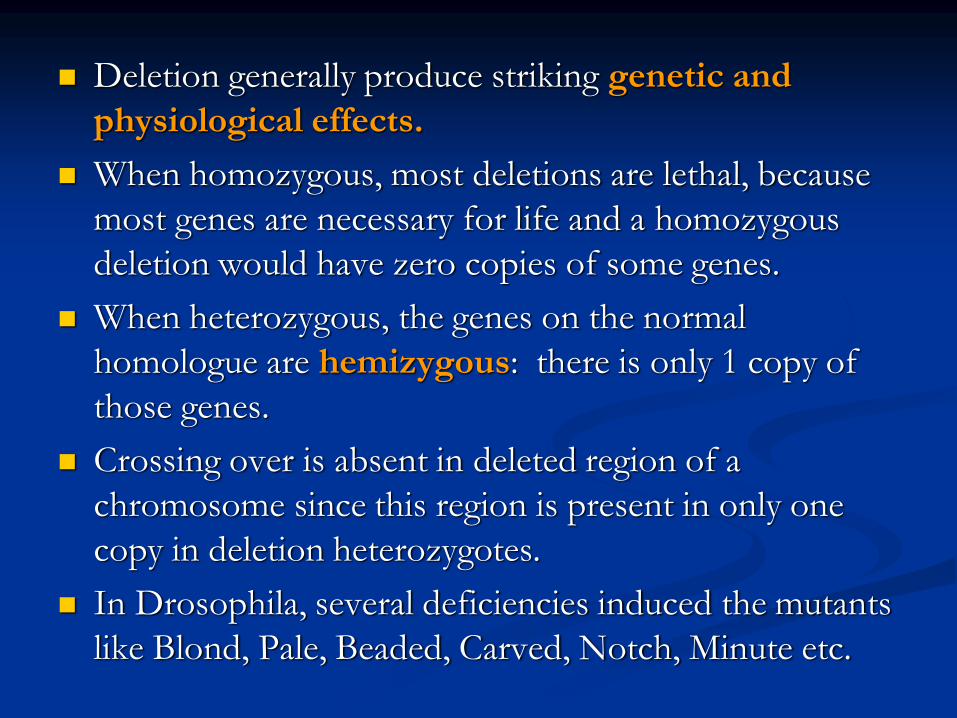
Deletion generally produce striking genetic and
physiological effects.
When homozygous, most deletions are lethal, because
most genes are necessary for life and a homozygous
deletion would have zero copies of some genes.
When heterozygous, the genes on the normal
homologue are hemizygous: there is only 1 copy of
those genes.
Crossing over is absent in deleted region of a
chromosome since this region is present in only one
copy in deletion heterozygotes.
In Drosophila, several deficiencies induced the mutants
like Blond, Pale, Beaded, Carved, Notch, Minute etc.
Deletion in Prokaryotes:
Deletions are found in prokaryotes as well, e.g., E.coli,
T4 phage and Lambda phage.
In E.coli, deletions of up to 1 % of the bacterial chromosome
are known.
In lambda phage, however 20% of the genome may be missing
in some of the deletions.
Deletion in Human:
Chromosome deletions are usually lethal even as
heterozygotes, resulting in zygotic loss, stillbirths, or infant
death.
Sometimes, infants with small chromosome deficiencies
however, survive long enough to permit the abnormal
phenotype they express.
Cri-du-chat (Cat cry syndrome):
The name of the syndrome came from a catlike mewing cry from small weak infants with the disorder.
Other characteristics are microcephaly (small head), broad face and saddle nose, physical and mental retardation.
Cri-du-chat patients die in infancy or early childhood.
The chromosome deficiency is in the short arm of chromosome 5 .
Myelocytic leukemia
Another human disorder that is associated with a chromosome abnormality is chronic myelocytic leukemia.
A deletion of chromosome 22 was described by P.C.Nowell and Hungerford and was called “Philadelphia” (Ph’)chromosome after the city in which the discovery was made.

DuplicationThe presence of an additional chromosome
segment, as compared to that normally present in a nucleus is known as Duplication.
In a diploid organism, presence of a chromosome segment in more than two copies per nucleus is called duplication.
Four types of duplication:
1. Tandem duplication
2. Reverse tandem duplication
3. Displaced duplication
4. Translocation duplication

The extra chromosome segment may be located
immediately after the normal segment in precisely
the same orientation forms the tandem
When the gene sequence in the extra segment of a
tandem in the reverse order i.e, inverted , it is
known as reverse tandem duplication
In some cases, the extra segment may be located in
the same chromosome but away from the normal
segment – termed as displaced duplication
The additional chromosome segment is located in
a non-homologous chromosome is translocation
duplication.
Origin Origin of duplication involves chromosome breakage and
reunion of chromosome segment with its homologous chromosome.
As a result, one of the two homologous involved in the production of a duplication ends up with a deficiency, while the other has a duplication for the concerned segment.
Another phenomenon, known as unequal crossing over, also leads to exactly the same consequences for small chromosome segments.
For e.g., duplication of the band 16A of X chromosome of Drosophila produces Bar eye.
This duplication is believed to originate due to unequal crossing over between the two normal X chromosomes of female.


Inversion When a segment of chromosome is oriented in the reverse
direction, such segment said to be inverted and the phenomenon is termed as inversion.
The existence of inversion was first detected by Strutevant and Plunkett in 1926.
Inversion occur when parts of chromosomes become detached , turn through 1800 and are reinserted in such a way that the genes are in reversed order.
For example, a certain segment may be broken in two places, and the breaks may be in close proximity because of chance loop in the chromosome.
When they rejoin, the wrong ends may become connected.
The part on one side of the loop connects with broken end different from the one with which it was formerly connected.
This leaves the other two broken ends to become attached.
The part within the loop thus becomes turned around or inverted.

Inversion may be classified into two types:
Pericentric - include the centromere
Paracentric - do not include the centromere

An inversion consists of two breaks in one
chromosome.
The area between the breaks is inverted (turned
around), and then reinserted and the breaks then
unite to the rest of the chromosome.
If the inverted area includes the centromere it is
called a pericentric inversion.
If it does not, it is called a paracentric inversion.

Inversions in natural populations
In natural populations, pericentric inversions are
much less frequent than paracentric inversions.
In many sp, however, pericentric inversions are
relatively common, e.g., in some grasshoppers.
Paracentric inversions appear to be very frequent
in natural populations of Drosophila.

Translocation
Integration of a chromosome segment into a
nonhomologous chromosome is known as
translocation.
Three types:
1. simple translocation
2. shift
3. reciprocal translocation.

Simple translocation: In this case, terminal segment of a chromosome is integrated at one end of a non-homologous region. Simple translocations are rather rare.
Shift: In shift, an intercalary segment of a chromosome is integrated within a non-homologous chromosome. Such translocations are known in the populations of Drosophila, Neurospora etc.
Reciprocal translocation: It is produced when two non-homologous chromosomes exchange segments – i.e., segments reciprocally transferred.
Translocation of this type is most common

Non-Disjunction
Generally during gametogenesis the homologous
chromosomes of each pair separate out
(disjunction) and are equally distributed in the
daughter cells.
But sometime there is an unequal distribution of
chromosomes in the daughter cells.
The failure of separation of homologous
chromosome is called non-disjunction.
This can occur either during mitosis or meiosis
or embryogenesis.


Mitotic non-disjunction: The failure of separation of homologous chromosomes during mitosis is called mitotic non-disjunction.
It occurs after fertilization.
May happen during first or second cleavage.
Here, one blastomere will receive 45 chromosomes, while other will receive 47.
Meiotic non-disjunction: The failure of separation of homologous chromosomes during meiosis is called mitotic non-disjunction
Occurs during gametogensis
Here, one type contain 22 chromosome, while other will be 24.

Variation in chromosome number
Organism with one complete set of chromosomes is said to be euploid (applies to haploid and diploid organisms).
Aneuploidy - variation in the number of individual chromosomes (but not the total number of sets of chromosomes).
The discovery of aneuploidy dates back to 1916 when Bridges discovered XO male and XXY female Drosophila, which had 7 and 9 chromosomes respectively, instead of normal 8.

Nullisomy - loss of one homologous chromosome pair. (e.g., Oat )
Monosomy – loss of a single chromosome (Maize).
Trisomy - one extra chromosome. (Datura)
Tetrasomy - one extra chromosome pair.
More about Aneuploidy

Uses of Aneuploidy
They have been used to determine the phenotypic effect of loss or gain of different chromosome
Used to produce chromosome substitution lines. Such lines yield information on the effects of different chromosomes of a variety in the same genetic background.
They are also used to produce alien addition and alien substitution lines. These are useful in gene transfer from one species to another.
Aneuploidy permits the location of a gene as well as of a linkage group onto a specific chromosome.

Trisomy in Humans
Down Syndrome
The best known and most common chromosome related syndrome.
Formerly known as “Mongolism”
1866, when a physician named John Langdon Down published an essay in England in which he described a set of children with common features who were distinct from other children with mental retardation he referred to as “Mongoloids.”
One child in every 800-1000 births has Down syndrome
250,000 in US has Down syndrome.
The cost and maintaining Down syndrome case in US is estimated at $ 1 billion per year.

Patients having Down syndrome will Short in stature
(four feet tall) and had an epicanthal fold, broad
short skulls, wild nostrils, large tongue, stubby hands
Some babies may have short necks, small hands, and
short fingers.
They are characterized as low in mentality.
Down syndrome results if the extra chromosome is
number 21.

Amniocentesis for Detecting Aneuploidy
Chromosomal abnormalities are sufficiently well understood to permit genetic counseling.
A fetus may be checked in early stages of development by karyotyping the cultured cells obtained by a process called amniocentesis.
A sample of fluid will taken from mother and fetal cells are cultured and after a period of two to three weeks, chromosomes in dividing cells can be stained and observed.
If three No.21 chromosomes are present, Down syndrome confirmed.

The risk for mothers less than 25 years of age to
have the trisomy is about 1 in 1500 births.
At 40 years of age, 1 in 100 births
At 45 years 1 in 40 births.
Other Syndromes
Chromosome Nomenclature: 47, +13
Chromosome formula: 2n+1
Clinical Syndrome: Trisomy-13
Estimated Frequency Birth: 1/20,000
Main Phenotypic Characteristics:
Mental deficiency and deafness, minor
muscle seizures, cleft lip, cardiac anomalies

Other Syndromes
Chromosome Nomenclature: 47, +18
Chromosome formula: 2n+1
Clinical Syndrome: Trisomy-18
Estimated Frequency Birth: 1/8,000
Main Phenotypic Characteristics:
Multiple congenital malformation of many organs, malformed ears, small mouth and nose with general elfin appearance.
90% die in the first 6 months.

Other Syndromes
Chromosome Nomenclature: 45, X
Chromosome formula: 2n - 1
Clinical Syndrome: Turner
Estimated Frequency Birth: 1/2,500 female
Main Phenotypic Characteristics:
Female with retarded sexual development,
usually sterile, short stature, webbing of skin in
neck region, cardiovascular abnormalities,
hearing impairment.

Other Syndromes
Chromosome Nomenclature: 47, XXY, 48, XXXY,
48,XXYY, 49,
XXXXY, 50, XXXXXY
Chromosome formula: 2n+1; 2n+2; 2n+2; 2n+3; 2n+4
Clinical Syndrome: Klinefelter
Estimated Frequency Birth: 1/500 male borth
Main Phenotypic Characteristics:
Pitched voice, Male, subfertile with small
testes, developed breasts, feminine, long limbs.

Found in certain tissues e.g., salivary glands of larvae, gut epithelium, Malphigian tubules and some fat bodies, of some Diptera (Drosophila, Sciara, Rhyncosciara)
These chromosomes are very long and thick (upto 200 times their size during mitotic metaphase in the case of Drosophila)
Hence they are known as Giant chromosomes.
Giant chromosomes

They are first discovered by Balbiani in 1881 in
dipteran salivary glands and thus also known as
salivary gland chromosomes.
But their significance was realized only after the
extensive studies by Painter during 1930’s.
Giant chromosomes have also been discovered
in suspensors of young embryos of many plants,
but these do not show the bands so typical of
salivary gland chromosomes.

He described the morphology in detail and
discovered the relation between salivary gland
chromosomes and germ cell chromosomes.
Slides of Drosophila giant chromosomes are
prepared by squashing in acetocarmine the
salivary glands dissected out from the larvae.
The total length of D.melanogater giant
chromosomes is about 2,000µ.
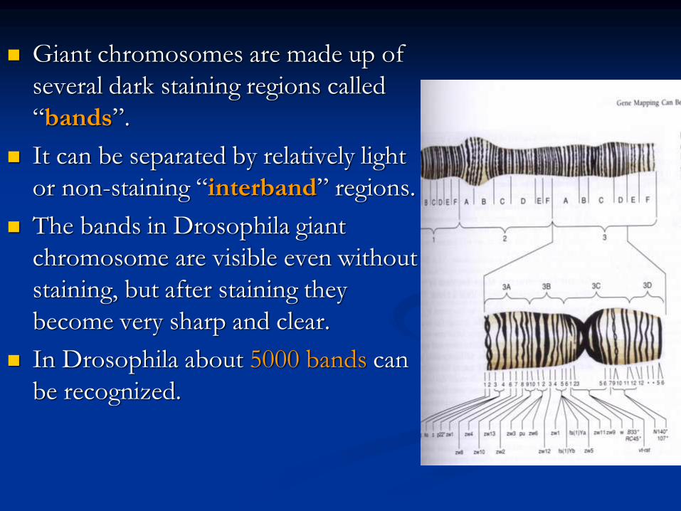
Giant chromosomes are made up of
several dark staining regions called
“bands”.
It can be separated by relatively light
or non-staining “interband” regions.
The bands in Drosophila giant
chromosome are visible even without
staining, but after staining they
become very sharp and clear.
In Drosophila about 5000 bands can
be recognized.

Some of these bands are as thick as 0.5µ, while some may be only 0.05µ thick.
About 25,000 base-pairs are now estimated for each band.
All the available evidence clearly shows that each giant chromosome is composed of numerous strands, each strand representing one chromatid.
Therefore, these chromosomes are also known as “Polytene chromosome”, and the condition is referred to as “Polytene”

The numerous strands of these chromosomes are produced due to repeated replication of the paired chromosomes without any nuclear or cell division.
So that the number of strands (chromatids) in a chromosome doubles after every round of DNA replication
It is estimated that giant chromosomes of Drosophila have about 1,024 strands
In the case of Chironomous may have about 4,096 strands.
The bands of giant chromosomes are formed as a result of stacking over one another of the chromomeres of all strands present in them.

Since chromatin fibers are highly coiled in
chromosomes, they stain deeply.
On the other hand, the chromatin fibers in the
interband regions are fully extended, as a result
these regions take up very light stain.
In Drosophila the location of many genes is
correlated with specific bands in the connected
chromosomes.
In interband region do not have atleast
functional genes

During certain stages of development, specific
bands and inter band regions are associated with
them greatly increase in diameter and produced
a structure called Puffs or Balbiani rings.
Puffs are believed to be produced due to
uncoiling of chromatin fibers present in the
concerned chromomeres.
The puffs are sites of active RNA synthesis.
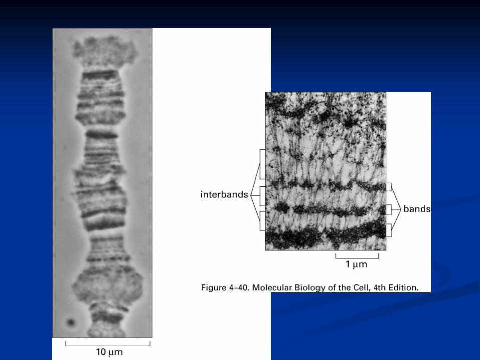

Figure 3. Polytene chromosome map of Anopheles gambiae

Lampbrush Chromosome
It was given this name because it is similar in appearance to the brushes used to clean lamp chimneys in centuries past.
First observed by Flemming in 1882.
The name lampbrush was given by Ruckert in 1892.
These are found in oocytic nuclei of vertebrates (sharks, amphibians, reptiles and birds)as well as in invertebrates (Sagitta, sepia, Ehinaster and several species of insects).
Also found in plants – but most experiments in oocytes.

Lampbrush chromosomes are up to 800 µm long; thus
they provide very favorable material for cytological
studies.
The homologous chromosomes are paired and each has
duplicated to produce two chromatids at the lampbrush
stage.
Each lampbrush chromosome contains a central axial
region, where the two chromatids are highly condensed
Each chromosome has several chromomeres
distributed over its length.
From each chromomere, a pair of loops emerges in the
opposite directions vertical to the main chromosomal
axis.
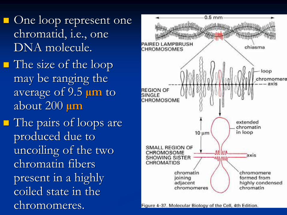
One loop represent one chromatid, i.e., one DNA molecule.
The size of the loop may be ranging the average of 9.5 µm to about 200 µm
The pairs of loops are produced due to uncoiling of the two chromatin fibers present in a highly coiled state in the chromomeres.

One end of each loop is thinner (thin end) than
the other end (thick end).
There is extensive RNA synthesis at the thin end
of the loops, while there is little or no RNA
synthesis at the thick end.

Phase-contrast and fluorescent micrographs of
lampbrush chromosomes

Dosage Compensation
Sex Chromosomes: females XX, males XY
Females have two copies of every X-linked gene;
males have only one.
How is this difference in gene dosage
compensated for? OR
How to create equal amount of X chromosome
gene products in males and females?

Levels of enzymes or proteins encoded by
genes on the X chromosome are the same in
both males and females
Even though males have 1 X chromosome
and females have 2.

G6PD, glucose 6 phosphate dehydrogenase,
gene is carried on the X chromosome
This gene codes for an enzyme that breaks
down sugar
Females produce the same amount of G6PD
enzyme as males
XXY and XXX individuals produce the same
about of G6PD as anyone else
In cells with more than two X chromosomes,
only one X remains genetically active and all the
others become inactivated.
In some cells the paternal allele is expressed
In other cells the maternal allele is expressed
In XXX and XXXX females and XXY males
only 1 X is activated in any given cell the rest are
inactivated


Barr Bodies
1940’s two Canadian scientists noticed a
dark staining mass in the nuclei of cat brain
cells
Found these dark staining spots in female
but not males
This held for cats and humans
They thought the spot was a tightly
condensed X chromosome

Barr bodies represent the inactive X chromosome and
are normally found only in female somatic cells.
Barr Bodies
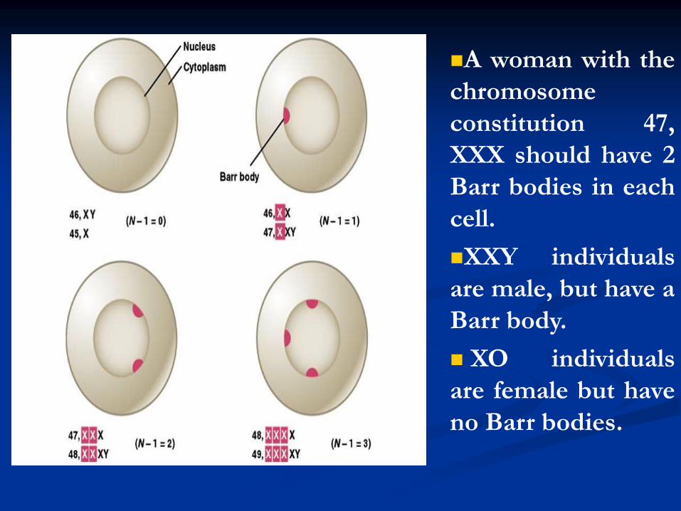
A woman with the
chromosome
constitution 47,
XXX should have 2
Barr bodies in each
cell.
XXY individuals
are male, but have a
Barr body.
XO individuals
are female but have
no Barr bodies.
Which chromosome is inactive is a matter of
chance, but once an X has become inactivated ,
all cells arising from that cell will keep the same
inactive X chromosome.
In the mouse, the inactivation apparently occurs
in early in development
In human embryos, sex chromatin bodies have
been observed by the 16th day of gestation.

Mechanism of X-chromosome Inactivation
A region of the p arm of the X chromosome
near the centromere called the X-inactivation
center (XIC) is the control unit.
This region contains the gene for X-inactive
specific transcript (XIST). This RNA
presumably coats the X chromosome that
expresses it and then DNA methylation locks
the chromosome in the inactive state.

This occurs about 16 days after fertilization in a
female embryo.
The process is independent from cell to cell.
A maternal or paternal X is randomly chosen to be
inactivated.

Rollin Hotchkiss first discovered methylated DNA in 1948.
He found that DNA from certain sources contained, in addition to the standard four bases, a fifth: 5-methyl cytosine.
It took almost three decades to find a role for it.
In the mid-1970s, Harold Weintraub and his colleagues noticed that active genes are low in methyl groups or under methylated.
Therefore, a relationship between under methylation and gene activity seemed likely, as if methylation helped repress genes.


This would be a valuable means of keeping genes
inactive if methylation passed on from parent to
daughter cells during cell division.
Each parental strand retains its methyl groups,
which serve as signals to the methylating
apparatus to place methyl groups on the newly
made progeny strand.
Thus methylation has two of the requirements for
mechanism of determination:
1. It represses gene activity
2. It is permanent.
Strictly speaking, the DNA is altered, since methyl groups are attached, but because methyl cytosine behaves the same as ordinary cytosine, the genetic coding remain same.
A striking example of such a role of methylation is seen in the inactivation of the X chromosome in female mammal.
The inactive X chromosome become heterochromatic and appears as a dark fleck under the microscope – this chromosome said to be lyonized, in honor of Mary Lyon who first postulated the effect in mice.

An obvious explanation is that the DNA in the
lyonized X chromosome is methylated, where as
the DNA in the active, X chromosome is not.
To check this hypothesis Peter Jones and Lawrence
Shapiro grew cells in the presence of drug 5-
azacytosine, which prevents DNA methylation.
This reactivated the lyonized the X chromosome.
Furthermore, Shapiro showed these reactivated
chromosomes could be transferred to other cells
and still remain active.


Reading assignment
Grewal and Moazed (2003) “Heterochromatin
and epigenetic control of gene expression”
Science 301:798
Goldmit and Bergman (2004) “Monoallelic gene
expression: a repertoire of recurrent themes”
Immunol Rev 200:197