Embed Size (px)
Citation preview

Cigarette smoke free radicals and BPDE-DNA adduct Kroum Alexandrov et al.
1
Cigarette smoke free radicals and BPDE-DNA adduct Kroum Alexandrov et al.
2
DNA damage by benzo(a)pyrene in human cells is increased by cigarette
smoke and decreased by a filter containing rosemary extract which lowers
free radicals
Kroum Alexandrov,1 Margarita Rojas,1 and Christian Rolando1
1Université des Sciences et Technologies de Lille (Lille 1), UMR CNRS 8009, Chimie
Organique et Macromoléculaire and IFR 118, Protéomique, Modifications Post-
trductionnelles et Glycobiologie, 59655 Villeneuve d’Ascq Cedex, France
Running Title: Cigarette smoke free radicals and BPDE-DNA adduct
Key Words: Cigarette smoke, free radicals, BPDE-DNA adduct
This work was conducted at the German Cancer Research Center (DKFZ), Division of
Toxicology and Cancer Risk Factors, Heidelberg, Germany with the collaboration of the
Organic and Macromolecular Chemistry Laboratory of the Université des Sciences et
Technologies de Lille (Lille 1), Villeneuve d’Ascq, France.
Requests for reprints: Kroum Alexandrov, Université des Sciences et Technologies de Lille,
Lille 1, UMR CNRS 8009, Chimie Organique et Macromoléculaire, 59655 Villeneuve d’Ascq
Cedex, France. Tel +33 1 60 10 44 97; Fax + 33 3 20 33 61 36; E-mail:
Cigarette smoke free radicals and BPDE-DNA adduct Kroum Alexandrov et al.
3
ABSTRACT
We previously found that the human lung benzo(a)pyrene diol epoxide-dG (BPDE-dG)
adduct concentrate in the target bronchial cells. This adduct now is considered to be critical
event in tumorigenesis by benzo(a)pyrene. In this study we investigate the contribution of
cigarette smoke on the BPDE-dG formation. In a cell free system the amount of (-)-anti-
BPDE-dG adduct increased linearly with concentration of cigarette smoke in the presence of
(+)-BP-7,8-diol. Catalase and superoxide dismutase inhibited its formation by more than 80%.
When MCF-7 cells were treated for 2 hours with the (+)-BP-7,8-diol, cigarette smoke
increased dose dependently the formation of (-)-anti-BPDE-dG and decreased the CYPs
dependent formation of (+)-syn-BPDE the adduct. Then cells were treated for up to one day
with benzo(a)pyrene and then exposed for 2 hours with cigarette smoke. During these 2 hours,
there are twice the increase in the adduct formation in cells treated with cigarette smoke in
comparison to levels in non treated cells due to CYPs activity. Thus, cigarette smoke by
reactive oxygen species which it contains may activate the second step of benzo(a)pyrene
metabolic way leading to the formation of BPDE-dG adduct. Cigarette smoke appears thus
may be in part responsible for the formation of the critical lung tumorigenic adduct. Finally
modified cigarette filter containing rosemary extract decreases by more than 70% of the
BPDE-dG adducts level due to the cigarette smoke in MCF-7 cells. This approach may lead to
decreasing lung cancer risk in addicted smokers.
Cigarette smoke free radicals and BPDE-DNA adduct Kroum Alexandrov et al.
4
INTRODUCTION
Cigarette smoking is causally associated with a large number of human cancers (1).
Tobacco use is by far the most widespread link between exposure to known carcinogens and
death from cancer, and is therefore a model for understanding mechanisms of cancer
induction. Benzo(a)pyrene (BP) is a highly carcinogenic polycyclic aromatic hydrocarbon
(PAH) present in emission exhausts, charbroiled food and in small quantity in cigarette smoke
(2-5), typically less than 10 ng per cigarette (6). BP is one of more than 60 carcinogens in
cigarette smoke that is involved in the aetiology of lung cancer (7). It is metabolically
activated into benzo(a)pyrene-7,8-diol-9,10-epoxide (BPDE) which reacts with DNA
predominantly at the N2-position of guanine to produce primarily N2-guanine lesions, e.g.
benzo(a)pyrene-7,8-diol-9,10-epoxide-N2-deoxyguanosine (BPDE-dG) adduct (8). The
presence of BPDE-DNA adducts in human tissues has been conclusively established (9) and
BPDE-dG adduct concentrated exclusively in bronchial cells and thus implicated in the
initiation of human lung cancer (10). Although considerable evidence implicates BP as
important causative agent of smoking-related cancers, its role is clearly not exclusive.
This carcinogen is metabolized by phase I enzymes to a large number of metabolites
including phenols, arene oxides, quinones, dihydrodiols, and diol epoxides (11). An overview
of BP metabolic way leading to the formation of (+)-anti-BPDE-dG adduct is presented in
Fig. 1.
< Insert Figure 1 here >
The ultimate carcinogen (+)-anti-BPDE is formed from BP by two rounds of cytochrome
P450-mediated oxidation. The first step of this oxidation leads preferentially to (-)-7,8-
dihydro-7,8-dihydrobenzo(a)pyrene [(-)BP -7,8-diol]. The diol is further oxidised primarily to
the highly mutagenic (+)-r-7,t-8-dihydroxy-t-9,10-oxy-7,8,9,10-tetrahydro-BP [(+)-anti-
BPDE] (12). Numerous studies have clearly identified the [(+)-anti-BPDE] as the primary
Cigarette smoke free radicals and BPDE-DNA adduct Kroum Alexandrov et al.
5
carcinogenic metabolite of BP exhibiting enhanced mutagenic activity in vitro and in vivo
(13-17). Most previous studies of genetic variation in metabolism of lung carcinogens have
focused on metabolic activation by various cytochromes P450s, although expression of these
enzymes in lung is generally low (18). The activation of BP-7,8-diol by lung epithelial cells is
not caused solely by classic CYPs/GSTs dependent biotransformation processes, but also
involves several metabolic routes other than CYPs. These include, lipooxygenase, lipid-
peroxidation products and peroxidase-dependent pathways (19-23), COX-1 and COX-2 (24).
Increasing evidence suggests the causal significance of tobacco free radicals in lung
cancer induction in smokers (6, 7, 25-27). Each puff of smoke contains over 10 trillion free
radicals, which may contribute to both tumor initiation as well as promotion (28, 29) of
various forms of human cancer caused by repeated attacks from ROS on cellular
macromolecules. The major free radical species was postulated to be an equilibrium mixture
of semiquinones, hydroquinones and quinones (25). It is suggested that this free radical
complex causes redox cycling that generates superoxide anion from molecular oxygen and
leads to the formation of hydrogen peroxide and hydroxyl radical (27). These reactive species
cause DNA nicking (27) and single-strand breaks in DNA of cultured rodent and human cells
(30, 31). Quinone-associated redox cycling may also be involved in these effects;
hydroquinone and catechol are believed to play a major role.
Our study was designed to (i) determine the relative contribution of ROS in the
cigarette smoke on the activation of BP-7,8-diol in comparison with cytochrome P450; (ii) to
apply the method used in this study to answer the following questions; (iii) does cigarette
smoke’s ROS promote the carcinogenic process by contributing to the metabolism of BP-7,8-
diol resulting in an increase in the formation of the critical lung BPDE-dG? (iv) Can a filter
containing a scavenger of cigarette free radicals affect the formation of BPDE-dG?
Cigarette smoke free radicals and BPDE-DNA adduct Kroum Alexandrov et al.
6
MATERIALS AND METHODS
Chemicals. Proteinase K (EC 3.4.21.64, from Tritirachium album) was purchased
from Sigma (St. Louis, MO), RNase T1 (EC 3.1.21.3, from Aspergillus oryzae) and RNase
(DNase free, heterogeneous mixture of ribonucleases from bovine pancreas) were obtained
from Roche Diagnostics (Meylan, France). Phosphate-buffered saline (PBS) adjusted at pH
7.4 contained 3.0 mM KCl, 1.5 mM KH2HPO4, 140 mM NaCl, 8.0 mM Na2HPO4. HPLC-
grade water, MeOH, diethylether and ethanol (Merck, for spectroscopy grade) were obtained
VWR International (Fontenay-sous-Bois). If not stated otherwise, the others chemicals were
purchased from Sigma (L’Isle d’Abeau Chesnes, France). All BP metabolite standards were
obtained from National Cancer Institute, Chemical Carcinogen Reference Standard
Repository Midwest Research Institute (Kansas City, MO).
Apparatus. High-performance liquid chromatography (HPLC) was carried out with a
Agilent high pressure isocratic and gradient systems (Massy, France) equipped with a
Shimadzu RF-10AXL (Champs-sur-Marne, France) fluorescence detector linked to Agilent
integrator.
Preparation of Cigarette Smoke/PBS Solution (CSS). Smoking was performed
according to Pryor et al (25) without the Cambridge filter. Essentially the same smoke
collecting method has been used earlier by Nakayama et al (30, 31). The smoke from burning
8 cm of one cigarette (Marlboro) during 3.8 min with the help of constant vacuum generated
from a water pump was bubbled through 10 ml of phosphate-buffered saline (PBS) solution
which traps both the gas-phase and tar cigarette smoke chemicals. As there were no water-
insoluble tar compounds present on the walls of the wash bottles, a major part of the water-
soluble compounds from the smoke of a single cigarette was contained in the 10 ml PBS
solution. This aqueous solution named cigarette smoke solution (CSS) was reacted
immediately with exogenous DNA or added to MCF-7 cells in culture in the presence of
Cigarette smoke free radicals and BPDE-DNA adduct Kroum Alexandrov et al.
7
benzo(a)pyrene or its proximate metabolite (+)-BaP-7,8-diol. Different dilutions of CSS were
used (see below).
Incorporation of a Rosemary Powder Extract into the Cigarette Filter. The filter
of the conventional cigarette was removed and 40 mg of a rosemary powder extract kindly
provided by Biosynthec company and prepared according to the Dr Iman Emami, patent
process (32) was introduced in the place liberated by the removal of the filter near to the
cigarette itself. After this operation, the filter was reinstalled. The effect of this filter was
evaluated by mass spectrometry (33). Briefly the cigarette smoke was bubbled in an organic
solution containing 3,3,5,5-tetramethyl-1-pyrroline-N-oxide (TMPO) a spin trap adduct. The
amount of hydroxyl radical adduct was then quantified using liquid chromatography, mass
spectrometry.
Reaction of Exogenous DNA with (+)-BP-7,8-Diol in Presence of Diluted
Cigarette Smoking Solution (CSS). 2 ml calf thymus DNA (3 mg/ml) was added to 5 ml
diluted 20 times CSS and reacted for 2 hours at room temperature with (+)-BP-7,8-diol (final
concentration 3.6 µM) according to the following reaction:
DNA + [(+)-BP-7,8-diol] + CSS → (-)-anti-BPDE-N2-dG
The level of the resulting (-)-anti BPDE-dG adduct was measured (see below). As control an
experiment without CSS was performed.
Cell Culture Conditions and Treatment. The human mammary carcinoma cell line
MCF-7 was grown in 150-cm2 cell culture flasks in a total volume of 20 ml minimal essential
medium E-MEM supplemented with 10% FCS, 15 mM Hepes buffer, and antibiotics (200
units/ml penicillin, 200 µg/ml streptomycin, and 25 µg/ml ampicillin). Cells were maintained
and treated at 37 °C in 5% CO2/95% air atmosphere.
After MCF-7 cells had covered 90% of the surface area of the flasks, (2-3 days after
splitting of a confluent culture), the medium was replaced with 20 ml of fresh medium
Cigarette smoke free radicals and BPDE-DNA adduct Kroum Alexandrov et al.
8
containing 10% serum. Twenty four hours later, near-confluent cells e.g. more than 90% of
cells in G0/G1 phase were treated with DMSO alone or with carcinogen (see below) dissolved
in DMSO and cigarette smoke/PBS (CSS see above). The final concentration of DMSO did
not exceed 0.1% of the total incubation volume. Control samples included in each incubation
set were treated with DMSO alone.
a) Treatment of MCF-7 Cells with (+)-BP-7,8-Diol and Cigarette Smoke. Cells were
treated for 2 hours with (+)-BP-7,8-diol (0.2 µM) alone or in presence of different dilutions of
CSS. The (+)-BaP-7,8-diol was activated by ROO° generated from the CS and cell CYP’s to
form (-)-anti-BPDE-dG and (+)-syn-BPDE-dG respectively. Their levels were measured by
the formation of BP-tetrol I-1 and BP-tetrol II-2 (see bellow ).
b) Time/Dose Exposure Experiments with BP. To characterize time/dose exposure to
BP and BPDE-dG level, cells (10x106 cells /150 cm2 flask, total volume of 20 ml) were
treated with medium containing final concentration of 1.25, 2.5 and 5.0 µM each for 6, 12, 18
and 24 hours (two flasks/dose/time point). BPDE-dG adduct formed in the cells increase
linearly in a dose-and time-dependent manner to as was shown also by others (34). On the
basis of the results obtained we choose 2.5 µM as working concentration for BP.
c) Treatment of MCF-7 Cells with BP and Cigarette Smoke. To see the effect of CS
concentration, the cells were treated with different CSS dilutions (1:79; 1:39; 1:19; 1:9
vol/vol). On the basis of the results obtained from this experience we choose 1:19 (vol/vol)
dilution as working CS concentration. Thus, the cells (see above “Cell Culture and
Treatment”) were treated with BP (2.5 µM) for 12 hours (group A) or 18 hours (group B) then
exposed during the last 2 hours to standard CSS or rosemary filtered CSS (dilution 1:19
vol/vol); cells treated only with BP during 14 hours (respectively 20 hours) were used as
control.
Cigarette smoke free radicals and BPDE-DNA adduct Kroum Alexandrov et al.
9
All incubation sets were repeated 2-3 times with duplicate samples. At the end of
treatment, cells were examined microscopically for morphological changes, then harvested
by trypsinization with 0.05% trypsin-EDTA (0.05% trypsin, 0.14 M NaCl, 3 mM KCl, 0.1 M
Na2HPO4, 1.5 mM K2HPO4, 0.5 mM EDTA). After addition of an equal volume of medium
containing 10% FCS, the cells were centrifuged at 1000 g, washed three times with PBS, and
the cell pellet then stored frozen at –20°C. The viability of the cells treated with BP and
cigarette smoke or (+)-BP-7,8-diol and cigarette smoke was roughly 90% at the time of
harvesting as determined by a Trypan blue exclusion assay. The doses used did not show any
cytotoxicity as measured by lactate dehydrogenase activity assay (ELISA Kit, Boehringer,
Mannheim).
DNA Preparation and Hydrolysis. DNA isolation from MCF-7 cell pellets was
carried out by treatment with RNase, proteinase K, salting procedure (35) and chloroform.
Briefly, the cell pellets were resuspended in EDTA-sodium dodecyl sulfate (SDS) buffer [10
mM Tris buffer, 1 mM Na2EDTA, 1% SDS (w/v), pH 8.0] incubated for 1 h at 37°C with
RNase T1 (2000 U/ml) and RNase A (DNase free; 100 µg/ml) on a shaker (100 rpm). Then
proteinase K (300 µg/ml) was added and the incubation continued overnight at 37°C. After
digestion, 6M NaCl was added to have final concentration of 1M followed by a
centrifugation at 10000 g. DNA in the supernal was precipitated with 2 vol. ethanol, washed
with 70%, 100% ethanol, ether, dried and dissolved in 10 mM Tris buffer. Again, RNase A
(100 µg/ml) and RNase T1 (2000 U/ml) were added and the solution incubated at 37°C for 1
h. followed by proteinase K (100 µg/ml) for another 2 hours at 37°C. The solution was
extracted once with chloroform, centrifuged and the solution was made1M NaCl. DNA was
precipitated with 2 vol. cold ethanol. The portion of DNA to be hydrolyzed was rinsed with
100% ethanol to remove unbound BP-tetrols. The DNA, free of unbound BP-tetrols, was
dissolved in water and the DNA concentration was determined by A260 nm.. The purity was
Cigarette smoke free radicals and BPDE-DNA adduct Kroum Alexandrov et al.
10
ascertained by the ratios at A260/A280 and A260/A230. The amount of DNA for analysis was
hydrolyzed as described previously (36, 37) by incubation at 90°C for 4 hour in a final
concentration of 0.1 N HCl. This releases tetrols from BPDE-DNA adducts with > 90%
recovery (36). The volume of the hydrolysate for injection was made 700 µl containing 5-10
µg DNA.
Determinations of BPDE-N2-dG adduct level. The adduct levels were
determined by HPLC-FD as previously described (36, 37) using r-7,c-9,t-8,t-10-
tetrahydroxy-7,8,9,10-tetrahydrobenzo(a)pyrene (BP-tetrol II-1) as an internal standard
(38). The hydrolysate was loaded onto a precolumn containing 5 µm C18 reverse-phase
material (Nucleosil 100) equilibrated with 10% MeOH and washed for 20 min with 12 ml
10% MeOH. Subsequently, the pre-column was switched by a Valco Instruments
switching valve to flow over a 4.6 mm x 25 cm 5 µm C18 reverse-phase (Nucleosil 100)
analytical column (Alltech, Templemars, France). The products obtained by hydrolysis
were eluted with the following MeOH/H2O gradient: 50%, 0-17 min; 50 to 60%, 17-32
min; 60%, 32-42 min; 60 to 100%, 42-57 min. Retention times of the BP tetrols were: BP
tetrol I-1 (trans-anti-BP-tetrol) (35.2 min); BP-tetrol II-1 (trans-syn-BP-tetrol), internal
standard (36.9 min); BP tetrol II-2 (cis-syn-BP-tetrol) (42.3 min). Fluorescence was
assessed at an excitation wavelength of 344 nm and emission wavelength of 398 nm. As
we did not detect the formation of BP tetrol II-1 in separate analysis of MCF-7 samples,
we used it as internal standard (2 pg added to each HPLC run) for verification of the
relative retention time. The detection limit was 0.5 pg of BP tetrol I-1 and BP-tetrol II-1.
The level of each BP-tetrol was determined by using a standard curve generated from the
fluorescence peak area of authentic BP-tetrol standard analyzed just before the analysis of
MCF-7 samples. The BP-tetrol-I-1 detected is derived after hydrolysis of (+)-anti-BPDE-
DNA adduct. The hydrolysis of (-)-anti-BPDE-dG leads to the formation of BP-tetrol I-2,
Cigarette smoke free radicals and BPDE-DNA adduct Kroum Alexandrov et al.
11
which however is unstable and is converted in BP-tetrol I-1 (39). Thus, the level of the
formed (-)-anti-BPDE-dG was measured by the quantity of BP-tetrol I-1 found on HPLC
runs. Based on the finding that BPDE reacting with DNA produce primarily BPDE-N2-
dG (8), we assumed that BP-tetrol-I-1 level corresponds to this of BPDE-N2-dG. The
level of BPDE bound to MCF-7 DNA was quantified in duplicates. The adduct level was
calculated from the equation 1 pmol/mg DNA / 3.125 = 1 adduct per 106 nucleotide. The
HPLC runs were quantitatively reproducible, and variability between the two assays was
lower than 5%.
RESULTS AND DISCUSSIONS
The mechanism of mutagenesis by BP is sufficiently well defined to be used as a
“molecular signature” to establish the causal nature between particular genetic events in
development of tumors and carcinogenic exposure (the “smoking gun”). The “BP molecular
signature” has major implication for pinpointing the tobacco smoke as the cause of human
lung cancer, and for the elaboration of specific strategies to minimize tobacco smoking, or
introduce preventive measures. Specific agents used in cancer chemoprevention appear to act
by inhibiting carcinogen damage to DNA, mutagenesis, tumor promotion and/or tumor
progression.
This investigation was designed to investigate the relative role of CS on the
biotransformation of BP-7,8-diol to BPDE capable of forming stable DNA adduct in human
cells. Numerous studies have demonstrated that stable PAH-DNA adducts can lead to
mutations through mis-incorporation of nucleotides or deletion (40). CS is an aerosol of
complex chemical composition containing both organic and inorganic compounds, of which
4800 have been identified so far (41). Both vapor phase and particulate phase of smoke are
known to possess free radicals (27, 42). While the gas phase radicals are generally short-lived
Cigarette smoke free radicals and BPDE-DNA adduct Kroum Alexandrov et al.
12
the radicals in the particulate phase are relatively stable and consist of a hydroquinone,
semiquinone, quinone complex (25), this complex is an active redox system capable of
reducing molecular oxygen to produce superoxide, eventually leading to hydrogen peroxide
and hydroxyl radicals. In addition, at least 60 different CS carcinogens have been implicated
in tumor initiation and promotion; the most potent carcinogens agent contained in CS are BP
and NNK (4-(methylnitrosamino)-1-(3-pyridyl)-1-butanone) (7).
The Effect of Cigarette Smoke on (+)-anti-BPDE-dG Using a Cell-Free System
Concomitant with DNA Adduction. To elucidate the mechanism of BPDE-dG formation
dependent from active oxygen generated from cigarette smoke, we looked for this adduct
in cell-free in vitro system concomitant with DNA adduction. The CSS solution
containing the gas-phase and tar cigarette smoke radicals was immediately reacted with
DNA in the presence of (+)-BP-7,8-diol (see protocol above). The results from this
experiment show that CS can oxidize the (+)-BP-7,8 diol to (-)-anti-BPDE which in turn
form the (-)-anti-BPDE-dG adduct. The amount of (-)-anti-BPDE-dG increased linearly
and dose dependently (Fig. 2).
< Insert Figure 2 here >
Previously was found that large amounts of active oxygen such as H2O2 and
O2- were generated from cigarette smoke after trapping the smoke in PBS (31). This
active oxygen generated from cigarette smoke might be responsible for the observed
formation of (-)-anti-BPDE-dG. To verify this, we checked the effect of catalase and
superoxide dismutase (SOD) on the (-)-anti-BPDE-dG produced, and found that the
both enzymes inhibited the formation of the adduct. Inactivated catalase showed no
effect (Table 1). From these results we concluded that cigarette smoke can oxidize (+)-
BP-7,8-diol, thus forming (-)-anti-BPDE-dG, and that such capacity can be explained
mainly by the action of oxygen generated from cigarette smoke.
Cigarette smoke free radicals and BPDE-DNA adduct Kroum Alexandrov et al.
13
The Effect of Cigarette Smoke on (-)-anti-BPDE-dG Adduct Formed in
MCF-7 Cells Treated with (+)-BP-7,8-diol. Two independent pathways have been
shown to participate in the metabolism of BP-7,8-diol to BPDE (Fig. 3) (20-23). The
cytochrome P450 dependent metabolism of the (+)-enantiomer leads preferentially to
(+)-syn-BPDE whereas the pathway involving haem-containing proteins in conjunction
with a peroxide (e.g. lipid peroxide) preferentially results in (-)-anti-BPDE (20). The (-)-
BP-7,8-diol on the other hand, may be metabolized by both pathways and results in the
formation of (+)-anti-BPDE, the ultimate form of BP, and (-)-syn-BPDE (11). The
different pathways can be distinguished by HPLC analysis since the tetrols derived from
anti- and syn-BPDE respectively are clearly separated under our conditions.
< Insert Figure 3 here >
To investigate further the role of cigarette smoke dependent epoxidation of (+)-
BP-7,8-diol leading to the formation of (-)-anti-BPDE that form with DNA (-)-anti-
BPDE-dG adduct, human mammary cell line MCF-7 was used. The reason for which we
used MCF-7 cells to see the effect of cigarette smoke ROS on the activation of (+)-BP-
7, 8 diol was that these cells have little peroxidase activity. The cells were treated with
the (+)-BP-7,8-diol, a stereochemical probe which can distinguish the adducts formed
by ROS and CYPs dependent ways (Fig. 3). Two distinct peaks were observed on the
chromatograms corresponding to BP-tetrol I and BP-tetrol II derived from (-)-anti-
BPDE-dG and (+)-syn-BPDE-dG respectively (36-38). Cigarette smoke increased
linearly and dose dependently the ROS dependent formation of (-)-anti-BPDE-dG (Fig.
4a) and decreased the CYPs dependent formation of (+)-syn-BPDE-dG adduct measured
by the formation of BP-tetrol II. This decrease is also dose dependant and the inverse of
DNA adducts increased linearly with cigarette smoke concentration (Fig. 4b). The
inhibitory effect of CS on CYPs dependent formation of (+)-syn-BPDE-dG and
Cigarette smoke free radicals and BPDE-DNA adduct Kroum Alexandrov et al.
14
increased formation of (-)-anti-BPDE-dG adduct confirm the role of the oxygen
generated from the cigarette smoke in the formation of (-)-anti-BPDE-dG adduct. Other
studies showed that induced CYPs activity was impaired by the oxidative challenge. The
mechanism underlying such a phenomenon could be a down-regulation of cytochrome
P4501A1 gene (36-38, 43, 44). Having little peroxidase activity may lead MCF cells
under “stress” conditions to increased DNA damage and reduced repair capacity.
Consequently this may cause an increase of BPDE-DNA adducts independently from
BP-7,8-diol activation.
< Insert Figure 4 here >
The Effect of Cigarette Smoke on BPDE-dG Adduct Formed in Cells
Treated with BP. Previous studies with MCF-7 cell cultures revealed that these cells
possess inducible P4501B1 and P4501A1 activity (45). The presence of P450 catalyzed
metabolic turnover of BP and the absence of detectable peroxidase activity in MCF-7
cells, allowed the evaluation of the role of cigarette smoke oxygene radicals on BP
activation in human cell cultures. MCF-7 cells have high CYP1A1 enzyme activity for
the metabolic activation of BP leading to the formation of (-)-BP-7,8-diol and
consequently to (+)-anti-BPDE-dG. The level of adduct formation at 6 hours was
considerably lower than that observed after 12 and 24 hours of exposure. After treatment
with 2.5 µM BP for 6 hours approximately 2000 pg adducts per mg DNA were formed,
whereas more than 11000 pg and more than 20000 pg adducts per mg DNA were
present after 12 hours and 24 hours respectively (Wilcoxon Rank Sum Test gives
p=0.0022).
The cells was treated for 12 and 18 hours with BP to induce the formation of (-)-
BP-7,8 diol which is substrate for ROS. Indirect confirmation for the preferentially
formation of (-)-BP-7,8-diol is the absence of BP-tetrol II derived from syn-BPDE on
Cigarette smoke free radicals and BPDE-DNA adduct Kroum Alexandrov et al.
15
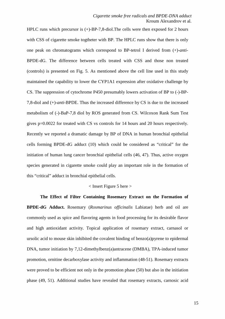
HPLC runs which precursor is (+)-BP-7,8-diol.The cells were then exposed for 2 hours
with CSS of cigarette smoke togtheter with BP. The HPLC runs show that there is only
one peak on chromatograms which correspond to BP-tetrol I derived from (+)-anti-
BPDE-dG. The difference between cells treated with CSS and those non treated
(controls) is presented on Fig. 5. As mentioned above the cell line used in this study
maintained the capability to lower the CYP1A1 expression after oxidative challenge by
CS. The suppression of cytochrome P450 presumably lowers activation of BP to (-)-BP-
7,8-diol and (+)-anti-BPDE. Thus the increased difference by CS is due to the increased
metabolism of (-)-BaP-7,8 diol by ROS generated from CS. Wilcoxon Rank Sum Test
gives p=0.0022 for treated with CS vs controls for 14 hours and 20 hours respectively.
Recently we reported a dramatic damage by BP of DNA in human bronchial epithelial
cells forming BPDE-dG adduct (10) which could be considered as “critical” for the
initiation of human lung cancer bronchial epithelial cells (46, 47). Thus, active oxygen
species generated in cigarette smoke could play an important role in the formation of
this “critical” adduct in bronchial epithelial cells.
< Insert Figure 5 here >
The Effect of Filter Containing Rosemary Extract on the Formation of
BPDE-dG Adduct. Rosemary (Rosmarinus officinalis Labiatae) herb and oil are
commonly used as spice and flavoring agents in food processing for its desirable flavor
and high antioxidant activity. Topical application of rosemary extract, carnasol or
ursolic acid to mouse skin inhibited the covalent binding of benzo(a)pyrene to epidermal
DNA, tumor initiation by 7,12-dimethylbenz(a)antracene (DMBA), TPA-induced tumor
promotion, ornitine decarboxylase activity and inflammation (48-51). Rosemary extracts
were proved to be efficient not only in the promotion phase (50) but also in the initiation
phase (49, 51). Additional studies have revealed that rosemary extracts, carnosic acid
Cigarette smoke free radicals and BPDE-DNA adduct Kroum Alexandrov et al.
16
and carnosol strongly inhibited phase I enzyme, CYP 450 activities and induced the
expression of the phase II enzyme, glutathione S-transferase (GST) and quinone
reductase activities (52-54). Carnosol was stated to inhibit nitric oxide (NO) production
in activated macrophage (55). The antioxidant property had been refereed to as the
mechanistic basis of their protective effects.
With the aim of removing free radicals and reactive oxygen species in the
cigarette smoke a small amount of rosemary powder was incorporated in a standard
filter (see Materials). The decrease in free radicals in the condensate induced by the
filters incorporating a rosemary extract was estimated by the quantitation of the
hydroxyl radical content of CSS with a spin trap (TMPO) using LC-ESI-MS/MS (33).
Under the smoking conditions used a 30% decrease in the hydroxyl radical was
observed. Due to the efficiency of this filter to reduce the level of the free radicals in
cigarette smoke, as compared to a comparable standard Marlboro filter without the
additive, we compared the effect of CS passed through this filter in comparison to the
standard filter on the formation of BPDE-dG using MCF-7 cells.
< Insert Figure 6 here >
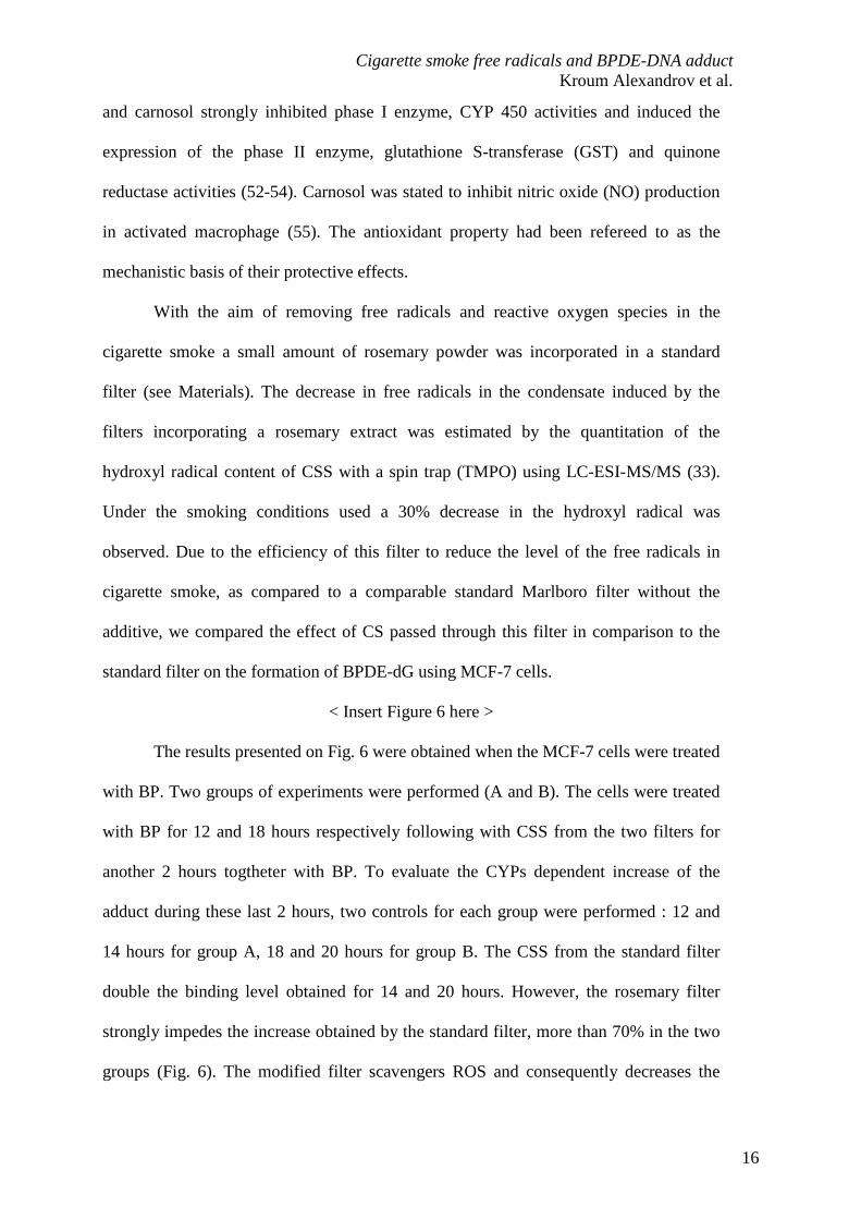
The results presented on Fig. 6 were obtained when the MCF-7 cells were treated
with BP. Two groups of experiments were performed (A and B). The cells were treated
with BP for 12 and 18 hours respectively following with CSS from the two filters for
another 2 hours togtheter with BP. To evaluate the CYPs dependent increase of the
adduct during these last 2 hours, two controls for each group were performed : 12 and
14 hours for group A, 18 and 20 hours for group B. The CSS from the standard filter
double the binding level obtained for 14 and 20 hours. However, the rosemary filter
strongly impedes the increase obtained by the standard filter, more than 70% in the two
groups (Fig. 6). The modified filter scavengers ROS and consequently decreases the
Cigarette smoke free radicals and BPDE-DNA adduct Kroum Alexandrov et al.
17
activation of (-)-BP-7,8-diol. Aside the reduction of the free radicals, rosemary powder
may have also other mechanisms to reduce BPDE-dG formation. Another study using
whole rosemary extract (6 µg.ml-1) inhibited CYP1A1 activity and DNA adduct
formation by 80% after 6 hours co-incubation with 1.5 µM BP in human bronchial
epithelial cells (BEAS-2B) (56). Thus, it is possible using the filters which decrease the
amount of the free radicals to reduce the formation of the critical tumorogenic adduct
which will be in benefit for the addicted smokers. Our study identify rosemary cigarette
filter as promising candidate for chemopreventive programs with the aim to reduce
BPDE-dG adducts in bronchial epithelial cells.
Concluding remarks
In conclusion, this study shows: (1) cigarette smoke can activate by its oxygen
generated radicals the second step of BP metabolic way leading to the formation of
BPDE-dG adduct, presumably by metabolism of the formed in the cells (-)-BP-7,8-diol
to (+)-r-7,t-8-dihydroxy-t-9, 10-oxy-7,8,9,10-tetrahydro-BP [(+)-anti-BPDE]; (2) this
activation is at least twice higher than this obtained with CYPs machinery; (3) ROS
from the cigarette smoke may be in part responsible for the increased BPDE-dG adduct
formation; (4) a filter containing a formulated rosemary powder can reduce considerably
the BPDE-dG level due CS oxygen generated radicals; (5) the methodology in this study
can be use for the search of cigarette filters to reduce the formation of carcinogenic
BPDE-dG adduct in bronchial epithelial cells.
BP is considered to be important carcinogen involved in lung cancer induction in
smokers (7, 10) and, as is shown in this study, reactive oxygen species contribute
substantially in the formation of the critical lung tumorigenic adduct. Whilst it is both
critical to prevent addiction to tobacco and to enhance the efficacy of smoking cessation
Cigarette smoke free radicals and BPDE-DNA adduct Kroum Alexandrov et al.
18
and reduction programs, these approaches have had little impact. The prevention of the
formation of BPDE-dG adduct is one approach which may lead to decreasing lung
cancer risk in addicted smokers.
Acknowledgements
The authors gratefully acknowledged Dr Hans-Rudolf Scherf for the MCF-7 cell culture, Dr
Helmut Bartsch from DKFZ for the support given to this research project and Dr Imam
Emami (Biosynthec, Paris) for providing antioxidant rosemary extract powder. The Mass
Spectrometry facilities used for this study funded by the European community (FEDER), the
Région Nord-Pas de Calais (France), the Réseau National des Génopoles (RNG), the CNRS
and the Université des Sciences et Technologies de Lille.
Cigarette smoke free radicals and BPDE-DNA adduct Kroum Alexandrov et al.
19
References
1. IARC Working Group on the Evaluation of Carcinogenic Risks to Humans. Tobacco
smoke and involuntary smoking. IARC Monogr Eval Carcinog Risks Hum 2004;83:1-1438.
2. Van Duuren BL. Polynuclear aromatic hydrocarbons in cigaret-smoke condensate. II. J
Natl Cancer Inst 1958;21:623-30.
3. Van Duuren BL. Identification of some polynuclear aromatic hydrocarbons in cigaret-
smoke condensate. J Natl Cancer Inst 1958;21:1-16.
4. Wynder EL, Hoffmann D. Tobacco carcinogenesis. VII. The role of higher polycyclic
hydrocarbons. Cancer 1959;12:1079-86.
5. IARC Working Group on the Evaluation of Carcinogenic Risks to Humans.
Polynuclear aromatic compounds, Part 1, Chemical, environmental and experimental data.
IARC Monogr Eval Carcinog Risk Chem Hum 1983;32:1-453.
6. Hecht SS. Tobacco carcinogens, their biomarkers and tobacco-induced cancer. Nat
Rev Cancer 2003;3(10):733-44.
7. Hecht SS. Tobacco smoke carcinogens and lung cancer. J Natl Cancer Inst
1999;91(14):1194-210.
8. Osborne MR, Beland FA, Harvey RG, Brookes P. The reaction of (+-)-7a,8b-
dihydroxy-9b,10b-epoxy-7,8,9,10-tetrahydrobenzo[a]pyrene with DNA. Int J Cancer
1976;18(3):362-8.
9. Boysen G, Hecht SS. Analysis of DNA and protein adducts of benzo[a]pyrene in
human tissues using structure-specific methods. Mutat Res 2003;543(1):17-30.
10. Rojas M, Marie B, Vignaud JM, Martinet N, Siat J, Grosdidier G, et al. High DNA
damage by benzo[a]pyrene 7,8-diol-9,10-epoxide in bronchial epithelial cells from patients
with lung cancer: comparison with lung parenchyma. Cancer Lett (Amsterdam, Neth)
2004;207(2):157-63.
Cigarette smoke free radicals and BPDE-DNA adduct Kroum Alexandrov et al.
20
11. Gelboin HV. Benzo[a]pyrene metabolism, activation, and carcinogenesis: role and
regulation of mixed-function oxidases and related enzymes. Physiol Rev 1980;60(4):1107-66.
12. Levin W, Wood A, Chang R, Ryan D, Thomas P, Yagi H, et al. Oxidative metabolism
of polycyclic aromatic hydrocarbons to ultimate carcinogens. Drug Metab Rev
1982;13(4):555-80.
13. Borgen A, Darvey H, Castagnoli N, Crocker TT, Rasmussen RE, Wang IY. Metabolic
conversion of benzo[a]pyrene by Syrian hamster liver microsomes and binding of metabolites
to deoxyribonucleic acid. J Med Chem 1973;16(5):502-6.
14. Huberman E, Sachs L, Yang SK, Gelboin HV. Identification of mutagenic metabolites
of benzo[a]pyrene in mammalian cells. Proc Natl Acad Sci U S A 1976;73(2):607-11.
15. Newbold RF, Brookes P. Exceptional mutagenicity of a benzo[a]pyrene diol epoxide
in cultured mammalian cells. Nature (London, U K) 1976;261(5555):52-4.
16. Slaga TJ, Viaje A, Berry DL, Bracken W, Buty SG, Scribner JD. Skin tumor initiating
ability of benzo(a)pyrene 4,5- 7,8- and 7,8-diol-9,10-epoxides and 7,8-diol. Cancer Lett
(Shannon, Irel) 1976;2(2):115-21.
17. Yang SK, McCourt DW, Leutz JC, Gelboin HV. Benzo[a]pyrene diol epoxides:
mechanism of enzymic formation and optically active intermediates. Science (Washington,
DC, U S) 1977;196(4295):1199-201.
18. Ding X, Kaminsky LS. Human extrahepatic cytochromes P450: Function in xenobiotic
metabolism and tissue-selective chemical toxicity in the respiratory and gastrointestinal tracts.
Annu Rev Pharmacol Toxicol 2003;43:149-73.
19. Borm PJ, Knaapen AM, Schins RP, Godschalk RW, Schooten FJ. Neutrophils amplify
the formation of DNA adducts by benzo[a]pyrene in lung target cells. Environ Health
Perspect 1997;105 Suppl 5:1089-93.
Cigarette smoke free radicals and BPDE-DNA adduct Kroum Alexandrov et al.
21
20. Marnett LJ. Prostaglandin synthase-mediated metabolism of carcinogens and a
potential role for peroxyl radicals as reactive intermediates. Environ Health Perspect
1990;88:5-12.
21. Pruess-Schwartz D, Nimesheim A, Marnett LJ. Peroxyl radical- and cytochrome P-
450-dependent metabolic activation of (+)-7,8-dihydroxy-7,8-dihydrobenzo(a)pyrene in
mouse skin in vitro and in vivo. Cancer Res 1989;49(7):1732-7.
22. Trush MA, Kensler TW. Role of free radicals in carcinogen activation. Oxid Stress
1991:277-318.
23. Trush MA, Kensler TW. An overview of the relationship between oxidative stress and
chemical carcinogenesis. Free Radic Biol Med 1991;10(3-4):201-9.
24. Wiese FW, Thompson PA, Kadlubar FF. Carcinogen substrate specificity of human
COX-1 and COX-2. Carcinogenesis 2001;22(1):5-10.
25. Pryor WA. Cigaret smoke radicals and the role of free radicals in chemical
carcinogenicity. Environmental Health Perspectives Supplements 1997;105(4):875-82.
26. Pryor WA, Hales BJ, Premovic PI, Church DF. The radicals in cigarette tar: their
nature and suggested physiological implications. Science 1983;220(4595):425-7.
27. Pryor WA, Stone K, Zang L-Y, Bermudez E. Fractionation of Aqueous Cigarette Tar
Extracts: Fractions That Contain the Tar Radical Cause DNA Damage. Chem Res Toxicol
1998;11(5):441-8.
28. Cerutti PA. Prooxidant states and tumor promotion. Science 1985;227(4685):375-81.
29. Kozumbo WJ, Trush MA, Kensler TW. Are free radicals involved in tumor
promotion? Chem-Biol Interact 1985;54(2):199-207.
30. Nakayama T, Kaneko M, Kodama M, Nagata C. Cigarette smoke induces DNA
single-strand breaks in human cells. Nature (London, U K) 1985;314(6010):462-4.
Cigarette smoke free radicals and BPDE-DNA adduct Kroum Alexandrov et al.
22
31. Nakayama T, Kodama M, Nagata C. Generation of hydrogen peroxide and superoxide
anion radical from cigarette smoke. Gann 1984;75(2):95-8.
32. Emami I, inventor; Francaise d'Aromes et Parfums, assignee. Utilisation de composés
polyphénoliques ou de leur dérivés comme capteurs de radicaux libres dans les filtres de
cigarette [Use of polyphenol compounds or their derivatives as scavengers of free radicals in
cigarette filters]. FR patent 2772561 1997 Dec 24.
33. Masselot D. Identification et quantification des radicaux libres par spectrométrie de
masse. Application à l'étude et à la dimminution des radicaux libres contenus dans les fumées
de cigarette [Identification and quantitation of free radicals by mass spectrometry. Application
to the study and to the decrease of free radicals contained in cigarette smoke] [Dissertation]:
Université des Sciences et Technologies de Lille (Lille 1); 2004.
34. Kuljukka-Rabb T, Peltonen K, Isotalo S, Mikkonen S, Rantanen L, Savela K. Time-
and dose-dependent DNA binding of PAHs derived from diesel particle extracts,
benzo[a]pyrene and 5-methylchrysene in a human mammary carcinoma cell line (MCF-7).
Mutagenesis 2001;16(4):353-8.
35. Miller SA, Dykes DD, Polesky HF. A simple salting out procedure for extracting
DNA from human nucleated cells. Nucleic Acids Res 1988;16(3):1215.
36. Alexandrov K, Rojas M, Geneste O, Castegnaro M, Camus AM, Petruzzelli S, et al.
An improved fluorometric assay for dosimetry of benzo[a]pyrenediol epoxide-DNA adducts
in smokers' lung: comparisons with total bulky adducts and aryl hydrocarbon hydroxylase
activity. Cancer Res 1992;52(22):6248-53.
37. Rojas M, Alexandrov K, van Schooten FJ, Hillebrand M, Kriek E, Bartsch H.
Validation of a new fluorometric assay for benzo[a]pyrenediol epoxide-DNA adducts in
human white blood cells: comparisons with 32P-postlabeling and ELISA. Carcinogenesis
1994;15(3):557-60.
Cigarette smoke free radicals and BPDE-DNA adduct Kroum Alexandrov et al.
23
38. Rojas M, Godschalk R, Alexandrov K, Cascorbi I, Kriek E, Ostertag J, et al.
Myeloperoxidase - 463A variant reduces benzo[a]pyrene diol epoxide DNA adducts in skin of
coal tar treated patients. Carcinogenesis 2001;22(7):1015-8.
39. Jansen EHJM, van den Berg RH, Kroese ED. Liquid chromatographic analysis and
stability of benzo[a]pyrene-tetrols in blood protein adducts in rats after exposure to
benzo[a]pyrene. Anal Chim Acta 1994;290(1-2):86-93.
40. Loechler EL. The role of adduct site-specific mutagenesis is understanding how
carcinogen - DNA adducts cause mutations: perspective, prospects and problems.
Carcinogenesis 1996;17(5):895-902.
41. Rodgman A, Smith CJ, Perfetti TA. The composition of cigarette smoke: A
retrospective, with emphasis on polycyclic components. Hum Exp Toxicol 2000;19(10):573-
95.
42. Shinagawa K, Tokimoto T, Shirane K. Spin trapping of nitric oxide in aqueous
solutions of cigarette smoke. Biochem Biophys Res Commun 1998;253(1):99-103.
43. Barouki R, Morel Y. Repression of cytochrome P450 1A1 gene expression by
oxidative stress: mechanisms and biological implications. Biochem Pharmacol
2001;61(5):511-6.
44. Bassi AM, Ledda S, Penco S, Menini S, Muzio G, Canuto R, et al. Changes of
CYP1A1, GST, and ALDH3 enzymes in hepatoma cell lines undergoing enhanced lipid
peroxidation. Free Radic Biol Med 2000;29(11):1186-96.
45. Spink DC, Spink BC, Cao JQ, DePasquale JA, Pentecost BT, Fasco MJ, et al.
Differential expression of CYP1A1 and CYP1B1 in human breast epithelial cells and breast
tumor cells. Carcinogenesis 1998;19(2):291-8.
Cigarette smoke free radicals and BPDE-DNA adduct Kroum Alexandrov et al.
24
46. Hainaut P, Pfeifer GP. Patterns of p53 G->T transversions in lung cancers reflect the
primary mutagenic signature of DNA-damage by tobacco smoke. Carcinogenesis
2001;22(3):367-74.
47. Pfeifer GP, Denissenko MF, Olivier M, Tretyakova N, Hecht SS, Hainaut P. Tobacco
smoke carcinogens, DNA damage and p53 mutations in smoking-associated cancers.
Oncogene 2002;21(48):7435-51.
48. Tokuda H, Ohigashi H, Koshimizu K, Ito Y. Inhibitory effects of ursolic and oleanolic
acid on skin tumor promotion by 12-O-tetradecanoylphorbol 13-acetate. Cancer Lett
(Shannon, Irel) 1986;33(3):279-85.
49. Singletary KW, Nelshoppen JM. Inhibition of 7,12-dimethylbenz[a]anthracene
(DMBA)-induced mammary tumorigenesis and of in vivo formation of mammary DMBA-
DNA adducts by rosemary extract. Cancer Lett (Shannon, Irel) 1991;60(2):169-75.
50. Huang MT, Ho CT, Wang ZY, Ferraro T, Lou YR, Stauber K, et al. Inhibition of skin
tumorigenesis by rosemary and its constituents carnosol and ursolic acid. Cancer Res
1994;54(3):701-8.
51. Singletary K, MacDonald C, Wallig M. Inhibition by rosemary and carnosol of 7,12-
dimethylbenz[a]anthracene (DMBA)-induced rat mammary tumorigenesis and in vivo
DMBA-DNA adduct formation. Cancer Lett (Shannon, Irel) 1996;104(1):43-8.
52. Debersac P, Heydel JM, Amiot MJ, Goudonnet H, Artur Y, Suschetet M, et al.
Induction of cytochrome P450 and/or detoxication enzymes by various extracts of rosemary:
description of specific patterns. Food Chem Toxicol 2001;39(9):907-18.
53. Debersac P, Vernevaut MF, Amiot MJ, Suschetet M, Siess MH. Effects of a water-
soluble extract of rosemary and its purified component rosmarinic acid on xenobiotic-
metabolizing enzymes in rat liver. Food Chem Toxicol 2001;39(2):109-17.
Cigarette smoke free radicals and BPDE-DNA adduct Kroum Alexandrov et al.
25
54. Offord EA, Mace K, Avanti O, Pfeifer AMA. Mechanisms involved in the
chemoprotective effects of rosemary extract studied in human liver and bronchial cells.
Cancer Lett (Shannon, Irel) 1997;114(1,2):275-81.
55. Chan MM-Y, Ho C-T, Huang H-I. Effects of three dietary phytochemicals from tea,
rosemary and turmeric on inflammation-induced nitrite production. Cancer Lett (Shannon,
Irel) 1995;96(1):23-9.
56. Offord EA, Mace K, Ruffeux C, Malnoe A, Pfeifer AMA. Rosemary components
inhibit benzo[a]pyrene-induced genotoxicity in human bronchial cells. Carcinogenesis
1995;16(9):2057-62.
Cigarette smoke free radicals and BPDE-DNA adduct Kroum Alexandrov et al.
26
Abbreviations:
BP: benzo(a)pyrene; (-)-BP-7,8-diol: (-)-trans-7,8-dihydroxy-7,8-
dihydrobenzo(a)pyrene; (+)-anti-BPDE: (+)-r-7,t-8-dihydroxy-t-9,10-oxy-7,8,9,10-
tetrahydro-BP; (+)-syn-BPDE: (+)-r-7,t-8-dihydroxy-c-9,10-oxy-7,8,9,10-tetrahydro-
BP; BP-tetrol I-1 or trans-anti-BP tetrol: r-7,c-10,t-8,t-9-tetrahydroxy-7,8,9,10-
tetrahydro -BP; BP- tetrol I-2,r-7: t-8,t-9-t-10-tetrahydroxy-7,8,9,10-tetrahydro-BP;
BP-tetrol II-1 or trans-syn-BP tetrol: r-7,c-9,t-8,t-10-tetrahydroxy-7,8,9,10-tetrahydro-
BP; BP-tetrol II-2 or cis-syn-BP tetrol: r-7,c-9,c-10,t-8,-tetrahydroxy-7,8,9,10-
tetrahydro-BP; (+)-anti-BPDE-dG: (+)-r-7,t-8-dihydroxy-t-9,10-oxy-7,8,9,10-
tetrahydro-BP-N2-deoxyguanosine; CS: cigarette smoke; CSS: cigarette smoke
solution; HPLC: high-performance liquid chromatography; MS: mass spectrometry;
ESI: electrospay ionization; TMPO: 3,3,5,5-tetramethyl-1-pyrroline-N-oxide.

Cigarette smoke free radicals and BPDE-DNA adduct Kroum Alexandrov et al.
27
Descriptions of the figures
Figure 1. Principal metabolic pathway and DNA binding of the carcinogen benzo(a)pyrene.
Benzo(a)pyrene is a tobacco carcinogen that may be converted in vivo enzymatically or by
oxygen reactive species to yield DNA-reactive dihydrodiol epoxides. Stereoselective
generation of the mutagenic (+)-r-7,t-8-dihydroxy-t-9,10-oxy-7,8,9,10-tetrahydro-BP [(+)-
anti-BPDE] from (-)-BP-7,8-dihydrodiol is catalyzed by cytochrome-P450-dependent
monooxygenases (P450) or reactive oxygen species. Subsequent reaction of this electrophilic
intermediate with genomic DNA produce stable adduct between dihydrodiol epoxide and the
exocyclic amino group of guanosine. This kind of DNA lesion may be converted into
mutations within the following replication cycle unless repair of this adduct is produced.
Figure 2. Results obtained by using a cell-free system concomitant with DNA adduction: 6
mg calf thymus DNA in 2 ml water was added to 5 ml CSS with different dilutions and
reacted for 2 hours at room temperature with (+)-BP-7,8-diol (final concentration 3.6 µM).
DNA was hydrolyzed and the released BP-tetrol I were measured as outlined in Materials and
Methods.
Figure 3. (a) Stereochemistry of BP-7,8-diol epoxidation by peroxyl radicals and cytochrome
P450 to reactive species (anti-BPDE and syn-BPDE) that can bind to DNA, and (b) acid
hydrolysis of DNA to BP-tetrols measured in this study. The hydrolysis of (-)-anti-BPDE-dG
and (-)-syn-BPDE leads to the formation of BP-tetrol I-2 and BP-tetrol II-1 which however
are unstable and are converted into BP-tetrol I-1and BP-tetrol II-2.
Cigarette smoke free radicals and BPDE-DNA adduct Kroum Alexandrov et al.
28
Figure 4. Results obtained using MFC-7 cells. 10 x 106 cells/150 cm2 flask in a total volume
of 20 ml were treated for 2 hrs with (+)-BP-7,8-diol (0.2 µM) alone or in presence of different
dilutions of CSS. DNA was isolated, hydrolyzed and the released BP-tetrols were measured
and the binding levels determined as outlined in Materials and Methods. Two distinct peaks
were observed on the chromatograms corresponding to BP-tetrol I and BP-tetrol II derived
from (-)-anti-BPDE-dG and (+)-syn-BPDE-dG respectively (refs.32-34). Values represent the
means plus Std of two independent experiments with 3-4 HPLC runs. Figure 4a, upper panel,
increases of (-)-anti-BPDE-dG adducts with CSS dilution; Figure 4b, lower panel, increases
of 1/(+)-syn-BPDE-dG adduct with CSS dilution.
Figure 5. BPDE-dG binding in MCF-7 cells after exposure to BP 2.5 µM or BP 2.5 µM + CS
solution (dilution 20 times) for the time indicated. Cigarette smoke solution was added the last
2 hours during the exposure to BP. Analysis of BPDE-dG was performed as described in
Materials and Methods. Values represent the means of two independent experiments with 4-6
HPLC runs plus Standard. The BPDE-dG value were 11.7 ± 0.5 (mean ± s.d.) µg of adducts
per mg DNA after 12 hours of incubation and 17.6 ± 0.4, 26.1 ± 0.9 after 18 hours and 24
hours respectively. The CYP spontaneous metabolism increased theses values to 17.2 ± 0.5,
27.8 ± 0.8 and 42.2 ± 1.0 two hours after the reference time at 12, 18 and 24 hours
respectively. The addition of cigarette smoke solution (CSS) induced a much more dramatic
change during the same two hours period leading to a final BPDE-dG value of 36.9 ± 1.2,
56.7 ± 0.9 and 80.2 ± 1.2 (mean ± s.d.) µg of adducts per mg after 12, 18 and 24 hours
respectively.
Figure 6. BPDE-dG binding in MCF-7 cells after exposure to BP 2.5 µM or BP 2.5 µM + CS
solution obtained from standard filter and filter containing rosemary extract. The cigarette
Cigarette smoke free radicals and BPDE-DNA adduct Kroum Alexandrov et al.
29
smoke solution was added for the last 2 hours during the exposure to BP for the time
indicated. Analysis of BPDE-dG was performed as described in Materials and Methods. The
HPLC runs show that there is only one peak on chromatograms which correspond to BP-tetrol
I derived from (+)-anti-BPDE-dG. Values represent the means of two independent
experiments with 4-6 HPLC runs plus Std.

Cigarette smoke free radicals and BPDE-DNA adduct Kroum Alexandrov et al.
30
Table 1. Effect of scavengers on BPDE-dG level caused by cigarette smoke in a cell-free
system
_____________________________________________________________________
% BPDE-dG relative
Treatment to standard CS
____________________________________________________________
Standard CSS 100
+ Superoxide Dismutase SOD (20 µg) 16
+ Catalase (4 µg) 12
+ Inactivated Catalase (4µg) 100
Romarin filtered CSS instead Standard CSS 42
______________________________________________________________________
The standard CSS system includes 2 ml calf thymus DNA (3 mg/ml), 600 µl 30 µM (+) BaP-
7,8-diol and 5 ml of diluted CSS with PBS (1:19) at pH 7.4, and was incubated at room
temperature fir 2h. Each value was obtained from three independent experiments performed in
duplicate. The average error was about 12% in each duplicate experiment. The BPDE-dG
value of the standard was 56 ± 6.3 (mean ± s.d.) adducts per mg DNA.