Embed Size (px)
Citation preview

Cigna Medical Coverage Policies – Radiology Abdomen Imaging
Effective July 31, 2020
____________________________________________________________________________________ Instructions for use The following coverage policy applies to health benefit plans administered by Cigna. Coverage policies are intended to provide guidance in interpreting certain standard Cigna benefit plans and are used by medical directors and other health care professionals in making medical necessity and other coverage determinations. Please note the terms of a customer’s particular benefit plan document may differ significantly from the standard benefit plans upon which these coverage policies are based. For example, a customer’s benefit plan document may contain a specific exclusion related to a topic addressed in a coverage policy.
In the event of a conflict, a customer’s benefit plan document always supersedes the information in the coverage policy. In the absence of federal or state coverage mandates, benefits are ultimately determined by the terms of the applicable benefit plan document. Coverage determinations in each specific instance require consideration of:
1. The terms of the applicable benefit plan document in effect on the date of service2. Any applicable laws and regulations3. Any relevant collateral source materials including coverage policies4. The specific facts of the particular situation
Coverage policies relate exclusively to the administration of health benefit plans. Coverage policies are not recommendations for treatment and should never be used as treatment guidelines.
This evidence-based medical coverage policy has been developed by eviCore, Inc. Some information in this coverage policy may not apply to all benefit plans administered by Cigna.
These guidelines include procedures eviCore does not review for Cigna. Please refer to the Cigna CPT code list for the current list of high-tech imaging procedures that eviCore reviews for Cigna.
CPT® (Current Procedural Terminology) is a registered trademark of the American Medical Association (AMA). CPT® five digit codes, nomenclature and other data are copyright 2017 American Medical Association. All Rights Reserved. No fee schedules, basic units, relative values or related listings are included in the CPT® book. AMA does not directly or indirectly practice medicine or dispense medical services. AMA assumes no liability for the data contained herein or not contained herein.

Abdomen Imaging Guidelines Abbreviations for Abdomen Imaging Guidelines 4AB-1: General Guidelines 6AB-2: Abdominal Pain 12AB-3: Abdominal Sepsis (Suspected Abdominal Abscess) 22AB-4: Flank Pain, Rule Out or Known Renal/Ureteral Stone 24AB-5: Gastroenteritis 28AB-6: Mesenteric/Colonic Ischemia 30AB-7: Post-Operative Pain Within 60 Days Following Abdominal Surgery – Abdominal Procedure 34AB-8: Abdominal Lymphadenopathy 36AB-9: Bariatric Surgery 40AB-10: Blunt Abdominal Trauma 42AB-11: Gaucher Disease and Hemochromatosis 44AB-12: Hernias 48AB-13: Abdominal Mass 51AB-14: Lower Extremity Edema 54AB-15: Zollinger-Ellison Syndrome (ZES-Gastrinoma) 55AB-16: Adrenal Cortical Lesions 57AB-17: Abdominal Aortic Aneurysm (AAA), Iliac Artery Aneurysm (IAA), and Visceral Artery Aneurysms Follow-Up of Known Aneurysms and Pre-Op Evaluation 65AB-18: Abdominal Aortic Aneurysm (AAA) and Iliac Artery Aneurysm (IAA)-Post Endovascular or Open Aortic Repair 67AB-19: Aortic Dissection and Imaging for Other Aortic Conditions 69AB-20: Bowel Obstruction and Gastroparesis 71AB-21: Diarrhea, Constipation, and Irritable Bowel 76AB-22: GI Bleeding 81AB-23: Inflammatory Bowel Disease Rule Out Crohn’s Disease or Ulcerative Colitis 84AB-24: Celiac Disease (Sprue) 88AB-25: CT Colonography (CTC) 90AB-26: Cirrhosis and Liver Screening for Hepatocellular Carcinoma (HCC); Ascites and Portal Hypertension 93AB-27: MR Cholangiopancreatography (MRCP) 99AB-28: Gallbladder Polyps 102AB-29: Liver Lesion Characterization 104AB-30: Abnormal Liver Chemistries 111
Page 2 of 164
Abdomen Imaging Guidelines V2.0
______________________________________________________________________________________________________ ©2020 eviCore healthcare. All Rights Reserved. 400 Buckwalter Place Boulevard, Bluffton, SC 29910 (800) 918-8924 www.eviCore.com

Abd
omen
Imag
ing
AB-31: Pancreatic Lesion 114 AB-32: Pancreatic Pseudocysts 119 AB-33: Pancreatitis 121 AB-34: Spleen 127 AB-35: Indeterminate Renal Lesion 130 AB-36: Renal Failure 135 AB-37: Renovascular Hypertension 137 AB-38: Polycystic Kidney Disease 139 AB-39: Hematuria and Hydronephrosis 141 AB-40: Urinary Tract Infection (UTI) 144 AB-41: Patent Urachus 147 AB-42: Transplant 149 AB-43: Hepatic and Abdominal Arteries 156 AB-44: This section intentionally left blank 159 AB-45: Liver Elastography 160 AB-46:Hiccups 162 AB-47: Retroperitoneal Fibrosis 163
Page 3 of 164
Abdomen Imaging Guidelines V2.0
______________________________________________________________________________________________________ ©2020 eviCore healthcare. All Rights Reserved. 400 Buckwalter Place Boulevard, Bluffton, SC 29910 (800) 918-8924 www.eviCore.com

Abd
omen
Imag
ing
Abbreviations for Abdomen Imaging Guidelines AAA abdominal aortic aneurysm
AASLD American Association for the Study of Liver Diseases
ACE angiotensin-converting enzyme
ACG American College of Gastroenterology
ACR American College of Radiology
ACTH adrenocorticotropic hormone
AFP alpha-fetoprotein
AGA American Gastroenterological Association
ALT alanine aminotransferase
ASGE American Society for Gastrointestinal Endoscopy
AST aspartate aminotransferase
AUA American Urological Association
BEIR Biological Effects of Ionizing Radiation
BUN blood urea nitrogen
CAG Canadian Association of Gastroenterology
CNS central nervous system
CT computed tomography
CTA computed tomography angiography
CTC computed tomography colonography (aka: virtual colonoscopy)
DVT deep vein thrombosis
ERCP endoscopic retrograde cholangiopancreatography
FNH focal nodular hyperplasia
GFR glomerular filtration rate
GGT gamma glutamyltransferase
GI gastrointestinal
HCC hepatocellular carcinoma
HCPCS Healthcare Common Procedural Coding System (commonly pronounced: “hix pix”)
HU Hounsfield units
Page 4 of 164
Abdomen Imaging Guidelines V2.0
______________________________________________________________________________________________________ ©2020 eviCore healthcare. All Rights Reserved. 400 Buckwalter Place Boulevard, Bluffton, SC 29910 (800) 918-8924 www.eviCore.com

Abd
omen
Imag
ing
IAA iliac artery aneurysm
IV intravenous
KUB kidneys, ureters, bladder (plain frontal supine abdominal radiograph)
LFT liver function tests
MRCP magnetic resonance cholangiopancreatography
MRA magnetic resonance angiography
MRI magnetic resonance imaging
mSv millisievert
NAFLD nonalcoholic fatty liver disease
PA posteroanterior projection
PET positron emission tomography
RAS renal artery stenosis
RBC red blood cell
SBFT small bowel follow through
SPECT single photon emission computed tomography
VC virtual colonoscopy (CT colonography)
PFT pulmonary function tests
WBC white blood cell
ZES Zollinger-Ellison Syndrome
Page 5 of 164
Abdomen Imaging Guidelines V2.0
______________________________________________________________________________________________________ ©2020 eviCore healthcare. All Rights Reserved. 400 Buckwalter Place Boulevard, Bluffton, SC 29910 (800) 918-8924 www.eviCore.com

AB-1: General Guidelines AB-1.1: Overview 7 AB-1.2: CT Imaging 7 AB-1.3: MR Imaging 8 AB-1.4: MR Enterography and Enteroclysis Coding Notes 8 AB-1.5: Ultrasound 9 AB-1.6: Abdominal Ultrasound 9 AB-1.7: Retroperitoneal Ultrasound 10 AB-1.8: This section intentionally left blank 10 AB-1.9: Contrast-Enhanced Ultrasound 10 AB-1.10: This section intentionally left blank 10 AB-1.11: RADCAT Grading System 11
Page 6 of 164
Abdomen Imaging Guidelines V2.0
______________________________________________________________________________________________________ ©2020 eviCore healthcare. All Rights Reserved. 400 Buckwalter Place Boulevard, Bluffton, SC 29910 (800) 918-8924 www.eviCore.com

Abd
omen
Imag
ing
AB-1.1: Overview A current clinical evaluation (within 60 days) is generally required before advanced
imaging can be considered. The clinical evaluation may include a relevant history and physical examination, appropriate laboratory studies, and non-advanced imaging modalities such as plain X-ray or ultrasound. Other meaningful contact (telephone call, electronic mail or messaging) by an established individual can substitute for a face-to-face clinical evaluation.
GI Specialist evaluations can be helpful, particularly in determining mesenteric/colonic ischemia, diarrhea/constipation, irritable bowel syndrome (IBS), or need for MRCP.
Abdominal imaging begins at the diaphragm and extends to the umbilicus or iliac crest.
Pelvic imaging begins at the iliac crest and extends to the pubis. Clinical concerns at the dividing line can be providers’ choice (abdomen and pelvis;
abdomen or pelvis).
AB-1.2: CT Imaging CT imaging is a more generalized modality. CT Abdomen is usually performed with
contrast (CPT® 74160): Oral contrast has no relation to the IV contrast administered. Coding for contrast
only refers to IV contrast. There is no coding for oral contrast. Exceptions are noted in these guidelines, and include:
CT Abdomen with contrast (CPT® 74160) or without and with contrast (CPT®
74170) with suspicion of a solid organ lesion (liver, kidney, pancreas, spleen). CT Abdomen without contrast (CPT® 74150) or CT Abdomen and Pelvis
without contrast (CPT® 74176) if there is renal insufficiency/failure, or a documented allergy to contrast. It can also be considered for diabetics or the very elderly.
Shellfish allergy: It is commonly assumed that an allergy to shellfish infers iodine allergy, and
that this implies an allergy to CT iodinated contrast media. However, this is NOT true. Shellfish allergy is due to tropomyosins. Iodine plays no role in these allergic reactions. Allergies to shellfish do not increase the risk of reaction to IV contrast any more than that of other allergens.
CT Abdomen and Pelvis, usually with contrast (CPT® 74177), should be considered when signs or symptoms are generalized, or involve a lower quadrant of the abdomen.
CT Enterography (CPT® 74177) combines CT imaging with large volumes of ingested neutral bowel contrast material to allow visualization of the small bowel.
CT Enteroclysis A tube is placed through the nose or mouth and advanced into the duodenum
or jejunum. Bowel contrast material is infused through the tube and CT imaging is performed either with or without intravenous contrast.
Page 7 of 164
Abdomen Imaging Guidelines V2.0
______________________________________________________________________________________________________ ©2020 eviCore healthcare. All Rights Reserved. 400 Buckwalter Place Boulevard, Bluffton, SC 29910 (800) 918-8924 www.eviCore.com

Abd
omen
Imag
ing
CT Enteroclysis is used to allow visualization of the small bowel wall and lumen. CT Enteroclysis may allow better or more consistent distention of the small bowel than CT Enterography.
Report by assigning: CPT® 74176 or CPT® 74177 Triple-phase CT
3 phases of a triple-phase CT are: 1) Hepatic arterial phase, 2) Portal venous phase, and 3) Washout or delayed acquisitions phase.
It should be noted that, in general, a precontrast or noncontrast CT is usually not needed, in a standard triple-phase CT, except in those individuals previously treated with locoregional embolic or ablative therapies. Thus, for the evaluation of liver lesions EITHER a CT Abdomen with contrast (CPT®
74160) or CT Abdomen without and with contrast (CPT® 74170) can be approved. This is in contradistinction to MRI, in which precontrast imaging is needed.
AB-1.3: MR Imaging MRI may be preferred as a more targeted study in cases of renal failure in
individuals allergic to intravenous CT contrast, and as noted in these guidelines. MRI Abdomen with contrast only is essentially never performed. If contrast is
indicated, MRI Abdomen without and with contrast (CPT® 74183) should be performed.
For pregnant women ultrasound or MRI without contrast should be used to avoid radiation exposure. The use of gadolinium contrast agents is contraindicated during pregnancy, as gadolinium contrast agents cross the placenta and enter the amniotic fluid with unknown long term effects on the fetus.
MR Elastography (CPT® 76391) replaces MRI Abdomen (CPT® 74183 or CPT® 74181) for requests for MR Elastography liver (See AB-45: Liver Elastography)
AB-1.4: MR Enterography and Enteroclysis Coding Notes MRI Enterography or Enteroclysis is reported in one of two ways:
MRI Abdomen without and with contrast (CPT® 74183), or MRI Abdomen without and with contrast (CPT® 74183) and MRI Pelvis with and
without contrast (CPT® 72197)
Page 8 of 164
Abdomen Imaging Guidelines V2.0
______________________________________________________________________________________________________ ©2020 eviCore healthcare. All Rights Reserved. 400 Buckwalter Place Boulevard, Bluffton, SC 29910 (800) 918-8924 www.eviCore.com

Abd
omen
Imag
ing
AB-1.5: Ultrasound Ultrasound, also called sonography, uses high frequency sounds waves to image
body structures. The routine use of 3D and 4D rendering, (post-processing), in conjunction with
ultrasound is considered investigational. All ultrasound studies require permanently recorded images either stored on film
or in a Picture Archiving and Communication System (PACS). The use of a hand-held or any Doppler device that does not create a hard-copy
output is considered part of the physical examination and is not separately billable. This exclusion includes devices that produce a record that does not permit analysis of bi-directional vascular flow.
Duplex scan describes an ultrasonic scanning procedure for characterizing the pattern and direction of blood flow in arteries and veins with the production of real-time images integrating B-mode 2D vascular structures, Doppler spectral analysis, and color flow Doppler imaging. The minimal use of color Doppler alone, when performed for anatomical structure
identification during a standard ultrasound procedure, is not separately reimbursable.
AB-1.6: Abdominal Ultrasound Complete abdominal ultrasound (CPT® 76700) includes all of the following required
elements: Liver, gallbladder, common bile duct, pancreas, spleen, kidneys, upper
abdominal aorta, and inferior vena cava. If a particular structure or organ cannot be visualized, the report should
document the reason. Limited abdominal ultrasound (CPT® 76705) is without all of these required elements
and can refer to a specific study of a single organ, a limited area of the abdomen, or a follow-up study. Further, CPT® 76705 should:
Be assigned to report follow-up studies once a complete abdominal ultrasound (CPT® 76700) has been performed; and
Be assigned to report ultrasonic evaluation of diaphragmatic motion; and Be reported only once per individual imaging session; and
Not be reported with CPT® 76700 for the same individual for the same imaging session.
Page 9 of 164
Abdomen Imaging Guidelines V2.0
______________________________________________________________________________________________________ ©2020 eviCore healthcare. All Rights Reserved. 400 Buckwalter Place Boulevard, Bluffton, SC 29910 (800) 918-8924 www.eviCore.com

Abd
omen
Imag
ing
AB-1.7: Retroperitoneal Ultrasound Complete retroperitoneal ultrasound (CPT® 76770) includes all of the following
required elements: Kidneys, lymph nodes, abdominal aorta, common iliac artery origins, inferior
vena cava. For urinary tract indications, a complete study can consist of kidneys and
bladder. Limited retroperitoneal ultrasound (CPT® 76775) studies are without all of these
required elements and can refer to a specific study of a single organ, a limited area of the abdomen, or a follow-up study. Further, CPT® 76775 should:
Be assigned to report follow-up studies once a complete retroperitoneal ultrasound (CPT® 76770) has been performed; and
Be reported only once per individual imaging session; and Not be reported with CPT® 76770 for the same individual for the same
imaging session.
AB-1.8: This section intentionally left blank
AB-1.9: Contrast-Enhanced Ultrasound Ultrasound with contrast (CEUS, CPT® 76978, CPT® 76979) is only considered when MRI or CT cannot be performed, and the clinical situation requires ultrasound contrast to further delineate the nature of the lesion. CEUS of the liver is otherwise considered investigational or experimental at this time.
AB-1.10: This section intentionally left blank
Page 10 of 164
Abdomen Imaging Guidelines V2.0
______________________________________________________________________________________________________ ©2020 eviCore healthcare. All Rights Reserved. 400 Buckwalter Place Boulevard, Bluffton, SC 29910 (800) 918-8924 www.eviCore.com

Abd
omen
Imag
ing
AB-1.11: RADCAT Grading System The RADCAT (Radiology Report Categorization) Grading System was developed in
order to communicate to ordering physicians (most commonly in the ER setting), the relative urgency of a radiologic finding. It is not related to the LIRADs reporting system, nor does it necessarily imply the need for follow-up imaging, as opposed to clinical follow-up. The rating system is as follows: RADCAT 1: Normal Result RADCAT 2: Routine Result RADCAT 3: Result with recommendation for non-urgent routine follow-up RADCAT 4: Priority Result RADCAT 5: Critical Result
References Faerber EN, Benator RM, Browne LP, et al. ACR–SPR Practice Parameter For The Safe And
Optimal Performance Of Fetal Magnetic Resonance Imaging (MRI) American College of Radiology. Published 2014.
ACR Practice Guideline for Imaging Pregnant or Potentially Pregnant Adolescents and Women with Ionizing Radiation. American College of Radiology. Published 2014.
Runyon BA. Management of adult patients with ascites due to cirrhosis: An update. Hepatology. 2009;49(6):2087-2107.(revised 2012).
Berzigotti A, Ashkenazi E, Reverter E, et al. Non-Invasive Diagnostic and Prognostic Evaluation of Liver Cirrhosis and Portal Hypertension. Disease Markers. 2011;31(3):129-138. Expert opinion.
Choi J-Y, Lee J-M, Sirlin CB. CT and MR Imaging Diagnosis and Staging of Hepatocellular Carcinoma: Part II. Extracellular Agents, Hepatobiliary Agents, and Ancillary Imaging Features. Radiology. 2014;273(1):30-50. doi:10.1148/radiol.14132362.
Chiorean L, Tana C, Braden B, et al. Advantages and Limitations of Focal Liver Lesion Assessment with Ultrasound Contrast Agents: Comments on the European Federation of Societies for Ultrasound in Medicine and Biology (EFSUMB) Guidelines. Medical Principles and Practice. 2016;25(5):399-407. doi:10.1159/000447670.
Claudon M, Dietrich C, Choi B, et al. Guidelines and Good Clinical Practice Recommendations for Contrast Enhanced Ultrasound (CEUS) in the Liver – Update 2012. Ultraschall in der Medizin - European Journal of Ultrasound. 2012;34(01):11-29. doi:10.1055/s-0032-1325499.
Beyer L, Wassermann F, Pregler B, et al. Characterization of Focal Liver Lesions using CEUS and MRI with Liver-Specific Contrast Media: Experience of a Single Radiologic Center. Ultraschall in der Medizin - European Journal of Ultrasound. 2017;38(06):619-625. doi:10.1055/s-0043-105264.
Trillaud H, Bruel J-M, Valette P-J, et al. Characterization of focal liver lesions with SonoVue®-enhanced sonography: International multicenter-study in comparison to CT and MRI. World Journal of Gastroenterology. 2009;15(30):3748. doi:10.3748/wjg.15.3748.
Marrero JA, Kulik LM, Sirlin CB, et al. Diagnosis, Staging, and Management of Hepatocellular Carcinoma: 2018 Practice Guidance by the American Association for the Study of Liver Diseases. Hepatology. 2018;68(2):723-750. doi:10.1002/hep.29913.
Baig, Mudassar. "Shellfish Allergy and Relation to Iodinated Contrast Media: United Kingdom Survey." World Journal of Cardiology 6, no. 3 (2014): 107-111. doi:10.4330/wjc.v6.i3.107.
Schabelman, Esteban, and Michael Witting. "The Relationship of Radiocontrast, Iodine, and Seafood Allergies: A Medical Myth Exposed." The Journal of Emergency Medicine 39, no. 5 (2010): 701-07. doi:10.1016/j.jemermed.2009.10.014.
Beckett, Katrina R., Andrew K. Moriarity, and Jessica M. Langer. "Safe Use of Contrast Media: What the Radiologist Needs to Know." RadioGraphics 35, no. 6 (2015): 1738-750. doi:10.1148/rg.2015150033.
Swenson DW, Baird GL, Portelli DC, Mainiero MB, Movson JS. Pilot study of a new comprehensive radiology report categorization (RADCAT) system in the emergency department. Emergency Radiology. 2017;25(2):139-145. doi:10.1007/s10140-017-1565-8.
Page 11 of 164
Abdomen Imaging Guidelines V2.0
______________________________________________________________________________________________________ ©2020 eviCore healthcare. All Rights Reserved. 400 Buckwalter Place Boulevard, Bluffton, SC 29910 (800) 918-8924 www.eviCore.com

AB-2: Abdominal Pain AB-2.1: General Information 13 AB-2.2: Abdominal Pain 14 AB-2.3: Right Upper Quadrant Pain including Suspected Gallbladder Disease 16 AB-2.4: Left Upper Quadrant (LUQ) Pain 16 AB-2.5: Epigastric Pain and Dyspepsia 17 AB-2.6: Chronic Abdominal Pain 18 AB-2.7: Non-operative Treatment of Acute Appendicitis 19 AB-2.8: Non-chronic Nonspecific Abdominal Pain With No Localizing Findings 19
Page 12 of 164
Abdomen Imaging Guidelines V2.0
______________________________________________________________________________________________________ ©2020 eviCore healthcare. All Rights Reserved. 400 Buckwalter Place Boulevard, Bluffton, SC 29910 (800) 918-8924 www.eviCore.com

Abd
omen
Imag
ing
AB-2.1: General Information The tables in AB-2.2: Abdominal Pain provide imaging guidance for generalized and quadrant specific abdominal pain. The column headers are defined as the following:
AB-2.2 Abdominal Pain
Pain Location
Initial Ultrasound?
Conservative Treatment?
Advanced Imaging Indicated? Comments
Location/type of abdominal pain
Is an initial US required before advanced imaging?
Is conservative treatment required before advanced imaging?
Advanced imaging indicated for the specific abdominal pain
Additional comments related to indication
Red Flag Signs and Symptoms In “red flag” situations, the imaging indications may vary from the usual imaging
pathway. A red flag situation is described as the following: Persistent abdominal pain and at least ONE of the following:
History of malignancy with a likelihood or propensity to metastasize to abdomen
Fever (≥101) Mass GI bleeding Moderate to severe abdominal tenderness Guarding, rebound tenderness, or other peritoneal signs Elevated WBC as per the testing laboratory’s range History of bariatric surgery
Please note, that when any one red flag is present with abdominal pain, the initial ultrasound is not required. Please proceed to the imaging indications under the “Advanced Imaging” column.
Pregnant Women Abdominal ultrasound (CPT® 76700), and/or Pelvic ultrasound (if below the
umbilicus) (CPT® 76856) and/or TV ultrasound (CPT® 76830) should be performed first. If ultrasound is equivocal or red flags are present, proceed to: MRI Abdomen without contrast (CPT® 74181) and/or MRI Pelvis without contrast
(CPT® 72195) (if below the umbilicus).
Page 13 of 164
Abdomen Imaging Guidelines V2.0
______________________________________________________________________________________________________ ©2020 eviCore healthcare. All Rights Reserved. 400 Buckwalter Place Boulevard, Bluffton, SC 29910 (800) 918-8924 www.eviCore.com

Abd
omen
Imag
ing
AB-2.2: Abdominal Pain Pain Location Initial
Ultrasound? Conservative Treatment?
Advanced Imaging Indicated?
Comments
Generalized, men and also women
not of childbearing age
See AB-2.8: Non-chronic Nonspecific Abdominal
Pain With No Localizing Findings
Generalized, women of
childbearing age, not pregnant,
See AB-2.8: Non-chronic Nonspecific Abdominal
Pain With No Localizing Findings
Generalized, pregnant
See AB-2.8: Non-chronic Nonspecific Abdominal
Pain With No Localizing Findings
Left Lower Quadrant, rule out diverticulitis – ALL men and non-pregnant women
No No CT Abdomen and
Pelvis with contrast
Left Lower Quadrant, suspected or known intraabdominal abscess – ALL men and non-pregnant women
No No CT Abdomen and/or
Pelvis with contrast. See AB-3- Abdominal Sepsis (Suspected Abdominal Abscess)
Left Lower Quadrant, follow-up known intraabdominal abscess – ALL men and non-pregnant women
No No Serial abdominal
and/or pelvic ultrasound (CPT ®76700 and/or CPT ®76856) or CT Abdomen and/or Pelvis with contrast:
See AB-3- Abdominal Sepsis (Suspected Abdominal Abscess)
Page 14 of 164
Abdomen Imaging Guidelines V2.0
______________________________________________________________________________________________________ ©2020 eviCore healthcare. All Rights Reserved. 400 Buckwalter Place Boulevard, Bluffton, SC 29910 (800) 918-8924 www.eviCore.com

Abd
omen
Imag
ing
Pain Location Initial Ultrasound?
Conservative Treatment?
Advanced Imaging Indicated?
Comments
The interval can be days, weeks, or months
Left Upper Quadrant – ALL men and non-pregnant women
See AB-2.4 Left Upper Quadrant (LUQ) Pain
See AB-2.4 Left Upper Quadrant (LUQ) Pain
See AB-2.4 Left Upper Quadrant (LUQ) Pain
Right Lower Quad, rule out appendicitis in – ALL men and non-pregnant women
Ultrasound complete or limited may be performed but is not required prior to performing a CT Abdomen and Pelvis with contrast or without contrast.
No CT Abdomen and
Pelvis either with contrast or without contrast.
Right Upper Quadrant, rule out cholecystitis - ALL men and non-pregnant women
See AB-2.3: Right Upper Quadrant Pain including Suspected Gallbladder Disease
See AB-2.3: Right Upper Quadrant Pain including Suspected Gallbladder Disease
See AB-2.3: Right Upper Quadrant Pain including Suspected Gallbladder Disease
Epigastric pain, dyspepsia, gastritis, and postprandial fullness – ALL men and non-pregnant women
See AB-2.5: Epigastric Pain and Dyspepsia
See AB-2.5: Epigastric Pain and Dyspepsia
See AB-2.5: Epigastric Pain and Dyspepsia
Acute epigastric pain with any red flag symptoms – ALL men and non-pregnant women
See AB-2.5: Epigastric Pain and Dyspepsia
See AB-2.5: Epigastric Pain and Dyspepsia
See AB-2.5: Epigastric Pain and Dyspepsia
Page 15 of 164
Abdomen Imaging Guidelines V2.0
______________________________________________________________________________________________________ ©2020 eviCore healthcare. All Rights Reserved. 400 Buckwalter Place Boulevard, Bluffton, SC 29910 (800) 918-8924 www.eviCore.com

Abd
omen
Imag
ing
CPT® Codes for AB 2.2
CPT® 74150 CT Abdomen without contrast
CPT® 76700 Ultrasound, complete Abdomen
CPT® 74160 CT Abdomen with contrast CPT® 76705 Ultrasound, limited Abdomen
CPT® 74176 CT Abdomen and Pelvis without contrast
CPT® 76830 Ultrasound, Transvaginal
CPT® 74177 CT Abdomen and Pelvis with contrast
CPT® 76856 Ultrasound, complete Pelvis
CPT® 74181 MRI Abdomen without contrast
CPT® 72195 MRI Pelvis without contrast
CPT® 74183 MRI Abdomen without and with contrast
CPT® 72197 MRI Pelvis without and with contrast
AB-2.3: Right Upper Quadrant Pain including Suspected Gallbladder Disease For Pregnant Women, See AB-2.1: General Information For all others:
Abdominal ultrasound (complete or limited) is the initial diagnostic test in the absence of red flags
CT Abdomen with contrast, or MRI Abdomen without or without and with contrast if ultrasound is equivocal, nondiagnostic, or red flags present
AB-2.4: Left Upper Quadrant (LUQ) Pain Most common causes which may be more specifically evaluated:
Splenic etiologies: Suspected trauma, or splenomegaly
See AB-34: Spleen Suspected infarct or abscess (severe pain and tenderness, fever, history of
atrial fibrillation) CT Abdomen without and with contrast or with contrast (CPT® 74170 or
CPT® 74160) Pancreatic etiologies:
Suspected pancreatitis See AB-33.1: Acute Pancreatitis
Renal etiologies Suspected nephrolithiasis
See AB-4.2: Suspected Renal Stone Suspected pyelonephritis or abscess
See AB-40.1: Upper (Pyelonephritis)
Page 16 of 164
Abdomen Imaging Guidelines V2.0
______________________________________________________________________________________________________ ©2020 eviCore healthcare. All Rights Reserved. 400 Buckwalter Place Boulevard, Bluffton, SC 29910 (800) 918-8924 www.eviCore.com
Abd
omen
Imag
ing
Suspected small or large bowel etiologies (e.g., ischemia, obstruction, volvulus, diverticulitis) CT Abdomen (CPT® 74160) or CT Abdomen and Pelvis (CPT® 74177)
Gastric etiologies If there is concern for peptic ulcer disease, or if the complaint is dyspepsia,
without any red flags suggesting possible perforation or penetration, endoscopy would be the best study for assessing these potential conditions. See EGD-1 in the EGD guidelines
If there is concern for a more urgent gastric problem, such as perforation, or any red flag is present, then a CT Abdomen (CPT® 74160) or CT Abdomen and Pelvis (CPT® 74177) can be approved.
Suspected aortic dissection See PVD-6.7: Aortic Dissection and Other Aortic Conditions in the
Peripheral Vascular Disease Imaging Guidelines Unknown etiology, simply reported as LUQ pain
LUQ pain with any red flag: CT Abdomen or CT Abdomen and Pelvis (CPT®
74160 or CPT® 74177) can be approved. LUQ pain without any red flags
Prior to advanced imaging, an adequate history and physical examination, with lab work to include: CBC, chemistry profile including electrolytes, BUN, creatinine, LFTs (ALT, AST, alkaline phosphatase and bilirubin) lipase, amylase, and urinalysis, should be performed with the intention of trying to establish a potential etiology.
If these evaluations and lab studies are negative or inconclusive for establishing a potential etiology which can be more specifically evaluated as described above, a CT Abdomen or CT Abdomen and Pelvis (CPT®
74160 or CPT® 74177) can be approved.
Background and Supporting Information LUQ pain is more difficult to categorize with regard to imaging as there are many
potential etiologies, which might be better evaluated with different imaging procedures.
AB-2.5: Epigastric Pain and Dyspepsia Epigastric pain with red flags: (non-pregnant individuals)
ANY of the following: Ultrasound Abdomen (CPT® 76700 or CPT® 76705) CT Abdomen with contrast (CPT® 74160) MRI Abdomen with and without contrast (CPT® 74183)
Epigastric pain without red flags and dyspepsia (Note: Those individuals with abnormal laboratory tests or physical findings should also be assessed under the appropriate guidelines for those findings, e.g. LFTs, jaundice, etc.)
Page 17 of 164
Abdomen Imaging Guidelines V2.0
______________________________________________________________________________________________________ ©2020 eviCore healthcare. All Rights Reserved. 400 Buckwalter Place Boulevard, Bluffton, SC 29910 (800) 918-8924 www.eviCore.com

Abd
omen
Imag
ing
Ultrasound Abdomen (CPT® 76700 or CPT® 76705) to assess for biliary/pancreatic disease
CT Abdomen (CPT® 74160) or MRI Abdomen (CPT® 74183), or MRCP (CPT®
74181 or CPT® 74183), may be appropriate to evaluate positive findings on ultrasound. The use of these advanced imaging procedures to evaluate the ultrasound findings may be specifically addressed in the dedicated guideline. For example, the use of MRCP to evaluate potential pathology in the biliary tree or pancreatic duct is addressed in AB-27: MR Cholangiopancreatography (MRCP)
CT Abdomen (CPT® 74160), or MRI Abdomen (CPT® 74183) for persistent symptoms after a negative or inconclusive upper gastrointestinal endoscopy and ultrasound as well as ONE of the following: Test and treat for Helicobacter pylori (H. pylori) and a trial of acid suppression
with a proton pump inhibitor (PPI) for 4–8 weeks if eradication is successful, but symptoms do not resolve OR
An empiric trial of acid suppression with a PPI for 4–8 weeks
NOTE: See imaging for pregnant women AB-2.1: General Information
Background and Supporting Information Epigastric pain without red flags and dyspepsia: defined by the ACG and CAG as predominant epigastric pain lasting at least one month and can be associated with any upper gastrointestinal symptoms such as epigastric fullness, nausea, vomiting, or heartburn
AB-2.6: Chronic Abdominal Pain Evaluation of Chronic Abdominal Pain (continuous or intermittent symptoms >6
months) If red flag symptoms are present:
CT Abdomen with contrast (CPT® 74160) or CT Abdomen and Pelvis with contrast (CPT® 74177)
In the absence of red flag symptoms: Epigastric Pain and Dyspepsia
See AB-2.5: Epigastric Pain and Dyspepsia Right Upper Quadrant Pain
See AB-2.3: Right Upper Quadrant Pain including Suspected Gallbladder Disease
Left Upper Quadrant Pain See AB-2.4: Left Upper Quadrant (LUQ) Pain
Nonspecific, generalized or lower abdominal pain CT Abdomen with contrast (CPT® 74160) or CT Abdomen and Pelvis with
contrast (CPT® 74177) as requested (include pelvis for lower abdominal complaints or findings) for the following: Initial laboratory assessment is negative or does not provide specific
findings for more directed workup (for example, colonoscopy or EGD if
Page 18 of 164
Abdomen Imaging Guidelines V2.0
______________________________________________________________________________________________________©2020 eviCore healthcare. All Rights Reserved.400 Buckwalter Place Boulevard, Bluffton, SC 29910 (800) 918-8924 www.eviCore.com

Abd
omen
Imag
ing
iron deficiency anemia is found, or CT Urogram if urinalysis shows hematuria) CBC with differential, chemistry profile including electrolytes,
glucose, creatinine, BUN and liver chemistries, ESR, urinalysis amylase and lipase (for generalized or upper abdominal complaints), thyroid function tests and serology testing for celiac (if celiac disease is suspected).
AB-2.7: Non-operative Treatment of Acute Appendicitis Recurrent symptoms or routine post-treatment follow-up, if requested:
CT Abdomen and Pelvis with contrast (CPT® 74177)
(Note: Non-operative treatment of acute appendicitis is increasingly utilized. There is an approximately 2% chance of a pathologic finding not initially identified prior to treatment (e.g. Crohn’s Disease or an appendiceal neoplasm such as a carcinoid). In view of this, some authors suggest a follow-up imaging study in asymptomatic individuals, post-antibiotic treatment.)
AB-2.8: Non-chronic Nonspecific Abdominal Pain With No Localizing Findings
Nonspecific abdominal pain can have multiple etiologies and be a diagnostic dilemma. Often, the history, physical examination, and laboratory data can guide subsequent workup in individuals presenting with abdominal pain (e.g. RUQ pain would lead to US for the evaluation of cholecystitis). If, despite an initial history and physical examination the clinical suspicion cannot be localized, and there is no specific indication of a significant concern for serious pathology (e.g., no red flags) then further workup and appropriate imaging may be directed by the results of initial lab studies or the results of non-advanced imaging relevant to and ordered for the evaluation of the current complaint being investigated.
When possible, please use the more specific guideline, depending on clinical presentation and the differential diagnosis offered by the provider: AB-2.3: Right Upper Quadrant Pain including Suspected Gallbladder
Disease AB-2.4: Left Upper Quadrant (LUQ) Pain AB-2.5: Epigastric Pain and Dyspepsia AB-2.6:Chronic Abdominal Pain AB-4: Flank Pain, Rule Out or Known Renal/Ureteral Stone AB-5.1: Gastroenteritis AB-6: Mesenteric/Colonic Ischemia AB-7: Post-Operative Pain With-in 60 Days Following Abdominal Surgery –
Abdominal Procedure AB-20: Bowel Obstruction and Gastroparesis AB-21: Diarrhea, Constipation, and Irritable Bowel
Page 19 of 164
Abdomen Imaging Guidelines V2.0
______________________________________________________________________________________________________ ©2020 eviCore healthcare. All Rights Reserved. 400 Buckwalter Place Boulevard, Bluffton, SC 29910 (800) 918-8924 www.eviCore.com
Abd
omen
Imag
ing
AB-23: Inflammatory Bowel Disease Rule Out Crohn’s Disease or Ulcerative Colitis
AB-33: Pancreatitis Evaluation of Nonspecific Abdominal Pain:
US Abdomen and/or Pelvis CT Abdomen and Pelvis with contrast:
Age ≥65 ANY Red Flag If a prior US Abdomen and/or Pelvis performed for the current complaint is
unrevealing or does not explain the pain Preliminary labs such as CBC, electrolytes, lipase or amylase, urinalysis,
ESR or CRP, or LFT’s are unrevealing or do not point to a specific etiology that would otherwise direct more appropriate imaging (such as findings suggestive of pancreatitis or biliary tract disease). Note: All the specific laboratory studies listed are not required, but there should be some studies performed relating to the current episode in order to help direct imaging appropriately. (Note: Pregnancy test should be performed prior to CT in all appropriate reproductive age females)
Special Populations: Pregnant women:
US Abdomen and/or Transvaginal and/or complete Pelvis as the initial study
If US is equivocal OR ANY Red Flag: MRI Abdomen and/or Pelvis without contrast
Page 20 of 164
Abdomen Imaging Guidelines V2.0
______________________________________________________________________________________________________ ©2020 eviCore healthcare. All Rights Reserved. 400 Buckwalter Place Boulevard, Bluffton, SC 29910 (800) 918-8924 www.eviCore.com

Abd
omen
Imag
ing
References 1. Cartwright S and Knudsen M. Evaluation of Acute Abdominal Pain in Adults. Am Fam Physician.
2008 Apr 1:77(7)971-978. Moayyedi PM, Lacy BE, Andrews CN, et al. ACG and CAG Clinical Guideline: Management of
Dyspepsia. Am J Gastroenterol. Fashier J and Gitu A.Diagnosis and Treatment of Peptic Ulcer Disease and H. pylori infection. Am
Fam Physician 2015 Feb 15:91 (4): 236-242. Birchard KR, Brown MA, First WB, et al. MRI of Acute Abdominal and Pelvic Pain in Pregnant
Patients. American Journal of Roentgenology. 2005;184:452-458. Prospective study with finding. Hui J, Sheth S, Kim DU, et al. Practice Guidelines for the performance of an ultrasound examination
of the abdomen and/or retroperitoneum, amended 2014(Revised 2017). Talley NJ, Vakil N, and the Practice Parameters Committee of the American College of
Gastroenterology.Guidelines for the management of dyspepsia. American Journal of Gastroenterology, 2005; 100:2324–2337.
ACR Appropriateness Criteria® left lower quadrant pain — suspected diverticulitis The American College of Radiology. Revised 2014). National Guideline Clearinghouse.
Rosen MP, Ding A, Blake MA, et al. ACR appropriateness Criteria® right lower quadrant pain suspected appendicitis. Journal of the American College of Radiology. Published June 19, 2011.
Doria AS, Moineddin R, Kellenberger CJ, et al. US or CT for diagnosis of appendicitis in children and adults? A meta-analysis. Radiology, 2006; 24(1):83-94
Yarmish GM, Smith MP, Rosen MP, et al. Expert Panel on Gastrointestinal Imaging. ACR Appropriateness Criteria® right upper quadrant pain. J Am Coll Radiol. 2014; 11(3): 316–32.
Feingold D, Steele SR, Lee S, et al. Practice Parameters for the Treatment of Sigmoid Diverticulitis. Diseases of the Colon & Rectum. 2014;57(3):284-294. doi:10.1097/dcr.0000000000000075.
Continuing Medical Education: July 2017: ACG and CAG Clinical Guideline: Management of Dyspepsia. The American Journal of Gastroenterology. 2017;112(7):987-987. doi:10.1038/ajg.2017.190.
Ringel-Kulka, Tamar, et. al. Evaluation of Chronic Abdominal Pain in Adults. Nov 28, 2018. Epocrates (Content by British Medical Journal).
Charles, G, Chery, M, King Channell, M. Chronic Abdominal Pain: Tips for the Primary Care Provider. Osteopathic Family Physician; Jan/Feb, 2019.11(1).
Mendelson R. Diagnostic tests: Imaging for chronic abdominal pain in adults. Australian Prescriber. 2015;38(2):49-54. doi:10.18773/austprescr.2015.019.
Sakorafas GH. Interval routine appendectomy following conservative treatment of acute appendicitis: Is it really needed. World Journal of Gastrointestinal Surgery. 2012;4(4):83. doi:10.4240/wjgs.v4.i4.83.
Talan DA, Saltzman DJ, Deugarte DA, Moran GJ. Methods of conservative antibiotic treatment of acute uncomplicated appendicitis. Journal of Trauma and Acute Care Surgery. 2019;86(4):722-736. doi:10.1097/ta.0000000000002137.
Jang T, Chauhan V, Cundiff C, Kaji AH. Assessment of emergency physician–performed ultrasound in evaluating nonspecific abdominal pain. The American Journal of Emergency Medicine. 2014;32(5):457-460. doi:10.1016/j.ajem.2014.01.004.
Gans SL, Pols MA, Stoker J, Boermeester MA. Guideline for the Diagnostic Pathway in Patients with Acute Abdominal Pain. Digestive Surgery. 2015;32(1):23-31. doi:10.1159/000371583.
Lameris W, Randen AV, Es HWV, et al. Imaging strategies for detection of urgent conditions in patients with acute abdominal pain: diagnostic accuracy study. Bmj. 2009;338(jun26 2). doi:10.1136/bmj.b2431.
American College of Radiology. ACR Appropriateness Criteria. Acute Nonlocalized Abdominal Pain. 2018.
Page 21 of 164
Abdomen Imaging Guidelines V2.0
______________________________________________________________________________________________________ ©2020 eviCore healthcare. All Rights Reserved. 400 Buckwalter Place Boulevard, Bluffton, SC 29910 (800) 918-8924 www.eviCore.com

AB-3: Abdominal Sepsis (Suspected Abdominal Abscess)
AB-3.1: Abdominal Sepsis 23
Page 22 of 164
Abdomen Imaging Guidelines V2.0
______________________________________________________________________________________________________ ©2020 eviCore healthcare. All Rights Reserved. 400 Buckwalter Place Boulevard, Bluffton, SC 29910 (800) 918-8924 www.eviCore.com

Abd
omen
Imag
ing
AB-3.1: Abdominal Sepsis CT Abdomen and/or Pelvis with contrast (CPT® 74160, or CPT® 72193, or CPT®
74177) for abdominal symptoms associated with fever and/or elevated white blood cell count.1
CT Abdomen and Pelvis with contrast (CPT® 74177) interval imaging as requested for intraperitoneal abscess.
Serial Ultrasound (CPT® 76705) or CT Abdomen and/or Pelvis with contrast (CPT®
74160, or CPT® 72193, or CPT® 74177) studies may be performed for follow-up of known abnormal fluid collections, especially following catheter drainage. The interval can be days, weeks, or months, based on the clinical course of the individual.
Reference Yaghmai V, Rosen MP, Lalani T, et al. ACR Appropriateness Criteria® acute (non localized)
abdominal pain and fever or suspected abdominal abscess. American College of Radiology, Published 2012.
Page 23 of 164
Abdomen Imaging Guidelines V2.0
______________________________________________________________________________________________________ ©2020 eviCore healthcare. All Rights Reserved. 400 Buckwalter Place Boulevard, Bluffton, SC 29910 (800) 918-8924 www.eviCore.com

AB-4: Flank Pain, Rule Out or Known Renal/Ureteral Stone
AB-4.0: Ultrasound 25 AB-4.1: Suspected Renal Stone(s) 25 AB-4.2: Observation of Known Ureteral Stone(s) 25 AB-4.3: Follow-Up of Treated Ureteral Stone(s) 26 AB-4.4: Annual Surveillance 27 AB-4.5: This section intentionally left blank 27
Page 24 of 164
Abdomen Imaging Guidelines V2.0
______________________________________________________________________________________________________ ©2020 eviCore healthcare. All Rights Reserved. 400 Buckwalter Place Boulevard, Bluffton, SC 29910 (800) 918-8924 www.eviCore.com

Abd
omen
Imag
ing
AB-4.0: Ultrasound Retroperitoneal ultrasound (CPT® 76770 or CPT® 76775) can be used in place of CT
Abdomen and Pelvis at any of the initial or follow-up indications, if requested by Provider.
AB-4.1: Suspected Renal Stone(s) Suspected renal stone with symptoms in non-pregnant adults (flank pain/renal
colic).1,2 CT Abdomen and Pelvis without contrast (CPT® 74176)
Suspected renal stone in pregnant women (flank pain/renal colic)3,4 Ultrasound (CPT® 76770 or CPT® 76775) or MRI Abdomen and Pelvis without
contrast (CPT® 74181 and CPT® 72195) Suspected renal stone in children (flank pain/renal colic)
See PEDAB-4: Flank Pain, Renal Stone in the Pediatric Abdomen Imaging Guidelines
Suspicion renal stones (flank pain/renal colic) with hematuria CT Abdomen and Pelvis without contrast (CPT® 74176) or CT Urogram (CPT®
74178)
AB-4.2: Observation of Known Ureteral Stone(s) Radiopaque Stones
Initial follow-up imaging: Retroperitoneal ultrasound (CPT® 76770 or CPT® 76775) and KUB X-ray
Subsequent follow-up imaging: If initial follow-up ultrasound and KUB are negative, and there is no hematuria
and individual is asymptomatic: See AB-4.4: Annual Surveillance
If initial follow-up ultrasound and KUB demonstrates hydronephrosis, retained stone, or if the individual has persistent hematuria, or is symptomatic: CT Abdomen and Pelvis without contrast (CPT® 74176)
Non-radiopaque Stones Initial follow-up imaging:
CT Abdomen and Pelvis without contrast (CPT® 74176) Subsequent follow-up imaging:
If CT is negative: See AB-4.4: Annual Surveillance
If CT demonstrates a retained stone, hydronephrosis, or if the individual is being evaluated for surgery: Further imaging can be considered on an individual basis
Page 25 of 164
Abdomen Imaging Guidelines V2.0
______________________________________________________________________________________________________ ©2020 eviCore healthcare. All Rights Reserved. 400 Buckwalter Place Boulevard, Bluffton, SC 29910 (800) 918-8924 www.eviCore.com
Abd
omen
Imag
ing
AB-4.3: Follow-Up of Treated Ureteral Stone(s) Post-shock wave lithotripsy (SWL):
Retroperitoneal ultrasound (CPT® 76770 or CPT® 76775) is the appropriate initial follow-up imaging.
Retroperitoneal ultrasound (CPT® 76770 or CPT® 76775) and/or CT Abdomen and Pelvis (contrast as requested) may be indicated for: Individuals who are symptomatic Individuals with hydronephrosis Individuals who have residual fragments
Individuals treated by SWL who have passed fragments, are asymptomatic and without hydronephrosis: can be followed according to AB-4.4: Annual Surveillance.
Post-medical expulsive therapy (MET): Retroperitoneal ultrasound for individuals treated by MET who have passed a
stone and are symptomatic. CT Abdomen and Pelvis (contrast as requested) if hydronephrosis is
demonstrated with ultrasound. Individuals treated by MET who have passed a stone and are asymptomatic can
be followed according to AB-4.4: Annual Surveillance. Post-ureteroscopic extraction with an intact stone:
Retroperitoneal ultrasound for individuals without symptoms CT Abdomen and Pelvis with contrast (CPT® 74177) for individuals with
symptoms or hydronephrosis demonstrated on ultrasound Individuals without symptoms or hydronephrosis demonstrated on ultrasound can
be followed according to AB-4.4: Annual Surveillance. Post-ureteroscopic extraction requiring fragmentation of the stone(s):
Retroperitoneal ultrasound for individuals without symptoms CT Abdomen and Pelvis without contrast (CPT® 74176) for individuals without
symptoms, but hydronephrosis demonstrated on ultrasound. Individuals without symptoms or without hydronephrosis demonstrated on
ultrasound can be followed according to AB-4.4: Annual Surveillance. Retroperitoneal ultrasound and KUB for individuals with symptoms and a
radiopaque stone. CT Abdomen and Pelvis without contrast (CPT® 74176) for individuals with
symptoms and a non-radiopaque stone. Retroperitoneal ultrasound and/or CT Abdomen and Pelvis (contrast as requested)
may be indicated for individuals with persistent symptoms and/or hydronephrosis.
Page 26 of 164
Abdomen Imaging Guidelines V2.0
______________________________________________________________________________________________________ ©2020 eviCore healthcare. All Rights Reserved. 400 Buckwalter Place Boulevard, Bluffton, SC 29910 (800) 918-8924 www.eviCore.com

Abd
omen
Imag
ing
AB-4.4: Annual Surveillance Annual surveillance for stable individuals who have a history of stones may be
indicated to assess for stone growth or formation of new stones: Plain X-ray (KUB) should be performed for individuals with radiopaque stones Retroperitoneal ultrasound (CPT® 76770 or CPT® 76775) is the preferred
modality for individuals with non-radiopaque stones
AB-4.5: This section intentionally left blank
References Fulgham PF, Assimos DG, Pearle MS, et al. Clinical Effectiveness Protocols for Imaging in the
Management of Ureteral Calculous Disease: AUA Technology Assessment. The Journal of Urology. 2013;189(4):1203-1213.
Dubinsky TJ, Sadro CT. Acute Onset Flank Pain–Suspicion of Stone Disease. Ultrasound Quarterly. 2012;28(3):239-240.
Faerber EN, Benator RM, Browne LP, et al. ACR–SPR Practice Parameter for the Safe and Optimal Performance of Fetal Magnetic Resonance Imaging (MRI). American College of Radiology.(Revised 2015).
Faerber EN, Abramson SJ, Benator RM, et al. ACR Practice Guideline for Imaging Pregnant or Potentially Pregnant Adolescents and Women with Ionizing Radiation. American College of Radiology. (Revised 2013). .
Banks KP, Green ED, Brown RKJ, et al. ACR–SPR Practice Guideline for the Performance of Renal Scintigraphy. (Revised 2017). American College of Radiology.
Remer EM, Papanicolaou N, Casalino DD,et al. American College of Radiology Appropriateness Criteria – Renal Failure. American College of Radiology. (Revised 2013).
Pearle MS, Godfarb DS, Assimos DG. Medical management of kidney stones: AUA guideline. American Urological Association (AUA). 2014.
Assimos D, Krambeck A, Miller NL, et al. Surgical Management of Stones: American Urological Association/Endourological Society Guideline, PART I. Journal of Urology. 2016;196(4):1153-1160. doi:10.1016/j.juro.2016.05.090.
Assimos D, Krambeck A, Miller NL, et al. Surgical Management of Stones: American Urological Association/Endourological Society Guideline, PART II. Journal of Urology. 2016;196(4):1161-1169. doi:10.1016/j.juro.2016.05.091.
Page 27 of 164
Abdomen Imaging Guidelines V2.0
______________________________________________________________________________________________________ ©2020 eviCore healthcare. All Rights Reserved. 400 Buckwalter Place Boulevard, Bluffton, SC 29910 (800) 918-8924 www.eviCore.com

AB-5: Gastroenteritis AB-5.1: Gastroenteritis 29
Page 28 of 164
Abdomen Imaging Guidelines V2.0
______________________________________________________________________________________________________ ©2020 eviCore healthcare. All Rights Reserved. 400 Buckwalter Place Boulevard, Bluffton, SC 29910 (800) 918-8924 www.eviCore.com

Abd
omen
Imag
ing
AB-5.1: Gastroenteritis CT Abdomen and Pelvis with contrast (CPT® 74177) if:
Acute abdomen suggesting bowel obstruction, toxic megacolon (abdominal swelling, fever, tachycardia, elevated white blood cell count), or perforation
Bloody stools Immunocompromised Previous gastric bypass Persistent abdominal pain and at least ONE of the following:
History of malignancy with a likelihood or propensity to metastasize to abdomen
Fever (≥101) Mass GI bleeding Moderate to severe abdominal tenderness Guarding, rebound tenderness, or other peritoneal signs WBC 10,000 or greater History of bariatric surgery
Background and Supporting Information Gastroenteritis is a nonspecific term which denotes a constellation of symptoms including, to a varying degree, nausea, vomiting, diarrhea, and abdominal pain. It is usually caused by infectious agents such as norovirus. The broad differential of such symptoms evades establishing a guideline to evaluate gastroenteritis, as a specific entity, from an imaging standpoint.
References Scorza K, Williams A, Phillips D, et al. Evaluation of Nausea and Vomiting. American Family
Physician, 2007, 76(1):76-84. Vol. 92, No. 11. DuPont Hl, Practice Parameters of the American College of Gastroenterology. Guideline on acute
infectious diarrhea in adults. The American Journal of Gastroenterology, 1997; 92: 1962-1975.
Page 29 of 164
Abdomen Imaging Guidelines V2.0
______________________________________________________________________________________________________ ©2020 eviCore healthcare. All Rights Reserved. 400 Buckwalter Place Boulevard, Bluffton, SC 29910 (800) 918-8924 www.eviCore.com

AB-6: Mesenteric/Colonic Ischemia AB-6.1: Mesenteric Ischemia 31 AB-6.2: Colonic ischemia (including ischemic colitis) 32
Page 30 of 164
Abdomen Imaging Guidelines V2.0
______________________________________________________________________________________________________ ©2020 eviCore healthcare. All Rights Reserved. 400 Buckwalter Place Boulevard, Bluffton, SC 29910 (800) 918-8924 www.eviCore.com

Abd
omen
Imag
ing
AB-6.1: Mesenteric Ischemia Suspicion of acute mesenteric ischemia ONE of the following:
CTA Abdomen and/or Pelvis (Mesenteric) (CPT® 74174 or CPT® 74175 or CPT®
72191) (preferable), or MRA Abdomen and/or Pelvis (CPT® 72198 and/or CPT® 74185), or CT Abdomen and Pelvis with contrast (CPT® 74177).
Post-procedure surveillance imaging following invasive treatment for mesenteric ischemia (celiac, superior mesenteric, and inferior mesenteric angioplasty with or without stenting, or mesenteric artery bypass grafting): Baseline Duplex ultrasound (CPT® 93975 or CPT® 93976) within 1 month of the
procedure Duplex ultrasound (CPT® 93975 or CPT® 93976) at 6 months, 12 months, 18
months, and then annually thereafter CT Abdomen or Abdomen and Pelvis with contrast (CPT® 74160 and CPT®
74177) or CTA Abdomen or Abdomen and Pelvis (CPT® 74174 or CPT® 74175) or MRA Abdomen (CPT® 74185) and if requested, MRA Pelvis (CPT® 72198): For symptoms suggesting recurrent ischemia OR In the absence of symptoms, following a Duplex Ultrasound if, on the Duplex
study: Celiac axis:
PSV >370 cm/s or a substantial increase from the post-treatment baseline PSV (substantial increase has not been defined) or demonstration of restenosis ≥70%
Superior mesenteric artery: PSV >420 cm/s, or a substantial increase from the post-treatment
baseline PSV (substantial increase has not been defined) or demonstration of restenosis of ≥70%
Inferior mesenteric artery: Substantial increase from the post treatment baseline PSV (substantial
increase has not been defined).
Background and Supporting Information Typical presentation of mesenteric ischemia is based on severe abdominal pain out
of proportion to findings on physical exam, usually in individuals with underlying risk factors including cardiovascular disease, atrial fibrillation, hypertension, etc.
Page 31 of 164
Abdomen Imaging Guidelines V2.0
______________________________________________________________________________________________________ ©2020 eviCore healthcare. All Rights Reserved. 400 Buckwalter Place Boulevard, Bluffton, SC 29910 (800) 918-8924 www.eviCore.com
Abd
omen
Imag
ing
AB-6.2: Colonic ischemia (including ischemic colitis) CT Abdomen and Pelvis with contrast (CPT® 74177) is considered the first imaging
modality in order to assess the distribution and phase of the colitis, and it can be performed if abdominal pain and: Lower GI bleed; or Moderate or severe tenderness; or Fever (≥101 degrees); or Guarding , rebound tenderness, or other peritoneal signs; or Elevated WBC as per the testing laboratory’s range
Repeat imaging for asymptomatic or unchanged symptoms, including routine post-operative imaging, is generally not needed.
CTA Abdomen (CPT® 74175) or CTA Abdomen and Pelvis (CPT® 74174) or MRA Abdomen (CPT® 74185) and if requested MRA Pelvis (CPT® 72198) can be performed for suspicion of right sided or pancolonic ischemia (as suggested on the initial CT Abdomen and Pelvis or by history/physical examination)
Background and Supporting Information Suspicion of colonic ischemia based on sudden cramping abdominal pain
accompanied by urgency to defecate and passage of bright red blood, maroon blood, or bloody diarrhea, with risk factors including cardiovascular disease, diabetes mellitus, kidney disease, previous abdominal surgery, use of constipating medications, COPD, and atrial fibrillation.
As noted in the ACG Clinical Guideline: “ In contrast to AMI (acute mesenteric ischemia) in which conventional
mesenteric angiography or CTA plays an essential role, vascular imaging studies are not indicated in most patients with suspected CI (colonic ischemia) because by the time of presentation, colon blood flow has usually returned to normal and the observed changes are not from ongoing ischemia but rather reflect the ischemic insult with or without reperfusion injury”
Page 32 of 164
Abdomen Imaging Guidelines V2.0
______________________________________________________________________________________________________ ©2020 eviCore healthcare. All Rights Reserved. 400 Buckwalter Place Boulevard, Bluffton, SC 29910 (800) 918-8924 www.eviCore.com

Abd
omen
Imag
ing
References 1. Fidelman N, Funaki BS, Ray CE , et al. Expert Panel on Interventional Radiology. ACR
Appropriateness Criteria® radiologic management of mesenteric ischemia. American College of Radiology (ACR); 2011 (Revised 2016).
2. Menke J. Diagnostic Accuracy Of Multidetector CT In Acute Mesenteric Ischemia: Systematic Review And Meta-Analysis. Radiology, 2010; 256: 93-101.
3. Olivia IB, Davarpanah AH, Rybicki FJ, et. Al ACR Appropriateness Criteria- Imaging of Mesenteric Ischemia 2012. The American College of Radiology.
4. Brandt LJ, Feuerstadt P, Longstreth GF, et al. Epidemiology, Risk Factors, Patterns of Presentation, Diagnosis, and Management of Colonic Ischemia. American College of Gastroenterology. 2015; 110: 18-44.
5. Bala M, Kashuk J, Moore EE, et al. Acute mesenteric ischemia: guidelines of the World Society of Emergency Surgery. World Journal of Emergency Surgery. 2017;12(1). doi:10.1186/s13017-017-0150-5.
6. Zierler RE, Jordan WD, Lal BK, et al. The Society for Vascular Surgery practice guidelines on follow-up after vascular surgery arterial procedures. Journal of Vascular Surgery. 2018;68(1):256-284. doi:10.1016/j.jvs.2018.04.018.
7. Peck MA, Conrad MF, Kwolek CJ, Lamuraglia GM, Paruchuri V, Cambria RP. Intermediate-term outcomes of endovascular treatment for symptomatic chronic mesenteric ischemia. Journal of Vascular Surgery. 2010;51(1). doi:10.1016/j.jvs.2009.06.064.
8. Cai W, Li X, Shu C, et al. Comparison of Clinical Outcomes of Endovascular Versus Open Revascularization for Chronic Mesenteric Ischemia: A Meta-analysis. Annals of Vascular Surgery. 2015;29(5):934-940. doi:10.1016/j.avsg.2015.01.010.
9. Alahdab F, Arwani R, Pasha AK, et al. A systematic review and meta-analysis of endovascular versus open surgical revascularization for chronic mesenteric ischemia. Journal of Vascular Surgery. 2018;67(5):1598-1605. doi:10.1016/j.jvs.2017.12.046.
Page 33 of 164
Abdomen Imaging Guidelines V2.0
______________________________________________________________________________________________________ ©2020 eviCore healthcare. All Rights Reserved. 400 Buckwalter Place Boulevard, Bluffton, SC 29910 (800) 918-8924 www.eviCore.com

AB-7: Post-Operative Pain Within 60 Days Following Abdominal Surgery – Abdominal
Procedure AB-7.1: Post-Op Pain within 60 Days 35
Page 34 of 164
Abdomen Imaging Guidelines V2.0
______________________________________________________________________________________________________ ©2020 eviCore healthcare. All Rights Reserved. 400 Buckwalter Place Boulevard, Bluffton, SC 29910 (800) 918-8924 www.eviCore.com

Abd
omen
Imag
ing
AB-7.1: Post-Op Pain within 60 Days CT Abdomen and/or Pelvis with contrast (CPT® 74177 or CPT® 74160 or CPT®
72193) for suspected postoperative/post procedure complications (For example: bowel obstruction, abscess or anastomotic leak).1,2
Beyond 60 days postoperatively, See AB-2: Abdominal Pain See AB-42.3: Post-Transplant Imaging for post-transplant indications and imaging
References Small WC, Rose TA , Rosen MP, et al. Expert Panel on Gastrointestinal Imaging. ACR
Appropriateness Criteria® suspected small-bowel obstruction. American College of Radiology (ACR); 2010. (Revised 2013).
Yaghmai V, Rosen MP, Lalani T, et al. ACR Appropriateness Criteria® acute (nonlocalized) abdominal pain and fever or suspected abdominal abscess. ACR Appropriateness Criteria® acute (nonlocalized) abdominal pain and fever or suspected abdominal abscess. American College of Radiology. Published 2012.
Page 35 of 164
Abdomen Imaging Guidelines V2.0
______________________________________________________________________________________________________ ©2020 eviCore healthcare. All Rights Reserved. 400 Buckwalter Place Boulevard, Bluffton, SC 29910 (800) 918-8924 www.eviCore.com
AB-8: Abdominal Lymphadenopathy AB-8.1: Abdominal Lymphadenopathy 37 AB-8.2: Inguinal Lymphadenopathy 37 AB-8.3: Sclerosing Mesenteritis and Mesenteric Panniculitis 38
Page 36 of 164
Abdomen Imaging Guidelines V2.0
______________________________________________________________________________________________________ ©2020 eviCore healthcare. All Rights Reserved. 400 Buckwalter Place Boulevard, Bluffton, SC 29910 (800) 918-8924 www.eviCore.com

Abd
omen
Imag
ing
AB-8.1: Abdominal Lymphadenopathy
History of malignancy Refer to oncology guidelines specific for that known malignancy Biopsy may be considered
Clinical or lab findings suggesting a lymphoproliferative disorder: Biopsy PET/CT (CPT® 78815) may be considered prior to biopsy in order to determine a
more favorable site for biopsy, when a prior biopsy was nondiagnostic, or a relatively inaccessible site is contemplated which would require invasive surgical intervention for biopsy attempt. Clinical note: Due to its relative lack of specificity as well as higher cost, PET is a less efficient alternative to biopsy.
Clinical or laboratory findings suggesting benign etiology, and no history of malignancy: CT Abdomen and Pelvis (CPT® 74177) for 3-month follow-up. If no changes at 3 months, 2 additional follow-up scans (at 6 months and one
year) can be approved. If no changes by one year, the finding can be considered benign. No further
imaging. If a follow-up CT demonstrates a concerning change, biopsy should be performed. If
biopsy is inconclusive, PET/CT (CPT® 78815) can be approved
AB-8.2: Inguinal Lymphadenopathy There is no evidence-based support for advanced imaging of clinically evidenced inguinal lymph adenopathy without biopsy. High suspicion of lymphoma See ONC-27: Non-Hodgkin Lymphomas; ONC-28:
Hodgkin Lymphomas in the Oncology Imaging Guidelines Prior history of malignancy See ONC-31: Metastatic Cancer, Carcinoma of
Unknown Primary Site, and Other Types of Cancer in the Oncology Imaging Guidelines
Background and Supporting Information Localized inguinal lymphadenopathy should prompt:
Search for adjacent extremity injury or infection 3 to 4 weeks of observation if clinical picture is benign Excisional biopsy of most abnormal lymph node if condition persists or
malignancy suspected Generalized inguinal lymphadenopathy should prompt:
Diagnostic work-up, including serological tests, for systemic diseases and Excisional or image-guided core needle biopsy
Page 37 of 164
Abdomen Imaging Guidelines V2.0
______________________________________________________________________________________________________ ©2020 eviCore healthcare. All Rights Reserved. 400 Buckwalter Place Boulevard, Bluffton, SC 29910 (800) 918-8924 www.eviCore.com

Abd
omen
Imag
ing
AB-8.3: Sclerosing Mesenteritis and Mesenteric Panniculitis For new or worsening clinical symptoms, or if not previously performed:
CT Abdomen and Pelvis without and with contrast (CPT® 74178) Requests for follow-up imaging in asymptomatic individuals or for sequential imaging
to monitor for the development of malignancy: Further imaging in these scenarios is not supported in the absence of worsening
or new clinical symptoms. PET imaging is not indicated for the evaluation of Sclerosing Mesenteritis or
Mesenteric Panniculitis
Background and Supporting Information Sclerosing mesenteritis and mesenteric panniculitis are rare, incompletely
understood entities that are characterized by an idiopathic inflammatory condition of the mesentery, with radiologic findings including : Fatty mass lesion in the small intestinal mesentery “Halo” (fat ring) surrounding lymph nodes or vessels Lymph nodes in the fatty mass A “pseudocapsule” “Misty” mesentery Calcifications from fat necrosis
Sclerosing mesenteritis may represent a spectrum of diseases (retractile mesenteritis, mesenteric panniculitis, and mesenteric lipodystrophy), or may be stages of one disease with progression.
The chronic inflammation may result in fibrosis with a mass effect and can involve the gut (causing obstruction), the mesenteric vessels, and other intra-abdominal or retroperitoneal organs. The etiology is uncertain, but may be secondary to trauma (previous abdominal surgery), an autoimmune process, ischemia, infection, and possibly may represent a paraneoplastic syndrome secondary to a malignancy, though this is controversial.
There is an increased prevalence of malignancy in individuals with sclerosing mesenteritis, and this has resulted in requested for sequential imaging in stable or asymptomatic individuals. In addition, requests may be made to assess the clinical response in those undergoing active treatment.
However, studies have reported that the data on potentially developing a subsequent malignancy is inconclusive and thus “it does not seem justified to subject individuals with MP, especially those in whom other associations such as abdomino-pelvic surgery may explain the MP findings, to multiple follow-up CT scans with the aim of detecting a future malignancy”1. This is supported by other authors.2,3,4,5
In addition, there is no correlation between radiolologic and clinical findings, and management decisions are guided by the severity and type of symptoms. Thus, sequential radiologic imaging to assess treatment response is not recommended.2
Page 38 of 164
Abdomen Imaging Guidelines V2.0
______________________________________________________________________________________________________ ©2020 eviCore healthcare. All Rights Reserved. 400 Buckwalter Place Boulevard, Bluffton, SC 29910 (800) 918-8924 www.eviCore.com
Abd
omen
Imag
ing
References 1. Nyberg L, Björk J, Björkdahl P, Ekberg O, Sjöberg K, Vigren L. Sclerosing mesenteritis and
mesenteric panniculitis – clinical experience and radiological features. BMC Gastroenterology. 2017;17(1). doi:10.1186/s12876-017-0632-7.
2. Akram S, Pardi DS, Schaffner JA, Smyrk TC. Sclerosing Mesenteritis: Clinical Features, Treatment, and Outcome in Ninety-Two Patients. Clinical Gastroenterology and Hepatology. 2007;5(5):589-596. doi:10.1016/j.cgh.2007.02.032.
3. Green MS, Chhabra R, Goyal H. Sclerosing mesenteritis: a comprehensive clinical review. Annals of Translational Medicine. 2018;6(17):336-336. doi:10.21037/atm.2018.07.01.
4. Catlow J, Twemlow M, Lee T. PWE-141 Should we reimage mesenteric panniculitis? Small Bowel. 2017. doi:10.1136/gutjnl-2017-314472.386.
5. Halligan S, Plumb A, Taylor S. Mesenteric panniculitis: systematic review of cross-sectional imaging findings and risk of subsequent malignancy. European Radiology. 2016;26(12):4531-4537. doi:10.1007/s00330-016-4298-2.
6. Protin-Catteau L, Thiéfin G, Barbe C, Jolly D, Soyer P, Hoeffel C. Mesenteric panniculitis: review of consecutive abdominal MDCT examinations with a matched-pair analysis. Acta Radiologica. 2016;57(12):1438-1444. doi:10.1177/0284185116629829.
7. Bazemore AW and Smucker DR. Lymphadenopathy and malignancy. American Family 2002, 66(1), 2103-2111.
8. Heller M, Harisinghani M, Neitlich J, et al. Managing Incidental Findings on Abdominal and Pelvic CT and MRI, Part 3: White Paper of the ACR Incidental Findings Committee II on Splenic and Nodal Findings. American College of Radiology, Volume 10, Issue 11, Pages 833-839, November 2013.
9. Gaddey HL, Riegel AM. Unexplained Lymphadenopathy: Evaluation and Differential Diagnosis. American Family Physician. Am Fam Physician. 2016 Dec 1;94(11):896-903.
Page 39 of 164
Abdomen Imaging Guidelines V2.0
______________________________________________________________________________________________________ ©2020 eviCore healthcare. All Rights Reserved. 400 Buckwalter Place Boulevard, Bluffton, SC 29910 (800) 918-8924 www.eviCore.com

AB-9: Bariatric Surgery AB-9.1: Bariatric Surgery 41
Page 40 of 164
Abdomen Imaging Guidelines V2.0
______________________________________________________________________________________________________ ©2020 eviCore healthcare. All Rights Reserved. 400 Buckwalter Place Boulevard, Bluffton, SC 29910 (800) 918-8924 www.eviCore.com

Abd
omen
Imag
ing
AB-9.1: Bariatric Surgery Pre-operative Assessment:
Abdominal ultrasound (CPT® 76700 or CPT® 76705) to assess the liver and gallbladder
Post-operative complications: CT Abdomen and Pelvis with contrast (CPT® 74177) or CT Abdomen with
contrast (CPT® 74160) for individuals who have had weight loss surgery and who present with suspected complications including: Weight loss failure Heartburn Nausea or vomiting Abdominal pain Fever Abdominal distension Suspected hernia
Note: Internal hernias in individuals who have had Roux-En-Y gastric bypasses may have intermittent and relatively mild abdominal symptoms which require immediate evaluation with CT imaging.
See AB-7: Post-Operative Pain With-in 60 Days Following Abdominal Surgery – Abdominal Procedure
Background and Supporting Information Bariatric procedures include gastric banding, gastric bypass, sleeve gastrectomy,
and biliopancreatic diversion procedures. Though abdominal pain in post-operative bariatric individuals may be gallbladder-
induced and an ultrasound would be helpful for this diagnosis, the complications of bariatric surgery can become quickly life-threatening, and so any request for CT imaging in the post-operative bariatric individual should not be delayed with recommendations for ultrasound, even if the examination does not indicate any “red flags”.
References 1. Gaetke-Udager K, Wasnik A, Kaza R, et al. A Guide To Imaging In Bariatric Surgery. Emergency
Radiology, June 2014; 21(3):309-319. 2. Levine MS and Carucci LR. Imaging of Bariatric Surgery: Normal Anatomy and Postoperative
Complications. Radiology. 2014;270(2):327-341. 3. Varghese JC and Roy-Choudhury SH. Radiological imaging of the GI tract after bariatric surgery.
Gastrointestinal Endoscopy. 2009;70(6):1176-1181. 4. Schneider R, Lazaridis I, Kraljević M, Beglinger C, Wölnerhanssen B, Peterli R. The impact of
preoperative investigations on the management of bariatric patients; results of a cohort of more than 1200 cases. Surgery for Obesity and Related Diseases. 2018;14(5):693-699. doi:10.1016/j.soard.2018.01.009.
Page 41 of 164
Abdomen Imaging Guidelines V2.0
______________________________________________________________________________________________________ ©2020 eviCore healthcare. All Rights Reserved. 400 Buckwalter Place Boulevard, Bluffton, SC 29910 (800) 918-8924 www.eviCore.com

AB-10: Blunt Abdominal Trauma AB-10.1: Blunt Abdominal Trauma 43
Page 42 of 164
Abdomen Imaging Guidelines V2.0
______________________________________________________________________________________________________ ©2020 eviCore healthcare. All Rights Reserved. 400 Buckwalter Place Boulevard, Bluffton, SC 29910 (800) 918-8924 www.eviCore.com

Abd
omen
Imag
ing
AB-10.1: Blunt Abdominal Trauma Abdominal and/or Pelvic ultrasound (CPT® 76700 and/or CPT® 76856) can be
approved for the evaluation of blunt abdominal trauma when requested. CT Abdomen and/or Pelvis with contrast (CPT® 74160, or CPT® 72193, or CPT®
74177): High probability intra-abdominal injury
Abdominal pain or tenderness Pelvic or femur fracture Lower rib fracture Costal margin tenderness or evidence of thoracic wall trauma Diminished breath sounds Vomiting Pneumothorax Hematocrit <30% Hematuria Elevated AST Non-examinable individual (intoxicated, less than fully conscious, Glasgow
Coma Scale Score >13, etc.) Evidence of abdominal wall trauma or seat-belt sign
If ultrasound demonstrates any positive finding(s)
References 1. ACR Appropriateness Criteria® blunt abdominal trauma Clinical Practice Guidelines. Guideline Central. 2. Soto JA and Anderson SW. Multidetector CT of Blunt Abdominal Trauma. Radiology. 2012;265(3):678-
693. 3. Nishijima DK, Simel DL, Wisner DH, et al. Does this adult patient have a blunt intra-abdominal injury?
JAMA 2012; 307:1517. 4. Washington State Department of Health Office of Community Health Systems: Trauma Clinical
Guideline. May 2017. https://www.doh.wa.gov/Portals/1/Documents/Pubs/530168.pdf 5. Jansen JO, Yule SR, Loudon MA. Investigation of blunt abdominal trauma. Bmj. 2008;336(7650):938-
942. doi:10.1136/bmj.39534.686192.80. 6. Diercks DB, Mehrotra A, Nazarian DJ, Promes SB, Decker WW, Fesmire FM. Clinical Policy: Critical
Issues in the Evaluation of Adult Patients Presenting to the Emergency Department With Acute Blunt Abdominal Trauma. Annals of Emergency Medicine. 2011;57(4):387-404. doi:10.1016/j.annemergmed.2011.01.013.
Page 43 of 164
Abdomen Imaging Guidelines V2.0
______________________________________________________________________________________________________ ©2020 eviCore healthcare. All Rights Reserved. 400 Buckwalter Place Boulevard, Bluffton, SC 29910 (800) 918-8924 www.eviCore.com

AB-11: Gaucher Disease and Hemochromatosis AB-11.1: Gaucher Disease 45 AB-11.2: Hereditary (Primary) Hemochromatosis (HH) and Other Iron Storage Diseases 45
Page 44 of 164
Abdomen Imaging Guidelines V2.0
______________________________________________________________________________________________________ ©2020 eviCore healthcare. All Rights Reserved. 400 Buckwalter Place Boulevard, Bluffton, SC 29910 (800) 918-8924 www.eviCore.com

Abd
omen
Imag
ing
AB-11.1: Gaucher Disease MRI Abdomen without contrast (CPT® 74181) and MRI Lower Extremity without
contrast (CPT® 73718) for: Individuals not on enzyme therapy every 12 to 24 months1 Individuals on enzyme therapy every 12 months:
For change in dose of medication, complication from medication specific for treatment of Gaucher disease or clinical complication, individuals with active bone disease may require more frequent monitoring than once a year.
See PEDPN-4: Gaucher Disease in the Pediatric Peripheral Nerve Disorders (PND) Imaging Guidelines
Background and Supporting Information Gaucher disease is a lysosomal storage disease characterized by glucosylceramide
accumulation in the spleen, liver, kidneys, lung, brain, and bone marrow
AB-11.2: Hereditary (Primary) Hemochromatosis (HH) and Other Iron Storage Diseases MRI Abdomen without contrast (CPT® 74181) for iron quantification
If transferrin iron saturation (TS) ≥45% OR Elevated serum ferritin (males >300 ng/ml, females >200 ng/ml) AND
Genetic studies for hemochromatosis have been performed and results are ANY of the following: Negative for hemochromatosis C282Y/H63D compound heterozygote C282Y heterozygote Non-C282Y homozygote
Note: For C282Y/C282Y homozygote, iron quantification generally not indicated.
Workup is as follows: If serum ferritin >1000 ug/L or elevated liver enzymes:
Liver biopsy for fibrosis staging and rule out concurrent liver disease If serum ferritin <1000 ug/L and normal liver enzymes:
Therapeutic phlebotomy
(Note: Studies indicate that measurements of hepatic iron concentration by MRI may be more useful in ruling out than diagnosing clinically significant iron overload. MRI can distinguish between primary and secondary iron overload based on iron uptake in the reitculoendothelial system.)
For the evaluation of suspected hepatic iron overload in chronic transfusional states (e.g., sickle cell disease, thalassemia, oncology individuals, bone marrow failure, and stem cell transplant individuals): MRI Abdomen without contrast (CPT® 74181) for iron quantification can be
performed annually
Page 45 of 164
Abdomen Imaging Guidelines V2.0
______________________________________________________________________________________________________ ©2020 eviCore healthcare. All Rights Reserved. 400 Buckwalter Place Boulevard, Bluffton, SC 29910 (800) 918-8924 www.eviCore.com
Abd
omen
Imag
ing
See PEDAB-18.2: Transfusion-Associated (Secondary) Hemochromatosis in the Pediatric Abdomen Imaging Guidelines regarding transfusion-associated hepatic iron deposition.
If clinical, biopsy, or radiological findings suggest advanced fibrosis or cirrhosis and HCC surveillance is requested, then follow HCC Screening Guidelines (See AB-26.1: Chronic Liver Disease, Cirrhosis and Screening for HCC).
Background and Supporting Information An elevated serum ferritin >1000 mcg/l is associated with an increased risk of
cirrhosis and mortality in C282 homozygotes, while a serum ferritin <1000 mcg/l is associated with a very low likelihood of cirrhosis.
The role of serial MRI for monitoring hepatic iron concentration in hemochromatosis has not been defined. Treatment is phlebotomy and results are monitored by serum ferritin.
The most recent ACG guideline (2019) noted that transient elastography has not been validated to assess fibrosis stage in hereditary hemochromatosis. The guideline further instructs that “if there is a concomitant need to stage hepatic fibrosis or evaluate for alternate liver diseases, then liver biopsy is the preferred method”.14
Page 46 of 164
Abdomen Imaging Guidelines V2.0
______________________________________________________________________________________________________ ©2020 eviCore healthcare. All Rights Reserved. 400 Buckwalter Place Boulevard, Bluffton, SC 29910 (800) 918-8924 www.eviCore.com
Abd
omen
Imag
ing
References 1. Zoller H and Henninger B. Pathogenesis, Diagnosis, and Treatment of Hemochromatosis. Digestive
Diseases. 2016;34:364-373. Accessed October 19, 2017. https://www.karger.com/Article/PDF/444549.
2. Weinreb NJ, Aggio MC, Andersson HC, et al. Gaucher disease type 1: revised recommendations on evaluations and monitoring for adult patients. Seminars in Hematology, 2004, 41(4 Suppl 5), 15-22.
3. Bacon BR, Adams PC, Kowdley KV, et al. Diagnosis and management of hemochromatosis: 2011 Practice Guideline by the American Association for the Study of Liver Diseases. Hepatology. 2011;54(1):328-343.
4. Taouli B, Ehman RL, Reeder SB. Advanced MRI Methods for Assessment of Chronic Liver Disease. American Journal of Roentgenology. 2009;193(1):14-27.
5. Penugonda N. Cardiac MRI in Infiltrative Disorders: A Concise Review. Current Cardiology Reviews, 2010, 6(2), 134-136.
6. Chavhan GB, Babyn PS, Thomas B, et al. Principles, Techniques, and Applications of T2*-based MR Imaging and Its Special Applications. RadioGraphics. 2009;29(5):1433-1449.
7. Bacon BR, Adams PC, Kowdley KV, et al. Diagnosis and management of hemochromatosis: 2011 Practice Guideline by the American Association for the Study of Liver Diseases. Hepatology. 2011;54(1):328-343.
8. Sarigianni M, Liakos A, Vlachaki E, et. al. Exam 1: Accuracy of Magnetic Resonance Imaging in Diagnosis of Liver Iron Overload: A Systematic Review and Meta-analysis. Clinical Gastroenterology and Hepatology. 2015;13(1).
9. Zoller H, and Henninger B. Pathogenesis, Diagnosis, and Treatment of Hemochromatosis: Dig Dis 2016;34:364-373
10. Kanwar P, Kowdley KV. Diagnosis and treatment of hereditary hemochromatosis: an update. Expert Review of Gastroenterology & Hepatology. 2013;7(6):517-530. doi:10.1586/17474124.2013.816114.
11. EASL clinical practice guidelines for HFE hemochromatosis. Journal of Hepatology. 2010;53(1):3-22. doi:10.1016/j.jhep.2010.03.001.
12. Bacon BR, Adams PC, Kowdley KV, Powell LW, Tavill AS. Diagnosis and management of hemochromatosis: 2011 Practice Guideline by the American Association for the Study of Liver Diseases. Hepatology. 2011;54(1):328-343. doi:10.1002/hep.24330.
13. Initial TS% > 45% Hemochromatosis Diagnosis Algorithm. http://www.irondisorders.org/Websites/idi/files/Content/863362/HHC_Both_April_16_2017.pdf. Accessed September 6, 2018.
14. Kowdley KV, Brown KE, Ahn J, Sundaram V. ACG Clinical Guideline. Hereditary Hemochromatosis. The American Journal of Gastroenterology. 2019:1. doi:10.14309/ajg.0000000000000315.
15. Degnan AJ, Ho-Fung VM, Ahrens-Nicklas RC, et al. Imaging of non-neuronopathic Gaucher disease: recent advances in quantitative imaging and comprehensive assessment of disease involvement. Insights into Imaging. 2019;10(1). doi:10.1186/s13244-019-0743-5.
Page 47 of 164
Abdomen Imaging Guidelines V2.0
______________________________________________________________________________________________________ ©2020 eviCore healthcare. All Rights Reserved. 400 Buckwalter Place Boulevard, Bluffton, SC 29910 (800) 918-8924 www.eviCore.com

AB-12: Hernias AB-12.1: Inguinal or Femoral Hernia 49 AB-12.2: Spigelian, Ventral, Umbilical, or Incisional Hernia 49 AB-12.3: Hiatal Hernia 50 AB-12.4: Indeterminate Groin Pain 50
Page 48 of 164
Abdomen Imaging Guidelines V2.0
______________________________________________________________________________________________________ ©2020 eviCore healthcare. All Rights Reserved. 400 Buckwalter Place Boulevard, Bluffton, SC 29910 (800) 918-8924 www.eviCore.com

Abd
omen
Imag
ing
AB-12.1: Inguinal or Femoral Hernia Clinical examination alone is usually sufficient for confirming the diagnosis of an
evident groin hernia Ultrasound, pelvic limited (CPT® 76857) or pelvic complete (CPT® 76856) is the initial
imaging study if: Vague groin swelling with diagnostic uncertainty Poor localization of swelling (as might be seen with a small hernia and prominent
overlying fat) Intermittent swelling not present on examination Other groin complaints without swelling
CT Pelvis with contrast (CPT® 72193) or without contrast (CPT® 72192) If ultrasound is indeterminate or non-diagnostic For suspected incarceration or strangulation
MRI Pelvis without contrast (CPT® 72195) or with and without contrast (CPT® 72197) If ultrasound is indeterminate or non-diagnostic, and musculoskeletal ailments
such as osteitis pubis, or athletic pubalgia are in the differential, See MS-23: Pelvis in the Musculoskeletal Imaging Guidelines for applicability of MRI.
For chronic post-surgical groin pain (after hernia repair): Pelvic ultrasound (CPT® 76856 or CPT® 76857) or US-guided nerve block CT Pelvis with contrast (CPT® 72193) or without contrast (CPT® 72192) or MRI
Pelvis without contrast (CPT® 72195) or without and with contrast (CPT® 72197) can be approved if either ultrasound or ultrasound-guided nerve block is indeterminate or non-diagnostic, to assess for other non-neuropathic causes.
AB-12.2: Spigelian, Ventral, Umbilical, or Incisional Hernia Known or suspected primary or recurrent Spigelian hernia (anterior abdominal wall
hernia through the semilunar line), ventral hernia, umbilical, or incisional hernia: CT Abdomen without or with contrast (if above the umbilicus) (CPT® 74150 or
CPT® 74160) or CT Pelvis without or with contrast (if below the umbilicus) (CPT® 72192 or CPT®
72193) or CT Abdomen and Pelvis without or with contrast (if above and below the
umbilicus) (CPT® 74176 or CPT® 74177)
Page 49 of 164
Abdomen Imaging Guidelines V2.0
______________________________________________________________________________________________________ ©2020 eviCore healthcare. All Rights Reserved. 400 Buckwalter Place Boulevard, Bluffton, SC 29910 (800) 918-8924 www.eviCore.com

Abd
omen
Imag
ing
AB-12.3: Hiatal Hernia CT Chest and/or Abdomen with contrast (CPT® 71260 and/or CPT® 74160) to
evaluate ANY of the following: GI specialist or surgeon request for treatment/pre-operative planning. Suspected complication of primary disease or surgery.
Background and Supporting Information Some complications might include suspicion of a gastric volvulus (torsion) within the
chest cavity, vomiting, chest pain, and difficulty in swallowing
AB-12.4: Indeterminate Groin Pain See MS-23: Pelvis in the Musculoskeletal Imaging Guidelines
References Yaghmai V, Yee J, Cash B, Expert Panel on Gastrointestinal Imaging. ACR Appropriateness Criteria®
palpable abdominal mass. American College of Radiology. Published 2014. LeBlanc KE, LeBlanc LL, LeBlanc KA. Inguinal hernias: Diagnosis and Management. Am Fam Physician, 2013;87(12):844-848.
Hartman S, Leyendecker JR, Friedman B, et al., Expert Panel on Urologic Imaging. ACR Appropriateness Criteria® acute onset of scrotal pain -- without trauma, without antecedent mass. Reston (VA): American College of Radiology (ACR); Last review date, 2014.
International guidelines for groin hernia management. Hernia. 2018;22(1):1-165. doi:10.1007/s10029-017-1668-x.
Murphy KP, Oconnor OJ, Maher MM. Adult Abdominal Hernias. American Journal of Roentgenology. 2014;202(6). doi:10.2214/ajr.13.12071.
Page 50 of 164
Abdomen Imaging Guidelines V2.0
______________________________________________________________________________________________________ ©2020 eviCore healthcare. All Rights Reserved. 400 Buckwalter Place Boulevard, Bluffton, SC 29910 (800) 918-8924 www.eviCore.com
AB-13: Abdominal Mass AB-13.1: Abdominal Wall Mass 52 AB-13.2: Intra-Abdominal Mass 52 AB-13.3: Abnormal Findings on Endoscopy/Colonoscopy 52
Page 51 of 164
Abdomen Imaging Guidelines V2.0
______________________________________________________________________________________________________ ©2020 eviCore healthcare. All Rights Reserved. 400 Buckwalter Place Boulevard, Bluffton, SC 29910 (800) 918-8924 www.eviCore.com

Abd
omen
Imag
ing
AB-13.1: Abdominal Wall Mass Abdominal ultrasound and/or Pelvic ultrasound (CPT® 76700 or CPT® 76705 and/or
CPT® 76856) is the initial imaging study to assess an abdominal wall or subcutaneous mass.
MRI Abdomen without and with contrast (CPT® 74183) or CT Abdomen with contrast (CPT® 74160) to assess a suspected malignant or indeterminate mass detected on ultrasound (Pelvis imaging can be included depending on the location of the mass).
AB-13.2: Intra-Abdominal Mass Palpable abdominal mass on physical examination:
CT Abdomen with contrast (CPT® 74160) or if extending below the umbilicus or involving the pelvis, CT Abdomen and Pelvis with contrast (CPT® 74177)
Abdominal ultrasound (CPT® 76700) may be approved if requested MRI Abdomen without and with contrast (CPT® 74183) may be approved to
evaluate indeterminate findings on a prior CT or ultrasound (Pelvic imaging may be included if the mass extends below the umbilicus or involves the pelvis.)
For a pulsatile abdominal mass, suspected aortic aneurysm: See PVD-6.3: Abdominal Aortic Aneurysm (AAA) in the Peripheral Vascular Disease (PVD) Imaging Guidelines
For females with a suspected adnexal mass or fibroid: See PV-5: Adnexal Mass/Ovarian Cysts or PV-12: Leiomyomata/Uterine Fibroids in the Pelvis Imaging Guidelines
Pregnant individual: Abdominal and/or Pelvic and/or Transvaginal ultrasound (CPT® 76700 and/or
CPT® 76856 and/or CPT® 76830) is appropriate for initial imaging Follow-up Imaging if ultrasound findings are indeterminate: See AB-2.1: General
Information
AB-13.3: Abnormal Findings on Endoscopy/Colonoscopy Submucosal colonic lesions above the rectum or unexplained colonic extrinsic
compression above the rectum, or for the pre-operative planning of anticipated surgical or endoscopic resection of a previously identified polypoid mass above the rectum (not for routine colonoscopic polypectomy): CT Abdomen and Pelvis with contrast (CPT® 74177)
Submucosal gastric lesions: CT Abdomen with contrast (CPT® 74160) or CT Abdomen and Pelvis with
contrast (CPT® 74177) If endoscopic ultrasound with or without fine-needle aspiration, which is the
preferred initial imaging modality for further characterize submucosal gastric lesions cannot be performed, is indeterminate, or if the findings of the endoscopic ultrasound indicate a need for further imaging.
Gastric extrinsic compression:
Page 52 of 164
Abdomen Imaging Guidelines V2.0
______________________________________________________________________________________________________ ©2020 eviCore healthcare. All Rights Reserved. 400 Buckwalter Place Boulevard, Bluffton, SC 29910 (800) 918-8924 www.eviCore.com

Abd
omen
Imag
ing
CT Abdomen with contrast (CPT® 74160) or CT Abdomen and Pelvis with contrast (CPT® 74177)
Submucosal rectal lesions or unexplained extrinsic compression in the rectum: MRI Pelvis without and with contrast (CPT® 72197), or, if requested, MRI Pelvis
without contrast (CPT® 72195) If rectal endoscopic ultrasound, which is the preferred initial imaging study,
cannot be performed (e.g. anal stricture, or severe inflammatory process prohibiting passage of probe etc.), is indeterminate, or, if based on endoscopic ultrasound findings, additional imaging is needed for further characterization
For the pre-operative planning of anticipated surgical or endoscopic resection of a polypoid mass (not for routine colonoscopic polypectomy). CT Abdomen and Pelvis with contrast (CPT® 74177) if requested for pre-operative planning.
For further imaging of a documented colonic or rectal malignancy, See ONC-16.2: Initial Work-Up/Staging in the Oncology Imaging Guidelines
For further imaging of a suspected Gastrointestinal Stromal Tumor (GIST), See ONC-12.5: Gastrointestinal Stromal Tumor (GIST) in the Oncology Imaging Guidelines
For further imaging of gastric cancer, See ONC-14.9: Gastric Cancer - Initial Work-Up/Staging in the Oncology Imaging Guidelines
References 1. Lakkaraju A, Sinha R, Garikipati Ret al. Ultrasound for initial evaluation and triage of clinically
suspicious soft-tissue masses. ClinRadiol, 2009; 64: 615-621. 2. Gaskin CM, Helms CA. Lipomas, Lipoma Variants, and Well-Differentiated Liposarcomas (Atypical
Lipomas):Results of MRI Evaluations of 126 Consecutive Fatty Masses. American Journal of Roentgenology. 2004;182(3):733-739.
3. Einarsdottir H, Söderlund V, Larsson O, et al. 110 Subfascial Lipomatous Tumors. Acta Radiologica. 1999;40(6):603-609.
4. Zoga AC, Weissman BN, Kransdorf MJ, et al. ACR Appropriateness Criteria: Soft Tissue Masses. American College of Radiology, 2012.
5. ACR Appropriateness Criteria for Colorectal Cancer. American College of Radiology. 2016. 6. NCCN Guidelines Colon Cancer Version 4.2018. Referenced with permission from the NCCN Clinical
Practice Guidelines in Oncology (NCCN Guidelines™) for Colon cancer V 4.2018. ©2018 National Comprehensive Cancer Network, Inc. All rights reserved. The NCCN Guidelines™ and illustrations herein may not be reproduced in any form for any purpose without the express written permission of the NCCN. To view the most recent and complete version of the NCCN Guidelines™, go online to NCCN.org
7. Evans JA, Chandrasekhara V, Chathadi KV, et al. The role of endoscopy in the management of premalignant and malignant conditions of the stomach. Gastrointestinal Endoscopy. 2015;82(1):1-8. doi:10.1016/j.gie.2015.03.1967.
8. Faulx AL, Kothari S, Acosta RD, et al. The role of endoscopy in subepithelial lesions of the GI tract. Gastrointestinal Endoscopy. 2017;85(6):1117-1132. doi:10.1016/j.gie.2017.02.022.
Page 53 of 164
Abdomen Imaging Guidelines V2.0
______________________________________________________________________________________________________ ©2020 eviCore healthcare. All Rights Reserved. 400 Buckwalter Place Boulevard, Bluffton, SC 29910 (800) 918-8924 www.eviCore.com

AB-14: Lower Extremity Edema See the Peripheral Vascular Disease Imaging Guidelines.
Page 54 of 164
Abdomen Imaging Guidelines V2.0
______________________________________________________________________________________________________ ©2020 eviCore healthcare. All Rights Reserved. 400 Buckwalter Place Boulevard, Bluffton, SC 29910 (800) 918-8924 www.eviCore.com
AB-15: Zollinger-Ellison Syndrome (ZES-Gastrinoma)
AB-15.1: Zollinger-Ellison Syndrome (ZES-Gastrinoma) 56
Page 55 of 164
Abdomen Imaging Guidelines V2.0
______________________________________________________________________________________________________ ©2020 eviCore healthcare. All Rights Reserved. 400 Buckwalter Place Boulevard, Bluffton, SC 29910 (800) 918-8924 www.eviCore.com

Abd
omen
Imag
ing
AB-15.1: Zollinger-Ellison Syndrome (ZES-Gastrinoma) See ONC-15: Neuroendocrine Cancers and Adrenal Tumors in the Oncology
Imaging Guidelines
Page 56 of 164
Abdomen Imaging Guidelines V2.0
______________________________________________________________________________________________________ ©2020 eviCore healthcare. All Rights Reserved. 400 Buckwalter Place Boulevard, Bluffton, SC 29910 (800) 918-8924 www.eviCore.com

AB-16: Adrenal Cortical Lesions AB-16.1: Adrenal Cortical Lesions 58 AB-16.2: Adrenal Insufficiency 63 AB-16.3: This section intentionally left blank 63
CPT® 74150 CT Abdomen without contrast
CPT® 74160 CT Abdomen with contrast
CPT® 74170 CT Abdomen without and with contrast
CPT® 74181 MRI Abdomen without contrast
CPT® 74183 MRI Abdomen without and with contrast
CPT® 78812 PET, Skull Base to Mid-Thigh
CPT® 78815 PET/CT, Skull Base to Mid-Thigh
Page 57 of 164
Abdomen Imaging Guidelines V2.0
______________________________________________________________________________________________________ ©2020 eviCore healthcare. All Rights Reserved. 400 Buckwalter Place Boulevard, Bluffton, SC 29910 (800) 918-8924 www.eviCore.com

Abd
omen
Imag
ing
AB-16.1: Adrenal Cortical Lesions Imaging Decision Tree: Incidentally Discovered Adrenal Mass1,2,3,4
Mass Details Primary Study Additional Studies
Incidental adrenal lesion discovered on US Note: US is not a prerequisite study for advanced imaging in the evaluation of any adrenal abnormality
CT Abdomen without contrast (CPT® 74150)
Incidental adrenal mass <1 cm in short axis, on any CT or MRI Abdomen or Abdomen and Pelvis
Need not be pursued with further imaging, as it is uncertain as to whether subcentimeter nodularity or adrenal thickening qualifies as an adrenal mass on radiology reports.
Asymptomatic adrenal mass ≥1 cm
No history of cancer
Incidentally detected on any CT or MRI Abdomen or Abdomen and Pelvis with definitive benign findings
No further imaging, regardless of size, if imaging is diagnostic for benign findings, including ANY of the following: Myelolipoma (macroscopic fat)
or Calcified mass or ≤10 HU on CT or decreased
signal on Chemical Shift MRI (CS-MRI, CPT® 74181) consistent with benign adenoma, or
If imaging was completed with and without contrast and no enhancement (defined as <10 HU change between unenhanced and enhanced/contrasted CT e.g. cyst, hemorrhage)*
1 to <4 cm No history of cancer Asymptomatic
Incidentally detected and Indeterminate on any initial CT or MRI Abdomen or
1 to 2 cm: Very next study is 12 months from the initial indeterminate study, as follows: CT Abdomen without and with
contrast (adrenal protocol), or
Page 58 of 164
Abdomen Imaging Guidelines V2.0
______________________________________________________________________________________________________ ©2020 eviCore healthcare. All Rights Reserved. 400 Buckwalter Place Boulevard, Bluffton, SC 29910 (800) 918-8924 www.eviCore.com

Abd
omen
Imag
ing
Imaging Decision Tree: Incidentally Discovered Adrenal Mass1,2,3,4
Mass Details Primary Study Additional Studies
No prior imaging for comparison
Abdomen and Pelvis
may consider CS-MRI (chemical shift MRI, CPT® 74181), especially if CT contraindicated If stable ≥1 year, no further
imaging-likely benign If enlarging (or new lesion
present): biochemical evaluation; consider resection for
possible primary adrenocortical carcinoma;
exclude pheochromocytoma prior to resection
˃2 cm to <4 cm: Very next study after initial indeterminate finding is done immediately, as follows: CT Abdomen without and with
contrast (adrenal protocol); may consider CS-MRI (chemical shift MRI, CPT® 74181), especially if CT contraindicated No further follow up imaging
if: Absolute Percentage
Washout/Relative Percentage Washout (APW/RPW) ≥60/40%: Benign adenoma;
No enhancement (defined as change in pre- and post-contrast imaging of <10 HU Cyst or hemorrhage)
If APR/RPW <60/40%: Consider 6-12 month
follow up imaging, or Resection for possible
primary adrenocortical carcinoma, with biochemical evaluation to
Page 59 of 164
Abdomen Imaging Guidelines V2.0
______________________________________________________________________________________________________ ©2020 eviCore healthcare. All Rights Reserved. 400 Buckwalter Place Boulevard, Bluffton, SC 29910 (800) 918-8924 www.eviCore.com

Abd
omen
Imag
ing
Imaging Decision Tree: Incidentally Discovered Adrenal Mass1,2,3,4
Mass Details Primary Study Additional Studies
determine functional status and to exclude pheochromocytoma prior to resection
If not resected, follow-up CT Abdomen with and without contrast in 6 – 12 months. May consider CS-MRI (chemical shift MRI, CPT®
74181), especially if CT contraindicated. If enlarging on follow up
imaging: Consider resection for possible primary adrenocortical carcinoma; biochemical evaluation to determine functional status and to exclude pheochromocytoma prior to resection.
≥4 cm No history of cancer
Incidentally detected and Indeterminate on any initial CT or MRI Abdomen or Abdomen and Pelvis
Biochemical assays to determine functional status to exclude pheochromocytoma prior to resection
Consider resection for possible primary adrenocortical carcinoma
History of cancer with a likelihood or propensity to metastasize to the adrenal gland or abdomen
Incidentally detected and Indeterminate on any initial CT or MRI Abdomen or Abdomen and Pelvis
See ONC-31.4: Adrenal Gland Metastases in the Oncology Imaging Guidelines
Page 60 of 164
Abdomen Imaging Guidelines V2.0
______________________________________________________________________________________________________ ©2020 eviCore healthcare. All Rights Reserved. 400 Buckwalter Place Boulevard, Bluffton, SC 29910 (800) 918-8924 www.eviCore.com

Abd
omen
Imag
ing
Suspected Condition Initial Imaging Additional Information
Suspected Cushing’s Syndrome, or virilizing adrenal tumors
CT Abdomen without contrast*
Laboratory: dexamethasone suppression, serum ACTH level, virilizing hormone levels, salivary cortisol, and/or 24 hour urine for adrenal hormones confirm adrenal cortical endocrine syndrome
Suspected Pheochromocytoma or
Paraganglioma (PPGL)
CT Abdomen and Pelvis without and with contrast (preferred study) (CPT® 74178); or
CT Abdomen and Pelvis with contrast (CPT® 74177); or
MRI Abdomen (CPT® 74183) and Pelvis (CPT® 72197) without and with contrast (if CT is contraindicated***)
CECT (contrast enhanced CT) is preferred over MRI due to superior spatial resolution in evaluation of PPGL.
Imaging to locate PPGL is indicated once biochemical evidence of PPGL is supported by plasma free metanephrine or urinary fractionated metanephrine testing.
Conn’s Syndrome (hyperaldosteronism)
CT Abdomen without contrast
If PAC (plasma aldosterone concentration) >20ng/dl plus undetectable PRA (plasma renin activity), plus spontaneously low potassium level (e.g. not diuretic-induced): proceed with advanced imaging.
If PAC 15-19ng/dl plus low PRA plus PAC/PRA ratio >20: Confirmatory testing demonstrating lack of aldosterone suppression needed prior to advanced imaging (See Practice Note).
If initial CT Abdomen without contrast is indeterminate, CT Abdomen with and without contrast (CPT® 74170) with adrenal protocol
Page 61 of 164
Abdomen Imaging Guidelines V2.0
______________________________________________________________________________________________________ ©2020 eviCore healthcare. All Rights Reserved. 400 Buckwalter Place Boulevard, Bluffton, SC 29910 (800) 918-8924 www.eviCore.com

Abd
omen
Imag
ing
Suspected Condition Initial Imaging Additional Information
is indicated or MRI Abdomen (contrast as requested), if CT contrast is contraindicated.
If adrenal vein sampling (AVS) is planned once primary aldosteronism is confirmed on biochemical and/or suppression testing: CT Abdomen with contrast is indicated after initial CT Abdomen without has been performed.
Background and Supporting Information Above imaging can be applied to individuals with bilateral adrenal masses, with each
lesion addressed separately. Benign calcified mass, such as and old hematoma or calcification from prior
granulomatous infection needs no further imaging. Both benign and malignant adrenal masses may enlarge over time; there is not a
known growth-rate threshold to differentiate benign from malignant adrenal masses. *If an adrenal mass does not demonstrate enhancement (defined as <10 HU change
between unenhanced and enhanced/contrasted CT scan), mass represents a cyst or hemorrhage and no further imaging is needed. Conversely, when an adrenal mass shows avid enhancement (>110 – 120 HU), a pheochromocytoma should be considered and biochemical evaluation with serum catecholamines is recommended.
**The most commonly used Confirmatory Aldosterone Suppression tests include: Sodium loading testing (oral or IV), Fludrocortisone Suppression Test (FST) and Captopril Challenge Test.
***MRI is recommended in individuals with clips that cause artifacts when using CT, in individuals with an allergy to CT contrast, and in individuals in whom radiation exposure should be limited (children, pregnant women, individuals with known germline mutations, and those with recent excessive radiation exposure), and for detection of skull base and neck paragangliomas, as skull base and neck paragangliomas are often biochemically silent and imaging represents the principal means for diagnosis.
For additional imaging regarding continued suspicion with negative/inconclusive CT scan or MRI and for metastatic tumors, See ONC-15.10: Adrenal Tumors - Initial Work-Up/Staging in the Oncology Imaging Guidelines
The laboratory’s reference range performing renin (PRA) and serum potassium levels should be used for determining abnormalities of these levels
Page 62 of 164
Abdomen Imaging Guidelines V2.0
______________________________________________________________________________________________________ ©2020 eviCore healthcare. All Rights Reserved. 400 Buckwalter Place Boulevard, Bluffton, SC 29910 (800) 918-8924 www.eviCore.com

Abd
omen
Imag
ing
AB-16.2: Adrenal Insufficiency CT Abdomen without contrast (CPT® 74150) or MRI Abdomen without contrast
(CPT® 74181) to determine the cause of primary adrenal insufficiency. Imaging is necessary if testing has confirmed adrenal insufficiency or adrenomyeloneuropathy.6,7
Background and Supporting Information The majority of “incidentalomas” are benign adenomas. The risk of primary adrenal
carcinoma is as high as 5%. Metastases with history of malignancy are 25-75%. Routine screening for endocrine function is recommended since 5%-23% will be hormone secreting.
Resection or biopsy is often considered for mass lesions larger than 4 cm or hormone-secreting tumors.*
Biopsy is often considered if pheochromocytoma is excluded. Signs and symptoms of pheochromocytoma:
Flushing spells and/or poorly controlled hypertension. Elevated plasma or urine metanephrines support the diagnosis of
pheochromocytoma with sensitivity for diagnosis at 99.7% If plasma metanephrines are not elevated, a 24-hour urine for catecholamine and
metanephrine levels should be obtained prior to considering advanced imaging. If catecholamine and metanephrine levels are not elevated in a 24-hour urine
test, then no advanced imaging is indicated unless unexplained symptoms suggestive of pheochromocytoma persist.
Endocrine guidelines recommend biochemical evaluation in all incidental adrenal lesions with the exception of myelolipomas and cysts.
Adenoma imaging characteristics:
Findings consistent with Adenoma Indeterminate for Adenoma
CT Abdomen without contrast ≤10 Hounsfield Units >10 Hounsfield Units
CT with contrast with washout (calculated)
≥60% absolute washout or ≥40% relative washout
<60% absolute washout <40% relative washout
Chemical Shift MRI Signal drop out Lack of signal drop out *Size >4 cm or growth of a lesion are concerning for malignancy (though occasionally adenomas can demonstrate very slight growth on 6 to 12 month follow up imaging).
AB-16.3: This section intentionally left blank
Page 63 of 164
Abdomen Imaging Guidelines V2.0
______________________________________________________________________________________________________ ©2020 eviCore healthcare. All Rights Reserved. 400 Buckwalter Place Boulevard, Bluffton, SC 29910 (800) 918-8924 www.eviCore.com

Abd
omen
Imag
ing
References 1. Hamrahian AH, Ioachimescu AG, Remer EM, et al. Clinical utility of noncontrast computed
tomography attenuation value (Hounsfield Units) to differentiate adrenal adenomas/hyperplasias from nonadenomas: Cleveland Clinic Experience. Journal of Clinical Endocrinology & Metabaolism, 2005 Feb;90(2):871-877.
Remer EM, Casalino DD, Bishoff JT, et al. Expert Panel on Urologic Imaging. ACR Appropriateness Criteria® incidentally discovered adrenal mass. American College of Radiology (ACR); 2012.
Song JH, Chaudry FS, Mayo-Smith WW. The incidental indeterminate adrenal mass on CT (> 10 H) in individuals without cancer: is further imaging necessary? Follow-up of 321 consecutive indeterminate adrenal masses. Am J Reontgenol. 2007;189:1119-1123.
Song JH, Chaudry FS, Mayo-Smith WW. The incidental adrenal mass on CT: praevalence of adrenal disease in 1,049 consecutive adrenal masses in individuals with no known malignancy. American Journal of Reontgenology, 2008 May;190:1163-1168.
Funder JW, Carey RM, Fardella C, et al. Case Detection, Diagnosis, and Treatment of Individuals with Primary Aldosteronism: An Endocrine Society Clinical Practice Guideline. Journal of Clinical Endocrinology and Metabolism, 2008 Sept;93(9):3266-3281.
Wallace I, Cunningham S, Lindsay J. The diagnosis and investigation of adrenalin sufficiency in adults. AnnClinBiochem. 2009 Sep;46(Pt 5):351-67.
Arlt W, Allolio B. Adrenal insufficiency. Lancet. 2003 May 31;361(9372):1881-93. Raman, Siva P, Lessne, M, et al. Diagnostic Performance of Multidetector Computed Tomography in
Distinguishing Unilateral From Bilateral Abnormalities in Primary Hyperaldosteronism: Comparison of Multidetector Computed Tomography with Adrenal Vein Sampling. JCAT, 2015, 39: 414-418.
Boland GW, Blake MA, Hahn PF, et al. Incidental Adrenal Lesions: Principles, Techniques, and Algorithms for Imaging Characterization. RSNA, 2008,49: 756-775.
Seo JM, Park BK, Park SY, et al. Characterization of Lipid-Poor Adrenal Adenoma: Chemical-Shift MRI and Washout CT. AJR, 2014, 202: 1043-1050.
Scheida N, Arrashad A, Flood TA. Comparison of Quantitative MRI and CT Washout Analysis for Differentiation of Adrenal Pheochromocytoma from Adrenal Adenoma. Am J Roentgenol. 2016 Jun; 206(6):1141-1148.
Johnson PT, Horton KM, Fishman, EK. Adrenal Imaging with Multidetector CT: Evidence-based Protocol Optimization and Interpretative Practice. Radiographics, 2009, 29: 1319-1330; 1333-1350.
Mayo-Smith WW, Song JH, Boland GL, et al. Management of incidental adrenal masses: a white paper of the ACR Incidental Findings Committee. JACR J Am Coll Radiol. 2017 Aug;14(8):1038-1044.
Funder JW, Carey RM, Mantero F, et al. The Management of Primary Aldosteronism: Case Detection, Diagnosis, and Treatment: An Endocrine Society Clinical Practice Guideline. The Journal of Clinical Endocrinology & Metabolism. 2016;101(5):1889-1916. doi:10.1210/jc.2015-4061.
Zeiger M, Thompson G, Duh Q-Y, et al. American Association of Clinical Endocrinologists and American Association of Endocrine Surgeons Medical Guidelines for the Management of Adrenal Incidentalomas: Executive Summary of Recommendations. Endocrine Practice. 2009;15(5):450-453. doi:10.4158/ep.15.5.450.
Lenders JWM, Duh Q-Y, Eisenhofer G, et al. Pheochromocytoma and Paraganglioma: An Endocrine Society Clinical Practice Guideline. The Journal of Clinical Endocrinology & Metabolism. 2014;99(6):1915-1942. doi:10.1210/jc.2014-1498.
Page 64 of 164
Abdomen Imaging Guidelines V2.0
______________________________________________________________________________________________________ ©2020 eviCore healthcare. All Rights Reserved. 400 Buckwalter Place Boulevard, Bluffton, SC 29910 (800) 918-8924 www.eviCore.com

AB-17: Abdominal Aortic Aneurysm (AAA), Iliac Artery Aneurysm (IAA), and Visceral Artery Aneurysms Follow-Up of Known Aneurysms
and Pre-Op Evaluation AB-17.1: Abdominal Aortic Aneurysm (AAA) 66 AB-17.2: Iliac Artery Aneurysm (IAA) 66 AB-17.3: Visceral Artery Aneurysm 66
Page 65 of 164
Abdomen Imaging Guidelines V2.0
______________________________________________________________________________________________________ ©2020 eviCore healthcare. All Rights Reserved. 400 Buckwalter Place Boulevard, Bluffton, SC 29910 (800) 918-8924 www.eviCore.com

Abd
omen
Imag
ing
AB-17.1: Abdominal Aortic Aneurysm (AAA) See PVD-6: Aortic Disorders, Renal Vascular Disorders, and Visceral Artery
Aneurysms in the Peripheral Vascular Disease Imaging Guidelines.
AB-17.2: Iliac Artery Aneurysm (IAA) See PVD-6: Aortic Disorders, Renal Vascular Disorders, and Visceral Artery
Aneurysms in the Peripheral Vascular Disease Imaging Guidelines.
AB-17.3: Visceral Artery Aneurysm See PVD-6: Aortic Disorders, Renal Vascular Disorders, and Visceral Artery
Aneurysms in the Peripheral Vascular Disease Imaging Guidelines.
Page 66 of 164
Abdomen Imaging Guidelines V2.0
______________________________________________________________________________________________________ ©2020 eviCore healthcare. All Rights Reserved. 400 Buckwalter Place Boulevard, Bluffton, SC 29910 (800) 918-8924 www.eviCore.com

AB-18: Abdominal Aortic Aneurysm (AAA) and Iliac Artery Aneurysm (IAA)-Post
Endovascular or Open Aortic Repair AB-18.1: AAA, IAA, Post Endovascular or Open Aortic Repair 68
Page 67 of 164
Abdomen Imaging Guidelines V2.0
______________________________________________________________________________________________________ ©2020 eviCore healthcare. All Rights Reserved. 400 Buckwalter Place Boulevard, Bluffton, SC 29910 (800) 918-8924 www.eviCore.com

Abd
omen
Imag
ing
AB-18.1: AAA, IAA, Post Endovascular or Open Aortic Repair See PVD-6: Aortic Disorders, Renal Vascular Disorders, and Visceral Artery
Aneurysms in the Peripheral Vascular Disease Imaging Guidelines.
Page 68 of 164
Abdomen Imaging Guidelines V2.0
______________________________________________________________________________________________________ ©2020 eviCore healthcare. All Rights Reserved. 400 Buckwalter Place Boulevard, Bluffton, SC 29910 (800) 918-8924 www.eviCore.com

AB-19: Aortic Dissection and Imaging for Other Aortic Conditions
AB-19.1: Aortic Dissection and Other Aortic Conditions 70 AB-19.2: Imaging for Other Aortic Conditions 70
Page 69 of 164
Abdomen Imaging Guidelines V2.0
______________________________________________________________________________________________________ ©2020 eviCore healthcare. All Rights Reserved. 400 Buckwalter Place Boulevard, Bluffton, SC 29910 (800) 918-8924 www.eviCore.com

Abd
omen
Imag
ing
AB-19.1: Aortic Dissection and Other Aortic Conditions See PVD-6: Aortic Disorders, Renal Vascular Disorders, and Visceral Artery
Aneurysms in the Peripheral Vascular Disease Imaging Guidelines
AB-19.2: Imaging for Other Aortic Conditions See PVD-6: Aortic Disorders, Renal Vascular Disorders, and Visceral Artery
Aneurysms in the Peripheral Vascular Disease Imaging Guidelines
Page 70 of 164
Abdomen Imaging Guidelines V2.0
______________________________________________________________________________________________________ ©2020 eviCore healthcare. All Rights Reserved. 400 Buckwalter Place Boulevard, Bluffton, SC 29910 (800) 918-8924 www.eviCore.com

AB-20: Bowel Obstruction and Gastroparesis AB-20.1: Bowel Obstruction 72 AB-20.2: This section intentionally left blank 72 AB-20.3: Nausea and Vomiting as the Primary Symptom 73 AB-20.4: Superior Mesenteric Artery (SMA) Syndrome 74
Page 71 of 164
Abdomen Imaging Guidelines V2.0
______________________________________________________________________________________________________ ©2020 eviCore healthcare. All Rights Reserved. 400 Buckwalter Place Boulevard, Bluffton, SC 29910 (800) 918-8924 www.eviCore.com

Abd
omen
Imag
ing
AB-20.1: Bowel Obstruction Suspected high-grade bowel obstruction:
CT Abdomen and Pelvis with contrast (CPT® 74177) Pediatric individuals:
MRI Abdomen and Pelvis without and with contrast (CPT® 74183 and CPT® 72197) can be approved if requested
Pregnant individuals: MRI Abdomen and Pelvis without contrast (CPT® 74181 and CPT® 72195)
Suspected intermittent or low-grade small bowel obstruction CT Abdomen and Pelvis with contrast (CPT® 74177) Pediatric individuals:
MRI Abdomen and Pelvis without and with contrast (CPT® 74183 and CPT® 72197) can be approved if requested
Pregnant individuals: MRI Abdomen and Pelvis without contrast (CPT® 74181 and CPT® 74195)
If the etiology or level of suspected intermittent or low-grade small bowel obstruction remains undetermined and additional imaging is needed after CT Abdomen and Pelvis: CT Enteroclysis (CPT® 74176 or CPT® 74177) or CT Enterography (CPT® 74177) or MR Enteroclysis (CPT® 74183 and CPT® 72197) or MR Enterography (CPT® 74183 and CPT® 72197)
If there is a suspected small bowel tumor as a cause of the small bowel obstruction (including a history of no prior abdominal or pelvic surgery, no known hernia and/or concomitant obscure GI bleeding): CT Enterography (CPT® 74177)
Small bowel obstruction suspected to be secondary to Crohn’s Disease: See AB-23.1: IBD Rule out Crohn’s Disease or Ulcerative Colitis and AB-
23.2: Known IBD For bariatric surgery individuals, See AB-9.1: Bariatric Surgery
AB-20.2: This section intentionally left blank
Page 72 of 164
Abdomen Imaging Guidelines V2.0
______________________________________________________________________________________________________ ©2020 eviCore healthcare. All Rights Reserved. 400 Buckwalter Place Boulevard, Bluffton, SC 29910 (800) 918-8924 www.eviCore.com

Abd
omen
Imag
ing
AB-20.3: Nausea and Vomiting as the Primary Symptom Nausea and vomiting as the Primary Symptom
An initial assessment should be performed prior to imaging requests. The initialassessment should include a history with a delineation of the duration, frequency, and severity of symptoms, including a description of their characteristics and any associated symptoms. The purpose of the initial assessment is to define whether the symptom complex suggests a central (neurologic), endocrine (e.g. pregnancy, thyroid disorder), iatrogenic (chemotherapy/medication-induced), obstructive (e.g., low-grade small bowel obstruction), or a mucosal (gastritis, peptic ulcer disease) etiology. Diagnostic testing for nausea and vomiting should be targeted at finding the etiology suggested by a thorough history and physical examination. In the absence of “red flags”, if the cause is not obvious or suggestive from the history and physical, laboratory data including a CBC, chemistry profile, and, in a reproductive-age female, pregnancy testing, should be performed prior to advanced radiographic imaging. Imaging is based on the findings of the initial evaluation as follows: Symptoms suggesting an intracranial etiology (vertigo/nystagmus, associated
headache, or neurogenic vomiting suggested by a positional nature and/or associated with other neurologic signs and symptoms): See HD-11: Headache, HD-23: Dizziness, Vertigo and Syncope, or
other Head Imaging Guidelines depending on the predominant neurologic presentation Nausea and vomiting associated with RUQ pain and suspicion of gallbladder
disease See AB-2.3: Right Upper Quadrant Pain including Suspected Gallbladder Disease
Nausea and vomiting associated with dyspeptic symptoms, or epigastric pain,
See AB-2.5: Epigastric Pain and Dyspepsia Symptoms suggesting an obstructive etiology (e.g. abdominal pain preceding
vomiting, distension, feculent vomiting), for abdominal red flags, if the initial assessment does not suggest a specific cause, or if the evaluation proves unproductive: CT Abdomen and Pelvis with contrast (CPT® 74177)
Symptoms suggesting mucosal disease (e.g. GERD, suspicion of ulcer disease): EGD prior to advanced imaging
If nausea and vomiting remains unexplained despite workup and CT Abdomen and Pelvis is negative: Gastric emptying study (CPT® 78264)
Page 73 of 164
Abdomen Imaging Guidelines V2.0
______________________________________________________________________________________________________©2020 eviCore healthcare. All Rights Reserved.400 Buckwalter Place Boulevard, Bluffton, SC 29910 (800) 918-8924 www.eviCore.com

Abd
omen
Imag
ing
AB-20.4: Superior Mesenteric Artery (SMA) Syndrome In individuals with the clinical suspicion of SMA syndrome with risk factors or
radiographic/EGD findings as noted below, or other radiologic findings or history suggestive of duodenal obstruction: CTA Abdomen (CPT® 74175) or MRA Abdomen (CPT® 74185)
Note: CTA Abdomen (CPT® 74175) or MRA Abdomen (CPT® 74185) may also be appropriate if there is a failure to diagnose either persistent nausea and vomiting despite the workup as outlined in AB-20.3: Nausea and Vomiting as the Primary Symptom, or epigastric pain as indicated in AB-2.5: Epigastric Pain and Dyspepsia, and there is a clinical suspicion of SMA syndrome
Background and Supporting Information
SMA syndrome is a rare cause of duodenal obstruction in which there is a decrease in the aortomesenteric angle with resulting compression of the duodenum by the SMA.
Risk factors: Recent significant weight loss which leads to a loss of retroperitoneal fat Presence of a severe debilitating illness such as malignancy, malabsorption
syndromes, AIDS, trauma, and burns. History of corrective spine surgery for scoliosis Anorexia Nervosa Abdominal surgery Congenital short ligament of Treitz
The typical clinical scenario includes an episode of weight loss followed by chronic food intolerance with nausea and vomiting, further weight loss, and epigastric pain, and can be relieved by lying prone or in the left lateral decubitus position.
The diagnosis can be suspected with barium studies demonstrating delayed passage of contrast beyond the duodenum, dilatation of the first and second portions of the duodenum, anti-persistaltic flow of barium proximal to the obstruction, and relief of obstruction when placed in the prone, knee-chest, or left lateral position, or with an upper endoscopy revealing pulsatile extrinsic compression of the duodenum, or plain films suggesting duodenal obstruction.
Page 74 of 164
Abdomen Imaging Guidelines V2.0
______________________________________________________________________________________________________ ©2020 eviCore healthcare. All Rights Reserved. 400 Buckwalter Place Boulevard, Bluffton, SC 29910 (800) 918-8924 www.eviCore.com

Abd
omen
Imag
ing
References Expert Panel on Gastrointestinal Imaging. ACR Appropriateness Criteria® suspected small-bowel
obstruction. American College of Radiology (ACR); 2013 Donohoe KJ, Maurer AH, Ziessman HA. Society of Nuclear Medicine Procedure Guideline for Gastric
Emptying and Motility, Version 2.0. Society of NuclearMedicine and Molecular Imaging. Published June 6, 2004.
Parkman HP, Hasler WL, RS Fisher. American Gastroenterological Association Medical Position Statement: diagnosis and treatment of gastroparesis.Gastroenterology, 2004; 127:1589-1591.
Abell TL, Camilleri M, Donohoe KJ, et al. Consensus recommendations for gastric emptying scintigraphy: A joint report of the American Neurogastroenterology and Motility Society and the Society of Nuclear Medicine, Am J Gastroenterol, 2008; 103:753-763.
Sarnelli G, Caenepeel P, Geypens B, et al. Symptoms associated with impaired gastric emptying of solids and liquids in Functional dyspepsia, Am J Gastroenterol, 2003; 98:783-788.
Parkman HP, Hasler WL, RS Fisher. American Gastroenterological Association technical review on the diagnosis and treatment of gastroparesis, Gastroenterology, 2004; 127:1592-1622.
Lawal A, Barboi A, Krasnow A, et al. Rapid gastric emptying is more common than gastroparesis in individuals with autonomic dysfunction, Am J Gastroenterol, 2007; 102:618-623.
Chial HJ, Camilleri M, Williams DE, et al. Rumination Syndrome in Children and Adolescents: Diagnosis, Treatment, and Prognosis, Pediatrics 2003;111(1)158-62.
Maglinte DD, Howard TJ, Lillemoe KD, Sandrasegaran K, Rex DK. Small-Bowel Obstruction: State-of-the-Art Imaging and Its Role in Clinical Management. Clinical Gastroenterology and Hepatology. 2008;6(2):130-139. doi:10.1016/j.cgh.2007.11.025.
Paulson EK, Thompson WM. Review of Small-Bowel Obstruction: The Diagnosis and When to Worry. Radiology. 2015;275(2):332-342. doi:10.1148/radiol.15131519.
Mullan CP, Siewert B, Eisenberg RL. Small Bowel Obstruction. American Journal of Roentgenology. 2012;198(2). doi:10.2214/ajr.10.4998.
American Gastroenterological Association medical position statement: Nausea and vomiting. Gastroenterology. 2001;120(1):261-262. doi:10.1053/gast.2001.20515.
Scorza K, Williams A, Phillips JD, Shaw J. Evaluation of Nausea and Vomiting, American Family Physician, 2007; 76(1)76-84.
Quigley EM, Hasler WL, Parkman HP. AGA technical review on nausea and vomiting. Gastroenterology. 2001;120(1):263-286. doi:10.1053/gast.2001.20516.
Sinagra E, Raimondo D, Albano D, et al. Superior Mesenteric Artery Syndrome: Clinical, Endoscopic, and Radiological Findings. Gastroenterology Research and Practice. 2018;2018:1-7. doi:10.1155/2018/1937416.
Zaraket V, Deeb L. Wilkies Syndrome or Superior Mesenteric Artery Syndrome: Fact or Fantasy. Case Reports in Gastroenterology. 2015;9(2):194-199. doi:10.1159/000431307.
Merrett ND, Wilson RB, Cosman P, Biankin AV. Superior Mesenteric Artery Syndrome: Diagnosis and Treatment Strategies. Journal of Gastrointestinal Surgery. 2008;13(2):287-292. doi:10.1007/s11605-008-0695-4.
Page 75 of 164
Abdomen Imaging Guidelines V2.0
______________________________________________________________________________________________________ ©2020 eviCore healthcare. All Rights Reserved. 400 Buckwalter Place Boulevard, Bluffton, SC 29910 (800) 918-8924 www.eviCore.com

AB-21: Diarrhea, Constipation, and Irritable Bowel
AB-21.1: Acute and Persistent Diarrhea (up to 30 days 77 AB-21.2: Chronic Diarrhea (more than 30 days) 77 AB-21.3: Constipation 78 AB-21.4: Bloating and/or Irritable Bowel Syndrome 78
Page 76 of 164
Abdomen Imaging Guidelines V2.0
______________________________________________________________________________________________________ ©2020 eviCore healthcare. All Rights Reserved. 400 Buckwalter Place Boulevard, Bluffton, SC 29910 (800) 918-8924 www.eviCore.com

Abd
omen
Imag
ing
AB-21.1: Acute and Persistent Diarrhea (up to 30 days Routine advanced imaging is not supported for acute, or persistent (up to 30 days)
uncomplicated, including infectious diarrhea. CT Abdomen and Pelvis with contrast (CPT® 74177) can be used if:
Suspected ischemia (See AB-6: Mesenteric/Colonic Ischemia) Older (>50) individuals with significant abdominal pain Acute abdomen suggesting bowel obstruction, toxic megacolon (abdominal
swelling, fever, tachycardia, elevated white blood cell count), or perforation Bloody stools Immunocompromised Previous gastric bypass Persistent abdominal pain and at least one of the following: History of malignancy with a likelihood or propensity to metastasize to abdomen Fever (≥101 degrees) Mass GI bleeding Moderate to severe abdominal tenderness Guarding, rebound tenderness, or peritoneal signs WBC 10,000 or greater
Background and Supporting Information Travel and dysenteric (including bloody) diarrhea should undergo biological
assessment and antimicrobial treatment.9,10,11 (See AB-2.1: General Information)
AB-21.2: Chronic Diarrhea (more than 30 days) Basic lab work including routine CBC, chemistries, as well as stool tests for
pathogens should be done prior to advanced imaging. If diarrhea is watery – a secretory or osmotic etiology should be identified. If diarrhea is bloody, it is inflammatory – requiring colonoscopy.
CT Abdomen with contrast (CPT® 74160), CT Abdomen and Pelvis with contrast
(CPT® 74177), CT Enterography (CPT® 74177), or MR Enterography (CPT® 74183 or CPT® 74183 and CPT® 72197), can be considered if both basic lab work and colonoscopy are negative.
See AB-23.1: IBD Rule out Crohn’s Disease or Ulcerative Colitis for concerns regarding inflammatory bowel disease
Page 77 of 164
Abdomen Imaging Guidelines V2.0
______________________________________________________________________________________________________ ©2020 eviCore healthcare. All Rights Reserved. 400 Buckwalter Place Boulevard, Bluffton, SC 29910 (800) 918-8924 www.eviCore.com

Abd
omen
Imag
ing
AB-21.3: Constipation CT Abdomen and Pelvis with contrast (CPT® 74177) if:
Acute abdomen suggesting bowel obstruction, toxic megacolon (abdominal swelling, fever, tachycardia, elevated white blood cell count), or perforation
Bloody stools Immunocompromised Previous gastric bypass Persistent abdominal pain and at least one of the following:
Failure of conservative treatment for 4 weeks History of cancer Fever (101 degrees or greater) Mass GI bleeding Moderate to severe abdominal tenderness Guarding, rebound tenderness, or peritoneal signs WBC 10,000 or greater History of bariatric surgery
MRI Defecography (MRI pelvis without contrast CPT® 72195) can be considered if the following conditions are met: Individual has undergone ano-rectal manometry and a balloon-expulsion test,
and the results confirm a defecatory disorder or are inconclusive and the individual has failed a trial of biofeedback or other conservative therapy.
or Balloon expulsion test is normal and there is a need to identify structural lesions
or To guide planned surgical therapy for rectoceles, cystoceles, or uterine prolapse.
Background and Supporting Information The workup and treatment of constipation usually proceeds with a history and
physical followed by empiric medication or dietary trials. In general, a colonoscopy is performed prior to advanced imaging in an individual
presenting with chronic constipation if the alarm symptoms of blood in the stool, anemia, or weight loss are present.
AB-21.4: Bloating and/or Irritable Bowel Syndrome Colonoscopy should be performed prior to advanced imaging to rule out microscopic
colitis or inflammatory bowel disease in individuals with IBS-D. Advanced imaging in the absence of alarm symptoms has a very low yield, but can
be considered in the following circumstances (The ACG Task Force recommends against the routine use of abdominal imaging in individuals with IBS symptoms and no alarm features): CT Abdomen (CPT® 74160) or CT Abdomen and Pelvis (CPT® 74177) can be
considered in the following circumstances: Presence of alarm symptoms
Page 78 of 164
Abdomen Imaging Guidelines V2.0
______________________________________________________________________________________________________ ©2020 eviCore healthcare. All Rights Reserved. 400 Buckwalter Place Boulevard, Bluffton, SC 29910 (800) 918-8924 www.eviCore.com

Abd
omen
Imag
ing
Weight loss Frequent nocturnal awakenings due to gastrointestinal symptoms Fever Blood in the stool (See AB-22: GI Bleeding) New onset and progressive symptoms Onset of symptoms after age 50 Recent antibiotic use Family history of colon cancer or inflammatory bowel disease Findings of an abdominal mass Presence of lymphadenopathy See AB-2.1: General Information for Red flag signs and symptoms
Positive findings on blood work including CBC (elevated WBC count), elevated CRP (CRP ≤0.5 essentially excludes inflammatory bowel disease in individuals with IBS symptoms), and celiac testing
Positive fecal calprotectin (Note: a fecal calprotectin level <40mcg/g virtually excludes inflammatory bowel disease in individuals with IBS) (See Practice Note in AB-23.1: IBD Rule out Crohn’s Disease or Ulcerative Colitis
Background and Supporting Information Irritable bowel syndrome is characterized by abdominal pain associated with altered
bowel habits, abdominal distention, and bloating. Subtypes include IBS-C(constipation-predominant), IBS-D (diarrhea-predominant) and IBS-M (mixed). RomeIV Criteria for the diagnosis of irritable bowel syndrome are: Recurrent abdominal pain, on average ≥1 d/wk in the past 3 months, related to
≥2 of the following: Defecation Change in stool frequency Change in stool appearance (form)
Page 79 of 164
Abdomen Imaging Guidelines V2.0
______________________________________________________________________________________________________©2020 eviCore healthcare. All Rights Reserved.400 Buckwalter Place Boulevard, Bluffton, SC 29910 (800) 918-8924 www.eviCore.com

Abd
omen
Imag
ing
References O’Connor OJ, McSweeney SE, McWiliams S, et al. Role of radiologic imaging in irritable bowel
syndrome: Evidence-based review. Radiology, 2012;262(2): 485-494. Holten KB, Wetherington A, Bankston L. Diagnosing the individual with abdominal pain and altered
bowel habits: Is it Irritable Bowel Syndrome? Am Fam Physician,2003; 67(10):2157-2162. American Gastroenterological Association. AGA medical position statement: Guidelines on
constipation. Gastroentero 2000;119:1761-1778. Cortes E, Reid WMN, Singh K, Berger L. Clinical examination and dynamic magnetic resonance
imaging in vaginal vault prolapse. ObstetGynecol 2004; 103:41-46. Riddle MS, Dupont HL, Connor BA. ACG Clinical Guideline: Diagnosis, Treatment, and Prevention of
Acute Diarrheal Infections in Adults. The American Journal of Gastroenterology. 2016;111(5):602-622.
Thomas PD, Forbes A, Green J, et al. Guidelines for the investigation of chronic diarrhea. Gut. 2003;52(Suppl V):v1-v5.
American Gastroenterological Association medical position statement: Guidelines for the evaluation and management of chronic diarrhea. Gastroenterology. 1999;116(6):1461-1463.
Bharucha A. Exam 3: American Gastroenterological Association Technical Review on Constipation. Gastroenterology. 2013;144(1).
van Iersel JJ, Jonkers F, Verheijen PMm et al. (2017), Comparison of dynamic magnetic resonance defaecography with rectal contrast and conventional defaecography for posterior pelvic floor compartment prolapse. Colorectal Dis, 19: O46–O53.
Wald A, Bharucha AE, Cosman BC, et.al. Clinical Guideline: Management of Benign Anorectal Disorders. Am. J. Gastroenterol. 15 July2014.
Menees SB, Powell C, Kurlander J, Goel A, Chey WD. A Meta-Analysis of the Utility of C-Reactive Protein, Erythrocyte Sedimentation Rate, Fecal Calprotectin and Fecal Lactoferrin to Exclude Inflammatory Bowel Disease in Adults With IBS. The American Journal of Gastroenterology. 2015;110(3):444-454. doi:10.1038/ajg.2015.
Sultan S, Malhotra A. Irritable Bowel Syndrome. Annals of Internal Medicine. 2017;166(11). doi:10.7326/aitc201706060.
An Evidence-Based Position Statement on the Management of Irritable Bowel Syndrome. The American Journal of Gastroenterology. 2008;104(S1). doi:10.1038/ajg.2008.122.
O’Connor OJ, Mcsweeney SE, Mcwilliams S, et al. Role of Radiologic Imaging in Irritable Bowel Syndrome: Evidence-based Review. Radiology. 2012;262(2):485-494. doi:10.1148/radiol.11110423.
Ford AC, Moayyedi P, Lacy BE, et al. American College of Gastroenterology Monograph on the Management of Irritable Bowel Syndrome and Chronic Idiopathic Constipation. The American Journal of Gastroenterology. 2014;109(S1). doi:10.1038/ajg.2014.187.
Page 80 of 164
Abdomen Imaging Guidelines V2.0
______________________________________________________________________________________________________ ©2020 eviCore healthcare. All Rights Reserved. 400 Buckwalter Place Boulevard, Bluffton, SC 29910 (800) 918-8924 www.eviCore.com
AB-22: GI Bleeding AB-22.1: GI Bleeding 82 AB-22.2: Small Bowel Bleeding Suspected 82
Page 81 of 164
Abdomen Imaging Guidelines V2.0
______________________________________________________________________________________________________ ©2020 eviCore healthcare. All Rights Reserved. 400 Buckwalter Place Boulevard, Bluffton, SC 29910 (800) 918-8924 www.eviCore.com

Abd
omen
Imag
ing
AB-22.1: GI Bleeding Endoscopy for upper GI bleeding as initial evaluation Colonoscopy for lower GI bleeding as initial evaluation CTA Abdomen (CPT® 74175) or CTA Abdomen and Pelvis (CPT® 74174) or CT
Abdomen and Pelvis with contrast (CPT® 74177): Active bleeding and if endoscopy is negative If conventional angiography is being considered If surgery is being considered If colonoscopy cannot be performed in an individual with GI bleeding GI bleeding and severe abdominal pain GI bleeding and hemodynamic instability (shock) If there is concern for an aorto-enteric fistula (known or suspected aortic
aneurysm, history of any type of aortic aneurysm repair). Meckel’s scan (CPT® 78290) can be approved if bleeding is suspected from a
Meckel’s diverticulum Gastrointestinal Bleeding Scintigraphy (CPT® 78278) can be considered if there is
brisk active bleeding with negative endoscopy For TIPS placement, See AB-26.3: Portal Hypertension
AB-22.2: Small Bowel Bleeding Suspected If small bowel bleeding is suspected as the source of bleeding, and if upper and
lower endoscopies are negative: Video capsule endoscopy (VCE) is performed prior to advanced imaging.
VCE is not required prior to advanced imaging if small bowel obstruction or stricture of the gastrointestinal tract is suspected, or if there is dysphagia. In addition there are theoretical concerns in individuals with implantable devices such as pacemakers or defibrillators.
CT Enterography (CPT® 74177) if upper and lower endoscopy are negative and if VCE is negative. If there is a contraindication to CT Enterography, MR Enterography (CPT® 74183 or CPT® 74183 and CPT® 72197) may be performed.
Note: Providers occasionally request a CT or MR Enterography prior to the administration of a VCE, in order to assess whether there is pathology that might impede passage of the capsule and cause retention. This is not supported as a routine procedure prior to VCE. However, guidance from the consensus group of the American College of Gastroenterology recommends that in individuals with obstructive symptomatology, imaging (MR Enterography or CT Enterography) should be performed prior to VCE. This group would also include high risk individuals with a known history of Crohn’s Disease, known history of strictures or other obstruction, history of previous pelvic or abdominal radiation, or suspected tumor.
Iron Deficiency Anemia If the bleeding is determined to be non-gastrointestinal (e.g. hematuria or vaginal
bleeding), refer to the appropriate guideline for these conditions.
Page 82 of 164
Abdomen Imaging Guidelines V2.0
______________________________________________________________________________________________________ ©2020 eviCore healthcare. All Rights Reserved. 400 Buckwalter Place Boulevard, Bluffton, SC 29910 (800) 918-8924 www.eviCore.com

Abd
omen
Imag
ing
If the source is determined to be gastrointestinal: Upper endoscopy and colonoscopy should be performed, unless contra-
indicated. Small bowel video capsule endoscopy is next, if endoscopies are negative
(unless contraindicated). CT Abdomen and Pelvis with contrast (CPT® 74177), CT Enterography (CPT®
74177), or MR Enterography (CPT® 74183 or CPT® 74183 and CPT® 72197) (if CT Enterography is contraindicated) can be performed, if small bowel video capsule endoscopy is negative, or for further evaluation of abnormal video capsule findings. CT Enterography should be considered the test of choice given the lack of motion artifact and its superior spatial resolution.
References ACR Appropriateness Criteria®. Radiologic Management of Upper Gastrointestinal Bleeding. 2016. Laing CJ, Tobias T, Rosenblum DI, Banker WL, et al. Acute gastrointestinal bleeding: emerging role
of multidetector CT angiography and review of current imaging Techniques.Radiographics,2007;27:1055-1070.
American Gastroenterological Association Medical Position Statement: Evaluation And Management Of Occult And Obscure Gastrointestinal Bleeding. Gastroenterology, 2000; 118(1):197-200.
Barkun AN, Bardou M, Kuipers EJ, et al. International Consensus Upper Gastrointestinal Bleeding Conference Group. International Consensus Recommendations on the Management of Individuals with Nonvariceal Upper Gastrointestinal Bleeding. Ann Intern Med. 2010 Jan 19;152(2):101-13.
Wilkins T, Khan N, Nabh A, et al. Diagnosis and Management of Upper Gastrointestinal Bleeding. Am Fam Physician. 2012 Mar 1;85(5):469-76.
Strate LL, Gralnek IM. ACG Clinical Guideline. Management of Individuals with Acute Lower Gastrointestinal Bleeding. Amer. J. Gastroenterol. Advance Online Publication 1 March 2016.
Gerson l, et al. ACG Clinical Guideline: Diagnosis and Management of Small Bowel Bleeding. Amer J Gastroenterol, 2015;110:1265-1287.
Laine L, Jensen D. Management of Individuals with Ulcer Bleeding. Am J. Gastroenterol 2012; 107:345-360.
Garcia-Tsao G, et al. Prevention and Management of Gastroesophageal Varices and Variceal Hemorrhage in Cirrhosis. Amer J Gastroenterol, 2007;102:2086-2102.
10. Short M and Domagalski J, Iron deficiency Anemia: Evaluation and Management. Am. Fam. Physician, 2013 Jan 15;87 (2): 98-104.
11. Garcia-Lopez S, Bermejo F. A guide to diagnosis of iron deficiency and iron deficiency anemia in Digestive Diseases. World Journal of Gastroenterology, 2009 Oct 7; 15 (37): 4638-4643.
12. Ghosh S. Investigating Iron Deficiency Anemia without Clinical Evidence of Gastrointestinal Blood Loss. Canadian Journal of Gastroenterology. 2012;26(10):686-686.
13. American Gastroenterological Association Medical Position Statement: Evaluation and management of occult and obscure gastrointestinal bleeding. Gastroenterology, 2000; 118:197-200.
14. Raju GS, Gerson L, Das A, et al. American Gastroenterological Association (AGA) Institute medical position statement on obscure gastrointestinal bleeding. Gastroenterology, 2007; 133:1694-1696.
15. Zuckerman GR, Prakash C, Askin MP, et al. AGA Technical review on the evaluation and management of occult and obscure gastrointestinal bleeding. Gastroenterology, 2000; 118:201-221.
16. Enns RA, Hookey L, Armstrong D, et al. Clinical Practice Guidelines for the Use of Video Capsule Endoscopy. Gastroenterology. 2017;152(3):497-514. doi:10.1053/j.gastro.2016.12.032.
17. Flemming J, Cameron S. Small bowel capsule endoscopy. Medicine. 2018;97(14). doi:10.1097/md.0000000000010148.
18. Technology status evaluation report on wireless capsule endoscopy. Gastrointestinal Endoscopy. 2014;79(5):805-815.
Page 83 of 164
Abdomen Imaging Guidelines V2.0
______________________________________________________________________________________________________ ©2020 eviCore healthcare. All Rights Reserved. 400 Buckwalter Place Boulevard, Bluffton, SC 29910 (800) 918-8924 www.eviCore.com

AB-23: Inflammatory Bowel Disease Rule Out Crohn’s Disease or Ulcerative Colitis
AB-23.1: IBD Rule out Crohn’s Disease or Ulcerative Colitis 85 AB-23.2: Known IBD 86 AB-23.3: Perirectal/Perianal Disease 86 AB-23.4: Primary Sclerosing Cholangitis (PSC) 86 AB-23.5: Special Considerations 87
Page 84 of 164
Abdomen Imaging Guidelines V2.0
______________________________________________________________________________________________________ ©2020 eviCore healthcare. All Rights Reserved. 400 Buckwalter Place Boulevard, Bluffton, SC 29910 (800) 918-8924 www.eviCore.com

Abd
omen
Imag
ing
AB-23.1: IBD Rule out Crohn’s Disease or Ulcerative Colitis Suspected Crohn’s Disease or Ulcerative Colitis
Chronic diarrhea without “Red Flags” (See AB-2.1: General Information and AB-21: Diarrhea, Constipation, and Irritable Bowel)
CT Abdomen and Pelvis with contrast (CPT® 74177) or CT Enterography (CPT® 74177) or MR Enterography (CPT® 74183 and CPT® 72197) if ANY of the following: Acute abdomen suggesting bowel obstruction, toxic megacolon (abdominal
swelling, fever, tachycardia, elevated white blood cell count), or perforation Bloody stools Immunocompromised Previous gastric bypass Persistent abdominal pain and at least one of the following:
History of malignancy with a likelihood or propensity to metastasize to abdomen
Fever (≥101 degrees) Mass GI bleeding Moderate to severe abdominal tenderness Guarding, rebound tenderness, or other peritoneal signs WBC 10,000 or greater History of bariatric surgery
CT Enterography (CPT® 74177) or MR Enterography (CPT® 74183 or CPT®
74183 and CPT® 72197) can be approved if no red flag is present and request is for the evaluation of chronic abdominal pain associated with diarrhea due to a concern for inflammatory bowel disease if: There is a positive family history of inflammatory bowel disease, or There are endoscopy or colonoscopy findings suggestive of inflammatory
bowel disease, or Elevated inflammatory markers (CRP or fecal calprotectin) Note: If the CRP is ≤0.5 mg/dl, OR fecal calprotectin is <40 mcg/g, then IBD is
effectively excluded and enterography would not be indicated to exclude IBD. NOTE: Serologic markers Serologic and genetic markers are currently under investigation with regards to their value in diagnosing inflammatory bowel disease, and are sometimes used as a screening test for IBD in which other examinations are negative. At the current time they are not considered suitable as a screening test for inflammatory bowel disease in individuals with GI symptoms, and the routine use of serologic or genetic markers for the diagnosis of IBD is not indicated, Thus, an isolated positive marker result in a individual without any other findings to suggest IBD, especially in the presence of negative inflammatory markers and endoscopic examinations, is not, in and of itself, an indication for advanced imaging. Note: Serologic markers include anti-glycan antibodies, such as ASCA, ACCA, ALCA, AMCA, Anti-L, Anti-C), Anti-OmpC, Anti-Is, Anti-Cbir, pANCA, PAB, GAB
Page 85 of 164
Abdomen Imaging Guidelines V2.0
______________________________________________________________________________________________________ ©2020 eviCore healthcare. All Rights Reserved. 400 Buckwalter Place Boulevard, Bluffton, SC 29910 (800) 918-8924 www.eviCore.com

Abd
omen
Imag
ing
Background and Supporting Information Studies have demonstrated the negative predictive value of a low fecal calprotectin and CRP with regards to inflammatory bowel disease. Chey, et. al. in a meta-analysis demonstrated that a fecal calprotectin <40mcg/g or a CRP ≤0.5 mg/dl effectively excludes inflammatory bowel disease in individuals with IBS. Katsinelos, et. a. reviewed wireless capsule endoscopy results in individuals with abdominal pain and diarrhea, The diagnostic yield of capsule endoscopy in individuals with abdominal pain and diarrhea with positive inflammatory markers was 90.1%, and 0% in individuals with abdominal pain and diarrhea with negative inflammatory markers. This led the Canadian Association of Gastroenterology to recommend against the use of capsule endoscopy in persons with chronic abdominal pain or diarrhea as their only symptoms and no evidence of biomarkers associated with Crohn’s Disease, stating “CE (capsule endoscopy) is not warranted in most individuals who present with chronic abdominal pain in the absence of positive tests for inflammatory markers or abnormal findings on endoscopy or imaging
AB-23.2: Known IBD Known Crohn’s Disease or Ulcerative Colitis with suspected complications including
abscess, perforation, fistula or obstruction, or monitoring response to therapy: CT Abdomen and Pelvis (CPT® 74177), CT Enterography (CPT® 74177), or MR
Enterography (CPT® 74183 or CPT® 74183 and CPT® 72197) MR Enterography is the test of choice for the follow up of young individuals with
IBD given the lack of ionizing radiation and the need for lifetime follow up in many individuals.
AB-23.3: Perirectal/Perianal Disease Perirectal/Perianal Fistula:
MRI Pelvis without and with contrast (CPT® 72197) Endoscopic ultrasound is preferential to CT in this setting CT Pelvis with contrast (CPT® 72193) is an inferior study in this setting, and
should be used when MRI or Endoscopic ultrasound cannot be performed. Perirectal/Perianal Abscess:
MRI Pelvis without and with contrast (CPT® 72197) CT Pelvis with contrast (CPT® 72193) is inferior but can be approved as an
alternative if desired.
AB-23.4: Primary Sclerosing Cholangitis (PSC) Primary Sclerosing Cholangitis:
MRCP can be considered to assess for PSC in those: With IBD and any elevated liver study (including alkaline phosphatase,
GGTP, bilirubin, AST, or ALT). Without IBD, but with persistent cholestatic liver tests. (See AB-30:
Abnormal Liver Chemistries) Ultrasound or MRI/MRCP can be done as surveillance for cholangiocarcinoma in
individuals with PSC every 6 months.
Page 86 of 164
Abdomen Imaging Guidelines V2.0
______________________________________________________________________________________________________ ©2020 eviCore healthcare. All Rights Reserved. 400 Buckwalter Place Boulevard, Bluffton, SC 29910 (800) 918-8924 www.eviCore.com

Abd
omen
Imag
ing
Background and Supporting Information Primary sclerosing cholangitis (PSC) is a chronic liver and biliary tract disease that can result in stricturing and fibrosis of the intra- and extra- hepatic biliary ducts, as well as end-stage liver disease. It is most often associated with inflammatory bowel disease. Biliary obstruction can occur anywhere along the biliary tree, resulting in cholangitis, and there is a high risk of the development of cholangiocarcinoma, which must be strongly considered in individuals with PSC and a dominant stricture, as well as an increased risk of gallbladder polyps and other malignancies. As such, imaging plays an important role in the diagnosis and follow-up of PSC.6,7,8 See AB-26.1: Chronic Liver Disease, Cirrhosis and Screening for HCC Practice Notes PSC (Primary Sclerosing Cholangitis) vs PBC (Primary Biliary Cholangitis)
AB-23.5: Special Considerations CT Abdomen and Pelvis either with or without contrast (CPT® 74177 or CPT® 74176)
prior to endoscopy if requested by the physician who will be performing the endoscopy, especially if there is suspected inflammatory bowel disease.
References Lichtenstein GR, Loftus EV, Isaacs KL, Regueiro MD, Gerson LB, Sands BE. ACG Clinical Guideline:
Management of Crohnʼs Disease in Adults. American Journal of Gastroenterology. 2018;113(4):481-517. doi:10.1038/ajg.2018.27.
Hara AK, Leighton JA, Heigh RI, et al. Crohn Disease of the Small Bowel: Preliminary Comparison among CT Enterography, Capsule Endoscopy, Small-Bowel Follow-through, and Ileoscopy | Radiology.
Lin MF and Narra V. Developing role of magnetic resonance imaging in Crohn's disease. Current Opinion in Gastroenterology. 2008, 24(2):135-140.
Expert Panel on Gastrointestinal Imaging. ACR Appropriateness Criteria® Crohn disease. American College of Radiology (ACR); Reviewed 2014.
Linder KD et al. ACG Clinical Guideline: Primary Sclerosing Cholangitis. Amer J Gastroenterol. 2015;110:646-659.
Razumilava, N. et al. Cancer Surveillance in individuals with primary sclerosing cholangitis. Hepatology, 2011; 54: 1842-1852.
Chapman R, Fevery J, Kalloo A, et al. Diagnosis and Management of Primary Sclerosing Cholangitis. Hepatology, 2010;51(2).
Katsinelos P, Fasoulas K, Beltsis A, et al. Diagnostic yield and clinical impact of wireless capsule endoscopy in patients with chronic abdominal pain with or without diarrhea: A Greek multicenter study. European Journal of Internal Medicine. 2011;22(5). doi:10.1016/j.ejim.2011.06.012.
Enns RA, Hookey L, Armstrong D, et al. Clinical Practice Guidelines for the Use of Video Capsule Endoscopy. Gastroenterology. 2017;152(3):497-514. doi:10.1053/j.gastro.2016.12.032.
Menees SB, Powell C, Kurlander J, Goel A, Chey WD. A Meta-Analysis of the Utility of C-Reactive Protein, Erythrocyte Sedimentation Rate, Fecal Calprotectin and Fecal Lactoferrin to Exclude Inflammatory Bowel Disease in Adults With IBS. The American Journal of Gastroenterology. 2015;110(3):444-454. doi:10.1038/ajg.2015.6.
Ziech M, Felt–Bersma R, Stoker J. Imaging of Perianal Fistulas. Clinical Gastroenterology and Hepatology. 2009;7(10):1037-1045. doi:10.1016/j.cgh.2009.06.030.
Berman L. Utility of magnetic resonance imaging in anorectal disease. World Journal of Gastroenterology. 2007;13(23):3153. doi:10.3748/wjg.v13.i23.3153.
Vogel JD, Johnson EK, Morris AM, et al. Clinical Practice Guideline for the Management of Anorectal Abscess, Fistula-in-Ano, and Rectovaginal Fistula. Diseases of the Colon & Rectum. 2016;59(12):1117-1133. doi:10.1097/dcr.0000000000000733.
Page 87 of 164
Abdomen Imaging Guidelines V2.0
______________________________________________________________________________________________________ ©2020 eviCore healthcare. All Rights Reserved. 400 Buckwalter Place Boulevard, Bluffton, SC 29910 (800) 918-8924 www.eviCore.com

AB-24: Celiac Disease (Sprue) AB-24.1: Celiac Disease 89
Page 88 of 164
Abdomen Imaging Guidelines V2.0
______________________________________________________________________________________________________ ©2020 eviCore healthcare. All Rights Reserved. 400 Buckwalter Place Boulevard, Bluffton, SC 29910 (800) 918-8924 www.eviCore.com
Abd
omen
Imag
ing
AB-24.1: Celiac Disease Endoscopy and biopsy of the small bowel is performed to confirm the diagnosis if the
anti-tTG and EMA tests are positive. CT Abdomen and Pelvis with contrast (CPT® 74177), CT Enteroclysis (CPT® 74176
or CPT® 74177), or CT Enterography (CPT® 74177) is appropriate for: One time study after initial, confirmed diagnosis of Celiac Disease. Confirmed Celiac disease and despite adherence to a gluten free diet the
individual is experiencing new or continued weight loss, diarrhea, abdominal distention, anemia, or other symptoms suggesting complications of celiac disease.
Background and Supporting Information Celiac is an autoimmune disease in which the villi of the small intestine are damaged
from eating gluten (found in wheat, barley, and rye). Complications of celiac disease include ulcerative jejunitis, lymphoma, and small
intestinal adenocarcinoma. Diagnosis is made by blood testing1:
Anti-tissue transglutaminase antibody (anti-tTg), anti-endomysium antibody (EMA), total IgA count, CBS to detect anemia, iron studies, ESR, C-reactive protein, complete metabolic panel, vitamin D, vitamin E, vitamin B12 levels
References Rubio-Tapia A, Hill ID, Kelly CP, Calderwood AH, Murray JA. ACG Clinical Guidelines: Diagnosis and
Management of Celiac Disease. The American Journal of Gastroenterology. 2013;108(5):656-676. Weyenberg SJV, Mulder CJ, Waesberghe JHTV. Small Bowel Imaging in Celiac Disease. Digestive
Diseases. 2015;33(2):252-259. doi:10.1159/000369516. Radmard AR, Taheri APH, Nik ES, et al. MR enterography in nonresponsive adult celiac disease:
Correlation with endoscopic, pathologic, serologic, and genetic features. Journal of Magnetic Resonance Imaging. 2017;46(4):1096-1106. doi:10.1002/jmri.25646.
Elsayes KM, Al-Hawary MM, Jagdish J, Ganesh HS, Platt JF. CT Enterography: Principles, Trends, and Interpretation of Findings. RadioGraphics. 2010;30(7):1955-1970. doi:10.1148/rg.307105052.
Page 89 of 164
Abdomen Imaging Guidelines V2.0
______________________________________________________________________________________________________ ©2020 eviCore healthcare. All Rights Reserved. 400 Buckwalter Place Boulevard, Bluffton, SC 29910 (800) 918-8924 www.eviCore.com

AB-25: CT Colonography (CTC) AB-25.1: CTC 91
Page 90 of 164
Abdomen Imaging Guidelines V2.0
______________________________________________________________________________________________________ ©2020 eviCore healthcare. All Rights Reserved. 400 Buckwalter Place Boulevard, Bluffton, SC 29910 (800) 918-8924 www.eviCore.com

Abd
omen
Imag
ing
AB-25.1: CTC Screening CTC (CPT® 74263) for colorectal cancer is NOT indicated if:
FIT-DNA [multi-targeted stool DNA test, i.e., Cologuard] within the last 3 years, OR
Colonoscopy within the last 10 years
Screening CTC (CPT® 74263) for colorectal cancer is NOT* indicated if: Individual with hereditary syndromes such as hereditary nonpolyposis colorectal
cancer (HNPCC), OR Personal history of ulcerative colitis or Crohn colitis *The individual should be screened via colonoscopy
Screening CTC (CPT® 74263) for colorectal cancer can be performed as follows:
Every 5 years in average-risk non-African American individuals ages 50 to 75 (average risk is defined as no previously diagnosed colorectal cancer, colonic adenomas, or inflammatory bowel disease involving the colon)
Every 5 years in African-Americans beginning at age 45 One-time screening CTC can be performed in individuals between age 76 to 85 if
there is no history of a previously negative colonoscopy or CTC Individual with a SINGLE first-degree relative diagnosed at age >60 years with
colorectal cancer or an advanced adenoma can be screened every 5 years with CTC beginning at age 40. (If there are 2 or more first degree relatives at any age with CRC or an advanced adenoma, or a first degree relative <60, the individual should be screened via colonoscopy, not CTC).
Diagnostic CTC without contrast (CPT® 74261) for: Failed conventional colonoscopy (e.g. due to a known colonic lesion, structural
abnormality, or technical difficulty), and/or Conventional colonoscopy is medically contraindicated. Contraindications may
include:4 Coagulopathy Intolerance to sedation Elderly ≥80 years of age Recent (within the last 60 days) myocardial infarction (MI)
Diagnostic CTC with contrast (CPT® 74262) can be approved if: There is a known obstructing colorectal malignancy so that staging prior to
surgery can be performed, if desired. There is a clearly stated indication for IV contrast to evaluate extra-colonic
organs.
Background and Supporting Information CT Colonography is routinely performed without contrast, and IV contrast is not needed in most cases
Page 91 of 164
Abdomen Imaging Guidelines V2.0
______________________________________________________________________________________________________ ©2020 eviCore healthcare. All Rights Reserved. 400 Buckwalter Place Boulevard, Bluffton, SC 29910 (800) 918-8924 www.eviCore.com
Abd
omen
Imag
ing
References Lin JS, Piper MA, Perdue LA, et al. Screening for Colorectal Cancer. JAMA. 2016;315(23):2576.
doi:10.1001/jama.2016.3332. Yee J, Kim DH, Rosen MP, et al. ACR Appropriateness Criteria® Colorectal cancer screening. Last
review date: 2018. Rex DK, Boland CR, Dominitz JA, et al. Colorectal Cancer Screening: Recommendations for
Physicians and Patients From the U.S. Multi-Society Task Force on Colorectal Cancer. Gastroenterology. 2017;153(1):307-323. doi:10.1053/j.gastro.2017.05.013.
Yau TY, Alkandari L, Haaland B, Low W, Tan CH. Is intravenous contrast necessary for detection of clinically significant extracolonic findings in patients undergoing CT colonography? The British Journal of Radiology. 2014;87(1036):20130667. doi:10.1259/bjr.20130667.
Spada C, Stoker J, Alarcon O, et al. Clinical indications for computed tomographic colonography: European Society of Gastrointestinal Endoscopy (ESGE) and European Society of Gastrointestinal and Abdominal Radiology (ESGAR) Guideline. Endoscopy. 2014;46(10):897-915. doi:10.1055/s-0034-1378092.
ACR-SAR-SCBT-MR: Practice Parameter for the Performance of Computed Tomography (CT) Colonography in Adults. 2014.
Scalise P, Mantarro A, Pancrazi F, Neri E. Computed tomography colonography for the practicing radiologist: A review of current recommendations on methodology and clinical indications. World Journal of Radiology. 2016;8(5):472. doi:10.4329/wjr.v8.i5.472.
Page 92 of 164
Abdomen Imaging Guidelines V2.0
______________________________________________________________________________________________________ ©2020 eviCore healthcare. All Rights Reserved. 400 Buckwalter Place Boulevard, Bluffton, SC 29910 (800) 918-8924 www.eviCore.com

AB-26: Cirrhosis and Liver Screening for Hepatocellular Carcinoma (HCC); Ascites and
Portal Hypertension AB-26.1: Chronic Liver Disease, Cirrhosis and Screening for HCC 94 AB-26.2: Ascites 96 AB-26.3: Portal Hypertension 96 AB-26.4: Monitoring After Fontan Procedure 97
Page 93 of 164
Abdomen Imaging Guidelines V2.0
______________________________________________________________________________________________________ ©2020 eviCore healthcare. All Rights Reserved. 400 Buckwalter Place Boulevard, Bluffton, SC 29910 (800) 918-8924 www.eviCore.com
Abd
omen
Imag
ing
AB-26.1: Chronic Liver Disease, Cirrhosis and Screening for HCC Screening for HCC in individuals with chronic liver disease or cirrhosis:
Ultrasound (CPT® 76700 or CPT® 76705) every 6 months in the presence of chronic liver disease, regardless of etiology (See exception for AB-26.4: Monitoring After Fontan Procedure) If liver nodule is identified:
Less than 1 cm Repeat US in 3 months, then every 3 to 6 months. If stable for 2 years, then return to US every 6 months.
Greater than or equal to 1 cm
Multiphase CT Liver (either CPT® 74160 or CPT® 74170) or MRI Abdomen (CPT® 74183) should be performed If negative, return to routine surveillance via US in 6 months. Li-RADS NC (non-categorizable): repeat the same study or an
alternative diagnostic imaging ≤3 months. (Note: non-categorizable refers to a technical problem with the study, such as image omission or severe degradation).
Li-RADS 1 (definitely benign): Return to routine surveillance via US in 6 months.
Li-RADS 2 (probably benign): CT or MRI in 6 months can be approved (US requests are approvable if desired). If unchanged, return to routine surveillance via US.
Li-RADS 3 (intermediate): CT or MRI in 6 months, and can be repeated every 6 months 2 more times, for a total of 18 months from the initial finding. If no change by 18 months, return to US surveillance every 6 months.
Li-RADS 4 (probable HCC): Repeat or alternative imaging in ≤3 months. If HCC confirmed: See ONC-14: Upper GI Cancers in the Oncology Imaging Guidelines.
Li-RADS 5 (HCC confirmed): See ONC-14: Upper GI Cancers in the Oncology Imaging Guidelines.
Li-RADS M (Malignant, not definitely HCC): Repeat or alternative imaging in ≤3 months, and follow appropriate Oncology guidelines upon diagnosis.
Exceptions to the above algorithms: Advanced imaging for surveillance may be substituted for US in the following
circumstances: Obesity (BMI >35) Marked parenchymal heterogeneity noted on US. Other specifically noted technical limitations of US such as obscuration by
intestinal gas, chest wall deformity, etc. For individuals on the Liver Transplant list: See AB-42.1: Liver Transplant,
Pre-Transplant
Alpha-fetoprotein ≥20 ng/mL: Multiphasic CT or MRI Abdomen:
Page 94 of 164
Abdomen Imaging Guidelines V2.0
______________________________________________________________________________________________________ ©2020 eviCore healthcare. All Rights Reserved. 400 Buckwalter Place Boulevard, Bluffton, SC 29910 (800) 918-8924 www.eviCore.com

Abd
omen
Imag
ing
Further imaging should follow the above algorithm, depending on the findings of the CT or MRI.
If the initial CT or MRI do not reveal a lesion, but the AFP increases on subsequent testing, additional advanced imaging by CT or MRI may be approved if laboratory results demonstrate an increase in AFP by ≥7ng/mL/month on at least 3 determinations.
Contrast-Enhanced Ultrasound (CEUS)
Further studies are needed to assess the value of CEUS in this setting, and it should be considered investigational and experimental at this time.
Background and Supporting Information When performed for liver lesion evaluation, a multiphase CT protocol may include non-contrast imaging as well as arterial, portal venous, and delayed-phase post-contrast imaging. However, these protocols do not always require non-contrast imaging which may not provide additional information in many scenarios. Therefore, a multiphase CT for liver lesion evaluation can be requested as CPT® 74160 (CT Abdomen with contrast) or CPT® 74170 (CT Abdomen without and with contrast).
The American Association for the Study of Liver Diseases (AASLD) revised its guidelines with respect to surveillance for HCC in individuals with cirrhosis in 2017. The recommended algorithm now includes either US alone or US with serum AFP every 6 months. It should be noted that “modification of this surveillance strategy based on the etiology of liver diseases or risk stratification models cannot be recommended at this time”.1
In addition, the AASLD also issued a subsequent Practice Guidance in 2018 and this document forms the basis of Cigna-eviCore’s guidelines. The AASLD has adopted the Li-RADS classification of liver lesions with respect to HCC surveillance imaging for individuals with advanced liver disease, and follow-up imaging protocols are based on this system. In view of this, the Li-RADS classification now informs imaging protocols used by Cigna-eviCore. Note: PSC (Primary Sclerosing Cholangitis) vs. PBC (Primary Biliary Cholangitis) These 2 entities sound similar, and both are cholestatic, but they are different diseases, and as such have different monitoring requirements. PSC is an idiopathic cholestatic disease characterized by chronic inflammation, progressive fibrosis, and stricturing of the medium and large-sized extra-hepatic or intra-hepatic bile ducts. Segmental bile duct dilation proximal to areas of stricturing creates the characteristic beaded appearance on a cholangiogram, such as MRCP. This may progress and eventually lead to cirrhosis as well. It is most commonly associated with inflammatory bowel disease. From a surveillance standpoint, PSC may be complicated by disease-associated malignancies, including cholangiocarcinoma, hepatocellular carcinoma, and pancreatic cancer. Thus, follow-up imaging in this setting is generally via MRCP +/- MRI Abdomen (CPT® 74181 or CPT® 74183) - See AB-23.4: Primary Sclerosing Cholangitis. PBC is a complex, chronic, and slowly progressive autoimmune liver disease that predominately affects women, and is characterized by cholestatic liver biochemistries as
Page 95 of 164
Abdomen Imaging Guidelines V2.0
______________________________________________________________________________________________________ ©2020 eviCore healthcare. All Rights Reserved. 400 Buckwalter Place Boulevard, Bluffton, SC 29910 (800) 918-8924 www.eviCore.com

Abd
omen
Imag
ing
well as the presence of AMA (Anti-Mitochondrial Antibodies), and results in T-lymphocyte-mediated destruction of small intrahepatic bile ducts. This may ultimately lead to cirrhosis, and thus an increased risk of hepatocellular carcinoma. Because of this, surveillance via US screening protocols for HCC are followed in PBC. It may be necessary, when the diagnosis of PBC is uncertain, for an MRCP to be performed in order to distinguish between PBC and PSC. However, MRI or MRCP is not used for serial monitoring for PBC, once the diagnosis is established. This is in contradistinction to PSC, in which MRCP is used to surveil for cholangiocarcinoma, as discussed above.
AB-26.2: Ascites Abdominal ultrasound (CPT® 76700 or CPT® 76705) with diagnostic paracentesis
required for all initial evaluations to determine the need for advanced imaging. Peritoneal-venous shunt patency study (CPT® 78291) is considered for evaluation of
shunt patency and function in an individual with ascites
AB-26.3: Portal Hypertension For noninvasive abdominal imaging:
Abdominal US (CPT® 76700 or CPT® 76705) (Duplex Doppler US [CPT® 93975] of the liver and upper abdomen) is required for all initial evaluations to assist in determining the cause (pre-hepatic [e.g. portal vein thrombosis, extrinsic compression from a tumor], intrahepatic [e.g. cirrhosis], and post-hepatic [e.g. hepatic vein thrombosis]).
For inconclusive US or further evaluation of US findings: Multiphase CT Abdomen (CPT® 74160 or CPT® 74170), Multiphase CTA
Abdomen (CPT® 74175), Multiphase MRA Abdomen (CPT® 74185), or MRI Abdomen liver protocol (CPT® 74183)
TIPS (transjugular intrahepatic portosystemic shunt) Pre-procedure evaluation:
Abdominal US, including Doppler (CPT® 76700 and/or CPT® 93975), Multiphase CT Abdomen (CPT® 74160 or CPT® 74170), Multiphase CTA Abdomen (CPT® 74175), Multiphase MRA Abdomen (CPT® 74185), or MRI Abdomen liver protocol (CPT® 74183) See AB-43.1: Hepatic Arteries and Veins
For routine follow-up to monitor stent patency: US with Doppler (CPT® 93975) 7-14 days after shunt creation, and then at 3
months, 6 months, and then every 6 months thereafter (Note: Doppler can be approved if requested earlier than the above
intervals because of a clinical deterioration or suspicion of stent occlusion.)
If Doppler imaging is indeterminate or if there is a negative Doppler with clinical signs of worsening portal hypertension:
Page 96 of 164
Abdomen Imaging Guidelines V2.0
______________________________________________________________________________________________________ ©2020 eviCore healthcare. All Rights Reserved. 400 Buckwalter Place Boulevard, Bluffton, SC 29910 (800) 918-8924 www.eviCore.com

Abd
omen
Imag
ing
Multiphase CT Abdomen (CPT® 74160 or CPT® 74170), Multiphase CTA Abdomen (CPT® 74175), Multiphase MRA Abdomen (CPT® 74185), or MRI Abdomen liver protocol (CPT® 74183)
Certain requests are made for advanced imaging to evaluate an individual with cirrhosis for the presence of esophageal varices. In general, and in the absence of a contraindication, endoscopy should be performed in individuals to assess for the presence of varices.
Background and Supporting Information Hepatic Venous Pressure Gradient (HPVG [pressure gradient between portal vein
and the inferior vena cava]), is an invasive test and is the gold standard for the assessment for portal hypertension.
Most cases of portal hypertension are caused by cirrhosis, and the most feared complication is that of esophageal variceal hemorrhage. Causes of portal hypertension can be divided into prehepatic (e.g. portal vein thrombosis, extrinsic compression from a tumor), intrahepatic (e.g. cirrhosis) and post-hepatic (e.g. hepatic vein thrombosis) causes. The differentiation of some of these causes may require workup which includes measurement of the hepatic venous pressure gradient (HVPG) which is considered the gold standard for the evaluation of portal hypertension.
AB-26.4: Monitoring After Fontan Procedure Abdominal ultrasound and Doppler yearly Transient Elastography yearly (CPT® 91200) If any sized lesions are detected on ultrasound:
MRI Abdomen without or without and with contrast (CPT® 74181 or CPT® 74183) and then follow AB-26.1: Chronic Liver Disease, Cirrhosis and Screening for HCC timeframes for follow-up based on Li-RADS classification, with the exception that all future follow-up imaging can be with MRI Abdomen without or without and with contrast (CPT® 74181 or CPT® 74183) if requested
If advanced fibrosis or cirrhosis is detected: HCC monitoring every 6 months with MRI Abdomen without or without and with
contrast (CPT® 74181 or CPT® 74183) is indicated
Background and Supporting Information Individuals with single-ventricle physiology who have undergone the Fontan
Procedure which redirects venous blood flow to the pulmonary circulation invariably develop liver complications, which can include the development of nodules and cirrhosis secondary to the altered vascular anatomy, and thus are at risk for hepatocellular carcinoma. In addition, the congestive hepatopathy associated with the Fontan procedure makes differentiation of focal liver lesions from congestive changes more challenging than other cirrhotic conditions. Thus most institutions use MRI rather than US for monitoring in the setting of cirrhosis. There are no current
Page 97 of 164
Abdomen Imaging Guidelines V2.0
______________________________________________________________________________________________________ ©2020 eviCore healthcare. All Rights Reserved. 400 Buckwalter Place Boulevard, Bluffton, SC 29910 (800) 918-8924 www.eviCore.com

Abd
omen
Imag
ing
society-endorsed guidelines and institutions may vary in the monitoring of chronic liver disease in this individual population. The above algorithm represents an accepted approach and is consistent with the consensus from the Fontan-Associated Liver Disease proceedings from the American College of Cardiology Shareholders Meeting (2015) as well as an institutional algorithm.10
References Heimbach JK, Kulik LM, Finn RS, et al. AASLD guidelines for the treatment of hepatocellular
carcinoma. Hepatology. 2017;67(1):358-380. doi:10.1002/hep.29086. Benson AB, D’Angelica MI, Abbot D et al. National Comprehensive Cancer Network (NCCN)
Guidelines Version 2.2018 – June 7, 2018. Hepatobiliary cancers, available at: https://www.nccn.org/professionals/physician_gls/pdf/hepatobiliary.pdf Referenced with permission from the NCCN Clinical Practice Guidelines in Oncology (NCCN Guidelines™) for Hepatobiliary cancers, V 2.2018 – June 7, 2018. ©2018 National Comprehensive Cancer Network, Inc. All rights reserved. The NCCN Guidelines™ and illustrations herein may not be reproduced in any form for any purpose without the express written permission of the NCCN. To view the most recent and complete version of the NCCN Guidelines™, go online to NCCN.org.
Ascites SR, Katz J. Portal Hypertension Imaging: Practice Essentials, Radiography, Computed Tomography. Published June 9, 2017.
Khanna R, Sarin SK. Non-cirrhotic portal hypertension – Diagnosis and management. Journal of Hepatology. 2014;60(2):421-441.
Gore RM, Pickhardt PJ, Mortele KJ, et al. Management of incidental liver lesions on CT: a white paper of the ACR Incidental Findings Committee. JACR J Am Coll Radiol. 2017 Nov:14(11):1429-1437.
Boyer TD, Haskal ZJ. AASLD practice guidelines the role of transjugular intrahepatic portosystemic shunt (TIPS) in the management of portal hypertension update 2009. Hepatology. 2009. doi:10.1002/hep.23392.
Kapoor B, Sands M, Copelan A. Transjugular Intrahepatic Portosystemic Shunt: Indications, Contraindications, and Patient Work-Up. Seminars in Interventional Radiology. 2014;31(03):235-242. doi:10.1055/s-0034-1382790.
Dariushnia SR, Haskal ZJ, Midia M, et al. Quality Improvement Guidelines for Transjugular Intrahepatic Portosystemic Shunts. Journal of Vascular and Interventional Radiology. 2016;27(1):1-7. doi:10.1016/j.jvir.2015.09.018.
Marrero JA, Kulik LM, Sirlin CB, et al. Diagnosis, Staging, and Management of Hepatocellular Carcinoma: 2018 Practice Guidance by the American Association for the Study of Liver Diseases. Hepatology. 2018;68(2):723-750. doi:10.1002/hep.29913.
Diamond T, Ovchinsky N. Fontan-associated liver disease: Monitoring progression of liver fibrosis. Clinical Liver Disease. 2018;11(1):1-5. doi:10.1002/cld.681.
Daniels CJ, Bradley EA, Landzberg MJ, et al. Fontan-Associated Liver Disease. Journal of the American College of Cardiology. 2017;70(25):3173-3194. doi:10.1016/j.jacc.2017.10.045.
Munsterman ID, Duijnhouwer AL, Kendall TJ, et al. The clinical spectrum of Fontan-associated liver disease: results from a prospective multimodality screening cohort. European Heart Journal. 2018;40(13):1057-1068. doi:10.1093/eurheartj/ehy620.
Lindor KD, Bowlus CL, Boyer J, Levy C, Mayo M. Primary Biliary Cholangitis: 2018 Practice Guidance from the American Association for the Study of Liver Diseases. Hepatology. June 2018. doi:10.1002/hep.30145.
Primary Biliary Cirrhosis and Primary Sclerosing Cholangitis: a Review Featuring a Womens Health Perspective. Journal of Clinical and Translational Hepatology. 2014;2(4). doi:10.14218/jcth.2014.00024.
Page 98 of 164
Abdomen Imaging Guidelines V2.0
______________________________________________________________________________________________________ ©2020 eviCore healthcare. All Rights Reserved. 400 Buckwalter Place Boulevard, Bluffton, SC 29910 (800) 918-8924 www.eviCore.com

AB-27: MR Cholangiopancreatography (MRCP) AB-27: MR Cholangiopancreatography (MRCP) - General 100 AB-27.1: MRCP 100
Page 99 of 164
Abdomen Imaging Guidelines V2.0
______________________________________________________________________________________________________ ©2020 eviCore healthcare. All Rights Reserved. 400 Buckwalter Place Boulevard, Bluffton, SC 29910 (800) 918-8924 www.eviCore.com

Abd
omen
Imag
ing
AB-27: MR Cholangiopancreatography (MRCP) - General MRCP is an alternative to endoscopic retrograde cholangiopancreatography (ERCP) for evaluating the biliary system and pancreatic ducts.
AB-27.1: MRCP MRCP (Magnetic Resonance CholangioPancreatography) is a non-invasive imaging
procedure which is used to visualize the biliary system or pancreatic ductal system. It is used most often in the following circumstances: Suspected gallstone pancreatitis (See AB-33: Pancreatitis) Suspected biliary pain (See AB-2.3: Right Upper Quadrant Pain including
Suspected Gallbladder Disease and AB-2.5: Epigastric Pain and Dyspepsia) Pancreatic cyst and pseudocyst evaluation (See AB-31: Pancreatic Lesion) Evaluation of abnormal liver chemistries (See AB-30.1: Abnormal Liver
Chemistries) Evaluation of the pancreas secondary to abdominal trauma with suspected duct
injury or pseudocyst Recurrent pancreatitis of unknown etiology (See AB-33: Pancreatitis) Evaluation and follow-up of Primary Sclerosing Cholangitis (See AB-23.4:
Primary Sclerosing Cholangitis (PSC)) Evaluation of jaundice (See AB-30.1: Abnormal Liver Chemistries) Evaluation of congenital anomalies of the cystic and hepatic ducts Post-surgical biliary anatomy and complications (See AB-42.3: Liver
Transplant, Post-Transplant) Code assignment for MRCP
In general, there is no specific CPT code to describe MRCP. To report an MRCP, one of the MRI Abdomen codes should be selected, depending on contrast needs (CPT® 74181, CPT® 74182, or CPT® 74183).
There is a Level ll HCPCS code for MRCP, S8037, used by some insurers. However this code (and any other code beginning with the letter “S”) is not payable by Medicare.
Reporting or billing a second MRI code to represent the “MRCP portion” of the study is not supported. When this occurs, it is usually seen as 2 simultaneous MRI requests, an MRI Abdomen without and with contrast (CPT® 74183) AND an additional MRI Abdomen without contrast (CPT® 74181). This second MRI code, as noted, is not supported. Both the primary MRI Abdomen AND the MRCP portion of the study are covered by the single MRI Abdomen code (CPT® 74183).
Requests for 3D rendering (either CPT® 76376 or CPT® 76377) are approvable, if requested, in addition to the primary MRI Abdomen code (CPT® 74181, CPT® 74182, or CPT® 74183).
Page 100 of 164
Abdomen Imaging Guidelines V2.0
______________________________________________________________________________________________________ ©2020 eviCore healthcare. All Rights Reserved. 400 Buckwalter Place Boulevard, Bluffton, SC 29910 (800) 918-8924 www.eviCore.com

Abd
omen
Imag
ing
References Faerber EN, Benator RM, Browne LP, et al. American College of Radiology. ACR practice guideline
for the performance of magnetic resonance imaging (MRI) of the abdomen. Reston (VA): American College of Radiology (ACR); 2010 (revised 2015).
Kaltenthaler EC, Walters SJ, Chilcott J, et al. MRCP compared to diagnostic ERCP for diagnosis when biliary obstruction is suspected: a systematic review. BMC Medical Imaging. 2006;6(1.
Griffin N, Charles-Edwards G, Grant LA. Magnetic resonance cholangiopancreatography: the ABC of MRCP. Insights into Imaging. 2011;3(1):11-21. doi:10.1007/s13244-011-0129-9.
Page 101 of 164
Abdomen Imaging Guidelines V2.0
______________________________________________________________________________________________________ ©2020 eviCore healthcare. All Rights Reserved. 400 Buckwalter Place Boulevard, Bluffton, SC 29910 (800) 918-8924 www.eviCore.com
AB-28: Gallbladder Polyps AB 28.1: Gallbladder Polyps 103
Page 102 of 164
Abdomen Imaging Guidelines V2.0
______________________________________________________________________________________________________ ©2020 eviCore healthcare. All Rights Reserved. 400 Buckwalter Place Boulevard, Bluffton, SC 29910 (800) 918-8924 www.eviCore.com

Abd
omen
Imag
ing
AB 28.1: Gallbladder Polyps Individuals at increased risk for gallbladder malignancy (if surgery not chosen):
Age >50 Primary Sclerosing Cholangitis Indian ethnicity Sessile polyp or gallbladder wall thickening >4 mm
Increased risk for gallbladder malignancy: Polyp <6 mm
Ultrasound at 6 months, then yearly for 5 years Polyp 6-9 mm (If cholecystectomy is not chosen)
Ultrasound at 6 months, then yearly for 5 years No increased risk for gallbladder malignancy:
Polyp <6 mm Ultrasound at 1, 3, and 5 years
Polyp 6-9 mm Ultrasound at 6 months, and then yearly for 5 years
Gallbladder polyp ≥10 mm: Surgery recommended. If surgery not performed, follow guidelines for increased
risk of gallbladder malignancy as noted above. Alternative Imaging:
Endoscopic ultrasound (EUS) may provide additional information in the diagnosis of gallbladder polyps. There is insufficient data that advanced imaging (CT or MRI) should be used ahead of conventional ultrasound in the investigation of gallbladder polyps.1
Findings on ultrasound or EUS suspicious for malignancy: CT Abdomen with or without and with contrast (CPT® 74160 or CPT® 74170)
For confirmed gallbladder malignancy: ONC-14.6: Gallbladder and Biliary Tumors – Initial Work-Up/Staging in the
Oncology Imaging Guidelines
References Wiles R, Thoeni RF, Barbu ST, et al. Management and follow-up of gallbladder polyps. European
Radiology. 2017;27(9):3856-3866. doi:10.1007/s00330-017-4742-y. Andrén-Sandberg Å. Diagnosis and Management of Gallbladder Polyps. North American Journal of
Medical Sciences. 2012;4(5):203. doi:10.4103/1947-2714.95897. Mccain RS, Diamond A, Jones C, Coleman HG. Current practices and future prospects for the
management of gallbladder polyps: A topical review. World Journal of Gastroenterology. 2018;24(26):2844-2852. doi:10.3748/wjg.v24.i26.2844.
Anderson MA, Appalaneni V, Ben-Menachem T, et al. The role of endoscopy in the evaluation and treatment of patients with biliary neoplasia. Gastrointestinal Endoscopy. 2013;77(2):167-174. doi:10.1016/j.gie.2012.09.029.
Page 103 of 164
Abdomen Imaging Guidelines V2.0
______________________________________________________________________________________________________ ©2020 eviCore healthcare. All Rights Reserved. 400 Buckwalter Place Boulevard, Bluffton, SC 29910 (800) 918-8924 www.eviCore.com

AB-29: Liver Lesion Characterization AB-29.1: Liver Lesion Characterization 105
Page 104 of 164
Abdomen Imaging Guidelines V2.0
______________________________________________________________________________________________________ ©2020 eviCore healthcare. All Rights Reserved. 400 Buckwalter Place Boulevard, Bluffton, SC 29910 (800) 918-8924 www.eviCore.com

Abd
omen
Imag
ing
AB-29.1: Liver Lesion Characterization Note: Advanced imaging approvals in this section refers to MRI Abdomen without and with contrast (CPT® 74183) and CT Abdomen with contrast (CPT® 74160) or CT Abdomen without and with contrast (CPT® 74170).
Low-risk individuals defined as: No known primary malignancy No hepatic dysfunction (abnormal liver tests) No known underlying chronic liver disease No history of alcoholism, sclerosing cholangitis, choledochal cysts,
hemochromatosis, or anabolic steroid use2 Incidental Liver Lesion discovered on US:
No further imaging: Asymptomatic simple hepatic cyst1 Fatty liver (steatosis) without findings suspicious for focal liver lesion4 or
technical limitation of the study** MRI Abdomen without and with contrast (CPT® 74183) or CT Abdomen (CPT®
74160 or CPT® 74170): Indeterminate findings, or hepatic cyst with septations, fenestrations, irregular
walls, or daughter cysts3 For liver lesions detected on US in individual with underlying chronic liver disease
or cirrhosis, See AB-26.1: Chronic Liver Disease, Cirrhosis and Screening for HCC
Incidental Liver Lesion discovered on CT:2 <1cm:
Low-risk individual: No further advanced imaging
MRI Abdomen approvable for: High-risk individual with known primary malignancy with a propensity to
metastasize to the liver (NOTE: For additional considerations in individuals with a known malignancy, please refer to ONC-31.2: Liver Metastases or malignancy-specific guidelines in the Oncology Imaging Guidelines).
High-risk individual with history of alcoholism, elevated liver enzymes, sclerosing cholangitis*, choledchal cysts, hemochromatosis, or anabolic steroid use
Suspicious imaging features noted by radiologist For high-risk individuals with underlying chronic liver disease
See AB-26.1: Chronic Liver Disease, Cirrhosis and Screening for HCC
If a specific focal lesion is identified, refer to guidelines below regarding specific focal liver lesions.
(*See AB-23.4: Primary Sclerosing Cholangitis (PSC))
Page 105 of 164
Abdomen Imaging Guidelines V2.0
______________________________________________________________________________________________________ ©2020 eviCore healthcare. All Rights Reserved. 400 Buckwalter Place Boulevard, Bluffton, SC 29910 (800) 918-8924 www.eviCore.com

Abd
omen
Imag
ing
1.0-1.5cm: No further advanced imaging
Benign imaging features including sharp margins, homogeneous low attenuation (<20 Hounsfield Units on noncontrast and/or portal-venous phase imaging), characteristic features of hemangiomas (See below for incompletely characterized hemangiomas), focal fatty sparing or deposition, or perfusional changes, and in low-risk individuals with “Flash-filling” imaging features (uniform hyper-enhancement relative to hepatic parenchyma or arterial-phase postcontrast imaging)2
MRI Abdomen approvable for: Suspicious imaging features (ill-defined margins, heterogeneous density,
mural thickening or nodularity, thick septa, intermediate to high attenuation on portal-venous-phase imaging (>20 HU, in the absence of pseuodenhancement), or if pre- and post-contrast imaging demonstrates enhancement >20 HU)2
Any high-risk individual if there is any doubt that the mass is benign1 If radiologist reports that imaging is inadequate to ascertain the presence
of benign vs. suspicious features (indeterminate) If a specific focal lesion is identified, refer to guidelines below regarding
specific focal liver lesions. >1.5cm:2
Benign Imaging Features: No further imaging
MRI Abdomen approvable for: Suspicious or “Flash-Filling” imaging features Radiologist reports that imaging is inadequate to ascertain the presence of
benign vs. suspicious features (indeterminate) Any high-risk individual if there is any doubt that the mass is benign1
If a specific focal lesion is identified, refer to guidelines below regarding specific focal liver lesions.
Additional follow-up imaging for an Indeterminate lesion2: Indeterminate lesion <1cm, low-risk or average risk individual
No further imaging Indeterminate lesion <1cm in high-risk individuals with known extra-hepatic
malignancy, or other high-risk individuals other than chronic liver disease (See AB-26.1: Chronic Liver Disease, Cirrhosis and Screening for HCC) not fully characterized after initial MRI: See ONC-31.2: Liver Metastases or malignancy-specific guidelines in the
Oncology Imaging Guidelines If lesion remains indeterminate, and biopsy cannot be performed, follow-up
MRI can be obtained in 3-6 months. Additional imaging in this setting can be considered on an individual basis.
Indeterminate lesion <1cm in high-risk individuals with known underlying chronic liver disease or cirrhosis See AB-26.1: Chronic Liver Disease, Cirrhosis and Screening for HCC
Page 106 of 164
Abdomen Imaging Guidelines V2.0
______________________________________________________________________________________________________ ©2020 eviCore healthcare. All Rights Reserved. 400 Buckwalter Place Boulevard, Bluffton, SC 29910 (800) 918-8924 www.eviCore.com

Abd
omen
Imag
ing
Most lesions ≥1cm can be categorized by MRI or histology. For lesions which have been categorized, regardless of size, see below.
For the imaging of specific focal liver lesions: Suspected hepatic adenoma:3
MRI is considered the best technique for characterization. Follow-up imaging can be CT or MRI Abdomen every 6 months for 2 years, and then annually, to establish any growth patterns and assess for malignant transformation.
Hepatic Hemangioma (if not completely characterized on initial CT without a liver protocol):3 Multiphase CT Abdomen (CPT® 74160) or MRI Abdomen (CPT® 74183) Additional follow-up imaging is not required if the advanced imaging study
demonstrates classic features of hemangioma with the following exception: Giant hemangiomas (>4cm) can be followed by limited abdominal US in 6-
12 months. If no change in size, no further follow-up is indicated, unless it becomes symptomatic.
See below for pre-operative considerations Focal Nodular Hyperplasia (FNH):3
MRI Abdomen (CPT® 74183) or CT Abdomen (CPT® 74160 or CPT® 74170) to confirm a diagnosis of FNH. The use of Eovist contrast is often diagnostic in differentiating FNH from other lesions seen on MRI or CT.
Additional follow-up is annual US for 2 to 3 years in women diagnosed with FNH who are continuing to use oral contraceptives. Follow-up with CT or MRI can be done if the lesion is not adequately visualized on US.
Hepatic cysts:3 Asymptomatic, simple cysts do not require additional follow-up. For complicated cysts (US shows internal septations, fenestrations,
calcifications, irregular walls, as well as the presence of daughter cysts): CT Abdomen or MRI Abdomen can be performed
Additional indications for advanced imaging (MRI Abdomen or CT Abdomen): If documented that a percutaneous liver biopsy is to be considered if imaging is
atypical or inconclusive.1 Fatty liver on US with a focal liver lesion. **If there is a technical limitation to US (e.g. marked heterogeneity, or other
specifically noted technical limitations of US such as obscuration by intestinal gas, chest wall deformity, etc.)5
For suspected liver metastases, See ONC-31.2: Liver Metastases in the Oncology Imaging Guidelines
Preoperative studies for individuals with large hemangiomas or adenomas considered for resection: MRA Abdomen (CPT® 74185) or CTA Abdomen (CPT® 74175) can be
considered For Indeterminate Lesions ≥1cm in categories for which defined guidelines do not
exist (i.e., underlying chronic liver disease, AB-26.1: Chronic Liver Disease, Cirrhosis and Screening for HCC, underlying malignancy, ONC-31.2: Liver Metastases or the specific malignancy, hepatic adenoma, etc.) a biopsy should be
Page 107 of 164
Abdomen Imaging Guidelines V2.0
______________________________________________________________________________________________________ ©2020 eviCore healthcare. All Rights Reserved. 400 Buckwalter Place Boulevard, Bluffton, SC 29910 (800) 918-8924 www.eviCore.com

Abd
omen
Imag
ing
considered when the findings from advanced imaging are inconclusive. In clinical situations when a biopsy cannot be performed (medical contraindication or a liver transplant candidate due to the risk of needle-tract seeding), or is inconclusive, a short-term surveillance MRI can be performed in 3-4 months to monitor lesion stability. This can be repeated every 6 months, as necessary in this scenario.1
Incidental fatty liver without a focal lesion or technical limitation, discovered on abdominal imaging (US, CT, MRI): No further advanced imaging except as indicated in AB-45: Liver Elastography,
or in the above guideline. Requests for imaging studies to screen individuals at high-risk for NALFD (e.g.,
diabetes or obesity) or for screening family members of individuals with NALFD is not approvable at this time.4
Polycystic Liver Disease Defined as >20 cysts, or the presence of cysts occupying ½ the volume of the
hepatic parenchyma Most commonly seen as an extra-renal manifestation of Autosomal Dominant
Polycystic Kidney Disease, though may occur as Autosomal Dominant Polycystic Liver Disease.
Imaging: For prognostication purposes MRI Abdomen (CPT® 74183) or CT Abdomen
(CPT® 74160 or CPT® 74170) can be performed initially to assess liver volume.
At this time, there is no evidence that the asymptomatic individuals requires surveillance imaging or monitoring.
Suspected complications such as cyst rupture or hemorrhage (manifested by acute pain in the upper abdomen): MRI Abdomen (CPT® 74183) or CT Abdomen (CPT® 74160 or CPT®
74170) Contrast-Enhanced Ultrasound (CEUS, CPT® 76978 and CPT®76979)
Is only considered when MRI or CT cannot be performed, and the clinical situation requires ultrasound contrast to further delineate the nature of the lesion. CEUS of the liver is otherwise considered investigational or experimental at this time
Background and Supporting Information As noted by the AASLD “…imaging tests, such as ultrasound, computed tomography
(CT), and MR, do not reliably reflect the spectrum of liver histology in patients with NAFLD.” In addition, “MR imaging, either by spectroscopy or by proton density fat fraction is an excellent noninvasive modality for quantifying hepatic fat and is being widely used in NAFLD clinical trials…..However, the utility of noninvasively quantifying HS (hepatic steatosis) in patients with NAFLD in routine clinical care is limited”.4
Hints for liver lesion imaging: Imaging accuracy:
Page 108 of 164
Abdomen Imaging Guidelines V2.0
______________________________________________________________________________________________________ ©2020 eviCore healthcare. All Rights Reserved. 400 Buckwalter Place Boulevard, Bluffton, SC 29910 (800) 918-8924 www.eviCore.com

Abd
omen
Imag
ing
A non-contrast CT is less sensitive than ultrasound A non-contrast MRI is better than a non-contrast CT, but inadequate to define
the etiology of a lesion Triple-phase scanning is essential in characterizing a liver lesion
How to interpret the radiologist’s descriptors: Hemangioma:
Hyperechoic Peripheral nodular enhancement Fills in from the periphery (nodular centripedal fill-in on venous and delayed
phases) Focal nodular hyperplasia:
Homogenous enhancement Washout. No delayed rim enhancement Central scar (with fibrous-appearing septae radiating from the scar) MRI specifics:
Homogenous on T1 Scar hyperintense on T2 Uniformly hyperintense with contrast
Hepatic adenoma: Irregular enhancement Fat-containing Washout Central hemorrhage No rim enhancement No central scar MRI specifics: Hyperintense signal on T1 and T2-weighted imaging with intra-
lesional lipid Hepatocellular carcinoma:
HCC’s are hypervascular and receive 100% of their blood supply from the hepatic artery, whereas the liver parenchyma receives 30% from the hepatic artery and 70% from the portal vein, and this discrepancy can be exploited during imaging.
Dynamic imaging via MRI and CT follows tumor density with time after IV contrast bolus.
During the early arterial phase: HCC appears brighter than surrounding liver (hyperintense) due to hepatic arterial supply.
May have a necrotic central region Washes out rapidly Delayed post-contrast phase: rim enhancement (a “tumor capsule”)
Focal fat (pseudo-mass) Area with sharply demarcated borders Absence of mass effect of surrounding architecture Vessels can course through the region No rim enhancement No central scar
Page 109 of 164
Abdomen Imaging Guidelines V2.0
______________________________________________________________________________________________________ ©2020 eviCore healthcare. All Rights Reserved. 400 Buckwalter Place Boulevard, Bluffton, SC 29910 (800) 918-8924 www.eviCore.com

Abd
omen
Imag
ing
References Lalani T, Rosen MP, Blake MA, Baker ME, et al. Expert Panel on Gastrointestinal Imaging. ACR
Appropriateness Criteria® liver lesion -- initial characterization. American College of Radiology (ACR), 2014.
1. Gore RM, Pickhardt PJ, Mortele KJ, et al. Management of Incidental Liver Lesions on CT: A White Paper of the ACR Incidental Findings Committee. Journal of the American College of Radiology. 2017;14(11):1429-1437. doi:10.1016/j.jacr.2017.07.018.
2. Marrero JA, Ahn J, Reddy KR. ACG Clinical Guideline: The Diagnosis and Management of Focal Liver Lesions. The American Journal of Gastroenterology. 2014;109(9):1328-1347. doi:10.1038/ajg.2014.213.
3. Chalasani N, Younossi Z, Lavine JE, et al. The diagnosis and management of nonalcoholic fatty liver disease: Practice guidance from the American Association for the Study of Liver Diseases. Hepatology. 2017;67(1):328-357. doi:10.1002/hep.29367.
4. Heimbach JK, Kulik LM, Finn RS, et al. AASLD guidelines for the treatment of hepatocellular carcinoma. Hepatology. 2017;67(1):358-380. doi:10.1002/hep.29086.
5. Albrecht T. Dynamic Vascular Pattern of Focal Liver Lesions with Contrast-Enhanced Ultrasound: Latest Results with SonoVue. Contrast-Enhanced Ultrasound in Clinical Practice.:1-22. doi:10.1007/88-470-0357-1_1.
6. Nolsøe CP, Lorentzen T. International guidelines for contrast-enhanced ultrasonography: ultrasound imaging in the new millennium. Ultrasonography. 2016;35(2):89-103. doi:10.14366/usg.15057.
7. Greenbaum LD. Foreword to Guidelines and Good Clinical Practice Recommendations for Contrast Enhanced Ultrasound (CEUS) in the Liver – Update 2012. Ultrasound in Medicine & Biology. 2013;39(2):186. doi:10.1016/j.ultrasmedbio.2012.09.021.
8. Chalasani N, Younossi Z, Lavine JE, et al. The Diagnosis and Management of Non-alcoholic Fatty Liver Disease: Practice Guideline by the American Gastroenterological Association, American Association for the Study of Liver Diseases, and American College of Gastroenterology. Gastroenterology. 2012;142(7):1592-1609. doi:10.1053/j.gastro.2012.04.001.
9. Chandok N. Polycystic liver disease: a clinical review. Annals of Hepatology. 2012;11(6):819-826. doi:10.1016/s1665-2681(19)31406-1.
10. Cnossen WR, Drenth JP. Polycystic liver disease: an overview of pathogenesis, clinical manifestations and management. Orphanet Journal of Rare Diseases. 2014;9(1):69. doi:10.1186/1750-1172-9-69.
11. Aerts RMV, Laarschot LFVD, Banales JM, Drenth JP. Clinical management of polycystic liver disease. Journal of Hepatology. 2018;68(4):827-837. doi:10.1016/j.jhep.2017.11.024.
12. Schiffman, Mitchell. Director, Liver Institute of Virginia. Assessment of Liver Masses. Presentation at 2019 American College of Gastroenterology Hepatology School and Eastern Regional Postgraduate Course. Washington, DC, June 7-9, 2019.
13. Aytaman, Ayse. Hepatocellular Carcinoma. Presentation at 2019 American College of Gastroenterology Hepatology School and Eastern Regional Postgraduate Course. Washington, DC, June 7-9, 2019.
14. Singal, Amit. Approach to Liver Lesions: Abnormal Sonogram, Please Evaluate. Medical Director, Liver Tumor Program, UT Southwestern Medical College. Presentation at 2019 American College of Gastroenterology Hepatology School and Eastern Regional Postgraduate Course. Washington, DC, June 7-9, 2019.
15. Bell, Daniel. Et. al. Hepatocellular Carcinoma Radiopedia 16. Marrero JA, Kulik LM, Sirlin CB, et al. Diagnosis, Staging, and Management of Hepatocellular
Carcinoma: 2018 Practice Guidance by the American Association for the Study of Liver Diseases. Hepatology. 2018;68(2):723-750. doi:10.1002/hep.29913.
Page 110 of 164
Abdomen Imaging Guidelines V2.0
______________________________________________________________________________________________________ ©2020 eviCore healthcare. All Rights Reserved. 400 Buckwalter Place Boulevard, Bluffton, SC 29910 (800) 918-8924 www.eviCore.com

AB-30: Abnormal Liver Chemistries AB-30.1: Abnormal Liver Chemistries 112
Page 111 of 164
Abdomen Imaging Guidelines V2.0
______________________________________________________________________________________________________ ©2020 eviCore healthcare. All Rights Reserved. 400 Buckwalter Place Boulevard, Bluffton, SC 29910 (800) 918-8924 www.eviCore.com

Abd
omen
Imag
ing
AB-30.1: Abnormal Liver Chemistries The major patterns of elevation which affect workup are:
Hepatocellular (AST and ALT disproportionately elevated to ALKP) Cholestatic (ALKP elevated disproportionately to AST and ALT) Mixed pattern (ALKP, AST, and ALT all elevated) Isolated hyperbilirubinemia (elevated bilirubin and normal ALKP, ALT and AST) “R” Ratio
“R” Ratio: The so-called “R” ratio can be used to determine whether a pattern of multiple elevated liver chemistries is predominately cholestatic or hepatocellular in origin
R=(ALT/Upper limit of normal (ULN))/(ALKPH/ULN ALKPH) If the “R” ratio:
>5 = hepatocellular <2 = cholestatic 2-5 = mixed pattern
For hepatocellular, use AST or ALT elevation guidelines For cholestatic, use ALKPH elevation guidelines Use ULN for ALT as noted below, and ULN for alkphos based on the
individual lab report For elevated ALT and/or AST above the upper limit of normal (ULN) per the lab
where it was drawn and other LFTs are normal: <2X normal:
Repeat lab after 3 weeks and discontinuation of medications associated with elevated LFTs (such as statins, niacin, sulfa, rifampin, tetracycline, estrogen) if applicable.
Abdominal US (CPT®76700 or CPT® 76705) if LFTs remain elevated 2 to 15X normal:
Abdominal US (CPT® 76700 or CPT® 76705) >15X normal:
Abdominal US with Doppler (CPT® 76700 or CPT® 76705 and CPT® 93975) Elevated alkaline phosphatase level, and other LFTs are normal
Etiology of elevated ALKP should be determined prior to imaging. If isolated ALKP elevation, GGT should be obtained for confirmation of
hepatic etiology, prior to imaging. If ALKP is elevated with other LFTs, no confirmatory test is necessary.
Abdominal or RUQ ultrasound (CPT® 76700 or CPT® 76705) for confirmed hepatic etiology of elevated ALKP MRCP if dilated biliary ducts on US
If no dilated biliary ducts: anti-mitochondrial antibody (AMA) should be checked prior to advanced imaging. MRCP if AMA is negative, and ALKP >2X ULN If AMA is negative, and ALKP 1 to 2X ULN: observe for 6 months, MRCP
if ALKP remains elevated Isolated elevated bilirubin (no other LFTs elevated).
Page 112 of 164
Abdomen Imaging Guidelines V2.0
______________________________________________________________________________________________________ ©2020 eviCore healthcare. All Rights Reserved. 400 Buckwalter Place Boulevard, Bluffton, SC 29910 (800) 918-8924 www.eviCore.com

Abd
omen
Imag
ing
An isolated elevated bilirubin should be fractionated into direct (conjugated) and indirect (unconjugated) levels. No advanced imaging if elevation is unconjugated, and no other LFT
elevations RUQ ultrasound if elevation is conjugated
MRCP if biliary ducts dilated Check AMA prior to advanced imaging if biliary ducts not dilated
MRCP or liver biopsy can be considered if negative and elevation persists or is unexplained.
For individuals with elevated LFTs and suspicion of sclerosing cholangitis, such as those with IBD, See AB-23.4: Primary Sclerosing Cholangitis (PSC).
For individuals with elevated LFTs and history of underlying malignancy, please refer to the specific oncology guidelines, when appropriate.
Requests for additional advanced imaging (CT, MRI, etc.) are based on the US or MRCP results, as appropriate to the finding (for example, if a lesion is identified that needs further characterization, refer to liver lesion imaging as per AB-29.1: Liver Lesion Characterization).
Clinical jaundice, no known predisposing condition Abdominal ultrasound (CPT® 76700 or CPT® 76705) For further imaging, follow guideline for elevated bilirubin
Clinical jaundice, suspected mechanical obstruction based on clinical condition or laboratory values (e.g., known choledocholithiasis, acute and chronic pancreatitis, suspected stricture from a recent invasive procedure, previous biliary surgery, suspected tumor), or US findings suggesting mechanical biliary obstruction, non-diagnostic or technically limited US (e.g., large amounts of intestinal gas, obesity with BMI >35): CT Abdomen with contrast (CPT® 74160) or MRI and/or MRCP (CPT® 74183 or CPT® 74181)
Background and Supporting Information The standard laboratory tests commonly referred to as “LFTs” include bilirubin, alkaline phosphatase (alk phos or ALKP), aspartate transaminase (AST), alanine transaminase (ALT), and gamma-glutamyl transferase (GGT).
References Kwo, Paul, et al. ACG Clinical Guideline: Evaluation of Abnormal Liver Chemistries. Am J
Gastroenterol 2017; 112:18-35. O’Shea RS, Dasarathy S, McCullough AJ. ACG practice guidelines: alcoholic liver disease. American
Journal of Gastroenterology. 2010, 105: 14-32. American College of Radiology ACR Appropriateness Criteria® Jaundice, Revised 2018. EASL Clinical Practice Guidelines: Management of cholestatic liver diseases. Journal of Hepatology.
2009;51(2):237-267. doi:10.1016/j.jhep.2009.04.009. Fargo, MV, et.al, Evaluation of Jaundice in Adults. Am Fam Physician, 2017;95(3):164-68. Aronsohn A, Gondal B. A Systematic Approach to Patients with Jaundice. Seminars in Interventional
Radiology. 2016;33(04):253-258. doi:10.1055/s-0036-1592331.
Page 113 of 164
Abdomen Imaging Guidelines V2.0
______________________________________________________________________________________________________ ©2020 eviCore healthcare. All Rights Reserved. 400 Buckwalter Place Boulevard, Bluffton, SC 29910 (800) 918-8924 www.eviCore.com

AB-31: Pancreatic Lesion AB-31.1: Pancreatic Cystic Lesions 115 AB-31.2: Incidental Pancreatic Mass or Suspected Metastatic Disease to Pancreas 117
Page 114 of 164
Abdomen Imaging Guidelines V2.0
______________________________________________________________________________________________________ ©2020 eviCore healthcare. All Rights Reserved. 400 Buckwalter Place Boulevard, Bluffton, SC 29910 (800) 918-8924 www.eviCore.com
Abd
omen
Imag
ing
AB-31.1: Pancreatic Cystic Lesions Screening studies for pancreatic cancer can be considered in those who are considered high risk in the following guideline: ONC-13: Pancreatic Cancer in the Oncology Imaging Guidelines. Note:
Individuals who are not medically fit for surgery should not undergo further surveillance of incidentally found pancreatic cysts, irrespective of size.
Surveillance should be discontinued if an individual is no longer a surgical candidate. However, follow-up imaging can be performed if requested for a symptomatic cyst (such as the development of jaundice secondary to cyst), in which palliative treatment might be available.
This guideline applies to the following pancreatic cystic lesions: Intraductal papillary mucinous neoplasms (IPMN) Mucinous cystic neoplasms (MCN) Serous Cystadenomas (SCA) Solid-pseudopapillary neoplasms (SPN)
Pancreatic Cyst seen on Imaging-Initial Management: MRI Abdomen (CPT® 74183) and/or MRCP are the tests of choice for initial
evaluation. CT Pancreatic protocol (CPT® 74170) or EUS are alternatives in individuals who
are unable to undergo MRI. Indeterminate cysts may benefit from a second imaging modality or EUS prior to
proceeding with surveillance. MRI/MRCP can be approved to better characterize the lesion, without reference to the timeframe for follow-up imaging, if a previous US or CT Abdomen has been performed.
Radiographic diagnosis of a non-neoplastic cyst or classic features of a serous cystadenoma No further imaging
If any of the following are present the individual should proceed to EUS + FNA and depending on findings, surgical consultation: Main duct >5mm Cyst ≥3cm Change in main duct caliber with upstream atrophy
If EUS does not reveal findings of main duct involvement, patulous ampulla, cytology with high-grade dysplasia or pancreatic malignancy, or a mural nodule, then follow up MRI should performed in 6 months.
Pancreatic Cyst Follow up Imaging If high risk features (See below High Risk Considerations and Features) are not
present, then the next follow-up imaging proceeds as follows: Cyst <1cm: MRI in 2 years Cyst 1-<2cm: MRI in 1 year Cyst 2-3cm: if cyst is not clearly an IPMN or MCN then proceed with EUS. If it
is an IPMN or MCN, then MRI at 6-12 months.
Page 115 of 164
Abdomen Imaging Guidelines V2.0
______________________________________________________________________________________________________ ©2020 eviCore healthcare. All Rights Reserved. 400 Buckwalter Place Boulevard, Bluffton, SC 29910 (800) 918-8924 www.eviCore.com

Abd
omen
Imag
ing
If the cyst is determined to be a serous cystadenoma, then no further evaluation unless symptomatic.
Additional Surveillance for a presumed IPMN or MCN (imaging from time of presentation):
(Note: MRCP or MRI/MRCP is the preferred modality for surveillance due to non-invasiveness, lack of radiation, and improved delineation of the main pancreatic duct. In addition, since the timeframes for surveillance imaging are based on the size of the cyst as well as characteristics such as the presence or absence of high-risk features, it is necessary to have an adequate description of these findings from the previous imaging study, either by inclusion of the previous imaging report, or an adequate description of the findings. Finally, the date of the previous study is needed so that the appropriate timing for the next study can be determined.)
Cyst <1cm MRI every 2 years for 4 years. If stable after 4 years consider lengthening of interval imaging. If increase in cyst size, then MRI or EUS in 6 months. If stable, repeat again in 1 year and if stable return to MRI every 2 years.
Cyst 1-<2cm MRI yearly for 3 years If stable for 3 years, then change to MRI every 2 years for 4 years If stable after the additional 4 years, consider lengthening of interval for
surveillance. If increase in cyst size, repeat MRI in 6 months. If stable, repeat MRI in 1 year
and if remains stable, resume original surveillance schedule. Cyst 2-<3cm
MRI every 6-12 months for 3 years If stable after 3 years, change to MRI every year for 4 years If remains stable, consider lengthening of surveillance interval
Cyst ≥3cm MRI alternating with EUS every 6 months for 3 years If stable for 3 years, increase interval to MRI alternating with EUS yearly for 4
years. If remains stable, consider lengthening of surveillance interval. If increase in cyst size, EUS + FNA
Additional considerations Individuals with asymptomatic cysts that are diagnosed as pseudocysts on
initial imaging and clinical history, or are determined to be serous cystadenomas, do not require further evaluation.
High-Risk Considerations and Features Individuals with IPMNs or MCNs with new onset or worsening diabetes Rapid increase in cyst size (>3mm/year) during surveillance may have an
increased risk of malignancy and should undergo a short-interval MRI or EUS.
Additional features which may prompt early evaluation are: Jaundice secondary to the cyst Acute pancreatitis secondary to the cyst
Page 116 of 164
Abdomen Imaging Guidelines V2.0
______________________________________________________________________________________________________ ©2020 eviCore healthcare. All Rights Reserved. 400 Buckwalter Place Boulevard, Bluffton, SC 29910 (800) 918-8924 www.eviCore.com

Abd
omen
Imag
ing
Significantly elevated CA 19-9 Presence of a mural nodule or solid component either within the cyst or
in the pancreatic parenchyma Dilation of the main pancreatic duct >5mm Focal dilation of the pancreatic duct concerning for main duct IPMN or
an obstructing lesion IPMNs or MCNs measuring ≥3cm in diameter Presence of high-grade dysplasia or pancreatic cancer on cytology. In
this circumstance, imaging should be at the discretion of the provider. Post-op surveillance
Surgically resected serous cystadenomas, pseudocyst, or other benign cyst: No additional imaging after resection
Surgically resected mucinous cystic neoplasms (MCNs) without an associated pancreatic malignancy (can have low, intermediate, or high-grade dysplasia): No additional post-op surveillance
Surgically resected MCNs with invasive cancer: Standard surveillance-based pancreatic cancer guidelines (See ONC-13.5:
Surveillance/Follow-Up in the Oncology Imaging Guidelines) for 5 years. No surveillance required after 5 years.
Surgically resected IPMNs IPMN with cancer
Pancreatic cancer surveillance guidelines (See ONC-13.5: Surveillance/Follow-Up in the Oncology Imaging Guidelines)
IPMN with high-grade dysplasia MRI Abdomen (CPT® 74183) or EUS every 6 months
IPMN with low- or intermediate-grade dysplasia MRI Abdomen (CPT® 74183) every 2 years
Surgically resected solid-pseudopapillary neoplasm with negative margins: MRI Abdomen (CPT® 74183) yearly for 5 years.
See AB-27: MR Cholangiopancreatography (MRCP) for coding guidelines for MRCP.
AB-31.2: Incidental Pancreatic Mass or Suspected Metastatic Disease to Pancreas CT Abdomen with contrast with dual phase imaging (CPT® 74160), or CT Abdomen
without and with contrast (CPT® 74170) (dedicated pancreatic protocol) since the majority of pancreatic tumors will enhance following IV contrast.2
Page 117 of 164
Abdomen Imaging Guidelines V2.0
______________________________________________________________________________________________________ ©2020 eviCore healthcare. All Rights Reserved. 400 Buckwalter Place Boulevard, Bluffton, SC 29910 (800) 918-8924 www.eviCore.com

Abd
omen
Imag
ing
References Vege SS, Ziring B, Jain R, et al. and the Clinical Guidelines Committee Guideline American
Gastroenterological Association Institute guideline on the diagnosis and management of asymptomatic neoplastic pancreatic cysts. Gastroenterol. 2015 Apr;148(4):819-822.
Elta GH, Enestvedt BK, Sauer BG, Lennon AM. ACG Clinical Guideline: Diagnosis and Management of Pancreatic Cysts. The American Journal of Gastroenterology. 2018;113(4):464-479. doi:10.1038/ajg.2018.14.
Page 118 of 164
Abdomen Imaging Guidelines V2.0
______________________________________________________________________________________________________ ©2020 eviCore healthcare. All Rights Reserved. 400 Buckwalter Place Boulevard, Bluffton, SC 29910 (800) 918-8924 www.eviCore.com

AB-32: Pancreatic Pseudocysts AB-32.1: Pancreatic Pseudocysts 120
Page 119 of 164
Abdomen Imaging Guidelines V2.0
______________________________________________________________________________________________________ ©2020 eviCore healthcare. All Rights Reserved. 400 Buckwalter Place Boulevard, Bluffton, SC 29910 (800) 918-8924 www.eviCore.com

Abd
omen
Imag
ing
AB-32.1: Pancreatic Pseudocysts See AB-33.1: Acute Pancreatitis
Page 120 of 164
Abdomen Imaging Guidelines V2.0
______________________________________________________________________________________________________©2020 eviCore healthcare. All Rights Reserved.400 Buckwalter Place Boulevard, Bluffton, SC 29910 (800) 918-8924 www.eviCore.com

AB-33: Pancreatitis AB-33.1: Acute Pancreatitis 122 AB-33.2: Chronic Pancreatitis 124
Page 121 of 164
Abdomen Imaging Guidelines V2.0
______________________________________________________________________________________________________ ©2020 eviCore healthcare. All Rights Reserved. 400 Buckwalter Place Boulevard, Bluffton, SC 29910 (800) 918-8924 www.eviCore.com
Abd
omen
Imag
ing
AB-33.1: Acute Pancreatitis Knowledge base:
Acute pancreatitis (2 of 3 of the following criteria): Characteristic abdominal pain (typically epigastric or left upper quadrant pain
with radiation to the back, chest, or flank) Amylase or lipase >3 times the upper limit of normal Radiographic evidence of pancreatitis on cross-sectional imaging
Early Phase takes place in the first week
Goals of imaging:1 Establish the correct diagnosis or provide an alternative diagnosis Establish the etiology Stage the morphologic severity Assess for complications in individuals who deteriorate or fail to improve
Late phase can last weeks to months thereafter
Goals of imaging:1 Monitor established pancreatic collections Delineate the presence of symptomatic and asymptomatic complications Guide interventional procedures
Etiologies of pancreatitis:
Gallstones and alcohol account for 75-80% of all causes1 Hypercalcemia, hypertriglyceridemia, medications, a benign or malignant
obstruction, pancreatic mass, genetic causes (hereditary pancreatitis), autoimmune pancreatitis (IgG4), infectious etiologies, ischemia secondary to vascular disease, anatomic abnormalities (e.g., pancreas divisum), physiologic abnormalities (Sphincter of Oddi dysfunction), idiopathic causes.
Complications: Early Phase:2
Generally manifests as a systemic inflammatory response In the first week, imaging findings correlate poorly with clinical severity1 Advanced imaging is most useful when performed 5-7 days after
admission, when local complications have developed and pancreatic necrosis can be clearly defined.
IEP = acute interstitial edematous pancreatitis Necrotizing Pancreatitis
Late Phase:2
AFPC (Acute peripancreatic fluid collection) occurs during the first 4 weeks. If it does not resolve within 4 weeks, it can become organized and develop into a pseudocyst, which contains only fluid with no nonliquefied components
Walled-off necrosis (sequelae of necrotizing pancreatitis): inhomogenous nonliquefied components, encapsulated with a wall
Page 122 of 164
Abdomen Imaging Guidelines V2.0
______________________________________________________________________________________________________ ©2020 eviCore healthcare. All Rights Reserved. 400 Buckwalter Place Boulevard, Bluffton, SC 29910 (800) 918-8924 www.eviCore.com

Abd
omen
Imag
ing
Note: Most cases of pancreatitis are mild. More severe cases are usually hospitalized and imaging performed in that setting is generally not managed by eviCore. The majority of imaging requests are for the initial evaluation of suspected pancreatitis in individuals with epigastric pain, and then the follow-up imaging of discharged individuals with respect to complications experienced during the hospitalization, to further elucidate the etiology of the pancreatitis if this was not previously established, or to evaluate continued post-discharge symptoms.
Imaging: Initial imaging for suspicion of pancreatitis (typical symptoms, <48 to 72 hours,
first-time presentation)3 Abdominal ultrasound (CPT® 76700 or CPT® 76705)
Purpose is to establish the presence/absence of gallstones and biliary ductal dilation.
Doppler ultrasound (CPT® 93975) can be approved to assess vasculature, if requested
If ultrasound performed and is nondiagnostic due to technical limitation (obesity, overlying gas, etc.): MRI/MRCP (CPT® 74183 or CPT® 74181) CT Abdomen and Pelvis with contrast (CPT® 74177) if ultrasound is
nondiagnostic and MRI/MRCP cannot be performed. In suspected acute biliary pancreatitis and/or cholangitis (dilated ducts or
choledocholithiasis on ultrasound, elevated liver chemistries with a negative ultrasound, suspicion of cholangitis (classic triad is RUQ pain, fever, and jaundice))4 MRI/MRCP (CPT® 74183 or CPT® 74181)
Initial imaging with atypical signs and symptoms when diagnoses other than
pancreatitis are being considered (e.g., bowel perforation, bowel ischemia): (Note: This would apply generally if RED FLAGS are present See AB-2.1: General Information) CT Abdomen and Pelvis with contrast (CPT® 74177)
NOTE: While MRI/MRCP will give better evaluation of the pancreatic parenchyma as well as biliary and pancreatic ducts, it does NOT provide coverage and adequate evaluation of the bowel to assess alternative diagnoses such as bowel ischemia or perforation.
MRI/MRCP (CPT® 74181 or CPT® 74183) can be considered for pregnant individuals (non-contrast), or those with renal insufficiency (without or without and with depending on request)
Follow-up imaging (late phase and thereafter): Continued or worsening symptoms:
CT Abdomen and Pelvis with contrast (CPT® 74177) or MRI and/or MRCP (CPT® 74183 or CPT® 74181)
Follow-up of known pancreatic or peri-pancreatic fluid collections (including pseudocysts), to follow-up symptomatic collections, or for interventional planning:
Page 123 of 164
Abdomen Imaging Guidelines V2.0
______________________________________________________________________________________________________ ©2020 eviCore healthcare. All Rights Reserved. 400 Buckwalter Place Boulevard, Bluffton, SC 29910 (800) 918-8924 www.eviCore.com

Abd
omen
Imag
ing
MRI/MRCP (CPT® 74183 or CPT® 74181) or CT Abdomen and Pelvis (CPT® 74177) Note: If requested, CT Abdomen with or without and with (CPT® 74160
or CPT® 74170) or Abdominal ultrasound (CPT® 76705 or CPT® 76700) can be approved (Note: Frequency or intervals for additional follow-up is not defined and depends on clinical circumstances, response to therapy, etc.)
If, despite initial imaging, the etiology of the pancreatitis is still in doubt: MRI/MRCP (CPT® 74183 or CPT® 74181) or CT Abdomen and Pelvis with
(CPT® 74177) Note: If requested, CT Abdomen with or without and with (CPT® 74160
or CPT® 74170) can be approved.
Acute recurrent pancreatitis Abdominal ultrasound (CPT® 76705 or CPT® 76700) MRI/MRCP (CPT® 74183 or CPT® 74181) CT Abdomen and Pelvis with contrast (CPT® 74177) See AB-33.2: Chronic Pancreatitis.
AB-33.2: Chronic Pancreatitis If chronic pancreatitis is suspected:
Initial imaging: CT Abdomen with or without and with contrast (CPT® 74160 or CPT® 74170)
If diagnostic criteria are met (pancreatic calcification in combination with pancreatic atrophy and/or dilated pancreatic duct): No further imaging indicated (see below regarding worsening
symptoms) If initial CT is inconclusive or nondiagnostic of chronic pancreatitis:
MRI/MRCP with secretin enhancement (CPT® 74183 or CPT® 74181) If MRI/MRCP are inconclusive or nondiagnostic of chronic pancreatitis:
Endoscopic ultrasound (EUS) is the appropriate next imaging study If EUS is inconclusive, pancreatic function testing and/or ERCP can be
performed Note: If abdominal ultrasound is requested at any stage for evaluation of
chronic pancreatitis, this can be approved in lieu of advanced imaging If initial imaging fails to confirm chronic pancreatitis, but the clinical suspicion
remains, the above testing can be repeated in 6 months.
Known chronic pancreatitis with worsening symptoms or pain CT Abdomen with or without and with contrast (CPT® 74160 or CPT® 74170),
MRI/MRCP (CPT® 74183 or CPT® 74181) or Abdominal ultrasound (CPT® 76700 or CPT® 76705) can be approved
Note: Possible etiologies of worsening pain include: Peptic ulcer disease
Page 124 of 164
Abdomen Imaging Guidelines V2.0
______________________________________________________________________________________________________ ©2020 eviCore healthcare. All Rights Reserved. 400 Buckwalter Place Boulevard, Bluffton, SC 29910 (800) 918-8924 www.eviCore.com

Abd
omen
Imag
ing
GI cancers Pseudocysts Duodenal or common bile duct obstruction Pancreatic duct stone or strictures Inflammatory masses at the head of the pancreas
For pre-surgical planning or post-surgical evaluation for treatment of complications
of chronic pancreatitis CT Abdomen with or without and with contrast (CPT® 74160 or CPT® 74170), or
MRI/MRCP (CPT® 74183 or CPT® 74181) or Abdominal ultrasound (CPT® 76700 or CPT® 76705)
Routine screening for pancreatic cancer in chronic pancreatitis Chronic pancreatitis is a risk factor for the development of pancreatic cancer.
However, there is no current consensus on whether or how to conduct pancreatic screening in this population. For pancreatic cancer screening guidelines in inherited syndromes, including hereditary pancreatitis, See ONC-13.1: Screening Studies for Pancreatic Cancer in the Oncology Imaging Guidelines
Background and Supporting Information Clinical signs of chronic pancreatitis include history of alcohol use, abdominal pain,
weight loss, steatorrhea, malabsorption, recurrent pancreatitis, fatty food intolerance, low fecal elastase.
Page 125 of 164
Abdomen Imaging Guidelines V2.0
______________________________________________________________________________________________________ ©2020 eviCore healthcare. All Rights Reserved. 400 Buckwalter Place Boulevard, Bluffton, SC 29910 (800) 918-8924 www.eviCore.com

Abd
omen
Imag
ing
References Imaging Assessment of Etiology and Severity of Acute Pancreatitis. The Pancreapedia: Exocrine
Pancreas Knowledge Base. doi:10.3998/panc.2016.31. Foster BR, Jensen KK, Bakis G, Shaaban AM, Coakley FV. Revised Atlanta Classification for Acute
Pancreatitis: A Pictorial Essay—Erratum. RadioGraphics. 2019;39(3):912-912. doi:10.1148/rg.2019194003.
ACR Appropriateness Criteria: Acute Pancreatitis. Rev. 2019. Greenberg JA, Hsu J, Bawazeer M, et al. Clinical practice guideline: management of acute
pancreatitis. Canadian Journal of Surgery. 2016;59(2):128-140. doi:10.1503/cjs.015015. Testoni PA. Acute recurrent pancreatitis: Etiopathogenesis, diagnosis and treatment. World Journal of
Gastroenterology. 2014;20(45):16891. doi:10.3748/wjg.v20.i45.16891. Pan G, Wan MH, Xie K-L, et al. Classification and Management of Pancreatic Pseudocysts. Medicine.
2015;94(24). doi:10.1097/md.0000000000000960. Oconnor OJ, Buckley JM, Maher MM. Imaging of the Complications of Acute Pancreatitis. American
Journal of Roentgenology. 2011;197(3). doi:10.2214/ajr.10.4339. Conwell DL, Lee LS, Yadav D, et al. American Pancreatic Association Practice Guidelines in Chronic
Pancreatitis. Pancreas. 2014;43(8):1143-1162. doi:10.1097/mpa.0000000000000237. Löhr JM, Dominguez-Munoz E, Rosendahl J, et al. United European Gastroenterology evidence-
based guidelines for the diagnosis and therapy of chronic pancreatitis (HaPanEU). United European Gastroenterology Journal. 2017;5(2):153-199. doi:10.1177/2050640616684695.
Forsmark CE. Management of Chronic Pancreatitis. Gastroenterology. 2013;144(6). doi:10.1053/j.gastro.2013.02.008.
Duggan SN, Chonchubhair HMN, Lawal O, O’Connor DB, Conlon KC. Chronic pancreatitis: A diagnostic dilemma. World Journal of Gastroenterology. 2016;22(7):2304-2313. doi:10.3748/wjg.v22.i7.2304.
Conwell DL, Wu BU. Chronic Pancreatitis: Making the Diagnosis. Clinical Gastroenterology and Hepatology. 2012;10(10):1088-1095. doi:10.1016/j.cgh.2012.05.015
Page 126 of 164
Abdomen Imaging Guidelines V2.0
______________________________________________________________________________________________________ ©2020 eviCore healthcare. All Rights Reserved. 400 Buckwalter Place Boulevard, Bluffton, SC 29910 (800) 918-8924 www.eviCore.com
AB-34: Spleen AB-34.1: Spleen 128 AB-34.2: Trauma - Spleen 129
Page 127 of 164
Abdomen Imaging Guidelines V2.0
______________________________________________________________________________________________________ ©2020 eviCore healthcare. All Rights Reserved. 400 Buckwalter Place Boulevard, Bluffton, SC 29910 (800) 918-8924 www.eviCore.com

Abd
omen
Imag
ing
AB-34.1: Spleen Incidental splenic findings on US:
CT Abdomen (CPT® 74170) or MRI Abdomen (CPT® 74183) can be obtained. Incidental splenic findings on CT or MRI:
Imaging is diagnostic of a benign lesion (simple cyst, hemangioma) or characteristics are benign-appearing (homogeneous, low attenuation, no enhancement, smooth margins): No follow-up imaging.
Imaging characteristics are not diagnostic: Prior imaging available:
One year stability: no follow up imaging Lack of stability: consider MRI if not done, biopsy, or PET/CT (CPT®
78815). No prior imaging:
No known malignancy: Suspicious imaging features: (suggesting possible malignancy) MRI Abdomen (CPT® 74183) if not already done or biopsy If MRI still inconclusive and biopsy is not feasible then PET/CT
(CPT® 78815) can be considered Indeterminate imaging features: (equivocal but not suspicious for
malignancy) Follow up MRI Abdomen (CPT® 74183) in 6 and 12 months.
Known malignancy: <1 cm: follow up MRI Abdomen (CPT® 74183) in 6 and 12 months. ≥1 cm: consider MRI Abdomen (CPT® 74183) if not done, biopsy If MRI still inconclusive and biopsy is not feasible then PET/CT
(CPT® 78815) can be considered (See diagnosis-specific in the Oncology Imaging Guidelines)
Clinically detected splenomegaly Abdominal US (CPT® 76700 or CPT® 76705) should be the first imaging study to
evaluate splenic size. If splenomegaly is confirmed, the following evaluation is indicated prior to
advanced imaging: CBC, evaluation of the peripheral blood smear, LFTs, UA, chest x-ray, HIV
testing. CT Abdomen without and with contrast or with (CPT® 74170 or CPT®
74160) can be performed if the etiology of the splenomegaly remains unexplained.
MRI Abdomen (CPT® 74183) can be considered for pregnant individuals, or individuals with iodinated contrast allergy.
Page 128 of 164
Abdomen Imaging Guidelines V2.0
______________________________________________________________________________________________________ ©2020 eviCore healthcare. All Rights Reserved. 400 Buckwalter Place Boulevard, Bluffton, SC 29910 (800) 918-8924 www.eviCore.com

Abd
omen
Imag
ing
AB-34.2: Trauma - Spleen Ultrasound Abdomen (CPT® 76700 or CPT® 76705) and Pelvis (CPT® 76856 or
CPT® 76857) or CT3,4,5 Abdomen and Pelvis without and with contrast (CPT® 74178) or with contrast (CPT® 74177) are indicated in individuals with blunt abdominal trauma with suspected splenic rupture or in individuals with penetrating trauma to the left upper quadrant. See AB-10: Blunt Abdominal Trauma
Background and Supporting Information Splenomegaly is usually the result of systemic disease, and diagnostic studies are directed toward identifying the causative disease. Complete blood count with differential, LFT’s, and peripheral blood smear examination are often performed prior to considering advanced imaging. There is no evidence-based data to support performing serial CT or MRI to follow individuals with incidental splenic lesions.
References Heller, Matthew, et. al. Managing Incidental Findings on Abdominal and Pelvic CT and MRI, Part
3.Journal of the American College of Radiology, Vol. 10, Issue 11, Pages 833-839, Nov. 2013. Thut, Daniel, et. al. A diagnostic approach to splenic lesions. Appl. Radiology 2017; 46 (2): 7-22(B) Saboo SS, Krajewski KM, O’Regan KN, et al. Spleen in haematological malignancies: spectrum of
imaging findings. British Journal of Radiology , 2012; 85: 81-92 2012. Benter T, Klühs L, Teichgräber U. Sonography of the spleen. J Ultrasound Med., 2011; 30:1281-93. Killeen KL, Shanmuganathan K, Boyd-Kranis R, et al. CT findings after embolization for blunt splenic
trauma. J VascIntervRadiol. Feb 2001;12(2):209-14. Naulet P, Wassel J, Gervaise A, et al. Evaluation of the value of abdominopelvic acquisition without
contrast injection when performing a whole body CT scan in a patient who may have multiple trauma. DiagnInterv Imaging. Apr 2013;94(4):410-7.
Boscak AR, Shanmuganathan K, Mirvis SE, et al. Optimizing trauma multidetector CT protocol for blunt splenic injury: need for arterial and portal venous phase scans. Radiology. Jul 2013;268(1):79-88.
Page 129 of 164
Abdomen Imaging Guidelines V2.0
______________________________________________________________________________________________________ ©2020 eviCore healthcare. All Rights Reserved. 400 Buckwalter Place Boulevard, Bluffton, SC 29910 (800) 918-8924 www.eviCore.com

AB-35: Indeterminate Renal Lesion AB-35: Indeterminate Renal Lesion– General Information 131 AB-35.1: Indeterminate Renal Lesion 131 AB-35.2: Pre-operative Assessment 134
Page 130 of 164
Abdomen Imaging Guidelines V2.0
______________________________________________________________________________________________________ ©2020 eviCore healthcare. All Rights Reserved. 400 Buckwalter Place Boulevard, Bluffton, SC 29910 (800) 918-8924 www.eviCore.com

Abd
omen
Imag
ing
AB-35: Indeterminate Renal Lesion– General Information For acute flank pain, rule out renal stone, See AB-4: Flank Pain, Rule Out or Known Renal/Ureteral Stone
AB-35.1: Indeterminate Renal Lesion Incidental Renal Mass on Ultrasound
If categorized as simple cyst or Bosniak I or II, no further imaging. Otherwise, CT Abdomen without and with contrast (CPT® 74170), or MRI
Abdomen without and with contrast (CPT® 74183). CT Abdomen without and with contrast (CPT® 74170) or MRI Abdomen without and
with contrast (CPT® 74183) can be approved for further characterization if the original study reveals incomplete visualization of a renal lesion (for example, if only partially visualized on a CT Chest).
Incidental Renal Mass on Non-Contrast CT If characterized as heterogeneous (thick or irregular wall, mural nodule, septa or
calcification: Considered indeterminate. MRI Abdomen without and with contrast (CPT®
74183) or CT Abdomen without and with contrast (CPT® 74170) If characterized as homogeneous (thin or imperceptible wall, NO mural nodule,
septa or calcification): 10 to 20 HU (Hounsfield units)
Likely benign, not fully characterized: no further workup 21 to 69 HU
Indeterminate: MRI or CT Abdomen without and with contrast (CPT®
74183 or CPT® 74170) ≥70 HU
Hemorrhagic or proteinaceous cyst, unlikely to be neoplastic: no further workup
If characterized as TSTC (too small to characterize) and homogeneous: If labelled likely benign cyst, not fully characterized:
No further workup If labelled inconclusive based on subjective evaluation:
Considered indeterminate. MRI Abdomen without and with contrast (CPT®
74183) (preferred) or CT Abdomen without and with contrast (CPT®
74170) within 6-12 months Incidental Renal Mass on Contrast-Enhanced CT
If characterized as heterogeneous: thick or irregular wall, mural nodule, septa or calcification: Considered indeterminate. MRI Abdomen without and with contrast (CPT®
74183) or CT Abdomen without and with contrast (CPT® 74170) If characterized as homogeneous: thin or imperceptible wall, NO mural nodule,
septa or calcification: 10 to 20 HU
No further workup
Page 131 of 164
Abdomen Imaging Guidelines V2.0
______________________________________________________________________________________________________ ©2020 eviCore healthcare. All Rights Reserved. 400 Buckwalter Place Boulevard, Bluffton, SC 29910 (800) 918-8924 www.eviCore.com

Abd
omen
Imag
ing
>20 HU (solid or complicated cystic mass Considered indeterminate. MRI Abdomen without and with contrast (CPT®
74183) or CT Abdomen without and with contrast (CPT® 74170) If characterized as TSTC, homogeneous:
If labelled likely benign cyst, not fully characterized: No further workup
If labelled inconclusive based on subjective evaluation: Considered indeterminate. MRI Abdomen without and with contrast (CPT®
74183) (preferred), or CT Abdomen without and with contrast (CPT®
74170) within 6-12 months Incidental cystic renal mass on CT or MRI without and with contrast (completely
characterized, and does NOT contain fat) Bosniak I (benign simple) or II (minimally complicated)
No further workup Bosniak IIF
CT Abdomen without and with contrast (CPT® 74170) or MRI Abdomen without and with contrast (CPT® 74183) at 6 and 12 months, then yearly for 5 years
If no changes for 5 years, cyst is considered benign and of no clinical significance
Bosniak III or IV should be referred for additional management or if chosen, active surveillance (See ONC-17.4: Surveillance in the Oncology Imaging)
Incidental solid renal mass or incidental mass too small to characterize evaluated on CT or MRI without and with contrast and does NOT contain fat TSTC
If labelled likely benign cyst: No further workup
If labelled inconclusive based on subjective evaluation: MRI Abdomen without and with contrast (CPT® 74183) (preferred), or CT
Abdomen without and with contrast (CPT® 74170) within 6-12 months If solid mass <1.0cm
MRI Abdomen without and with contrast (CPT® 74183) (preferred), or CT Abdomen without and with contrast (CPT® 74170) beginning at 6 months, then yearly for 5 years
If stable at 5 years (average growth ≤3mm per year): No further workup If mass shows growth (≥4mm per year) or morphologic change: refer for
management, consider renal biopsy. If biopsy is technically challenging or relatively contraindicated, a T2 weighted image MRI Abdomen without and with contrast (CPT® 74183) can be performed
Solid mass 1.0-4.0cm: Considered a small renal neoplasm: refer for management, consider biopsy. If
biopsy is technically challenging or relatively contraindicated, a T2 weighted imaging MRI Abdomen without and with contrast (CPT® 74183) can be performed. If active surveillance chosen due to limited life expectancy or co-morbidities, See ONC-17.4: Surveillance in the Oncology Imaging.
Solid renal mass >4.0cm
Page 132 of 164
Abdomen Imaging Guidelines V2.0
______________________________________________________________________________________________________ ©2020 eviCore healthcare. All Rights Reserved. 400 Buckwalter Place Boulevard, Bluffton, SC 29910 (800) 918-8924 www.eviCore.com

Abd
omen
Imag
ing
Considered a renal neoplasm: refer for management, or biopsy. If biopsy is technically challenging or relatively contraindicated, a T2 weighted image MRI Abdomen without and with contrast (CPT® 74183) can be performed. If active surveillance chosen due to limited life expectancy or co-morbidities, See ONC-17.4: Surveillance in the Oncology Imaging
Incidental renal mass containing fat (contains a region of interest measuring < -10 HU) No calcification angiomyolipoma (AML)
Solitary and without documentation of growth: <4cm: no further workup
If no prior imaging study for comparison, one follow-up MRI Abdomen (CPT® 74183) or CT Abdomen (CPT® 74170) can be repeated in 6-12 months to assess for any growth.
≥4cm, and considered an AML with potential for clinical symptoms: refer for management.
Multiple lesions or growth documented based on old studies: Refer for management. If active surveillance chosen due to limited life
expectancy or co-morbidities, See ONC-17.4: Surveillance in the Oncology Imaging.
With calcification (suspected renal cell carcinoma): CT Abdomen without and with contrast (CPT® 74170) or MRI Abdomen
without and with contrast (CPT® 74183) if only a non-contrast CT has been performed. If active surveillance chosen due to limited life expectancy or co-morbidities, See ONC-17.4: Surveillance in the Oncology Imaging.
Active Surveillance: For all Active Surveillance indications, See ONC-17.4: Surveillance in the Oncology Imaging Guidelines
NOTE: PET/CT or PET/MRI are not recommended because their role evaluating
the incidental renal mass is limited.1
Bosniak Classification: I- Benign simple cyst with a hairline thin wall without septa, calcification, or solid component. Homogeneous near-water attenuation density (10 to 20 HU) without enhancement. II- Benign minimally complicated cyst that may contain a few hairline thin septa that may have “perceived” but not measurable enhancement. Fine calcification or a segment of slightly thickened calcification may be present in the wall or septa. Also, a well-marginated non-enhancing homogeneous mass <3cm with density above simple fluid attenuation (hyperdense cyst). III- Usually benign complicated renal cyst with multiple hairline thin septa or minimal smooth thickening of the wall or septa. Wall or septa may contain thick and nodular calcification and may have “perceived” but not measurable enhancement. Also, a well-marginated intrarenal non-enhancing mass >3cm with density above simple fluid.
Page 133 of 164
Abdomen Imaging Guidelines V2.0
______________________________________________________________________________________________________ ©2020 eviCore healthcare. All Rights Reserved. 400 Buckwalter Place Boulevard, Bluffton, SC 29910 (800) 918-8924 www.eviCore.com
Abd
omen
Imag
ing
IV -Indeterminate complicated cystic renal mass with thickened irregular walls or septa that have measurable enhancement. V-Malignant cystic renal mass with enhancing soft tissue components (cystic renal cell carcinoma). From the Journal of the American College of Radiology1
AB-35.2: Pre-operative Assessment Pre-operative assessment for robotic kidney surgery
If not previously performed: CT Abdomen without and with contrast (CPT® 74170) OR MRI Abdomen without and with contrast (CPT® 74183)
CTA Abdomen (CPT® 74175) or CTA Abdomen and Pelvis (CPT® 74174) OR MRA Abdomen (CPT® 74185), or MRA Abdomen and Pelvis (CPT® 74185 and
CPT® 72198)
References Herts BR, Silverman SG, Hindman NM, et al. Management of the Incidental Renal Mass on CT: A
White Paper of the ACR Incidental Findings Committee. Journal of the American College of Radiology. 2018;15(2):264-273. doi:10.1016/j.jacr.2017.04.028.
Finelli A, Ismaila N, Russo P. Management of Small Renal Masses: American Society of Clinical Oncology Clinical Practice Guideline Summary. Journal of Oncology Practice. 2017;13(4):276-278. doi:10.1200/jop.2016.019620.
Campbell S, Uzzo RG, Allaf ME, et al. Renal Mass and Localized Renal Cancer: AUA Guideline. The Journal of Urology. 2017;198(3):520-529. doi:10.1016/j.juro.2017.04.100.
Zhao PT, Richstone L, Kavoussi LR. Laparoscopic partial nephrectomy. International Journal of Surgery. 2016;36:548-553. doi:10.1016/j.ijsu.2016.04.028.
Lane BR, Campbell SC, Gill IS. 10-Year Oncologic Outcomes After Laparoscopic and Open Partial Nephrectomy. Journal of Urology. 2013;190(1):44-49. doi:10.1016/j.juro.2012.12.102.
Page 134 of 164
Abdomen Imaging Guidelines V2.0
______________________________________________________________________________________________________ ©2020 eviCore healthcare. All Rights Reserved. 400 Buckwalter Place Boulevard, Bluffton, SC 29910 (800) 918-8924 www.eviCore.com

AB-36: Renal Failure AB-36.1: Renal Failure 136
Page 135 of 164
Abdomen Imaging Guidelines V2.0
______________________________________________________________________________________________________ ©2020 eviCore healthcare. All Rights Reserved. 400 Buckwalter Place Boulevard, Bluffton, SC 29910 (800) 918-8924 www.eviCore.com
Abd
omen
Imag
ing
AB-36.1: Renal Failure Ultrasound kidney and bladder (CPT® 76770 or CPT® 76775), preferably with
Doppler (CPT® 93975 or CPT® 93976), is the preferred imaging study for the evaluation of acute or chronic renal failure1.
MRA Abdomen (CPT® 74185) when there is suspected1: Renal vein/caval thrombosis Renal artery stenosis as cause of renal failure MRA with contrast may be contraindicated in severe renal failure or individuals
on dialysis due to the risk of gadolinium agents in causing nephrogenic systemic sclerosis.
CT Abdomen without contrast (CPT® 74150) is not needed except to rule out ureteral obstruction or retroperitoneal mass.1
References Papnicolaou N, Francis IR, Casalino DD, Arellano RS, et al. Expert Panel on Urologic Imaging. ACR
Appropriateness Criteria® renal failure. American College of Radiology (ACR); 2008. National Kidney Foundation. KDOQI Clinical Practice Guidelines for Chronic Kidney Disease:
Evaluation, Classification, and Stratification. 2012. Am J Kidney Disease, 2002;39(2 Supp 1):S1-S266.
Kim C, Becker M, Grant F, et al., ACR–SPR Practice Guideline for the Performance of Renal Scintigraphy. Revised 2017. The American College of Radiology.
Expert Panel on Urologic Imaging. American College of Radiology Appropriateness Criteria – Renal Failure.
Page 136 of 164
Abdomen Imaging Guidelines V2.0
______________________________________________________________________________________________________ ©2020 eviCore healthcare. All Rights Reserved. 400 Buckwalter Place Boulevard, Bluffton, SC 29910 (800) 918-8924 www.eviCore.com
AB-37: Renovascular Hypertension AB-37.1: Renovascular Hypertension 138
Page 137 of 164
Abdomen Imaging Guidelines V2.0
______________________________________________________________________________________________________ ©2020 eviCore healthcare. All Rights Reserved. 400 Buckwalter Place Boulevard, Bluffton, SC 29910 (800) 918-8924 www.eviCore.com

Abd
omen
Imag
ing
AB-37.1: Renovascular Hypertension See PVD-6.6: Renovascular Hypertension/Renal Artery Stenosis in the
Peripheral Vascular Disease Imaging Guidelines.
Page 138 of 164
Abdomen Imaging Guidelines V2.0
______________________________________________________________________________________________________ ©2020 eviCore healthcare. All Rights Reserved. 400 Buckwalter Place Boulevard, Bluffton, SC 29910 (800) 918-8924 www.eviCore.com

AB-38: Polycystic Kidney Disease AB-38.1: Polycystic Kidney Disease 140
Page 139 of 164
Abdomen Imaging Guidelines V2.0
______________________________________________________________________________________________________ ©2020 eviCore healthcare. All Rights Reserved. 400 Buckwalter Place Boulevard, Bluffton, SC 29910 (800) 918-8924 www.eviCore.com

Abd
omen
Imag
ing
AB-38.1: Polycystic Kidney Disease Retroperitoneal ultrasound1 (CPT® 76770 or CPT® 76775) can be performed for:
Suspected polycystic kidney disease Screening individuals at risk for autosomal dominant polycystic disease (ADPKD)
In the absence of any clinical change, follow-up screening is not indicated if a screening ultrasound was performed at age 40 or later and was negative for any cysts (The negative predictive value of an ultrasound in this age group is 100% for both PKD1 and PKD2, if no cysts are identified.).
If an initial ultrasound is negative for any cysts, a follow-up ultrasound can be performed at the discretion of the ordering provider for individuals <40 years of age.
MRI Abdomen without contrast (CPT® 74181) can be performed: If a cystic renal lesion is detected in an individual at-risk of PKD, for prognostic
purposes For volume averaging (Total Kidney Volume – TKV) prior to treatment for PKD
(Jynarque, tolvaptan) Optimal follow-up imaging intervals in this setting have not yet been
established. Requests for follow-up imaging can be considered on a case-by-case basis.
Background and Supporting Information Ultrasound is very effective in establishing a diagnosis of ADPKD, though may miss
early small cysts. However, the negative predictive value in the various age groups of a negative ultrasound is as follows: ≥40: 100% for PKD1 and PKD2 30-39: 100% for PKD1 and 96.8% for PKD2 5-29: 99.1% for PKD1 and 83.5% for PKD2
In addition, the preferable advanced imaging study is MRI Abdomen without contrast (CPT® 74181). This is because of the increased risk of gadolinium-induced nephrogenic fibrosis in individuals with PKD.
References Chapman AB, Devuyst O, Eckardt K-U, et al. Autosomal-dominant polycystic kidney disease
(ADPKD): executive summary from a Kidney Disease: Improving Global Outcomes (KDIGO) Controversies Conference. Kidney International. 2015;88(1):17-27. doi:10.1038/ki.2015.59.
Belibi FA, Edelstein CL. Unified Ultrasonographic Diagnostic Criteria for Polycystic Kidney Disease. Journal of the American Society of Nephrology. 2008;20(1):6-8. doi:10.1681/asn.2008111164.
Chebib FT, Torres VE. Autosomal Dominant Polycystic Kidney Disease: Core Curriculum 2016. American Journal of Kidney Diseases. 2016;67(5):792-810. doi:10.1053/j.ajkd.2015.07.037.
Gastel MDAV, Messchendorp AL, Kappert P, et al. T1 vs. T2 weighted magnetic resonance imaging to assess total kidney volume in patients with autosomal dominant polycystic kidney disease. Abdominal Radiology. 2017;43(5):1215-1222. doi:10.1007/s00261-017-1285-2.
Alam A, Dahl NK, Lipschutz JH, et al. Total Kidney Volume in Autosomal Dominant Polycystic Kidney Disease: A Biomarker of Disease Progression and Therapeutic Efficacy. American Journal of Kidney Diseases. 2015;66(4):564-576. doi:10.1053/j.ajkd.2015.01.030.
Page 140 of 164
Abdomen Imaging Guidelines V2.0
______________________________________________________________________________________________________ ©2020 eviCore healthcare. All Rights Reserved. 400 Buckwalter Place Boulevard, Bluffton, SC 29910 (800) 918-8924 www.eviCore.com

AB-39: Hematuria and Hydronephrosis AB-39.1: Hematuria with Urinary Tract Infection (UTI) 142 AB-39.2: Hematuria, not Related to Urinary Tract Infection (UTI) or Flank Pain (Asymptomatic Hematuria) 142 AB-39.3: Hematuria and Flank Pain (suspicion for renal/ureteral stones) 142 AB-39.4:Hydronephrosis of unexplained or indeterminate cause 3, 4 143
Page 141 of 164
Abdomen Imaging Guidelines V2.0
______________________________________________________________________________________________________ ©2020 eviCore healthcare. All Rights Reserved. 400 Buckwalter Place Boulevard, Bluffton, SC 29910 (800) 918-8924 www.eviCore.com

Abd
omen
Imag
ing
AB-39.1: Hematuria with Urinary Tract Infection (UTI) Females ≤40 years of age should receive at least a 3-day regimen of antibiotics
followed by repeat dipstick urinalysis or complete urinalysis with microscopic exam. If the hematuria resolves, advanced imaging is not indicated. If symptoms persist, CT Urogram (CPT® 74178) is indicated.
CT Urogram1 (CPT® 74178) for females >40 years of age Males with UTI should be imaged, See AB-40: Urinary Tract Infection (UTI) NOTE: 3-D Reconstruction enhances a CT Urogram. Requests for 3-D
reconstruction (CPT® 76377) for a CT Urogram can be approved.
Background and Supporting Information Signs and symptoms of UTI (urinary frequency, burning on urination, urgency, dysuria, positive urine leukocyte esterase, presence of WBCs in the urine, fever, elevated WBC as per the testing laboratory’s range.
AB-39.2: Hematuria, not Related to Urinary Tract Infection (UTI) or Flank Pain (Asymptomatic Hematuria) Multiphasic CT Urogram (CPT® 74178) If CT contraindicated (renal insufficiency, contrast allergy):
MR Urography without and with contrast (CPT® 74183 and CPT® 72197) or MR Urography without contrast (CPT® 74181 and CPT® 72195) if contrast contraindicated (e.g. pregnancy)
If both Multiphase CT and MRI are contraindicated: CT Urography without contrast (CPT® 74176) or Renal US (CPT® 76775 or CPT®
76770) can be approved If persistent or recurrent asymptomatic hematuria with an initial negative urologic
workup, repeat imaging within 3 to 5 years should be considered. NOTE: 3-D Reconstruction enhances a CT Urogram. Requests for 3-D
reconstruction (CPT® 76377) for a CT Urogram can be approved.
AB-39.3: Hematuria and Flank Pain (suspicion for renal/ureteral stones) CT Abdomen and Pelvis without contrast (CPT® 74176) or CT Urogram (CPT®
74178) NOTE: 3-D Reconstruction enhances a CT Urogram. Requests for 3-D
reconstruction (CPT® 76377) for a CT Urogram can be approved.
Page 142 of 164
Abdomen Imaging Guidelines V2.0
______________________________________________________________________________________________________ ©2020 eviCore healthcare. All Rights Reserved. 400 Buckwalter Place Boulevard, Bluffton, SC 29910 (800) 918-8924 www.eviCore.com
Abd
omen
Imag
ing
AB-39.4:Hydronephrosis of unexplained or indeterminate cause 3, 4 CT Urogram (CPT® 74178) NOTE: 3-D Reconstruction enhances a CT Urogram. Requests for 3-D
reconstruction (CPT® 76377) for a CT Urogram can be approved. Individuals with known uncomplicated hydronephrosis, neurogenic bladder,
myelomeningocele (open spinal dysraphism), or spina bifida can have follow-up/surveillance imaging with Retroperitoneal Ultrasound (CPT® 76770) every 6 to 12 months
References Ramchandani P, Kisler T, Francis IR, Casalino DD, et al. Expert Panel on Urologic Imaging. ACR
Appropriateness Criteria® hematuria. American College of Radiology (ACR); 2014. Cohen RA, Brown RS. Microscopic hematuria. New England Journal of Medicine, 2003; 348:2330-
2338. Kolbeck K, Ray C Jr, Lorenz J, et al. Expert Panel on Interventional Radiology. ACR Appropriateness
Criteria® radiologic management of urinary tract obstruction. American College of Radiology (ACR); 2013.
Expert Panel on Urologic Imaging. ACR Appropriateness Criteria® acute onset flank pain - suspicion of stone disease (urolithiasis). American College of Radiology (ACR), 2015:11.
Raman SP, Horton KM, Fishman EK. MDCT Evaluation of Ureteral Tumors: Advantages of 3D Reconstruction and Volume Visualization. American Journal of Roentgenology. 2013;201(6):1239-1247. doi:10.2214/ajr.13.10880.
Coplen D. Diagnosis, Evaluation and Follow-Up of Asymptomatic Microhematuria (AMH) in Adults: AUA Guideline. Yearbook of Urology. 2013;2013:1-2. Reviewed and Validity Confirmed 2016 doi:10.1016/j.yuro.2013.07.019.
Page 143 of 164
Abdomen Imaging Guidelines V2.0
______________________________________________________________________________________________________ ©2020 eviCore healthcare. All Rights Reserved. 400 Buckwalter Place Boulevard, Bluffton, SC 29910 (800) 918-8924 www.eviCore.com

AB-40: Urinary Tract Infection (UTI) AB-40: Urinary Tract Infection 145 AB-40.1: Upper (Pyelonephritis) 145 AB-40.2: Lower 145
Page 144 of 164
Abdomen Imaging Guidelines V2.0
______________________________________________________________________________________________________ ©2020 eviCore healthcare. All Rights Reserved. 400 Buckwalter Place Boulevard, Bluffton, SC 29910 (800) 918-8924 www.eviCore.com

Abd
omen
Imag
ing
AB-40: Urinary Tract Infection These guidelines refer to UTI without Hematuria. For UTI with Hematuria, See AB-39: Hematuria and Hydronephrosis
AB-40.1: Upper (Pyelonephritis) CT Abdomen and Pelvis without and with contrast (CPT® 74178) or CT Abdomen
and Pelvis with contrast (CPT® 74177) if1: Suspected complicated: diabetes, immune-compromised, history of stones, prior
renal surgery, or fever ≥101 F (≥38.5 C). Not responding to therapy after 3 days. Recurrent pyelonephritis (at least 1 prior pyelonephritis). Males with first time UTI, or recurrent UTI without etiology.
MRI Abdomen without or with and without contrast (CPT® 74181 or CPT® 74183) Elevated Creatinine
Pregnant women should be evaluated initially by renal ultrasound2 (CPT® 76770 or CPT® 76775) and if further imaging is necessary, MRI Abdomen and Pelvis3 without contrast (CPT® 74181 and CPT® 72195).
AB-40.2: Lower CT Abdomen and Pelvis without and with contrast (CPT® 74178) if3:
Suspected complicated: diabetes or immunocompromised or history of stones or prior renal surgery, or fever ≥101 F (≥38.5 C).
Not responding to therapy after 3 days. Males with first time UTI or recurrent UTI without etiology. Recurrent UTI ≥3 per year. Recommendation by urologist or specialists.
MRI Abdomen and MRI Pelvis without or with and without contrast (CPT® 74181 and CPT® 72195 or CPT® 74183 and CPT® 72197) Elevated Creatinine
See PV-13: Periurethral Cysts and Urethral Diverticula in the Pelvis Imaging Guidelines.
Page 145 of 164
Abdomen Imaging Guidelines V2.0
______________________________________________________________________________________________________ ©2020 eviCore healthcare. All Rights Reserved. 400 Buckwalter Place Boulevard, Bluffton, SC 29910 (800) 918-8924 www.eviCore.com

Abd
omen
Imag
ing
References Expert Panel on Urologic Imaging. ACR Appropriateness Criteria® acute pyelonephritis. American
College of Radiology (ACR); 2012. Delzell JE, Lefevre ML. Urinary tract infections during pregnancy. American Family Physician,
2000;61(3):713-720. Lazarus E, Casalino DD, Remer EM, Arellano RS, et al. Expert Panel on Urologic Imaging. ACR
Appropriateness Criteria® recurrent lower urinary tract infection in women. American College of Radiology (ACR); 2014.
Davis R, Jones JS, Barocas DA, et al. Diagnosis, Evaluation and Follow-Up of Asymptomatic Microhematuria (AMH) in Adults: AUA Guideline. Journal of Urology. 2012;188(6s):2473-2481. doi:10.1016/j.juro.2012.09.078.
Silverman SG, Leyendecker JR, Amis ES. What Is the Current Role of CT Urography and MR Urography in the Evaluation of the Urinary Tract? Radiology. 2009;250(2):309-323. doi:10.1148/radiol.2502080534.
Hooton TM. Uncomplicated Urinary Tract Infection. New England Journal of Medicine. 2012;366(11):1028-1037. doi:10.1056/nejmcp1104429.
Suskind AM, Saigal CS, Hanley JM, Lai J, Setodji CM, Clemens JQ. Incidence and Management of Uncomplicated Recurrent Urinary Tract Infections in a National Sample of Women in the United States. Urology. 2016;90:50-55. doi:10.1016/j.urology.2015.11.051.
Gupta K, Hooton TM, Naber KG, et al. International Clinical Practice Guidelines for the Treatment of Acute Uncomplicated Cystitis and Pyelonephritis in Women: A 2010 Update by the Infectious Diseases Society of America and the European Society for Microbiology and Infectious Diseases. Clinical Infectious Diseases. 2011;52(5). doi:10.1093/cid/ciq257.
Page 146 of 164
Abdomen Imaging Guidelines V2.0
______________________________________________________________________________________________________ ©2020 eviCore healthcare. All Rights Reserved. 400 Buckwalter Place Boulevard, Bluffton, SC 29910 (800) 918-8924 www.eviCore.com

AB-41: Patent Urachus AB-41.1: Patent Urachus 148
Page 147 of 164
Abdomen Imaging Guidelines V2.0
______________________________________________________________________________________________________ ©2020 eviCore healthcare. All Rights Reserved. 400 Buckwalter Place Boulevard, Bluffton, SC 29910 (800) 918-8924 www.eviCore.com

Abd
omen
Imag
ing
AB-41.1: Patent Urachus See PV-23.1 Patent Urachus in the Pelvis Imaging Guidelines
Page 148 of 164
Abdomen Imaging Guidelines V2.0
______________________________________________________________________________________________________ ©2020 eviCore healthcare. All Rights Reserved. 400 Buckwalter Place Boulevard, Bluffton, SC 29910 (800) 918-8924 www.eviCore.com

AB-42: Transplant AB-42.1: Liver Transplant, Pre-Transplant 150 AB-42.2: Liver Transplant, Living Donor Pre-Transplant (Donor Imaging) 151 AB-42.3: Liver Transplant, Post-Transplant Imaging 151 AB-42.4: Post-Transplant Lymphoproliferative Disorder (PTLD) 153 AB-42.5: Kidney Transplant, Pre-Transplant Imaging Studies 153 AB-42.6: Kidney Transplant, Post-transplant 154 AB-42.7: Heart Transplant 154
Page 149 of 164
Abdomen Imaging Guidelines V2.0
______________________________________________________________________________________________________ ©2020 eviCore healthcare. All Rights Reserved. 400 Buckwalter Place Boulevard, Bluffton, SC 29910 (800) 918-8924 www.eviCore.com

Abd
omen
Imag
ing
AB-42.1: Liver Transplant, Pre-Transplant Cardiac studies specific to liver transplantation:
Stress echocardiogram which should be pharmacologic, or MPI, initially and can be repeated annually prior to transplant. Requests for cardiac catheterization for an abnormal stress study should be reviewed by cardiologist.
Echocardiography or Echocardiography with bubble studies to exclude portopulmonary hypertension (POPH) and/or Hepatopulmonary Syndrome (HPS). Request for right heart catheterization if there is evidence of POPH should be reviewed by cardiologist. (Note: AASLD guidelines suggest right heart catheterization for POPH with RSVP ≥45)
See CD-1.6: Transplant Individuals in the Cardiac Imaging Guidelines Individuals on transplant list without Hepatocellular Carcinoma (HCC):
CT Chest with or without contrast (CPT® 71260 or CPT® 71250) for placement on the transplant list, with repeat studies based on clinical indications per Chest Imaging Guidelines.
CT or MRI Abdomen (CPT® 74160 or CPT® 74170 or CPT® 74183) for placement on the transplant list (i.e., initial placement or part of a transplant evaluation) and can be repeated annually.
Abdominal US (CPT® 76700 or CPT® 76705) and Doppler (CPT® 93975) every 6 months
MRI Bone Marrow Blood Supply (CPT® 77084) or bone-scan one time. Vascular evaluation in anticipation of transplant:
CTA or MRA Abdomen (CPT® 75175 or CPT® 74185) Immediately prior to transplant:
ANY of the above studies can be repeated immediately prior to transplant, if requested.
In addition, CT Abdomen and Pelvis (CPT® 74177) or CT Pelvis (CPT® 72193) if requested, can be performed
Individual on transplant list with known HCC: CT or MRI Abdomen (CPT® 74170 or CPT® 74160, or CPT® 74183) every 3
months. CT Chest (CPT® 71260) every 6 months. Bone scan every 6 months. If under active locoregional therapy to control tumor growth in waitlisted
individuals (i.e., tumor ablation), CT or MRI Abdomen (CPT® 74160, or CPT® 74170, or CPT® 74183) and CT Chest (CPT® 71260) as requested according to the transplant center’s protocol.
Abdominal US (CPT® 76700 or CPT® 76705) with Doppler (CPT® 93975) every 6 months
MRI Bone Marrow Blood Supply (CPT® 77084), CTA or MRA Abdomen (CPT® 74175 or CPT® 74185) and imaging immediately prior to transplant, as per the guideline note above in individuals without HCC
Individual on transplant list with known cholangiocarcinoma
Page 150 of 164
Abdomen Imaging Guidelines V2.0
______________________________________________________________________________________________________ ©2020 eviCore healthcare. All Rights Reserved. 400 Buckwalter Place Boulevard, Bluffton, SC 29910 (800) 918-8924 www.eviCore.com
Abd
omen
Imag
ing
As per guidelines for individuals without HCC except that CT or MRI Abdomen (CPT® 74160, or CPT® 74170, or CPT® 74183) and CT Chest (CPT® 71260) can be repeated according to the transplant institution’s protocol
Individual on the transplant list with known Primary Sclerosing Cholangitis (PSC): In addition to the standard studies for an individual on the transplant list without
HCC MRCP (See AB-27: MRCP for acceptable CPT Codes) can be requested as
per the transplant institution’s protocol.
AB-42.2: Liver Transplant, Living Donor Pre-Transplant (Donor Imaging) CT Abdomen or MRI Abdomen (CPT® 74170 or CPT® 74160, or CPT® 74183) to
assess liver anatomy and volumetrics. MRCP to assess biliary anatomy (See AB-27.1: MRCP for proper coding) CTA or MRA Abdomen (CPT® 74175 or CPT® 74185) to assess vascular
anatomyAB-42.3: Liver Transplant, Post-Transplant Imaging Cardiac Imaging:
See CD-1.6: Transplant Individuals in the Cardiac Imaging Guidelines Suspected post-operative complications :
Vascular thrombosis (suspected hepatic artery thrombosis) Doppler ultrasound (CPT® 93975). CTA or MRA Abdomen (CPT® 74175 or CPT® 74185)
Suspicion of biliary anastomotic strictures: MRCP (See AB-27.1: MRCP for appropriate CPT codes) Vascular imaging as above for vascular thrombosis may also be requested
and approved for this indication Other suspected post-operative complications (e.g., infection, etc.)
Imaging as requested by the transplant institution or team Transplant individuals without prior HCC or cholangiocarcinoma:
Routine post-transplant imaging is not indicated If cirrhosis develops post-transplant:
See AB-26: Cirrhosis and Liver Screening for Hepatocellular Carcinoma (HCC); Ascites and Portal Hypertension for HCC screening guidelines
Fibrosis assessment post-liver transplant: Transient elastography (CPT® 91200), which is the most studied modality in
this setting. Surveillance after transplant for HCC:
Based on RETREAT score 0 points: No additional screening needed 1-3 points: CT or MRI Abdomen (CPT® 74160, or CPT® 74170, or CPT®
74183) and CT Chest (CPT® 71260 or CPT® 71250) every 6 months for 2 years.
Page 151 of 164
Abdomen Imaging Guidelines V2.0
______________________________________________________________________________________________________©2020 eviCore healthcare. All Rights Reserved.400 Buckwalter Place Boulevard, Bluffton, SC 29910 (800) 918-8924 www.eviCore.com

Abd
omen
Imag
ing
4 points: CT or MRI Abdomen (CPT® 74160, or CPT® 74170, or CPT® 74183) and CT Chest (CPT® 71260 or CPT® 71250) every 6 months for 5 years
≥5 points: CT or MRI Abdomen (CPT® 74160, or CPT® 74170, or CPT® 74183) and CT Chest (CPT® 71260 or CPT® 71250) every 3 months for 2 years, then every 6 months between the 2nd and 5th years.
If there is a suspicion of recurrent tumor based on clinical findings and/or sequentially increasing AFP: CT Abdomen (CPT® 74160 or CPT® 74170) or MRI Abdomen (CPT® 74183)
Imaging after transplant for primary sclerosing cholangitis (PSC): Suspected recurrence of PSC;
MRCP (See AB-27: MRCP for proper coding) Imaging after transplant for cholangiocarcinoma:
Liver ultrasound (CPT® 76705 or CPT® 76700) or MRI Abdomen and MRCP (CPT® 74183) every 6 months for 5 years post-transplantation.
CT Chest (CPT® 71260 or CPT® 71260) every 6 months for 5 years post-transplantation
Background and Supporting Information Consensus guidelines regarding post-transplant surveillance imaging have not yet been established. There have been recent attempts to establish evidence-based guidelines, including the development of the RETREAT score, validated recently in a study conducted at University of California, San Francisco, Mayo Clinic-Rochester, and Mayo Clinic-Jacksonville. This scoring system has been adopted for use by UCSF and guides post-transplant imaging for individuals who have undergone transplant for HCC. The RETREAT score is a protocol used to estimate the risk of tumor recurrence after liver transplantation in patients who have been transplanted for the treatment of hepatocellular carcinoma. It is comprised of 3 factors which are assessed before and after transplant. Points are assigned based on criteria which include the alpha-fetoprotein level before liver transplantation, the presence or absence of microvascular invasion, and the sum of the diameter of the largest viable tumor and the number of viable nodules on pathologic examination of the explant liver. The RETREAT score is calculated as follows:
Page 152 of 164
Abdomen Imaging Guidelines V2.0
______________________________________________________________________________________________________ ©2020 eviCore healthcare. All Rights Reserved. 400 Buckwalter Place Boulevard, Bluffton, SC 29910 (800) 918-8924 www.eviCore.com

Abd
omen
Imag
ing
Risk Factor Score Alpha-fetoprotein level before LT 0-20 0 21-99 1 100-999 2 ≥1000 3 Microvascular invasion present 2 Sum of the diameter of the largest viable tumor and the number of viable nodules
0 0 1.1-4.9 1 5.0-9.9 2 ≥10 3
AB-42.4: Post-Transplant Lymphoproliferative Disorder (PTLD) CT Chest/Abdomen/Pelvis with contrast (CPT® 71260 and CPT® 74177) for known or
suspected PTLD. Additional evaluation of suspected PTLD is the same as the evaluation of
lymphoma. See ONC-27.2: Diffuse Large B Cell Lymphoma (DLBCL) in the Oncology Imaging Guidelines for further recommendations
There is insufficient evidence to support the routine use of imaging to screen for PTLD.
Background and Supporting Information Post-transplant lymphoproliferative disease (PTLD) is a major complication of solid organ transplantation and the spectrum ranges from benign hyperplasia to malignant lymphoma. It has an incidence of 1-20%, and is usually related to Epstein-Barr virus infection in the setting of immunosuppression.
AB-42.5: Kidney Transplant, Pre-Transplant Imaging Studies See CD-1.6: Transplant Individuals in the Cardiac Imaging Guidelines for guidelines on cardiac stress testing. Individuals on the kidney transplant waiting list can undergo advanced imaging per
that institution’s protocol as long as the studies do not exceed the following: Diagnostic left heart catheterization if stress test is positive for reversible
ischemia, or if duration of diabetes is >25 years and individual has additional cardiac risk factors.
Carotid duplex study (CPT® 93880 bilateral study or CPT® 93882 unilateral study) if there is history of stroke, TIA, or if carotid bruit is present on exam.
CT Abdomen and Pelvis (CPT® 74176 or CPT® 74177) or CTA Abdomen (CPT®
74175) one time. Donor Transplant Imaging Studies
Page 153 of 164
Abdomen Imaging Guidelines V2.0
______________________________________________________________________________________________________ ©2020 eviCore healthcare. All Rights Reserved. 400 Buckwalter Place Boulevard, Bluffton, SC 29910 (800) 918-8924 www.eviCore.com

Abd
omen
Imag
ing
CTA Abdomen (CPT® 74175) is the study of choice prior to transplant to evaluate donors
MRI Abdomen without and with contrast (CPT® 74183) can be substituted for a CTA in individuals with contrast allergy
AB-42.6: Kidney Transplant, Post-transplant Ultrasound of transplanted kidney:
Current ultrasound imaging protocols of the transplanted kidney commonly include a Doppler study and are coded as CPT® 76776. Do not report non-invasive vascular codes CPT® 93975 and CPT® 93976 in
conjunction with CPT® 76776. Ultrasound of the transplanted kidney performed without duplex Doppler should
be reported as a limited retroperitoneal ultrasound (CPT® 76775).
AB-42.7: Heart Transplant See CD-1.6: Transplant Individuals in the Cardiac Imaging Guidelines
Page 154 of 164
Abdomen Imaging Guidelines V2.0
______________________________________________________________________________________________________ ©2020 eviCore healthcare. All Rights Reserved. 400 Buckwalter Place Boulevard, Bluffton, SC 29910 (800) 918-8924 www.eviCore.com

Abd
omen
Imag
ing
References Carruso S, Miraglia R, et al. Imaging in liver transplantation. World Journal of
Gastroenterology.2009;15(6):675-683. PomfretE, Washburn K, Wald C, et al. Report of a national conference on liver allocation in patients
with hepatocellular carcinoma in the United States. Liver Transplant. 2010;16(3):262-78. Sahani D, Mehta A, Blake M, et al. Preoperative hepatic vascular evaluation with CT and MR
angiography: implications for surgery. RadioGraphics. 2004;24:1367-1380. Cincinnati Children's Hospital Medical Center. Evidence based clinical practice guideline for
management of EBV-associated post-transplant lymphoproliferative disease (PTLD) in solid organ transplant.
Liu, D. et al. Evidence-Based Surveillance Imaging Schedule After Liver Transplantation for Hepatocellular Carcinoma Recurrence. Transplantation 2017. Jan;101(1): 107-111. http://journals.lww.com/transplantjournal/Abstract/2017/01000/Evidence_Based_Surveillance_Imaging_Schedule_After.23.asp.
Lucey, Michael, et al. Long-Term Management of the Successful Adult Liver Transplant: 2012 Practice Guideline by AASLD and the American Society of Transplantation.
Mehta N, Heimbach J, Harnois DM, et al. Validation of a Risk Estimation of Tumor Recurrence After Transplant (RETREAT) Score for Hepatocellular Carcinoma Recurrence After Liver Transplant. JAMA Oncology. 2017;3(4):493. doi:10.1001/jamaoncol.2016.5116.
Filgueira NA. Hepatocellular carcinoma recurrence after liver transplantation: Risk factors, screening and clinical presentation. World Journal of Hepatology. 2019;11(3):261-272. doi:10.4254/wjh.v11.i3.261.
Xu M, Doyle MM, Banan B, et al. Neoadjuvant Locoregional Therapy and Recurrent Hepatocellular Carcinoma after Liver Transplantation. Journal of the American College of Surgeons. 2017;225(1):28-40. doi:10.1016/j.jamcollsurg.2017.03.015.
Cai L, Yeh BM, Westphalen AC, Roberts JP, Wang ZJ. Adult living donor liver imaging. Diagnostic and Interventional Radiology. 2016;22(3):207-214. doi:10.5152/dir.2016.15323.
Liu D, Chan ACY, Fong DYT, Lo C-M, Khong P-L. Evidence-Based Surveillance Imaging Schedule After Liver Transplantation for Hepatocellular Carcinoma Recurrence. Transplantation. 2017;101(1):107-111. doi:10.1097/tp.0000000000001513.
Bajer L, Slavcev A, Macinga P, et al. Risk of recurrence of primary sclerosing cholangitis after liver transplantation is associated with de novo inflammatory bowel disease. World Journal of Gastroenterology. 2018;24(43):4939-4949. doi:10.3748/wjg.v24.i43.4939.
Ligeti K, Müller LP, Müller-Tidow C, Weber T. Risk factors, diagnosis, and management of posttransplant lymphoproliferative disorder: improving patient outcomes with a multidisciplinary treatment approach. Transplant Research and Risk Management. 2017;Volume 9:1-14. doi:10.2147/trrm.s84744.
Aghayev A, Gupta S, Dabiri BE, Steigner ML. Vascular imaging in renal donors. Cardiovascular Diagnosis and Therapy. 2019;9(S1). doi:10.21037/cdt.2018.11.02.
Sawinski D, Locke JE. Evaluation of Kidney Donors: Core Curriculum 2018. American Journal of Kidney Diseases. 2018;71(5):737-747. doi:10.1053/j.ajkd.2017.10.018. .
Page 155 of 164
Abdomen Imaging Guidelines V2.0
______________________________________________________________________________________________________ ©2020 eviCore healthcare. All Rights Reserved. 400 Buckwalter Place Boulevard, Bluffton, SC 29910 (800) 918-8924 www.eviCore.com

AB-43: Hepatic and Abdominal Arteries AB-43.1: Hepatic Arteries and Veins 157 AB-43.2: Abdominal Veins other than Hepatic and Portal Veins 158 AB-43.3: Renal Vein Thrombosis 158
Page 156 of 164
Abdomen Imaging Guidelines V2.0
______________________________________________________________________________________________________ ©2020 eviCore healthcare. All Rights Reserved. 400 Buckwalter Place Boulevard, Bluffton, SC 29910 (800) 918-8924 www.eviCore.com
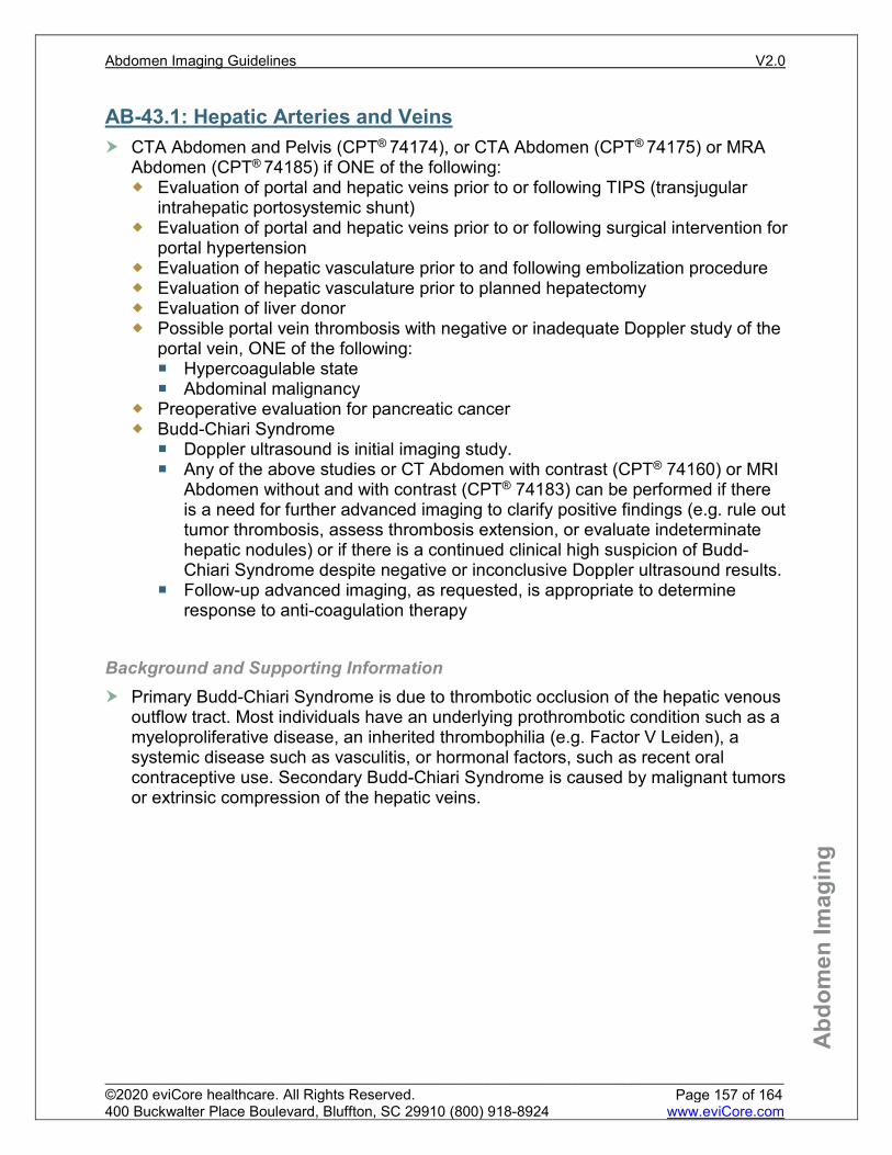
Abd
omen
Imag
ing
AB-43.1: Hepatic Arteries and Veins CTA Abdomen and Pelvis (CPT® 74174), or CTA Abdomen (CPT® 74175) or MRA
Abdomen (CPT® 74185) if ONE of the following: Evaluation of portal and hepatic veins prior to or following TIPS (transjugular
intrahepatic portosystemic shunt) Evaluation of portal and hepatic veins prior to or following surgical intervention for
portal hypertension Evaluation of hepatic vasculature prior to and following embolization procedure Evaluation of hepatic vasculature prior to planned hepatectomy Evaluation of liver donor Possible portal vein thrombosis with negative or inadequate Doppler study of the
portal vein, ONE of the following: Hypercoagulable state Abdominal malignancy
Preoperative evaluation for pancreatic cancer Budd-Chiari Syndrome
Doppler ultrasound is initial imaging study. Any of the above studies or CT Abdomen with contrast (CPT® 74160) or MRI
Abdomen without and with contrast (CPT® 74183) can be performed if there is a need for further advanced imaging to clarify positive findings (e.g. rule out tumor thrombosis, assess thrombosis extension, or evaluate indeterminate hepatic nodules) or if there is a continued clinical high suspicion of Budd-Chiari Syndrome despite negative or inconclusive Doppler ultrasound results.
Follow-up advanced imaging, as requested, is appropriate to determine response to anti-coagulation therapy
Background and Supporting Information Primary Budd-Chiari Syndrome is due to thrombotic occlusion of the hepatic venous
outflow tract. Most individuals have an underlying prothrombotic condition such as a myeloproliferative disease, an inherited thrombophilia (e.g. Factor V Leiden), a systemic disease such as vasculitis, or hormonal factors, such as recent oral contraceptive use. Secondary Budd-Chiari Syndrome is caused by malignant tumors or extrinsic compression of the hepatic veins.
Page 157 of 164
Abdomen Imaging Guidelines V2.0
______________________________________________________________________________________________________ ©2020 eviCore healthcare. All Rights Reserved. 400 Buckwalter Place Boulevard, Bluffton, SC 29910 (800) 918-8924 www.eviCore.com

Abd
omen
Imag
ing
AB-43.2: Abdominal Veins other than Hepatic and Portal Veins CTA Abdomen and Pelvis (CPT® 74174), or CTA Abdomen (CPT® 74175) or MRA
Abdomen (CPT® 74185) if ONE of the following: Nephrotic syndrome Suspicion of iliac vein thrombus Suspicion of inferior vena cava thrombus Renal vein thrombosis Mesenteric vein thrombosis
AB-43.3: Renal Vein Thrombosis MRA Abdomen (CPT® 74185) if ONE of the following:
Nephrotic syndrome Proteinuria – 3 grams or more in 24 hours Lupus nephritis Hypercoagulable state, ONE of the following:
Antiphospholipid antibodies Behçet’s syndrome Protein C deficiency Protein S deficiency
References American College of Radiology (ACR), North American Society for Cardiovascular Imaging (NASCI),
Society for Pediatric Radiology (SPR). ACR-NASCI-SPR practice guideline for the performance of pediatric and adult body magnetic resonance angiography (MRA). Am Coll Radiol. Revised 2015.
Nghiem HV, Winter TC III, Mountford MC, et al. Evaluation of the portal venous system before liver transplantation: value of phase-contrast MR angiography. AJR. 1995;164:871-878.
American Association for the Study of Liver Disease (AASLD). ASSLD practice guidelines: the role of transjugular intrahepatic protosystemic shunt (TIPS) in the management of portal hypertension. Hepatology, 2010;51:1-16.
Lee SS, Kim TK, Byun JH, et al. Hepatic arteries in potential donors for living related liver transplantation: evaluation with multi–detector row CT angiography. Radiology. 2003; 227:391-399.
Simonetto DA, Singal AK, Garcia-Tsao G, Caldwell SH, Ahn J, Kamath PS. ACG Clinical Guideline. The American Journal of Gastroenterology. 2020;115(1):18-40. doi:10.14309/ajg.0000000000000486.
Page 158 of 164
Abdomen Imaging Guidelines V2.0
______________________________________________________________________________________________________ ©2020 eviCore healthcare. All Rights Reserved. 400 Buckwalter Place Boulevard, Bluffton, SC 29910 (800) 918-8924 www.eviCore.com

Abd
omen
Imag
ing
AB-44: This section intentionally left blank
Page 159 of 164
Abdomen Imaging Guidelines V2.0
______________________________________________________________________________________________________ ©2020 eviCore healthcare. All Rights Reserved. 400 Buckwalter Place Boulevard, Bluffton, SC 29910 (800) 918-8924 www.eviCore.com

Abd
omen
Imag
ing
AB-45: Liver Elastography Vibration-Controlled Transient Elastography (VCTE) (e.g. Fibroscan, CPT® 91200)
may be considered appropriate to assess for advanced fibrosis and cirrhosis in the following conditions: Hepatitis C Hepatitis B Chronic alcoholic liver disease All other chronic liver diseases
Special consideration for Magnetic Resonance Elastography (MRE, CPT® 76391): Suspected NAFLD (Non-alcoholic fatty liver disease)
Transient Elastography (CPT® 91200) is the initial imaging modality to stage fibrosis
If Transient Elastography failure despite use of an XL-probe, OR BMI ≥35 in individuals with suspected NAFLD
For NAFLD in low risk populations (e.g. signs of fatty liver found on imaging only) MRE would be considered investigational.
The use of VCTE and MRE are considered experimental and investigational for all other indications with regards to liver disease
The use of other ultrasound elastographic techniques (CPT® 76981, CPT® 76982, and CPT® 76983), including but not limited to acoustic radiation force impulse imaging or real-time tissue elastography for any indication is considered experimental or investigational at this time
Background and Supporting Information For the assessment of cirrhosis in individuals with hepatitis C, the AGA noted that MRE has little to no increase in identifying cirrhosis, but had poorer specificity and thus higher false-positive rates than VCTE. In view of this, the AGA concluded that MRE has a poorer diagnostic performance in this setting, compared to VCTE. In their recommendations for the assessment of fibrosis in chronic liver disease, VCTE was recommended over MRE with the exception of NAFLD in high risk populations, in which MRE resulted in a lower rate of false positives compared to VCTE. This was considered a conditional recommendation with a low quality of evidence. The role of MRE was reviewed again in 2019 (Castera, et. al.) in Gastroenterology8 and the pathway recommendations form the basis of our current guideline with respect to the role of MRE in fatty liver disease.
Page 160 of 164
Abdomen Imaging Guidelines V2.0
______________________________________________________________________________________________________ ©2020 eviCore healthcare. All Rights Reserved. 400 Buckwalter Place Boulevard, Bluffton, SC 29910 (800) 918-8924 www.eviCore.com

Abd
omen
Imag
ing
References American Gastroenterologic Association Institute guideline on the role of elastography in the
evaluation of liver fibrosis. Gastroenterology. 2017:152:1536-1543. Conti CB, Cavalcoli F, Fraquelli M, Conte D, Massironi S. Ultrasound elastographic techniques in
focal liver lesions. World Journal of Gastroenterology. 2016;22(9):2647. doi:10.3748/wjg.v22.i9.2647. The diagnosis and management of nonalcoholic fatty liver disease: Practice guidance from the
American Association for the Study of Liver Diseases. Clinical Liver Disease. 2018;11(4):81-81. doi:10.1002/cld.722.
Li Q, Dhyani M, Grajo JR, Sirlin C, Samir AE. Current status of imaging in nonalcoholic fatty liver disease. World Journal of Hepatology. 2018;10(8):530-542. doi:10.4254/wjh.v10.i8.530.
Imajo K, Kessoku T, Honda Y, et al. Magnetic Resonance Imaging More Accurately Classifies Steatosis and Fibrosis in Patients With Nonalcoholic Fatty Liver Disease Than Transient Elastography. Gastroenterology. 2016;150(3). doi:10.1053/j.gastro.2015.11.048.
Chalasani N, Younossi Z, Lavine JE, et al. The diagnosis and management of nonalcoholic fatty liver disease: Practice guidance from the American Association for the Study of Liver Diseases. Hepatology. 2017;67(1):328-357. doi:10.1002/hep.29367.
Vuppalanchi R, Siddiqui MS, Natta MLV, et al. Performance characteristics of vibration-controlled transient elastography for evaluation of nonalcoholic fatty liver disease. Hepatology. 2017;67(1):134-144. doi:10.1002/hep.29489,
Castera L, Friedrich-Rust M, Loomba R. Noninvasive Assessment of Liver Disease in Patients With Nonalcoholic Fatty Liver Disease. Gastroenterology. 2019;156(5). doi:10.1053/j.gastro.2018.12.036.
Page 161 of 164
Abdomen Imaging Guidelines V2.0
______________________________________________________________________________________________________ ©2020 eviCore healthcare. All Rights Reserved. 400 Buckwalter Place Boulevard, Bluffton, SC 29910 (800) 918-8924 www.eviCore.com

Abd
omen
Imag
ing
AB-46:Hiccups Hiccups <48 hours without any localizing or specific symptoms:
No advanced imaging Hiccups ≥48 hours:
History and physical examination, laboratory and CMP and baseline chest x-ray Abnormal or negative chest x-ray with symptoms referable to the chest:
CT Chest with contrast (CPT® 71260) Lab or history/physical findings suggest a gastrointestinal etiology:
CT Abdomen with contrast (CPT® 74160)
References 1. British Journal of General Practice. Hiccups. A Common Problem with Some Unusual Causes and
Cures: 2016;66(652):584-586. 2. Steger M, Schneemann M, Fox M. Systemic review: the pathogenesis and pharmacological treatment
of hiccups. Alimentary Pharmacology & Therapeutics. 2015;42(9):1037-1050. doi:10.1111/apt.13374.
Page 162 of 164
Abdomen Imaging Guidelines V2.0
______________________________________________________________________________________________________ ©2020 eviCore healthcare. All Rights Reserved. 400 Buckwalter Place Boulevard, Bluffton, SC 29910 (800) 918-8924 www.eviCore.com
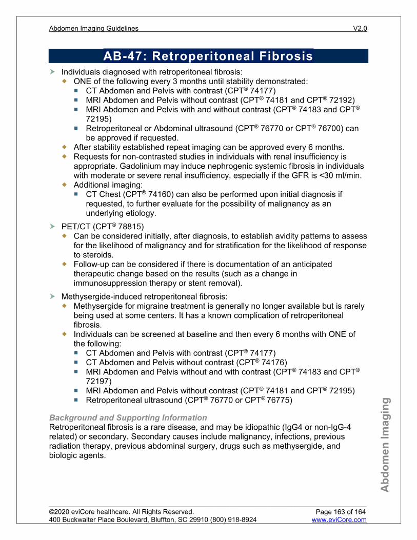
Abd
omen
Imag
ing
AB-47: Retroperitoneal Fibrosis Individuals diagnosed with retroperitoneal fibrosis:
ONE of the following every 3 months until stability demonstrated: CT Abdomen and Pelvis with contrast (CPT® 74177) MRI Abdomen and Pelvis without contrast (CPT® 74181 and CPT® 72192) MRI Abdomen and Pelvis with and without contrast (CPT® 74183 and CPT®
72195) Retroperitoneal or Abdominal ultrasound (CPT® 76770 or CPT® 76700) can
be approved if requested. After stability established repeat imaging can be approved every 6 months. Requests for non-contrasted studies in individuals with renal insufficiency is
appropriate. Gadolinium may induce nephrogenic systemic fibrosis in individuals with moderate or severe renal insufficiency, especially if the GFR is <30 ml/min.
Additional imaging: CT Chest (CPT® 74160) can also be performed upon initial diagnosis if
requested, to further evaluate for the possibility of malignancy as an underlying etiology.
PET/CT (CPT® 78815) Can be considered initially, after diagnosis, to establish avidity patterns to assess
for the likelihood of malignancy and for stratification for the likelihood of response to steroids.
Follow-up can be considered if there is documentation of an anticipated therapeutic change based on the results (such as a change in immunosuppression therapy or stent removal).
Methysergide-induced retroperitoneal fibrosis: Methysergide for migraine treatment is generally no longer available but is rarely
being used at some centers. It has a known complication of retroperitoneal fibrosis.
Individuals can be screened at baseline and then every 6 months with ONE of the following: CT Abdomen and Pelvis with contrast (CPT® 74177) CT Abdomen and Pelvis without contrast (CPT® 74176) MRI Abdomen and Pelvis without and with contrast (CPT® 74183 and CPT®
72197) MRI Abdomen and Pelvis without contrast (CPT® 74181 and CPT® 72195) Retroperitoneal ultrasound (CPT® 76770 or CPT® 76775)
Background and Supporting Information Retroperitoneal fibrosis is a rare disease, and may be idiopathic (IgG4 or non-IgG-4 related) or secondary. Secondary causes include malignancy, infections, previous radiation therapy, previous abdominal surgery, drugs such as methysergide, and biologic agents.
Page 163 of 164
Abdomen Imaging Guidelines V2.0
______________________________________________________________________________________________________ ©2020 eviCore healthcare. All Rights Reserved. 400 Buckwalter Place Boulevard, Bluffton, SC 29910 (800) 918-8924 www.eviCore.com

Abd
omen
Imag
ing
References 1. Retroperitoneal Fibrosis Clinical Presentation: History and Physical Examination. Retroperitoneal
Fibrosis Clinical Presentation: History and Physical Examination. https://emedicine.medscape.com/article/458501-clinical. Published May 30, 2019.
2. Vaglio A, Maritati F. Idiopathic Retroperitoneal Fibrosis. Journal of the American Society of Nephrology. 2016;27(7):1880-1889. doi:10.1681/asn.2015101110.
3. Runowska M, Majewski D, Puszczewicz M. Retroperitoneal fibrosis – the state-of-the-art. Reumatologia/Rheumatology. 2016;5:256-263. doi:10.5114/reum.2016.63667.
4. Urban M, Palmisano A, Nicastro M, Corradi D, Buzio C, Vaglio A. Idiopathic and secondary forms of retroperitoneal fibrosis: A diagnostic approach. La Revue de Médecine Interne. 2015;36(1):15-21. doi:10.1016/j.revmed.2014.10.008.
5. EMA restricts methysergide use, concern over fibrosis. Reactions Weekly. 2014;1491(1):2-2. doi:10.1007/s40278-014-9172-x.
6. Fendler WP, Eiber M, Stief CG, Herrmann K. A PET for All Seasons: 18 F-Fluorodeoxyglucose to Characterize Inflammation and Malignancy in Retroperitoneal Fibrosis? European Urology. 2017;71(6):934-935. doi:10.1016/j.eururo.2017.01.019.
7. Gu L, Wang Y, Zhang X. Re: Archie Fernando, James Pattison, Catherine Horsfield, David D’Cruz, Gary Cook, Tim O’Brien. [18F]-Fluorodeoxyglucose Positron Emission Tomography in the Diagnosis, Treatment Stratification, and Monitoring of Patients with Retroperitoneal Fibrosis: A Prospective Clinical Study. Eur Urol 2017;71:926–33. European Urology. 2017;72(2). doi:10.1016/j.eururo.2017.02.029.
Page 164 of 164
Abdomen Imaging Guidelines V2.0
______________________________________________________________________________________________________ ©2020 eviCore healthcare. All Rights Reserved. 400 Buckwalter Place Boulevard, Bluffton, SC 29910 (800) 918-8924 www.eviCore.com