Embed Size (px)
Citation preview
Cigna Medical Coverage Policies – Radiology Peripheral Nerve Disorders Imaging
Effective November 15, 2018
______________________________________________________________________________________ Instructions for use The following coverage policy applies to health benefit plans administered by Cigna. Coverage policies are intended to provide guidance in interpreting certain standard Cigna benefit plans and are used by medical directors and other health care professionals in making medical necessity and other coverage determinations. Please note the terms of a customer’s particular benefit plan document may differ significantly from the standard benefit plans upon which these coverage policies are based. For example, a customer’s benefit plan document may contain a specific exclusion related to a topic addressed in a coverage policy. In the event of a conflict, a customer’s benefit plan document always supersedes the information in the coverage policy. In the absence of federal or state coverage mandates, benefits are ultimately determined by the terms of the applicable benefit plan document. Coverage determinations in each specific instance require consideration of: 1. The terms of the applicable benefit plan document in effect on the date of service 2. Any applicable laws and regulations 3. Any relevant collateral source materials including coverage policies 4. The specific facts of the particular situation Coverage policies relate exclusively to the administration of health benefit plans. Coverage policies are not recommendations for treatment and should never be used as treatment guidelines. This evidence-based medical coverage policy has been developed by eviCore, Inc. Some information in this coverage policy may not apply to all benefit plans administered by Cigna. These guidelines include procedures eviCore does not review for Cigna. Please refer to the Cigna CPT code list for the current list of high-tech imaging procedures that eviCore reviews for Cigna. CPT® (Current Procedural Terminology) is a registered trademark of the American Medical Association (AMA). CPT® five digit codes, nomenclature and other data are copyright 2017 American Medical Association. All Rights Reserved. No fee schedules, basic units, relative values or related listings are included in the CPT® book. AMA does not directly or indirectly practice medicine or dispense medical services. AMA assumes no liability for the data contained herein or not contained herein.
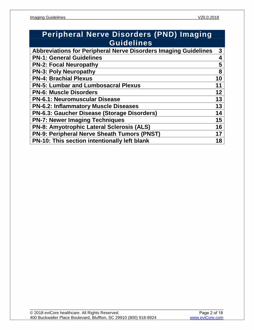
Peripheral Nerve Disorders (PND) Imaging Guidelines
Abbreviations for Peripheral Nerve Disorders Imaging Guidelines 3PN-1: General Guidelines 4PN-2: Focal Neuropathy 5PN-3: Poly Neuropathy 8PN-4: Brachial Plexus 10PN-5: Lumbar and Lumbosacral Plexus 11PN-6: Muscle Disorders 12PN-6.1: Neuromuscular Disease 13PN-6.2: Inflammatory Muscle Diseases 13PN-6.3: Gaucher Disease (Storage Disorders) 14PN-7: Newer Imaging Techniques 15PN-8: Amyotrophic Lateral Sclerosis (ALS) 16PN-9: Peripheral Nerve Sheath Tumors (PNST) 17PN-10: This section intentionally left blank 18
Imaging Guidelines V20.0.2018
______________________________________________________________________________________________________ © 2018 eviCore healthcare. All Rights Reserved. 400 Buckwalter Place Boulevard, Bluffton, SC 29910 (800) 918-8924 www.eviCore.com
Page 2 of 18
Perip
hera
l Ner
ve D
isor
ders
(PN
D)
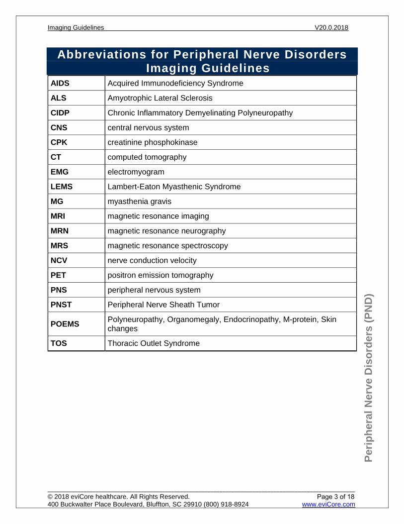
Abbreviations for Peripheral Nerve Disorders Imaging Guidelines
AIDS Acquired Immunodeficiency Syndrome
ALS Amyotrophic Lateral Sclerosis
CIDP Chronic Inflammatory Demyelinating Polyneuropathy
CNS central nervous system
CPK creatinine phosphokinase
CT computed tomography
EMG electromyogram
LEMS Lambert-Eaton Myasthenic Syndrome
MG myasthenia gravis
MRI magnetic resonance imaging
MRN magnetic resonance neurography
MRS magnetic resonance spectroscopy
NCV nerve conduction velocity
PET positron emission tomography
PNS peripheral nervous system
PNST Peripheral Nerve Sheath Tumor
POEMS Polyneuropathy, Organomegaly, Endocrinopathy, M-protein, Skin changes
TOS Thoracic Outlet Syndrome
Imaging Guidelines V20.0.2018
______________________________________________________________________________________________________ © 2018 eviCore healthcare. All Rights Reserved. 400 Buckwalter Place Boulevard, Bluffton, SC 29910 (800) 918-8924 www.eviCore.com
Page 3 of 18

Perip
hera
l Ner
ve D
isor
ders
(PN
D)
PN-1: General Guidelines A current clinical evaluation (within 60 days) is required before advanced imaging can be considered. The clinical evaluation may include a relevant history and physical examination, including a neurological examination, appropriate laboratory studies, non-advanced imaging modalities, electromyography and nerve conduction (EMG/NCV) studies. Other meaningful contact (telephone call, electronic mail or messaging) by an established individual can substitute for a face-to-face clinical evaluation. MRI is, most often, preferable to CT.
References 1. Bowen BC, Maravilla KR, Saraf-Lavi. Magnetic Resonance Imaging of the Peripheral Nervous
System. In Latchaw RE, Kucharczyk J, Moseley ME. Imaging of the Nervous System. Diagnostic and Therapeutic Applications. Vol 2, Mosby, Philadelphia, 2005, pp.1479-1497.
2. Walker WO. Ultrasonography in peripheral nervous system diagnosis. Continuum. 2017 Oct; 23 (5, Peripheral Nerve and Motor Neuron Disorders):1276-1294. Accessed November 21, 2017. https://insights.ovid.com/crossref?an=00132979-201710000-00009 Systematic Review.
3. Ohana M, Moser T, Moussaouï A, et al. Current and future imaging of the peripheral nervous system. Diagnostic and Interventional Imaging. 2014;95(1):17-26. Accessed November 21, 2017. http://www.sciencedirect.com/science/article/pii/S2211568413001976
4. Stoll G, Bendszuz M, Perez J, et al. Magnetic resonance imaging of the peripheral nervous system. J Neurol. 2009 Jul;256(7):1043-51. Accessed November 21, 2017. https://link.springer.com/article/10.1007/s00415-009-5064-z Systematic Review.
5. Stoll G, Wilder-Smith E, and Bendszus M. Imaging of the peripheral nervous system. Handb Clin Neurol. 2013;115:137-153. Accessed November 21, 2017. http://www.sciencedirect.com/science/article/pii/B9780444529022000084 Systematic Review.
6. Kim S, Choi J-Y, Huh Y-M, et al. Role of magnetic resonance imaging in entrapment and compressive neuropathy—what, where, and how to see the peripheral nerves on the musculoskeletal magnetic resonance image: part 1. Overview and lower extremity. Eur Radiol. 2007 Jan;17(1):139-149. Accessed November 21, 2017. https://link.springer.com/article/10.1007%2Fs00330-006-0179-4 Systematic Review.
Imaging Guidelines V20.0.2018
______________________________________________________________________________________________________ © 2018 eviCore healthcare. All Rights Reserved. 400 Buckwalter Place Boulevard, Bluffton, SC 29910 (800) 918-8924 www.eviCore.com
Page 4 of 18
Perip
hera
l Ner
ve D
isor
ders
(PN
D)
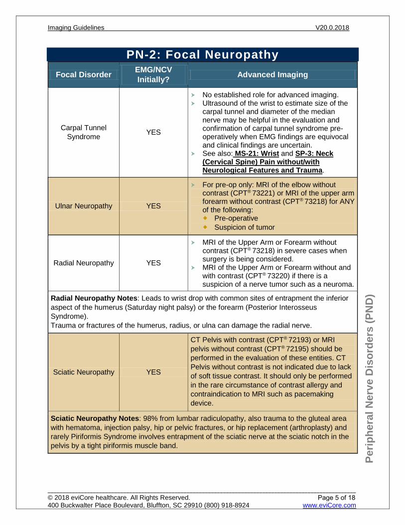
PN-2: Focal Neuropathy Focal Disorder EMG/NCV
Initially? Advanced Imaging
Carpal Tunnel Syndrome YES
No established role for advanced imaging. Ultrasound of the wrist to estimate size of the
carpal tunnel and diameter of the mediannerve may be helpful in the evaluation andconfirmation of carpal tunnel syndrome pre-operatively when EMG findings are equivocaland clinical findings are uncertain.
See also: MS-21: Wrist and SP-3: Neck(Cervical Spine) Pain without/withNeurological Features and Trauma.
Ulnar Neuropathy YES
For pre-op only: MRI of the elbow withoutcontrast (CPT® 73221) or MRI of the upper armforearm without contrast (CPT® 73218) for ANYof the following: Pre-operative Suspicion of tumor
Radial Neuropathy YES
MRI of the Upper Arm or Forearm withoutcontrast (CPT® 73218) in severe cases whensurgery is being considered.
MRI of the Upper Arm or Forearm without andwith contrast (CPT® 73220) if there is asuspicion of a nerve tumor such as a neuroma.
Radial Neuropathy Notes: Leads to wrist drop with common sites of entrapment the inferior aspect of the humerus (Saturday night palsy) or the forearm (Posterior Interosseus Syndrome). Trauma or fractures of the humerus, radius, or ulna can damage the radial nerve.
Sciatic Neuropathy YES
CT Pelvis with contrast (CPT® 72193) or MRI pelvis without contrast (CPT® 72195) should be performed in the evaluation of these entities. CT Pelvis without contrast is not indicated due to lack of soft tissue contrast. It should only be performed in the rare circumstance of contrast allergy and contraindication to MRI such as pacemaking device.
Sciatic Neuropathy Notes: 98% from lumbar radiculopathy, also trauma to the gluteal area with hematoma, injection palsy, hip or pelvic fractures, or hip replacement (arthroplasty) and rarely Piriformis Syndrome involves entrapment of the sciatic nerve at the sciatic notch in the pelvis by a tight piriformis muscle band.
Imaging Guidelines V20.0.2018
______________________________________________________________________________________________________ © 2018 eviCore healthcare. All Rights Reserved. 400 Buckwalter Place Boulevard, Bluffton, SC 29910 (800) 918-8924 www.eviCore.com
Page 5 of 18
Perip
hera
l Ner
ve D
isor
ders
(PN
D)
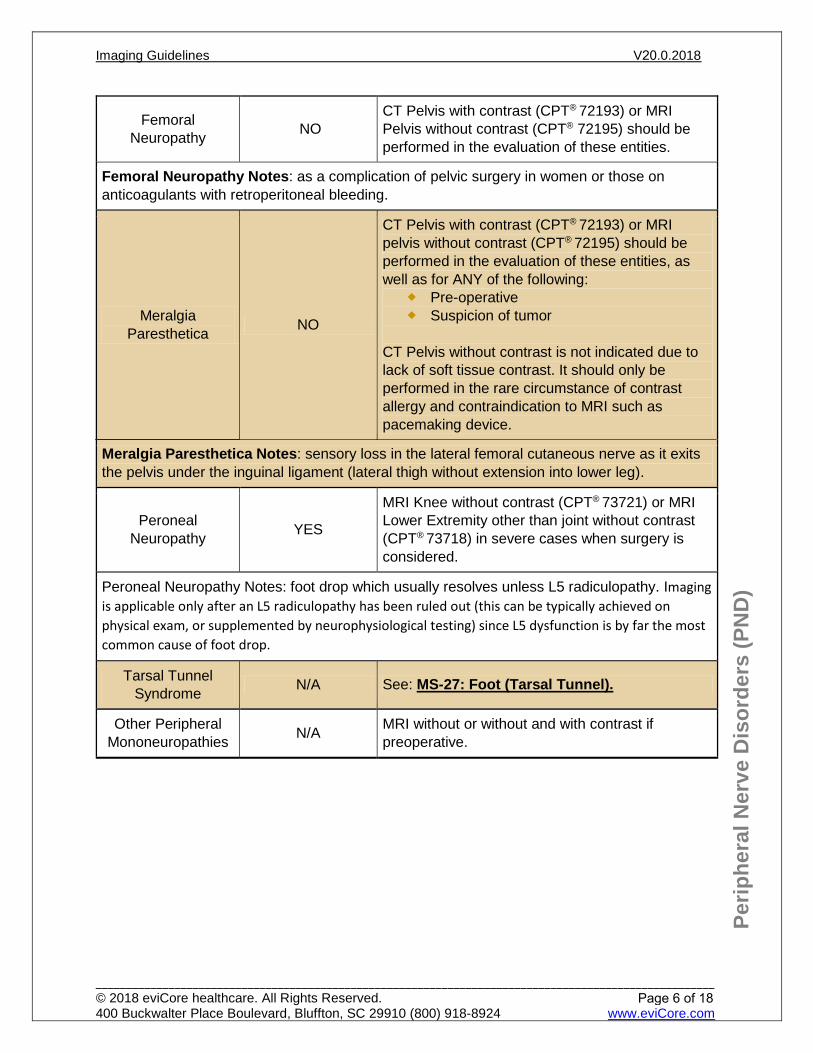
Femoral Neuropathy NO
CT Pelvis with contrast (CPT® 72193) or MRI Pelvis without contrast (CPT® 72195) should be performed in the evaluation of these entities.
Femoral Neuropathy Notes: as a complication of pelvic surgery in women or those on anticoagulants with retroperitoneal bleeding.
Meralgia Paresthetica NO
CT Pelvis with contrast (CPT® 72193) or MRI pelvis without contrast (CPT® 72195) should be performed in the evaluation of these entities, as well as for ANY of the following:
Pre-operative Suspicion of tumor
CT Pelvis without contrast is not indicated due to lack of soft tissue contrast. It should only be performed in the rare circumstance of contrast allergy and contraindication to MRI such as pacemaking device.
Meralgia Paresthetica Notes: sensory loss in the lateral femoral cutaneous nerve as it exits the pelvis under the inguinal ligament (lateral thigh without extension into lower leg).
Peroneal Neuropathy YES
MRI Knee without contrast (CPT® 73721) or MRI Lower Extremity other than joint without contrast (CPT® 73718) in severe cases when surgery is considered.
Peroneal Neuropathy Notes: foot drop which usually resolves unless L5 radiculopathy. Imaging is applicable only after an L5 radiculopathy has been ruled out (this can be typically achieved on physical exam, or supplemented by neurophysiological testing) since L5 dysfunction is by far the most common cause of foot drop.
Tarsal Tunnel Syndrome N/A See: MS-27: Foot (Tarsal Tunnel).
Other Peripheral Mononeuropathies N/A MRI without or without and with contrast if
preoperative.
Imaging Guidelines V20.0.2018
______________________________________________________________________________________________________ © 2018 eviCore healthcare. All Rights Reserved. 400 Buckwalter Place Boulevard, Bluffton, SC 29910 (800) 918-8924 www.eviCore.com
Page 6 of 18
Perip
hera
l Ner
ve D
isor
ders
(PN
D)
References 1. Andreisek G, Crook DW, Burg D, et al. Peripheral neuropathies of the median, radial, and ulnar
nerves: MR imaging features. RadioGraphics. 2006 Sep-Oct;26(5):1267-1287. Accessed October 12, 2017. http://pubs.rsna.org/doi/10.1148/rg.265055712?url_ver=Z39.88-2003&rfr_id=ori:rid:crossref.org&rfr_dat=cr_pub%3dpubmed
2. Iverson DJ. MRI detection of cysts of the knee causing common peroneal neuropathy. Neurology. 2005 Dec 13;65(11):1829-1831. Accessed October 12, 2017. http://www.neurology.org/content/65/11/1829
3. Cartwright MS, Walker FO. Neuromuscular ultrasound in common entrapment neuropathies. Muscle & Nerve. 2013 Sep 2;48(5):696-704. Accessed October 12, 2017. http://onlinelibrary.wiley.com/doi/10.1002/mus.23900/abstract;jsessionid=04686029379E194020A4795DFFFB31D0.f02t03
4. Linda DD, Harish S, Stewart BG, et al. Multimodality imaging of peripheral neuropathies of the upper limb and brachial plexus. RadioGraphics. 2010 Sep;30(5):1373-1400. Accessed October 12, 2017. http://pubs.rsna.org/doi/10.1148/rg.305095169
5. Hobson-Webb LD and Juel VC. Common Entrapment Neuropathies. Continuum. 2017 Apr;23(2):487-511. Accessed October 29, 2017. http://journals.lww.com/continuum/Abstract/2017/04000/Common_Entrapment_Neuropathies.12.aspx Systematic Review.
6. Tsivgoulis G and Alexandrov AV. Ultrasound in neurology. Continuum. 2016 Oct;22(5)Neuroimaging:1655-1677. Accessed November 21, 2017. https://insights.ovid.com/crossref?an=00132979-201610000-00018 Systematic Review.
Imaging Guidelines V20.0.2018
______________________________________________________________________________________________________ © 2018 eviCore healthcare. All Rights Reserved. 400 Buckwalter Place Boulevard, Bluffton, SC 29910 (800) 918-8924 www.eviCore.com
Page 7 of 18
Perip
hera
l Ner
ve D
isor
ders
(PN
D)
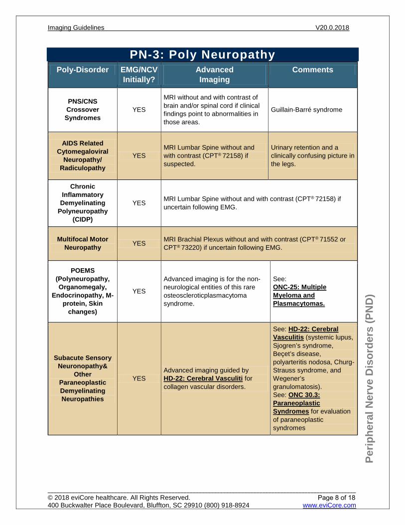
PN-3: Poly Neuropathy Poly-Disorder EMG/NCV
Initially? Advanced Imaging
Comments
PNS/CNS Crossover Syndromes
YES
MRI without and with contrast of brain and/or spinal cord if clinical findings point to abnormalities in those areas.
Guillain-Barré syndrome
AIDS Related Cytomegaloviral
Neuropathy/ Radiculopathy
YES MRI Lumbar Spine without and with contrast (CPT® 72158) if suspected.
Urinary retention and a clinically confusing picture in the legs.
Chronic Inflammatory Demyelinating
Polyneuropathy (CIDP)
YES MRI Lumbar Spine without and with contrast (CPT® 72158) if uncertain following EMG.
Multifocal Motor Neuropathy YES MRI Brachial Plexus without and with contrast (CPT® 71552 or
CPT® 73220) if uncertain following EMG.
POEMS (Polyneuropathy, Organomegaly,
Endocrinopathy, M-protein, Skin
changes)
YES
Advanced imaging is for the non-neurological entities of this rare osteoscleroticplasmacytoma syndrome.
See: ONC-25: Multiple Myeloma and Plasmacytomas.
Subacute Sensory Neuronopathy&
Other Paraneoplastic Demyelinating Neuropathies
YES Advanced imaging guided by HD-22: Cerebral Vasculiti for collagen vascular disorders.
See: HD-22: Cerebral Vasculitis (systemic lupus, Sjogren’s syndrome, Beçet’s disease, polyarteritis nodosa, Churg-Strauss syndrome, and Wegener’s granulomatosis). See: ONC 30.3: Paraneoplastic Syndromes for evaluation of paraneoplastic syndromes
Imaging Guidelines V20.0.2018
______________________________________________________________________________________________________ © 2018 eviCore healthcare. All Rights Reserved. 400 Buckwalter Place Boulevard, Bluffton, SC 29910 (800) 918-8924 www.eviCore.com
Page 8 of 18

Perip
hera
l Ner
ve D
isor
ders
(PN
D)
References 1. Anders HJ, Goebel FD. Cytomegalovirus polyradiculopathy in patients with AIDS. Clin Infect Dis.
1998 Aug 27;27(2):345-352. Accessed October 12, 2017. https://www.ncbi.nlm.nih.gov/pubmed/9709885
2. Duggins AJ, McLoed JG, Pollard JD, et al. Spinal root and plexus hypertrophy in chronic inflammatory demyelinating polyneuropathy. Brain. 1999 July 1;122(7):1383-1390. Accessed October 12, 2017. https://academic.oup.com/brain/article-lookup/doi/10.1093/brain/122.7.1383
3. Amato AA, Barohn RJ, Katz JS, et al. Clinical spectrum of chronic acquired demyelinating polyneuropathies. Muscle & Nerve. 2001 Mar;24(3):311-324. Accessed October 12, 2017. http://onlinelibrary.wiley.com/doi/10.1002/1097-4598(200103)24:3%3C311::AID-MUS1001%3E3.0.CO;2-A/abstract
4. Darnell RB, Posner JB. Paraneoplastic Syndromes Involving the Nervous System. N Engl J Med. 2003;349:1543-1554. Accessed October 12, 2017. http://www.nejm.org/doi/full/10.1056/NEJMra023009
5. Antoine JC, Bouhour F, Camdessanche JP. [18F] fluorodeoxyglucose positron emission tomography in the diagnosis of cancer in patients with paraneoplastic neurological syndrome and anti-Hu antibodies. Ann Neurol. 2000 July;48(1):105-108. Accessed October 12, 2017. https://www.ncbi.nlm.nih.gov/pubmed/10894223
Imaging Guidelines V20.0.2018
______________________________________________________________________________________________________ © 2018 eviCore healthcare. All Rights Reserved. 400 Buckwalter Place Boulevard, Bluffton, SC 29910 (800) 918-8924 www.eviCore.com
Page 9 of 18
Perip
hera
l Ner
ve D
isor
ders
(PN
D)
PN-4: Brachial Plexus Upper extremity other than joint MRI without or without and with contrast (CPT®
73218 or CPT® 73220), Chest MRI without or without and with contrast (CPT® 71550or CPT® 71552) or Neck MRI without (CPT® 70540) or without and with contrast(CPT® 70543) (if upper trunk) after EMG/NCV examination for: Malignant infiltration (EMG not required) Radiation plexitis to r/o malignant infiltration Brachial plexitis (Parsonage-Turner Syndrome or painful brachial amyotrophy).
Self-limited syndrome characterized by initial shoulder region pain followed by weakness of specific muscles in a pattern which does not conform to involvement of a single root or distal peripheral nerve
Consider MRI of the cervical spine if radiculopathy. See: SP-3: Neck (Cervical Spine) Pain without/with Neurological
Features and Trauma Traumatic injury Neurogenic Thoracic Outlet Syndrome (TOS) failed a 2 to 3 month trial of
conservative management and are being considered for surgical treatment. See: CH-31: Thoracic Outlet Syndrome (TOS) Preoperative study which requires evaluation of the brachial plexus
References 1. Adkins MC, Wittenberg KH. MR imaging of nontraumatic brachial plexopathies: frequency and
spectrum of findings. RadioGraphics. 2000 July;20(4):1023-1032. Accessed October 12, 2017.http://pubs.rsna.org/doi/10.1148/radiographics.20.4.g00jl091023
2. Bykowski J, Aulino JM, Berger KL, et al. (2016). ACR Appropriateness Criteria® Plexopathy.American College of Radiology (ACR). Accessed October 12, 2017.https://acsearch.acr.org/docs/69487/Narrative/
3. Van Es HW. MRI of the brachial plexus. Eur Radiol. 2001 Jan;11(2):325-336. Accessed October 12,2017. https://link.springer.com/article/10.1007%2Fs003300000644
4. Foley KM, Kori SH, Posner JB. Brachial plexus lesions in patients with cancer: 100 cases. Neurology.1981 Jan;31(1):45-50. Accessed October 12, 2017.https://www.ncbi.nlm.nih.gov/pubmed/6256684
5. Cascino TL, Harper CM, Thomas JE, et al. Distinction between neoplastic and radiation-inducedbrachial plexopathy, with emphasis on the role of EMG. Neurology. 1989 April;39(4):502-506.Accessed October 12, 2017. http://www.neurology.org/content/39/4/502
6. Husband JE, MacVicar AD, Padhani AR, et al. Symptomatic brachial plexopathy following treatmentfor breast cancer: Utility of MR imaging with surface-coil techniques. Radiology. 2000March;214(3):837-842. Accessed October 12, 2017.http://pubs.rsna.org/doi/10.1148/radiology.214.3.r00mr11837
7. McDonald TJ, Miller JD, Pruitt S. Acute brachial plexus neuritis: an uncommon cause of shoulderpain. Am Fam Physician. 2000 Nov 1;62(9):2067-2072. Accessed October 12, 2017.http://www.aafp.org/afp/2000/1101/p2067.html
Imaging Guidelines V20.0.2018
______________________________________________________________________________________________________ © 2018 eviCore healthcare. All Rights Reserved. 400 Buckwalter Place Boulevard, Bluffton, SC 29910 (800) 918-8924 www.eviCore.com
Page 10 of 18
Perip
hera
l Ner
ve D
isor
ders
(PN
D)
PN-5: Lumbar and Lumbosacral Plexus MRI Pelvis without and with contrast with fat suppression imaging (CPT® 72197) OR
MRI Abdomen and Pelvis without and with contrast with fat suppression imaging (CPT® 74183 and CPT® 72197) OR if MRI is not available, CT Pelvis with contrast (CPT® 72193) OR CT Abdomen and Pelvis with contrast (CPT® 74177) after EMG/NCV based on whether the upper lumbar plexus (abdominal retroperitoneal space) or the lumbosacral plexus (pelvis), respectively, is involved based on: Malignant infiltration (EMG not required) Radiation plexopathy to r/o malignant infiltration Traumatic injury Concern for retroperitoneal hematoma in patients on anticoagulation or who have
other thrombotic disorders (EMG/NCV not necessary)
References 1. Brejt N, Berry J, Nisbet A, et al. Pelvic radiculopathies, lumbosacral plexopathies, and neuropathies in
oncologic disease: A multidisciplinary approach to a diagnostic challenge. Cancer Imaging. 2013 Dec 30;13(4):591-601. Accessed October 12, 2017. https://www.ncbi.nlm.nih.gov/pmc/articles/PMC3893894/
2. McDonald JW, Sadwosky C. Spinal-cord injury. The Lancet. 2002 Feb 2;359(9304):417-425. Accessed October 12, 2017. https://www.ncbi.nlm.nih.gov/pubmed/11844532
Imaging Guidelines V20.0.2018
______________________________________________________________________________________________________ © 2018 eviCore healthcare. All Rights Reserved. 400 Buckwalter Place Boulevard, Bluffton, SC 29910 (800) 918-8924 www.eviCore.com
Page 11 of 18
Perip
hera
l Ner
ve D
isor
ders
(PN
D)
PN-6: Muscle DisordersPN-6.1: Neuromuscular Disease 13PN-6.2: Inflammatory Muscle Diseases 13PN-6.3: Gaucher Disease (Storage Disorders) 14
Imaging Guidelines V20.0.2018
______________________________________________________________________________________________________ © 2018 eviCore healthcare. All Rights Reserved. 400 Buckwalter Place Boulevard, Bluffton, SC 29910 (800) 918-8924 www.eviCore.com
Page 12 of 18
Perip
hera
l Ner
ve D
isor
ders
(PN
D)
PN-6.1: Neuromuscular Disease Myasthenia Gravis (MG) is associated with thymic disease and can undergo:
Chest CT with contrast (CPT® 71260) after an established diagnosis of MG. Can be repeated if initial CT previously negative and now symptoms of chest
mass, rising anti-striated muscle antibody titers, or need for preoperative evaluation (clinical presentation, electro-diagnostic studies, and antibody titers).
Chest CT without contrast (CPT® 71250) may be used if there is concern regarding adverse effects of contrast in individuals with MG.
Lambert–Eaton myasthenic syndrome (LEMS) is associated with small cell lungcancer and can undergo: Chest CT with contrast (CPT® 71260) with a suspected diagnosis (CXR,
symptoms of lung mass, clinical presentation, electro-diagnostic studies, and antibody titers). Can be repeated if initial CT previously negative after 3 months with
persistent suspicion. Stiff man syndrome is associated with small cell lung cancer and breast cancer
Chest CT with contrast (CPT® 71260) if Stiff Man Syndrome is suspected based on clinical findings.
PN-6.2: Inflammatory Muscle Diseases MRI without contrast (CPT® 73218 and CPT® 73718) or MRI without and with
contrast (CPT® 73220 and CPT® 73720) for: Additional evaluation of myopathy or myositis (based on clinical exam and
adjunct testing with EMG/NCV and labs) To plan muscle biopsy Treatment monitoring See also: PEDMS-10.3: Pediatric Inflammatory Muscle Diseases
All cases with dermatomyositis and polymyositis can undergo search for occultneoplasm (See ONC–30.3: Paraneoplastic Syndromes): Chest CT with contrast (CPT® 71260) for lung cancer and pelvic ultrasound (in
women) (CPT® 76856 or CPT® 76857 and/or CPT® 76830 [transvaginal]) for ovarian cancer should be done initially
Abdomen and Pelvis CT with contrast (CPT® 74177) if the above fail to make a diagnosis
Background and Supporting Information MRI and ultrasound are increasingly being used in the evaluation of muscle disease.
MRI may be helpful in demonstrating abnormalities in muscles that are difficult toexamine or not clinically weak, and MRI can also help distinguish between differenttypes of muscle disease. MRI is also useful in determining sites for muscle biopsy.
Imaging Guidelines V20.0.2018
______________________________________________________________________________________________________ © 2018 eviCore healthcare. All Rights Reserved. 400 Buckwalter Place Boulevard, Bluffton, SC 29910 (800) 918-8924 www.eviCore.com
Page 13 of 18
Perip
hera
l Ner
ve D
isor
ders
(PN
D)
PN-6.3: Gaucher Disease (Storage Disorders) See AB-11: Gaucher Disease and Hemochromatosis in the Abdomen Imaging
Guidelines. See PEDPN-4: Gaucher Disease in the pediatric PND Imaging Guidelines.
References 1. Darnell R, Posner J. Paraneoplastic syndromes involving the nervous system. N Engl J Med. 2003
Oct;349:1543-1554. Accessed October 12, 2017.http://www.nejm.org/doi/full/10.1056/NEJMra023009?keytype2=tf_ipsecsha&ijkey=b07ae6f203aa48e0f1c6d61624a159b87084997a
2. Schweitzer M, Fort J. Cost-effectiveness of MR imaging in evaluating polymyositis. Am J Roentgenol.1995;165:1469-1471. Accessed October 12, 2017.http://www.ajronline.org/doi/abs/10.2214/ajr.165.6.7484589
3. Adams E, Chow C, Premkumar A, Plotz P. The idiopathic inflammatory myopathies: spectrum of MRimaging findings. RadioGraphics. 1995;15(3):563-574. Accessed October 12, 2017.http://pubs.rsna.org/doi/pdf/10.1148/radiographics.15.3.7624563
4. Park J, Olsen N. Utility of magnetic resonance imaging in the evaluation of patients with inflammatorymyopathies. Curr Rheumatol Reports. 2001 Aug;3(4):334-345. Accessed October 12, 2017.https://link.springer.com/article/10.1007%2Fs11926-001-0038-x
5. Sekul E, Chow C, Dalakas M. Magnetic resonance imaging of the forearm as a diagnostic aid inpatients with sporadic inclusion body myositis. Neurolog. 1997 April;48(4):863-866. AccessedOctober 12, 2017. http://www.neurology.org/content/48/4/863.abstract
6. Lundberg I, Chung Y. Treatment and investigation of idiopathic inflammatory myopathies.Rheumatology. 2000 Jan;39(1):7-17. Accessed October 12, 2017.https://academic.oup.com/rheumatology/articlelookup/doi/10.1093/rheumatology/39.1.7
7. Park J, Olsen N. Utility of magnetic resonance imaging in the evaluation of patients with inflammatorymyopathies. Curr Rheumatol Reports. 2001 Aug;3(4):334-345. Accessed October 12, 2017.https://link.springer.com/article/10.1007%2Fs11926-001-0038-x
8. Hill C, Zhang Y, Sigurgeirsson B, et al. Frequency of specific cancer types in dermatomyositis andpolymyositis: a population-based study. Lancet. 2001 Jan 13;357(9250):96-100. Accessed October12, 2017. http://www.thelancet.com/journals/lancet/article/PIIS0140-6736(00)03540-6/fulltext
9. Maas M, Poll L, Terk M. Imaging and quantifying skeletal involvement in Gaucher disease. B JRadiol. 2002;75 suppl1:A13-A24. Accessed October 12, 2017.http://www.birpublications.org/doi/full/10.1259/bjr.75.suppl_1.750013
10. Giraldo P, Pocovi M, Perez-Calvo J, et al. Report of the Spanish Gaucher's disease registry: clinicaland genetic characteristics. Haematologica. 2000 Jan;85:792-799. Accessed October 12, 2017.http://www.haematologica.org/content/85/8/792
11. Rosow et al. The Role of Electrodiagnostic Testing, Imaging, and Muscle Biopsy in the Investigationof Muscle Disease. Continuum. 2016 Dec;22(6):1787-1802. Accessed October 12, 2017.https://www.ncbi.nlm.nih.gov/pubmed/27922493
12. Somashekar DK, Davenport MS, Cohan RH, et al. Effect of intravenous low-osmolality iodinatedcontrast media on patients with myasthenia gravis. Radiology. 2013 Jun;267(3):727-734. AccessedNovember 21, 2017. http://pubs.rsna.org/doi/full/10.1148/radiol.12121508.
Imaging Guidelines V20.0.2018
______________________________________________________________________________________________________ © 2018 eviCore healthcare. All Rights Reserved. 400 Buckwalter Place Boulevard, Bluffton, SC 29910 (800) 918-8924 www.eviCore.com
Page 14 of 18
Perip
hera
l Ner
ve D
isor
ders
(PN
D)
PN-7: Newer Imaging Techniques See: HD-24.6 Magnetic Resonance Neurography (MRN).
Imaging Guidelines V20.0.2018
______________________________________________________________________________________________________ © 2018 eviCore healthcare. All Rights Reserved. 400 Buckwalter Place Boulevard, Bluffton, SC 29910 (800) 918-8924 www.eviCore.com
Page 15 of 18
Perip
hera
l Ner
ve D
isor
ders
(PN
D)
PN-8: Amyotrophic Lateral Sclerosis (ALS) MRI Brain, Cervical, Thoracic, and Lumbar Spine most often without contrast, but
may be without and with contrast with meningeal symptoms. Can be considered when ALS is suspected (combination of upper and lower
motor neuron findings) to establish a diagnosis. Repeat imaging can be evaluated based on the appropriate Spine Imaging
Guidelines.
References 1. Agosta F, Chio A, Cosottini M, et al. The present and the future of neuroimaging in amyotrophic
lateral scoliosis. Am J Neuroradiol. 2010 Nov;31(10):1769-1777. Accessed October 12, 2017.http://www.ajnr.org/content/31/10/1769.long
2. Kollewe K, Korner S, Dengler R, et al. Magnetic resonance imaging in amyotrophic lateral sclerosis.Neurology Research International. 2012;v2012. Accessed October 12,2017.https://www.hindawi.com/journals/nri/2012/608501/
3. Filippi M, Agosta F, Abrahams S, et al. EFNS guidelines on the use of neuroimaging in themanagement of motor neuron diseases. Eur J Neurol. 2010 Apr;17(4):526-e20. Accessed October12, 2017. https://www.ncbi.nlm.nih.gov/pmc/articles/PMC3154636/
4. Wang S, Melhem ER, Poptani H, et al. Neuroimaging in amyotrophic lateral sclerosis.Neurotherapeutics. 2011 Jan;8(1):63-71. Accessed October 12, 2017.https://link.springer.com/article/10.1007%2Fs13311-010-0011-3
Imaging Guidelines V20.0.2018
______________________________________________________________________________________________________ © 2018 eviCore healthcare. All Rights Reserved. 400 Buckwalter Place Boulevard, Bluffton, SC 29910 (800) 918-8924 www.eviCore.com
Page 16 of 18
Perip
hera
l Ner
ve D
isor
ders
(PN
D)
PN-9: Peripheral Nerve Sheath Tumors (PNST) Tumors (Schwannomas or Neurofibromas) that arise from Schwann cells or other
connective tissue of the nerve are located anywhere in the body and can undergoadvanced imaging when suspected, which may include: MRI Brain without and with contrast (CPT® 70553). Cervical, thoracic, and lumbar spine MRI without and with contrast (CPT® 72156,
CPT® 72157, and CPT® 72158) if paraspinalneurofibroma is found any spine level or multiple simplex perineuralneurofibromas.
Follow-up imaging is not needed unless: New symptoms or neurological findings. CT Chest and Abdomen with contrast (CPT® 71260 and CPT® 74160) if
malignant transformation (5 %) is known or suspected See: PEDONC-2.3: Neurofibromatosis, 1 and 2 (NF1 and NF2) (Type 1).
References 1. Riccardi V. The genetic predisposition to and histogenesis of neurofibromas and neurofibrosarcoma
in neurofibromatosis type 1. Neurosurg Focus. 2007 Jun 15;22(6):E3. Accessed October 12, 2017.https://www.ncbi.nlm.nih.gov/pubmed/17613220
2. Li C, Huang G, Wu H, et al. Differentiation of soft tissue benign and malignant peripheral nervesheath tumors with magnetic resonance imaging. Clin Imaging. 2008 Mar-Apr;32(2):121-127.Accessed October 12, 2017. http://www.clinicalimaging.org/article/S0899-7071(07)00135-0/fulltext
3. Murovic J, Kim D, Kline D. Neurofibromatosis-associated nerve sheath tumors. Case report andreview of the literature. Neurosurg Focus. 2006 Jan;20(1):1-10 Accessed October 12, 2017.http://thejns.org/doi/abs/10.3171/foc.2006.20.1.2
Imaging Guidelines V20.0.2018
______________________________________________________________________________________________________ © 2018 eviCore healthcare. All Rights Reserved. 400 Buckwalter Place Boulevard, Bluffton, SC 29910 (800) 918-8924 www.eviCore.com
Page 17 of 18
Perip
hera
l Ner
ve D
isor
ders
(PN
D)
PN-10: This section intentionally left blank
Imaging Guidelines V20.0.2018
______________________________________________________________________________________________________ © 2018 eviCore healthcare. All Rights Reserved. 400 Buckwalter Place Boulevard, Bluffton, SC 29910 (800) 918-8924 www.eviCore.com
Page 18 of 18