Embed Size (px)
Citation preview

Ciliary Reversal without Rotation of Axonemal
Structures in Ctenophore Comb Plates
SIDNEY L. TAMM and SIGNHILD TAMMBoston University Marine Program, Marine Biological Laboratory, Woods Hole, Massachusetts 02543
ABSTRACT
We have used a newly discovered reversal response of ctenophore comb plates toinvestigate the structural mechanisms controlling the direction of ciliary bending. High K+concentrations cause cydippid larvae of the ctenophore Pleurobrachia to swim backward .High-speed cine films of backward-swimming animals show a 180° reversal in beat directionof the comb plates . Ion substitution and blocking experiments with artificial seawaters dem-onstrate that ciliary reversal is a Ca'-dependent response . Comb plate cilia possess uniquemorphological markers for numbering specific outer-doublet microtubules and identifying thesidedness of the central pair . Comb plates of forward- and backward-swimming ctenophoreswere frozen in different stages of the beat cycle by an "instantaneous fixation" method .Analysis of transverse and longitudinal sections of instantaneously fixed cilia showed that theassembly of outer doublets does not twist during ciliary reversal . This directly confirms theexistence of a radial switching mechanism regulating the sequence of active sliding on oppositesides of the axoneme.We also found that the axis of the central pair always remains perpendicular to the plane of
bending; more importantly, the ultrastructural marker showed that the central pair does notrotate during a 180° reversal in beat direction . Thus, the orientation of the central pair does notcontrol the direction of ciliary bending (i .e ., the pattern of active sliding around the axoneme) .We discuss the validity of this finding for three-dimensional as well as two-dimensional ciliarybeat cycles and conclude that models of central-pair function based on correlative data alonemust now be re-examined in light of these new findings on causal relations.
Various modifications in the pattern of ciliary and flagellarbeating are known to be caused by transient increases in freeCa" acting directly on the 9 + 2 axoneme itself (3-6, 15, 17,20, 24, 34, 54, 56). These Ca'-dependent motor responsesinclude changes in the direction of ciliary beating in protozoa(8, 10, 26, 29, 34), reversal of the direction of flagellar bendpropagation in trypanosomes (20), alterations in he symmetryof flagellar wave forms in sperm and algae (3-6, 24, 45),regulation of ciliary beat frequency (8, 9, 30, 31), and arrest ofciliary beating in various animals (12, 17, 32, 54, 56) .Despite their common ionic basis, little is known about the
molecular mechanisms and axonemal components responsiblefor these motor responses . We investigate here the ultrastruc-tural basis of one aspect of the Ca"-regulatory system incilia-the control of bend direction . This problem is closelyrelated to the basic mechanism of how cilia and flagella beat .Bending is the result of active sliding between adjacent doubletmicrotubules, driven by dynein ATPase arms (16, 43, 46, 57),coupled with resistance to sliding, which converts translational
THE JOURNAL OF CELL BIOLOGY " VOLUME 89 JUNE 1981 495-509©The Rockefeller University Press - 0021-9525/81/06/0495/15 $1 .00
movement into local bending (58, 59). All nine doublets appearto be functionally equivalent with respect to sliding, except, incertain cases, for doublets 5 and 6 (25, 35) . Because thedirection of dynein force generation is unipolar (40), arms onopposite sides of the axoneme would, if active simultaneously,act antagonistically with respect to creating bends . Conse-quently, only the dynein arms (or resistive elements) on oneside of the axoneme are believed to be activated at any onetime to produce bends. The direction of bending is thereforeassumed to be regulated, directly or indirectly, by some type ofswitching mechanism which activates or changes the effective-ness of doublet sliding at different sites around the axoneme(5, 7, 29, 36, 37, 39, 40, 44, 53, 55) .
However, there is no evidence for such a radial regulatorymechanism operating within a fixed array of outer doublets .The alternative possibility, that changes in bend direction areaccompanied by a corresponding rotation of the assembly ofdoublets, has never been ruled out . Indeed, several reportssuggest that axonemal twisting may occur (14, 19, 37).
495
on January 23, 2019jcb.rupress.org Downloaded from http://doi.org/10.1083/jcb.89.3.495Published Online: 1 June, 1981 | Supp Info:

In previous work on protozoan cilia with a variable directionof beat, rapid chemical fixation was used to "freeze" the ciliafor electron microscopy (23, 36, 37, 53) . These studies showedthat the plane of the central-pair microtubules changes ori-entation depending on the direction of the effective stroke andstage in the three-dimensional beat cycle .
However, because markers for numbering the doublets arenot available in protozoan cilia, the relationship between theorientation of specific doublets and the variable direction ofbending could not be determined in these studies . Hence, itcould not be decided whether the observed shifts in central-pair orientation were due to rotation of this part alone, or ofthe entire 9 + 2 as a unit .The former possibility, if found to be true, would provide
circumstantial evidence for a switching mechanism in cilia .Assuming this to be the case, it was suggested that activerotation of the central pair serves to regulate the pattern ofdoublet sliding (36, 37, 53) . However, the correlation observedbetween central-pair orientation and bend direction (53) isequally consistent with a passive role for the central pair (53) .
Thus, due largely to the inherent limitations and technicaldifficulties associated with protozoan cilia, it has not beenpossible to demonstrate the existence of a radial switchingmechanism coordinating doublet sliding, nor to determinewhether the central pair plays an active role in regulating beatdirection .We therefore developed a new and more advantageous
system for analyzing the structural control of bend direction .This system is based on our discovery that comb plate cilia ofctenophores undergo a Ca'-dependent reversal in beat direc-tion . Comb plate cilia possess unique morphological markers,not present in protozoan cilia, for numbering the outer doubletsand identifying the sidedness of the central pair . By applyingthe instantaneous fixation method to the ctenophore ciliaryreversal system, we show here that the array of outer doubletsdoes not twist when beat direction reverses . This result providesstrong support for the existence of a switching mechanism incilia .
In addition, because the beat cycle of comb plates is planar,and the directional shift is 180°, this system allows us todistinguish an active (i .e ., rotational) response of the centralpair from a passive (i .e ., no rotation) one . By finding that thecentral pair does not change orientation during ciliary reversal,we conclude that the switching mechanism that signals thepattern of doublet sliding does not involve rotation of thecentral pair . A preliminary account ofsome of these results hasbeen presented previously (49, 50, 52) .
MATERIALS AND METHODS
CtenophoresThis report deals with cydippid larvae of Pleurobrachiapileus. Similar but less
extensive observations were made on cydippid larvae of Mnemiopsis leidyi. Inaddition, behavioral observations were also made on adult ctenophores of bothspecies, as well as on Lampea pancerina, a ctenophore from the open ocean.
Sexually mature Pleurobrachia were dipped from the sea at Woods Hole,Mass ., during early summer, 1978 and 1979 . Mnemiopsis were collected at WoodsHole during late summer, 1979 and 1980 . Lampea pancerina were collected in theSargasso Sea in June 1980 during scuba dives from the R. V. Oceanus, operatedby the Woods Hole Oceanographic Institution.
To obtain cydippid larvae, freshly collected Pleurobrachia orMnemiopsiswereplaced in large glass bowls of seawater at ambient ocean temperatures . By thefollowing morning, large numbers ofeggs in early cleavage stages were found atthe bottom ofthe bowls. Theeggs were transferred to fresh seawater and allowedto develop into free-swimming cydippid larvae . 2- to 3-d-old larvae were typicallyused for most experiments .
496
THE JOURNAL Of CELL BIOLOGY " VOLUME 89, 1981
FIGURE 1 Cydippid larva of Pleurobrachia swimming forward inseawater . The statocyst (s) and mouth (m) define the aboral-oralaxis (a-o) of the larva. At this stage of development, the eight rowsof comb plates (cp) are grouped into four pairs. On either side ofthe larva, a pair of closely spaced rows is caught in profile byelectronic flash . Because the metachronal waves in each pair arealmost in phase, they appear as a "double image." The plates nearestthe statocyst have already performed an effective stroke, and arebeginning to unroll toward the mouth in the recovery stroke . Platescloser to the oral end are in the late-to-middle phases of theeffective stroke . The tentacles (t) are partially retracted. Zeiss No-marski, 1/3000 s . Bar, 30 )um .
Artificial Sea WatersThe ionic basis of the ciliary responses was tested with artificial seawaters
(ASW) of different compositions (Table I) .'
Ciliary ResponsesCydippid larvae were collected from the bowls with a braking pipet . Several
hundred larvae in excellent condition could be concentrated in 5-10 min by thismethod .
Ciliary responses of the larvae to different ionic conditions were determinedby pipetting 20-30 larvae (in about 1 drop of seawater) into a depression, andthen adding an excess volume (-0.5 ml) of the desired ASW. The direction andspeed of swimming, as well as the frequency of beating of the comb plates, werefollowed under a dissecting microscope and quantified by cinemicrography.
CinemicrographyThe patterns of ciliary beating in forward- and backward-swimming larvae
were recorded on Plus X Negative film at 200 or 400 frames/s with a Locam l6-mm movie camera (Redlake Laboratories, Santa Clara, Calif) and a ZeissUniversal microscope (x 16 objective, Nomarski optics) . Frame-by-frame analysisof the films was performed with a L-W Photo Optical projector (L-W Interna-tional, Woodland Hills, Calif.) .
Electron MicroscopyArapid osmium tetroxide fixation method (38), modified for our study, was
used to freeze the movementsofcombplates in forward- and backward swimmingPleurobrachia larvae . This "instantaneous" fixation method has been shown topreserve the pattern of ciliary coordination and the form of ciliary beat in bothprotozoa and metazoa (28, 41, 47, 53). Relative sliding displacement betweenouter doublets (42), changes in radial spoke configuration (58), and shifts in
' See also G . M . Cavanaugh, editor . 1965 . Formulae and Methods VI,Marine Biological Laboratory, Chemical Room .

orientation of the central pair (36, 37, 53) are also dynamically preserved by thistechnique . However, instantaneous fixation does not reveal dynein arm cross-bridging between doublet microtubules, as has been visualized by other methods(14, 61). Rapid fixation appears therefore to preserve the effect of the ciliary
machine but not the working of its parts . Osmium probably acts primarily on theciliary membrane, leading to a secondary immobilization of axonemal compo-nents via the various connections between these structures and the membrane .
About six drops of either Marine Biological Laboratory (MBL) ASW (for
FIGURE 2
Synapse (s) of a neurite onto a cell bearing comb plate cilia (cp) in a Pleurobrachia larva. Note the giant mitochondria(m) in the cytoplasm of the comb plate cells . The base of the lower comb plate is bent in the oral direction (a-o, aboral-oral axis) .Note that the central-pair microtubules are stacked so that only one tubule is seen in longitudinal sections through the center ofthe cilia (arrowheads) . The plane of the central pair is therefore oriented perpendicular to the direction of bending (see also, Fig .12) . x 16,600. Inset : a neurociliary synapse at higher magnification . Ctenophore synapses characteristically consist of a synapticcleft with a thickened postsynaptic membrane (pm), a single layer of synaptic vesicles (sv), a sac of smooth endoplasmic reticulum(er) immediately behind the vesicles, and one or two closely apposed mitochondria (m) . x 41,600.
TAMM AND TAMM
Ciliary Reversal in Ctenophores
497

forward locomotion) or high-K' ASW (to induce backward swimming) wereadded to several dozen larvae in 1 drop of seawater in a glass depression .Swimming behavior was observed immediately under a dissecting microscope,and 15-30 s later a tenfold excess of instantaneous fixative was rapidly pipettedonto the swimming larvae . Instantaneous fixative consisted of 2.5% glutaralde-hyde, 2% osmium tetroxide, 0.16 M sodium cacodylate buffer, pH 7.4, and 0.16M NaCl (room temperature) . Microscopic observations showed that almost allthe larvae in high-K' ASW were swimming backward at the time of fixation,whereas those in MBLASWwere swimming forward .
Larvae were fixed for 5-10 min, then washed in buffer (0 .2 M sodiumcacodylate [pH 7.41, 0.3 MNaCI) for 30-60 min, and postfixed in 2.5% glutaral-dehyde, 0.2 M sodium cacodylate buffer, pH 7.4, 0.14 M NaCl for 60-90 min.Following a second buffer rinse, larvae were dehydrated rapidly in acetone, andflat-embedded in a thin layer of Araldite. At each step of the procedure, solutionswere changed in the depression slide without centrifugation .
Individual flat-embedded larvae were examined with a light microscope .Larvae were selected with comb plates fixed in metachronal patterns resemblingthose seen in single frames from high-speed cine films of forward- and backward-swimming animals (Figs. 3-5) . It was necessary to confirm the qualityof fixationin this way because not all cilia are instantaneously stopped by osmium, even inthe best preparations (27) . This variability is well known to workers in the field,and may be due to slight spatial and temporal variations in the concentration offixative reaching different organisms, or different cilia on the surface of the sameindividual (28) .
Once selected for analysis, the beat pattern of the larvae was recorded, andthe animals were cut out of the Araldite and mounted on stubs in knownorientation for transverse or longitudinal sections through comb plates in specificstages of the beat cycle . The blocks were trimmed so that the asymmetry ofthetrapezoid bore a constant relationship to the aboral-oral axis of the larva. Thisallowed the body axis of the animal, and thus the direction of the effective stroke,to be determined in cross sections through comb plates. Sections were picked upon formvar-coated grids, stained with uranyl and lead salts, and viewed in aPhilips 300 electron microscope at 80 kV .
RESULTS
General Morphology
Free-swimming cydippid larvae closely resemble adult cten-ophores of the order Cydippida (i .e ., Pleurobrachia) . Themouth defines the oral end of the body, and a prominentstatocyst is located at the opposite, aboral end (Fig. 1). 2- to 3-d-old cydippid larvae of Pleurobrachia are 200-300 ,um long ;those of Mnemiopsis are about twice as large .The ciliary system of cydippid larvae is disproportionately
large in relation to the body size . Like adults, larvae possesseight rows of ciliary comb plates which run in an aboral-oraldirection (Fig . 1) . Each comb row contains about half a dozencomb plates at this stage . A single plate consists of hundreds oflong cilia, 50-80 ltm in length, which beat together as a unit .A nervous system is already present in larval stages. Synapses
with the characteristic ultrastructure of synaptic contacts inadult ctenophores (18, 52) are found onto the comb plate cellsof cydippid larvae (Fig . 2) .
498
Pattern of Ciliary ActivityThe beat cycle of comb plates takes place entirely in one
plane, parallel to the aboral-oral axis. The effective stroke is arapid swing of the plates toward the aboral end, propelling theanimal mouth foremost . In the recovery stroke, the platesunroll in the opposite direction by propagating a bend distallyup the cilia (Figs . 1, 3, 5 a-f, and 6) .
Beat frequency of the comb rows is commonly controlled bythe aboral statocyst (Fig . 1), which mediates geotactic responsesofthe animals (50, 52) . Four groups of motile mechanoreceptorcilia in the statocyst act as pacemakers for the four pairs ofcomb rows (50, 52) . Beating starts at the pacemaker cilia andtravels as a wave of activity down the comb rows in an oraldirection (Figs . 3 and 5 af) . Consequently, the direction ofthemetachronal waves is opposite to that of the effective stroke(antiplectic metachronal coordination) . The comb plates ofcydippid larvae are thought to be coordinated by mechanical(hydrodynamic) interaction, as shown to be the case for adultPleurobrachia (48, 50, 52) .
Beat frequency of the comb plates may also be regulated bypathways independent of the statocyst (52) . For example, tem-porary inhibition of beating or increases in beat frequency canbe elicited by appropriate mechanical stimulation of the larvae .In adult ctenophores, these motor responses are thought to becontrolled by nerves (52) . The neurociliary synapses in larvaemay serve similar functions, but may also be involved intriggering ciliary reversal (next section) .
In undisturbed larvae swimming forward in a horizontalplane, the beat frequency of the comb plates is typically 3-5Hz (cf. Fig. 3) .
Ciliary Reversal in Pleurobrachia Larvae
We discovered initially that 50-100 mM KCl in sea watercauses cydippid larvae of Pleurobrachia to swim backwards fora brief time . Subsequently, isotonic artificial sea water contain-ing 100 mM KCl (high-K+ ASW) was routinely used to evokethis response.
Backward locomotion in high-K+ ASW is accompanied byan increased beat frequency of 20-25 Hz (Fig . 4), but not byan increase in swimming speed . After 3-5 min in high-K'ASW, the larvae gradually resume forward swimming; how-ever, high beat frequency continues for >30 min.
High-speed cinemicrography shows that backward swim-ming is due to a 180' reversal in the beat direction of all combplates (Figs . 4, 5g-1, and 6) . The effective stroke is directedtoward the mouth, and the plates unroll in the aboral direction
FIGURE 3
Pleurobrachia larva swimming forward in seawater, from a cine film taken at 200 frames/s (fps) and printed at 30-msintervals (ms at lower right of each print) . Note statocyst (s) at aboral end (a-o, aboral-oral axis) . The two comb rows of a pair areseen in profile on each side, but they are not beating in phase. However, the in-focus rows on either side are in phase, and theserows are followed here . The figure shows one complete beat cycle, beginning with plates at the end of the recovery stroke,pointing toward the mouth (time 0) . The effective stroke takes place in the aboral direction, with the plates nearest the statocystperforming a beat first (60 ms print), followed by plates more orally (arrows) . The plates unroll toward the mouth in the recoverystroke (lower row), also in an aboral-to-oral sequence . x 135.
FIGURE 4
Pleurobrachia larva swimming backwards in high-K' seawater, from a cine film taken at 400 fps and printed at 5-msintervals to show one complete beat cycle. The two comb rows on each side are out of phase. At time 0, the plates of the in-focusrows on either side are at the end of the effective stroke, pointing toward the mouth. The plates unroll toward the statocyst duringthe recovery stroke, with those closest to the mouth initiating the recovery stroke first (5 and 10 ms) . The effective stroke (lowerrow) is reversed 180' toward the mouth (arrows, 30 ms) . As in the recovery stroke, plates are triggered to beat in an oral-to-aboral(o-a) sequence (30-40 ms), opposite to the sequence during forward swimming (cf. Fig. 3) . x 135.
THE JOURNAL OF CELL BIOLOGY " VOLUME 89, 1981

TAMM AND TAMM
Ciliary Reversal in Ctenophores
499

during the recovery stroke . The form of the beat cycle duringbackward swimming is similar to that in forward-swimminglarvae (Fig . 6) . Intermediate concentrations of K and Ca (seebelow) do not elicit graded shifts in beat direction ; instead,ciliary reversal in ctenophores is always a 180° all-or-nothingresponse.During backward swimming, the plates are triggered to beat
in an oral-aboral sequence, opposite to the usual direction ofcoordination (Figs . 4 and 5 g-1). Therefore, the type ofmetach-rony does not change, but remains antiplectic during ciliaryreversal.
Cydippid larvae of Mnemiopsis also show ,a 180° ciliaryreversal, accompanied by backward locomotion . However, thereversal response in Mnemiopsis larvae is not triggered by anincrease in KCI concentration, as in Pleurobrachia, but by anincrease in the external concentration of Ca" (50 mM), evenat normal KCI levels .
Ion Dependence of Ciliary ResponsesThe ionic basis of ciliary reversal in Pleurobrachia larvae
was investigated by ion substitution and competition experi-ments using artificial seawaters.
Table I shows that KCI-induced reversal does not occur inCa'-free high-K+ ASW. Ciliary reversal is also prevented bythe addition of Ca" competitors, such as Mg Mn", andCo++, to high-K+ seawater. Replacing external Na+ with cho-line chloride or Tris-HCI, however, does not prevent backwardswimming . Thus, ciliary reversal in Pleurobrachia larvae isCa++ dependent, but not Na" dependent .The close association between ciliary reversal and increased
beat frequency (Table I) indicates that beat frequency is alsoa Ca'-dependent response . Indeed, increasing the externalCa" concentration ofhigh-K+ ASW appears to cause a greaterfrequency of reversed beating than does high-K+ ASW alone .However, increasing the Mg++ concentration ofhigh-K+ ASWresults in high beat frequency without ciliary reversal. Inaddition, the increased frequency of beating in high-K+ ASWpersists longer than does the reversed beating, These examples
500
THE JOURNAL Or CELL BIOLOGY " VOLUME 89, 1981
SW
o-a
KCI-SW
a-oFIGURE 6 Sequence of profiles showing one complete beat cycleof a comb plate from a forward-swimming (top, drawn at 20-msintervals) and a backward-swimming (bottom, drawn at 2.5-msintervals) Pleurobrachia larva . Both plates are oriented with theeffective stroke directed to the reader's right, and the recoverystroke to the left . Note that the form of reversed beating is similarto that of normal beating. SW, seawater; KCI-SW, high-K + seawater.
FIGURE 5
Tracings of in-focus rows on either side of the forward (upper row) and backward (lower row) swimming larvae shownin Figs . 3 and 4, respectively . a-f are drawn at 60-ms intervals ; g-I are drawn at 8-ms intervals . Orientation of the a-o axis is thesame as in Figs . 3 and 4 . The direction of effective stroke (arrows c and k) is reversed 180° towards the mouth during backwardswimming . Adjacent plates are triggered to beat in an a-o order during forward swimming (a-f), but in an o-a sequence duringbackward swimming (g-1) . Therefore, the pattern of metachronal coordination remains antiplectic during ciliary reversal .

of uncoupling of the directional and frequency responses in-dicate that the two parameters may be regulated by Ca" indifferent ways (see Discussion) .
Ultrastructural Markers
The comb plate cilia of larvae, like those of adults, possessunique morphological markers for identifying specific outerdoublet microtubules and defining sidedness ofthe central pair(Figs . 7-11) . Flangelike longitudinal connections extend fromdoublets 3 and 8 to the ciliary membrane, linking adjacentaxonemes into rows running normal to the plane of bending(Fig . 7) . These connections are called compartmenting lamellae(1) because they divide the cilium into two unequal compart-ments, one containing three doublets (9, l, 2), and the othercontaining four doublets (4, 5, 6, 7). The compartmenting
Reversed beating
-
+
-
+High frequency
-
+
-
+
FIGURE 7
tes
a
MBL
lamellae run the entire length of the cilia, and thus provideunambiguous markers for numbering the nine outer doubletsat any level within the comb plate .The compartmenting lamellae probably help to synchronize
the beating of all the cilia within a comb plate, inasmuch asmicrosurgery on single plates shows that adjacent cilia aresynchronized by hydrodynamic coupling between them (51) .An electron-dense body, called a midfilament (1), is found
on only one side of the central pair (Figs. 7-11) . This structureis generally round or oval in outline, and is present along thewhole length of the cilia (Figs . 8-11) . A similar but smallerdense dot, termed a midfiber, has been observed in associationwith the central pair in mussel gill cilia (13) . In both cases, itis unclear whether these structures represent continuous lon-gitudinal elements or periodic projections from the central pair.On the opposite side of the central pair, each tubule bears a
TABLE I
Effects of Different Ions on the Beat Pattern of Comb Plates of Pleurobrachia Cydippid Larvae
Artificial Sea Waters*
High-K'
High-K' High-K' High-K' High-K' High-K'High-K' Mg"-
High-K'
High-
High-
High- Na'-free Na'-free High-High-K+ Ca"-free
free
High-Ca"
Mg"
Mn++Co ++Choline
Tris
Mg++
+ and - indicate the presence or absence, respectively, of ciliary reversal (i .e ., backward swimming) or of a frequency of beating higher than in MBL ASW.* Composition (mM) .
(1) 423 NaCl, 9 KCI, 9 CaC12, 23 MgCl 2 , 26 MgS04, 2 NaHCO3.(2) 333 NaCl, 100 KCI, 9 CaClz 23 MgCl2, 26 MgS04, 2 NaHCC)3 .(3) 347 NaC1, 100 KC1, 23 MgC12, 26 MgS04, 2 NaHC03.(4) 391 NaCl, 100 KCI, 9 CaC12, 51 Na 2SO 4 , 2 NaHCO3.(5) 273 NaCl, 100 KCI, 50 CaCl 2 , 23 MgC12, 26 MgS0 4 , 2 NaHC03 .(6) 170 NaCl, 100 KCI, 9 CaCl 2, 100 MgC1 2, 100 MgS04, 2 NaHCOs.(7) 333 NaCl, 100 KCI, 9 CaCl 2, 20 MnC1 2 , 23 MgC1 2 , 26 MgS04, 2 NaHC03 .(8) 333 NaCl, 100 KCI, 9 CaCl 2, 20 COCl2, 23 MgC12, 26 MgS04, 2 NaHC03 .(9) 333 Choline CI, 100 KCI, 9 CaC1 2, 23 MgC1 2 , 26 MgS04, 2 NaHC03.
(10) 380 Tris-HCI (pH 7.0), 100 KCI, 9 CaC12, 23 M9Cl 2 , 26 MgS04, 2 NaHC03.(11) 270 NaCl, 9 KCI, 9 CaC1 2 , 100 MgC1 2 , 100 MgS04, 2 NaHCO3.
Diagrammatic cross-section through comb plate cilia of a Cydippid larva. During forward swimming the effective stroke(es) is directed aborally (a) ; during backward swimming it is reversed 180° toward the oral end (o) . Compartmenting lamellae (cl)join doublets 3 and 8 of adjacent cilia into rows running normal to the plane of bending. The cilia are thus divided into a threedoublet side (9, 1, 2) and a four-doublet side (4, 5, 6, 7) . A dense midfilament (arrowhead in central cilium) lies on only one sideof the central pair . These axonemal markers do not change their orientation when beat direction reverses (cf . Figs. 8-11) .
TAMM AND TAMM
Ciliary Reversal in Ctenophores
501

502
THE JOURNAL OF CELL BIOLOGY " VOLUME 89, 1981
single row of projections which point toward one another,forming an arch (Figs. 7-11) . The midfilament in comb platecilia thus provides a convenient marker for distinguishing oneside of the central pair from the other, and allows us todetermine whether or not the central pair rotates during ciliaryreversal .
Relation between Axonemal Structures andBeat DirectionTo determine whether the orientation of the central pair or
the outer doublets is correlated with beat direction, the ori-entation of the axonemal markers was compared in crosssections through plates "frozen" at known stages of the beatcycle in forward- vs . backward-swimming Pleurobrachia larvae(Figs . 8 vs. 9, 10 vs . 11) . Sections through plates fixed atdifferent stages of the beat cycle (i.e ., effective vs . recoverystrokes) in forward- and backward-swimming larvae were alsoexamined . In addition, cross sections at different levels frombase to tip of the plates were compared (Figs . 8 vs. 10, 9 vs,11) . In contrast to protozoan cilia, the relatively large size andwide spacing of the comb plates allowed direct visualization ofthe direction and location of bend regions in flat-embeddedcilia that were subsequently cross-sectioned .We found that the axis of the central pair is always perpen-
dicular to the plane of bending regardless of (a) the directionof effective stroke, (b) the stage of the beat cycle, (c) the levelfrom base to tip along the cilia, and (d) whether activelybending vs . straight regions of the plate are examined.In addition, longitudinal sections were cut through distally
propagated recovery stroke bends (Fig . 12), and though thesharply bent region at the base of resting and recovery strokeplates (Fig . 2). Longitudinal sections through the center of theaxoneme and parallel to the bend direction show only onetubule of the central pair (Figs. 2 and 12) . This confirms thatthe two central tubules are aligned on a plane perpendicular,not parallel, to the direction of bending . These findings contra-dict those of Omoto and Kung (37) on Paramecium cilia, butagree with most other studies of central-pair orientation inprotozoan (53) and metazoan cilia (1, 11, 13, 14, 21) .More importantly, we found no change in orientation of the
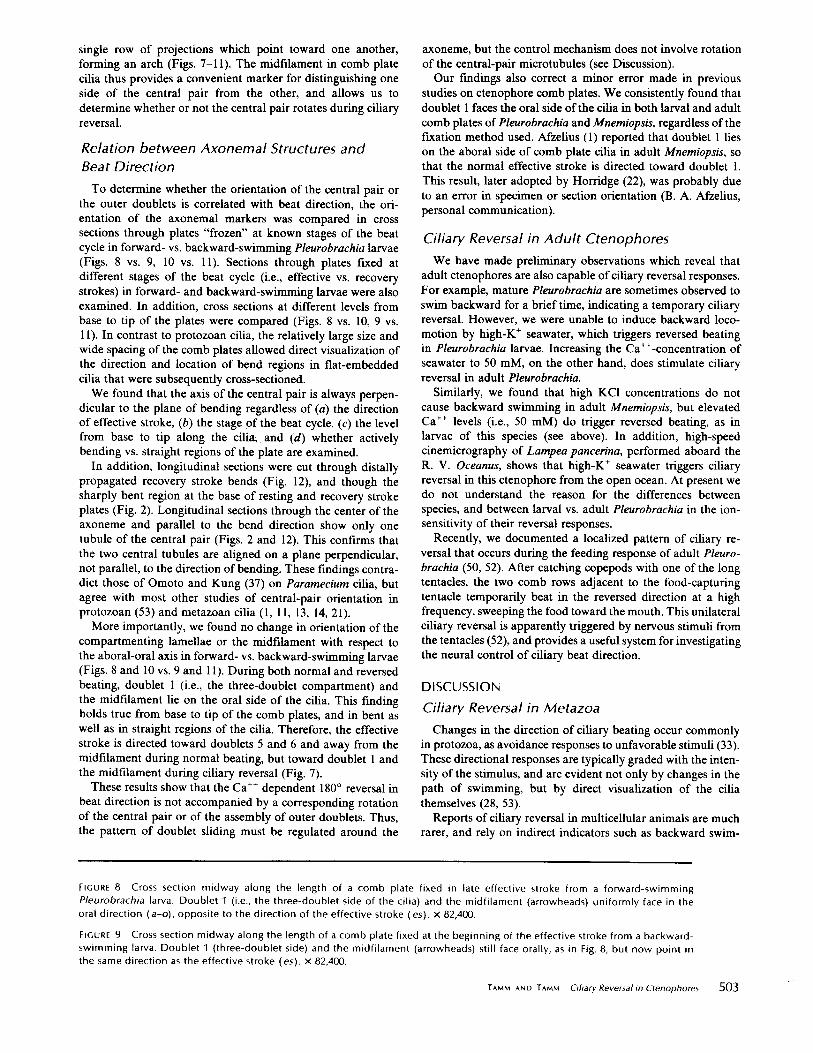
compartmenting lamellae or the midfilament with respect tothe aboral-oral axis in forward- vs. backward-swimming larvae(Figs . 8 and 10 vs. 9 and 11) . During both normal and reversedbeating, doublet 1 (i.e ., the three-doublet compartment) andthe midfilament lie on the oral side of the cilia . This findingholds true from base to tip of the comb plates, and in bent aswell as in straight regions of the cilia . Therefore, the effectivestroke is directed toward doublets 5 and 6 and away from themidfilament during normal beating, but toward doublet 1 andthe midfilament during ciliary reversal (Fig . 7).
These results show that the Ca" dependent 180° reversal inbeat direction is not accompanied by a corresponding rotationof the central pair or of the assembly of outer doublets . Thus,the pattern of doublet sliding must be regulated around the
axoneme, but the control mechanism does not involve rotationof the central-pair microtubules (see Discussion) .Our findings also correct a minor error made in previous
studies on ctenophore comb plates. We consistently found thatdoublet 1 faces the oral side ofthe cilia in both larval and adultcomb plates ofPleurobrachia and Mnemiopsis, regardless of thefixation method used. Afzelius (1) reported that doublet 1 lieson the aboral side of comb plate cilia in adult Mnemiopsis, sothat the normal effective stroke is directed toward doublet 1 .This result, later adopted by Horridge (22), was probably dueto an error in specimen or section orientation (B . A . Afzelius,personal communication) .
Ciliary Reversal in Adult CtenophoresWe have made preliminary observations which reveal that
adult ctenophores are also capable ofciliary reversal responses.For example, mature Pleurobrachia are sometimes observed toswim backward for a brief time, indicating a temporary ciliaryreversal . However, we were unable to induce backward loco-motion by high-K+ seawater, which triggers reversed beatingin Pleurobrachia larvae . Increasing the Ca'-concentration ofseawater to 50 mM, on the other hand, does stimulate ciliaryreversal in adult Pleurobrachia .
Similarly, we found that high KCl concentrations do notcause backward swimming in adult Mnemiopsis, but elevatedCa" levels (i .e., 50 mM) do trigger reversed beating, as inlarvae of this species (see above) . In addition, high-speedcinemicrography of Lampea pancerina, performed aboard theR . V. Oceanus, shows that high-K+ seawater triggers ciliaryreversal in this ctenophore from the open ocean. At present wedo not understand the reason for the differences betweenspecies, and between larval vs . adult Pleurobrachia in the ion-sensitivity of their reversal responses.
Recently, we documented a localized pattern of ciliary re-versal that occurs during the feeding response of adult Pleuro-brachia (50, 52) . After catching copepods with one of the longtentacles, the two comb rows adjacent to the food-capturingtentacle temporarily beat in the reversed direction at a highfrequency, sweeping the food toward the mouth . This unilateralciliary reversal is apparently triggered by nervous stimuli fromthe tentacles (52), and provides a useful system for investigatingthe neural control of ciliary beat direction .
DISCUSSION
Ciliary Reversal in MetazoaChanges in the direction ofciliary beating occur commonly
in protozoa, as avoidance responses to unfavorable stimuli (33) .These directional responses are typically graded with the inten-sity of the stimulus, and are evident not only by changes in thepath of swimming, but by direct visualization of the ciliathemselves (28, 53) .
Reports of ciliary reversal in multicellular animals are muchrarer, and rely on indirect indicators such as backward swim-
FIGURE 8 Cross section midway along the length of a comb plate fixed in late effective stroke from a forward-swimmingPleurobrachia larva . Doublet 1 (i .e ., the three-doublet side of the cilia) and the midfilament (arrowheads) uniformly face in theoral direction (a-o), opposite to the direction of the effective stroke (es) . x 82,400 .
FIGURE 9
Cross section midway along the length of a comb plate fixed at the beginning of the effective stroke from a backward-swimming larva . Doublet 1 (three-doublet side) and the midfilament (arrowheads) still face orally, as in Fig . 8, but now point inthe same direction as the effective stroke (es) . x 82,400 .
TAMM AND TAMM
Ciliary Reversal in Ctenophores
503

504
THE JOURNAL OF CELL BIOLOGY " VOLUME 89, 1981

ming and changes in direction of particle transport or watercurrents (2) . In no case has reversal in the direction of theeffective stroke been visualized directly in any metazoan sys-tem, and alternative explanations have not been ruled out incertain cases (2) . In ctenophores, reversal in the beat directionof comb plates has been claimed by some workers, but deniedby others (cf. reference 52 for review). Although indirect andcontradictory, these reports strongly suggest that under certainconditions-not clearly defined-ciliary reversal can occur inctenophores .By devising ionic conditions under which ciliary reversal is
consistently and reproducibly obtained in ctenophore larvae,and by carefully documenting this response with high-speedcinemicrography, we have directly demonstrated the phenom-enon of ciliary reversal in ctenophores-and in metazoa-forthe first time . By so doing, we have been able to use the uniquefeatures ofcomb plate cilia to analyze the structural control ofciliary bend direction .
Ca ++ -Dependence of Directional andFrequency Responses
Ion substitution and blocking experiments show that externalCa" is required for KCl-induced reversal of beat direction inPleurobrachia larvae . Presumably, Ca" activates the ciliaryreversal mechanism itself, as in Paramecium (8, 10, 26, 29, 34) .The direct involvement of Ca" is now being tested by deter-mining the effects of Ca" on the beat direction of ATP-reactivated, demembranated comb plates (S . L. Tamm. Inpreparation) .
In addition, Ca" may also be necessary for synaptic trig-gering of reversal, because the comb plate cells are innervated(Fig . 2) . In adult ctenophores, the nervous system is thought tomediate both inhibitory and excitatory responses of combplates, as well as unilateral ciliary reversal which occurs duringfeeding of Pleurobrachia (52 ; see above) . The possible role ofneurociliary synapses in triggering the global ciliary reversal oflarvae is currently under investigation.The close association between ciliary reversal and increased
beat frequency, together with the even greater beat frequencycaused by high Ca' concentrations (Table I), indicate thatCa" regulates both parameters ofciliary activity in Pleurobra-chia larvae . Under certain conditions, however, the directionaland frequency responses of comb plates become uncoupled . Inhigh-K+ ASW, for example, ciliary reversal lasts only 3-5 min,but the increase in beat frequency persists for a longer time .Raising the Mg" concentration of high-K+ ASW causes anincreased beat frequency without an accompanying ciliaryreversal . Similarly, an increase in beat frequency and speed offorward swimming can be elicited by mechanical stimulationof the tentacles (52).
Therefore, the directional and frequency responses may havedifferent thresholds and/or sensitivities to Ca" concentration .High Mg" concentrations presumably compete with externalCa", thereby decreasing the Ca' influx into comb plate cells
and reducing the internal free Ca" concentration. This possi-bility suggests that beat frequency, but not ciliary reversal, mayhave a bimodal dependence on intracellular Ca" concentra-tion, with frequency maxima occurring at both low and highCa" concentrations. A similar bimodal relation between beatfrequency and internal Ca" concentration has been proposedto explain the frequency responses of living and demembra-nated Paramecium (10, 29, 30, 34) .The ionic basis of the reversal response in Mnemiopsis larvae
has not yet been investigated . Nor is it known why increasedconcentrations of KCl do not stimulate ciliary reversal in thisctenophore, as in Pleurobrachia larvae . However, the ability toinduce reversed beating in Mnemiopsis by an increase in exter-nal Ca" concentration indicates that ciliary reversal in thisspecies is also controlled by Ca".
Evidence for a Radial Switching Mechanismin Cilia
Because of the single polarity of active sliding betweenmicrotubule doublets (40), most investigators assume that beatdirection is regulated by some type of switching mechanismthat activates or changes the effectiveness of doublet slidingaround the axoneme (5, 7, 29, 36, 37, 39, 40, 44, 53, 55).
Such a regulatory mechanism has been invoked to explaindifferentiation of the beat cycle into effective and recoverystrokes, as well as angular shifts in the orientation ofthe entirecycle during ciliary reversal. The role of Ca" in the formercase is problematical: intracellular free Ca" concentrationdoes not appear to change during the normal beat cycle. Onthe other hand, Wais-Steider and Satir (55) have reported thatthere are two different switching mechanisms in gill cilia, withthe switch at the end of the recovery stroke being Ca"-sensitive . Because Ca" has clearly been shown to determinethe direction of the beat cycle (8, 10, 26, 29, 34), a switchingmechanism controlling ciliary reversal must be Ca" depend-ent, and is probably distinct from that governing the transitionbetween effective and recovery phases .
Regardless of the number and kinds of switching mecha-nisms postulated, an obvious prediction of this type of regula-tion is that the assembly of outer doublets should not rotatewith bend direction . This simple prediction has never beendemonstrated heretofore . To the contrary, Gibbons (14) re-ported a systematic twisting of the outer doublets and centralpair in sea urchin sperm, suggesting that the doublets twistwith respect to the plane of bending in different regions of theflagellum. Woolley (60) found that the plane of each successivebending cycle of golden hamster sperm tails twists as it prog-resses along the flagellum, and suggested that a twisted-planewave form containing no twist in the axoneme itself would, ifartifactually flattened, give Gibbon's results. Holwill et al . (19)recently reinterpreted Satir's (42) results on Elliptio cilia tosupport a model incorporating axonemal twist during a three-dimensional recovery stroke .Our earlier work on Opalina showed that the shifts in central-
FIGURE 10
Cross section through the base of a comb plate in mid-to-late effective stroke from a forward-swimming Pleurobrachialarva. The orientation of the outer doublets and midfilament (arrowheads) with respect to the direction of effective stroke (es) andthe aboral-oral axis (a-o) is the same as in Fig. 8. x 82,400 .
FIGURE 11
Cross section through the base of a comb plate at the beginning of the effective stroke from a backward-swimminglarva. The orientation of the outer doublets and midfilament (arrowheads) with respect to beat direction (es) and the aboral-oralaxis (a-o) is the same as in Fig. 9. x 82,400 .
TAMM AND TAMM
Ciliary Reversal in Ctenophores
505

506
THE JOURNAL OF CELL BIOLOGY " VOLUME 89, 1981

pair orientation that accompany changes in beat direction alsooccur near the base of the cilia (53) . Because neither rotationofthe basal bodies nor a great degree ofdoublet twisting withina very short basal segment of the cilia seemed likely to us, weinferred that only the central pair, not the entire 9 + 2, rotateswith beat direction . Omoto and Kung (37) did not find a largeamount of axonemal twisting just above the basal body inParamecium cilia; however, they reported that limited twistingof the outer doublets and central pair does occur, but inopposite senses, leading them also to rule out rotation of thewhole axoneme during beating . Nevertheless, the lack of struc-tural markers for numbering the doublet microtubules in pro-tozoan cilia prevented a conclusive answer to this question .We have overcome this difficulty by using the compart-
menting lamellae of comb plate cilia as markers for specificdoublets . We found that the array of outer doublets does nottwist during the effective and recovery phases of the planarbeat cycle, nor does it rotate during Ca'-dependent 180°reversals in beat direction .
Therefore, reversal of the direction of beating, as well astransitions from effective to recovery strokes, must involvechanges in activation or effectiveness of doublet sliding onopposite sides of the axoneme . This finding is direct evidencefor a radial switching mechanism regulating doublet sliding incilia .
Active or Passive Role of the Central Pair?A major result of this paper concerns the nature of the
switching mechanism that controls the direction of the ciliaryeffective stroke . A regulatory role for the central pair micro-tubules was first suggested by earlier studies on metazoan ciliawith a fixed direction of beat . In general, these studies showedthat the direction of the effective stroke was perpendicular tothe axis ofthe central pair (1, 11, 13, 21) .Tamm and Horridge (53) tested the validity of this relation-
ship by applying instantaneous fixation to Opalina cilia thatcan change their direction of beating . They found that thecentral pair was always perpendicular to the plane of bendingat any level along the cilium, regardless of 90° changes in thedirection of beat, and also during the three-dimensional recov-ery stroke . This correlation clearly showed that the orientationof the central pair is related to the plane of bending but leftunanswered the problem of causal relations . That is, the "angleofthe central fibers could be the cause [ofthe direction of beat]
. . . which by its rotation releases the bending forces sequentially. . . or rotation of the central pair could equally well be aconsequence of the bending" (53). Because the directionalresponses of protozoan cilia are usually <180°, the results wereconsistent with either an active or a passive role of the centralpair (Fig . 13) .
Recently, Omoto and Kung (36, 37) have extended theseobservations to show that the central pair near the base ofParamecium cilia also undergoes beat-dependent changes inorientation, and may in fact rotate 360° during each beat cycle .Although these authors did not relate central pair orientationto bend direction during specific parts of the beat cycle, they
reported that the central pair as seen in longitudinal sectionswas parallel, not perpendicular, to the plane of bending, andthus could not be passively twisted by the motion ofthe cilium .However, because the cilia in these sections were not identifiedwith respect to stage in the beat cycle or part ofthe metachronalwave, it could not be directly ascertained that they trulyrepresented instantaneous images of active bends (as in Fig .12) .
Nevertheless, it was argued that rotation of the central pair,rather than being a passive process, may be an active one, atleast in certain parts of the beat cycle, and that the orientationof the central pair determines the pattern of active slidingaround the axoneme (36, 37) .The ctenophore ciliary reversal system has allowed a direct
test of the active role of the central pair in regulating doubletsliding, without the ambiguities associated with correlativestudies on protozoan cilia (Fig. 13) . The planar beat cycle,ultrastructural markers, and 180° reversal response of combplates permits the relation between central pair orientation andbend direction to be analyzed on a causal basis for the firsttime (Fig . 13 a and b) . In comb plate cilia, as in most othercilia, the axis of the central pair is always perpendicular to theplane of bending. We reasoned that if the orientation of thecentral pair actively controls the direction of beating, then itshould rotate 180° when the direction ofthe effective stroke isreversed 180° (Fig. 13 a). If, however, the orientation of thecentral pair determines the plane of bending or only representsa passive mechanical response to bending, then it should notrotate during a 180° reversal in beat direction (Fig . 13 b) . Incontrast, the possible central-pair orientations during thegraded shifts in beat direction of protozoan cilia do not offeras conclusive a test for deciding cause-effect relations (Fig . 13 cand d) .We found that the central pair does not rotate during Ca"-
dependent ciliary reversal of comb plates. Therefore, the ori-entation of the central pair does not regulate the direction ofciliary beating in ctenophores. That is, rotation of the centralpair is not the switching mechanism that signals the sequenceofdoublet sliding around the axoneme . We cannot rule out thepossibility that the central pair determines the plane ofbendingin comb plate cilia, with some other process specifying whichdirection within this plane . However, such a mechanism seemsunnecessarily complex. It seems more likely that Ca" actsdirectly on the dynein arms themselves, or on the resistiveelements which convert sliding into bending, rather than on anintermediary step such as rotation of the central pair . Thefunctional target of Ca" in the axoneme is now being inves-tigated by examining the effects of Ca" on the pattern ofATP-induced tubule extrusion in trypsin-treated comb platecilia (S . L . Tamm . In preparation) . We also found that thecentral pair does not rotate during the transition betweeneffective and recovery strokes. The orientation of the centralpair therefore does not appear to be involved in regulatingalternate-side force generation during a single beat cycle .
These findings are relevant to the problem of asymmetry inthe bend patterns of cilia and flagella . Because neither the
FIGURE 12
Longitudinal section through a distally propagated recovery-stroke bend in a comb plate from a forward-swimminglarva . The profile of only one tubule of the central pair is visible in sections through the center of the axoneme (arrowheads) . Theplane of the central pair is therefore perpendicular to the direction of active bending (see Fig . 2) . x 39,100 . Inset : tracing of a lowmagnification micrograph of the proximal part of the plate, showing the location of the bend region (rectangle) illustrated at highmagnification .
TAMM AND TAMM
Ciliary Reversal in Ctenophores
507

outer doublet assembly nor the central pair rotates when thebeat cycle is reversed, the asymmetric form ofciliary beating-i.e., the effective and recovery strokes-cannot be due to thebilateral asymmetry of the outer doublet array or to structuralasymmetries built into the central pair-central sheath complex.This result argues against the view that central sheath asym-metry is responsible for asymmetric bending in Chlamydo-monas flagella (3) . Instead, the form of beat must also begoverned by a switching mechanism (55), but with an ionicsensitivity different from that controlling the direction of beat .What, then, is the functional significance of the beat-de-
pendent changes in central pair orientation observed in proto-zoan cilia (36, 37, 53)? Until it is shown conclusively that thesechanges represent rotation of the central pair alone, and am-biguities concerning the relation between the axis of the centralpair and bend direction are resolved, an active role of thecentral pair in regulating beat direction in protozoan cilia mustremain doubtful . We believe that our conclusions on cteno-phore cilia with a two-dimensional beat cycle also hold forprotozoan cilia with three-dimensional beat cycles. In both
508
THE JOURNAL OF CELL BIOLOGY " VOLUME 89, 1981
FIGURE 13
Functional significance of possible orientations of the central-pair microtubules during 180° (a and b) vs. graded (cand d) directional responses of cilia . The normal plane of the effective stroke is shown by the white cilium and arrow in eachdiagram; the angular shift in this plane during reversal (dashed arrow) is indicated by the black cilium and arrow. The onlyconstraint on the central pair is that its axis always remains perpendicular to the plane of effective stroke (see also reference 53) .
The recovery stroke is not considered here . An ultrastructural marker is used to define one side of the central pair (arrow in ciliarycross sections ; see also references 36 and 37) . For 180° ciliary reversals typical of ctenophore comb plates (a and b), a corresponding180° rotation of the central pair (a) would indicate an active response controlling bend direction, whereas no rotation of thecentral pair ( b) would rule out this possibility . We found the latter to be true . For graded reversals found in protozoan cilia (c andd), rotation of the central pair so as to coincide with beat direction (c) would be consistent with an active or a passive response,and therefore not be conclusive for deciding cause-effect relations ; however, rotation of the central pair in the opposite direction(d) would rule out an active process . It is not known whether c or d is true for protozoan cilia .
cases, the direction of active bending at any level within theaxoneme must be regulated, and it seems unlikely that thebasic mechanism controlling dynein arm activity or resistiveshear interaction would be different, depending on whether theoverall motion of the organelle takes place in two or threedimensions. The advantage of analyzing a two-dimensionalcase such as ctenophore comb plates is that it allows aninvestigation of causal relations.
We thank Dr. Gary G. Borisy, Laboratory of Molecular Biology,University ofWisconsin, Madison, for the use of electron microscopefacilities and general lab support .
Observations on oceanic ctenophores were made possible by thegenerous invitation of Dr . Lawrence P. Madin, Woods Hole Oceano-graphic Institution, Woods Hole, Mass ., to allow S. L. Tamm toparticipate on a cruise aboard the R. V . Oceanus (supported byNational Science Foundation grant OCE 80-25415) .
This research was supported by National Science Foundation grantsPCM 77-09880 and 79-26459, and National Institutes of Health grantGM 27903-01 .

Received for publication 12 November 1980, and in revised form0 February 1981 .
REFERENCES
1 . Afzelius, B. A . 1961 . The fine structure of the cilia from ctenophore swimming-plates . J.Biophys. Biochem. Cyol. 9 :383-394.
2 . Aiello, E . 1974. Control of ciliary activity in Metazoa . In Cilia and Flagella, M . A . Sleigh,editor. Academic Press Inc., New York . 353-376.
3 . Bessen, M ., R . B. Fay, and G . B. Witman . 1980 . Calciu m control of waveform in isolatedflagellar axonemes of Chlamydomonas. J Cell Biol. 86:446455 .
4 . Brokaw, C . 1 . 1979 . Calcium-induced asymmetrical beating of Triton-demembranated seaurchin sperm flagella. l. Cell Biol. 82 :401411 .
5 . Brokaw, C . 1 ., and 1 . R . Gibbons. 1975 . Mechanisms of movement in flagella and cilia. InSwimming and Flying in Nature. T. Y .T. Wu, C . J. Brokaw and C . Brennan, editors .Plenum Publishing Co., New York. 1 :89-126 .
6 . Brokaw, C. J ., R . Josslin, and L. Bobrow. 1974 . Calcium ion regulation of flagellar beatsymmetry in reactivated sea urchin spermatozoa . Biochem. Biophys. Res. Commun. 58 :795-800.
7 . Doughty, M . J . 1979 . Control of ciliary activity in Paramecium. IV. Cap* modification ofMgt* dependent dynein ATPase activity . Comp. Biochem. Physiol. 64B:255-266 .
8. Eckert, R., and P. Brehm . 1979 . Ionic mechanisms of excitation in Paramecium . Annu.Rev. Biophys. Bioeng. 8 :353-383 .
9 . Eckert, R., and H . Machemer. 1975. Regulation ofciliary beating frequency by the surfacemembrane . In Molecules and Cell Movement. S . Inoue and R. E. Stephens, editors. RavenPress, New York. 151-164 .
10 . Eckert, R ., Y . Naitoh, and H. Machemer. 1976 . Calcium in the bioelectric and motorfunctions of Paramecium . Symp. Soc. Exp. Biol. 30 :233-255 .
11 . Fawcett, D . W ., and K . R. Porter . 1954 . A study of the fine structure of ciliated epithelia .J. Morphol 94:221-281 .
12 . Gibbons, B . H . 1980 . Intermittent swimming in live sea urchin sperm . J. Cell Biol. 84 :1-12.
13 . Gibbons, 1 . R. 1961 . The relationship between the fine structure and direction of beat in .gill cilia of a lamellibranch mollusc. J. Biophys. Biochem. Cylol. 11 : 179-205 .
14 . Gibbons, I . R. 1975. The molecular basis of flagellar motility in sea urchin spermatozoa .In Molecules and Cell Movement . S. Inoue and R. E. Stephens, editors . Raven Press,New York . 207-232.
15 . Gibbons, 1 . R . 1977 . Structure and function of flagellar microtubules . In InternationalCell Biology 1976-1977 . B . R . Brinkley and K . R. Porter, editors . The RockefellerUniversity Press, New York. 348-357 .
16 . Gibbons, B . H ., and I. R. Gibbons. 1973. The effect of partial extraction of dynein armson the movement of reactivated sea urchin sperm . J. Cell Sci. 13:337-357 .
17 . Gibbons, B . H., and 1. R. Gibbons . 1980. Calcium-induced quiescence in reactivated seaurchin sperm. J. Cell Biol. 84:13-27 .
18 . Hernandez-Nicaise, M . L . 1973 . The nervous system of Ctenophora. 111 . Ultrastructure ofsynapses. J. Neurocyiol. 2 :249-263 .
19 . Holwill, M . E. J ., H . J . Cohen, and P. Satir . 1979. A sliding microtubule model incorpo-rating axonemal twist and compatible with three-dimensional ciliary bending . J. Exp. Biol.78:265-280 .
20. Holwill, M . E. J., and l . L . McGregor. 1976. Effect s of calcium on flagellar movement inthe trypanosome Crithidia oncopelti. J. Exp. Biol. 65:229-242 .
21 . Horridge, G . A . 1965 . Macrocilia with numerous shafts from the lips of the ctenophoreBeroe. Proc. Roy. Soc. Land. B Biol. Sci. 162 :351-364.
22. Horridge, G . A., and B . Mackay. 1964. Neurociliary synapses in Pleurobrachia (Cteno-phora). Q. J. Microsc. Sci. 105 :163-174.
23 . Horridge, G . A ., and S . L . Tamm. 1969. Critica l point drying for scanning electronmicroscopic study of ciliary motion . Science (Wash. D. C.) . 163 :817-818 .
24. Hyams, 1 . S., and G . G . Borisy . 1978 . Isolated flagellar apparatus of Chlamydomonas:characterization of forward swimming and alteration of waveform and reversal of motionby calcium ions in vitro. J. Cell Sci. 33 :235-253 .
25 . Kamimura, S ., and K. Takahashi. 1980. The velocity of microtubule sliding in reactivated,trypsin-treated axonemes of sea urchin sperm flagella. Eur. J. Cell Biol. 22:301 .
26. Kung, C ., S . Y . Chang, Y . Satow, J . Van Houten, and H . Hansma. 1975, Genetic dissectionof behavior in Paramecium. Science (Wash . D. C.) . 188:898-904.
27. Machemer, H . 1970 . Primate and induzierte Bewegungsstadien bei Osmium Saurefixi-erung vorwartsschwimmender Paramecien. Arta Protozool. 7:531-535 .
28 . Machemer, H . 1974 . Ciliary activity and metachronism in Protozoa. In Cilia and Flagella,M . A. Sleigh, editor. Academic Press Inc., New York . 199-286.
29 . Machemer, H . 1977 . Motor activity and bioelectric control of cilia. Forischr. Zool. 24:195-210 .
30. Machemer, H ., and R . Eckert . 1975 . Ciliary frequency and orientational responses toclamped voltage steps in Paramecium . J. Comp. Physiol. 104:247-260 .
31 . Machemer, H ., and J . De Peyer . 1977. Swimming sensory cells: electrical membraneparameters, receptor properties, and motor control in ciliated Protozoa . Verh . Disch. Zool.Ges. 1977:86-110.
32 . Murakami, A ., and K . Takahashi . 1975 . The role of calcium in the control of ciliarymovement in Mytilus. II . The effects of calcium ionophores X537A and A23187 on thelateral gill cilia. J. Fac. Sci. Univ. Tokyo Sect. IV Zool. 13 :251-256 .
33 . Naitoh, Y ., and R . Eckert. 1974 . The control of ciliary activity in Protozoa. In Cilia andFlagella, M . A . Sleigh, editor. Academic Press Inc., New York. 305-352.
34. Naitoh, Y ., and H. Kaneko. 1973 . Control of ciliary activities by adenosine-triphosphateand divalent cations in Triton-extracted models of Paramecium caudatum. J. Exp. Biol. 58 :657-676 .
35 . Ogawa, K ., T . Mohri, and H . Mohri . 1977 . Identification of dynein as the outer arms ofsea urchin sperm axonemes. Proc. Nod. Acad. Sci. U. S. A. 74 :5006-5010.
36. Omoto, C . K ., and C . Kung . 1979. The pair of central tubules rotates during ciliary beatin Paramecium. Nature (Land.) . 279:532-534.
37. Omoto, C . K ., and C. Kung. 1980 . Rotation and twist of the central-pair microtubules inthe cilia of Paramecium. J . Cell Biol. 87:33-46 .
38 . Parducz, B. 1967. Ciliary movement and coordination in ciliates. Int. Rev. Cylol. 21 :91-128 .
39. Rosenthal, E. T., and R. W . Linck . 1979 . Sequentia l regulation of doublet microtubulesliding in demembranated rat sperm models . J. Cell Biol. 83(2, Pt . 2):18 to (abstr.) .
40. Sale, W . S., and P. Satin 1977 . Direction of active sliding of microtubules in Tetrahymenacilia. Proc. Nall. Acad. Sci. U. S. A . 74:2045-2049.
41 . Satir, P. 1963 . Studies on cilia: the fixation of the metachronal wave. J. Cell Biol. 18:345-365 .
42 . Satir, P . 1968 . Studies on cilia. III . Further studies of the cilium tip and a "sliding filament"model of ciliary motility. J. Cell Biol. 39:77-94 .
43 . Satir, P . 1974. The present status of the sliding microtubule model of ciliary motion. InCilia and Flagella, M. A. Sleigh, editor . Academic Press Inc ., New York . 131-142 .
44. Satir, P., and W. Sale . 1977 . Tails of Tetrahymena. J, Protozool. 24 :498-501 .45 . Schmidt, J . A., and R . Eckert. 1976. Calcium couples flagellar reversal to photostimulation
in Chlamydomonas reinhardtii. Nature (Land.). 262 :713-715 .46 . Summers, K. E ., and I. R . Gibbons. 1971 . Adenosine triphosphate-induced sliding of
tubules in trypsin-treated flagella of sea urchin sperm. Proc. Nall. A cad. Sci. U. S . A . 68 :3092-3096 .
47, Tamm, S. L . 1972 . Ciliary motion in Paramecium . A scanning electron microscope study .J. Cell Biol. 55 :250-255 .
48. Tamm, S . L . 1973. Mechanisms of ciliary coordination in ctenophores . J. Exp. Biol. 59 :231-245.
49 . Tamm, S . L . 1979 . Ionic and structural basis of ciliary reversal in ctenophores . J. Cell Biol.83(2, Pt . 2) :174a (Abstr .).
50. Tamm, S . L . 1980 . Cilia and ctenophores . Oceanus. 23 :50-59.51 . Tamm, S . L. 1980 . The mechanism of infra-plate ciliary synchrony in ctenophores. Biol.
Bull. (Woods Hole) . 159:446 (Abstr .).52. Tamm, S . L . 1981 . Ctenophores. In Electrical Conduction and Behaviour in 'Simple'
Invertebrates . G . A . B. Shelton, editor. Oxford University Press, Oxford, England . Inpress.
53 . Tamm, S. L., and G . A . Horridge . 1970. The relation between the orientation of the centralfibrils and the direction of beat in cilia of Opalina. Proc. Roy. Soc. London Ser. B. Biol.Sci. 175:219-233 .
54. Tsuchiya, T . 1977 . Effects of calcium ion on Triton-extracted lamellibranch gill cilia :ciliary arrest response in a model system . Comp . Biochem . Physiol. 56A:353-361 .
55. Wais-Steider, J ., and P. Satin 1979 . Effect of vanadate on gill cilia: switching mechanismin ciliary beat. J. Supramol. Strict. 11 :339-347.
56 . Walter, M . F ., and P . Satir. 1978 . Calcium control of ciliary arrest in mussel gill cells . J.Cell Biol. 79:110-120 .
57 . Warner, F. D ., and D . R. Mitchell . 1980 . Dynein : the mechanochemical coupling adenosinetriphosphatase of microtubule-based sliding filament mechanisms. Int. Rev. Cylol. 66:1-43 .
58. Warner, F . D., and P . Satir. 1974 . The structural basis of ciliary bend formation . Radialspoke positional changes accompanying microtubule sliding. J. Cell Biol. 63 :35-63.
59 . Witman, G . B ., J . Plummer, and G . Sander . 1978 . Chlamydomonas flagellar mutantslacking radial spokes and central tubules. Structure, composition, and function of specificaxonemal components. J. Cell Biol. 76:729-747 .
60 . Woolley, D . M . 1977 . Evidence for "twisted plane" undulations in golden hamster spermtails. J. Cell Biol. 75:851-865 .
61 . Zanetti, N . C ., D. R . Mitchell, and F. D. Warner . 1979 . Effects of divalent cations ondynein cross bridging and ciliary microtubule sliding. J. Cell Biol. 80:573-588.
TAMM AND TAMM
Ciliary Reversal in Ctenophores
509