Embed Size (px)
Citation preview
870 Lorenzi et al. Macromolecules
(14) M. Kurata and H. Stockmayer, Fortschr. Hochpo1ym.-Forsch., 3, 196 (1963).
(15) K. Karunakaran and M. Santappa, J . Polym. Sci., Part A-2, 6,713 (1968).
(16) H. Matsuda, K. Yamano, and H. Inagaki, J . PolYm. SCi., Part A-2, 7, 609 (1969).
(17) K. Matsuzaki, T. Uryu, A. Ishida, T. Ohki, and M. Takeuchi, J. Polym. Sci., Part A-1, 5, 2167 (1967).
(18) R. J. W. Le Fevre and A. Sundaram, J. Chem. SOC., 3904 (1962). (19) R. F. Curl, Jr., J. Chem. Phys., 30, 1529 (1959). (20) C. P. Smyth, “Dielectric Behavior and Structure”, McGraw-Hill,
New York, 1955. 1059 (1966). (21) R. J. W. Le Feme and K. M. S. Sundaram, J . Chem. SOC., 3188
(1963).
(22) G. P. Mikhailov, L. L. Burshtein, and V. P. Malinosvskaya,
(23) A. Kotera, K. Shimomura, and M. Shima, J. Polym. Sci., Part
(24) R. M. Masegosa, I. Hernhdez-Fuentes, E. A. Ojalvo, and E.
(25) H. G. Clark, J. polym. sei., part c , 16, 3455 (1968).
Vysokomol. Soedin., Ser. A, 11, 548, (1969).
C, 30, 233 (1970).
Saiz, Macromolecules, preceding paper.
(26) D. Doskocilova, S. Sykora, H. Pivcova, B. Obereigner, and D.
(27) T. Yoshino, Y. Kikuchi, and J. Komiyama, J. Phys. Chem., 70,
(28) K. Matsuzaki, T. Uryu, A. Ishida, and M. Takeuchi, J. Polym.
Lim, J . Polym. Sci., Part C, 23, 365 (1968).
Sci., Part C, 16, 2099 (1967).
Circular Dichroism and Conformational Equilibrium of Homopoly-L-peptides with Alkyl Side Chains in Concentrated Sulfuric Acid
Gian Paolo Loremi,* Vincenzo Rizzo,l* Finn Thoresen,lb and Lera Tomasic Technisch-chemisches Laboratorium, ETH-Zentrum, 8092, Zurich, Switzerland. Received April 16, 1979
ABSTRACT: Differences have been observed between the CD spectrum of poly-L-alanine (PA) and those of poly-L-leucine (PL), poly-L-valine (PV), and poly-L-isoleucine (PI) in 95.2% sulfuric acid and are attributed to short-range conformational order which is absent in PA and present in PL, PV, and PI. PA exhibits a CD spectrum characterized by a single negative band with the maximum a t 195 nm, consistent with the conformation of an expanded random coil. PL, PV, and PI show in addition to a negative band centered around 197 nm a less intense positive band with the maximum slightly below 220 nm. The polymers appear to be essentially completely 0 protonated in sulfuric acid, so this positive band cannot be attributed to n--A* transitions of nonprotonated peptide groups. On the basis of a comparison with the CD spectra for a series of L-isoleucine oligomers in sulfuric acid, it is proposed that this positive band derives from the interaction of electronic transitions below 220 nm of the protonated peptide groups and that it reflects the presence of short, conformationally regular sequences in the polymer chains.
The relationship between the conformational equilib- rium and the CD pattern for polypeptides with protonated peptide groups in strong acid solvents is still unclear. Some authors2+ consider polypeptides in backbone-protonating solvents to have the conformation of a random coil, and more specifically4 that of an expanded one. Tiffany and Krimm’ assert that under these conditions polypeptides should favor the extended helix (EH) conformation. This disagreement reflects different conformational interpretationsa of the CD spectrum that is typical for fully ionized poly(L-glutamic acid)g or po ly-~- lys ine~~’~ in aqueous solution. CD spectra of this type, with a large negative band slightly below 200 nm and a weak positive band near 220 nm, have also been found for all synthetic poly-L-peptides investigated so far, when strongly pro- tonating solvents, such as sulfuric a ~ i d ~ - ~ or methane- sulfonic a ~ i d , ~ ? ~ have been employed. Among the poly- peptides which in sulfuric acid give CD spectra of this type, there are5 poly-L-leucine (PL) and poly-L-valine (PV). However, in trifluoroacetic acid solution, PL and PV show no evidence of a positive CD band around 220 nm,’l nor do either poly-L-alanine or poly-L-isoleucine (P1).l1 These polypeptides have alkyl lateral chains, and pro- tonation may occur only in the main chain. Furthermore, the lateral chains contain no chromophore which would contribute to the CD in the 185-250-nm region. On these grounds, these polypeptides seemed more suitable than others for a study of the relationship between the CD pattern and the conformational equilibria of protonated
0024-9297/79/2212-0870$01.00/0
polypeptides. With this intent, we have undertaken a CD, UV, and NMR spectroscopic study of PA, PL, PV, and PI, using concentrated, aqueous sulfuric acid as solvent. Since it was expected that similar observations of oligomeric peptides of increasing chain length would greatly assist in the interpretation of particular spectral properties of these polypeptides, a series of oligo-L-alanines and oligo-L-iso- leucines was also investigated. In this paper, we report and discuss the results obtained.
Experimental Section Materials. The samples of PA and PL used in this study were
purchased from Myles-Yeda Ltd. The degree of polymerization (i i) of the sample of PA, according to the specifications, was 46. The samples of PV (ii = 19) and of PI (ii = 20) were synthesized by the NCA method using n-butylamine as the initiator. The details of the synthesis of the sample of PI have been reported e1se~here.I~ The i i ’ s of these samples were determined from the NMR spectra of trifluoroacetic acid solutions by measuring the ratio of the NH3+ end group to the peptide NH proton resonance areas. The ii of the commercial sample of PL, not specified, was also investigated by NMR and estimated to be at least 50.
The Boc- and MeO-protected oligopeptides Boc-Ala,-OMe and Boc-Ile,-OMe (n = 2 4 ) were prepared in a stepwise fashion using equivalent, conventional solution methods. The synthetic procedures used are those described for the L-isoleucine oligomers in ref 13. All oligopeptides were found to be pure by thin-layer chromatographic criteria and gave correct elemental analyses.
HC1-H-Ala-OMe and HC1.H-Ile-OMe were Fluka products. Concentrated sulfuric acid, Merck “pro analysi”, with an acid
content of 95.2% by weight (base titration) and trifluoroacetic
0 1979 American Chemical Society
Vol. 12, No. 5, September-October 1979
acid, Merck "Uvasol", were the solvents used. Preparat ion of t h e Solutions a n d Reactions with t h e
Solvents. All solutions were prepared a t room temperature by adding the solvent to a weighed amount of substance. The CD and the NMR spectra of the sulfuric acid solutions of the po- lypeptides showed no appreciable changes with time, even after the solutions stood several days a t room temperature. Thus, substantial degradation of the polypeptides under these conditions seems ruled out. Peggion et al.4 made the same conclusion for poly-L-lysine in concentrated sulfuric acid solution. The Boc- oligopeptide methyl esters reacted through their terminal groups with the acid solvents. Complete removal of the Boc group by trifluoroacetic acid after 1-2 min a t room temperature has previously been reported.14 Our 'H NMR measurements made immediately after dissolving the protected oligopeptides in this solvent showed a signal attributable to a NH3+ group, with the intensity expected for peptides with a nonprotected N terminus. Similar NMR results indicating a very rapid cleavage of the Boc group were also obtained when sulfuric acid was used as the solvent. With this solvent and with mixtures of it and tri- fluoroacetic acid, after some time, a second singlet appeared on the low field side of the singlet of COOCH3 and slowly grew a t the cost of the latter. It was observed with sulfuric acid solutions that the position of the second peak (4.84 ppm) coincided with that of the singlet of (CH3)2S04. On this basis, we attribute this splitting to a transesterification reaction between sulfuric acid and the terminal carbomethoxy group of the oligopeptides. This reaction was rather slow under the conditions adopted. For a solution prepared from 0.04 g of Boc-AlaP-OMe and 0.5 mL of sulfuric acid, the two singlets reached equal heights within about 15 h. The CD data on the oligomers, as well as that on the amino acid methyl esters reported in this paper, derive from mea- surements done immediately, mostly within 0.5 h, after the preparation of the solutions. In the NMR spectra of the oli- gopeptides, which were also taken within this time period, the second singlet was either not visible or extremely weak compared to the signal of the COOCH3 group. Thus, the spectroscopic properties reported in this paper are those of N-deblocked oli- gopeptide methyl esters (H-Ala,-OMe or H-Ile,-OMe, n = 2-6) and of amino acid methyl esters (henceforth referred to as H- Alal-OMe and H-Ilel-OMe).
Measurements. The CD spectra of the polypeptides in sulfuric acid were measured a t 27 "C with a Cary 60 equipped with the Model 6002CD attachment. The CD data are expressed in terms of molar ellipticity per residue, [e],,, in deg cm2 dmol-', derived from the equation
''-
Homopoly-L-peptides 871
PA PL PV PI
In this equation 8 is the observed ellipticity, 1 is the optical path length in decimeters, c is the concentration in g/L, and MW,,, is the molecular weight of the pertinent amino acid residue. The CD spectra of the oligomers and of the amino acid esters in sulfwic acid were measured a t room temperature on a Jasco J-40AS instrument. The CD data of these compounds also are reported as molar ellipticity per residue. This was calculated by the equation
~ i
I I I I I I
100 = Ic/MW/n
; ; : - i I : ~ : * :
I b
I I I I I 1 I I 1 I I I 1 1 I / l /
where 0,1, and c have the same meaning as in eq 1, MW is the molecular weight of the substance treated with the solvent, and n is the number of amino acid residues in a molecule. The UV spectra of the polypeptides in sulfuric acid were recorded at room temperature with a Cary 14 spectrophotometer purged with nitrogen. The UV data are expressed as the molar absorptivity per residue (eres) in cm-' M-l. The molarity was calculated as c/MW,. Solutions having c around 0.25 g/L were used for both the CD and the W measurements. The weight of the polypeptide dissolved was corrected on the basis of the microanalysis value for the nitrogen content of the substance.
The recordings of the 'H NMR spectra were carried out a t 25 OC using a Bruker WH spectrometer operating a t 90 MHz. Solutions obtained from about 0.030 g of substance and 0.5 mL of solvent were used. MelSi and CD3COCD3 were used externally as reference standard and lock substance, respectively.
Wavelength l n m l
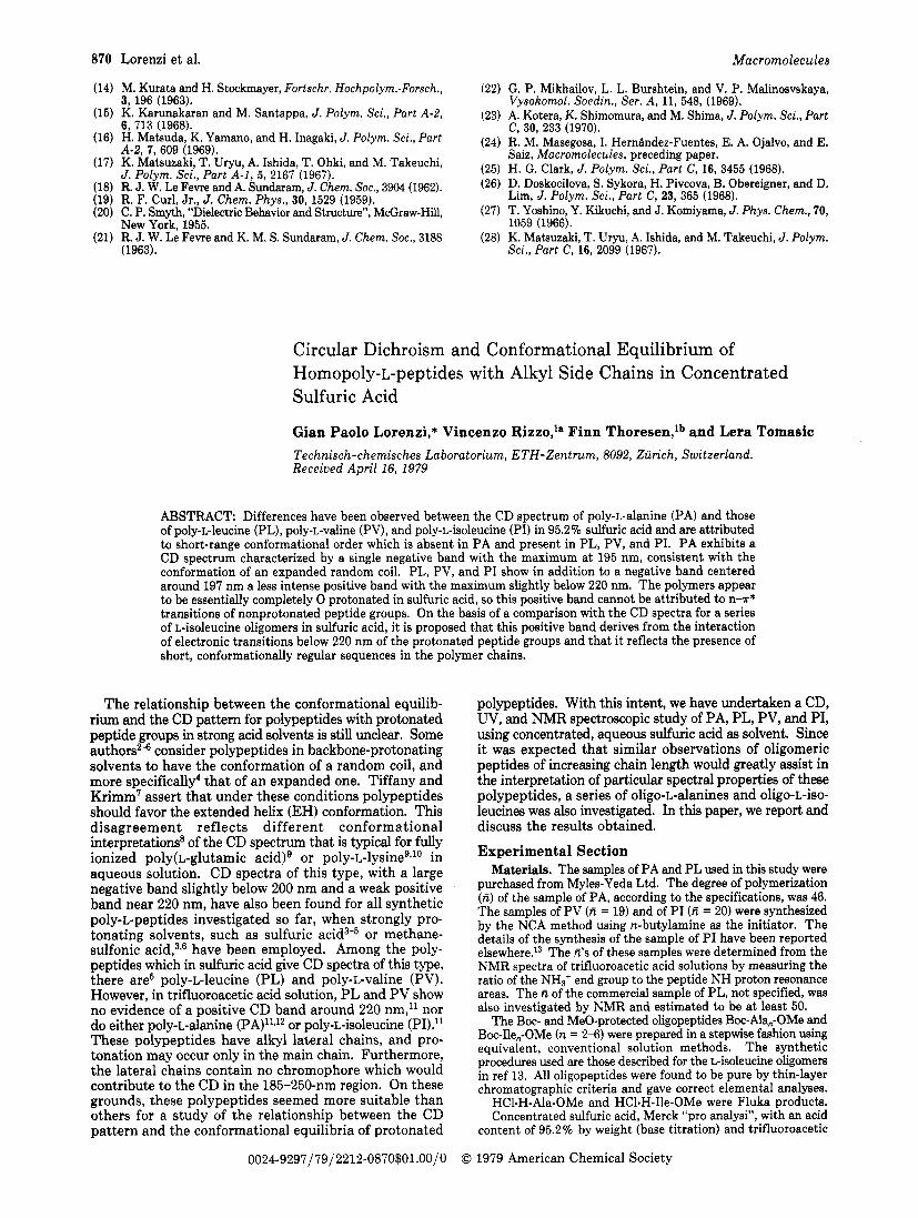
F igure 1. Circular dichroism spectra of PA, PL, PV, and P I in sulfuric acid.
Table I Position and Intensity of the Extrema of the CD Spectra
of PA, PL, PV, and PI in Sulfuric Acid Solution
poly- negative max cross- positive max P e p over, tide nm [o],, nm nm [@I, PA 195 -19 800 absent PL" 196 -18000 216 220 t 350 PVb 197 -15 200 212.5 218.5 +1100 PI 197.5 - 1 5 0 0 0 212.5 218.5 +1200
a Lit.' A E +0.14 (corresponding t o [ @ I r e s +463) at 219 nm (sample of unspecified molecular weight). Lit.S A e t 0 . 6 0 (corresponding to [o],, +1983) at 218 n m (sam- ple of unspecified molecular weight),
Results (a) CD Spectra of the Polypeptides in Sulfuric
Acid. The CD spectra of PA, PL, PV, and PI in sulfuric acid solution are shown in Figure 1. PA shows only a negative band with a maximum at 195 nm. PL, PV, and PI also show a similar negative band, but they exhibit in addition a region of weak positive ellipticities around 220 nm. The intensity and position of the CD extrema of PL are different from those of PV and PI. The CD spectra of PV and P I are almost identical. The main features of the CD spectra of Figure 1 are summarized in Table I.
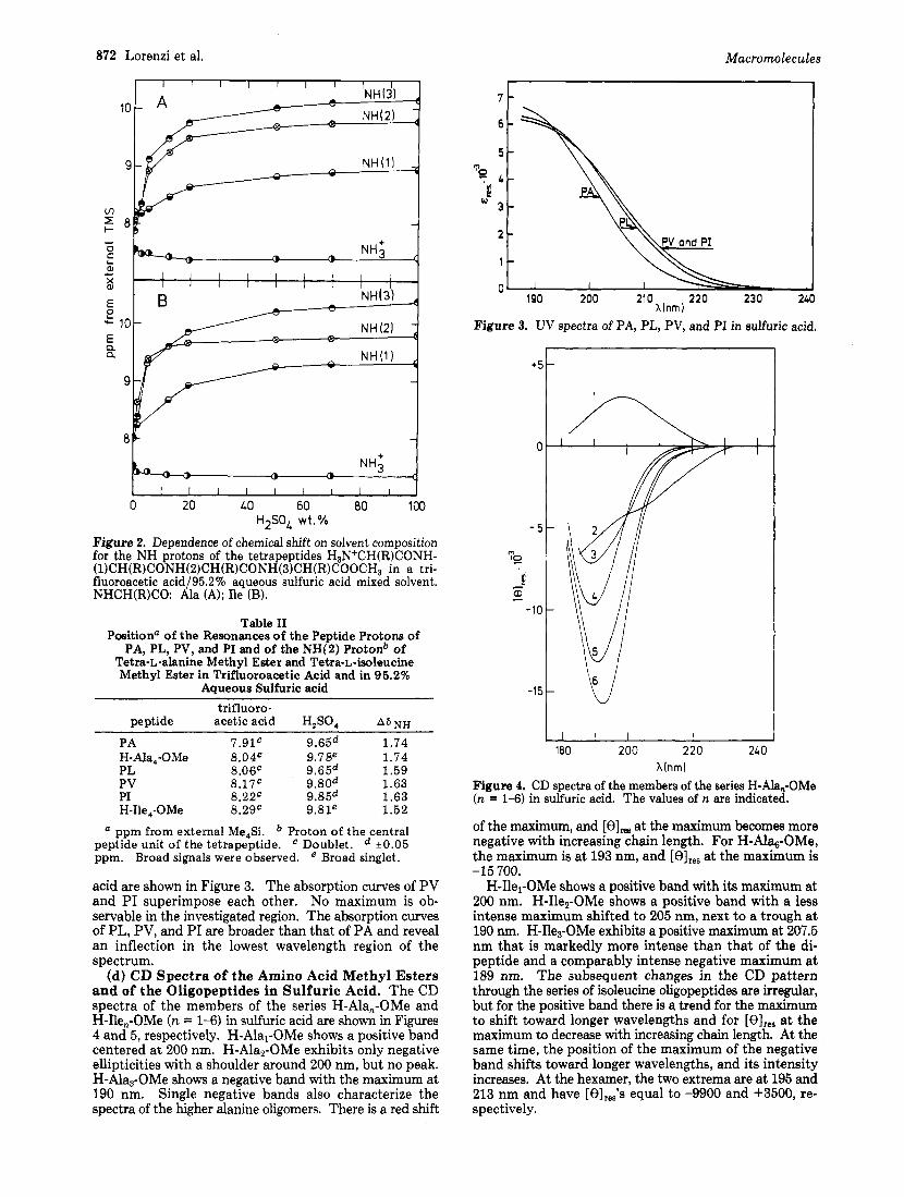
(b) 'H NMR Studies. By comparing the NMR spectra of different oligomeric peptides of the L-alanine or L- isoleucine series and by using reasonable assumptions, it was possible to assign the resonances of the various peptide protons of H-Ala4-OMe and of H-Ile,-OMe in trifluoro- acetic acid.15 Figure 2 shows that these resonances undergo a downfield shift as the solvent trifluoroacetic acid is gradually replaced with sulfuric acid. This downfield shift is initially very large but diminishes rapidly as the sulfuric acid concentration in the mixed solvent is progressively increased. About 90 to 95% of the overall shift is com- pleted when the weight concentration of sulfuric acid reaches 50%. Figure 2 also shows that the signal of the NH3+ end group protons experiences a small upfield shift of about 0.15 ppm upon changing from pure trifluoroacetic acid to a mixed solvent with 10-20% sulfuric acid but remains constant thereafter.
In the cases of PA, PL, PV, and PI, NMR measurements have been performed only with pure trifluoroacetic acid and with 95.2% aqueous sulfuric acid. The downfield shifts of the N H resonances of these polymers are com- pared in Table I1 with the overall shifts of the resonance of the proton of the central peptide unit of the two tet- ramers (NH(2), Figure 2).
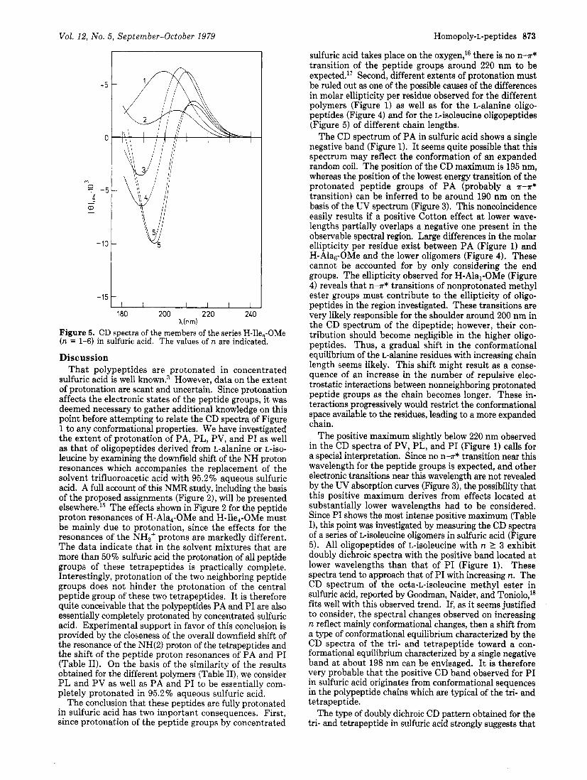
(c) UV Spectra of the Polypeptides in Sulfuric Acid. The W spectra between 187 nm (instrument cutoff wavelength) and 240 nm of the polypeptides in sulfuric
872 Lorenzi et al. Macromolecules
10
9
m 2 8
aJ .I-
8 E P - 10 E n n
9
8 Y J 0 20 LO 60 80 100
HzSOL wt .%
Figure 2. Dependence of chemical shift on solvent composition for the NH protons of the tetrapeptides H3N+CH(R)CONH- (1)CH(R)CONH(2)CH(R)CONH(3)CH(R)COOCH3 in a tri- fluoroacetic acid/95.2% aqueous sulfuric acid mixed solvent. NHCH(R)CO: Ala (A); Ile (B).
Table I1 Positiona of the Resonances of the Peptide Protons of
PA, PL, PV, and PI and of the "(2) Protonb of Tetra-L-alanine Methyl Ester and Tetra-L-isoleucine Methyl Ester in Trifluoroacetic Acid and in 95.2%
Aqueous Sulfuric acid trifluoro-
peptide acetic acid H,SO, A6 N n
PA 7.9lC 9.65d 1.74 H-Ala,-OMe 8.04c 9.78e 1.74 PL 8.06' 9.65d 1.59 PV 8.17c 9.80d 1.63 PI 8.22C 9.85d 1.63 H-Ile,-OMe 8.2gC 9.81e 1.52
ppm from external Me,Si. Proton of the central peptide unit of the tetrapeptide. ' Doublet. lt0.05 ppm. Broad signals were observed. e Broad singlet.
acid are shown in Figure 3. The absorption curves of PV and PI superimpose each other. No maximum is ob- servable in the investigated region. The absorption curves of PL, PV, and PI are broader than that of PA and reveal an inflection in the lowest wavelength region of the spectrum.
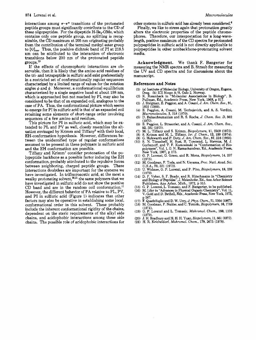
(d) CD Spectra of the Amino Acid Methyl Esters and of the Oligopeptides in Sulfuric Acid. The CD spectra of the members of the series H-Ala,-OMe and H-Ile,-OMe (n = 1-6) in sulfuric acid are shown in Figures 4 and 5, respectively. H-Alal-OMe shows a positive band centered a t 200 nm. H-Ala2-OMe exhibits only negative ellipticities with a shoulder around 200 nm, but no peak. H-Ala3-OMe shows a negative band with the maximum at 190 nm. Single negative bands also characterize the spectra of the higher alanine oligomers. There is a red shift
190 200 21 0 220 230 240 Xlnm)
Figure 3. UV spectra of PA, PL, PV, and PI in sulfuric acid.
I V -15
180 200 220 2LQ h(nrn)
Figure 4. CD spectra of the members of the series H-Ala,-OMe (n = 1-6) in sulfuric acid. The values of n are indicated.
of the maximum, and [e], at the maximum becomes more negative with increasing chain length. For H-Alh-OMe, the maximum is at 193 nm, and [e],,, at the maximum is
H-Ilel-OMe shows a positive band with its maximum at 200 nm. H-Ile2-OMe shows a positive band with a less intense maximum shifted to 205 nm, next to a trough a t 190 nm. H-Ile3-OMe exhibits a positive maximum at 207.5 nm that is markedly more intense than that of the di- peptide and a comparably intense negative maximum a t 189 nm. The subsequent changes in the CD pattern through the series of isoleucine oligopeptides are irregular, but for the positive band there is a trend for the maximum to shift toward longer wavelengths and for [e],,, at the maximum to decrease with increasing chain length. At the same time, the position of the maximum of the negative band shifts toward longer wavelengths, and its intensity increases. At the hexamer, the two extrema are at 195 and 213 nm and have [0lree's equal to -9900 and +3500, re- spectively.
-15 700.
Vol. 12, No. 5, September-October 1979 Homopoly-L-peptides 873
sulfuric acid takes place on the oxygen,16 there is no n--P* transition of the peptide groups around 220 nm to be expected.17 Second, different extents of protonation must be ruled out as one of the possible causes of the differences in molar ellipticity per residue observed for the different polymers (Figure 1) as well as for the L-alanine oligo- peptides (Figure 4) and for the L-isoleucine oligopeptides (Figure 5) of different chain lengths.
The CD spectrum of PA in sulfuric acid shows a single negative band (Figure 1). It seems quite possible that this spectrum may reflect the conformation of an expanded random coil. The position of the CD maximum is 195 nm, whereas the position of the lowest energy transition of the protonated peptide groups of PA (probably a -R--R*
transition) can be inferred to be around 190 nm on the basis of the UV spectrum (Figure 3). This noncoincidence easily results if a positive Cotton effect a t lower wave- lengths partially overlaps a negative one present in the observable spectral region. Large differences in the molar ellipticity per residue exist between PA (Figure 1) and H-Ala6-OMe and the lower oligomers (Figure 4). These cannot be accounted for by only considering the end groups. The ellipticity observed for H-Alal-OMe (Figure 4) reveals that n--R* transitions of nonprotonated methyl ester groups must contribute to the ellipticity of oligo- peptides in the region investigated. These transitions are very likely responsible for the shoulder around 200 nm in the CD spectrum of the dipeptide; however, their con- tribution should become negligible in the higher oligo- peptides. Thus, a gradual shift in the conformational equilibrium of the L-alanine residues with increasing chain length seems likely. This shift might result as a conse- quence of an increase in the number of repulsive elec- trostatic interactions between nonneighboring protonated peptide groups as the chain becomes longer. These in- teractions progressively would restrict the conformational space available to the residues, leading to a more expanded chain.
The positive maximum slightly below 220 nm observed in the CD spectra of PV, PL, and PI (Figure 1) calls for a special interpretation. Since no n l r* transition near this wavelength for the peptide groups is expected, and other electronic transitions near this wavelength are not revealed by the W absorption curves (Figure 3), the possibility that this positive maximum derives from effects located at substantially lower wavelengths had to be considered. Since PI shows the most intense positive maximum (Table I), this point was investigated by measuring the CD spectra of a series of L-isoleucine oligomers in sulfuric acid (Figure 5). All oligopeptides of L-isoleucine with n 2 3 exhibit doubly dichroic spectra with the positive band located at lower wavelengths than that of PI (Figure 1). These spectra tend to approach that of PI with increasing n. The CD spectrum of the octa-L-isoleucine methyl ester in sulfuric acid, reported by Goodman, Naider, and Toniolo,18 fits well with this observed trend. If, as it seems justified to consider, the spectral changes observed on increasing n reflect mainly conformational changes, then a shift from a type of conformational equilibrium characterized by the CD spectra of the tri- and tetrapeptide toward a con- formational equilibrium characterized by a single negative band at about 198 nm can be envisaged. I t is therefore very probable that the positive CD band observed for PI in sulfuric acid originates from conformational sequences in the polypeptide chains which are typical of the tri- and tetrapeptide.
The type of doubly dichroic CD pattern obtained for the tri- and tetrapeptide in sulfuric acid strongly suggests that
180 200 220 240 hinm)
Figure 5. CD spectra of the members of the series H-Ile,-OMe (n = 1-6) in sulfuric acid. The values of n are indicated.
Discussion That polypeptides are protonated in concentrated
sulfuric acid is well known.3 However, data on the extent of protonation are scant and uncertain. Since protonation affects the electronic states of the peptide groups, it was deemed necessary to gather additional knowledge on this point before attempting to relate the CD spectra of Figure 1 to any conformational properties. We have investigated the extent of protonation of PA, PL, PV, and PI as well as that of oligopeptides derived from L-alanine or L-iso- leucine by examining the downfield shift of the NH proton resonances which accompanies the replacement of the solvent trifluoroacetic acid with 95.2 70 aqueous sulfuric acid. A full account of this NMR study, including the basis of the proposed assignments (Figure 21, will be presented else~here. '~ The effects shown in Figure 2 for the peptide proton resonances of H-Ala4-OMe and H-Ile4-OMe must be mainly due to protonation, since the effects for the resonances of the NH3+ protons are markedly different. The data indicate that in the solvent mixtures that are more than 50% sulfuric acid the protonation of all peptide groups of these tetrapeptides is practically complete. Interestingly, protonation of the two neighboring peptide groups does not hinder the protonation of the central peptide group of these two tetrapeptides. I t is therefore quite conceivable that the polypeptides PA and PI are also essentially completely protonated by concentrated sulfuric acid. Experimental support in favor of this conclusion is provided by the closeness of the overall downfield shift of the resonance of the "(2) proton of the tetrapeptides and the shift of the peptide proton resonances of PA and PI (Table 11). On the basis of the similarity of the results obtained for the different polymers (Table 111, we consider P L and PV as well as PA and PI to be essentially com- pletely protonated in 95.2 70 aqueous sulfuric acid.
The conclusion that these peptides are fully protonated in sulfuric acid has two important consequences. First, since protonation of the peptide groups by concentrated
874 Lorenzi et al. Macromolecules
interactions among H-T* transitions of the protonated peptide groups must significantly contribute to the CD of these oligopeptides. For the dipeptide H-Ile2-OMe, which contains only one peptide group, no splitting is recog- nizable, the CD maximum at 205 nm originating probably from the contribution of the terminal methyl ester group to [e],,,. Thus, the positive dichroic band of PI a t 218.5 nm can be attributed to the interaction of electronic transitions below 200 nm of the protonated peptide groups.l9
If the effects of chromophoric interactions are ob- servable, then it is likely that the amino acid residues of the tri- and tetrapeptide in sulfuric acid exist preferentially in a restricted set of conformationally regular sequences characterized by a limited range of values for the rotation angles C#J and +. Moreover, a conformational equilibrium characterized by a single negative band at about 198 nm, which is approached but not reached by PI, may also be considered to be that of an expanded coil, analogous to the case of PA. Thus, the conformational picture which seems to emerge for PI in sulfuric acid is that of an expanded coil retaining some elements of short-range order involving sequences of a few amino acid residues.
This picture for PI in sulfuric acid, which may be ex- tended to PL and PV as well, closely resembles the sit- uation envisaged by Krimm and Tiffan? with their local, EH-conformation hypothesis. However, differences be- tween the unidentified type of regular conformations assumed to be present in these polymers in sulfuric acid and the EH conformation are possible.
Tiffany and Krimm’ consider protonation of the po- lypeptide backbone as a possible factor inducing the EH conformation, probably attributed to the repulsive forces between neighboring, charged peptide groups. These interactions doubtless are important for the systems we have investigated. In trifluoroacetic acid, at the most a wealky protonating so1vent,20~21 the same polymers that we have investigated in sulfuric acid do not show the positive CD band and are in the random coil conformation.” However, the different behavior of PA relative to PL, PV, and P I in sulfuric acid (Figure 1) indicates that other factors may also be operative in establishing some local, conformational order in this solvent. These probably include the inherent conformational rigidity of the chains, dependent on the steric requirements of the alkyl side chains, and acidophobic interactions among these side chains. The possible role of acidophobic interactions for
other systems in sulfuric acid has already been considered? Finally, we like to stress again that protonation greatly
alters the electronic properties of the peptide chromo- phores. Therefore, our interpretation for a long-wave- length, positive maximum of the CD spectra for protonated polypeptides in sulfuric acid is not directly applicable to polypeptides in other nonbackbone-protonating solvent media.
Acknowledgment. We thank F. Bangerter for measuring the NMR spectra and B. Straub for measuring the UV and CD spectra and for discussions about the manuscript.
References and Notes (a) Institute of Molecular Biology, University of Oregon, Eugene, Oreg. (b) IC1 Norge A/S, Oslo 2, Norway. K. Rosenheck in “Molecular Associations in Biology”, B. Pullman, Ed., Academic Press, New York, 1968, p 517. J. Steigman, E. Peggion, and A. Cosani, J. Am. Chem. Soc., 91, 1822 (1969). E. Peggion, A. Cosani, M. Terbojevich, and A. S. Verdini, Macromolecules, 3 , 318 (1970). D. Balasubramanian and R. S. Roche, J. Chem. SOC. D , 862 (1970). E. Peggion, L. Strasorier, and A. Cosani, J. Am. Chem. SOC., 92, 381 (1970). M. L. Tiffany and S. Krimm, Biopolymers, 11, 2309 (1972). S. Krimm and M. L. Tiffany, Zsr. J. Chem., 12, 189 (1974). G. Holzwarth and P. Doty, J. Am. Chem. SOC., 87,218 (1965). S. N. Timasheff, H. Susi, R. Townend, L. Stevens, M. J. Gorbunoff, and T. F. Kumosinski in “Conformation of Bio- Dolvmers”. Vol. 1. G. N. Ramachandran. Ed.. Academic Press, Ne; York, 1967,’~ 173. G. P. Lorenzi, G. Greco, and R. Mona, Biopolymers, 14, 237 . .
(1975). M. Goodman, F. Toda, and N. Ueyama, R o c . Natl. Acad. Sci. U.S.A., 70, 331 (1973). U. Widmer, G. P. Lorenzi, and P. Pino, Biopolymers, 18,239 (1979). D. F. Veber, S. F. Brady, and R. Hirschmann in “Chemistry and Biology of Peptides”, J. Meienhofer, Ed., Ann Arbor Science Publishers, Ann Arbor, Mich., 1972, p 315. G. P. Lorenzi, L. Tomasic, and F. Bangerter, to be published. M. Liler in “Advances in Physical Organic Chemistry”, Vol. 11, V. Gold and D. Bethell, Eds., Academic Press, New York, 1975, p 267. F. Quadrifoglio and D. W. Uny, J. Phys. Chem., 71,2364 (1967). M. Goodman, F. Naider, and C. Toniolo, Biopolymers, 10,1719 (1971). G. P. Lorenzi and L. Tomasic, Makromol. Chem., 180, 1103 (1979). J. H. Bradbury and H. H. H. Yuan, Biopolymers, 11,661 (1972). H. R. Kricheldorf, Makromol. Chem., 179, 2675 (1978).