Embed Size (px)
Citation preview
pubs.acs.org/Biochemistry Published on Web 02/17/2010 r 2010 American Chemical Society
2464 Biochemistry 2010, 49, 2464–2474
DOI: 10.1021/bi100036f
Circular Permutation of Bacillus circulans Xylanase: A Kinetic and Structural Study†
Stephan Reitinger,‡, ) Ying Yu,§ Jacqueline Wicki,‡, ) Martin Ludwiczek,^ Igor D’Angelo,^ Simon Baturin,‡,^
Mark Okon,‡,^ Natalie C. J. Strynadka,^,#,) Stefan Lutz,§ Stephen G. Withers,*,‡, ),^ and Lawrence P. McIntosh*,‡, ),^,#
‡Department of Chemistry, University of British Columbia, Vancouver, British Columbia, Canada V6T 1Z1, §Department ofChemistry, Emory University, 1515 Dickey Drive, Atlanta, Georgia 30322, )Centre for High Throughput Biology, University of BritishColumbia, Vancouver, British Columbia, CanadaV6T 1Z1, ^Department of Biochemistry andMolecular Biology, University of BritishColumbia, Vancouver, British Columbia, Canada V6T 1Z3, #Michael Smith Laboratory, University of British Columbia, Vancouver,British Columbia, Canada V6T 1Z4, and )Centre for Blood Research, University of British Columbia, Vancouver, British Columbia,
Canada V6T 1Z3
Received January 11, 2010; Revised Manuscript Received February 16, 2010
ABSTRACT: The 20 kDa Bacillus circulans Bcx is a well-studied endoxylanase with a β-jellyroll fold thatplaces its N- and C-termini in salt bridge contact. Initial experiments verified that Bcx could be circularlypermuted by PCRmethods to introduce new termini in loop regions while linking its native termini directlyor via one or two glycines. Subsequently, a library of circular permutants, generated by random DNasecleavage of the circularized Bcx gene, was screened for xylanase activity on xylan in Congo Red-stainedagar. Analysis of 35 unique active circular permutants revealed that, while many of the new termini wereintroduced in external loops as anticipated, a surprising number were also located within β-strands.Furthermore, several permutations placed key catalytic residues at or near the new termini with minimaldeleterious effects on activity and, in one case, a 4-fold increase. The structure of one permutant wasdetermined by X-ray crystallography, whereas three others were probed by NMR spectroscopy. Thesestudies revealed that the overall conformation of Bcx changed very little in response to circularpermutation, with effects largely being limited to increased local mobility near the new and the linkedold termini and to a decrease in global stability against thermal denaturation. This library of circularlypermuted xylanases provides an excellent set of new start points for directed evolution of this commerciallyimportant enzyme, as well as valuable constructs for intein-mediated replacement of key catalytic residueswith unnatural analogues. Such approaches should permit new insights into the mechanism of enzymaticglycoside hydrolysis.
The endoxylanase Bcx1 from Bacillus circulans is a 20 kDaglycoside hydrolase that has served as an excellentmodel systemfor understanding enzyme mechanisms in general and glyco-sidase mechanisms in particular. The insights obtained not onlyare of academic interest but also have substantial commercialimportance since xylanases are widely used in commercial foodpreparation, as well as in the kraft pulp and paper industryand potentially in biofuel generation. Consequently, Bcx and
other closely related GH family 11 xylanases (1) have beensubjected to numerous studies on the structural and functionallevel (2).
Bcx degrades xylan through a retaining, double-displacementmechanism involving a glycosyl-enzyme intermediate (3-6).Formation and hydrolysis of this covalent intermediate proceedsvia oxocarbenium ion-like transition states, with the assistance oftwo key catalytic glutamic acids. One residue (Glu78) acts as anucleophile, attacking the anomeric carbon of the substrate. Theother (Glu172) functions as a general acid/base catalyst, assistingin the formation of the intermediate by proton donation, as wellas in the subsequent hydrolysis step by deprotonating theattacking nucleophilic water. The active site residues of Bcx, aswell as the dynamic and electrostatic properties of the glycosyl-enzyme intermediate, have been characterized extensively byX-ray crystallography and NMR spectroscopy (5, 7-12).
Studies of Bcx, along with parallel investigations on anotherxylanase of a different tertiary fold (theGH family 10Cellulomonasfimi endoxylanase Cex) and continuing collaborative computa-tional and heavy atom isotope effect studies, have providedparticularly detailed insights into fundamental mechanisms ofcarbohydrate degradation (6, 13, 14). With this same set ofmechanistic tools, analyses have been performed on interes-ting mutants derived from site-directed and random mutagenesismethods, thereby providing additional insights into structure/function relationships. However, further understanding could be
†This researchwas supported by grants from theNatural Sciences andEngineering Research Council of Canada to L.P.M. and S.G.W. andfrom the National Science Foundation (CBET-0730312) and PetroleumResearch Fund of the American Chemical Society (PRF 47135-AC1) toS.L. Instrument support was provided by the Canadian Institutes forHealth Research, the Canadian Foundation for Innovation, the BritishColumbiaKnowledgeDevelopment Fund, the UBCBlusson Fund, andthe Michael Smith Foundation for Health Research. S.R. and M.L.acknowledge Erwin Schr€odinger Fellowships from the Austrian ScienceFund. I.D. acknowledges postdoctoral support from theMichael SmithFoundation for Health Research. S.G.W. thanks the Canada ResearchChairs program for salary support.*To whom correspondence should be addressed: L.P.M.: e-mail,
[email protected]; phone, 604-822-3341; fax, 604-822-5227. S.G.W.: e-mail, [email protected]; phone, 604-822-3042; fax, 604-822-8869.
1Abbreviations: Bcx, Bacillus circulans xylanase; cfu, colony formingunit; cp, cyclic permutant; 2F-DNPX2, 2,4-dinitrophenyl 2-deoxy-2-fluoro-β-xylobioside; HSQC, heteronuclear single-quantum correla-tion; ONPX2, 2-nitrophenyl β-xylobioside; DNPX2, 2,5-dinitrophenylβ-xylobioside; Tm, midpoint unfolding temperature.
Article Biochemistry, Vol. 49, No. 11, 2010 2465
obtained if these enzymes were amenable to circular permuta-tion, such that new start and finish positions for the proteinsequences could be generated. Not only would this allowinterrogation of the role of the dynamics of the “old” and“new” termini in enzyme structure and function but it wouldalso facilitate the specific incorporation of unnatural orlabeled amino acids into the protein by semisyntheticapproaches, for example, using intein methodologies (15).Bcx is particularly well suited to circular permutation sinceits β-jellyroll structure places its native N- and C-termini nextto each other, interacting directly via a salt bridge.
Systematic circular permutation of genes has emerged as auseful tool to conduct studies on polypeptide folding andstability (16-20). The overall tertiary structure of a protein isusually retained, since, with the exception of possible residuesintroduced to bridge the native termini, the amino acid sequenceis not changed but simply rearranged. Nevertheless, circularpermutation can significantly affect stability, dynamics, andfunction (21-27). According to the Circular Permutation Data-base (CPDB), more than 4000 naturally occurring or artificiallygenerated circularly permuted proteins have been identified todate (28). In the first reported circular permutation experiment,the termini of bovine pancreatic trypsin inhibitor (BPTI) werelinked chemically, and the circular polypeptide chain was subse-quently cleaved by proteolysis at a specific reactive site (29). Inrecent years, recombinant DNA technologies have been used togenerate circularly permuted variants of proteins such as phos-phoribosyl anthranilate isomerase (30), β-glucanases (31, 32),aspartate transcarbamoylase (33, 34), dual chain avidin (35), T4lysozyme (25), and Candida antarctica lipase B (24, 27, 36). Asortase-catalyzed transpeptidation approach to synthesize circu-lar proteins was also recently reported (37).
A particularly attractive approach, especially for the objectivesof our mechanistic study, involves the generation of libraries ofcircular permutants via random cleavage of the circularized gene.Expression of the resultant permuted gene library in Escherichiacoli, followed by activity-based screening, can lead to theidentification of active permuted enzymes. Application of thisapproach to C. antarctica lipase B not only generated a largelibrary of functional permutants but also identified some withsubstantially (up to 175-fold) increased activity (36). In thispaper, we present the enzymatic, structural, and dynamic char-acterization of a set of active, circularly permutated Bcx variantsgenerated by this random cleavage protocol. Most significantly,new termini could be introduced into the active site of the enzymewithout dramatically altering its catalytic activity, thus establish-ing the framework for future mechanistic studies, as proposedabove.
EXPERIMENTAL PROCEDURES
PCR Cloning of “Designed” Permutants. Initial clonesencoding Bcx permutants with a single glycine linker joining thenative ends (Ala1 and Trp185) and with new termini introducedat positions 102 (cpG102G1), 123 (cpA123G1), or 139 (cpG139G1)were generated by a four-primer PCR approach and placed in the
pET16b vector (Novagen) usingNcoI andXhoI restriction sites.2
Subsequent clones with linkers of zero (cpA123G0) and twoglycine residues (cpA123G2) were derived from the cpA123G1
template by PCR. Primer sequences are summarized in Support-ing Information Table S1, and the amino acid sequences of alldesigned circular permutants are listed in Supporting Informa-tion Table S2.Random Circular Permutation of Bcx. The random cir-
cular permutation of Bcx was performed as described previouslyfor C. antarctica lipase B (24). Briefly, the gene encodingcpA123G2 (with a two-glycine linker, deletion of Arg122, andthe Thr123Ala mutation) was permutated by PCRmethodologyto position the unique internalNsiI restriction site at both ends ofthe gene sequence, creating cpBcx_NsiI (Supporting InformationTable S1). In preparation for the permutation experiment,cpBcx_NsiI was cloned into the high-copy DNA plasmidpSTBlue (Novagen, Madison, WI). Following plasmid amplifi-cation inE. coliDH5R, the purified vectorwas digestedwithNsiI,and the desired 560-bp DNA fragment was isolated via agarosegel electrophoresis. Next, the linear fragment was circularized byintramolecular ligation (2 μg/mL DNA) with 30 units/mL T4DNA ligase (Promega, Madison, WI) in the manufacturer’sbuffer overnight at 16 �C. The reactionmixture was concentratedby ethanol precipitation, and the remaining linear DNA waseliminated by treatment with ExoIII (120 units/μg of DNA;Promega) for 30 min at 37 �C. After heat inactivation of ExoIII(10 min at 72 �C), the circular DNAwas recovered via QIAquickpurification (Qiagen, Valencia, CA).
The circularized DNA was linearized by limited DNase Idigestion (0.0005 units/μg of DNA; Roche, Indianapolis, IN) in50mMTris-HCl (pH 7.5) and 1mMMnCl2 at room temperaturefor 15 min. The reaction was quenched by addition of 10 μL ofEDTA (0.5M), followed by treatment with T4DNApolymerase(1 unit/μg of DNA; Promega), 150 μM dNTPs, and T4 DNAligase (2 units/μg of DNA; Promega) in T4 ligase buffer atambient temperature for 1 h to repair DNA nicks and to createblunt ends. The resulting cpBcx library was purified by agarosegel electrophoresis and cloned into pET27-PP, predigested withPstI and PacI. The pET27-PP vector is derived from pET-27b(Novagen) and carries unique PstI and PacI restriction sites(underlined), as well as three stop codons (bold) in all threereading frames in themultiple cloning site. The new cleavage siteswere introduced by primer overlap extension, using forwardprimer 50CTG CTC CTC GCT GCC CAG CCG GCG ATGGCC TGC AGA TGG ATA TCG GAA TTA ATT CG30 andreverse primer 50GAT CTC GAG TTA GTT AGT TAA TTAAGC GGC CGC AAG CTT GTC GAC30. In addition, thepET27-PP encodes a pelB leader sequence for protein secretion tothe periplasmic space.Library Screening Using a Congo Red Overlay Assay.
The circularly permuted Bcx gene library was transformed intoE. coliBL21(λDE3) and spread out on LB agar plates containing30 μg/mL kanamycin. After 12 h incubation at 37 �C, colonieswere replica-plated onto fresh LB agar media with 30 μg/mLkanamycin.While the master plate was stored at 4 �C, the replicaplates were incubated for 3 h at 37 �C prior to overlaying themwith molten agar containing 0.4% agar, 0.4% birchwood xylan(Sigma, St. Louis, MO), 30 μg/mL kanamycin, and 1 mM IPTG.Once the overlay agar had solidified, plates were incubated foranother 8 h at 37 �C. To visualize xylanase activity, the agarplates were stained with 0.5% Congo Red solution for 15 min,followed by destaining with 1 M NaCl for 30 min (38). Colonies
2Nomenclature of the circular permutants: Termini of circular per-mutantswere either joined directly (G0) or joined via the insertion of one(G1) or two (G2) glycine residues. All permutants and cpWT have theArg122 deletion and the Thr123Ala mutation except cpG102G1 andcpG139G1. Permutants, recloned to remove additional or replace miss-ing residues at the new termini, are indicated by a prime symbol (e.g.,cpN35G20).
2466 Biochemistry, Vol. 49, No. 11, 2010 Reitinger et al.
expressing functional glycoside hydrolases could be identified byformation of a clearing zone surrounding the host cells. Thecorresponding colonies on the master plate were picked andregrown for DNA sequence analysis. For initial characterizationof the resulting Bcx permutants, the pET27-PP constructs wereused (Supporting Information Table S3). For subsequent de-tailed kinetic analysis, genes of selected permutants were sub-cloned into pET-21b via the NdeI and HindIII restriction sites.The PCR primers for this step were also designed to removeadditional or replace missing residues at the new termini of thepermutants (Supporting Information Table S4),Protein Purification. Gene expression and protein purifica-
tion were carried out as described (12, 39). E. coli BL21(λDE3)cells were grown at 30 �C in LB media containing 100 μg/mLcarbenicillin (for pET16b and pET21b constructs) or 50 μg/mLkanamycin (for pET27-PP constructs), respectively, or in M9medium with 1 g/L 15NH4Cl or 1 g/L 15NH4Cl and 3 g/L[13C6]glucose (Spectra Stable Isotope Inc.). Protein expressionwas induced with 50 μM IPTG at OD600 ∼ 0.6, and cellsharvested 20 h later. For preliminary screening, proteins werepurified with only SP-Sepharose (HiTrap, 5 mL column; GEHealthcare) ion-exchange chromatography in 20 mM MESbuffer, pH 6.0, and eluted with a 0-1 M NaCl gradient. Theirconcentrations were determined by absorbance spectroscopywith a Unicam UV/vis spectrometer UV4 using the predictedε280 value of 81790 M-1 cm-1 (40) and corrected by purityestimated from SDS-PAGE images taken and analyzed withAlphaImager and AlphaEase FC software (Alpha Innotech,Corp.). For all other experiments, the permutants werefirst purified by ion-exchange chromatography, followed bySephacryl S-100 (HiPrep 16/60; GE Healthcare) size-exclusionchromatography using 20 mMMES buffer, pH 6.0, and 50 mMNaCl for activity assays or 10 mM phosphate buffer, pH 6.5, forstructural analyses. In these cases, enzyme concentrations weredetermined by active site titration.Active Site Titrations Using 2F-DNPX2. Enzyme (final
concentration of 5-20 μM) was added to a solution of 0.39 mM2,4-dinitrophenyl 2-deoxy-2-fluoro-β-xylobioside (2F-DNPX2),20 mM MES, pH 6.0, and 50 mM NaCl at 22 �C. The 2,4-dinitrophenolate released, due to covalent inactivation of theactive enzyme (41), was monitored at 400 nm. The increase in theA400 observed was corrected for the absorbance due to sponta-neous hydrolysis of 2F-DNPX2 and enzyme-catalyzed turnover.From the net ΔA400, which corresponds to the concentration of2,4-dinitrophenolate released (Δε400= 11.40mM-1 cm-1, whereΔε is the difference in molar absorptivity between 2,4-dinitro-phenol and its corresponding xylobioside at pH 6.0), the con-centration of active enzyme was determined.Steady-State Enzyme Kinetics. Two aryl β-xylobiosides
were used as substrates in the assays described below: 2,5-dinitrophenyl β-xylobioside (DNPX2), Δε440nm = 3.57 mM-1
cm-1, pKa of 2,5-dinitrophenol = 5.22, and 2-nitrophenylβ-xylobioside (ONPX2), Δε400nm = 1.07 mM-1 cm-1, pKa of2-nitrophenol = 7.22 (42). All substrates were synthesizedand characterized according to previously published proce-dures (43, 44). Spectrophotometric assays were performed usingeither a Cary 4000 or UnicamUV4 spectrophotometer in 200 μLmicroblack-walled quartz cuvettes or 1000 μL disposable metha-crylate cuvettes (42).
The second-order rate constants (kcat/Km) for the hydrolysis ofONPX2 and DNPX2 were determined from progress curvesat low substrate concentrations using the substrate depletion
method (6). Enzyme was added to reaction mixtures containingsubstrate concentrations less than (1/5)Km, and the release ofnitrophenolate was monitored until substrate depletion wasobserved. The change in absorbance with respect to time wasfitted to a first-order rate equation using the program GraFit5.0 (45).
Michaelis-Menten steady-state parameters for the hydrolysisof DNPX2 were determined at six different substrate concentra-tions ranging from 0.2 to 2 times the estimated Km value. Therelative insolubility of DNPX2 precluded study at higher sub-strate concentrations. From the experimental rate versus sub-strate concentration data, Km and kcat values were calculateddirectly using GraFit 5.0 (45).Thermal DenaturationMeasurements. Circular dichroism
(CD) spectra were measured on a JASCO J-810 spectropolari-meter. A 1 mm path length cell containing protein at a concen-tration of 8 μM in 20 mM sodium phosphate buffer, pH 7.0, wasused. The long-lived glycosyl-enzyme intermediate forms weregenerated by treatment with excess 2F-DNPX2 for at least 3 h.Thermal denaturation curves were recorded by monitoring thesignal at 219 nm as a function of temperature, increasing at a rateof 1 �C/min. The midpoint unfolding temperature, Tm, wascalculated by nonlinear least-squares data fitting to a standardequation describing a two-state conformational equilibrium (46).X-ray Crystallography. Crystals of cpA123G1 were grown
at 4 �C using the hanging drop method by equilibrating 2 mL ofpurified protein solution (25-30 mg/mL) against an equalvolume of 13-20% saturated (NH4)2SO4 in 40 mM Tris-HCl,pH 8.0. Prior to data collection, all crystals were seriallytransferred for a few seconds into cryoprotectant solutionscomposed of mother liquor supplemented with 5-25% glyceroland then frozen in liquid nitrogen. X-ray data collection wasperformed under cryogenic conditions (100 K) using an in-houserotating anode X-ray generator (Cu KR radiation λ= 1.541 A).Data were recorded using an image plate detector and processedusing HKL2000 (47). The wild-type Bcx coordinates 1HV1.pdbwere used as a search model for molecular replacement withPHASER (48). The correct solution was then used for modelrebuilding and structure refinement with CNS (simulated an-nealing) (49) and REFMAC (maximum likelihood functions)(50), alternating with manual adjustments using COOT (51). Arandom sample containing roughly 5% of the total number ofreflections was excluded from the refinement and used for thecalculation of the free R factor. Tight noncrystallographicsymmetry restraints and geometry were maintained throughoutall of the different steps of refinement and then partially relaxedat the final stage. Care was taken to avoid overfitting thestructure by reducing the X-ray data weighting term duringrefinement.Water molecules were assigned at 3σ residual Fo- Fc
electron density areas located within 3 A from the protein andconfirmed by visual inspection. All models displayed acceptablestereochemical geometries, with >95% of the residues in themost favorable regions of the Ramachadran plot. SupportingInformation Table S5 provides a summary of this crystallo-graphic analysis. The final coordinates of cpA123G1 were depos-ited in the RCSB Protein Data Bank under accession code 3LB9.Structural figures were made using PYMOL (52).NMR Spectroscopy. NMR spectra were recorded at 25 �C
with Varian Unity 500 MHz and cryoprobe-equipped Inova600 MHz spectrometers. Data were processed with NMRpipe(53) and analyzed using Sparky (54). Samples of 13C/15N-labeledcpWT (0.94 mM), 13C/15N-labeled cpN35G20 (0.7 mM), and
Article Biochemistry, Vol. 49, No. 11, 2010 2467
15N-labeled cpY94G20 (0.04 mM) were in 10 mM sodiumphosphate buffer, pH 6.5, with 5% D2O lock solvent. Thespectral assignments of main chain nuclei in cpWT and cpN35G20
were obtained from 15N-HSQC, HN(CA)CB, CBCACONH,HNCO, HN(CA)CO, and 15N-editted TOCSY- and NOESY-HSQC spectra (55), combined with knowledge of the previouslyreported assignments for the wild-type protein (9, 56).
Backbone amide 15N T1 and T2 relaxation and heteronuclear1H-15NNOE experiments were performedwith 13C/15N-labeledcpWT and cpN35G20 at 25 �C on an Inova 600 MHz spectro-meter (57). The T1 and T2 values were determined using anonlinear, least-squares fitting of the intensity of the cross-peaksto an exponential decay in the Sparky software. NOEvalues wereobtained by comparing the ratios of cross-peak intensities withand without proton saturation. Anisotropic tumbling and inter-nal dynamics parameters calculated from the relaxation datawere processed according to the model-free formalism of Lipariand Szabo (58), using TENSOR2 (59).
RESULTS
Creation of Rational Design Permutants. As an initialfeasibility study, three circular permutants (cpG102G1,cpA123G1, and cpG139G1) were generated by rational design.The 2.7 A distance between the native N- and C-termini of Bcxwas bridged with a single glycine residue, and new termini wereintroduced at the indicated positions (Supporting InformationTable S2). The sites were selected due to their locations in exposedsurface loops, minimizing the potential risk for disruption ofstructure. Following expression and purification of the threepermutants, steady-state kinetics revealed only minor differencesin kcat/Km for hydrolysis of ONPX2 (0.3-1.3 fold) compared towild-type enzyme (Table 1). Although all three variants werestable under laboratory conditions, thermal denaturation experi-ments demonstrated a substantial reduction in their midpointtemperatures of unfolding (Tm). For the apoproteins, the Tm
values dropped by 10-15 �C, whereas for their covalentlymodified 2-deoxy-2-fluoro-β-xylobiosyl-enzyme adducts, theTm valueswere lowered by 2-8 �C, respectively. Crystallographicstudies of cpA123G1, discussed below, confirmed the retentionof the permutant’s secondary and ternary structure relative towild-type enzyme. Only minimal structural perturbations wereobserved at the site of the glycine linker, as well as the position of
the new termini. These data demonstrated that Bcx is indeedamenable to circular permutation.Optimization of Linker Length. The length and composi-
tion of the peptide linker connecting the native termini can have asignificant effect on enzyme activity and stability. To explore theimpact of truncation or insertion of additional amino acids in thelinker region on the properties of Bcx, we chose cpA123G1 as atemplate for generating two additional variants. In one case, wedeleted the linker and directly joined the native termini(cpA123G0), and in the other, we extended the linker by a secondglycine residue (cpA123G2). Thermal denaturation curvesrevealed that cpA123G2 had a slightly higher Tm value thancpA123G0 and cpA123G1, whereas the enzymatic activities of allthree hydrolases measured with ONPX2 were essentially thesame (Table 1). Based on its slightly higher stability, cpA123G2
was chosen as the template for the subsequent generation ofrandom permutants.Random Circular Permutation of Bcx. Extending our
studies of circular permutated Bcx beyond the three designedvariants, we applied a random circular permutation protocol for
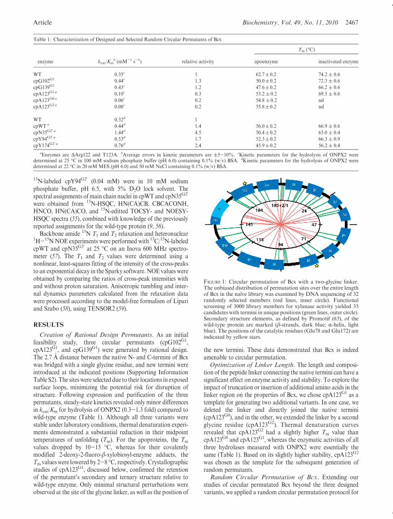
Table 1: Characterization of Designed and Selected Random Circular Permutants of Bcx
Tm (�C)
enzyme kcat/Kmb (mM-1 s-1) relative activity apoenzyme inactivated enzyme
WT 0.35c 1 62.7( 0.2 74.2 ( 0.6
cpG102G1 0.44c 1.3 50.0( 0.2 72.3 ( 0.6
cpG139G1 0.43c 1.2 47.6( 0.2 66.2 ( 0.6
cpA123G1 a 0.10c 0.3 53.2 ( 0.2 69.5 ( 0.6
cpA123G0 a 0.06c 0.2 54.8 ( 0.2 nd
cpA123G2 a 0.08c 0.2 55.8( 0.2 nd
WT 0.32d 1
cpWT a 0.44d 1.4 56.0( 0.2 66.9 ( 0.6
cpN35G20 a 1.44d 4.5 50.4( 0.2 63.0 ( 0.4
cpY94G20 a 0.53d 1.7 52.3( 0.2 66.3 ( 0.9
cpY174G20 a 0.76d 2.4 45.9( 0.2 56.2 ( 0.4
aEnzymes are ΔArg122 and T123A. bAverage errors in kinetic parameters are (5-10%. cKinetic parameters for the hydrolysis of ONPX2 weredetermined at 25 �C in 100 mM sodium phosphate buffer (pH 6.0) containing 0.1% (w/v) BSA. dKinetic parameters for the hydrolysis of ONPX2 weredetermined at 22 �C in 20 mM MES (pH 6.0) and 50 mM NaCl containing 0.1% (w/v) BSA.
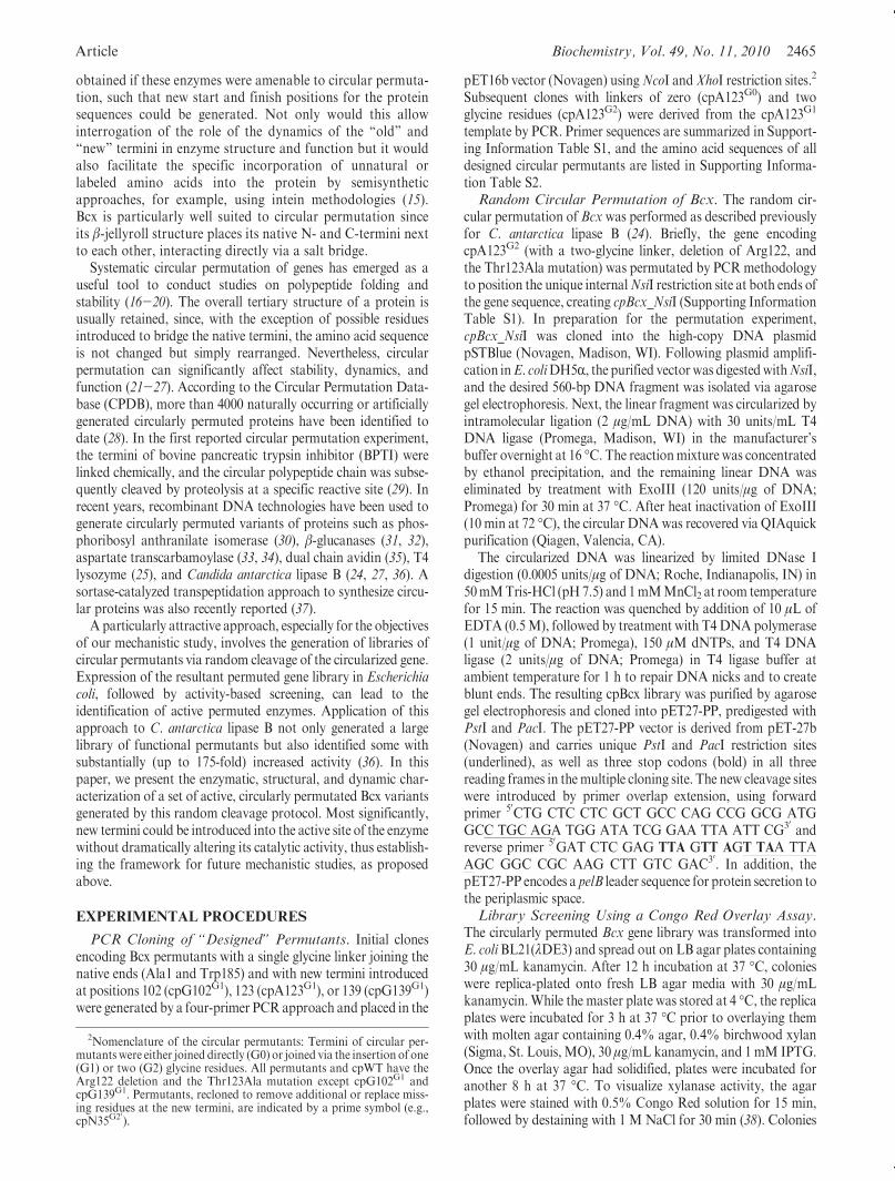
FIGURE 1: Circular permutation of Bcx with a two-glycine linker.The unbiased distribution of permutation sites over the entire lengthof Bcx in the naıve library was examined by DNA sequencing of 32randomly selected members (red lines, inner circle). Functionalscreening of 3000 library members for xylanase activity yielded 35candidates with termini in unique positions (green lines, outer circle).Secondary structure elements, as defined by Promotif (65), of thewild-type protein are marked (β-strands, dark blue; R-helix, lightblue). The positions of the catalytic residues (Glu78 and Glu172) areindicated by yellow stars.

2468 Biochemistry, Vol. 49, No. 11, 2010 Reitinger et al.
generating a comprehensive combinatorial library of all possibleBcx permutants. Briefly, we self-ligated the gene encoding forcpA123G2 to create circular DNA and then relinearized thesequence by limited digestion withDNase I. The resulting libraryof permutated genes was cloned into a modified pET expressionvector, allowing for blunt-end ligation of the gene inserts next to astart codon and stop codons in all three reading frames. Thiscloning strategy introduced two mutations: an extra methionineresidue at the N-terminus, which is accommodated by deletingArg122, and a substitution of Thr123 to Ala due to the PstIrestriction site. After transformation of the library into E. coliBL21 cells (2� 105 cfu), DNA sequence analysis of 32 randomlyselected members indicated a stochastic distribution of newtermini across the entire length of the protein sequence(Figure 1, red lines).
Enzyme variants with glycoside hydrolase activity were iden-tified by screening the library against xylan using a Congo Redoverlay assay. The examination of 3000 cfu yielded 59 activecandidates. Among them, DNA sequence analysis found 35hydrolases with unique termini (Figure 1, green lines, andSupporting Information Table S3). The new N- and C-terminiof these variants were distributed broadly across the sequence ofBcx.Whenmapped onto the tertiary structure of the protein, newtermini were found to occur not only in exposed loops but alsowithin β-strands and even surprisingly near several active siteresidues (Figure 2).Qualitative Analysis of Randomly Generated Bcx Per-
mutants. A preliminary qualitative analysis of these 35 permu-tants was performed to assess expression levels, purifiable yields,and specific activity of individual variants for hydrolysis ofDNPX2 (Table 2). As a reference, we used “cpWT”, one of thefunctional library members whose new termini coincide with thepositions in wild-type Bcx. cpWT is derived from cpA123G2 andhence carries theArg122 deletion and the T123Amutation. Thesechanges lower its Tm value by 7 �C relative to the wild-typeenzyme, yet lead to slightly higher activity with the test substrates(Tables 1 and 3). To assess protein expression levels and solubilityof all 35 Bcx permutants, culture samples of theE. coli expressionhost were analyzed for total and soluble target protein by ion-exchange chromatography and SDS-PAGE. Relative to cpWT,
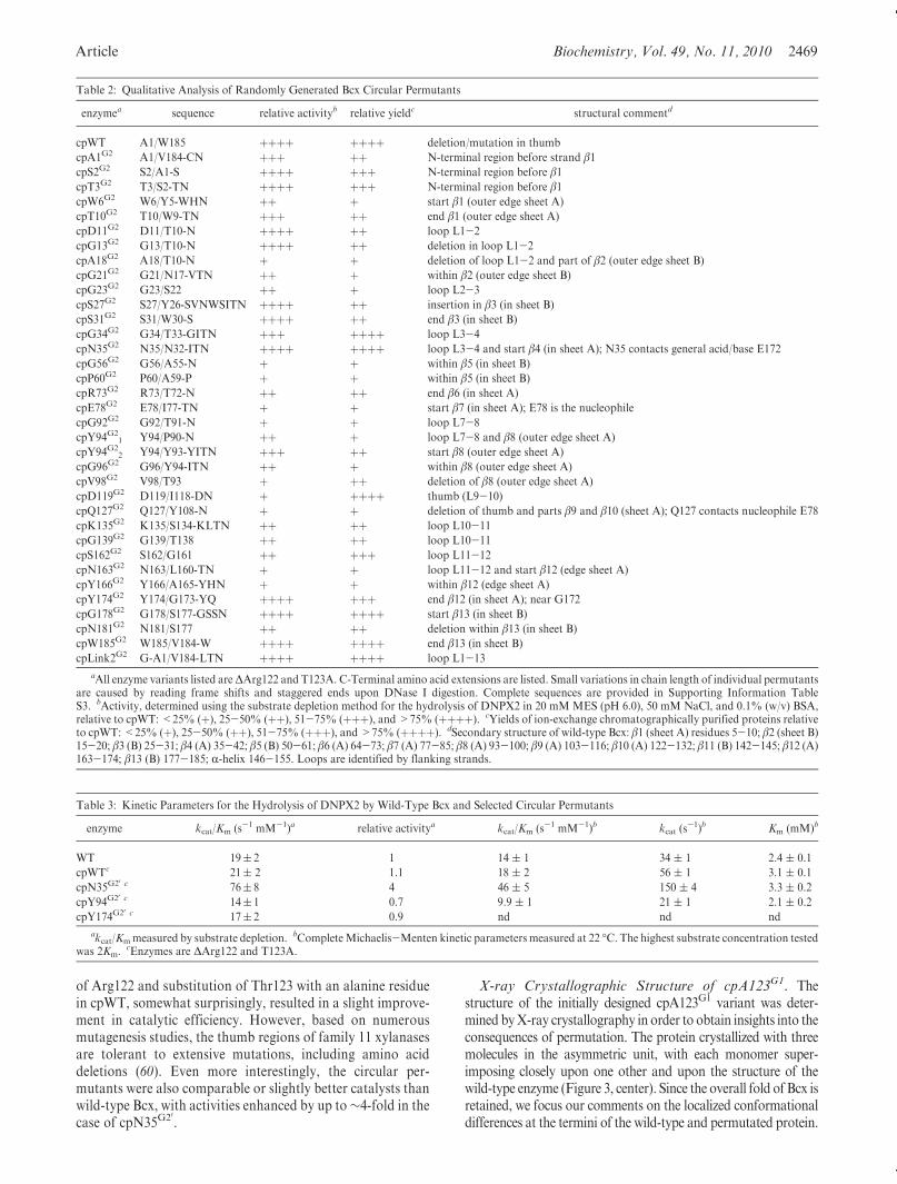
a majority of permutants showed lower amounts of solubleprotein and a tendency for formation of inclusion bodies. Thetrend likely reflects reduced overall protein stability and possiblyimpaired folding kinetics. The relative activities of the single-steppurified proteins, determined using a colorimetric substratedepletion assay, largely (though not in all cases) correlated withthe purified yields and thus likely with stability. As summarizedin Figure 2 and Table 2, several permutants exhibited wild-type-like activity, whereas activities of others were significantlyimpaired.Detailed Stability andActivity Studies onThree Selected
Permutants. Three permutants with new termini within or nearthe active site (cpN35G20, cpY94G20, and cpY174G20) were selectedfor detailed characterization. The genes encoding these permu-tants were corrected for missing or extra residues resulting fromthe random circular permutation protocol and subsequentlyrecloned into the pET21 vector without an encoded pelB leader.The expressed and purified enzymes were quantified by an activesite titration assay using the covalent inactivator 2F-DNPX2.
Initial measures of the effects of the permutations on thestructure and stability of Bcx were obtained using CD spectro-scopy. cpN35G20, cpY94G20, and cpY174G20 yielded CD spectrathat were practically identical to those of both cpWT and wild-type Bcx (not shown). Thus, each variant folded properly, andcircular permutation at the three sites had little impact on theoverall structures of the enzymes. Nonetheless, the stability ofthese variants was impaired since thermal denaturation studiesrevealed a significant drop in Tm values from 56 �C for cpWT to,in the worst case, 46 �C for cpY174G20 (Table 1). The structureand catalytic viability of each permutant were probed by treat-ment with the mechanism-based inhibitor 2F-DNPX2 to trapthem as their covalent glycosyl-enzyme intermediate. Indeed, ineach case the Tm value was raised by approximately 10 �C upontrapping of the intermediate, as also found for the wild typeand cpWT, therefore indicating correct folding and enzymaticmechanism.
Kinetic parameters for hydrolysis of ONPX2 and DNPX2by the selected permutants and wild-type Bcx are presented inTables 1 and 3, respectively. Kinetic properties of wild-typeBcx were consistent with previous reports (6, 12, 42). Deletion
FIGURE 2: “Heat map” summary of the active Bcx circular permutants (Table 2) plotted on rotated structural views of the wild-type protein. Thetwo glycines linking Ala1 and Trp185 are indicated as a dashed line. The positions of new N-termini are labeled, and a color code summarizesrelative activity. The catalytic Glu78 and Glu172 are displayed in stick mode (brown).
Article Biochemistry, Vol. 49, No. 11, 2010 2469
of Arg122 and substitution of Thr123 with an alanine residuein cpWT, somewhat surprisingly, resulted in a slight improve-ment in catalytic efficiency. However, based on numerousmutagenesis studies, the thumb regions of family 11 xylanasesare tolerant to extensive mutations, including amino aciddeletions (60). Even more interestingly, the circular per-mutants were also comparable or slightly better catalysts thanwild-type Bcx, with activities enhanced by up to∼4-fold in thecase of cpN35G20.
X-ray Crystallographic Structure of cpA123G1. Thestructure of the initially designed cpA123G1 variant was deter-mined byX-ray crystallography in order to obtain insights into theconsequences of permutation. The protein crystallized with threemolecules in the asymmetric unit, with each monomer super-imposing closely upon one other and upon the structure of thewild-type enzyme (Figure 3, center). Since the overall fold of Bcx isretained, we focus our comments on the localized conformationaldifferences at the termini of the wild-type and permutated protein.
Table 2: Qualitative Analysis of Randomly Generated Bcx Circular Permutants
enzymea sequence relative activityb relative yieldc structural commentd
cpWT A1/W185 þþþþ þþþþ deletion/mutation in thumb
cpA1G2 A1/V184-CN þþþ þþ N-terminal region before strand β1cpS2G2 S2/A1-S þþþþ þþþ N-terminal region before β1cpT3G2 T3/S2-TN þþþþ þþþ N-terminal region before β1cpW6G2 W6/Y5-WHN þþ þ start β1 (outer edge sheet A)
cpT10G2 T10/W9-TN þþþ þþ end β1 (outer edge sheet A)
cpD11G2 D11/T10-N þþþþ þþ loop L1-2
cpG13G2 G13/T10-N þþþþ þþ deletion in loop L1-2
cpA18G2 A18/T10-N þ þ deletion of loop L1-2 and part of β2 (outer edge sheet B)
cpG21G2 G21/N17-VTN þþ þ within β2 (outer edge sheet B)
cpG23G2 G23/S22 þþ þ loop L2-3
cpS27G2 S27/Y26-SVNWSITN þþþþ þþ insertion in β3 (in sheet B)
cpS31G2 S31/W30-S þþþþ þþ end β3 (in sheet B)
cpG34G2 G34/T33-GITN þþþ þþþþ loop L3-4
cpN35G2 N35/N32-ITN þþþþ þþþþ loop L3-4 and start β4 (in sheet A); N35 contacts general acid/base E172
cpG56G2 G56/A55-N þ þ within β5 (in sheet B)
cpP60G2 P60/A59-P þ þ within β5 (in sheet B)
cpR73G2 R73/T72-N þþ þþ end β6 (in sheet A)
cpE78G2 E78/I77-TN þ þ start β7 (in sheet A); E78 is the nucleophile
cpG92G2 G92/T91-N þ þ loop L7-8
cpY94G21 Y94/P90-N þþ þ loop L7-8 and β8 (outer edge sheet A)
cpY94G22 Y94/Y93-YITN þþþ þþ start β8 (outer edge sheet A)
cpG96G2 G96/Y94-ITN þþ þ within β8 (outer edge sheet A)
cpV98G2 V98/T93 þ þþ deletion of β8 (outer edge sheet A)
cpD119G2 D119/I118-DN þ þþþþ thumb (L9-10)
cpQ127G2 Q127/Y108-N þ þ deletion of thumb and parts β9 and β10 (sheet A); Q127 contacts nucleophile E78
cpK135G2 K135/S134-KLTN þþ þþ loop L10-11
cpG139G2 G139/T138 þþ þþ loop L10-11
cpS162G2 S162/G161 þþ þþþ loop L11-12
cpN163G2 N163/L160-TN þ þ loop L11-12 and start β12 (edge sheet A)
cpY166G2 Y166/A165-YHN þ þ within β12 (edge sheet A)
cpY174G2 Y174/G173-YQ þþþþ þþþ end β12 (in sheet A); near G172
cpG178G2 G178/S177-GSSN þþþþ þþþþ start β13 (in sheet B)
cpN181G2 N181/S177 þþ þþ deletion within β13 (in sheet B)
cpW185G2 W185/V184-W þþþþ þþþþ end β13 (in sheet B)
cpLink2G2 G-A1/V184-LTN þþþþ þþþþ loop L1-13
aAll enzyme variants listed areΔArg122 and T123A. C-Terminal amino acid extensions are listed. Small variations in chain length of individual permutantsare caused by reading frame shifts and staggered ends upon DNase I digestion. Complete sequences are provided in Supporting Information TableS3. bActivity, determined using the substrate depletion method for the hydrolysis of DNPX2 in 20 mM MES (pH 6.0), 50 mM NaCl, and 0.1% (w/v) BSA,relative to cpWT: <25% (þ), 25-50% (þþ), 51-75% (þþþ), and >75% (þþþþ). cYields of ion-exchange chromatographically purified proteins relativeto cpWT:<25% (þ), 25-50% (þþ), 51-75% (þþþ), and>75% (þþþþ). dSecondary structure of wild-type Bcx: β1 (sheet A) residues 5-10; β2 (sheet B)15-20; β3 (B) 25-31; β4 (A) 35-42; β5 (B) 50-61; β6 (A) 64-73; β7 (A) 77-85; β8 (A) 93-100; β9 (A) 103-116; β10 (A) 122-132; β11 (B) 142-145; β12 (A)163-174; β13 (B) 177-185; R-helix 146-155. Loops are identified by flanking strands.
Table 3: Kinetic Parameters for the Hydrolysis of DNPX2 by Wild-Type Bcx and Selected Circular Permutants
enzyme kcat/Km (s-1 mM-1)a relative activitya kcat/Km (s-1 mM-1)b kcat (s-1)b Km (mM)b
WT 19( 2 1 14 ( 1 34 ( 1 2.4 ( 0.1
cpWTc 21( 2 1.1 18 ( 2 56 ( 1 3.1 ( 0.1
cpN35G20 c 76( 8 4 46 ( 5 150 ( 4 3.3 ( 0.2
cpY94G20 c 14( 1 0.7 9.9 ( 1 21 ( 1 2.1 ( 0.2
cpY174G20 c 17( 2 0.9 nd nd nd
akcat/Kmmeasured by substrate depletion. bCompleteMichaelis-Menten kinetic parameters measured at 22 �C. The highest substrate concentration testedwas 2Km.
cEnzymes are ΔArg122 and T123A.
2470 Biochemistry, Vol. 49, No. 11, 2010 Reitinger et al.
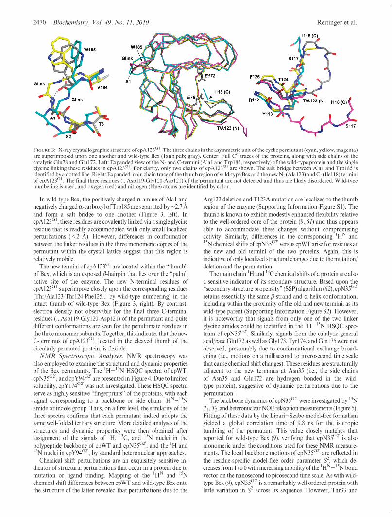
In wild-type Bcx, the positively charged R-amine of Ala1 andnegatively chargedR-carboxyl of Trp185 are separated by∼2.7 Aand form a salt bridge to one another (Figure 3, left). IncpA123G1, these residues are covalently linked via a single glycineresidue that is readily accommodated with only small localizedperturbations (<2 A). However, differences in conformationbetween the linker residues in the three monomeric copies of thepermutant within the crystal lattice suggest that this region isrelatively mobile.
The new termini of cpA123G1 are located within the “thumb”of Bcx, which is an exposed β-hairpin that lies over the “palm”active site of the enzyme. The new N-terminal residues ofcpA123G1 superimpose closely upon the corresponding residues(Thr/Ala123-Thr124-Phe125... by wild-type numbering) in theintact thumb of wild-type Bcx (Figure 3, right). By contrast,electron density not observable for the final three C-terminalresidues (...Asp119-Gly120-Asp121) of the permutant and quitedifferent conformations are seen for the penultimate residues inthe threemonomer subunits. Together, this indicates that the newC-terminus of cpA123G1, located in the cleaved thumb of thecircularly permuted protein, is flexible.NMR Spectroscopic Analyses. NMR spectroscopy was
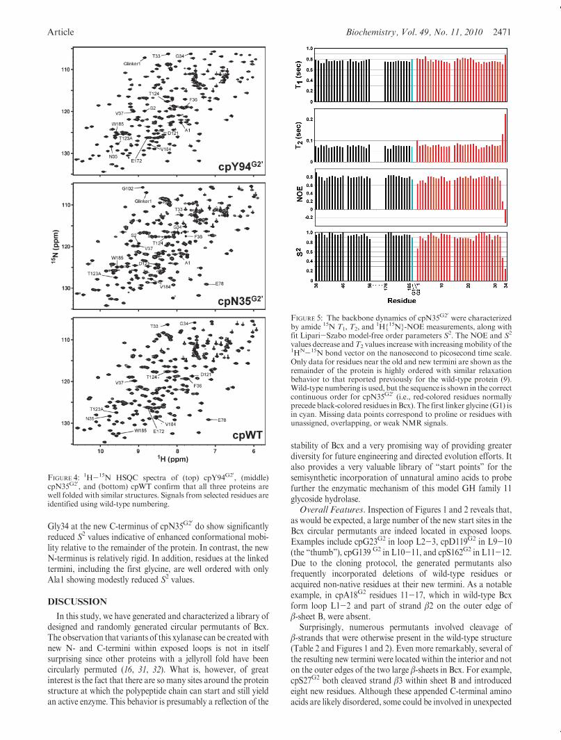
also employed to examine the structural and dynamic propertiesof the Bcx permutants. The 1H-15N HSQC spectra of cpWT,cpN35G20, and cpY94G20 are presented in Figure 4. Due to limitedsolubility, cpY174G20 was not investigated. These HSQC spectraserve as highly sensitive “fingerprints” of the proteins, with eachsignal corresponding to a backbone or side chain 1HN-15Namide or indole group. Thus, on a first level, the similarity of thethree spectra confirms that each permutant indeed adopts thesame well-folded tertiary structure. More detailed analyses of thestructures and dynamic properties were then obtained afterassignment of the signals of 1H, 13C, and 15N nuclei in thepolypeptide backbone of cpWT and cpN35G20, and the 1H and15N nuclei in cpY94G20, by standard heteronuclear approaches.
Chemical shift perturbations are an exquisitely sensitive in-dicator of structural perturbations that occur in a protein due tomutation or ligand binding. Mapping of the 1HN and 15Nchemical shift differences between cpWT and wild-type Bcx ontothe structure of the latter revealed that perturbations due to the
Arg122 deletion and T123A mutation are localized to the thumbregion of the enzyme (Supporting Information Figure S1). Thethumb is known to exhibit modestly enhanced flexibility relativeto the well-ordered core of the protein (9, 61) and thus appearsable to accommodate these changes without compromisingactivity. Similarly, differences in the corresponding 1HN and15N chemical shifts of cpN35G20 versus cpWT arise for residues atthe new and old termini of the two proteins. Again, this isindicative of only localized structural changes due to the mutation/deletion and the permutation.
Themain chain 1H and 13C chemical shifts of a protein are alsoa sensitive indicator of its secondary structure. Based upon the“secondary structure propensity” (SSP) algorithm (62), cpN35G20
retains essentially the same β-strand and R-helix conformation,including within the proximity of the old and new termini, as itswild-type parent (Supporting Information Figure S2). However,it is noteworthy that signals from only one of the two linkerglycine amides could be identified in the 1H-15N HSQC spec-trum of cpN35G20. Similarly, signals from the catalytic generalacid/baseGlu172 aswell asGly173, Tyr174, andGln175were notobserved, presumably due to conformational exchange broad-ening (i.e., motions on a millisecond to microsecond time scalethat cause chemical shift changes). These residues are structurallyadjacent to the new terminus at Asn35 (i.e., the side chainsof Asn35 and Glu172 are hydrogen bonded in the wild-type protein), suggestive of dynamic perturbations due to thepermutation.
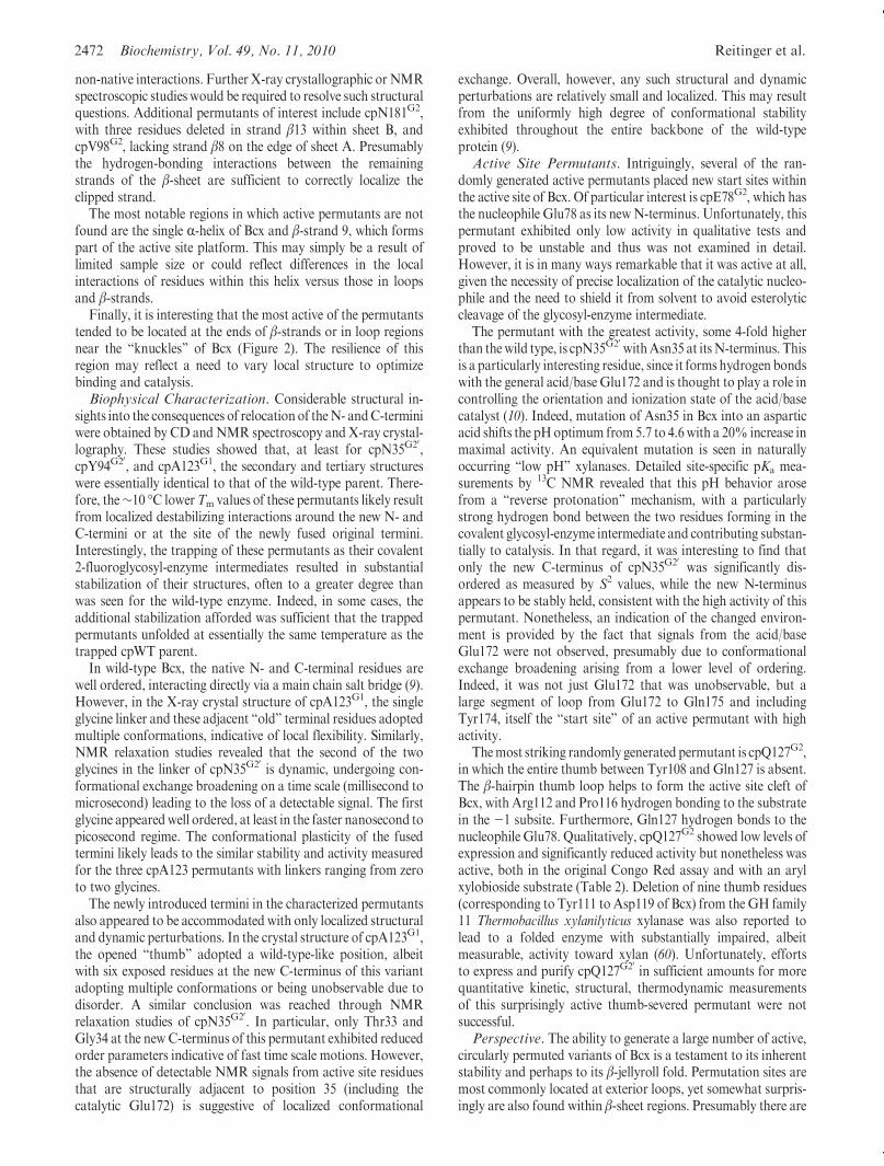
The backbone dynamics of cpN35G20 were investigated by 15NT1,T2, and heteronuclearNOE relaxationmeasurements (Figure 5).Fitting of these data by the Lipari-Szabo model-free formalismyielded a global correlation time of 9.8 ns for the isotropictumbling of the permutant. This value closely matches thatreported for wild-type Bcx (9), verifying that cpN35G20 is alsomonomeric under the conditions used for these NMR measure-ments. The local backbone motions of cpN35G20 are reflected inthe residue-specific model-free order parameter S2, which de-creases from1 to 0with increasingmobility of the 1HN-15Nbondvector on the nanosecond to picosecond time scale. As with wild-type Bcx (9), cpN35G20 is a remarkably well ordered protein withlittle variation in S2 across its sequence. However, Thr33 and
FIGURE 3: X-ray crystallographic structure of cpA123G1. The three chains in the asymmetric unit of the cyclic permutant (cyan, yellow,magenta)are superimposed upon one another and wild-type Bcx (1xnb.pdb; gray). Center: Full CR traces of the proteins, along with side chains of thecatalytic Glu78 and Glu172. Left: Expanded view of the N- and C-termini (Ala1 and Trp185, respectively) of the wild-type protein and the singleglycine linking these residues in cpA123G1. For clarity, only two chains of cpA123G1 are shown. The salt bridge between Ala1 and Trp185 isidentifiedbyadotted line.Right:Expandedmain chain trace of the thumb regionofwild-typeBcx and thenewN- (Ala123) andC- (Ile118) terminiof cpA123G1. The final three residues (...Asp119-Gly120-Asp121) of the permutant are not detected and thus are likely disordered. Wild-typenumbering is used, and oxygen (red) and nitrogen (blue) atoms are identified by color.
Article Biochemistry, Vol. 49, No. 11, 2010 2471
Gly34 at the new C-terminus of cpN35G20 do show significantlyreduced S2 values indicative of enhanced conformational mobi-lity relative to the remainder of the protein. In contrast, the newN-terminus is relatively rigid. In addition, residues at the linkedtermini, including the first glycine, are well ordered with onlyAla1 showing modestly reduced S2 values.
DISCUSSION
In this study, we have generated and characterized a library ofdesigned and randomly generated circular permutants of Bcx.The observation that variants of this xylanase can be created withnew N- and C-termini within exposed loops is not in itselfsurprising since other proteins with a jellyroll fold have beencircularly permuted (16, 31, 32). What is, however, of greatinterest is the fact that there are so many sites around the proteinstructure at which the polypeptide chain can start and still yieldan active enzyme. This behavior is presumably a reflection of the
stability of Bcx and a very promising way of providing greaterdiversity for future engineering and directed evolution efforts. Italso provides a very valuable library of “start points” for thesemisynthetic incorporation of unnatural amino acids to probefurther the enzymatic mechanism of this model GH family 11glycoside hydrolase.Overall Features. Inspection of Figures 1 and 2 reveals that,
as would be expected, a large number of the new start sites in theBcx circular permutants are indeed located in exposed loops.Examples include cpG23G2 in loop L2-3, cpD119G2 in L9-10(the “thumb”), cpG139 G2 in L10-11, and cpS162G2 in L11-12.Due to the cloning protocol, the generated permutants alsofrequently incorporated deletions of wild-type residues oracquired non-native residues at their new termini. As a notableexample, in cpA18G2 residues 11-17, which in wild-type Bcxform loop L1-2 and part of strand β2 on the outer edge ofβ-sheet B, were absent.
Surprisingly, numerous permutants involved cleavage ofβ-strands that were otherwise present in the wild-type structure(Table 2 and Figures 1 and 2). Even more remarkably, several ofthe resulting new termini were located within the interior and noton the outer edges of the two large β-sheets in Bcx. For example,cpS27G2 both cleaved strand β3 within sheet B and introducedeight new residues. Although these appended C-terminal aminoacids are likely disordered, some could be involved in unexpected
FIGURE 4: 1H-15N HSQC spectra of (top) cpY94G20, (middle)cpN35G20, and (bottom) cpWT confirm that all three proteins arewell folded with similar structures. Signals from selected residues areidentified using wild-type numbering.
FIGURE 5: The backbone dynamics of cpN35G20 were characterizedby amide 15N T1, T2, and
1H{15N}-NOE measurements, along withfit Lipari-Szabo model-free order parameters S2. The NOE and S2
values decrease andT2 values increase with increasingmobility of the1HN-15N bond vector on the nanosecond to picosecond time scale.Only data for residues near the old and new termini are shown as theremainder of the protein is highly ordered with similar relaxationbehavior to that reported previously for the wild-type protein (9).Wild-type numbering is used, but the sequence is shown in the correctcontinuous order for cpN35G20 (i.e., red-colored residues normallyprecede black-colored residues inBcx). The first linker glycine (G1) isin cyan. Missing data points correspond to proline or residues withunassigned, overlapping, or weak NMR signals.
2472 Biochemistry, Vol. 49, No. 11, 2010 Reitinger et al.
non-native interactions. Further X-ray crystallographic or NMRspectroscopic studies would be required to resolve such structuralquestions. Additional permutants of interest include cpN181G2,with three residues deleted in strand β13 within sheet B, andcpV98G2, lacking strand β8 on the edge of sheet A. Presumablythe hydrogen-bonding interactions between the remainingstrands of the β-sheet are sufficient to correctly localize theclipped strand.
The most notable regions in which active permutants are notfound are the single R-helix of Bcx and β-strand 9, which formspart of the active site platform. This may simply be a result oflimited sample size or could reflect differences in the localinteractions of residues within this helix versus those in loopsand β-strands.
Finally, it is interesting that the most active of the permutantstended to be located at the ends of β-strands or in loop regionsnear the “knuckles” of Bcx (Figure 2). The resilience of thisregion may reflect a need to vary local structure to optimizebinding and catalysis.Biophysical Characterization. Considerable structural in-
sights into the consequences of relocation of theN- andC-terminiwere obtained by CD andNMR spectroscopy and X-ray crystal-lography. These studies showed that, at least for cpN35G20,cpY94G20, and cpA123G1, the secondary and tertiary structureswere essentially identical to that of the wild-type parent. There-fore, the∼10 �C lowerTm values of these permutants likely resultfrom localized destabilizing interactions around the new N- andC-termini or at the site of the newly fused original termini.Interestingly, the trapping of these permutants as their covalent2-fluoroglycosyl-enzyme intermediates resulted in substantialstabilization of their structures, often to a greater degree thanwas seen for the wild-type enzyme. Indeed, in some cases, theadditional stabilization afforded was sufficient that the trappedpermutants unfolded at essentially the same temperature as thetrapped cpWT parent.
In wild-type Bcx, the native N- and C-terminal residues arewell ordered, interacting directly via a main chain salt bridge (9).However, in the X-ray crystal structure of cpA123G1, the singleglycine linker and these adjacent “old” terminal residues adoptedmultiple conformations, indicative of local flexibility. Similarly,NMR relaxation studies revealed that the second of the twoglycines in the linker of cpN35G20 is dynamic, undergoing con-formational exchange broadening on a time scale (millisecond tomicrosecond) leading to the loss of a detectable signal. The firstglycine appearedwell ordered, at least in the faster nanosecond topicosecond regime. The conformational plasticity of the fusedtermini likely leads to the similar stability and activity measuredfor the three cpA123 permutants with linkers ranging from zeroto two glycines.
The newly introduced termini in the characterized permutantsalso appeared to be accommodated with only localized structuraland dynamic perturbations. In the crystal structure of cpA123G1,the opened “thumb” adopted a wild-type-like position, albeitwith six exposed residues at the new C-terminus of this variantadopting multiple conformations or being unobservable due todisorder. A similar conclusion was reached through NMRrelaxation studies of cpN35G20. In particular, only Thr33 andGly34 at the new C-terminus of this permutant exhibited reducedorder parameters indicative of fast time scale motions. However,the absence of detectable NMR signals from active site residuesthat are structurally adjacent to position 35 (including thecatalytic Glu172) is suggestive of localized conformational
exchange. Overall, however, any such structural and dynamicperturbations are relatively small and localized. This may resultfrom the uniformly high degree of conformational stabilityexhibited throughout the entire backbone of the wild-typeprotein (9).Active Site Permutants. Intriguingly, several of the ran-
domly generated active permutants placed new start sites withinthe active site of Bcx. Of particular interest is cpE78G2, which hasthe nucleophile Glu78 as its newN-terminus. Unfortunately, thispermutant exhibited only low activity in qualitative tests andproved to be unstable and thus was not examined in detail.However, it is in many ways remarkable that it was active at all,given the necessity of precise localization of the catalytic nucleo-phile and the need to shield it from solvent to avoid esterolyticcleavage of the glycosyl-enzyme intermediate.
The permutant with the greatest activity, some 4-fold higherthan thewild type, is cpN35G20 withAsn35 at itsN-terminus. Thisis a particularly interesting residue, since it forms hydrogen bondswith the general acid/base Glu172 and is thought to play a role incontrolling the orientation and ionization state of the acid/basecatalyst (10). Indeed, mutation of Asn35 in Bcx into an asparticacid shifts the pHoptimum from5.7 to 4.6with a 20% increase inmaximal activity. An equivalent mutation is seen in naturallyoccurring “low pH” xylanases. Detailed site-specific pKa mea-surements by 13C NMR revealed that this pH behavior arosefrom a “reverse protonation” mechanism, with a particularlystrong hydrogen bond between the two residues forming in thecovalent glycosyl-enzyme intermediate and contributing substan-tially to catalysis. In that regard, it was interesting to find thatonly the new C-terminus of cpN35G20 was significantly dis-ordered as measured by S2 values, while the new N-terminusappears to be stably held, consistent with the high activity of thispermutant. Nonetheless, an indication of the changed environ-ment is provided by the fact that signals from the acid/baseGlu172 were not observed, presumably due to conformationalexchange broadening arising from a lower level of ordering.Indeed, it was not just Glu172 that was unobservable, but alarge segment of loop from Glu172 to Gln175 and includingTyr174, itself the “start site” of an active permutant with highactivity.
Themost striking randomly generated permutant is cpQ127G2,in which the entire thumb between Tyr108 and Gln127 is absent.The β-hairpin thumb loop helps to form the active site cleft ofBcx, with Arg112 and Pro116 hydrogen bonding to the substratein the -1 subsite. Furthermore, Gln127 hydrogen bonds to thenucleophile Glu78. Qualitatively, cpQ127G2 showed low levels ofexpression and significantly reduced activity but nonetheless wasactive, both in the original Congo Red assay and with an arylxylobioside substrate (Table 2). Deletion of nine thumb residues(corresponding to Tyr111 to Asp119 of Bcx) from the GH family11 Thermobacillus xylanilyticus xylanase was also reported tolead to a folded enzyme with substantially impaired, albeitmeasurable, activity toward xylan (60). Unfortunately, effortsto express and purify cpQ127G20 in sufficient amounts for morequantitative kinetic, structural, thermodynamic measurementsof this surprisingly active thumb-severed permutant were notsuccessful.Perspective. The ability to generate a large number of active,
circularly permuted variants of Bcx is a testament to its inherentstability and perhaps to its β-jellyroll fold. Permutation sites aremost commonly located at exterior loops, yet somewhat surpris-ingly are also found within β-sheet regions. Presumably there are

Article Biochemistry, Vol. 49, No. 11, 2010 2473
sufficient hydrogen-bonding interactions surrounding these“cleavage sites” to stabilize the new termini. Structural studies,both by X-ray crystallography and by NMR spectroscopy,confirmed the integrity of the core and the occurrence of onlylocalized perturbations in these native-like permutants.
These results open up several new directions of study. Forexample, it would be interesting to carry out comparativedirected evolution studies on some of the permutants of Bcx thatstart at Asn35 and Tyr174, both to see if activities can be evolvedmore readily than is the case with the wild-type enzyme and to seeif variants of higher stability can be identified (63, 64). Likewise, itwould be particularly interesting to carry out directed evolutionon permutants such as cpE78G2 and cpQ127G2 to try to improvetheir stabilities such that they could then be expressed, purified,and studied in detail.
This library also provides excellent potential starting points forsimplified intein-based approaches to chemobiological synthesesof variants in which key catalytic residues have been substitutedwith labeled or modified amino acids. Candidate permutants forsuch studies to substitute the catalytic nucleophile E78 includecpR73G2 and cpE78G2 for N-terminal substitution or possiblycpY94G2 for C-terminal substitution, though these may dependon prior stabilization through directed evolution. Likewise,variants containing unnatural acid/base residues (Glu172) couldbe generated starting from cpS162G2 or cpY174G2 for N- and C-terminal substitution, respectively. Analysis of variants generatedin this way, using the full repertoire of structural andmechanistictools that have been assembled, in conjunction with computa-tional approaches, should provide particularly deep insights intothe fundamentals of enzymatic catalysis.
SUPPORTING INFORMATION AVAILABLE
Additional tables summarizing cloning primers, permutantsequences, and X-ray crystallographic statistics, as well as figuresof NMR chemical shift perturbations and secondary structuralpredictions. This material is available free of charge via theInternet at http://pubs.acs.org.
REFERENCES
1. Cantarel, B. L., Coutinho, P. M., Rancurel, C., Bernard, T.,Lombard, V., and Henrissat, B. (2009) The Carbohydrate-ActiveEnZymes database (CAZy): an expert resource for glycogenomics.Nucleic Acids Res. 37, D233–D238.
2. Collins, T., Gerday, C., and Feller, G. (2005) Xylanases, xylanasefamilies and extremophilic xylanases.FEMSMicrobiol. Rev. 29, 3–23.
3. Gebler, J., Gilkes, N. R., Claeyssens, M., Wilson, D. B., Beguin, P.,Wakarchuk, W.W., Kilburn, D. G., Miller, R. C., Jr., Warren, R. A.,and Withers, S. G. (1992) Stereoselective hydrolysis catalyzed byrelated beta-1,4-glucanases and beta-1,4-xylanases. J. Biol. Chem.267, 12559–12561.
4. Miao, S., Ziser, L., Aebersold, R., and Withers, S. G. (1994) Identi-fication of glutamic acid 78 as the active site nucleophile in Bacillussubtilis xylanase using electrospray tandem mass spectrometry. Bio-chemistry 33, 7027–7032.
5. Wakarchuk, W. W., Campbell, R. L., Sung, W. L., Davoodi, J., andYaguchi, M. (1994) Mutational and crystallographic analyses of theactive site residues of the Bacillus circulans xylanase. Protein Sci. 3,467–475.
6. Wicki, J., Schloegl, J., Tarling, C. A., and Withers, S. G. (2007)Recruitment of both uniform and differential binding energy inenzymatic catalysis: xylanases from families 10 and 11. Biochemistry46, 6996–7005.
7. McIntosh, L. P., Hand, G., Johnson, P. E., Joshi, M. D., Korner, M.,Plesniak, L. A., Ziser, L., Wakarchuk, W. W., and Withers, S. G.(1996) The pKa of the general acid/base carboxyl group of a glyco-sidase cycles during catalysis: a 13C-NMR study of Bacillus circulansxylanase. Biochemistry 35, 9958–9966.
8. Sidhu, G., Withers, S. G., Nguyen, N. T., McIntosh, L. P., Ziser, L.,and Brayer, G. D. (1999) Sugar ring distortion in the glycosyl-enzymeintermediate of a family G/11 xylanase. Biochemistry 38, 5346–5354.
9. Connelly, G. P., Withers, S. G., and McIntosh, L. P. (2000) Analysisof the dynamic properties of Bacillus circulans xylanase upon forma-tion of a covalent glycosyl-enzyme intermediate. Protein Sci. 9, 512–524.
10. Joshi, M. D., Sidhu, G., Pot, I., Brayer, G. D., Withers, S. G., andMcIntosh, L. P. (2000) Hydrogen bonding and catalysis: a novelexplanation for how a single amino acid substitution can change thepH optimum of a glycosidase. J. Mol. Biol. 299, 255–279.
11. Joshi, M. D., Sidhu, G., Nielsen, J. E., Brayer, G. D., Withers, S. G.,and McIntosh, L. P. (2001) Dissecting the electrostatic interactionsand pH-dependent activity of a family 11 glycosidase. Biochemistry40, 10115–10139.
12. Ludwiczek, M. L., Heller, M., Kantner, T., and McIntosh, L. P.(2007) A secondary xylan-binding site enhances the catalytic activityof a single-domain family 11 glycoside hydrolase. J. Mol. Biol. 373,337–354.
13. White, A., Tull, D., Johns, K., Withers, S. G., and Rose, D. R. (1996)Crystallographic observation of a covalent catalytic intermediate in abeta-glycosidase. Nat. Struct. Biol. 3, 149–154.
14. Poon, D. K., Ludwiczek,M. L., Schubert, M., Kwan, E. M.,Withers,S. G., and McIntosh, L. P. (2007) NMR spectroscopic characteriza-tion of a beta-(1,4)-glycosidase along its reaction pathway: stabiliza-tion upon formation of the glycosyl-enzyme intermediate.Biochemistry 46, 1759–1770.
15. Kwon, J. S., Bal, J., Hwang, H.M., andKim, J. Y. (2008) A circularlypermuted beta-lactamase as a novel reporter for evaluation of proteincyclization efficiency. J. Microbiol. 46, 456–461.
16. Heinemann, U., and Hahn, M. (1995) Circular permutation ofpolypeptide chains: implications for protein folding and stability.Prog. Biophys. Mol. Biol. 64, 121–143.
17. Hennecke, J., Sebbel, P., and Glockshuber, R. (1999) Randomcircular permutation of DsbA reveals segments that are essential forprotein folding and stability. J. Mol. Biol. 286, 1197–1215.
18. Iwakura, M., Nakamura, T., Yamane, C., and Maki, K. (2000)Systematic circular permutation of an entire protein reveals essentialfolding elements. Nat. Struct. Biol. 7, 580–585.
19. Cellitti, J., Llinas, M., Echols, N., Shank, E. A., Gillespie, B., Kwon,E., Crowder, S. M., Dahlquist, F. W., Alber, T., and Marqusee, S.(2007) Exploring subdomain cooperativity in T4 lysozyme I: structur-al and energetic studies of a circular permutant and protein fragment.Protein Sci. 16, 842–851.
20. Haglund, E., Lindberg, M. O., and Oliveberg, M. (2008) Changes ofprotein folding pathways by circular permutation. Overlapping nucleipromote global cooperativity. J. Biol. Chem. 283, 27904–27915.
21. Fishburn, A. L., Keeffe, J. R., Lissounov, A. V., Peyton, D. H., andAnthony-Cahill, S. J. (2002) A circularly permuted myoglobin pos-sesses a folded structure and ligand binding similar to those of thewild-type protein but with a reduced thermodynamic stability. Bio-chemistry 41, 13318–13327.
22. Pieper, U., Hayakawa, K., Li, Z., and Herzberg, O. (1997) Circularlypermuted beta-lactamase from Staphylococcus aureus PC1. Biochem-istry 36, 8767–8774.
23. Piervincenzi, R. T., and Chilkoti, A. (2004) Effect of genetic circularpermutation near the active site on the activity and stability of anenzyme inhibitor. Biomol. Eng. 21, 33–42.
24. Qian, Z., and Lutz, S. (2005) Improving the catalytic activity ofCandida antarctica lipase B by circular permutation. J. Am. Chem.Soc. 127, 13466–13467.
25. Zhang, T., Bertelsen, E., Benvegnu, D., and Alber, T. (1993) Circularpermutation of T4 lysozyme. Biochemistry 32, 12311–12318.
26. Qian, Z., Horton, J. R., Cheng, X., and Lutz, S. (2009) Structuralredesign of Lipase B fromCandida antarctica by circular permutationand incremental truncation. J. Mol. Biol. 393, 191–201.
27. Yu, Y., and Lutz, S. (2010) Improved triglyceride transesterificationby circular permuted Candida antarctica lipase B. Biotechnol. Bioeng.105, 44–50.
28. Lo, W. C., Lee, C. C., Lee, C. Y., and Lyu, P. C. (2009) CPDB: adatabase of circular permutation in proteins. Nucleic Acids Res. 37,D328–332.
29. Goldenberg,D. P., andCreighton, T. E. (1983) Circular and circularlypermuted forms of bovine pancreatic trypsin inhibitor. J. Mol. Biol.165, 407–413.
30. Luger, K., Hommel, U., Herold, M., Hofsteenge, J., and Kirschner,K. (1989) Correct folding of circularly permuted variants of a betaalpha barrel enzyme in vivo. Science 243, 206–210.
2474 Biochemistry, Vol. 49, No. 11, 2010 Reitinger et al.
31. Hahn, M., Piotukh, K., Borriss, R., and Heinemann, U. (1994)Native-like in vivo folding of a circularly permuted jellyroll proteinshown by crystal structure analysis. Proc. Natl. Acad. Sci. U.S.A. 91,10417–10421.
32. Ay, J., Hahn, M., Decanniere, K., Piotukh, K., Borriss, R., andHeinemann, U. (1998) Crystal structures and properties of de novocircularly permuted 1,3-1,4-beta-glucanases. Proteins: Struct.,Funct., Genet. 30, 155–167.
33. Zhang, P., and Schachman, H. K. (1996) In vivo formation ofallosteric aspartate transcarbamoylase containing circularly per-muted catalytic polypeptide chains: implications for protein foldingand assembly. Protein Sci. 5, 1290–1300.
34. Graf, R., and Schachman,H.K. (1996) Random circular permutationof genes and expressed polypeptide chains: application of the methodto the catalytic chains of aspartate transcarbamoylase. Proc. Natl.Acad. Sci. U.S.A. 93, 11591–11596.
35. Hyt€onen, V. P., H€orh€a, J., Airenne, T. T., Niskanen, E. A., Helttunen,K. J., Johnson, M. S., Salminen, T. A., Kulomaa, M. S., andNordlund, H. R. (2006) Controlling quaternary structure assembly:subunit interface engineering and crystal structure of dual chainavidin. J. Mol. Biol. 359, 1352–1363.
36. Qian, Z., Fields, C. J., and Lutz, S. (2007) Investigating the structuraland functional consequences of circular permutation on lipase B fromCandida antarctica. ChemBioChem 8, 1989–1996.
37. Antos, J. M., Popp, M. W., Ernst, R., Chew, G. L., Spooner, E., andPloegh, H. L. (2009) A straight path to circular proteins. J. Biol.Chem. 284, 16028–16036.
38. Teather, R. M., and Wood, P. J. (1982) Use of Congo Red poly-saccharide interactions in enumeration and characterization of cellu-lolytic bacteria from the bovine rumen. Appl. Environ. Microbiol. 43,777–780.
39. Sung, W. L., Luk, C. K., Zahab, D. M., and Wakarchuk, W. (1993)Overexpression of the Bacillus subtilis and circulans xylanases inEscherichia coli. Protein Expression Purif. 4, 200–206.
40. Gasteiger, E., Hoogland, C., A., G., S., D., R.,W.,M., D., A., R., andA., B. (2005) Protein identification and analysis tools on the ExPASyServer, in The Proteomics Protocols Handbook (Walker, J. M., Ed.)pp 571-607, Humana Press, Totowa, NJ.
41. Withers, S. G., Rupitz, K., and Street, I. P. (1988) 2-Deoxy-2-fluoro-D-glycosyl fluorides;a new class of specific mechanism-based glyco-sidase inhibitors. J. Biol. Chem. 263, 7929–7932.
42. Lawson, S. L., Wakarchuk, W. W., and Withers, S. G. (1997)Positioning the acid/base catalyst in a glycosidase: studies withBacillus circulans xylanase. Biochemistry 36, 2257–2265.
43. Ziser, L., and Withers, S. G. (1994) A short synthesis of [beta]-xylobiosides. Carbohydr. Res. 265, 9–17.
44. Ziser, L., Setyawati, I., and Withers, S. G. (1995) Syntheses andtesting of substrates and mechanism-based inactivators for xylanases.Carbohydr. Res. 274, 137–153.
45. Leatherbarrow, R. J. (2001) GraFit Version 5, 5.0 ed., ErithacusSoftware Limited, Horley, U.K.
46. Pace, C. N., Shirley, B. A., and Thomson, J. A. (1989) Measuring theconformational stability of a protein, in Protein Structure: A PracticalApproach (Creighton, T. E., Ed.) pp 311-330, IRL Press, Oxford,U.K.
47. Otwinowski, Z., Minor, W., and Carter, Charles W., Jr. (1997)Processing of X-ray diffraction data collected in oscillation mode.Methods Enzymol. 276, 307–326.
48. Read, R. (2001) Pushing the boundaries of molecular replacementwith maximum likelihood. Acta Crystallogr., Sect. D 57, 1373–1382.
49. Br€unger, A. T., Adams, P. D., Clore, G.M., DeLano,W. L., Gros, P.,Grosse-Kunstleve, R. W., Jiang, J.-S., Kuszewski, J., Nilges, M.,
Pannu, N. S., Read, R. J., Rice, L. M., Simonson, T., andWarren, G.L. (1998) Crystallography & NMR System: a new software suite formacromolecular structure determination. Acta Crystallogr., Sect. D54, 905–921.
50. Vagin, A. A., Steiner, R. A., Lebedev, A. A., Potterton, L.,McNicholas,S., Long, F., and Murshudov, G. N. (2004) REFMAC5 dictionary:organization of prior chemical knowledge and guidelines for its use.ActaCrystallogr., Sect. D 60, 2184–2195.
51. Emsley, P., and Cowtan, K. (2004) COOT: model-building tools formolecular graphics. Acta Crystallogr., Sect. D 60, 2126–2132.
52. DeLano, W. (2002) PyMOL Release 0.99, DeLano Scientific LLC,Palo Alto, CA.
53. Delaglio, F., Grzesiek, S., Vuister, G. W., Zhu, G., Pfeifer, J., andBax, A. (1995) NMRPipe: a multidimensional spectral processingsystem based on UNIX pipes. J. Biomol. NMR 6, 277–293.
54. Goddard, T. D., and Kneeler, D. G. (1999) Sparky 3, 3rd ed.,University of California, San Francisco, CA.
55. Sattler, M., Schleucher, J., and Griesinger, C. (1999) Heteronuclearmultidimensional NMR experiments for the structure determinationof proteins in solution employing pulsed field gradients. Prog. Nucl.Magn. Res. Spectrosc. 34, 93–158.
56. Plesniak, L. A., Wakarchuk, W. W., and McIntosh, L. P. (1996)Secondary structure and NMR assignments of Bacillus circulansxylanase. Protein Sci. 5, 1118–1135.
57. Farrow,N. A.,Muhandiram, R., Singer, A. U., Pascal, S.M., Kay, C.M., Gish, G., Shoelson, S. E., Pawson, T., Formankay, J. D., andKay, L. E. (1994) Backbone dynamics of a free and a phosphopeptide-complexed Src homology-2 domain atudied by N-15 NMR relaxa-tion. Biochemistry 33, 5984–6003.
58. Lipari, G., and Szabo, A. (1982) Model-free approach to the inter-pretation of nuclear magnetic resonance relaxation in macromole-cules. 1. Theory and range of validity. J. Am. Chem. Soc. 104, 4546–4559.
59. Dosset, P., Hus, J.-C., Blackledge, M., and Marion, D. (2000)Efficient analysis of macromolecular rotational diffusion fromheteronuclear relaxation data. J. Biomol. NMR 16, 23–28.
60. Paes, G., Tran, V., Takahashi,M., Boukari, I., andO’Donohue,M. J.(2007) New insights into the role of the thumb-like loop in GH-IIxylanases. Protein Eng. Des. Select. 20, 15–23.
61. Pollet, A., Vandermarliere, E., Lammertyn, J., Strelkov, S. V.,Delcour, J. A., and Courtin, C. M. (2009) Crystallographic andactivity-based evidence for thumb flexibility and its relevance inglycoside hydrolase family 11 xylanases. Proteins: Struct., Funct.,Genet. 77, 395–403.
62. Marsh, J. A., Singh, V. K., Jia, Z. C., and Forman-Kay, J. D. (2006)Sensitivity of secondary structure propensities to sequence differencesbetween alpha- and gamma-synuclein: implications for fibrillation.Protein Sci. 15, 2795–2804.
63. Palackal, N., Brennan, Y., Callen, W. N., Dupree, P., Frey, G.,Goubet, F., Hazlewood, G. P., Healey, S., Kang, Y. E., Kretz, K.A., Lee, E., Tan, X. Q., Tomlinson, G. L., Verruto, J., Wong, V. W.K., Mathur, E. J., Short, J. M., Robertson, D. E., and Steer, B. A.(2004) An evolutionary route to xylanase process fitness. Protein Sci.13, 494–503.
64. Belien, T., Van Campenhout, S., Bosch, A. V., Bourgois, T. M.,Rombouts, S., Robben, J., Courtin, C. M., Delcour, J. A., andVolckaert, G. (2007) Engineering molecular recognition of endoxy-lanase enzymes and their inhibitors through phage display. J. Mol.Recognit. 20, 103–112.
65. Hutchinson, E. G., and Thornton, J. M. (1996) PROMOTIF;aprogram to identify and analyze structural motifs in proteins. ProteinSci. 5, 212–220.