Embed Size (px)
Citation preview

INFECTION AND IMMUNITY, Feb. 1992, p. 401-4050019-9567/92/020401-05$02.00/0Copyright C) 1992, American Society for Microbiology
Circulating and Localized Immune Complexes in ExperimentalMycoplasma-Induced Arthritis-Associated Ocular Inflammation
CHARLES E. THIRKILL,* NANCY K. TYLER, AND ALAN M. ROTH
Ophthalmology Research, University of California, Davis, Medical Center, 1603 Alhambra Boulevard,Sacramento, California 95816
Received 27 February 1991/Accepted 19 November 1991
Ocular deposits of immune complexes are believed to contribute to the anterior segment inflammationsobserved in association with the human arthritides. Arthritis-related ocular inflammations may be reproducedin animals by infection with certain species of mycoplasma. To evaluate the role of immune complexes in theproduction of ocular lesions, we studied their involvement in the rodent model of experimental arthritis-associated ocular inflammation induced by Mycoplasma arthritidis. Sprague-Dawley rats were infected withviable concentrates of M. arthritidis and monitored for the production of related circulating and intraocularimmune complexes. Circulating immune complexes were monitored by antigen capture systems, and localizedintraocular complexes were identified by indirect immunohistochemistry. Polyacrylamide gel immunoblotanalysis of captured complexes confirmed the antigen(s) involved as proteins derived from M. arthritidis.Indirect immunofluorescence revealed localized complexes containing mycoplasma antigens within the ciliary-iris vasculature. Concentrations of the generated complexes diminished rapidly over a 30-day period. Whilecomplex deposits within ocular tissues could represent a contributing cause to the localized anterior segmentinflammation reported in this rodent model, secondary challenge with viable M. arthritidis, which reproducedhigh concentrations of intraocular and circulating immune complexes, failed to elicit any ocular response.
Mycoplasma-induced ocular inflammation and arthritiscontinue to erupt in seasonal epidemics within herds ofcattle, goats, and sheep. Eye and joint lesions are suspectedto result from an inflammatory response to localized viablemycoplasmas and deposits of immune complexes. The initialinflammatory response to infection declines within 2 to 3weeks after onset, sometimes leaving ocular and joint tissuesirreparably damaged. Persistent carrier states can ensueduring which the disease can be transmitted to offspringthrough colostrum and milk. Carriers occasionally experi-ence relatively mild exacerbations of the disease indicatingsome, but incomplete, protection by the activated immuneresponse (4, 17).The rodent model of Mycoplasma arthritidis-induced pol-
yarthritis permits easy access to the multiple events thatresult in experimental arthritis, including ocular inflamma-tions (28, 29). Strains of M. arthritidis vary in their patho-genicity, however; virulent strains will induce symptons ofeye involvement and isolated joint swelling within 1 week(30, 32). The combination of limited joint involvement withanterior segment inflammation is not unlike that encounteredin the pauciarticular variety ofjuvenile rheumatoid arthritisin humans (18, 23, 25). The immune response of rats infectedwith M. arthritidis includes the production of complement-fixing and metabolism-inhibiting antibodies, but growth-inhibiting or rheumatoid factors have not been found (37-39). Antigenic mimicry, specific tissue interactions, andimmune complex involvement have been proposed as fac-tors contributing to the pathogenicity of M. arthritidis (31-33).To define the role of ocular immune complexes in this
model, we examined the eyes and sera of rats following bothprimary and secondary challenge with viable M. arthritidis.
* Corresponding author.
MATERIALS AND METHODS
Mycoplasma cultivation. M. arthritidis (strain 14152)(American Type Culture Collection) was cultured in GIBCOmycoplasma broth supplemented with 5% horse serum and0.5% arginine hydrochloride, with the addition of 0.001%phenol red. Agar plates for colony counts and isolation of M.arthritidis from blood samples were prepared with the sameadditives. No antibiotics were used in these culture media.Lysed extracts of M. arthritidis, to be used as test antigens,were prepared by centrifugation of broth cultures. Concen-trations were resuspended and washed in phosphate-buff-ered saline (pH 7.5) (PBS) and then briefly sonicated andstored for use at -20°C (31).Rodent inoculation and tissue preparation. On day zero, 36
6-week-old female Sprague-Dawley rats were infected withM. arthritidis via a single tail-vein inoculation with 1.0 ml ofa centrifuged culture concentrate containing approximately109 viable CFU, washed and resuspended in PBS as previ-ously described (32). Animals were treated in accord withthe Guiding Principles in the Care and Use ofAnimals (lla).A separate group of 12 rats each received a single tail-veininoculation of 1.0 ml of complete, serum-supplementedmycoplasma culture medium to act as uninfected, negativecomparison controls. Groups of one control and three testrats were exsanguinated at 5-day intervals following injec-tion, up to day 30. Viable mycoplasmas from blood sampleswere isolated by the direct inoculation of 0.25 ml of wholeblood from freshly killed animals onto plates and into 10-mlvolumes of mycoplasma isolation broth. These culture mediawere incubated aerobically at 37°C for 1 week and examinedfor evidence of mycoplasma growth. Eyes from the test andcontrol rats were promptly removed and fixed in 10%buffered formalin, dehydrated in ethanol, embedded in par-affin, and sectioned. Serum from each group of rats was
stored at -20°C for immunological examination.On day 35, the remaining 18 test rats, now recovered from
the acute phase of the disease, were challenged with a
401
Vol. 60, No. 2
Dow
nloa
ded
from
http
s://j
ourn
als.
asm
.org
/jour
nal/i
ai o
n 17
Jan
uary
202
2 by
45.
112.
166.
36.

402 THIRKILL ET AL.
second, identical intravenous dose of viable M. arthritidis,and the observations continued. Reinfected rats were sacri-ficed in groups of three along with one control at 5-dayintervals following challenge, and blood, eyes, and sera werecollected for study as described above.Hyperimmune rabbit serum. Two female New Zealand
albino rabbits were immunized with M. arthritidis washedand homogenized in PBS as previously described (31). Twoweeks following the last injection, the rabbits were exsan-guinated to provide antiserum which was quantified forimmunoglobulin concentration by quantitative radial immu-nodiffusion and stored for use at -20°C.ELISA antibody assay (36). The antibody response of each
pool of sera from infected rats to mycoplasma antigens wasevaluated and compared with the antibody reactivity ofuninfected control rats which had received a single injectionof complete culture medium. Microplates were coated with10 ,ug/ml of M. arthritidis test antigen per well, and serumsamples from each group were titrated for antibody contentby doubling dilutions, from 1:20 to 1:2,560, down individualcolumns. Antibody reactions with M. arthritidis antigenswere resolved through the application of rabbit anti-ratpolyvalent immunoglobulins (Sigma) and then alkaline phos-phatase-conjugated goat anti-rabbit immunoglobulin G (IgG)(Sigma) at a dilution of 1:1,000 each, using para-nitrophenylphosphate as a substrate. Overall average antibody responsethroughout the study period was obtained from duplicateassays. The relative antibody activity of each pool of testserum at 1:20 was recorded and plotted when the 1:20dilution of control serum reached an optical density of 0.3 at405 mm.ELISA antigen capture system. A modification of the
enzyme-linked immunosorbent assay (ELISA) method de-scribed by Swenson and Kaplan (27) was applied to detectmycoplasma antigens within circulating immune complexes.Microplates were coated overnight at 4°C with rabbit anti-M.arthritidis serum at a concentration of 10 jig of IgG per well,using bicarbonate coating buffer (pH 9.6). Rat serum fromboth test and control animals was double diluted downseparate columns of the microplates starting from 1:2 to1:256 and allowed to react at room temperature for 1 h. Anyavailable mycoplasma antigens were captured by the rabbitanti-M. arthritidis serum bound to the plate. Following athorough wash, rat immunoglobulins associated with cap-tured antigens were detected through the application of a1:1,000 dilution of goat anti-rat IgG-alkaline phosphataseconjugate reacting on para-nitrophenyl phosphate. Dupli-cate assays were performed from which the average relativeabsorption of the 1:2 dilution of serum from each test groupwas compared with that of the 1:2 dilution of control serumwhen it reached an optical density of 0.3 at 405 nm. Theresults were recorded and compared for variation over time.
Immunohistochemistry. Sections (6 ,um) of rat eyes fromeach group were deparaffinized and placed in normal phys-iological saline, in which all antiserum dilutions were pre-pared. Sections were probed with the following antisera todetect intraocular deposits of mycoplasma antigens, aggre-gates of rat complement, and immunoglobulins: (i) rabbitanti-M. arthritidis serum produced in this laboratory; (ii)fluorescein isothiocyanate-conjugated goat anti-rat comple-ment component C3 (U.S. Biochemicals) and (iii) fluoresceinisothiocyanate-conjugated goat anti-rat polyvalent immuno-globulins (U.S. Biochemicals), all used at a dilution of 1:100.Each of these sera was allowed to react on separate sectionsof eyes from each group at room temperature for 30 minbefore washing in fresh saline. Antibody binding to localized
mycoplasma antigens was resolved by indirect immunofluo-rescence with fluorescein isothiocyanate-labeled goat anti-rabbit IgG (Cappel). Sequential sections were examined forimmunofluorescence, and positive findings were photo-graphed. Results obtained from sensitized rats were com-pared with sections of the eyes of the uninfected control rats.Western immunoblot analysis of complexes. Antigens en-
trapped within circulating immune complexes produced inthe first set of rats were analyzed by using a polyacrylamidegel electrophoresis (PAGE) procedure based on those orig-inally described by Matsuo et al. (21, 22) and Vinayak et al.(35). The antibody content of 500-,ul samples of each set oftest and control rat sera was absorbed onto Pansorbin(Calbiochem). The mixture was incubated at room temper-ature with agitation for 1 h, and the Pansorbin was collectedby centrifugation. Bound immunoglobulins and their relatedantigens were eluted from the Pansorbin by boiling for 10min in PAGE sample buffer (13). The supernatant waselectrophoresed on a 1.5-mm-thick, 20-cm 12% polyacryl-amide gel and blot transferred to nitrocellulose (34). Thetransfer was blocked in PBS containing 0.5% Tween and0.05% nonfat milk before being probed with rabbit anti-M.arthritidis serum at a dilution of 1:1,000. Mycoplasma anti-gens, present in serum samples obtained over the initial30-day period prior to challenge, were subsequently resolvedby using goat anti-rabbit IgG-horseradish peroxidase conju-gate reacting on the substrate 3,3'-diaminobenzidine. Thistechnique was confined to the first set of rats that experi-enced a single infective dose, since its application to thosereceiving a secondary challenge would reflect that of previ-ously sensitized animals.The competence of the rabbit anti-M. arthritidis serum
prepared in this laboratory to identify the antigens of M.arthritidis in Western blot reactions was compared with theactivity of pooled rat sera obtained from day 30 of the study.Both were evaluated at a dilution of 1:1,000 in side-by-sidereactions on the same blot transfer of strain 14152 of M.arthritidis.
RESULTS
Conjunctivitis appeared in all test animals in the first setwithin in 24 h postinfection but was resolved completely byday 5. Intravitreal leukocyte infiltration, indicting intraocu-lar inflammation, was found histologically in only 5 of thefirst set of 18 test rats (3 on day 15 and 2 on day 20) and innone of the second set of challenged animals. Swollen jointsappeared in all test rats on day 5 and continued throughoutthe first 30 days of the study, following the same patternwhich we and others have previously described (28, 29).Viable mycoplasmas were present in blood samples obtainedfrom the first group on day 5, but not thereafter. Secondarychallenge on day 35 again produced a similar pattern ofsepticemia, with viable isolates present in blood only on day40, 5 days postchallenge, and not thereafter (Fig. 1). Noevidence of overt joint or eye involvement appeared in thesecondary challenge group of rats. Uninfected control rats,injected with complete serum-supplemented culture me-dium, showed no response in any of the parameters exam-ined throughout the study.ELISA antibody assay. Overall antibody response to infec-
tion showed an elevation in antibody reactions to M. arthri-tidis antigens which increased rapidly to peak and persistthroughout the first phase of the study period. Serum fromuninfected control rats assayed under the same conditionsgave background readings against which the antibody reac-
INFECT. IMMUN.
Dow
nloa
ded
from
http
s://j
ourn
als.
asm
.org
/jour
nal/i
ai o
n 17
Jan
uary
202
2 by
45.
112.
166.
36.

IMMUNE COMPLEXES IN MYCOPLASMA-INDUCED OCULAR LESIONS 403
1.61.51.41.31.2
E 1.1aLt) 111, 0.9
Ci 0.8
Oj 0.70.60.50.40.30.20.10
5 10 15 20 25 30 35 40 45 50 55 60 65 Days
FIG. 1. Comparative evaluations of the overall antibody re-sponse to mycoplasma infection (determined by ELISA) and therelative content of captured mycoplasma antigens (estimated by thecapture system) present in the blood sera of test animals. Hatchedcolumns represent relative concentrations of captured complexeswhen viable particles were present in cultured whole blood. Sero-logical activity was evaluated at a dilution of 1:20 for overallantibody content and 1:2 in the antigen capture system. RelativeA405 values were recorded when the control sera had an opticaldensity (O.D.) of 0.3, indicated by the dotted line labeled ELISAand capture normal.
tions of the infected rats were compared. There was noappreciable increase in the overall antibody response follow-ing challenge (Fig. 1).ELISA antigen capture system. Since living mycoplasmas
were demonstrable in the blood of test animals 5 daysfollowing both initial exposure and secondary challenge,complexes captured during the transient septicemia were notconsidered representative of true immune complexes. Cir-culating antibody complexed with non-viable M. arthritidisantigens was detected on day 10 and thereafter in decreasingconcentrations in all test serum samples until day 30 follow-ing initial infection. Secondary challenge resulted in thereappearance of high concentrations of circulating immunecomplexes, which again declined progressively to the end ofthe study (Fig. 1).
Immunohistochemistry. Examination of sectioned eyesfrom infected rats, obtained on days 5 through 30, showedthe early appearance of intense immunofluorescence indicat-ing the present of mycoplasma antigens, aggregated C3, andrat immunoglobulins entrapped within the iris-ciliary mi-
crovasculature (Fig. 2). These reactions were most intenseduring the first 15 days following infection, but declinedrapidly to day 30, when no immunofluorescence was detect-able. The second set of rats, reinfected on day 35, exhibiteda similar pattern of localized complexes. Reactions weremost intense on day 40 and gradually declined to zero at theend of the study on day 65. After challenge, secondarydeposits of mycoplasma antigens, complement, and immu-noglobulins occurred and declined in the complete absenceof arthritic and ocular signs. Eyes from uninfected controlrats showed no abnormalities.Western immunoblot analysis of complexes. M. arthritidis
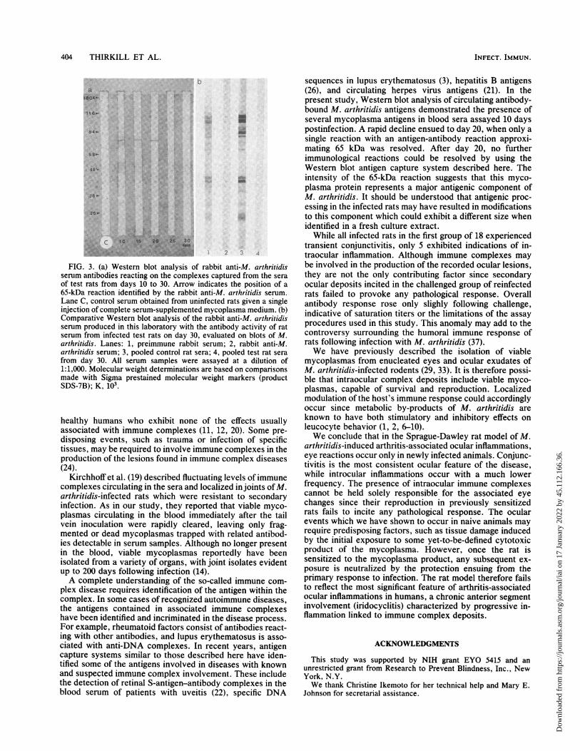
antigens present in captured immunoglobulin complexeswere detected in decreasing quantities on days 10, 15, and20. The principal antigen resolved by the rabbit anti-M.arthritidis serum exhibited a relative molecular mass ofapproximately 65 kDa and was present in serum samplesfrom days 10 to 20, after which no further reactions werefound by this method. Serum from uninfected control ratsgave no reaction in this assay (Fig. 3a). Western blot assaysof the rabbit anti-M. arthritidis sera and pooled rat seraobtained from test rats on day 30 showed similar immuno-reactivity when compared on a blot transfer of M. arthritidis(Fig. 3b).
DISCUSSION
Mycoplasmas produce an assortment of ocular inflamma-tions in a variety of animals, but the pathogenic process isnot fully understood. The M. arthritidis model of arthritis-associated ocular inflammation shares similarities with thehuman arthritides. Ocular lesions associated with rheuma-toid arthritis may result from soluble circulating immunecomplexes which eventually deposit within the blood vesselsof the eye and initiate localized inflammatory reaction (23).Char et al. (5) showed a correlation between the relativeconcentrations of circulating immune complexes and theseverity of ocular inflammation in patients with chronicuveitis and iridocyclitis. Howes and McKay (15) demon-strated increased vascular permeability in rabbits in whichthey had produced immune complexes and associated theoccurrence of localized lesions with intravascular deposits.However, the mere presence of intraocular immune com-plexes cannot be held solely responsible for the induction ofuveitis since Hylkema et al. (16) produced immune com-plexes within the eyes of mice with no resultant inflamma-tory reaction. There are also examples of high concentra-tions of circulating immune complexes found in normal
_1~~~~~~~~~~ C
FIG. 2. Indirect immunofluorescence of iris-ciliary tissue sections showing immunological reactions with antisera to M. arthritidis antigens(a), (b) aggregated rat complement, and accumulated immunoglobulins within the vascular bed (c). Sections were obtained from eyesharvested on day 20. Bars = 20 p.m.
Overall Antibody Response
culting
Complexeswith
.T~~~~~~~~~~~~~ibeIoae
VOL. 60, 1992
Dow
nloa
ded
from
http
s://j
ourn
als.
asm
.org
/jour
nal/i
ai o
n 17
Jan
uary
202
2 by
45.
112.
166.
36.
404 THIRKILL ET AL.
84b-a'T ~~~~~~~~...
~ ~ ~ ~~~~::.:
.~~~~ ~ ~ ~ ~~ ~ ~~ ~
,. :. ka
isn!~~~~~~~~
b
U-
.-
2 3 4
FIG. 3. (a) Western blot analysis of rabbit anti-M. arthritidisserum antibodies reacting on the complexes captured from the seraof test rats from days 10 to 30. Arrow indicates the position of a65-kDa reaction identified by the rabbit anti-M. arthritidis serum.Lane C, control serum obtained from uninfected rats given a singleinjection of complete serum-supplemented mycoplasma medium. (b)Comparative Western blot analysis of the rabbit anti-M. arthritidisserum produced in this laboratory with the antibody activity of ratserum from infected test rats on day 30, evaluated on blots of M.arthritidis. Lanes: 1, preimmune rabbit serum; 2, rabbit anti-M.arthritidis serum; 3, pooled control rat sera; 4, pooled test rat serafrom day 30. All serum samples were assayed at a dilution of1:1,000. Molecular weight determinations are based on comparisonsmade with Sigma prestained molecular weight markers (productSDS-7B); K, 103.
healthy humans who exhibit none of the effects usuallyassociated with immune complexes (11, 12, 20). Some pre-disposing events, such as trauma or infection of specifictissues, may be required to involve immune complexes in theproduction of the lesions found in immune complex diseases(24).
Kirchhoff et al. (19) described fluctuating levels of immunecomplexes circulating in the sera and localized in joints ofM.arthritidis-infected rats which were resistant to secondaryinfection. As in our study, they reported that viable myco-plasmas circulating in the blood immediately after the tailvein inoculation were rapidly cleared, leaving only frag-mented or dead mycoplasmas trapped with related antibod-ies detectable in serum samples. Although no longer presentin the blood, viable mycoplasmas reportedly have beenisolated from a variety of organs, with joint isolates evidentup to 200 days following infection (14).A complete understanding of the so-called immune com-
plex disease requires identification of the antigen within thecomplex. In some cases of recognized autoimmune diseases,the antigens contained in associated immune complexeshave been identified and incriminated in the disease process.For example, rheumatoid factors consist of antibodies react-ing with other antibodies, and lupus erythematosus is asso-
ciated with anti-DNA complexes. In recent years, antigencapture systems similar to those described here have iden-tified some of the antigens involved in diseases with knownand suspected immune complex involvement. These includethe detection of retinal S-antigen-antibody complexes in theblood serum of patients with uveitis (22), specific DNA
sequences in lupus erythematosus (3), hepatitis B antigens(26), and circulating herpes virus antigens (21). In thepresent study, Western blot analysis of circulating antibody-bound M. arthritidis antigens demonstrated the presence ofseveral mycoplasma antigens in blood sera assayed 10 dayspostinfection. A rapid decline ensued to day 20, when only asingle reaction with an antigen-antibody reaction approxi-mating 65 kDa was resolved. After day 20, no furtherimmunological reactions could be resolved by using theWestern blot antigen capture system described here. Theintensity of the 65-kDa reaction suggests that this myco-plasma protein represents a major antigenic component ofM. arthritidis. It should be understood that antigenic proc-essing in the infected rats may have resulted in modificationsto this component which could exhibit a different size whenidentified in a fresh culture extract.While all infected rats in the first group of 18 experienced
transient conjunctivitis, only 5 exhibited indications of in-traocular inflammation. Although immune complexes maybe involved in the production of the recorded ocular lesions,they are not the only contributing factor since secondaryocular deposits incited in the challenged group of reinfectedrats failed to provoke any pathological response. Overallantibody response rose only slighly following challenge,indicative of saturation titers or the limitations of the assayprocedures used in this study. This anomaly may add to thecontroversy surrounding the humoral immune response ofrats following infection with M. arthritidis (37).We have previously described the isolation of viable
mycoplasmas from enucleated eyes and ocular exudates ofM. arthritidis-infected rodents (29, 33). It is therefore possi-ble that intraocular complex deposits include viable myco-plasmas, capable of survival and reproduction. Localizedmodulation of the host's immune response could accordinglyoccur since metabolic by-products of M. arthritidis areknown to have both stimulatory and inhibitory effects onleucocyte behavior (1, 2, 6-10).We conclude that in the Sprague-Dawley rat model of M.
arthritidis-induced arthritis-associated ocular inflammations,eye reactions occur only in newly infected animals. Conjunc-tivitis is the most consistent ocular feature of the disease,while introcular inflammations occur with a much lowerfrequency. The presence of intraocular immune complexescannot be held solely responsible for the associated eyechanges since their reproduction in previously sensitizedrats fails to incite any pathological response. The ocularevents which we have shown to occur in naive animals mayrequire predisposing factors, such as tissue damage inducedby the initial exposure to some yet-to-be-defined cytotoxicproduct of the mycoplasma. However, once the rat issensitized to the mycoplasma product, any subsequent ex-posure is neutralized by the protection ensuing from theprimary response to infection. The rat model therefore failsto reflect the most significant feature of arthritis-associatedocular inflammations in humans, a chronic anterior segmentinvolvement (iridocyclitis) characterized by progressive in-flammation linked to immune complex deposits.
ACKNOWLEDGMENTS
This study was supported by NIH grant EYO 5415 and anunrestricted grant from Research to Prevent Blindness, Inc., NewYork, N.Y.We thank Christine Ikemoto for her technical help and Mary E.
Johnson for secretarial assistance.
INFECT. IMMUN.
Dow
nloa
ded
from
http
s://j
ourn
als.
asm
.org
/jour
nal/i
ai o
n 17
Jan
uary
202
2 by
45.
112.
166.
36.

IMMUNE COMPLEXES IN MYCOPLASMA-INDUCED OCULAR LESIONS 405
REFERENCES1. Atkin, C. L., B. C. Cole, G. J. Sullivan, L. R. Washburn, and
B. B. Wiley. 1986. Stimulation of mouse lymphocytes by amitogen derived from Mycoplasma arthritidis. V. A small basicprotein from culture supernatants is a potent T cell mitogen. J.Immunol. 137:1581-1589.
2. Bekoff, M. C., B. C. Cole, and H. M. Grey. 1987. Studies on themechanism of stimulation of T cells by the Mycoplasma arthri-tidis-derived mitogen. Role of class II IE molecules. J. Immu-nol. 139:3189-3194.
3. Brinkman, K., R. Termaat, J. H. Berden, and R. J. Smeenk.1990. Anti-DNA antibodies and the lupus nephritis: the com-plexity of crossreactivity. Immunol. Today 11:232-234.
4. Buchvarova, Y., and A. Vesselinova. 1986. Arthritis in calvescaused by mycoplasmas. Arch. Exp. Veterinaemed. 40:41-43.
5. Char, D. H., P. Stein, R. Masi, and M. Christensen. 1979.Immune complexes in uveitis. Am. J. Ophthalmol. 87:678-681.
6. Cole, B. C., B. A. Araneo, and G. J. Sullivan. 1986. Stimulationof mouse lymphocytes by a mitogen derived from Mycoplasmaarthritidis. IV. Murine T hybridoma cell exhibit differentialaccessory cell requirements for activation by M. arthritidis Tcell mitogen, concanavalin A, or hen egg-white lysozyme. J.Immunol. 136:3572-3578.
7. Cole, B. C., D. R. Kartchner, and D. J. Wells. 1989. Stimulationof mouse lymphocytes by a mitogen derived from Mycoplasmaarthritidis. VII. Responsiveness is associated with expression ofa product(s) of the V beta 8 gene family present on the T cellreceptor alpha/beta for antigen. J. Immunol. 142:4131-4137.
8. Cole, B. C., D. R. Kartchner, and D. J. Wells. 1990. Stimulationof mouse lymphocytes by a mitogen derived from Mycoplasmaarthritidis (MAM). VIII. Selective activation of T cells express-ing distinct V beta T cell receptors from various strains of miceby the "superantigen" MAM. J. Immunol. 144:425-431.
9. Cole, B. C., J. W. Tulier, and G. J. Sullivan. 1987. Stimulationof mouse lymphocytes by a mitogen derived from Mycoplasmaarthritidis. VI. Detection of a non-MHC gene(s) in the Ealpha-bearing RIIIS mouse strain that is associated with aspecific lack of T cell responses to the M. arthritidis solublemitogen. J. Immunol. 139:927-935.
10. Cole, B. C., and D. J. Wells. 1990. Immunosuppressive proper-ties of the Mycoplasma arthritidis T-cell mitogen in vivo:inhibition of proliferative responses to T-cell mitogens. Infect.Immun. 58:228-236.
11. Dayer, E., and P. H. Lambert. 1985. Immune complexes, p.680-690. In W. N. Kelly, E. D. Harris, S. Ruddy, andC. B. Sledge (ed.), Textbook of rheumatology, 2nd ed. TheW. B. Saunders Co., Philadelphia.
11a.Department of Health, Education, and Welfare. 1985. Guide forthe care and use of laboratory animals. NIH publication 86-23.National Institutes of Health, Bethesda, Md.
12. Dumonde, D. C., E. Kasp-Grochowska, E. Graham, M. D.Sanders, J. P. Faure, Y. de Kozak, and V. van Tuyen. 1982.Anti-retinal autoimmunity and circulating immune complexes inpatients with retinal vasculitis. Lancet ii:787-792.
13. Gershoni, J. M., and G. E. Palade. 1983. Protein blotting:principles and applications. Anal. Biochem. 131:1-15.
14. Hermanns, W., L. C. Schultz, H. Kirchhoff, and J. Heitmann.1983. Studies of polyarthritis caused by mycoplasma arthritidisin rats. III. Histopathological findings. Zentralbl. Bakteriol.Mikrobiol. Hyg. Ser. A 254:423-434.
15. Howes, E. L., Jr., and D. G. McKay. 1975. Circulating immunecomplexes. Effects on ocular vascular permeability in the rab-bit. Arch. Ophthalmol. 93:365-370.
16. Hylkema, H. A., W. M. Rathman, and A. Kijlstra. 1983.Deposition of immune complexes in the mouse eye. Exp. EyeRes. 37:257-265.
17. Jansson, E., A. Backman, K. Hakkarainen, A. Miettinen, and B.Seniusovf. 1983. Mycoplasmas and arthritis. Z. Rheumatol.42:315-319.
18. Key, S. N., and S. J. Kimura. 1975. Iridocyclitis associated withjuvenile rheumatoid arthritis. Am. J. Ophthalmol. 80:425-429.
19. Kirchhoff, H., A. Binder, M. Runge, B. Meier, R. Jacobs, and K.Busche. 1989. Pathogenetic mechanisms in the Mycoplasma
arthritidis polyarthritis of rats. Rheumatol. Int. 9:193-1%.20. Lowder, C. Y., and D. H. Char. 1985. Immune complexes in
ocular disease. Int. Ophthalmol. Clin. 25:143-151.21. Matsuo, T., T. Nakayama, N. Matsuo, and N. Koide. 1986.
Immunological studies of uveitis. 1. Immune complex contain-ing herpes virus antigens in four patients with acute retinalnecrosis syndrome. Jpn. J. Ophthalmol. 30:472-479.
22. Matsuo, T., T. Nakayama, T. Tsuji, T. Koyama, N. Matsuo, andN. Koide. 1986. Immunological studies of uveitis. 2. Immunecomplex containing retinal S antigen in patient with chronicintractable uveitis. Jpn. J. Ophthalmol. 30:480-486.
23. Moore, T. L., P. W. Sheridan, R. B. Traycoff, J. Zuckner, andR. W. Dorner. 1982. Immune complexes in juvenile rheumatoidarthritis: a comparison of four methods. J. Rheumatol. 9:395-401.
24. Peress, N. S. 1980. Immune complex deposition in the ciliaryprocess of rabbits with acute and chronic serum sickness. Exp.Eye Res. 30:371-378.
25. Petty, R. E., and D. W. Hunt. 1989. Immunity to ocular andcollagen antigens in childhood arthritis and uveitis. Int. Arch.Allergy Appl. Immunol. 89:31-37.
26. Surelia, P., and E. H. Boxall. 1990. Hepatitis B virus infection:detection of circulating HBsAg/IgM antibody immune com-plexes. Med. Lab. Sci. 47:204-209.
27. Swenson, P. D., and M. H. Kaplan. 1986. Rapid detection ofrespiratory syncytial virus in nasopharyngeal aspirates by acommercial enzyme immunoassay. J. Clin. Microbiol. 23:485-488.
28. Thirkill, C. E., and D. S. Gregerson. 1982. Mycoplasma arthri-tidis-induced ocular inflammatory disease. Infect. Immun. 36:775-781.
29. Thirkill, C. E., and D. S. Gregerson. 1985. Mycoplasma arthri-tidis-induced experimental uveitis, p. 231-236. In G. R. O'Con-ner and J. W. Chandler (ed.), Advances in immunology andimmunopathology of the eye. Year Book Medical Publishers,Inc., Chicago.
30. Thirkill, C. E., and D. S. Gregerson. 1986. Diphtheroid-likeproperties of Mycoplasma arthritidis. Med. Lab. Sci. 43:9-13.
31. Thirkill, C. E., H. G. Muchmore, R. M. Hyde, and L. V. Scott.1981. Immunologic reactions of rabbit anti-Mycoplasma arthri-tidis serum with in vitro cultivated rat synovial cells. In Vitro17:405-411.
32. Thirkill, C. E., A. M. Roth, R. J. Munn, P. Lee, and N. K. Tyler.1990. Interactions of cultured rat synovial and ocular ciliarybody cells with two strains of Mycoplasma arthritidis. In VitroCell Dev. Biol. 26:140-146.
33. Thirkill, C. E., D. Y. Song, and D. S. Gregerson. 1983. Appli-cation of monoclonal antibodies to detect intraocular myco-plasma antigens in Mycoplasma arthritidis-infected Sprague-Dawley rats. Infect. Immun. 40:389-397.
34. Towbin, H., T. Staehelin, and J. Gordon. 1979. Electrophoretictransfer of proteins from polyacrylamide gels to nitrocellulosesheets: procedure and some applications. Proc. Natl. Acad. Sci.USA 76:4350-4354.
35. Vinayak, V. K., Purnima, K. Singh, K. Venkatwswarlu, C. K.Nain, and S. K. Mehta. 1986. Specific circulating immunecomplexes in amoebic liver abscess. J. Clin. Microbiol. 23:1088-1090.
36. Voller, A., D. Bidweli, and A. Bartlett. 1976. Microplate enzymeimmunoassays for the immunodiagnosis of virus infections, p.506-512. In N. R. Ross and H. Friedman (ed.), Manual ofclinical immunology. American Society for Microbiology,Washington, D.C.
37. Washburn, L. R. 1987. The Derrick Edward Award Lecture.Immunologic aspects of Mycoplasma arthritidis-induced arthri-tis. Isr. J. Med. Sci. 23:326-333.
38. Washburn, L. R., B. C. Cole, and J. R. Ward. 1988. Expressionof metabolism-inhibition antibodies against Mycoplasma arthri-tidis in rats. Am. J. Vet. Res. 49:52-57.
39. Washburn, L. R., and J. R. Ramsay. 1989. Experimental induc-tion of arthritis in LEW rats and antibody response to fourMycoplasma arthritidis strains. Vet. Microbiol. 21:41-55.
VOL. 60, 1992
Dow
nloa
ded
from
http
s://j
ourn
als.
asm
.org
/jour
nal/i
ai o
n 17
Jan
uary
202
2 by
45.
112.
166.
36.