Embed Size (px)
Citation preview
Circulating and Intra-articular Immune Complexes in
Patients with Rheumatoid Arthritis
CORRELATIONOF 125IClq BINDING ACTIVITY WITH CLINICAL
ANDBIOLOGICAL FEATURESOF THE DISEASE
R. H. ZUBLER, U. NYDEGGER,L. H. PERRIN, K. FEmR, J. MCCORMICK,P. H. LAMBERT, and P. A. MEsOHER
From the World Health Organization Immunology Research and TrainingCenter, Geneva Blood Center and Department of Medicine, University ofGeneva, 1211 Geneva, Switzerland
A B S T R A C T The correlation between the incidenceand level of immune complexes in serum and synovialfluid and the various clinical and biological manifesta-tions of rheumatoid arthritis has been studied. Immunecomplexes were quantitated using a sensitive radioim-munoassay, the "I-Clq binding test, in unheated nativesera and synovial fluids from 50 patients with seroposi-tive (RA +) and 45 with seronegative (RA -) rheu-matoid arthritis, 17 with other inflammatory arthritis,and 37 with degenerative and post-traumatic joint dis-ease. The following observations were made: (a) whencompared to the results from patients with degenerativeand post-traumatic joint diseases, the l"I-Clq bindingactivity (Clq-BA) in synovial fluid was found to be in-creased (by more than 2 SD) in most of the patientswith RA+ (80%) and RA- (71%) and in 29% ofpatients with other inflammatory arthritis; the serumClq-BA was also frequently increased in both RA+(76%) and RA - (49%) patients, but only exception-ally in patients with other inflammatory arthritis (6%);(b) a significant negative correlation existed betweenthe Clq-BA and the immunochemical C4 level in syno-vial fluids from patients with RA+ and RA-; (c)neither the serum nor the synovial fluid Clq-BA inrheumatoid arthritis significantly correlated with theerythrocyte sedimentation rate, the clinical stage of thedisease, or the IgM rheumatoid factor titer; and (d)the serum Clq-BA in patients with rheumatoid arthritisand extra-articular disease manifestations (40±34%in those with RA+, 32±29% in those with RA-)
Received for publication 17 November 1975 and in revisedform 16 January 1976.
was significantly increased as compared to the serumClq-BA in patients with joint disease alone (24±30%in those with RA+, 10±13% in those with RA-).Experimental studies were carried out in order to char-acterize the Clq binding material in rheumatoid arthritis.This material had properties similar to immune com-plexes: it sedimented in a high molecular weight rangeon sucrose density gradients (10-30S) and lost the abil-ity to bind Clq after reduction and alkylation, or afteracid dissociation at pH 3.8, or after passage through an
anti-IgG immunoabsorbant. DNase did not affect theClq BA.
These results support the hypothesis that circulatingas well as intra-articular immune complexes may play an
important role in some pathogenetic aspects of rheuma-toid arthritis. The "'I-Clq binding test may also be ofsome practical clinical value in detecting patients whohave a higher risk of developing vasculitis.
INTRODUCTIONThe evidence for the involvement of immune complexesin the pathogenesis of rheumatoid arthritis (RA)' isbased on the direct demonstration of such complexes inthe synovium (1, 2) or in the synovial fluid (3-5), andon the observation of biological changes generally as-
'Abbreviations used in this paper: aggHGG, aggregatedhuman gammaglobulin; Clq-BA, 'I-Clq binding activity;DJD, degenerative joint disease; IA, inflammatory arthritis;NHS, normal human serum; PEG, polyethylene glycol;RA, rheumatoid arthritis; RA+, seropositive RA; RA-,seronegative RA; RF, IgM rheumatoid factor; TR, post-traumatic joint effusions; VBS, barbital buffered saline.
The Journal of Clinical Investigation Volume 57 May 1976 1308-13191308

sociated with the formation of immune complexes suchas an increased catabolism of complement components(6-9). Two major approaches were used in order todetect immune complexes in serum or in joint fluid.First, the existence of specific antigen-antibody systemswas investigated and immunoglobulin-anti-immunoglob-ulin complexes were particularly demonstrated (4, 5,10-15). Secondly, methods which are not specific forany particular antigen-antibody system were used. Thesestudies were mostly qualitative and led to the demon-stration of immune complex-like material such as cryo-globulins (16, 17), macromolecular complexes (3, 10,11), Clq precipitins (4, 5), complement fixing material(4, 16), or of complexes reacting with monoclonal rheu-matoid factor (5). Recently, quantitative investigationswere carried out using radioimmunological techniquessuch as for binding of complexes by rheumatoid factor(18, 19) and macrophages (20). These studies havesuggested a possible role of immune complexes in ar-ticular and extra-articular manifestations of RA.
The main purpose of this work was to define the cor-relation between the incidence and the level of immunecomplexes in serum and synovial fluid on one hand, andthe various forms of clinical and biological manifesta-tions of RA on the other. Immune complexes were quan-titated in the sera and the synovial fluids from patientswith various forms of arthritis, using a sensitive radio-immunoassay, the 'I-Clq binding test, which has beenpreviously applied to the study of patients with sys-temic lupus erythematosus (21) and which has beenrecently modified for the detection of complexes in un-heated native serum or synovial fluid samples (22).These studies confirmed the presence of immune com-plexes in synovial fluid but also led to the demonstra-tion of a high amount of immune complexes in serum.The Clq binding complexes have been further character-ized physicochemically and immunochemically. Theseclinical investigations suggested that the use of suchradioimmunoassays for immune complexes would be ofpractical value in the diagnosis and in follow up studiesof RA.
METHODSSelection of patients studied. The patients included in
this investigation were followed at the Universitits-Rheu-maklinik, Kantonsspital, Zurich, Switzerland (Dr. Fehr),and at the Rheumatic Disease Unit, Northern General Hos-pital, Edinburgh, Scotland (Dr. McCormick). Patients wereselected on the basis of availability of sufficient synovialfluid for arthrocentesis. 156 synovial fluids, 149 sera, and 115plasma samples were obtained from 149 patients with rheu-matoid and various nonrheumatoid joint diseases. Patientswith RA satisfied the American Rheumatism Association'scriteria for classical or definite RA (23). Clinical stagingwas performed according to Steinbrocker et al. (24). IgMrheumatoid factor was measured by the sensitized sheep cellagglutination technique (25). RA patients were considered
as seropositive when the rheumatoid factor titer was equalor higher than 1: 64. Nonrheumatoid patients satisfied gen-erally accepted diagnostic criteria. Synovial fluids of 15patients who suffered from post-traumatic joint effusions(7 with meniscus lesions, 7 with luxations or closed frac-tures, 1 with hydarthrosis) were used as controls for theother studies.
Collection and storage of synovial fluid serum and plasma.Synovial fluids were collected mostly from the knee andonly in some instances from the shoulder or elbow joints.The samples were immediately mixed with EDTA (20 mM,final concentration) then centrifuged at 1,500 g for 15 minat room temperature. The supernates were immediatelyfrozen and stored at - 70'C in portions of 0.5 ml. For thecollection of serum, blood was allowed to clot at roomtemperature for 60 min, was then centrifuged at 1,500 gfor 15 min and the serum was stored at - 70'C. To obtainplasma blood was collected in plastic tubes containing EDTA(20 mMfinal concentration), centrifuged at 1,500 g for 15min, and the supernate was immediately stored at - 700C.
".5I-Clq binding test. The basic principle of this test isoutlined on Fig. 1. For the present investigation, the tech-nique was performed as follows (22): Clq was isolated fromfresh normal human serum (NHS) by the method of Vo-lanakis and Stroud (26), but relative salt concentrations of0.04 and 0.078 M NaCI, respectively, were attained for thefirst and second precipitation steps. Radioiodination of Clqwith lactoperoxydase was performed according to Heusseret al. (27). The 'I-Clq had a sp act of 1 /sCi//Lg protein,and was stored in portions of 10 ,ug in 100 jul of barbitalbuffered saline (containing Ca and Mg) (VBS) (28). Onthe day of testing, a portion of isolated labeled Clq wasdiluted in 3-5 ml VBS containing 1% (wt/vol) bovineserum albumin (A grade, Calbiochem, San Diego, Calif.)and centrifuged at 18,000 g for 40 min at 4VC in order toremove aggregated Clq. The supernate was used for thetest. The test was carried out in duplicate, using Bio-Vialpolypropylene tubes (Beckman Instruments, Inc., Palo Alto,Calif.). For testing of patients' sera, 50 Al of tested serumwas mixed with 100 ul of Na2EDTA, 0.2 M, (adjusted topH 7.5 with NaOH) and incubated for 30 min at 37°C.Then, the mixture was transferred into an ice bath. 50 1A1of the '5I-Clq and 1 ml of 3% (wt/vol) PEG (polyethyleneglycol) (DAB-7, mol wt 6,000, Siegfried, Zofingen, Switzer-land) solution were added. PEG was dissolved in 0.1 Mboric acid, 0.025 M di-sodium-tetraborate, 0.075 M NaCl,pH 8.3. After the addition of PEG, the mixture was left onice for 60 min and centrifuged at 1,500 g for 20 min at 4°C.The supernate was completely discarded and the radioac-tivity was measured on the precipitate. Results were ex-pressed as percent 'I-Clq precipitated as compared with theradioactivity precipitated in a "trichloracetic acid control"tube in which 1 ml 20% trichloracetic acid was added to 100,ul of NHSmixed with 50 pIA of 'I-Clq.
Synovial fluids were tested as follows: 50 jul of freshlythawed synovial fluids were mixed with 100 tl of EDTAbuffer containing 20% (wt/vol) NHS heated for 30 min at560C (NHS 560C), incubated for 30 min, and then tested asfor serum. Sucrose gradient or immunoabsorbant columnfractions to be tested were also mixed with NHS 56°C inorder to maintain a suitable protein concentration. 100 "I ofthese EDTA containing fractions were mixed with 50 alNHS 560C and incubated and tested as for serum. Sucrose(DAB-7, Merck A.G., Inc., Darmstadt, W. Germany) aloneup to concentrations of 50% (wt/vol) in EDTA buffer didnot influence the results of the test.
In some experiments, the test was performed on synovial
Immune Complexes in Rheumatoid Arthritis 1309

Clq BINDING TEST
37 °C30 min
DISSOCIATION OFINTRINSIC C lqrs
1251Clq
( 5Ong)
PEG30/0
( 20 vol)
I-Clq BINDING
FIGURE 1 Basic principle of the '5I-Clq binding test: in a first step, the tested native serum
sample is incubated for 30 min at 370C with EDTA in order to prevent the integration of'"I-Clq into the intrinsic macromolecular Clqrs complex. In a second step, 1"I-Clq and PEGare added to this mixture which is further incubated at 40C for 1 h. Under these conditions,free Clq (0) remains soluble while Clq bound to macromolecular complexes (0) is precipi-tated. The radioactivity of the precipitates is then measured.
fluids treated with 2-mercaptoethanol or with DNAse; 100,"l of the mixtures were incubated with 50 ul EDTA bufferand tested as for serum.
For each test series NHS was used for negative controlsand various amounts of heat aggregated human gamma-
globulins (agg HGG) in NHS560C were used for positivecontrols. The agg HGGwere obtained by heating humanCohn fraction II (Globuman, Berna. Berne, Switzerland)at 630C for 20 min at a concentration of 6 mg/ml in 0.9%oNaCl. This preparation was centrifuged for 15 min at 1,500g and stored at - 700C. Reproducibility studies of the Clqbinding test have indicated that, within duplicates (50 sam-
ples), the variation coefficient was 3.4%o. When agg HGGpositive controls were tested on 10 separate days, the varia-tion coefficient was 6.2%o. As shown in previous investiga-tions (22), the minimal amount of agg HGGdetected inthis Clq binding test is 10 ,ug agg HGG/ml NHS 560C or
30-50 ,ug agg HGG/ml native serum.
Correction of the results for nonspecific 'I-Clq precipita-tion was performed by using a modified Farr's formula(29). The mean values for 'lI-Clq precipitation observed inpresence of synovial fluids from 15 patients with post-trau-matic joint effusions (7.1±+1.8 [mean±1 SD]) and of sera
from 30 healthy blood donors (5.8±1.5%) were used forthe calculation of the correction factors for synovial fluidand serum respectively. The Clq-binding activity (Clq-BA)of tested samples represents the corrected percentage of'I-Clq precipitated in these samples.
Enzymatic and chemical treatment of synovial fluids. ForDNase treatment, 0.2 ml of synovial fluid without EDTAwas mixed with 0.2 ml VBS containing Ca++ and Mg++ and50 ul DNase (Deoxyribonuclease I, Worthington Biochemi-
cal Corp., Freehold, N. J.) (0.5 mg/ml) in VBS and incu-bated at 370C for 3 h. Synovial fluid controls were incubatedwith VBS alone. For reduction and alkylation, 0.2 ml ofsynovial fluid was incubated with 0.2 ml of 0.4 M 2-mercaptoethanol at room temperature for 60 min andthereafter with an additional 0.2 ml of 0.66 M iodoacetamideat 4° C for 60 min. Control synovial fluids were incubatedwith 0.4 ml VBS. For control of the reduction and alkyla-tion procedure, 0.2 ml NHS containing agg HGG (1 mg/ml) was treated like synovial fluid. The mixtures were dia-lysed for 24 h against VBS, before testing the Clq-BA.
Density gradient studies. Ultracentrifugations were per-formed with a SW65-Ti rotor in a Spinco L2-65B prepara-tive ultracentrifuge (Beckman Instruments, Inc.). Linear10-40%o (wt/vol) sucrose gradients were performed witha gradient mixer simultaneously in three tubes, in eitherborate-NaCl buffer 0.1 M, pH 8.3 containing EDTA 0.1 Mor acetate buffer 0.1 M, pH 3.8. Acid gradient fractionswere neutralized by addition of 0.5 M Tris-NaOH, pH 12,and left on ice for 1 h before testing. 0.5 ml of synovialfluid, serum, or PEG fraction obtained from synovial fluidwere layered, undiluted or diluted 1 in 0.9% NaCI, on 4.5-ml gradients and centrifuged at 34,000 rpm for 18 h at 50C.In some experiments, 1 ml of a gradient fraction, dilutedone fourth in buffer, was layered on a 4-ml gradient anda second run was performed at 60,000 rpm for 5 h at 50C.Serial fractions were collected and OD patterns were ob-tained with a 0.3-mm flow cell (Uvicord, LKB-Produkter,Bromma, Sweden) and a logarithmic recorder (W + Welectronic A.G., Basel, Switzerland). The 19 S peak of syno-vial fluid or serum was used as one reference marker. LI-labeled IgG was centrifuged in control tubes and used as
1310 Zubler, Nydegger, Perrin, Fehr, McCormick, Lambert, and Miescher
iKII.
EDTA0.2 M(2vol)
Li

7S reference marker. The apparent S values were calculatedaccording to the method of Martin and Ames (30). In someexperiments, a PEG-precipitation was done in order to con-centrate the Clq binding material before the ultracentrifuga-tions: 1 vol of synovial fluid was mixed with 4 vol of a5% (wt/vol) PEG solution in borate-EDTA buffer, pH8.3. The mixture was kept at 4°C for 1 h, centrifuged at2,000 g for 30 min, the precipitate was homogenized andresuspended with borate-EDTA buffer in one fifth of theoriginal volume of synovial fluid.
Immunochemical techniques. Immunoabsorption: rabbitanti-human IgG (21) or bovine serum albumin were coupledto CNBr-activated Sepharose 4B (Pharmacia Fine Chemi-cals, Inc., Uppsala, Sweden) according to the manufacturer'srecommendation. Two identical 3 ml- (0.5 cm diameter)columns were prepared. The columns were washed withnormal rabbit serum to decrease the nonspecific binding andwere then equilibrated with borate-EDTA buffer, pH 8.3at 4VC. Thereafter, 0.4 ml of Clq binding gradient fractionwas applied to each column and the flow was stopped for30 min. Serial fractions of 0.2 ml were eluted and tested for'I-Clq BA.
Immunoelectrophoresis: gradient fractions were tested bythe method of Scheidegger (31) using commercial rabbitantiwhole human serum and rabbit antihuman IgG (-y chain)serum (Behringwerke AG., Marburg-Lahn, W. Germany).
Complement studies: these were carried out on eitherEDTA-synovial fluid or EDTA-plasma. Hemolytic activitywas quantitated in a continuous flow system (32), Clq, C4and C3 were quantitated by single radial immunodiffusionas described earlier (33). All values were expressed as apercentage of the values obtained with a pool of 30 blooddonors, which was stored in liquid nitrogen. These valueswere corrected for a protein concentration of 70 mg/ml.Protein concentration was assayed by the method of Lowryet al. (34).
Statistical evaluation. Statistical evaluation was carriedout according to Student's t test and by linear regressionanalysis by the method of the least squares.
RESULTSClq-BA in synovial fluid and serum samples. The
Clq-BA of synovial fluids from 149 patients with vari-ous joint diseases were studied. The patients were clas-sified according to the clinical diagnoses: seropositiverheumatoid arthritis (RA +), seronegative rheumatoidarthritis (RA -), various other inflammatory arthritis(IA), degenerative joint disease (DJD) and post-trau-
matic joint effusion (TR). The synovial fluids of 15 pa-tients with TR were used for the estimation of thenonspecific "I-Clq precipitation. The mean values forspecific Clq-BA in synovial fluids of the remaining fourpatient groups are listed in Table I. The following ob-servations were made: (a) the mean Clq-BA was sig-nificantly increased in RA+ and RA- as compared toDJD (P <0.0005) and to IA (P <0.0005); (b) therewas no significant difference in synovial fluid Clq-BAbetween RA+ and RA- (P < 0.1); and (c) the meanClq-BA in IA was significantly increased when com-pared to DJD (P <0.025). The values for Clq-BA insynovial fluids of individual patients are shown on Fig.2. When compared to DJD the synovial fluid Clq-BAwas increased by more than 2 SD in 80% of patientswith RA+, 71% with RA -, 29% with IA, and 0%with DJD. With respect to the IA group, it should bementioned that the values were increased by more than2 SD in three out of seven patients with infectious ar-thritis, two out of seven patients with gout, in a singlepatient with chondrocalcinosis, and in one of two pa-tients with Reiter's disease. Synovial fluids from sevenpatients with RA+ or RA- were obtained on the sameday from both knee joints. The mean Clq-BA was thesame for the right side joints (49±28%) and the leftside joints (49±25%), and the mean individual differ-ence between the right and the left knee joints was6±4% (mean+l1 SD).
The sera from the patients who underwent synovialfluid analysis were also tested. 30 sera from healthyblood donors were used for the estimation of nonspecific12I-Clq precipitation in serum. The mean values forspecific serum Clq-BA in the various patient groups arealso listed in Table I. The following observations weremade: (a) the mean Clq-BA in serum was significantlyincreased in RA+ and in RA- when compared toDJD (P <0.0005) and to IA (P <0.0005); (b) themean serum Clq-BA was significantly higher in RA+than in RA- (P < 0.0005). One should note that sucha difference was not observed in synovial fluid; and (c)the mean serum Clq-BA was not increased in the IA
TABLE I125I-Clq-BA in Synovial Fluids and Sera from Patients with Various Joint Diseases
No. of Sex Clq-BADiagnosis patients M F Age Synovial fluid Serum
yr % %Seropositive RA 50 20 30 55415* 40±22* 33±29*Seronegative RA 45 17 28 51±18 33±25 14±18Other IAj 17 14 3 43414 11i14 0.5±1DJD 22 10 12 58±20 3±5 2±4
* Mean±1 SD.Infectious arthritis (7), Reiter's disease (2), gout (7), chondrocalcinosis (1).
Immune Complexes in Rheumatoid Arthritis 1311

100-
"to- 50-
0-
50-
* :
I
RA+ RA- DJD IA
FIGURE 2 'I-Clq-BA in synovial fluid samples from pa-tients with RA+ and RA-, DJD, and various other (seetext) IA. The means (±1 SD) of the values from eachpatient group are indicated.
group as compared to DJD. It is important to notetherefore that only the mean Clq-BA in synovial fluidwas found to be increased in IA. The values for Clq-BAin sera from the individual patients are shown on Fig. 3.When compared to DJD, the serum Clq-BA was in-creased by more than 2 SD in 76% of patients withRA+, 49% with RA -, 6% with IA, and 2% withDJD. With respect to IA, it should be mentioned thatthe serum Clq-BA was only increased in the one pa-tient with Reiter's disease who also had an increasedsynovial fluid Clq-BA.
A significant correlation was found by regressionanalysis between the synovial fluid Clq-BA and the se-rum Clq-BA of patients with either RA+ or RA-,and this is shown on Fig. 4.
Correlation between Clq-BA and clinical and labora-tory features in RA. The Clq-BA in synovial fluids andsera from patients with RA was compared to various
100-
50-
0S".-W"I.-
zV..
u
AL
S
RA+ RA- DJ D I AFIGURE 3 '~I-Clq-BA in serum samples from patients withRA+, RA-, DJD, and various other (see text) IA. Themeans (±+1 SD) of the values from each patient group areindicated.
It-
.c
o)
-)
* s -./
* 0
*0
I-.r1001
50-
0.
I
*/ RA+0- rr=0O498
P<0.001
A *00
RA-* r= 0.555
**
Pc0.001
* 0
0. 0. : . .
50 100
Clq-BA in SYNOVIAL FLUID (M/*)FIGURE 4 Correlation between the 'I-Clq-BA in serumsamples and the Clq-BA in synovial fluid samples frompatients with RA+ and RA-.
clinical and laboratory parameters of RA. By linear re-gression analysis it appeared that the Clq-BA was notrelated to age, sex, or duration of the disease of indi-vidual patients nor to erythrocyte sedimentation rate,blood hemoglobin values and leukocyte counts in bloodor synovial fluid. Furthermore, despite the significantdifference in mean serum Clq-BA that was observed be-tween RA+ and RA -, no correlation was found be-tween serum or synovial fluid Clq-BA and the IgMrheumatoid factor titer in individual patients with RA+.The Clq-BA could not be related to disease stages ofRA+ or RA -. Most of the patients had stage II or IIIdisease. The Clq-BA in sera from three patients in stageI of RA+ was 21±20% (mean±1 SD) as compared tothe serum Clq-BA of 35±33% from patients in stage IIto IV, and the serum Clq-BA from eight patients in stageI of RA- was 5±12% as compared to the serum Clq-BAof 16+21% from patients in stage II to IV. The pa-tients were further characterized with respect to theoccurrence of extra-articular disease manifestations suchas subcutaneous nodules or other organ involvement(Table II). It appeared that the mean Clq-BA in serumwas significantly increased in either RA+ (P < 0.05)or RA - (P <0.05) patients with extra-articular dis-ease manifestations as compared to patients suffering
1312 Zubler, Nydegger, Perrin, Fehr, McCormick, Lambert, and Miescher
100-1.
09* 0
;e--. .--0 e

TABLE IICorrelation between Serum and Synovial Fluid 1251-Clq-BA and the Occurrence of Extra-articular
Disease Manifestations and other Clinical Features in Patients with RA
No. of Sex Disease Clq-BADiagnosis patients M F Age duration ESR* Synovial fluid Serum
yr yr mm/h % %RA+: no extra-articular manifestations 24 13 11 54±16t 9±6t 50428$ 37±29$ 24430t
with extra-articular manifestations 26 17 9 56±18 14A7 47418 43±26 40434-nodules only 21 14 7 57418 16±6 42417 42±25 40433-other extra-articular
manifestations§ 5 3 2 52±17 6±3 71410 46±21 41±28
RA -: no extra-articular manifestations 37 23 14 50±19 11±9 32 ±24 33±25 10±13with extra-articular manifestations 8 5 3 52±15 8±11 48±29 44±27 32±29
* ESR, erythrocyte sedimentation rate.t Meant1 SD.§ Erythema nodosum (1), polyneuritis (1), pulmonary fibrosis (1), endocarditis (1), lymphadenopathy (1).11 Siogren syndrome (2), Raynaud phenomenon (2), pleural effusion (1), episcleritis (1), lymphadenopathy (2).
from joint disease alone, whereas no significant differ-ence was found with respect to synovial fluid Clq-BA.There was no significant difference in the mean serumClq-BA between RA+ patients with nodules and RA+patients with other organ involvement, whereas in RA -where extra-articular symptoms occurred less frequently,nodules were not observed. Moreover, it appeared thatthere was no significant difference (P> 0.1) in meanserum Clq-BA between those RA+ and RA- patientswho presented extra-articular disease manifestations,whereas RA+ patients with joint disease alone had ahigher mean serum Clq-BA (P < 0.025) than RA-
patients with joint disease alone. When compared toDJD, the Clq-BA was found to be increased by morethan 2 SD in synovial fluids of most of the patients withRA and nodules or other organ involvement (92% inRA+; 88% in RA -) but also frequently in patientswith RA and joint disease alone (67% in RA+; 64%in RA -). The serum Clq-BA was increased by morethan 2 SD in most of the patients with RA and nodulesor other organ involvement (88% in RA+; 75% inRA -), and in 63% of patients with RA+ and jointdisease alone, but only in 38% of patients with RA -
and joint disease alone.Correlation between Clq-BA and complement levels.
Complement studies were performed in order to allowfor a correlation of the measured Clq-BA with comple-ment level in the various patient groups. Total hemolyticcomplement activity, intrinsic Clq, C4 and C3 were mea-sured in synovial fluids and plasma of 40 patients withRA+, 30 patients with RA -, 15 patients with IA, and30 patients with DJD, or TR. The mean values foundin synovial fluids were compared to the Clq-BA. It ap-peared that a high mean Clq-BA was associated with asignificant decrease of the mean hemolytic complementactivity (P < 0.025) and C4 (P < 0.0005) in RA+ andwith a significant decrease of C4 in RA- (P < 0.0005).
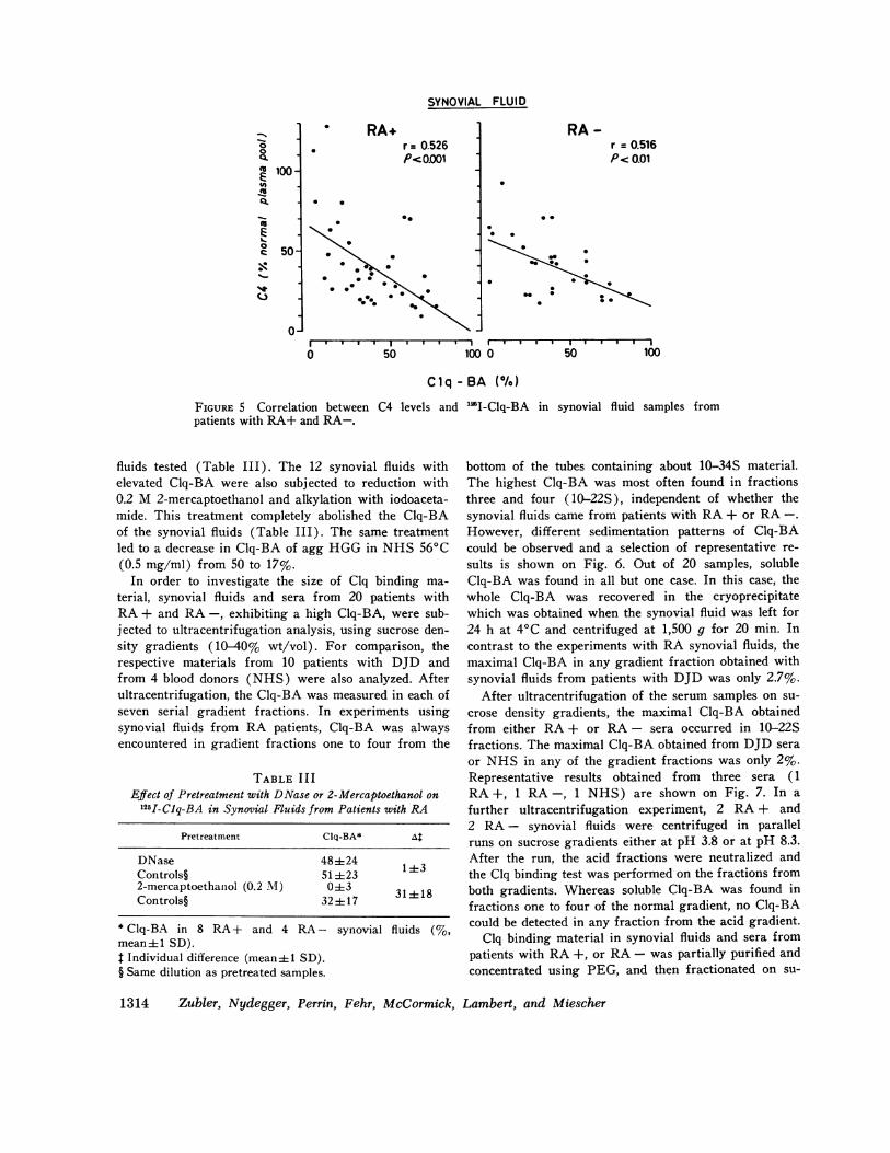
In contrast, a low mean Clq-BA was associated withnormal (DJD and TR) or even increased (IA) com-plement levels. Linear regression analysis gave signifi-cant negative correlations for Clq-BA and C4 levels insynovial fluids of patients with both RA+ (P < 0.001)and RA - (P < 0.01) (Fig. 5). No significant corre-lations were found for Clq-BA and intrinsic Clq, C3, andhemolytic complement activity, respectively. The hemo-lytic complement activity, intrinsic Clq, C4 and C3 werealso measured in the plasma of the same patients whounderwent synovial fluid complement analysis. With theexception of C4, the mean values of the different pa-tient groups were within the normal range. However,when compared to the C4 level of the DJD and TRgroup (100±23%; mean±1 SD), C4 was significantlydecreased in the RA+ group (85±37%, P < 0.005),whereas it was not significantly decreased in the RA-
group (95±34%, P > 0.1) and was slightly increasedin the IA group (107±30%). Therefore, in serum andplasma, respectively, a high mean Clq-BA was associ-ated with a decreased mean C4 level in the RA+ group,but by linear regression analysis no significant correla-tion was found.
Characterization of the Clq binding material in RA.A series of experiments was performed in order to fur-ther characterize the Clq binding material in RA. Forthese studies, synovial fluids and sera from patients withRA were selected for their high Clq-BA and subjectedto various treatments. It has been shown by others thatsynovial fluid from patients with RA may contain freeDNAand anti-DNA antibodies (16, 35). It was there-fore relevant to test if DNAwas involved in the Clq-BAof synovial fluids from RA patients. For this purpose,synovial fluids from 8 patients with RA+ and 4 withRA - were incubated with either DNase or buffer forcontrols. This treatment with DNase did not produceany significant reduction in Clq-BA of the synovial
Immune Complexes in Rheumatoid Arthritis 1313
SYNOVIAL FLUID
* .
1
*. ** -0
rI I I I I* I
100 0 50 100
Clq-BA (%)
FIGURE 5 Correlation between C4 levels andpatients with RA+ and RA-.
fluids tested (Table III). The 12 synovial fluids withelevated Clq-BA were also subjected to reduction with0.2 M 2-mercaptoethanol and alkylation with iodoaceta-mide. This treatment completely abolished the Clq-BAof the synovial fluids (Table III). The same treatmentled to a decrease in Clq-BA of agg HGGin NHS560C(0.5 mg/ml) from 50 to 17%.
In order to investigate the size of Clq binding ma-terial, synovial fluids and sera from 20 patients withRA+ and RA-, exhibiting a high Clq-BA, were sub-jected to ultracentrifugation analysis, using sucrose den-sity gradients (10-40% wt/vol). For comparison, therespective materials from 10 patients with DJD andfrom 4 blood donors (NHS) were also analyzed. Afterultracentrifugation, the Clq-BA was measured in each ofseven serial gradient fractions. In experiments usingsynovial fluids from RA patients, Clq-BA was alwaysencountered in gradient fractions one to four from the
TABLE IIIEffect of Pretreatment with DNase or 2-Mercaptoethanol on
1251-Clq-BA in Synovial Fluids from Patients with RA
Pretreatment Clq-BA* At
DNase 484±24 1±3Controls§ 51 ±232-mercaptoethanol (0.2 M) 0±3 31 + 18Controls§ 32±--17 318
* Clq-BA in 8 RA+ and 4 RA- synovial fluids (%,mean±1 SD).t Individual difference (mean±+ SD).§ Same dilution as pretreated samples.
'"I-Clq-BA in synovial fluid samples from
bottom of the tubes containing about 10-34S material.The highest Clq-BA was most often found in fractionsthree and four (10-22S), independent of whether thesynovial fluids came from patients with RA+ or RA -.
However, different sedimentation patterns of Clq-BAcould be observed and a selection of representative re-
sults is shown on Fig. 6. Out of 20 samples, solubleClq-BA was found in all but one case. In this case, thewhole Clq-BA was recovered in the cryoprecipitatewhich was obtained when the synovial fluid was left for24 h at 4VC and centrifuged at 1,500 g for 20 min. Incontrast to the experiments with RA synovial fluids, themaximal Clq-BA in any gradient fraction obtained withsynovial fluids from patients with DJD was only 2.7%.
After ultracentrifugation of the serum samples on su-
crose density gradients, the maximal Clq-BA obtainedfrom either RA+ or RA - sera occurred in 10-22Sfractions. The maximal Clq-BA obtained from DJD sera
or NHS in any of the gradient fractions was only 2%.Representative results obtained from three sera (1RA+, 1 RA-, 1 NHS) are shown on Fig. 7. In a
further ultracentrifugation experiment, 2 RA+ and2 RA - synovial fluids were centrifuged in parallelruns on sucrose gradients either at pH 3.8 or at pH 8.3.After the run, the acid fractions were neutralized andthe Clq binding test was performed on the fractions fromboth gradients. Whereas soluble Clq-BA was found infractions one to four of the normal gradient, no Clq-BAcould be detected in any fraction from the acid gradient.
Clq binding material in synovial fluids and sera frompatients with RA+, or RA- was partially purified andconcentrated using PEG, and then fractionated on su-
1314 Zubler, Nydegger, Perrin, Fehr, McCormick, Lambert, and Miescher
RA+r = 0.526P<0001
RA -00
'U
in
Q.
cx
m
EU0
(3
r = 0.516P< 0.01
100 ]
50-
0-
* -00 0
*. *
**0
% 9:--0 50
.
.

OD
0.5
As:
7S\
19 Su ,
io
1020
20
0 _20-
101
100S
100]
10I01
10nOl
10]
l- . . . . ~~~~~~~-
Ma. RA+
Key. RA+
Twe. RA+
Dav. RA-
Com. RA -
Cra. RA-
Bru. DJD
Cok. DJD
Lin. DJD
tions by double immunodiffusion. At pH 8.3 maximumIgG was found to be located in the 14S peak, whereasat pH 3.8 the maximum was found to be located in the7S peak. Immunoelectrophoretic analysis of this 7S peakfraction with antiserum to total human serum revealedonly IgG. Secondly, the concentrated Clq binding ma-terial was left to react in an immunoabsorbant system.Small columns of CNBr-sepharose 4B coupled with rab-bit antihuman IgG or with bovine serum albumin wereused. Concentrated Clq binding material from RA syn-ovial fluid was passed through these columns and theClq-BA of the effluents were compared. Clq-BA wasstill observed in the effluent of the bovine serum albu-min sepharose columns, while the effluent of the anti-IgG sepharose columns no longer exhibited Clq bind-ing activity.
DISCUSSIONThe occurrence of immune complexes during the courseof RA has been previously demonstrated by analyticalstudies of synovial tissue, synovial fluid, and se-rum samples (1-5, 10-15). These investigations havebeen conducted mainly on a qualitative basis. Mac-romolecular complexes containing immunoglobulins havebeen detected in synovial fluids and serum and, in someinstances, their size has been characterized (3-5, 10-15).
'76 5 4 3 2 1Top
Gradient fractions
FIGURE 6 'I-Clq-BA measured in individual sucrose gra-
dient fractions obtained by separation of synovial fluids frompatients with RA+ and RA- and DJD: on top of thefigure the ODpattern obtained with a 1:10 diluted synovialfluid and the positions of the 7S and 19S markers are indi-cated, Clq-BA is represented by the black columns on thelower part of the figure.
crose density gradients. This procedure led to a threeto four times increased Clq-BA in 14-24S gradientfractions when compared to the Clq-BA in respectivegradient fractions obtained by centrifugation of the orig-inal untreated synovial fluids. A representative experi-ment is shown on Fig. 8. Two experiments were per-
formed with the 14-24S concentrated Clq binding ma-
terial from 2 RA+ and 2 RA- synovial fluids: first,the influence of acidification was investigated by an ex-
periment involving parallel ultracentrifugation runs
either at pH 8.3 or at pH 3.8. The OD patterns fromthese two gradients were compared (Fig. 9). The ODpattern at pH 8.3 resulted in two peaks of similar size,one in the 19S region and another one in the 14-15Sregion. In contrast, the ODpattern at acid pH resultedin a major peak in the 7S region and a small peak in the19S region. IgG was quantitated in the gradient frac-
OD
1.5
40-302010
0
10 _0
lz
Wid. RA+
Key. RA-
NHS
17 6 05 4 13 2 1Top
Gradient fractions
FIGuRE 7 II-Clq-BA measured in individual sucrose gra-dient fractions obtained by separation of sera from a patientwith RA+ and one with RA- and from a healthy blooddonor (NHS): on top of the figure the OD pattern ob-tained with a 1: 10 diluted serum and the positions of the7S and 19S markers are indicated, Clq-BA is representedby the black columns on the lower part of the figure.
Immune Complexes in Rheumatoid Arthritis
-%1
qqcoI-
10of
1315

TOP 7 S
l I
4% PEGPDFDIrI TATF
80(/°PPO 60-
20
O- I I II
7 6 5 4 3 2
GRADIENT FRACTIONS
FIGURE 8 Concentration and partial purification of the Clq binding material from the synovialfluid of a patient with RA+: the curves represent the OD pattern obtained after sucrose
gradient separation either of synovial fluid (upper part of the figure) or of a- concentratedfraction obtained after incubation of that synovial fluid with PEG, (4% final concentration)(lower part of the figure). The black columns represent the percent specific l"I-Clq precipita-tion (%o ppt) in individual gradient fractions obtained from either sample.
The purpose of the present study was to investigate on
a quantitative basis the incidence of immune complexesin serum and in synovial fluids from patients with RA,using a newly developed methodology which allowed fora sensitive detection of such immune complexes. As withthe Clq precipitation test described by Agnello et al.(36), the radioimmunoassay used in this investigationis based on the higher avidity of Clq for aggregated or
complexed immunoglobulins as compared with mono-
meric immunoglobulins (37). However, the sensitivityof the method has been increased by a direct measure-
ment of the binding of Clq by immune complexes ratherthan by observing a secondary precipitation of thosecomplexes by Clq. This procedure also largely avoidedthe possible interference of some substances which can
nonspecifically fix Clq, such as DNA, bacterial lipo-polysaccharides (22), or calcium-dependent C reactiveprotein complexes (38). However, one cannot a priorirule out the possibility that other substances with similarproperties may influence these studies.
The results obtained confirm the existence of largeamounts of immune complex-like material in synovialfluids from patients with RA. Indeed, the Clq-BA was
significantly increased in those fluids when comparedto the Clq-BA observed in samples from patients withDJD or with TR. The physicochemical characterizationof the Clq binding material showed that it was similarto the 1O-30S macromolecular complexes observed insuch fluids by ultracentrifugation analysis (3-5, 14).Furthermore, the fact that this material lost its abilityto bind Clq after acid dissociation or reduction-alkyla-tion (39), or after passage through an anti-IgG im-munoabsorbant, demonstrates that it has properties sim-ilar to that of immune complexes. DNA or DNA-anti-DNA complexes do not contribute to this binding ofClq since DNase treatment did not reduce the Clq-BAof synovial fluids. The average Clq-BA observed insynovial fluids from patients with RA (37%) corre-
sponds to that obtained when heat-aggregated immuno-
globulin is added to native normal serum at a concen-
1316 Zubler, Nydegger, Perrin, Fehr, McCormick, Lambert, and Miescher
20 -
1D280
10-SYNOVIALFLUID
ofL
40
(°/° PPO20A0
10
n
II I I I I I I I
19 S
I I I I I I I
ranL%.Ow . ,.

ot )280TOP 7S 19 S
IO
_*
0.2- IgG
0.1 -
O-
0.3-
0.2 -
0.1 -
lgG +** 4 0 0
0J10 9 8 7 6 5 4 3 2 1
GRADIENT FRACTIONSFIGURE 9 Acid dissociation of Clq binding material: thecurves represent OD pattern obtained after separation ofconcentrated and partially purified Clq binding materialrecovered from the synovial fluid of a patient with RA+on sucrose gradients at either pH 8.3 or pH 3.8. Thehatched area represents the ODof a control tube containingonly sucrose. The top of the 10-40% sucrose gradient, thepositions of the 7S and 19S markers and the relative con-centration of IgG in various gradient fractions are indicatedon the figure.
tration of about 0.5 mg/ml (22). However, this valueshould be considered as approximate, since the size andthe nature of the detected complexes influence theirefficiency to bind Clq (37). It has been found previouslythat IgG-anti-IgG complexes can exhibit a higher Clq-BA than a similar amount of heat-aggregated IgG (21).
The participation of 19S rheumatoid factor (RF) inthe Clq binding complexes seemed relatively limitedsince (a) fluids from patients with RA- exhibitedsimilar levels of Clq-BA to those from patients withRA+, and (b) the maximum Clq binding material bysucrose density gradient analysis was most frequentlyobserved in the 14-15S range. An increased Clq-BAwas also observed in synovial fluids from some patientswith various IA. Although bacterial antigen-antibodycomplexes may be responsible for this binding in in-fectious arthritis, the occurrence of an increased Clq-BAin gout or chondrocalcinosis may correspond to a non-specific aggregation of IgG in synovial fluids.
The investigation of immune complexes in serum frompatients wtih RA led to some discrepancies accordingto various methods which had been previously used.
While 57% of RA+ reacted with monoclonal RF, noneof these sera precipitated with Clq in agarose (5). Re-cently, using a radioimmunoassay with monoclonal RF,Luthra, et al. (19) detected complexes in 24% of RAsera while in a similar assay, but using polyclonal RF,none of the RA sera studied exhibited a positive reac-tion (18). Cryoglobulins were also found in 25% ofsera (17). In the present study, Clq binding materialwas found in 76% of RA+ sera and in 49% of RA-sera. The differences observed between the results ob-tained in these various studies probably indicate differ-ences in the sensitivity of the methods used for the par-ticular complexes present in RA sera or in the suscepti-bility of each technique to the interfering effect of RF.This was apparent when the method using the inhibitionof the uptake of labeled agg HGGby macrophages wasapplied to RA sera (20).
Using the Clq binding test, the main finding was thatan increased Clq-BA in serum was observed frequentlyin both RA+ and RA- patients but occurred only ex-ceptionally in other IA. This result suggests that RA isa disease associated with circulating as well as articularimmune complexes, while other IA may be associatedwith complexes formed in the joint spaces but rarelywith circulating complexes. The size of the materialbinding Clq in RA sera is similar to that of the com-plexes which were detected with monoclonal rheumatoidfactors (5). It corresponds mostly to 15-20S intermedi-ate complexes and may represent IgG-anti-IgG com-plexes. However, some larger complexes were alsofound in these serum samples. One cannot exclude thepossibility that other antigen-antibody systems may bealso responsible for the increase in Clq-BA in RA. Thecriteria for seropositivity with respect to rheumatoidfactor was a positive sheep cell agglutination titer of1:64 or higher. This is arbitrary and therefore someseronegative patients' sera may indeed have had a lowIgM RF agglutinating activity. Moreover, it is wellknown that such "seronegative" patients usually exhibitthe presence of IgG RF.
The studies of the correlation between Clq-BA andother biological features of RA indicated that the pres-ence of immune complexes is not directly related to theconcentration of RF in serum. Furthermore, nonspecificchanges in the serum protein profile characterizing theinflammatory stimulus in RA were clearly not interfer-ing with the Clq binding test. However, there was anegative correlation between the level of C4 in synovialfluid and the Clq-BA indicating that the Clq bindingmaterial may be responsible for the activation of com-plement in the joint spaces and may play an importantrole in some pathogenic aspects of the disease. Suchsignificant correlation could not be demonstrated in se-rum samples and this may reflect a better efficiency of
Immune Complexes in Rheumatoid Arthritis 1317
I
modulating mechanisms such as that due to complementinhibitors in circulating blood rather than in the articu-lar cavity which is a relatively closed anatomical space.In addition, an increased synthesis of complement com-ponents, which is frequently associated with inflamma-tory syndromes (40) may mask the hypercatabolism ofthese complement components as suggested by the resultsof catabolic studies (41, 42). However, it is likely thatthe binding of Clq is not necessarily followed by an in-tense activation of the whole complement sequence.
The significant correlation between the values of Clq-BA in serum and the presence of extraarticular mani-festations of RA is indeed suggestive of the role of cir-culating immune complexes in the pathogenesis of theselesions. It confirms previous elution and immunofluores-cence studies demonstrating the deposition of immuno-globulins and complement in rheumatoid nodules (1),nerve lesions (43), and other types of vasculitis (44).The role of IgM RF in these manifestations is not clear,but it is possible that circulating intermediate complexesmay have an enhanced pathogenicity when IgM RF isavailable in the medium. IgM RF might also bind tointermediate complexes already localized in some inter-stitial tissues. Furthermore, the finding of a high Clq-BA in serum from patients with RA may be an indica-tor of a higher risk of those patients to develop vascu-litis and may be considered of some importance for themonitoring of the treatment of the disease. Long-termfollow up studies should provide further information onthat particular interpretation.
ACKNOWLEDGMENTSWe thank Dr. Marie Velvart, Kantonspital Zurich, for helpin providing clinical data. We thank Mrs. Ghislaine Langefor expert technical help. The secretarial assistance of Mrs.M. Devouge and Mrs. J. Ringrose is gratefully acknow-ledged.
This work was supported by the Swiss National ResearchFoundation (grant no. 3,2600,74) and by the World HealthOrganization.
REFERENCES1. Munthe, E., and J. B. Natvig. 1971. Characterization
of IgG complexes in eluates from rheumatoid tissue.Clin. Exp. Immunol. 8: 249-262.
2. Kinsella, T. D., J. Baum, and M. Ziff. 1969. Immuno-fluorescent demonstration of an IgG-t81. complex insynovial lining cells of rheumatoid synovial membrane.Clin. Exp. Immunol. 4: 265-271.
3. Hannestad, K. 1967. Presence of aggregated v-globulinin certain rheumatoid synovial effusions. Clin. Exp. Im-nunol. 2: 511-529.
4. Winchester, R. J., V. Agnello, and H. G. Kunkel. 1970.Gamma-globulin complexes in synovial fluids of patientswith rheumatoid arthritis. Partial characterization andrelationship to lowered complement levels. Clin. Exp.Immunol. 6: 689-706.
5. Winchester, R. J., H. G. Kunkel, and V. Agnello. 1971.Occurrence of 'y-globulin complexes in serum and joint
fluid of rheumatoid arthritis patients: use of mono-clonal rheumatoid factors as reagents for their demon-stration. J. Exp. Med. 134: 286s-295s.
6. Zvaifler, N. J. 1969. Breakdown products of C'3 inhuman synovial fluids. J. Clin. Invest. 48: 1532-1542.
7. Ruddy, S., H. J. Muller-Eberhard, and K. F. Austen.1971. Direct measurement of intra-articular hypercatab-olism of the third complement component C3 in rheu-matoid arthritis (RA) and systemic lupus erythematosus(SLE). Arthritis Rheum. 14: 410. (Abstr.)
8. Lambert, P. H., U. E. Nydegger, L. H. Perrin, J.McCormick, K. Fehr, and P. A. Miescher. 1975. Com-plement activation in seropositive and seronegative rheu-matoid arthritis. "I-Clq binding capacity and comple-ment breakdown products in serum and synovial fluid.Rheumatology. 6: 52-59.
9. Perrin, L. H., S. Shiraishi, R. M. Stroud, and P. H.Lambert. 1975. Detection and quantitation in plasma andsynovial fluid of a fragment of human C4 with amobility generated during the activation of the comple-ment system. J. Immunol. 115: 32-35.
10. Franklin, E. C., H. R. Holman, H. J. Muller-Eberhard,and H. G. Kunkel. 1957. An unusual protein componentof high molecular weight in the serum of certain patientswith rheumatoid arthritis. J. Exp. Med. 105: 425438.
11. Kunkel, H. G., H. J. Muller-Eberhard, H. H. Fuden-berg, and T. B. Tomasi. 1961. Gamma-globulin com-plexes in rheumatoid arthritis and certain other condi-tions. J. Clin. Invest. 40: 117-129.
12. Chodirker, W. B., and T. B. Tomasi, Jr. 1963. Low-molecular-weight rheumatoid factor. J. Clin. Invest. 42:876-884.
13. Schrohenloher, R. E. 1966. Characterization of the vy-globulin complexes present in certain sera having hightiters of anti-y-globulin activity. J. Clin. Invest. 45:501-512.
14. Hannestad, K. 1968. Rheumatoid factors reacting withautologous native yG-globulin and joint fluid -yG aggre-gates. Clin. Exp. Immunol. 3: 671-690.
15. Pope, R. M., M. Mannik, B. C. Gilliland, and D. C.Teller. 1975. The hyperviscosity syndrome in rheuma-toid arthritis due to intermediate complexes formed byself-association of IgG-rheumatoid factors. ArthritisRheum. 18: 97-106.
16. Marcus, R. L., and A. S. Townes. 1971. The occurrenceof cryoproteins in synovial fluid; the association of acomplement-fixing activity in rheumatoid synovial fluidwith cold-precipitable protein. J. Clin. Invest. 50: 282-293.
17. Weisman, M., and N. Zvaifler. 1975. Cryoimmunoglobu-linemia in rheumatoid arthritis. Significance in serum ofpatients with rheumatoid vasculitis. J. Clin. Invest. 56:725-739.
18. Cowdery, J. S., Jr., P. E. Treadwell, and R. B. Fritz.1975. A radioimmunoassay for human antigen-antibodycomplexes in clinical material. J. Immunol. 114: 5-9.
19. Luthra, H. S., F. C. McDuffie, G. G. Hunder, and E. A.Samayoa. 1975. Immune complexes in sera and synovialfluids of patients with rheumatoid arthritis. Radioimmu-noassay with monoclonal rheumatoid factor. J. Clin.Invest. 56: 458-466.
20. Onyewotu, I. I., P. M. Johnson, G. D. Johnson, and E.J. Holborow. 1975. Enhanced uptake by guinea-pig mac-rophages of radio-iodinated human aggregated immuno-globulin G in the presence of sera from rheumatoid pa-tients with cutaneous vasculitis. Clin. Exp. Immunol.19: 267-280.
1318 Zubler, Nydegger, Perrin, Fehr, McCormick, Lambert, and Miescher
21. Nydegger, U. E., P. H. Lambert, H. Gerber, and P. A.Miescher. 1974. Circulating immune complexes in theserum in systemic lupus erythematosus and in carriersof hepatitis B antigen. Quantitation by binding to radio-labeled Clq. J. Clin. Invest. 54: 297-309.
22. Zubler, R. H., G. Lange, P. H. Lambert, and P. A.Miescher. 1976. Detection of immune complexes in un-heated sera by a modified '5I-Clq binding test. Effectof heating on the binding of Clq by immune complexesand application of the test to systemic lupus erythemato-sus. J. Immunol. 116: 232-235.
23. Ropes, M. W., G. A. Bennett, S. Cobb, R. Jacox, andR. A. Jessar. 1958. Revision of diagnostic criteria forrheumatoid arthritis. Bull. Rheum. Dis. 9: 175-176.
24. Steinbrocker, O., C. H. Traeger, and R. C. Batterman.1949. Therapeutic criteria in rheumatoid arthritis. J.Amer. Med. Assoc. 140: 659-662.
25. Rose, H. M., C. Ragan, E. Pearce, and M. 0. Lipman.1948. Differential agglutination of normal and sensitizedsheep erythrocytes by sera of patients with rheumatoidarthritis. Proc. Soc. Exp. Biol. Med. 68: 1-6.
26. Volanakis, J. E., and R. M. Stroud. 1972. Rabbit Clq:Purification, functional and structural studies. J. Im-munol. Methods. 2: 25-34.
27. Heusser, C., M. Boesman, J. H. Nordin, and H. Isliker.1973. Effect of chemical and enzymatic radioiodinationon in vitro human Clq activities. J. Imm-unol. 110: 820-828.
28. Mayer, M. M. 1961. Experimental Immunochemistry. E.A. Kabat and M. M. Mayer, editors. Charles C Thomas,Publisher, Springfield, Ill. 2nd edition. 149 pp.
29. Mahieu, P., P. H. Lambert, and P. A. Miescher. 1974.Detection of antiglomerular basement membrane anti-bodies by a radioimmunological technique. Clinical ap-plication in human nephropathies. J. Clin. Invest. 54:128-137.
30. Martin, R. G., and B. N. Ames. 1961. A method fordetermining the sedimentation behavior of enzymes:application to protein mixtures. J. Biol. Chem. 236:1372-1379.
31. Scheidegger, J. J. 1955. Une micro-methode de l'immu-noelectrophorese. Int. Arch. Allergy AppL. Immunol. 7:103-110.
32. Nydegger, U. E., L. M. Achermann, P. H. Lambert,and P. A. Miescher. 1972. A simple automated methodfor complement estimation in a continuous flow system.J. Immunol. 109: 910-913.
33. Perrin, L. H., P. H. Lambert, and P. A. Miescher.1974. Properdin levels in systemic lupus erythematosusand membranoproliferative glomerulonephritis. Clin. Exp.Immunol. 16: 575-581.
34. Lowry, 0. H., N. J. Rosebrough, A. L. Farr, and R.J. Randall. 1951. Protein measurement with the Folinphenol reagent. J. Biol. Chem. 193: 265-275.
35. Robitaille, P., N. J. Zvaifler, and E. M. Tan. 1973.Antinuclear antibodies and nuclear antigens in rheuma-toid synovial fluids. Clin. Immunol. Immunopathol. 1:385-397.
36. Agnello, V., R. J. Winchester, and H. G. Kunkel. 1970.Precipitin reactions of the Clq component of comple-ment with aggregated y-globulin and immune complexesin gel diffusion. Immunology. 19: 909-919.
37. Augener, W., H. M. Grey, N. R. Cooper, and H. J.Muller-Eberhard. 1971. The reaction of monomeric andaggregated immunoglobulins with Cl. Immunochemistry.8: 1011-1020.
38. Kaplan, M. H., and J. E. Volanakis. 1974. Interactionof C-reactive protein complexes with the complementsystem. I. Consumption of human complement associatedwith the reaction of C-reactive protein with pneumo-coccal C-polysaccharide and with the choline phospha-tides, lecithin and sphingomyelin. J. Immunol. 112:2135-2147.
39. Wiedermann, G., P. A. Miescher, and E. C. Franklin.1963. Effect of mercaptoethanol on complement bindingability of human 7s gammaglobulin. Proc. Soc. Exp.Biol. Med. 113: 609-613.
40. Schur, P. H., and K. F. Austen. 1968. Complement inhuman disease. Annu. Rev. Med. 19: 1-24.
41. Weinstein, A., K. Peters, D. Brown, and R. Blue-stone. 1972. Metabolism of the third component of com-plement (C3) in patients with rheumatoid arthritis.Arthritis Rheum. 15: 49-56.
42. Perrin, L. H., P. H. Lambert, and P. A. Miescher.1975. Complement breakdown products in plasma frompatients with systemic lupus erythematosus and pa-tients with membranoproliferative or other glomerulo-nephritis. J. Clin. Invest. 56: 165-176.
43. Conn, D. L., F. C. McDuffie, and P. J. Dyck. 1972. Im-munopathologic study of sural nerves in rheumatoidarthritis. Arthritis Rheum. 15: 135-143.
44. Pernis, B., C. B. Ballabio, and G. Chiappino. 1963.Presence of the rheumatoid factor in vascular lesions ofaggravated (malignant) rheumatoid arthritis: fluores-cent antibody studies. Rheumatismo. 15: 187-199.
Immune Complexes in Rheumatoid Arthritis 1319


![Movilizacion Articular Del Complejo Articular Del Hombro [Compatibility Mode]](https://img.pdfslide.net/doc/110x75/54e27cb94a7959b7578b467a/movilizacion-articular-del-complejo-articular-del-hombro-compatibility-mode.jpg)