Embed Size (px)
Citation preview
Chapter 42
Circulation and Gas Exchange
Overview: Trading Places
• Every organism must exchange materials with its
environment
• Exchanges ultimately occur at the cellular level by
crossing the plasma membrane
• In unicellular organisms, these exchanges occur
directly with the environment
© 2011 Pearson Education, Inc.
• For most cells making up multicellular organisms,
direct exchange with the environment is not
possible
• Gills are an example of a specialized exchange
system in animals
– O2 diffuses from the water into blood vessels
– CO2 diffuses from blood into the water
• Internal transport and gas exchange are
functionally related in most animals
© 2011 Pearson Education, Inc.
Circulatory systems link exchange
surfaces with cells throughout the body
• Diffusion time is proportional to the square of the
distance (only works for a short distance)
• In small and/or thin animals, cells can exchange
materials directly with the surrounding medium
• In most animals, cells exchange materials with the
environment via a fluid-filled circulatory system
© 2011 Pearson Education, Inc.
Gastrovascular Cavities
• Some animals lack a circulatory system
• Some cnidarians, such as jellies, have elaborate
gastrovascular cavities
• A gastrovascular cavity functions in both digestion
and distribution of substances throughout the body
• The body wall that encloses the gastrovascular
cavity is only two cells thick
• Flatworms have a gastrovascular cavity and a
large surface area to volume ratio
© 2011 Pearson Education, Inc.
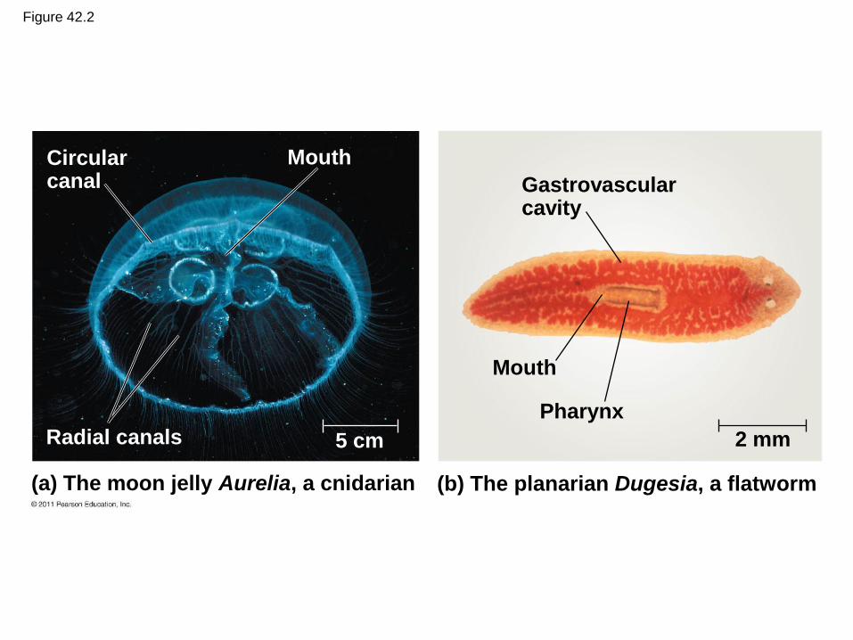
Figure 42.2
Circular canal
Mouth
Radial canals 5 cm
(a) The moon jelly Aurelia, a cnidarian (b) The planarian Dugesia, a flatworm
Gastrovascular cavity
Mouth
Pharynx
2 mm
Evolutionary Variation in Circulatory
Systems
• A circulatory system minimizes the diffusion
distance in animals with many cell layers
• Being thick means that you need a system
© 2011 Pearson Education, Inc.
General Properties of Circulatory Systems
• A circulatory system has
– A circulatory fluid
– A set of interconnecting vessels
– A muscular pump, the heart
• The circulatory system connects the fluid that
surrounds cells with the organs that exchange
gases, absorb nutrients, and dispose of wastes
• Circulatory systems can be open or closed, and
vary in the number of circuits in the body
© 2011 Pearson Education, Inc.
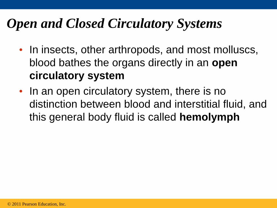
Open and Closed Circulatory Systems
• In insects, other arthropods, and most molluscs,
blood bathes the organs directly in an open
circulatory system
• In an open circulatory system, there is no
distinction between blood and interstitial fluid, and
this general body fluid is called hemolymph
© 2011 Pearson Education, Inc.
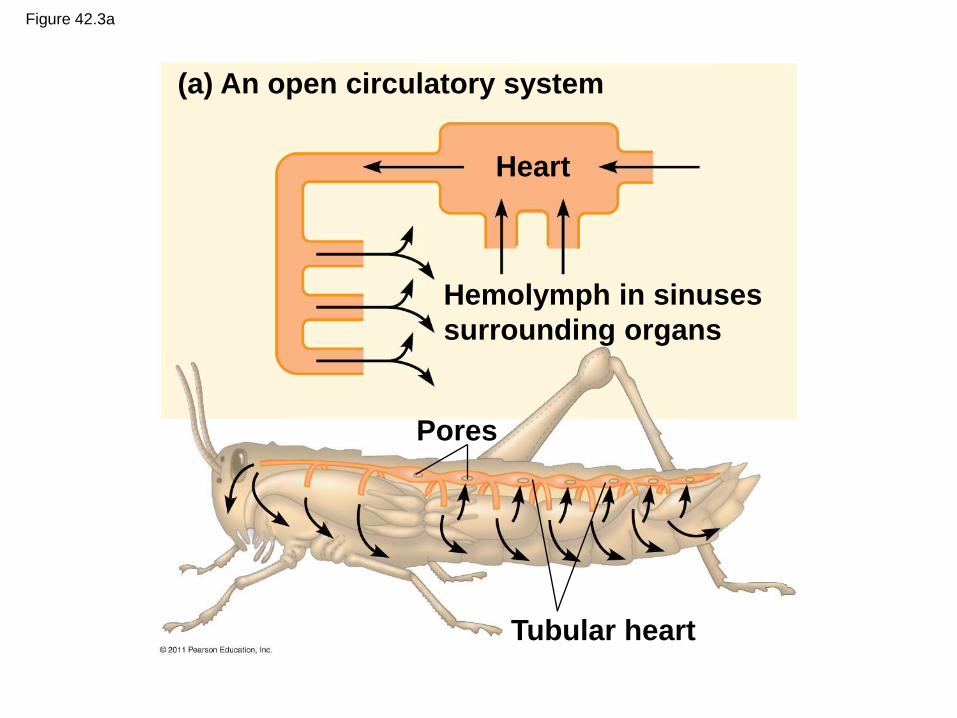
Figure 42.3a
(a) An open circulatory system
Heart
Hemolymph in sinuses
surrounding organs
Pores
Tubular heart
• In a closed circulatory system, blood is
confined to vessels and is distinct from the
interstitial fluid
• Closed systems are more efficient at transporting
circulatory fluids to tissues and cells
• Annelids, cephalopods, and vertebrates have
closed circulatory systems
© 2011 Pearson Education, Inc.
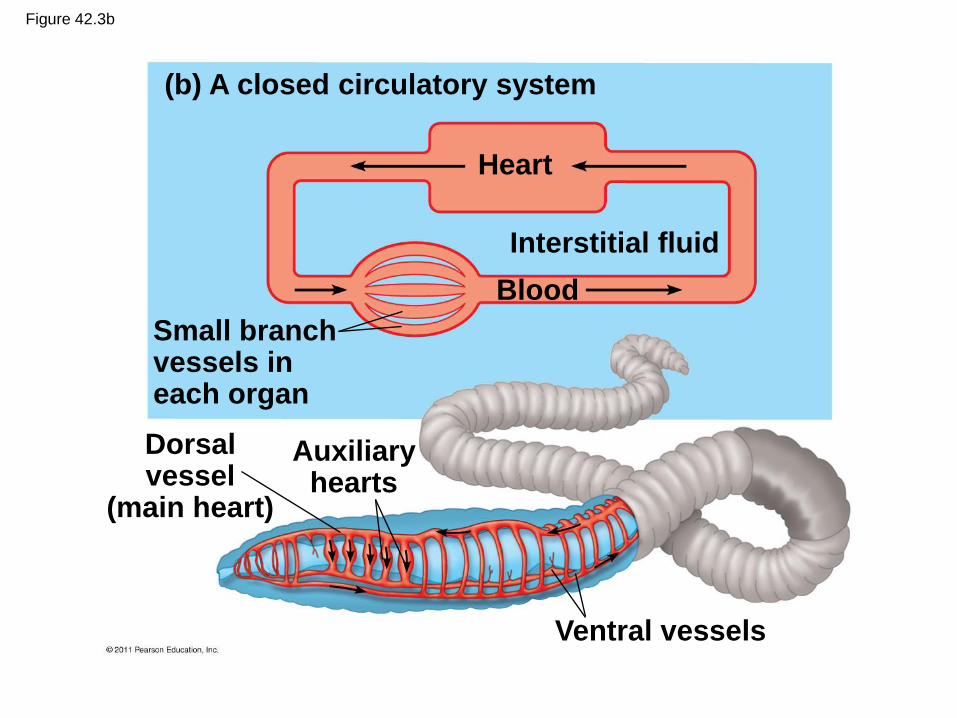
Figure 42.3b
(b) A closed circulatory system
Dorsal vessel
(main heart)
Auxiliary hearts
Small branch vessels in each organ
Ventral vessels
Blood
Interstitial fluid
Heart
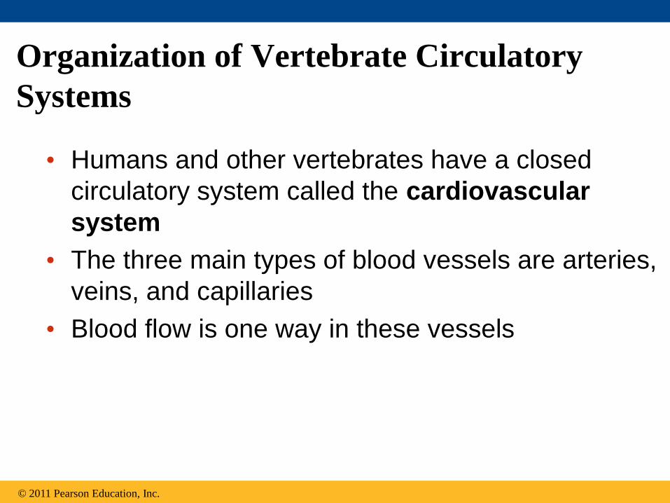
Organization of Vertebrate Circulatory
Systems
• Humans and other vertebrates have a closed
circulatory system called the cardiovascular
system
• The three main types of blood vessels are arteries,
veins, and capillaries
• Blood flow is one way in these vessels
© 2011 Pearson Education, Inc.
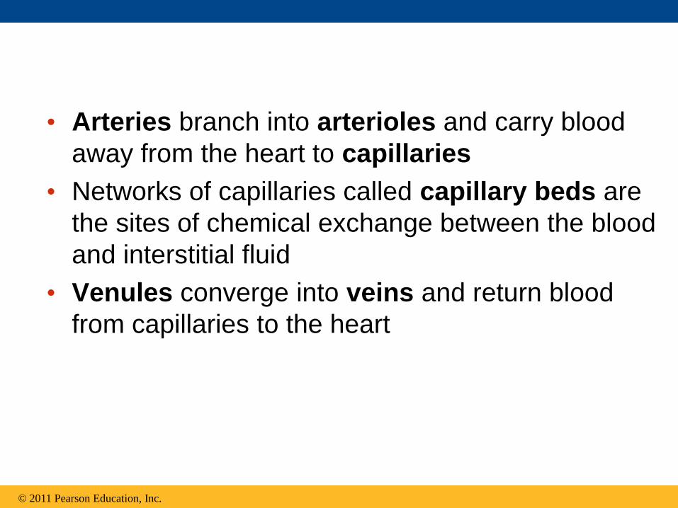
• Arteries branch into arterioles and carry blood
away from the heart to capillaries
• Networks of capillaries called capillary beds are
the sites of chemical exchange between the blood
and interstitial fluid
• Venules converge into veins and return blood
from capillaries to the heart
© 2011 Pearson Education, Inc. © 2011 Pearson Education, Inc.
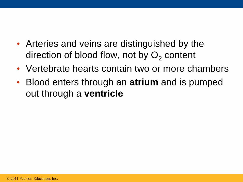
• Arteries and veins are distinguished by the
direction of blood flow, not by O2 content
• Vertebrate hearts contain two or more chambers
• Blood enters through an atrium and is pumped
out through a ventricle
© 2011 Pearson Education, Inc.
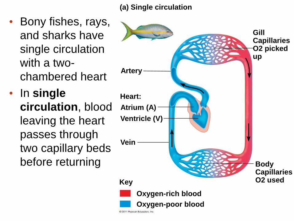
(a) Single circulation
Artery
Heart:
Atrium (A)
Ventricle (V)
Vein
Gill Capillaries O2 picked up
Body Capillaries O2 used Key
Oxygen-rich blood
Oxygen-poor blood
• Bony fishes, rays,
and sharks have
single circulation
with a two-
chambered heart
• In single
circulation, blood
leaving the heart
passes through
two capillary beds
before returning
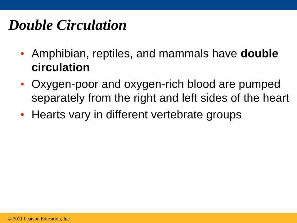
Double Circulation
• Amphibian, reptiles, and mammals have double
circulation
• Oxygen-poor and oxygen-rich blood are pumped
separately from the right and left sides of the heart
• Hearts vary in different vertebrate groups
© 2011 Pearson Education, Inc.
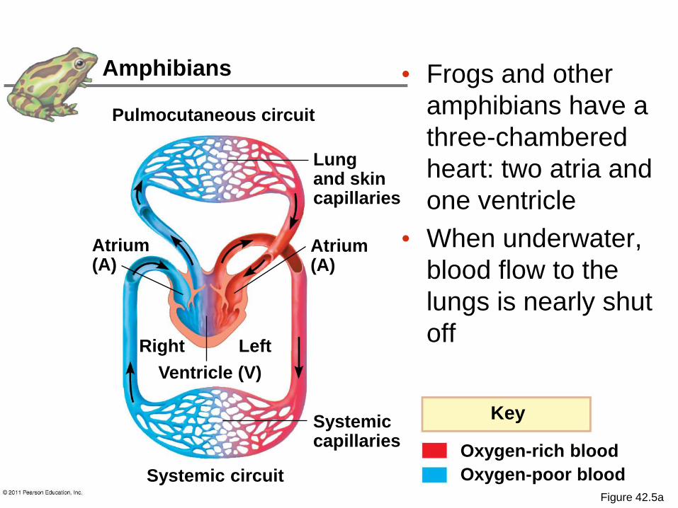
Amphibians
Pulmocutaneous circuit
Lung and skin capillaries
Atrium (A)
Atrium (A)
Left Right
Ventricle (V)
Systemic capillaries
Systemic circuit
Key
Oxygen-rich blood
Oxygen-poor blood Figure 42.5a
• Frogs and other
amphibians have a
three-chambered
heart: two atria and
one ventricle
• When underwater,
blood flow to the
lungs is nearly shut
off
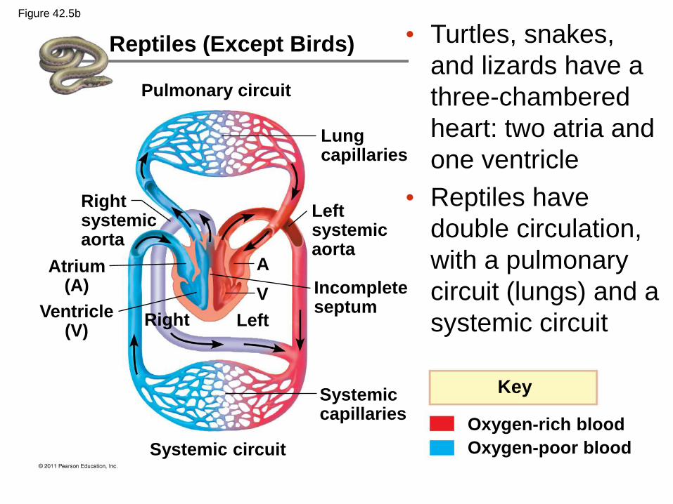
Figure 42.5b
Reptiles (Except Birds)
Pulmonary circuit
Systemic circuit
Systemic capillaries
Incomplete septum
Left systemic aorta
Left Right
Right systemic aorta
A
V
Lung capillaries
Atrium (A)
Ventricle (V)
Key
Oxygen-rich blood
Oxygen-poor blood
• Turtles, snakes,
and lizards have a
three-chambered
heart: two atria and
one ventricle
• Reptiles have
double circulation,
with a pulmonary
circuit (lungs) and a
systemic circuit
Mammals and Birds
• Mammals and birds have a four-chambered heart
with two atria and two ventricles
• The left side of the heart pumps and receives only
oxygen-rich blood, while the right side receives
and pumps only oxygen-poor blood
• Mammals and birds are endotherms and require
more O2 than ectotherms
© 2011 Pearson Education, Inc.
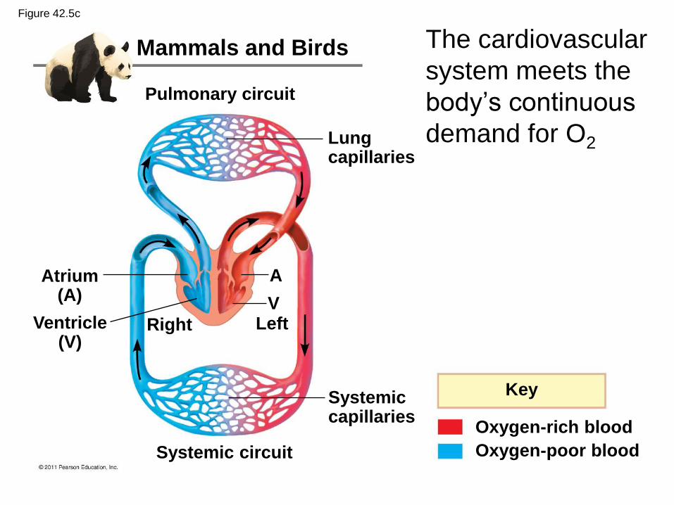
Systemic circuit
Lung capillaries
Pulmonary circuit
A
V Left Right
Systemic capillaries
Mammals and Birds
Atrium (A)
Ventricle (V)
Key
Oxygen-rich blood
Oxygen-poor blood
Figure 42.5c
The cardiovascular
system meets the
body’s continuous
demand for O2
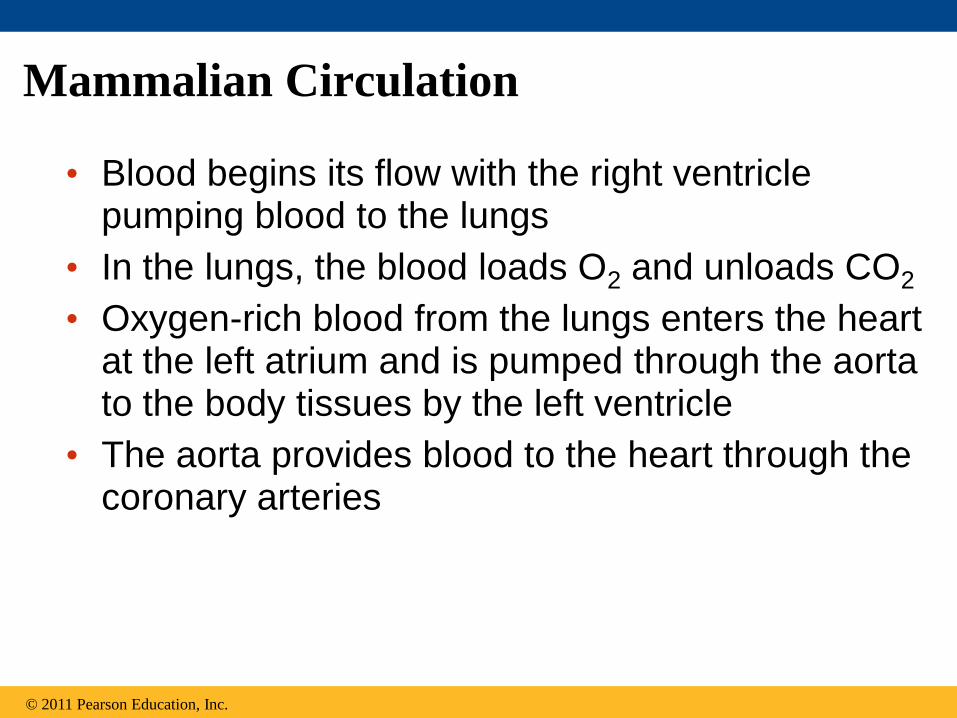
Mammalian Circulation
• Blood begins its flow with the right ventricle pumping blood to the lungs
• In the lungs, the blood loads O2 and unloads CO2
• Oxygen-rich blood from the lungs enters the heart at the left atrium and is pumped through the aorta to the body tissues by the left ventricle
• The aorta provides blood to the heart through the coronary arteries
© 2011 Pearson Education, Inc.
• Blood returns to the heart through the superior
vena cava (blood from head, neck, and forelimbs)
and inferior vena cava (blood from trunk and hind
limbs)
• The superior vena cava and inferior vena cava
flow into the right atrium
© 2011 Pearson Education, Inc.
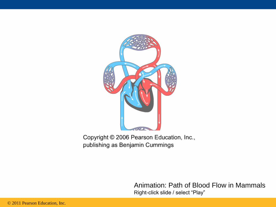
Animation: Path of Blood Flow in Mammals
© 2011 Pearson Education, Inc.
Animation: Path of Blood Flow in Mammals Right-click slide / select “Play”
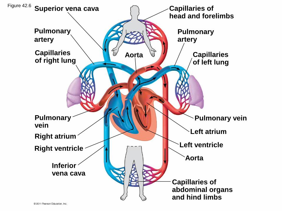
Superior vena cava
Pulmonary
artery
Capillaries of right lung
Pulmonary vein
Aorta
Inferior vena cava
Right ventricle
Capillaries of abdominal organs and hind limbs
Right atrium
Aorta
Left ventricle
Left atrium
Pulmonary vein
Pulmonary artery
Capillaries of left lung
Capillaries of head and forelimbs
Figure 42.6
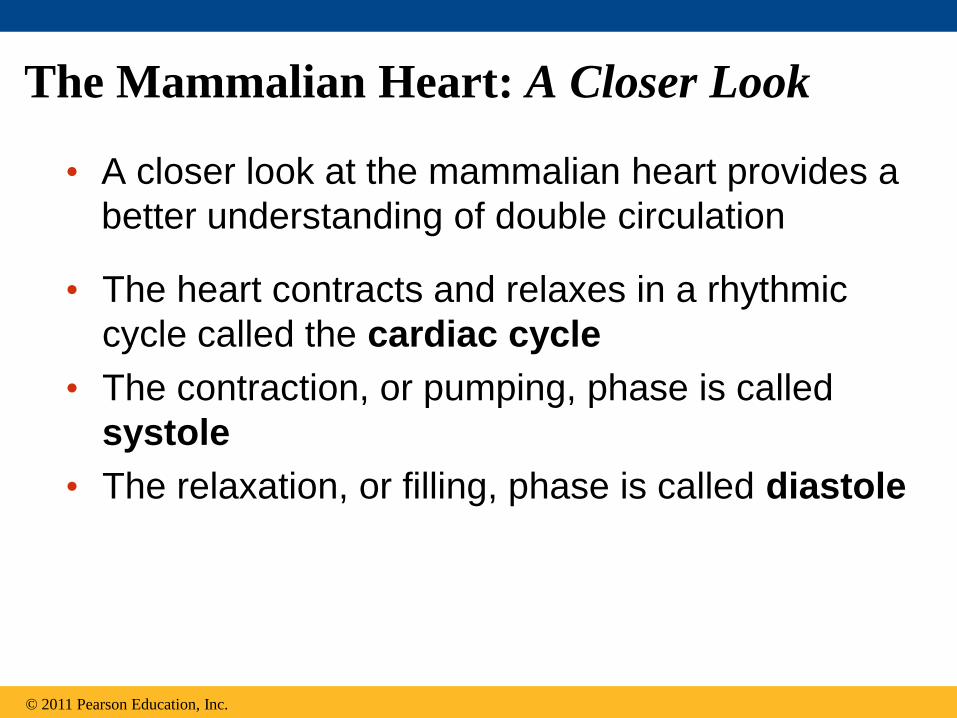
The Mammalian Heart: A Closer Look
• A closer look at the mammalian heart provides a
better understanding of double circulation
© 2011 Pearson Education, Inc.
• The heart contracts and relaxes in a rhythmic
cycle called the cardiac cycle
• The contraction, or pumping, phase is called
systole
• The relaxation, or filling, phase is called diastole
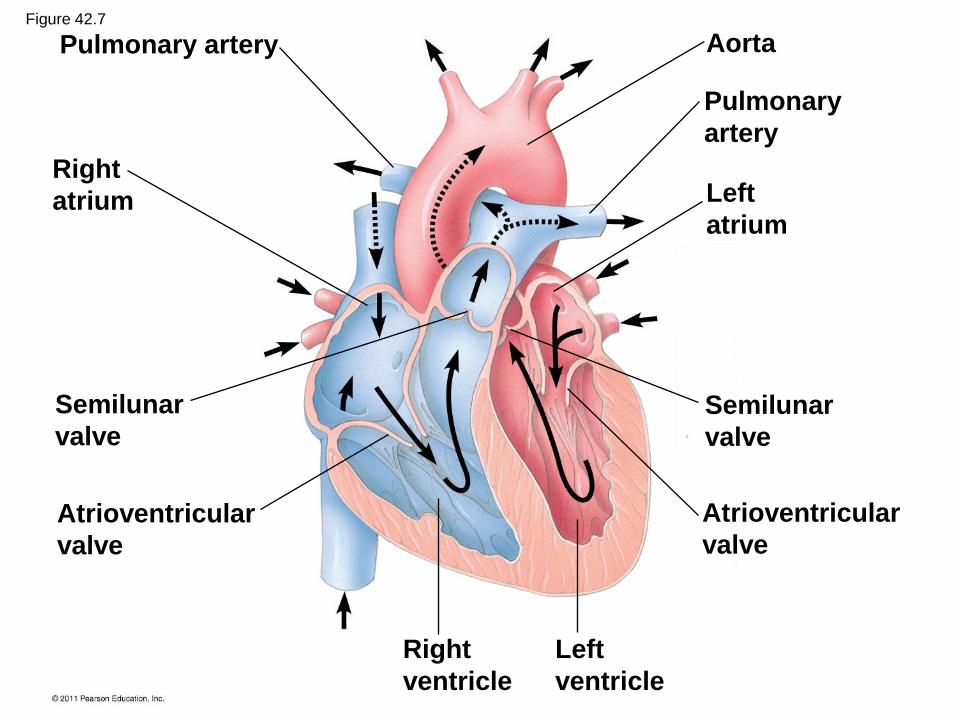
Figure 42.7
Pulmonary artery
Right
atrium
Semilunar
valve
Atrioventricular
valve
Right
ventricle
Left
ventricle
Atrioventricular
valve
Semilunar
valve
Left
atrium
Pulmonary
artery
Aorta
• The heart rate, also called the pulse, is the
number of beats per minute
• The stroke volume is the amount of blood
pumped in a single contraction
• The cardiac output is the volume of blood
pumped into the systemic circulation per minute
and depends on both the heart rate and stroke
volume
© 2011 Pearson Education, Inc.
• Four valves prevent backflow of blood in the heart
• The atrioventricular (AV) valves separate each
atrium and ventricle
• The semilunar valves control blood flow to the
aorta and the pulmonary artery
© 2011 Pearson Education, Inc.
• The “lub-dup” sound of a heart beat is caused by
the recoil of blood against the AV valves (lub) then
against the semilunar (dup) valves
• Backflow of blood through a defective valve
causes a heart murmur
© 2011 Pearson Education, Inc.
Heart sounds
Concept 42.3: Patterns of blood pressure and
flow reflect the structure and arrangement
of blood vessels
• The physical principles that govern
movement of water in plumbing systems also
influence the functioning of animal circulatory
systems
© 2011 Pearson Education, Inc.
Blood Vessel Structure and Function
• A vessel’s cavity is called the central lumen
• The epithelial layer that lines blood vessels is
called the endothelium
• The endothelium is smooth and minimizes
resistance
© 2011 Pearson Education, Inc.
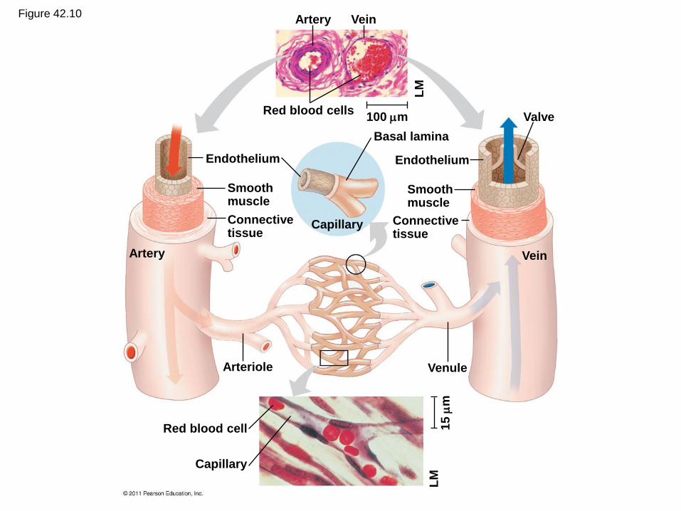
Figure 42.10 Artery
Red blood cells
Endothelium
Artery
Smooth muscle
Connective tissue
Capillary
Valve
Vein
Vein
Basal lamina
Endothelium
Smooth muscle
Connective tissue
100 m
LM
Venule
15
m
LM
Arteriole
Red blood cell
Capillary
• Capillaries have thin walls, the endothelium plus its basal lamina, to facilitate the exchange of materials
• Arteries and veins have an endothelium, smooth muscle, and connective tissue
• Arteries have thicker walls than veins to accommodate the high pressure of blood pumped from the heart
• In the thinner-walled veins, blood flows back to the heart mainly as a result of muscle action
© 2011 Pearson Education, Inc.
Blood Pressure
• Blood flows from areas of higher pressure to areas
of lower pressure
• Blood pressure is the pressure that blood exerts
against the wall of a vessel
• In rigid vessels blood pressure is maintained; less
rigid vessels deform and blood pressure is lost
© 2011 Pearson Education, Inc.
Changes in Blood Pressure During the
Cardiac Cycle
• Systolic pressure is the pressure in the arteries
during ventricular systole; it is the highest pressure
in the arteries
• Diastolic pressure is the pressure in the arteries
during diastole; it is lower than systolic pressure
• A pulse is the rhythmic bulging of artery walls with
each heartbeat
© 2011 Pearson Education, Inc.
Regulation of Blood Pressure
• Blood pressure is determined by cardiac output
and peripheral resistance due to constriction of
arterioles
• Vasoconstriction is the contraction of smooth
muscle in arteriole walls; it increases blood
pressure
• Vasodilation is the relaxation of smooth muscles
in the arterioles; it causes blood pressure to fall
© 2011 Pearson Education, Inc.
• Vasoconstriction and vasodilation help maintain
adequate blood flow as the body’s demands
change
• Nitric oxide is a major inducer of vasodilation
• The peptide endothelin is an important inducer of
vasoconstriction
© 2011 Pearson Education, Inc.
• Fainting is caused by inadequate blood flow to the head
• Animals with longer necks require a higher systolic pressure to pump blood a greater distance against gravity
• Blood is moved through veins by smooth muscle contraction, skeletal muscle contraction, and expansion of the vena cava with inhalation
• One-way valves in veins prevent backflow of blood
© 2011 Pearson Education, Inc.
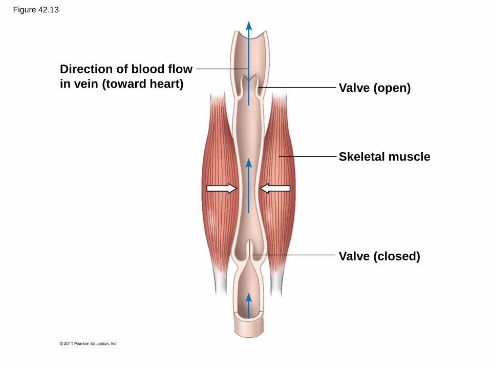
Direction of blood flow
in vein (toward heart) Valve (open)
Skeletal muscle
Valve (closed)
Figure 42.13
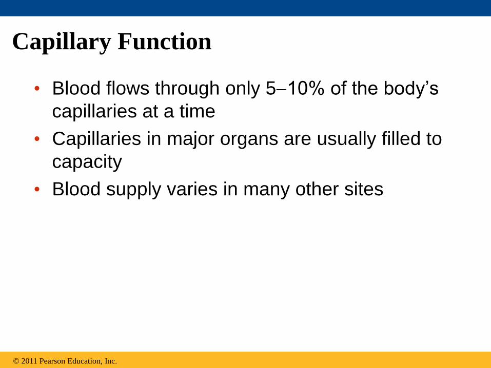
Capillary Function
• Blood flows through only 510% of the body’s
capillaries at a time
• Capillaries in major organs are usually filled to
capacity
• Blood supply varies in many other sites
© 2011 Pearson Education, Inc.
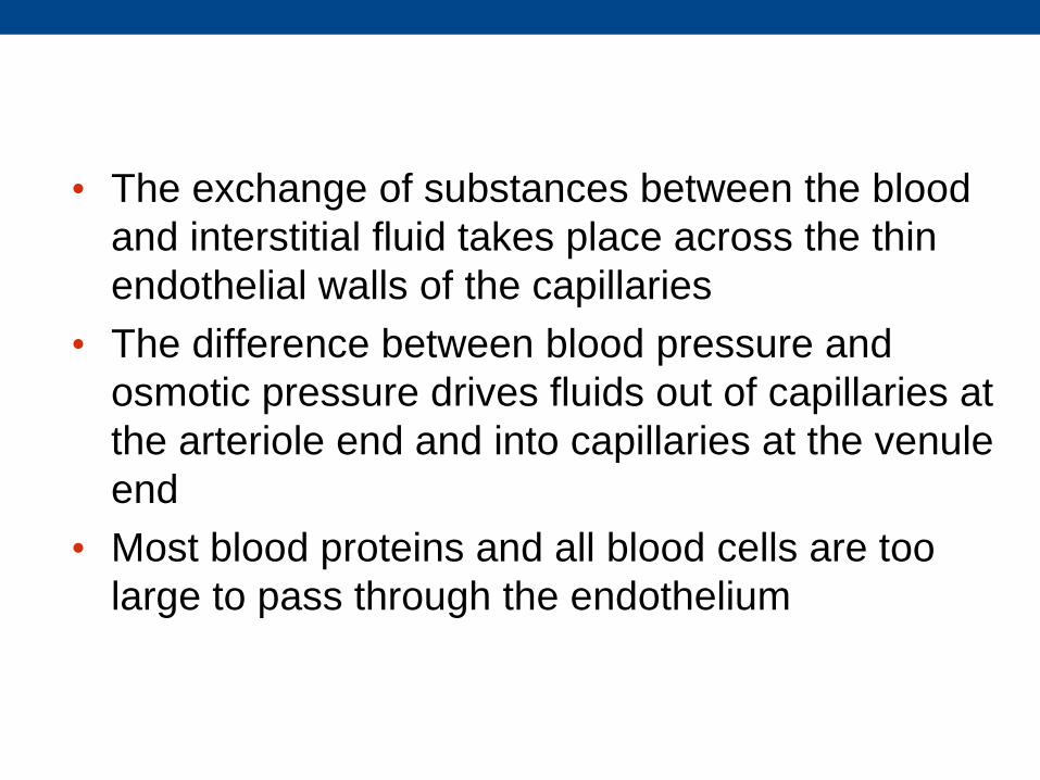
• The exchange of substances between the blood
and interstitial fluid takes place across the thin
endothelial walls of the capillaries
• The difference between blood pressure and
osmotic pressure drives fluids out of capillaries at
the arteriole end and into capillaries at the venule
end
• Most blood proteins and all blood cells are too
large to pass through the endothelium
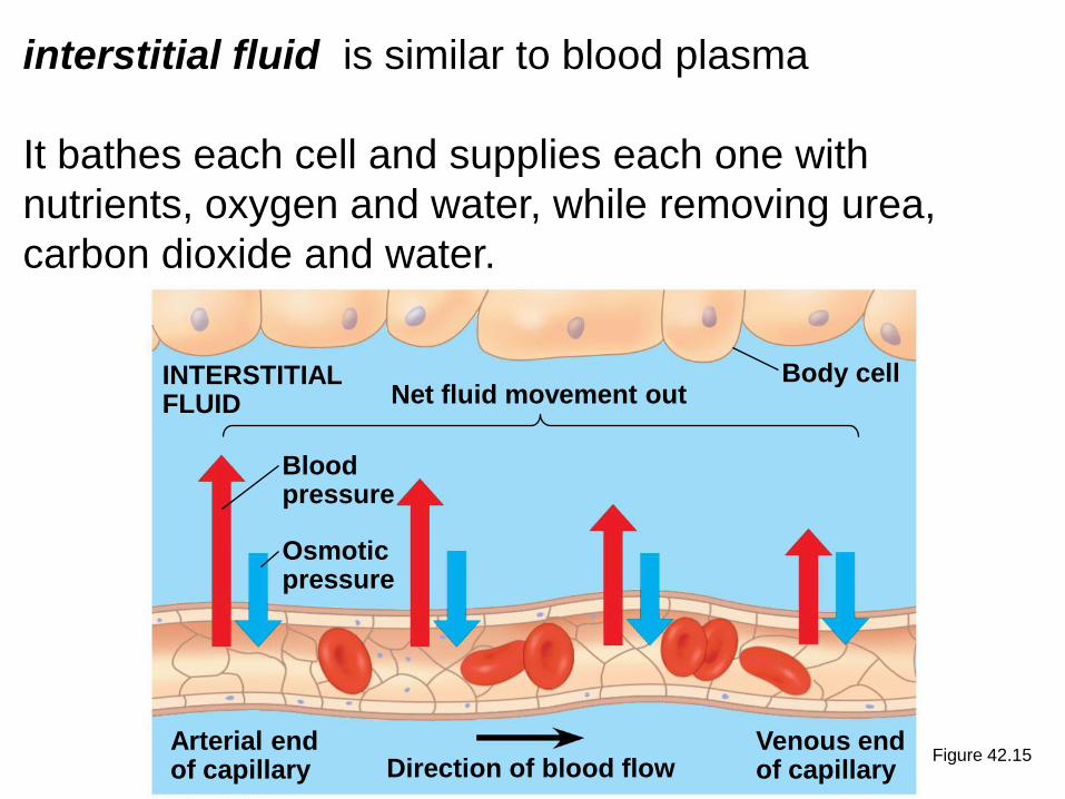
Figure 42.15
INTERSTITIAL FLUID Net fluid movement out
Blood pressure
Osmotic pressure
Arterial end of capillary Direction of blood flow
Venous end of capillary
Body cell
interstitial fluid is similar to blood plasma
It bathes each cell and supplies each one with
nutrients, oxygen and water, while removing urea,
carbon dioxide and water.

Fluid Return by the Lymphatic System
• The lymphatic system returns fluid that leaks out
from the capillary beds
• Fluid, called lymph, reenters the circulation
directly at the venous end of the capillary bed and
indirectly through the lymphatic system
• The lymphatic system drains into veins in the neck
• Valves in lymph vessels prevent the backflow of
fluid
© 2011 Pearson Education, Inc.
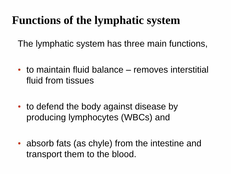
Functions of the lymphatic system
The lymphatic system has three main functions,
• to maintain fluid balance – removes interstitial
fluid from tissues
• to defend the body against disease by
producing lymphocytes (WBCs) and
• absorb fats (as chyle) from the intestine and
transport them to the blood.
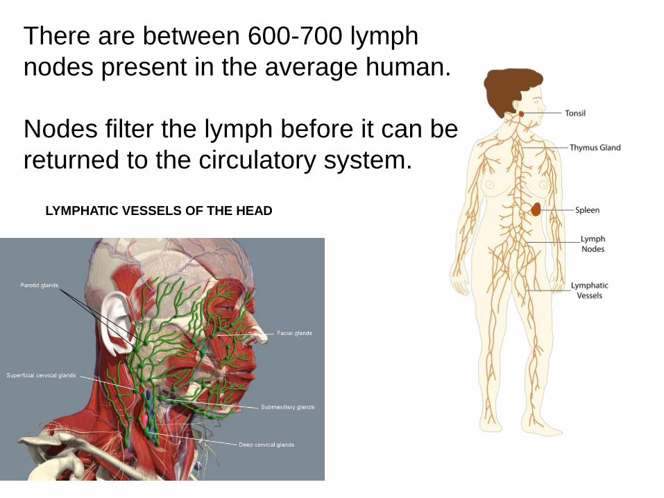
LYMPHATIC VESSELS OF THE HEAD
There are between 600-700 lymph
nodes present in the average human.
Nodes filter the lymph before it can be
returned to the circulatory system.
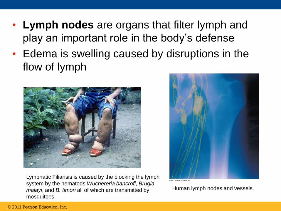
• Lymph nodes are organs that filter lymph and
play an important role in the body’s defense
• Edema is swelling caused by disruptions in the
flow of lymph
© 2011 Pearson Education, Inc.
Human lymph nodes and vessels.
Lymphatic Filiarisis is caused by the blocking the lymph
system by the nematods Wuchereria bancrofi, Brugia
malayi, and B. timori all of which are transmitted by
mosquitoes
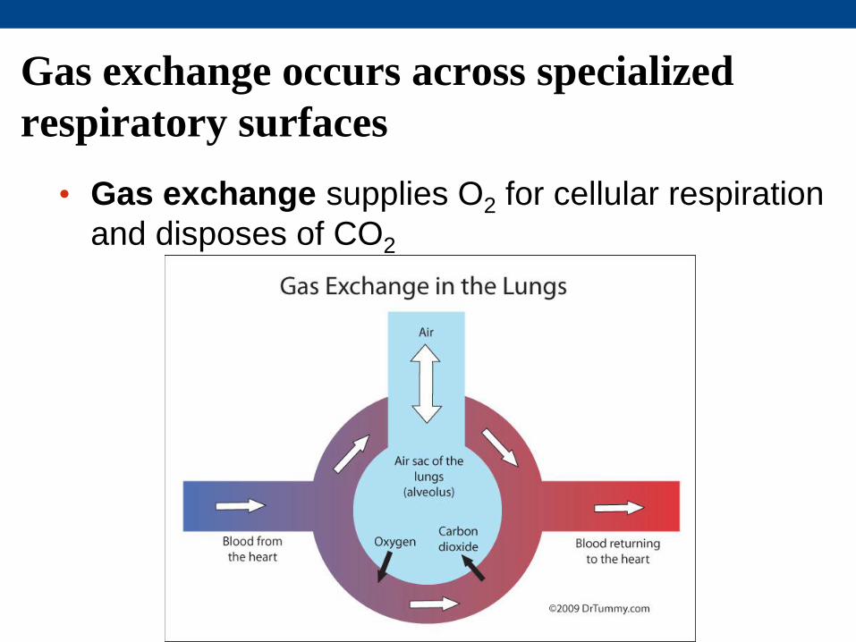
Gas exchange occurs across specialized
respiratory surfaces
• Gas exchange supplies O2 for cellular respiration
and disposes of CO2
Partial Pressure Gradients in Gas Exchange
• A gas diffuses from a region of higher partial
pressure to a region of lower partial pressure
• Partial pressure is the pressure exerted by a
particular gas in a mixture of gases
• Gases diffuse down pressure gradients in the
lungs and other organs as a result of differences in
partial pressure
© 2011 Pearson Education, Inc.
Respiratory Media
• Animals can use air or water as a source of O2, or
respiratory medium
• In a given volume, there is less O2 available in
water than in air
• Obtaining O2 from water requires greater
efficiency than air breathing
© 2011 Pearson Education, Inc.
Respiratory Surfaces
• Animals require large, moist respiratory surfaces
for exchange of gases between their cells and the
respiratory medium, either air or water
• Gas exchange across respiratory surfaces takes
place by diffusion
• Respiratory surfaces vary by animal and can
include the outer surface, skin, gills, tracheae, and
lungs
© 2011 Pearson Education, Inc.
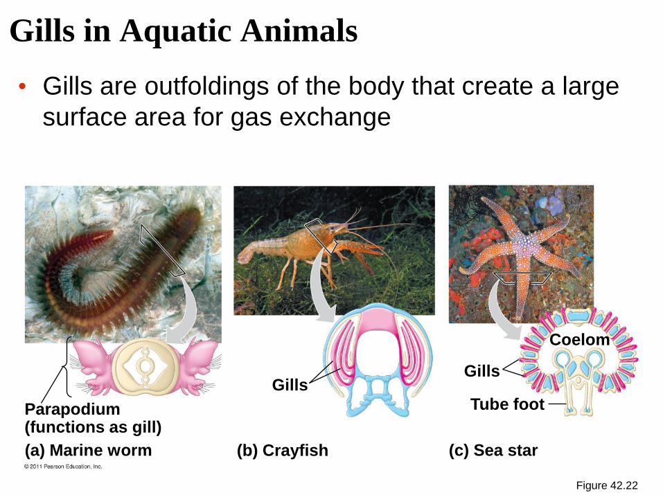
Figure 42.22
Parapodium (functions as gill)
(a) Marine worm (b) Crayfish
Gills Gills
Tube foot
(c) Sea star
Coelom
• Gills are outfoldings of the body that create a large
surface area for gas exchange
Gills in Aquatic Animals
• Ventilation moves the respiratory medium over
the respiratory surface
• Aquatic animals move through water or move
water over their gills for ventilation
• Fish gills use a countercurrent exchange
system, where blood flows in the opposite
direction to water passing over the gills; blood is
always less saturated with O2 than the water it
meets
© 2011 Pearson Education, Inc.
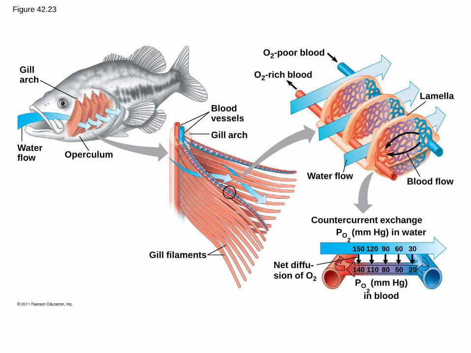
Figure 42.23
Gill arch
O2-poor blood
O2-rich blood
Blood vessels
Gill arch
Operculum Water flow
Water flow Blood flow
Countercurrent exchange
PO (mm Hg) in water 2
150
PO (mm Hg)
in blood 2
120 90 60 30
140 110 80 50 20 Net diffu- sion of O2
Lamella
Gill filaments
Tracheal Systems in Insects
• The tracheal system of insects consists of tiny
branching tubes that penetrate the body
• The tracheal tubes supply O2 directly to body cells
• The respiratory and circulatory systems are
separate
• Larger insects must ventilate their tracheal system
to meet O2 demands
© 2011 Pearson Education, Inc.
Tracheoles Mitochondria Muscle fiber
2.5
m
Tracheae
Air sacs
External opening
Trachea
Air sac Tracheole
Body cell
Air
Figure 42.24
Lungs
• Lungs are an infolding of the body surface
• The circulatory system (open or closed) transports
gases between the lungs and the rest of the body
• The size and complexity of lungs correlate with an
animal’s metabolic rate
© 2011 Pearson Education, Inc.
Mammalian Respiratory Systems: A Closer
Look
• A system of branching ducts conveys air to the lungs
• Air inhaled through the nostrils is warmed, humidified, and sampled for odors
• The pharynx directs air to the lungs and food to the stomach
• Swallowing tips the epiglottis over the glottis in the pharynx to prevent food from entering the trachea
© 2011 Pearson Education, Inc.
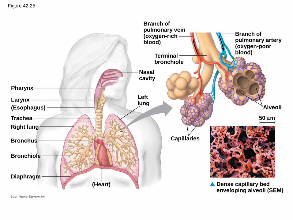
Figure 42.25
Pharynx
Larynx
(Esophagus)
Trachea
Right lung
Bronchus
Bronchiole
Diaphragm
(Heart)
Capillaries
Left lung
Dense capillary bed enveloping alveoli (SEM)
50 m
Alveoli
Branch of pulmonary artery (oxygen-poor blood)
Branch of pulmonary vein (oxygen-rich blood)
Terminal bronchiole
Nasal cavity
• Air passes through the pharynx, larynx, trachea, bronchi, and bronchioles to the alveoli, where gas exchange occurs
• Exhaled air passes over the vocal cords in the larynx to create sounds
• Cilia and mucus line the epithelium of the air ducts and move particles up to the pharynx
• This “mucus escalator” cleans the respiratory system and allows particles to be swallowed into the esophagus
© 2011 Pearson Education, Inc.
• Gas exchange takes place in alveoli, air sacs at the tips of bronchioles
• Oxygen diffuses through the moist film of the epithelium and into capillaries
• Carbon dioxide diffuses from the capillaries across the epithelium and into the air space
© 2011 Pearson Education, Inc.
Concept 42.6: Breathing ventilates the lungs
• The process that ventilates the lungs is breathing,
the alternate inhalation and exhalation of air
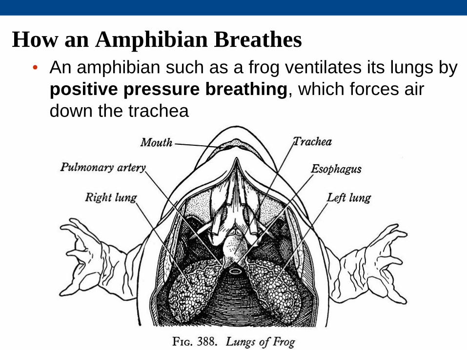
How an Amphibian Breathes
• An amphibian such as a frog ventilates its lungs by
positive pressure breathing, which forces air
down the trachea
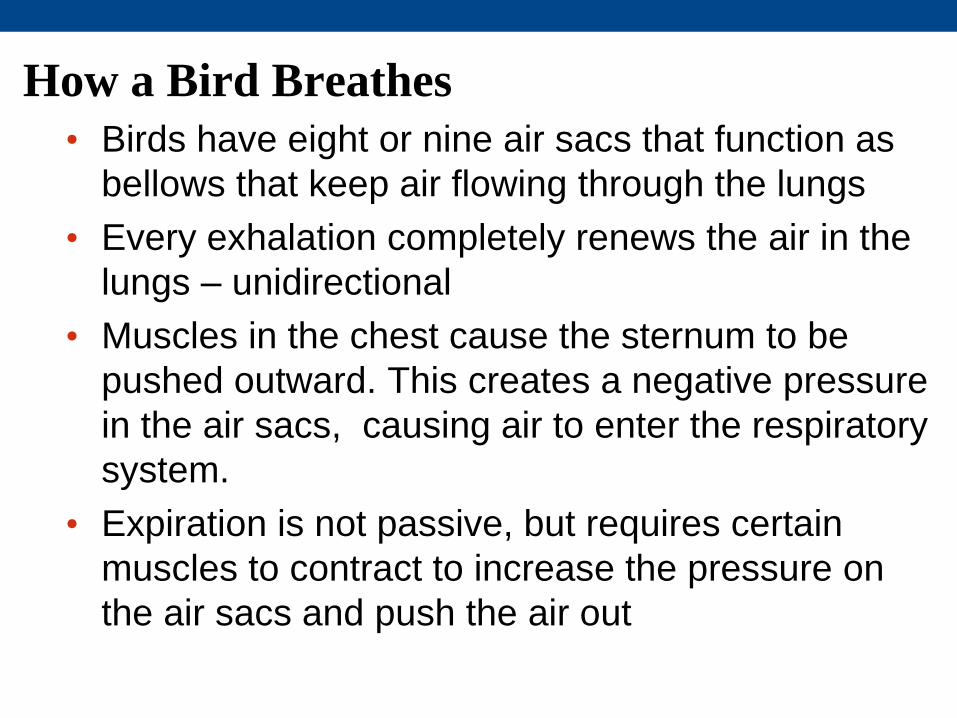
How a Bird Breathes
• Birds have eight or nine air sacs that function as
bellows that keep air flowing through the lungs
• Every exhalation completely renews the air in the
lungs – unidirectional
• Muscles in the chest cause the sternum to be
pushed outward. This creates a negative pressure
in the air sacs, causing air to enter the respiratory
system.
• Expiration is not passive, but requires certain
muscles to contract to increase the pressure on
the air sacs and push the air out
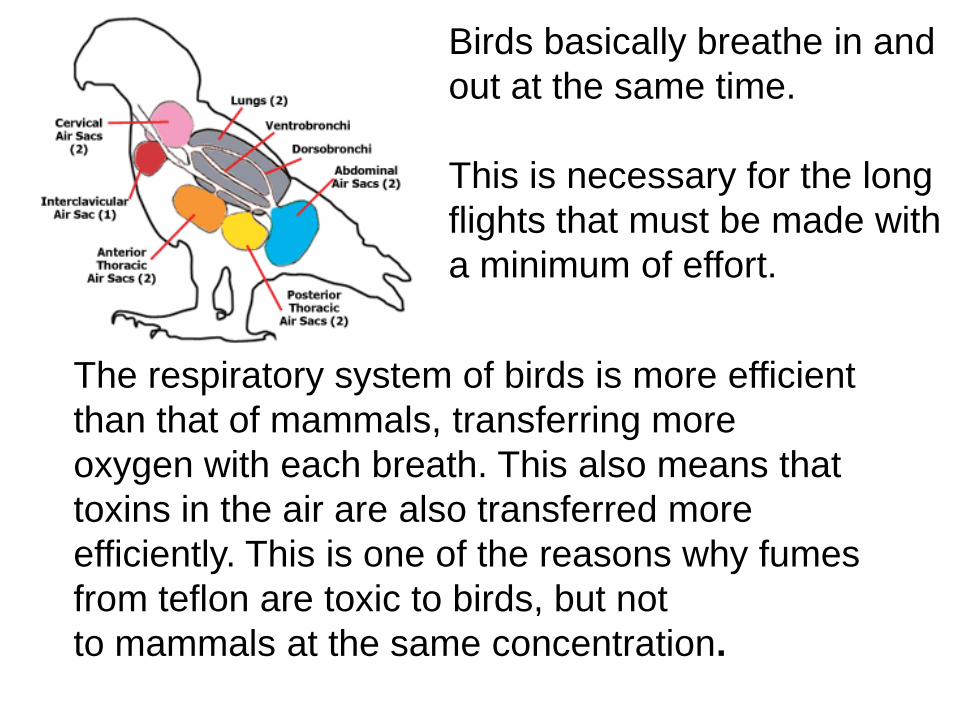
The respiratory system of birds is more efficient
than that of mammals, transferring more
oxygen with each breath. This also means that
toxins in the air are also transferred more
efficiently. This is one of the reasons why fumes
from teflon are toxic to birds, but not
to mammals at the same concentration.
Birds basically breathe in and
out at the same time.
This is necessary for the long
flights that must be made with
a minimum of effort.
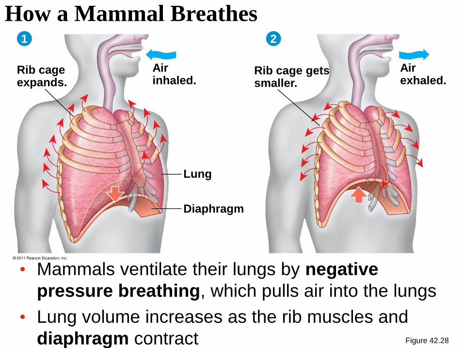
Figure 42.28
Rib cage expands.
Air inhaled.
Air exhaled.
Rib cage gets smaller.
1 2
Lung
Diaphragm
• Mammals ventilate their lungs by negative
pressure breathing, which pulls air into the lungs
• Lung volume increases as the rib muscles and
diaphragm contract
How a Mammal Breathes
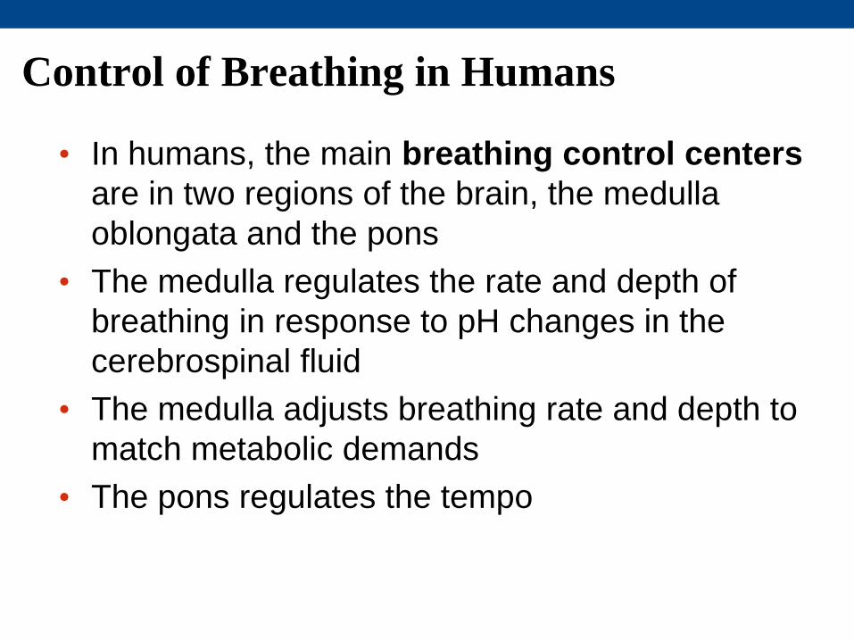
Control of Breathing in Humans
• In humans, the main breathing control centers
are in two regions of the brain, the medulla
oblongata and the pons
• The medulla regulates the rate and depth of
breathing in response to pH changes in the
cerebrospinal fluid
• The medulla adjusts breathing rate and depth to
match metabolic demands
• The pons regulates the tempo
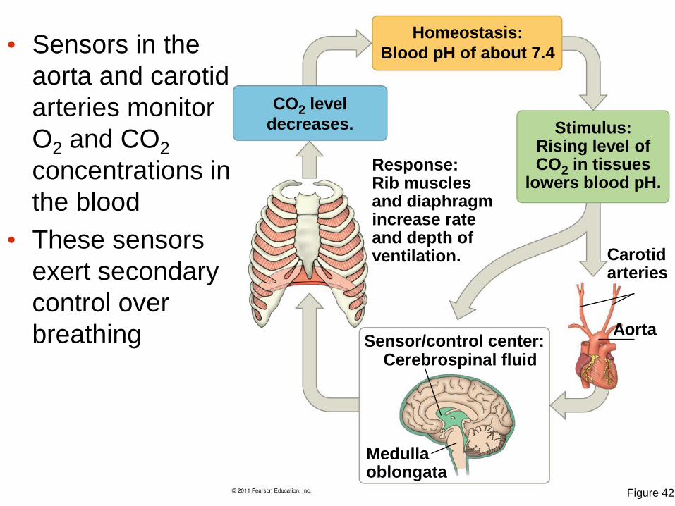
Homeostasis:
Blood pH of about 7.4
CO2 level
decreases. Stimulus: Rising level of CO2 in tissues
lowers blood pH. Response: Rib muscles and diaphragm increase rate and depth of ventilation. Carotid
arteries
Aorta Sensor/control center:
Cerebrospinal fluid
Medulla oblongata
Figure 42.29
• Sensors in the
aorta and carotid
arteries monitor
O2 and CO2
concentrations in
the blood
• These sensors
exert secondary
control over
breathing