Embed Size (px)
DESCRIPTION
1 2 3 4 5 6 7 8 9 10 11 12 13 14
Citation preview
CIRRHOSIS
Cirrhosis is a pathologically defined entity that is associated with a spec-
trum of characteristic clinical manifestations. The cardinal pathologic fea-
tures reflect irreversible chronic injury of the hepatic parenchyma and in-
clude extensive fibrosis in association with the formation of regenerative nod-
ules.
These features result from hepatocyte necrosis, collapse of the supporting
reticulin network with subsequent connective tissue deposition, distortion of the
vascular bed, and nodular regeneration of remaining liver parenchyma.
Classification of the various types of cirrhosis
based on either etiology or morphology alone is unsatisfactory. A single pathologic
pattern may result from a variety of insults, while the same insult may produce
several morphologic patterns. Nevertheless, most types of cirrhosis may be use-
fully classified by a mixture of etiologically and morphologically defined entities
as follows:
Etiology
1) alcoholic;
2) cryptogenic
3) posthepatitic;
4) biliary (primary biliary cirrhosis and secondary biliary cirrhosis);
5) cardiac;
6) metabolic,
7) inherited, and drug-related.
On the clinical findings:
Stages: initial, manifesting, terminal
Activity: Active C, non-active C.
Степень печеночно-клеточной функциональной недостаточности:
- легкая (компенсированная),
- средней тяжести (субкомпенсированая),
- тяжелая (декомпенсированая).
1
Форма портальной гипертензии: скрытая, умеренная, развернутая
(внутрипеченочная, подпеченочная, надпеченочная и смешанная).
Развитие сочетанных клинических синдромов:
- гепато-лиенальный (splenomegaly, hypersplenism),
- hepatopancreatic syndrome,
- hepatorenal syndrome.
COMPLICATIONS OF CIRRHOSIS
These include:
- portal hypertension and its consequences (e.g., gastroesophageal varices
and splenomegaly),
- ascites,
- hepatic encephalopathy,
- spontaneous bacterial peritonitis,
- and hepatocellular carcinoma.
Тhe most common histologic classification divides cirrhosis into micron-
odular, macronodular, and mixed forms.
- In micronodular cirrhosis, the regenerating nodules are no larger than the
original lobules, i.e. approximately 1 mm in diameter or less.
- Macronodular cirrhosis is characterized by larger nodules, which mea-
sure several centimeters in diameter and may contain central veins.
This form corresponds more or less to postnecrotic (posthepatic) cirrhosis
but does not necessarily follow episodes of massive necrosis and stromal collapse.
- Mixed macro- and micronodular cirrosis signifies that the features of
cirrhosis are variable.
There is a limited interrelationship among the histologic form of cirrhosis,
the etiology, and the prognosis.
2
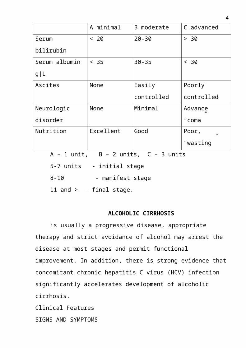
Child’s criteria for hepatic functional reserve
A minimal B moderate C advanced
Serum bilirubin < 20 20-30 > 30
Serum albumin g|L < 35 30-35 < 30
Ascites None Easily controlled Poorly controlled
Neurologic disorder None Minimal Advance “coma”
Nutrition Excellent Good Poor, “wasting”
A – 1 unit, B – 2 units, C – 3 units
5-7 units - initial stage
8-10 - manifest stage
11 and > - final stage.
ALCOHOLIC CIRRHOSIS
is usually a progressive disease, appropriate therapy and strict avoidance of
alcohol may arrest the disease at most stages and permit functional improvement.
In addition, there is strong evidence that concomitant chronic hepatitis C virus
(HCV) infection significantly accelerates development of alcoholic cirrhosis.
Clinical Features
SIGNS AND SYMPTOMS
Alcoholic cirrhosis may be clinically silent, and many cases (10 to 40%) are
discovered incidentally at laparotomy or autopsy. In many cases symptoms are in-
sidious in onset, occurring usually after ≥10 years of excessive alcohol use and
progressing slowly over subsequent weeks and months. Anorexia and malnutrition
lead to weight loss and a reduction in skeletal muscle mass. The patient may expe-
rience easy bruising, increasing weakness, and fatigue. Eventually the clinical
manifestations of hepatocellular dysfunction and portal hypertension ensue, includ-
ing progressive jaundice, bleeding from gastroesophageal varices, ascites, and en-
cephalopathy. The abrupt onset of one of these complications may be the first
event prompting the patient to seek medical attention. In other cases, cirrhosis first
3
becomes evident when the patient requires treatment of symptoms related to alco-
holic hepatitis.
A firm, nodular liver may be an early sign of disease; the liver may be either
enlarged, normal, or decreased in size. Other frequent findings include jaundice,
palmar erythema, spider angiomas, parotid and lacrimal gland enlargement, club-
bing of fingers, splenomegaly, muscle wasting, and ascites with or without periph-
eral edema. Men may have decreased body hair and/or gynecomastia and testicular
atrophy, which, like the cutaneous findings, result from disturbances in hormonal
metabolism, including increased peripheral formation of estrogen due to dimin-
ished hepatic clearance of the precursor androstenedione. Testicular atrophy may
reflect hormonal abnormalities or the toxic effect of alcohol on the testes. In
women, signs of virilization or menstrual irregularities may occasionally be en-
countered. Dupuytren's contractures resulting from fibrosis of the palmar fascia
with resulting flexion contracture of the digits are associated with alcoholism but
are not specifically related to cirrhosis.
Although the cirrhotic patient may stabilize if drinking is discontinued, over a pe-
riod of years, the patient may become emaciated, weak, and chronically jaundiced.
Ascites and other signs of portal hypertension may become increasingly promi-
nent. Ultimately, most patients with advanced cirrhosis die in hepatic coma, com-
monly precipitated by hemorrhage from esophageal varices or intercurrent infec-
tion. Progressive renal dysfunction often complicates the terminal phase of the ill-
ness.
BILIARY CIRRHOSIS
Biliary cirrhosis results from injury to or prolonged obstruction of either the intra-
hepatic or extrahepatic biliary system. It is associated with impaired biliary excre-
tion, destruction of hepatic parenchyma, and progressive fibrosis. Primary biliary
cirrhosis (PBC) is characterized by chronic inflammation and fibrous obliteration
of intrahepatic bile ductules. Secondary biliary cirrhosis (SBC) is the result of
long-standing obstruction of the larger extrahepatic ducts. Although primary and
4
secondary biliary cirrhosis are separate pathophysiologic entities with respect to
the initial insult, many clinical features are similar.
PRIMARY BILIARY CIRRHOSIS
Etiology and Pathogenesis
The cause of PBC remains unknown. Several observations suggest that a
disordered immune response may be involved. PBC is frequently associated with a
variety of disorders presumed to be autoimmune in nature, such as the syndrome of
calcinosis, Raynaud's phenomenon, esophageal dysmotility, sclerodactyly, telang-
iectasia (CREST); the sicca syndrome (dry eyes and dry mouth); autoimmune thy-
roiditis; type 1 diabetes mellitus; and IgA deficiency.
Most important, a circulating IgG antimitochondrial antibody (AMA) is de-
tected in >90% of patients with PBC and only rarely in other forms of liver dis-
ease.
Clinical Features
SIGNS AND SYMPTOMS
Among patients with symptomatic disease, 90% are women age 35 to 60.
Often the earliest symptom is pruritus, which may be either generalized or limited
initially to the palms and soles. In addition, fatigue is commonly a prominent early
symptom. After several months or years, jaundice and gradual darkening of the ex-
posed areas of the skin (melanosis) may ensue. Other early clinical manifestations
of PBC reflect impaired bile excretion. These include steatorrhea and the malab-
sorption of lipid-soluble vitamins. Protracted elevation of serum lipids, especially
cholesterol, leads to subcutaneous lipid deposition around the eyes (xanthelasmas)
and over joints and tendons (xanthomas). Over a period of months to years, the
itching, jaundice, and hyperpigmentation slowly worsen.
LABORATORY FINDINGS
PBC is increasingly diagnosed at a presymptomatic stage, prompted by the
finding of a twofold or greater elevation of the serum alkaline phosphatase during
routine screening. Serum 5′-nucleotidase activity and γ-glutamyl transpeptidase
levels are also elevated. In this setting, serum bilirubin is usually normal and
5
aminotransferase levels minimally increased. The diagnosis is supported by a posi-
tive AMA test (titer > 1:40).
TREATMENT
While there is no specific therapy for PBC, ursodiol has been shown to im-
prove biochemical and histologic features and might improve survival, particularly
liver transplantation–free survival (although this remains unproven). Ursodiol
should be given in doses of 13 to 15 mg/kg per day, but lower doses are sometimes
just as effective in reducing serum alkaline phosphatase and aminotransferase lev-
els. Ursodiol should be given with food and can be taken in a single dose daily.
Side effects are rare: gastrointestinal intolerance (diarrhea) and skin rashes occur
but are uncommon. Isolated instances of severe exacerbation of pruritus have been
reported in patients with advanced disease. Ursodiol probably works by replacing
the endogenously produced hydrophobic bile acids with urosdeoxycholate, a hy-
drophilic and relatively nontoxic bile acid.
SECONDARY BILIARY CIRRHOSIS
Etiology
SBC results from prolonged partial or total obstruction of the common bile
duct or its major branches. In adults, obstruction is most frequently caused by post-
operative strictures or gallstones, usually with superimposed infectious cholangitis.
Chronic pancreatitis may lead to biliary stricture and secondary cirrhosis.
Pathology and Pathogenesis
Unrelieved obstruction of the extrahepatic bile ducts leads to (1) bile stasis
and focal areas of centrilobular necrosis followed by periportal necrosis, (2) prolif-
eration and dilatation of the portal bile ducts and ductules, (3) sterile or infected
cholangitis with accumulation of polymorphonuclear infiltrates around bile ducts,
and (4) progressive expansion of portal tracts by edema and fibrosis.
Clinical Features
The symptoms, signs, and biochemical findings of SBC are similar to those of
PBC. Jaundice and pruritus are usually the most prominent features. In addition,
6
fever and/or right upper quadrant pain, reflecting bouts of cholangitis or biliary
colic, are typical.
TREATMENT
Relief of obstruction to bile flow, by either endoscopic or surgical means, is the
most important step in the prevention and therapy of SBC.
POSTHEPATITIC AND CRYPTOGENIC CIRRHOSIS
Posthepatitic or postnecrotic cirrhosis represents the final common pathway
of many types of chronic liver disease. Coarsely nodular cirrhosis and multilobular
cirrhosis are terms synonymous with posthepatitic cirrhosis. The term cryptogenic
cirrhosis has been used interchangeably with posthepatitic cirrhosis, but this desig-
nation should be reserved for those cases in which the etiology of cirrhosis is un-
known (approximately 10% of all patients with cirrhosis).
Posthepatitic cirrhosis is characterized morphologically by irregular nodules
of regenerating hepatocytes, varying in size from microscopic to several centime-
ters in diameter.
Clinical Features
In patients with cirrhosis of known etiology in whom there is progression to
a posthepatitic stage, the clinical manifestations are an extension of those resulting
from the initial disease process. Usually clinical symptoms are related to portal hy-
pertension and its sequelae, such as ascites, splenomegaly, hypersplenism, en-
cephalopathy, and bleeding gastroesophageal varices. The hematologic and liver
function abnormalities resemble those seen with other types of cirrhosis. In a few
patients with posthepatitic cirrhosis, the diagnosis may be made incidentally at op-
eration, at postmortem, or by a needle biopsy of the liver performed to investigate
abnormal liver function tests or hepatomegaly.
CARDIAC CIRRHOSIS
Prolonged, severe right-sided congestive heart failure may lead to chronic
liver injury and cardiac cirrhosis. The characteristic pathologic features of fibrosis
and regenerative nodules distinguish cardiac cirrhosis from both reversible passive
congestion of the liver due to acute heart failure and acute hepatocellular necrosis
7
(“ischemic hepatitis” or “shock liver”) resulting from systemic hypotension and
hypoperfusion of the liver.
The signs and symptoms of heart failure usually overshadow the liver dis-
ease. With prolonged right-sided heart failure the liver becomes enlarged, firm, and
usually nontender.
TREATMENT
Prevention or treatment of cardiac cirrhosis depends on the diagnosis and
therapy of the underlying cardiovascular disorder. Improvement in cardiac function
frequently results in improvement of liver function and stabilization of the liver
disease.
MAJOR COMPLICATIONS OF CIRRHOSIS
PORTAL HYPERTENSION
Clinical Features
The major clinical manifestations of portal hypertension include hemorrhage from
gastroesophageal varices, splenomegaly with hypersplenism, ascites, and acute and
chronic hepatic encephalopathy. These are related, at least in part, to the develop-
ment of portal-systemic collateral channels. The absence of valves in the portal ve-
nous system facilitates retrograde (hepatofugal) blood flow from the high-pressure
portal venous system to the lower-pressure systemic venous circulation. Major
sites of collateral flow involve the veins around the cardioesophageal junction
(esophagogastric varices), the rectum (hemorrhoids), retroperitoneal space, and the
falciform ligament of the liver (periumbilical or abdominal wall collaterals). Ab-
dominal wall collaterals appear as tortuous epigastric vessels that radiate from the
umbilicus toward the xiphoid and rib margins (caput medusae).
A frequent marker of the presence of cirrhosis in a patient being followed for
chronic liver disease is a progressive decrease in platelet count. A low-normal
platelet count can be the first clue to progression to cirrhosis. Ultimately, a marked
decrease in platelets (to 30,000 to 60,000/µL) and white blood cells can occur.
VARICEAL BLEEDING
8
Pathogenesis
While vigorous hemorrhage may arise from any portal-systemic venous collaterals,
bleeding is most common from varices in the region of the gastroesophageal junc-
tion. The factors contributing to bleeding from gastroesophageal varices are not en-
tirely understood but include the degree of portal hypertension (>12 mmHg) and
the size of the varices.
Clinical Features and Diagnosis
Variceal bleeding often occurs without obvious precipitating factors and usually
presents with painless but massive hematemesis with or without melena. Associ-
ated signs range from mild postural tachycardia to profound shock, depending on
the extent of blood loss and degree of hypovolemia. Endoscopy is the best ap-
proach to evaluate upper gastrointestinal hemorrhage in patients with known or
suspected portal hypertension
The medical management of acute variceal hemorrhage includes the use of
vasoconstrictors (somatostatin/octreotide or vasopressin), balloon tamponade, and
endoscopic variceal ligation (EVL) or sclerosis of varices (sclerotherapy). Intra-
venous infusion of vasopressin at a rate of 0.1 to 0.4 U/min results in generalized
vasoconstriction leading to diminished blood flow in the portal venous system. In-
travenous infusion of vasopressin is as effective as selective intraarterial adminis-
tration. Control of bleeding can be achieved in up to 80% of cases, but bleeding re-
curs in more than half after the vasopressin is tapered and discontinued. Further-
more, a number of serious side effects, including cardiac and gastrointestinal tract
ischemia, acute renal failure, and hyponatremia, may be associated with vaso-
pressin therapy. Concurrent use of venodilators such as nitroglycerin as an intra-
venous infusion or isosorbide dinitrate sublingually may enhance the effectiveness
of vasopressin and reduce complications. Somatostatin and its analogue, oc-
treotide, are direct splanchnic vasoconstrictors. In some studies somatostatin, given
as an initial 250-µg bolus followed by constant infusion (250 µg/h), has been
found to be as effective as vasopressin. Octreotide at doses of 50 to 100 µg/h is
also effective. These agents are preferable to vasopressin, offering equivalent effi-
9
cacy with fewer complications. If bleeding is too vigorous or endoscopy is not
available, balloon tamponade of the bleeding varices may be accomplished with a
triple-lumen (Sengstaken-Blakemore) or four-lumen (Minnesota) tube with
esophageal and gastric balloons.
Portal Hypertensive Gastropathy
Although variceal hemorrhage is the most commonly encountered bleeding com-
plication of portal hypertension, many patients will develop a congestive gastropa-
thy due to the venous hypertension. In this condition, identified by endoscopic ex-
amination, the mucosa appears engorged and friable. Indolent mucosal bleeding
occurs rather than the brisk hemorrhage typical of a variceal source. β-Adrenergic
blockade with propranolol (reducing splanchnic arterial pressure as well as portal
pressure) is sometimes effective in ameliorating this condition. Proton pump in-
hibitors or other agents useful in the treatment of peptic disease are usually not
helpful.
SPLENOMEGALY
Congestive splenomegaly is common in patients with severe portal hypertension.
Clinical Features
Although usually asymptomatic, splenomegaly may be massive and contribute to
the thrombocytopenia or pancytopenia of cirrhosis. In the absence of cirrhosis,
splenomegaly in association with variceal hemorrhage should suggest the possibil-
ity of splenic vein thrombosis.
ASCITES
Definition
Ascites is the accumulation of excess fluid within the peritoneal cavity.
ASCITES
Ascites is the accumulation of excess fluid within the peritoneal cavity.
Regardless of the initiating event, a number of factors contribute to accumulation
of fluid in the abdominal cavity (Fig. 282-2). Elevated levels of serum epinephrine
and norepinephrine have been well documented. Increased central sympathetic out-
flow is found in patients with cirrhosis and ascites but not in those with cirrhosis
10
alone. Increased sympathetic output results in diminished natriuresis by activation
of the renin-angiotensin system and diminished sensitivity to atrial natriuretic pep-
tide. Portal hypertension plays an important role in the formation of ascites by rais-
ing hydrostatic pressure within the splanchnic capillary bed. Hypoalbuminemia
and reduced plasma oncotic pressure also favor the extravasation of fluid from
plasma to the peritoneal cavity, and thus ascites is infrequent in patients with cir-
rhosis unless both portal hypertension and hypoalbuminemia are present. Hepatic
lymph may weep freely from the surface of the cirrhotic liver due to distortion and
obstruction of hepatic sinusoids and lymphatics and contributes to ascites forma-
tion.
Renal factors also play an important role in perpetuating ascites. Patients
with ascites have increased renal sodium reabsorption by both proximal and distal
tubules, the latter due largely to increased plasma renin activity and secondary hy-
peraldosteronism.
Treatment
Salt restriction is the cornerstone of therapy. A diet containing 800 mg sodium (2 g
NaCl) is often adequate to induce a negative sodium balance and permit diuresis.
Response to salt restriction alone is more likely to occur if the ascites is of recent
onset, the underlying liver disease is reversible, a precipitating factor can be cor-
rected, or the patient has a high urinary sodium excretion (>25 mmol/d) and nor-
mal renal function. Fluid restriction of approximately 1000 mL/d does little to en-
hance diuresis but may be necessary to correct hyponatremia. If sodium restriction
alone fails to result in diuresis and weight loss, diuretics should be prescribed. Be-
cause of the role of hyperaldosteronism in sustaining salt retention, spironolactone
or other distal tubule–acting diuretics (triamterene, amiloride) are the drugs of
choice. More potent, proximally acting diuretics (furosemide, bumetanide,
torasemide) may be added to the regimen. Thus, spironolactone is initially given at
a dose of 100 mg/d with or without furosemide, 40 mg/d, and both agents may be
increased by 100- and 40-mg increments respectively: total dose should not exceed
400 mg/d and 160 mg/d, respectively. An indication of the minimum effective dose
11
of spironolactone may be obtained by monitoring urinary electrolyte concentra-
tions for a rise in sodium and fall in potassium concentrations, reflecting effective
competitive inhibition of aldosterone. Great caution should be exercised to avoid
plasma volume depletion, azotemia, and hypokalemia, which may lead to en-
cephalopathy.
In patients with massive ascites, ascites refractory to diuretics, or intolerable
diuretic side-effects, large-volume (4-6 L) paracentesis is effective. When this is
done, it is safest to give intravenous albumin concomitantly at a dosage of 10 g|L
of ascites fluid removed to protect the intravascular volume. Large volume para-
centesis can be repeated daily until ascites is largely resolved. If possible, diuretics
should be continued in the hope of preventing recurrent ascites.
HEPATIC ENCEPHALOPATHY
Hepatic (portal-systemic) encephalopathy is a complex neuropsychiatric syndrome
characterized by disturbances in consciousness and behavior, personality changes,
fluctuating neurologic signs, asterixis or “flapping tremor,” and distinctive elec-
troencephalographic changes.
Pathogenesis
Perhaps the most common predisposing factor is gastrointestinal bleeding ,
which leads to an increase in the production of ammonia and other nitrogenous
substances, which are then absorbed. Similarly, increased dietary protein may pre-
cipitate encephalopathy as a result of increased production of nitrogenous sub-
stances by colonic bacteria. Electrolyte disturbances , particularly hypokalemic al-
kalosis secondary to overzealous use of diuretics, vigorous paracentesis, or vomit-
ing, may precipitate hepatic encephalopathy.
Clinical Features
Disturbances of sleep with reversal of sleep/wake cycles are among the ear-
liest signs of encephalopathy. Alterations in personality, mood disturbances, confu-
sion, deterioration in self-care and handwriting, and daytime somnolence are addi-
tional clinical features of encephalopathy. Fetor hepaticus, a unique musty odor of
12
the breath and urine believed to be due to mercaptans, may be noted in patients
with varying stages of hepatic encephalopathy.
TREATMENT.
Early recognition and prompt treatment of hepatic encephalopathy are essen-
tial. Patients with acute, severe hepatic encephalopathy (stage IV) require the usual
supportive measures for the comatose patient. Specific treatment of hepatic en-
cephalopathy is aimed at (1) elimination or treatment of precipitating factors and
(2) lowering of blood ammonia (and other toxin) levels by decreasing the absorp-
tion of protein and nitrogenous products from the intestine.
-In the setting of acute gastrointestinal bleeding, blood in the bowel should
be promptly evacuated with laxatives (and enemas if necessary) in order to reduce
the nitrogen load.
-Protein should be excluded from the diet, and constipation should be
avoided. Protein is restricted to 60-80 g|d, and vegetable protein is better tolerated
than meat protein.
-Acutely, lactulose syrup can be administered in a dose of 30 to 60 mL every
hour until diarrhea occurs; thereafter the dose is adjusted (usually 15 to 30 mL
three times daily) so that the patient has two to four soft stools daily. When rectal
use is indicated, because of the patient’s inability to take medicines orally, the dose
is 300 mL of lactulose in 700 mL of saline or sorbitol as a retention enema for 30-
60 minutes; it may be repeated every 4-6 hours.
-Intestinal ammonia production by bacteria can also be decreased by oral ad-
ministration of a “nonabsorbable” antibiotic such as neomycin (0.5 to 1.0 g every 6
h) for 5-7 days. However, despite poor absorption, neomycin may reach sufficient
concentrations in the bloodstream to cause renal toxicity. Equal benefits may be
achieved with broad-spectrum antibiotics such as metronidazole. Flumazenil, a
short-acting benzodiazepine antagonist, may have a role in management of hepatic
encephalopathy precipitated by use of benzodiazepines, if there is a need for urgent
therapy. Hemoperfusion to remove toxic substances and therapy directed primarily
toward coincident cerebral edema in acute encephalopathy are also of unproven
13
value. The efficacy of extracorporeal liver assist devices employing hepatocytes of
porcine or human origin to bridge patients to recovery or transplantation is as yet
unproven but is currently being studied.
Liver Transplantation
Liver Transplantation is indicated in selected cases of irreversible, progres-
sive chronic liver diseases, fulminant hepatic failure, and certain metabolic dis-
eases in which the metabolic defect is in the liver.
Absolute contraindications include:
- sepsis
- malignancy
- advanced cardiopulmonary disease
- HIV infection
- Lack of patient’s understanding.
Relative contraindications:
- portal and mesenteric vein thrombosis,
- chronic hepatitis B with viral replication,
- active alcohol or drug abuse. Alcoholics should be abstinent for 6
months.
Immunosurrression is achieved with cyclosporine, corticosteroids and aza-
thioprine and may be complicated by infections, renal failure, and neuro-
logic disorders as well as graft rejection, vascular occlusion, or bile leaks.
Prognosis
The prognosis in advanced cirrhosis has shown little change over the years.
14
15


![Cirrosis [1]](https://img.pdfslide.net/doc/110x75/554b3623b4c905d3088b5359/cirrosis-1.jpg)