Embed Size (px)
Citation preview
HLA Class I-restricted Cytotoxic T Lymphocytes Specific for Hepatitis C VirusIdentification of Multiple Epitopes and Characterization of Patterns of Cytokine Release
Margaret James Koziel,* Darryll Dudley,* Nezam Afdhal,* Arash Grakoui,' Charles M. Rice,* Qui-Lim Choo,"Michael Houghton,1' and Bruce D. Walker**Infectious Disease Unit, Massachusetts General Hospital and Harvard Medical School, Boston, Massachusetts 02114; *Division ofGastroenterology, Boston City Hospital and Boston University Medical School, Boston, Massachusetts 02118; §Department of MolecularMicrobiology, Washington University, St. Louis, Missouri 63110; and ItChiron Corporation, Emeryville, California 94608
Abstract
Cytotoxic T lymphocytes (CTL) are important to the con-trol of viral replication and their presence may be importantto disease outcome. An understanding of the spectrum ofproteins recognized by hepatitis C virus (HCV)-specificCTL and the functional properties of these cells is an im-portant step in understanding the disease process and themechanisms of persistent infection, which occurs in the ma-jority of HCV-infected individuals. In this report we identifyHCV-specific CTL responses restricted by the HLA class Imolecules A2, A3, All, A23, B7, B8, and B53. The epitopesrecognized by these intrahepatic CTL conform to publishedmotifs for binding to HLA class I molecules, although insome cases we have identified CTL epitopes for which nopublished motif exists. The use of vectors expressing twodifferent strains of HCV, HCV-1 and HCV-H, revealed bothstrain-specific and cross-reactive CTL. These HCV-specificCTL were shown to produce cytokines including IFN-y,TNF-a, GM-CSF, IL-8, and IL-10 in an antigen- and HLAclass I-specific manner. These studies indicate that the CTLresponse to HCVis broadly directed and that as many asfive different epitopes may be targeted in a single individual.The identification of minimal epitopes may facilitate pep-tide-specific immunization strategies. In addition, the re-lease of proinflammatory cytokines by these cells may con-tribute to the pathogenesis of HCV-induced liver damage.(J. Clin. Invest. 1995. 96:2311-2321.) Key words: hepatitisC virus * epitopes * cytotoxic T lymphocytes * cytokines.chronic hepatitis
Introduction
Infection with hepatitis C virus (HCV)' leads to chronic liverdisease in 50-70% of infected individuals (1). In chronically
This work was presented in part at the 44th annual meeting of theAmerican Society for Liver Disease in Chicago, IL, 1993.
Address correspondence to Margaret James Koziel, Infectious Dis-ease Unit, Massachusetts General Hospital, Fruit Street, Boston, MA02114. Phone: 617-724-7563; FAX: 617-726-7416.
Received for publication 29 August 1994 and accepted in revisedforn 6 July 1995.
1. Abbreviations used in this paper: aa, amino acid; B-LCL, B lymph-oblastoid cell line; con A, concanavalin A; CTL, cytotoxic T lympho-cytes; HBV, hepatitis B virus; HCV, hepatitis C virus; vv, vacciniavirus.
infected individuals, there is persistent viremia and liver damagedespite the presence of both humoral (2, 3) and cellular immuneresponses (4-7). The mechanism of persistent infection despitesuch vigorous immune responses is unknown. In hepatitis Bvirus (HBV) infection, cytotoxic T lymphocytes (CTL) arepresent in the acute phase of hepatitis (8, 9) but their role ininhibition of viral replication is unknown. In the chronic hepati-tis, the presence of virus-specific CTL is thought to lead totissue destruction in the absence of control of viral replica-tion (10).
Previously this laboratory has reported HLA class I-re-stricted CTL specific for the core, El, E2, and NS2 proteinsamong liver-infiltrating lymphocytes of persons with chronicHCV, establishing the existence of a CTL response to this virusin chronically infected humans (4, 5). In this report, we extendthe findings regarding cellular immunity to HCVby demonstra-ting that multiple proteins may serve as targets for HCV-specificCTL within the context of the immune response in an individual.Using vaccinia-HCV recombinant viruses which express twodifferent strains of HCV, we have also identified CTL whichrecognize only one strain of HCV, a finding which may haveimportant implications for vaccine development.
Despite the broadly directed CTL response, CTL would becapable of killing only a minority of hepatocytes, since in mostcases only a fraction of hepatocytes are infected with HCV(11).Recent studies in a transgenic mouse model of fulminant HBVinfection have shown that production of IFN-y and TNF-a byvirus-specific CTL may amplify the ability of the CTL to lyseinfected cells (12). Production of IFN-y and TNF-a by murineHBV-specific CTL leads to the recruitment of large numbersof inflammatory cells, and also serves to inhibit viral replication(13). The production of cytokines, based on patterns of cytokinerelease and functional studies, by murine CD4 lymphocyte pop-ulations has been well characterized (reviewed in reference 14).The cytokines released by human CD8+ CTL have been lesswell characterized, and may be important to our understandingof the balance between the tissue destruction initiated by virus-specific CTL and their role in controlling viral replication (15).Previously this laboratory has reported the production of IFN-y, TNF-a, and TNF-f3 by HIV-l-specific CTL (16). In thisreport we extend these findings to show that HCV-specific CTLproduce additional cytokines, including GM-CSF, IL-8, and, insome cases, IL-10 upon recognition of target cells.
Methods
Subjects. HCV-seropositive subjects with evidence of chronic liver dis-ease (at least fourfold elevations in serum transaminases for at least 6mo) underwent liver biopsy by modified Klatskin technique before a-
IFN therapy. All subjects were HBVand HIV-1 seronegative, and hadevidence of mild to moderate chronic active hepatitis on histologicexamination of liver tissue. All subjects gave informed consent, and the
Hepatitis C Virus-specific CTL: Epitopes and Cytokine Production 2311
J. Clin. Invest.X The American Society for Clinical Investigation, Inc.0021-9738/95/11/2311/1 1 $2.00Volume 96, November 1995, 2311-2321
1 191 384 730 1008 1657 2013 3011
5' 1 1 1 3'col El E2(NS1) NS2 NS3 NS4 NS5w-Core/El 34 2005 - 2396
347 906 vw-NS5aw-E2(NSI)/NS2
364 1619 2396 - 301w-E2/NS2/NS3
1590 2050w-NS4
w-NSSb1i
HCV-1
H Wain
w-1-966
w-827-301 1
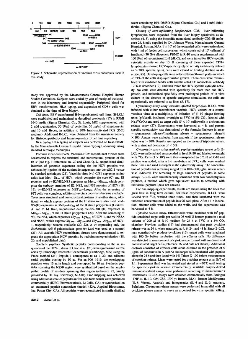
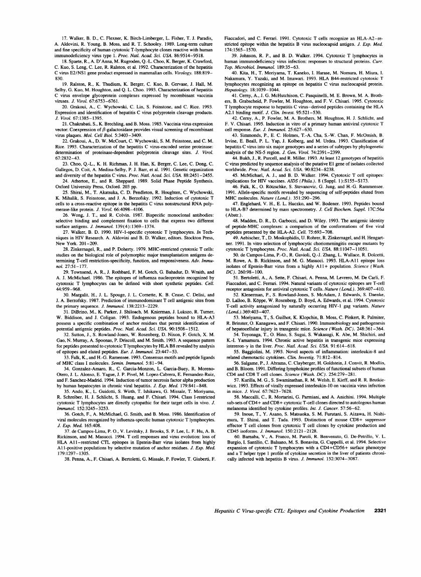
Figure 1. Schematic representation of vaccinia virus constructs used inthis study.
study was approved by the Massachusetts General Hospital HumanStudies Committee. Subjects were coded by year of receipt of the speci-men in the laboratory and lettered sequentially. Peripheral blood forEBV transformation, HLA typing, and expansion of CD8+ cells wasobtained at the time of liver biopsy.
Cell lines. EBV-transformed B-lymphoblastoid cell lines (B-LCL)were established and maintained as described previously (17) in RPMI1640 media (Sigma Chemical Co., St. Louis, MO) supplemented with2 mML-glutamine, 50 U/ml of penicillin, 50 pg/ml of streptomycin,and 10 mMHepes, in addition to 20% heat-inactivated FCS (R-20medium). Additional B-LCL were obtained from the American Societyfor Histocompatibility and Immunogenetics B cell line repository.
HLA typing. HLA typing of subjects was performed on fresh PBMCby the Massachusetts General Hospital Tissue Typing Laboratory, usingstandard serologic techniques.
Vaccinia virus constructs. Vaccinia-HCV recombinant viruses wereconstructed to express the structural and nonstructural proteins of theHCV(see Fig. 1; reference 18-20 and Choo, Q.-L., unpublished data).Insertion of genomic sequences coding for the HCV proteins wasachieved by ligation of the nucleotide sequences into the pSCl 1 vectorby standard techniques (21). Vaccinia virus (vv)-C/El expresses aminoacids (aa) Met1-Ile3Q of HCV, which comprise the core (C) and Elproteins; and vv-E2/NS21NS3 expresses aa Met364-Hisl619, which com-prise the carboxy terminus of E2, NS2, and NS3 proteins of HCV (18,19). vv-E2/NS2 expresses aa MET347-Leugw. After the screening of92T cells was complete, additional vaccinia constructs became available.To express structural and non-structural proteins of another strain, addi-tional vv which express proteins of the H strain were also used. vv-1-966(H) expresses aa Met, -Aspw of the H strain polyprotein (Grakovi,A. and C. M. Rice, unpublished data). vv-827-301 1(H) expresses aaMet827-Arg3011 of the H strain polyprotein (20). After the screening of93I, vv-NS4, which expresses Gly,590-Lys2050 of HCV-1, and vv-NS5Aand NS5B, which express Gly2005-Gly2396 and Gly2396-Arg3011 of HCV-1, respectively, became available (20, 22). A vv expressing only theEscherichia coli 6-galactosidase gene (vv-Lac) was used as a control(21). All vaccinia-HCV recombinant viruses were demonstrated to ex-press the appropriate HCVproteins by radioimmunoprecipitation (18,20, and unpublished data).
Synthetic peptides. Synthetic peptides corresponding to the aa se-quences of the HCV-1 strain of Choo et al. (23) were synthesized as freeacids by Cambridge Research Biochemicals (Cambridge, MA) using theFmoc method (24). Peptide 1 corresponds to aa 1-20, and adjacentserial peptides overlap by 10 aa. For aa 906-1619, the overlappingpeptides were 13 aa in length and overlapped by 10 aa. Synthetic pep-tides spanning the NS5B region were synthesized based on the amphi-pathic profile of residues spanning this region (reference 25, kindlyprovided by Dr. Jay Berzofsky, NIAID). Fine mapping was achievedusing additional smaller peptides in free acid form which were purchasedcommercially (IDEC Pharmaceuticals, La Jolla, CA) or synthesized onan automated peptide synthesizer (model 482A, Applied Biosystems,Inc. Foster City, CA). All peptides were reconstituted in sterile distilled
water containing 10% DMSO(Sigma Chemical Co.) and 1 mMdithio-threitol (Sigma Chemical Co.).
Cloning of liver-infiltrating lymphocytes. CD8+ liver-infiltratinglymphocytes were expanded from the liver biopsy specimens as de-scribed (4, 5), using the bispecific monoclonal antibody CD3,4B (refer-ence 26, kindly supplied by Dr. Johnson Wong, Massachusetts GeneralHospital, Boston, MA). 1 x 10⁶ of the expanded cells were restimulatedwith 4 ml of feeder cell suspension, which consisted of 106 cells/ml ofirradiated (30 Gy) allogeneic PBMCin R-10 media supplemented with100 U/ml of recombinant IL-2 (rIL-2), and were tested for HCV-specificcytolytic activity on day 10. If screening of these expanded CD8+lymphocytes showed HCV-specific cytolytic activity (arbitrarily definedas > 20% specific lysis), cells were cloned at limiting dilution as de-scribed (5). Developing cells were selected from 96-well plates in which< 33% of the cells displayed visible growth. These cells were restimu-lated with irradiated feeder cells and the anti-CD3 monoclonal antibody12F6 as described (17), and then tested for HCV-specific cytolytic activ-ity. No cells were detected with specificity for more than one HCVprotein, and maintained specificity over prolonged periods of in vitroculture in the absence of specific antigenic stimulation, the cells areoperationally are referred to as lines (5, 17).
Cytotoxicity assay using vaccinia-infected target cells. B-LCL wereinfected with either recombinant vaccinia-HCV vectors or a controlvaccinia virus at a multiplicity of infection of 5-10 plaque-formingunits (pfu)/cell, incubated overnight at 370C in 5% CO2, labeled withNa2(5"CrO4) and used as target cells (5 x 10³ cells/well) in a chromiumrelease assay (27). Supernatants were harvested at 4 h, and percentspecific cytotoxicity was determined by the formula [(release in assay- spontaneous release)/(maximum release - spontaneous release)]x 100. Assays were excluded from analysis if the spontaneous releasevalue was > 30%. Results are reported as the mean of triplicate values,with a standard deviation of < 5%.
Cytotoxicity assay using synthetic peptide-sensitized target cells. B-LCL were pelleted and resuspended in fresh R-20 and labeled overnightwith 5"Cr. Cells (1 x 10⁶) were then resuspended in 0.2 ml of R-10 andpeptide was added; after a 1-h incubation at 37°C, cells were washedthree times and used as targets in the cytotoxicity assay. The concentra-tion of peptides for screening ranged from 100-300 jsg/ml unless other-wise indicated. For screening of large numbers of peptides in someassays, B-LCL were simultaneously sensitized with two noncontiguouspeptides, a method which gave equivalent results to screening withindividual peptides (data not shown).
For fine mapping experiments, results are shown using the lines thatgrew best in long term culture. For these experiments, B-LCL werelabeled with 5"Cr, washed three times, and then incubated with theindicated concentration of peptide in a 96-well plate. After a 1-h incuba-tion, effector cells were added to the wells, and the supernatant washarvested at 4 h.
Cytokine release assay. Effector cells were incubated with 105 pep-tide-sensitized target cells per well in 96-well U-bottom plates in a totalvolume of 200 ,ul of R-10 medium for 24 h at 37°C in a 5% CO2chamber. Previous studies (16) had demonstrated that peak cytokinerelease was at 24 h, when measured at 4, 6, 24, and 48 h. Since B-LCLmay constitutively produce cytokines (16), target cells were irradiatedwith 100 Gy before incubation with the effector cells. No differencewas detected in measurement of cytokines performed with irradiated andnonirradiated target cells (reference 16, and data not shown). Additionalcontrols consisted of effector cells alone cultured in the presence of 5,ug/ml of concanavalin A (conA) and target cells incubated with peptidealone for 24 h and then lysed with 1%Triton X-100 before measurementof cytokine release. Lines were tested for cytokine release at an E/T of1:1. Supernatant fluid was harvested and stored at -70°C until testingfor specific cytokine release. Commercially available enzyme-linkedimmunoabsorbent assays were performed according to manufacturer'sinstructions. ELISA assays were obtained commercially from Endogen(TNF-a, IL-10, GM-CSF, IFN-y; Boston, MA); Bender MedSystems(1L-8; Vienna, Austria); and Innogenetics (IL-4 and 11-6; Antwerp,Belgium). Chromium release assays were performed in parallel with allcytokine release assays to serve as a control for virus specific killing.
2312 Koziel et al.
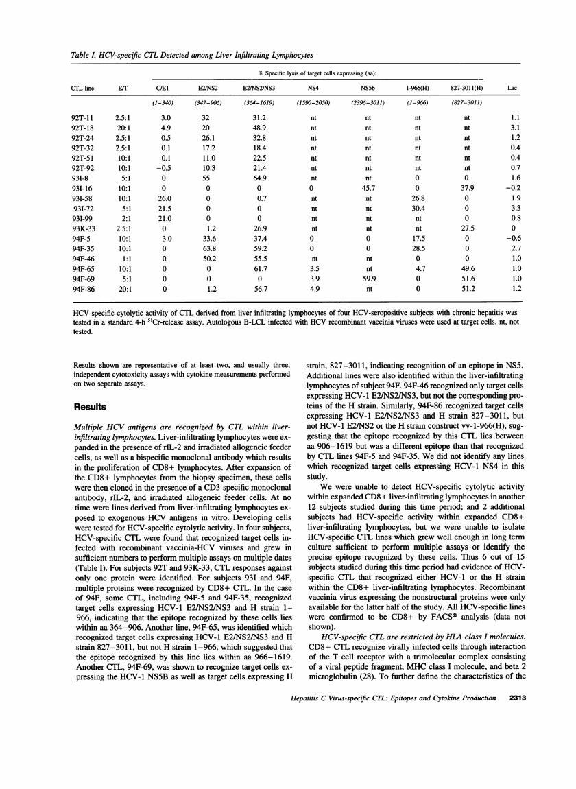
Table L HCV-specific CTL Detected among Liver Infiltrating Lymphocytes
%Specific lysis of target cells expressing (aa):
CTL line E/T C/El E2/NS2 E2/NS2/NS3 NS4 NS5b 1-966(H) 827-3011(H) Lac
(1-340) (347-906) (364-1619) (1590-2050) (2396-3011) (1-966) (827-3011)
92T-11 2.5:1 3.0 32 31.2 nt nt nt nt 1.192T-18 20:1 4.9 20 48.9 nt nt nt nt 3.192T-24 2.5:1 0.5 26.1 32.8 nt nt nt nt 1.292T-32 2.5:1 0.1 17.2 18.4 nt nt nt nt 0.492T-51 10:1 0.1 11.0 22.5 nt nt nt nt 0.492T-92 10:1 -0.5 10.3 21.4 nt nt nt nt 0.7931-8 5:1 0 55 64.9 nt nt 0 0 1.6931-16 10:1 0 0 0 0 45.7 0 37.9 -0.2931-58 10:1 26.0 0 0.7 nt nt 26.8 0 1.993I-72 5:1 21.5 0 0 nt nt 30.4 0 3.3931-99 2:1 21.0 0 0 nt nt nt 0 0.893K-33 2.5:1 0 1.2 26.9 nt nt nt 27.5 094F-5 10:1 3.0 33.6 37.4 0 0 17.5 0 -0.694F-35 10:1 0 63.8 59.2 0 0 28.5 0 2.794F-46 1:1 0 50.2 55.5 nt nt 0 0 1.094F-65 10:1 0 0 61.7 3.5 nt 4.7 49.6 1.094F-69 5:1 0 0 0 3.9 59.9 0 51.6 1.094F-86 20:1 0 1.2 56.7 4.9 nt 0 51.2 1.2
HCV-specific cytolytic activity of CTL derived from liver infiltrating lymphocytes of four HCV-seropositive subjects with chronic hepatitis wastested in a standard 4-h 5'Cr-release assay. Autologous B-LCL infected with HCVrecombinant vaccinia viruses were used at target cells. nt, nottested.
Results shown are representative of at least two, and usually three,independent cytotoxicity assays with cytokine measurements performedon two separate assays.
Results
Multiple HCVantigens are recognized by CTL within liver-infiltrating lymphocytes. Liver-infiltrating lymphocytes were ex-panded in the presence of rIL-2 and irradiated allogeneic feedercells, as well as a bispecific monoclonal antibody which resultsin the proliferation of CD8+ lymphocytes. After expansion ofthe CD8+ lymphocytes from the biopsy specimen, these cellswere then cloned in the presence of a CD3-specific monoclonalantibody, rIL-2, and irradiated allogeneic feeder cells. At notime were lines derived from liver-infiltrating lymphocytes ex-posed to exogenous HCVantigens in vitro. Developing cellswere tested for HCV-specific cytolytic activity. In four subjects,HCV-specific CTL were found that recognized target cells in-fected with recombinant vaccinia-HCV viruses and grew insufficient numbers to perform multiple assays on multiple dates(Table I). For subjects 92T and 93K-33, CTL responses againstonly one protein were identified. For subjects 931 and 94F,multiple proteins were recognized by CD8+ CTL. In the caseof 94F, some CTL, including 94F-5 and 94F-35, recognizedtarget cells expressing HCV-1 E2/NS2/NS3 and H strain 1-966, indicating that the epitope recognized by these cells lieswithin aa 364-906. Another line, 94F-65, was identified whichrecognized target cells expressing HCV-1 E2/NS2/NS3 and Hstrain 827-3011, but not H strain 1 -966, which suggested thatthe epitope recognized by this line lies within aa 966-1619.Another CTL, 94F-69, was shown to recognize target cells ex-pressing the HCV-1 NS5B as well as target cells expressing H
strain, 827-3011, indicating recognition of an epitope in NS5.Additional lines were also identified within the liver-infiltratinglymphocytes of subject 94F. 94F-46 recognized only target cellsexpressing HCV-1 E2/NS2/NS3, but not the corresponding pro-teins of the H strain. Similarly, 94F-86 recognized target cellsexpressing HCV-1 E2/NS2/NS3 and H strain 827-3011, butnot HCV-1 E2/NS2 or the H strain construct vv-1-966(H), sug-gesting that the epitope recognized by this CTL lies betweenaa 906-1619 but was a different epitope than that recognizedby CTL lines 94F-5 and 94F-35. Wedid not identify any lineswhich recognized target cells expressing HCV-1 NS4 in thisstudy.
Wewere unable to detect HCV-specific cytolytic activitywithin expanded CD8+ liver-infiltrating lymphocytes in another12 subjects studied during this time period; and 2 additionalsubjects had HCV-specific activity within expanded CD8+liver-infiltrating lymphocytes, but we were unable to isolateHCV-specific CTL lines which grew well enough in long termculture sufficient to perform multiple assays or identify theprecise epitope recognized by these cells. Thus 6 out of 15subjects studied during this time period had evidence of HCV-specific CTL that recognized either HCV-1 or the H strainwithin the CD8+ liver-infiltrating lymphocytes. Recombinantvaccinia virus expressing the nonstructural proteins were onlyavailable for the latter half of the study. All HCV-specific lineswere confirmed to be CD8+ by FACS® analysis (data notshown).
HCV-specific CTL are restricted by HLA class I molecules.CD8+ CTL recognize virally infected cells through interactionof the T cell receptor with a trimolecular complex consistingof a viral peptide fragment, MHCclass I molecule, and beta 2microglobulin (28). To further define the characteristics of the
Hepatitis C Virus-specific CTL: Epitopes and Cytokine Production 2313
A B53, C4-
B7, C7-
A3-
A23, B7, C4-
Autologous-
CAll
B7,8
A3,B7
Autologous
cmJ CO)
Io o
J
B A3
C7A3
B53, C4A23, B7, C7Autologous
D All
B7,8
A3,B7
Autologous
0 20 40 60 80
I.: ...::..>..
J
1:--:':': :::'' '- -:- ::-: -
Ji
1
0 10 20 30 40
K_.0 20 40 60
* w-E2/NS2/NS3I vv-H827-3011
o w-Lac
80
%Specific Lysis
Figure 2. HCV-specific CTL areHLA class I restricted. Lines weretested for their ability to lyse au-tologous and allogeneic targetcells expressing HCVantigens.Target cells were infected with thevv construct expressing the rele-vant antigen as well as the controlvv-lac. Shared HLAmolecules areshown. (A) Line 931-8 recognizedtarget cells expressing E2/NS2/NS3 in the context of HLA A23.The E/T was 5:1 (B) Line 93I-16recognized target cells expressingthe H strain NS3/NS4/NS5 in thecontext of HLA A3. The E/T was5:1 (C) Line 94F-35 recognizedtarget cells expressing E2/NS2/NS3 in the context of HLA A1.The E/T was 5:1 (D) Line 94F-65 recognized target cells express-ing E2/NS2/NS3 in the contextof HLAB8. The E/T was 5:1. Thecomplete HLA type of subject 931is A2, 23; B 7, 53: C4,5: DR8,13:DRw52, DQ 1,7. The completeHLA type of subject 94F is A3,11; B7,8; C7, Bw 6; DR3,15;DRw52; DQ1,2.
observed HCV-specific cytolytic activity, selected CTL lineswere tested for their ability to lyse allogeneic target cells ex-
pressing HCV antigens. The HCV-specific cytotoxic effectorcells recognized allogeneic target cells only in the context of a
shared HLA class I molecule, with different HLA moleculesrestricting recognition of different HCVantigens. Examples are
shown in Fig. 2. For example, the line 931-8 recognized targetcells expressing HCV-1 E2/NS2/NS3 only in the context of a
shared HLA A23 molecule, whereas for CTL 94F-35, recogni-tion was only present if HLA All was expressed. For line 94F-65, which also recognized target cells expressing HCV-1 E2/NS2/NS3, target cells were recognized only if HLA B8 was
expressed. The HLA restricting element of the other HCV-specific CTL identified are summarized in Table II. If multiplelines were identified in a single subject which recognized targetcells expressing the same HCVproteins and shared the same
HLA restriction, in general confirmation studies were done with
Table II. Summary of HLA Restriction and Epitope Mappingof CTL
CTL line HLA restriction Epitope recognized aa
92T-32 B53 RPLTDFDQGW 460-46993I-8 A23 YISWCLWW 838-845931-16 A3 RVCEKMALY 2588-2596931-58 B7 GPRLGVRAT 41-4993K-33 A2 CINGVCWTV 1073-108194F-35 All TINYTIFK 621-62894F-65 B8 HSKKKCDEL 1395-140394F-69 A3 NS5B - not defined
AA are numbered according to reference 23.
a single representative CTL line which grew well in long termculture. These studies show not only that multiple proteins are
recognized by HCV-specific CTL, but that multiple HLA mole-cules may participate in the presentation of HCVproteins forrecognition by HCV-specific CTL.
HCV-specific CTL recognize 8-10 aa epitopes in structuraland nonstructural proteins. Peptide fragments presented to classI-restricted CTL are typically 8-10 aa in length (29). Incubat-ing target cells with synthetic peptide has been shown to bypassthe normal requirements for endogenous processing of viralpeptide into fragments of the appropriate length (29), and thusenables definition of the epitopes recognized by virus-specificCTL. Overlapping 13-20 aa synthetic peptides spanning theappropriate viral proteins were used to map the regions con-
taining epitopes recognized by the HCV-specific CTL. Once a
region was identified, amino and carboxy terminal truncationsof the peptide containing this region were synthesized and usedto sensitize target cells for lysis. These results are shown inFigs. 3-8.
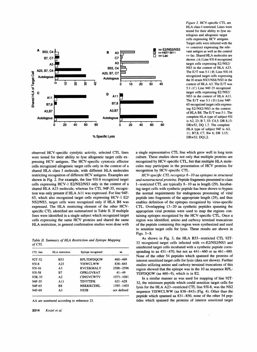
As shown in Fig. 3, the HLA B53-restricted CTL 92T-32 recognized target cells infected with vv-E2/NS2/NS3 anduninfected target cells incubated with a synthetic peptide corre-
sponding to aa 451-470, but not aa 441-460 or aa 461-480.None of the other 54 peptides which spanned the proteins ofinterest sensitized target cells for lysis (data not shown). Furtherstudies utilizing amino and carboxy terminal truncations of thisregion showed that the epitope was in the 10 aa sequence RPL-TDFDQGW(aa 460-9), which is in E2.
In a similar manner as was used for mapping of line 92T-32, the minimum peptide which could sensitize target cells forlysis for the HLA A23-restricted CTL line 93I-8, was the NS2sequence YISWCLWW(aa 838-845) (Fig. 4). Other than thepeptide which spanned aa 831-850, none of the other 54 pep-tides which spanned the proteins of interest sensitized target
2314 Koziel et al.
i)00
Is
10
CO
. Wee. ...7 ,,.e. .0. .0. .%,.,. 3
A481-500-471 490-461-480-
I
451-470441-460-431450-421-440-
w-Lac-
A861-880-851-870-841-860-
0 831 -850c 821-840-
811-830-801-820-
w-Lac-w-E2/NS2W-E2/NS2tNS3
I I I I
UII
II
w-E2/NS2/NS3 _. .y .is
%Specific Lysis
B
'am
-J
.9Cl)
0-
60-50-
40130-20-10-
..r -
0 V- _0 0V- 0
-0-- AA 451470
- CRPLTDFDOG
- CRPLTDFDQGW
* RPLTDFDQGWG- M461-480
---a--- Control
B80
.0w 60-
0.9 40-
ae 20
_ N CX) ) CD
%Specific Lysis
§7~~~~a- _U- RYISWCLWWL
* YISWCLWWL
----v-. ISWCLWWL
- YISWCLWW
YKRYISWCLW
-.-.- - Control
Conc Peptide (uM)
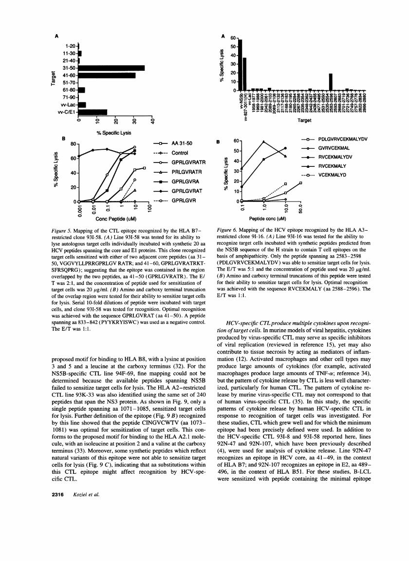
Figure 3. Mapping of the CTL epitope recognized by the HLA B53-restricted CTL clone 92T-32. (A) Line 92T-32 was tested for its abilityto lyse target cells sensitized with 20 aa peptides spanning the E2 andNS2 proteins. This clone recognized only target cells sensitized withthe peptide spanning aa 451-470 (GCPERLASRCRPLTDFDQGWG),but not target cells sensitized with the overlapping peptide spanning461-480 (PLTDFDQGWGPISYANGSGPD).Lysis of target cells in-fected with recombinant vv is shown as a control. The E/T was 5:1and the concentration of peptide used for sensitization of target cellswas 100 Ag/ml. (B) Line 92T-32 was also tested for its ability to lyseautologous target cells incubated with amino and carboxy terminal trun-cations of the region unique to this peptide. 10-fold serial dilutions ofthe amino and carboxy terminal truncations of peptide 46 were incubatedwith target cells, and clone 92T-32 was tested for its ability to lysetarget cells. The data indicate the epitope was in 10 aa sequence RPL-TDFDQGW(aa 460-9), which is in E2. The E/T was 5:1.
cells for lysis (data not shown). Target cells sensitized withpeptides lacking either the tyrosine (ISWCLWWLQ)or thecarboxy terminal tryptophan (RYISWCLW)were unable to sen-sitize target cells for lysis (data not shown). The B7-restrictedepitope recognized by the core-specific line 93I-58 lies withinthe overlap of two adjacent peptides spanning aa 31-50 and aa41-60 (Fig. 5). Fine mapping of this sequence within the HCVcore protein showed that optimal recognition was achieved withthe sequence GPRLGVRAT(aa 41-49), as evidenced by theability of this peptide to sensitize target cells for lysis at thelowest concentration of peptide.
Overlapping peptides representing either the HCV-1 or theH strain were not available for fine mapping of the epitoperecognized by line 931-16 (Fig. 6). However, synthetic peptideswhich contain potential T cell epitopes on the basis of theamphipathicity profile of NS5B were available (reference 30,generously provided by Dr. J. Berzofsky). These peptides arepartially overlapping and have been used to map an epitoperecognized by murine HCV-specific CTL (25). The HLA A3-restricted line 931-16 recognized target cells sensitized withthe peptide spanning aa 2588-2596 (RVCEKMALY), whichis consistent with the proposed motif for HLA A3 of a hy-drophobic aa at position 2 and a tyrosine or lysine at position
° Conc Peptide (uM)
Figure 4. Mapping of the CTL epitope recognized by the HLA A23-restricted CTL clone 931-8. (A) Line 931-8 was tested for its ability tolyse target cells sensitized with 20 aa peptides spanning the E2 and NS2proteins. This clone recognized only target cells sensitized with peptidespanning aa 831-850 (LSPYYKKYISWCLWWLQYFL),but not pep-tides spanning aa 821-840 or 841-860. Lysis of target cells infectedwith recombinant vv is shown as a control. The E/T was 5:1 and theconcentration of peptide used for sensitization of target cells was 50,ug/ml. (B) Line 931-8 was tested for its ability to lyse autologous cellssensitized with amino and carboxy terminal truncation of this peptide.Serial 10-fold dilutions of the amino and carboxy terminal truncationsof aa 831-850 were incubated with target cells, and clone 93I-8 wastested for its ability to lyse target cells sensitized with these truncations.The minimum peptide which could sensitize target cells for lysis wasYISWCLWW(aa 838-845). A peptide spanning aa 491-500 (PKP-CGIVPAK) was used as a negative control. The E/T was 1:1.
9 (31). Since these peptides are not overlapping and not all aaof NS5B are represented, the presence of another A3 epitopecannot be completely excluded.
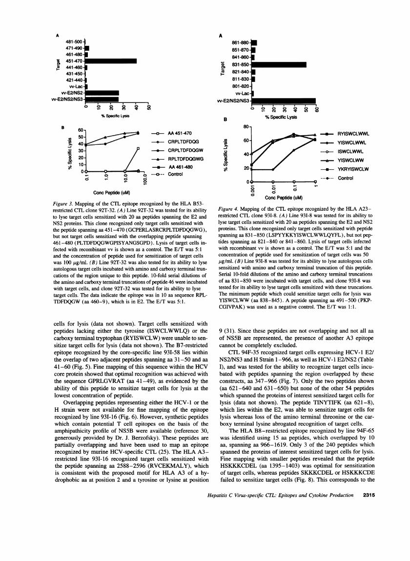
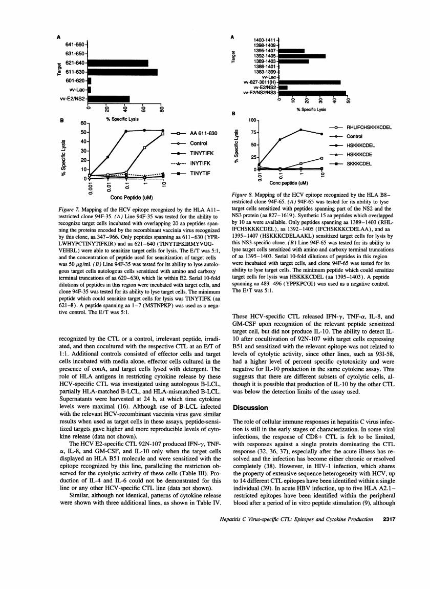
CTL 94F-35 recognized target cells expressing HCV-1 E2/NS2/NS3 and HStrain 1-966, as well as HCV-1 E2/NS2 (TableI), and was tested for the ability to recognize target cells incu-bated with peptides spanning the region overlapped by theseconstructs, aa 347-966 (Fig. 7). Only the two peptides shown(aa 621-640 and 631-650) but none of the other 54 peptideswhich spanned the proteins of interest sensitized target cells forlysis (data not shown). The peptide TINYTIFK (aa 621-8),which lies within the E2, was able to sensitize target cells forlysis whereas loss of the amino terminal threonine or the car-boxy terminal lysine abrogated recognition of target cells.
The HLA B8-restricted epitope recognized by line 94F-65was identified using 15 aa peptides, which overlapped by 10aa, spanning aa 966-1619. Only 3 of the 240 peptides whichspanned the proteins of interest sensitized target cells for lysis.Fine mapping with smaller peptides revealed that the peptideHSKKKCDEL(aa 1395-1403) was optimal for sensitizationof target cells, whereas peptides SKKKCDELor HSKKKCDEfailed to sensitize target cells (Fig. 8). This corresponds to the
Hepatitis C Virus-specific CTL: Epitopes and Cytokine Production 2315
l
A
1 -20.11-30-21-40*31-5041-6051-70-61-80*71 -90-
w-Lac-w-C/E1I
U
U
111 CM CII) ItJ
I ZW
.o
I0
8
6
4
2
%Specific Lysis
10"OI
/Ip
00-0c; 0-To
o Irl 0-0- (
--M-AA31-50
---@--- Control
GPRLGVRATR
I& PRLGVRATR
-in*- GPRLGVRA
---* GPRLGVRAT
---o--- GPRLGVR
Conc Peptide (uM)
Figure 5. Mapping of the CTL epitope recognized by the HLA B7-restricted clone 931-58. (A) Line 93I-58 was tested for its ability tolyse autologous target cells individually incubated with synthetic 20 aa
HCVpeptides spanning the core and El proteins. This clone recognizedtarget cells sensitized with either of two adjacent core peptides (aa 31-50, VGGVYLLPRRGPRLGVRATR; and 41-60, GPRLGVRATRKT-SFRSQPRG); suggesting that the epitope was contained in the regionoverlapped by the two peptides, aa 41-50 (GPRLGVRATR). The E/T was 2:1, and the concentration of peptide used for sensitization oftarget cells was 20 Mg/ml. (B) Amino and carboxy terminal truncationof the overlap region were tested for their ability to sensitize target cellsfor lysis. Serial 10-fold dilutions of peptide were incubated with targetcells, and clone 931-58 was tested for recognition. Optimal recognitionwas achieved with the sequence GPRLGVRAT(aa 41-50). A peptidespanning aa 833-842 (PYYKRYISWC) was used as a negative control.The E/T was 1:1.
proposed motif for binding to HLAB8, with a lysine at position3 and 5 and a leucine at the carboxy terminus (32). For theNS5B-specific CTL line 94F-69, fine mapping could not bedetermined because the available peptides spanning NS5Bfailed to sensitize target cells for lysis. The HLA A2-restrictedCTL line 93K-33 was also identified using the same set of 240peptides that span the NS3 protein. As shown in Fig. 9, only a
single peptide spanning aa 1071-1085, sensitized target cellsfor lysis. Further definition of the epitope (Fig. 9 B) recognizedby this line showed that the peptide CINGVCWTV(aa 1073-1081) was optimal for sensitization of target cells. This con-
forms to the proposed motif for binding to the HLA A2. 1 mole-cule, with an isoleucine at position 2 and a valine at the carboxyterminus (33). Moreover, some synthetic peptides which reflectnatural variants of this epitope were not able to sensitize targetcells for lysis (Fig. 9 C), indicating that aa substitutions withinthis CTL epitope might affect recognition by HCV-spe-cific CTL.
B
._ACO
.-oh
PDLGVRVCEKMALYDV
v GVRVCEKMAL
* RVCEKMALYDV
-*-- RVCEKMALY
--o-- VCEKMALYD
Peptide conc (uM)
Figure 6. Mapping of the HCVepitope recognized by the HLA A3-restricted clone 9I-16. (A) Line 931-16 was tested for the ability torecognize target cells incubated with synthetic peptides predicted fromthe NS5B sequence of the H strain to contain T cell epitopes on thebasis of amphipathicity. Only the peptide spanning aa 2583-2598(PDLGVRVCEKMALYDV)was able to sensitize target cells for lysis.The E/T was 5:1 and the concentration of peptide used was 20 ,ug/ml.(B) Amino and carboxy terminal truncations of this peptide were testedfor their ability to sensitize target cells for lysis. Optimal recognitionwas achieved with the sequence RVCEKMALY(aa 2588-2596). TheE/T was 1:1.
HCV-specific CTLproduce multiple cytokines upon recogni-tion of target cells. In murine models of viral hepatitis, cytokinesproduced by virus-specific CTL may serve as specific inhibitorsof viral replication (reviewed in reference 15), yet may alsocontribute to tissue necrosis by acting as mediators of inflam-mation (12). Activated macrophages and other cell types may
produce large amounts of cytokines (for example, activatedmacrophages produce large amounts of TNF-a; reference 34),but the pattern of cytokine release by CTL is less well character-ized, particularly for human CTL. The pattern of cytokine re-
lease by murine virus-specific CTL may not correspond to thatof human virus-specific CTL (35). In this study, the specificpatterns of cytokine release by human HCV-specific CTL inresponse to recognition of target cells was investigated. Forthese studies, CTL which grew well and for which the minimumepitope had been precisely defined were used. In addition tothe HCV-specific CTL 931-8 and 93I-58 reported here, lines92N-47 and 92N-107, which have been previously described(4), were used for analysis of cytokine release. Line 92N-47recognizes an epitope in HCVcore, aa 41-49, in the contextof HLA B7; and 92N-107 recognizes an epitope in E2, aa 489-496, in the context of HLA B51. For these studies, B-LCLwere sensitized with peptide containing the minimal epitope
2316 Koziel et al.
a,-0)
Cuco
B
Ca
0
COI0-0
Target
A
D
A641-660-
631-650-
621 -640
611-630-601-620-
w-Lac
w-E2INS2
02'co
0
B
.0
.540.
CO)
oR
B
(0.-
C)
C)I-o
1400-1411-1398-1409-1395-1407-1392-14051389-1403-1386-1401-1383-1399-
w-Lac-w-827-3011 (H)
w-E2INS2w-E2/NS21NS3 _ _ _
ro o o_.6 I
.4. ( 0G
%Specific Lysis
AA611-630
Control
-*- TINYTIFK
---A--- INYTIFK
* TINYTIF
o o0° o
C.onc Peptide (uM)
Figure 7. Mapping of the HCVepitope recognized by the HLA All -restricted clone 94F-35. (A) Line 94F-35 was tested for the ability torecognize target cells incubated with overlapping 20 aa peptides span-ning the proteins encoded by the recombinant vaccinia virus recognizedby this clone, aa 347-966. Only peptides spanning aa 611-630 (YPR-LWHYPCTINYTIFKIR) and aa 621-640 (TINYTIFKIRMYVGG-VEHRL) were able to sensitize target cells for lysis. The E/T was 5:1,and the concentration of peptide used for sensitization of target cellswas 50 fig/ml. (B) Line 94F-35 was tested for its ability to lyse autolo-gous target cells autologous cells sensitized with amino and carboxyterminal truncations of aa 620-630, which lie within E2. Serial 10-folddilutions of peptides in this region were incubated with target cells, andclone 94F-35 was tested for its ability to lyse target cells. The minimumpeptide which could sensitize target cells for lysis was TINYTIFK (aa621-8). A peptide spanning aa 1-7 (MSTNPKP) was used as a nega-tive control. The E/T was 5:1.
recognized by the CTL or a control, irrelevant peptide, irradi-ated, and then cocultured with the respective CTL at an EIT of1:1. Additional controls consisted of effector cells and targetcells incubated with media alone, effector cells cultured in thepresence of conA, and target cells lysed with detergent. Therole of HLA antigens in restricting cytokine release by theseHCV-specific CTL was investigated using autologous B-LCL,partially HLA-matched B-LCL, and HLA-mismatched B-LCL.Supernatants were harvested at 24 h, at which time cytokinelevels were maximal (16). Although use of B-LCL infectedwith the relevant HCV-recombinant vaccinia virus gave similarresults when used as target cells in these assays, peptide-sensi-tized targets gave higher and more reproducible levels of cyto-kine release (data not shown).
The HCVE2-specific CTL 92N-107 produced IFN-y, TNF-a, IL-8, and GM-CSF, and IL-10 only when the target cellsdisplayed an HLA B51 molecule and were sensitized with theepitope recognized by this line, paralleling the restriction ob-served for the cytolytic activity of these cells (Table III). Pro-duction of IL-4 and IL-6 could not be demonstrated for thisline or any other HCV-specific CTL line (data not shown).
Similar, although not identical, patterns of cytokine releasewere shown with three additional lines, as shown in Table IV.
I
%Specific Lysis
o o
RHLIFCHSKKKCDEL
Control
* HSKKKCDEL
-I- HSKKKCDE
-*-- SKKKCDEL
Cn pConc peptide (uM)
Figure 8. Mapping of the HCVepitope recognized by the HLA B8-restricted clone 94F-65. (A) 94F-65 was tested for its ability to lysetarget cells sensitized with peptides spanning part of the NS2 and theNS3 protein (aa 827-1619). Synthetic 15 aa peptides which overlappedby 10 aa were available. Only peptides spanning aa 1389-1403 (RHL-IFCHSKKKCDEL), aa 1392-1405 (IFCHSKKKCDELAA), and aa1395-1407 (HSKKKCDELAAKL) sensitized target cells for lysis bythis NS3-specific clone. (B) Line 94F-65 was tested for its ability tolyse target cells sensitized with amino and carboxy terminal truncationsof aa 1395-1403. Serial 10-fold dilutions of peptides in this regionwere incubated with target cells, and clone 94F-65 was tested for itsability to lyse target cells. The minimum peptide which could sensitizetarget cells for lysis was HSKKKCDEL(aa 1395-1403). A peptidespanning aa 489-496 (YPPKPCGI) was used as a negative control.The E/T was 5:1.
These HCV-specific CTL released IFN--y, TNF-a, IL-8, andGM-CSFupon recognition of the relevant peptide sensitizedtarget cell, but did not produce IL-10. The ability to detect IL-10 after cocultivation of 92N-107 with target cells expressingB51 and sensitized with the relevant epitope was not related tolevels of cytolytic activity, since other lines, such as 931-58,had a higher level of percent specific cytotoxicity and werenegative for IL-10 production in the same cytokine assay. Thissuggests that there are different subsets of cytolytic cells, al-though it is possible that production of IL-10 by the other CTLwas below the detection limits of the assay used.
Discussion
The role of cellular immune responses in hepatitis C virus infec-tion is still in the early stages of characterization. In some viralinfections, the response of CD8+ CTL is felt to be limited,with responses against a single protein dominating the CTLresponse (32, 36, 37), especially after the acute illness has re-solved and the infection has become either chronic or resolvedcompletely (38). However, in HIV-1 infection, which sharesthe property of extensive sequence heterogeneity with HCV, upto 14 different CTL epitopes have been identified within a singleindividual (39). In acute HBVinfection, up to five HLAA2. 1-restricted epitopes have been identified within the peripheralblood after a period of in vitro peptide stimulation (9), although
Hepatitis C Virus-specific CTL: Epitopes and Cytokine Production 2317
A
SD.cmIII
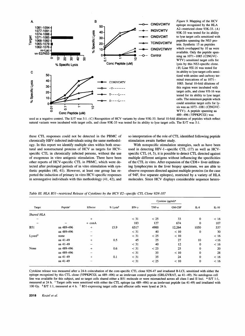
A B Figure 9. Mapping of the HCV
1081-1094 50 ° CINGVCWrV epitope recognized by the HLA1081-1094 ~~~~~~~~~~~~~~~~~~~~~A2-restrictedclone 93K-33. (A)1074-10881 40 93K-33 was tested for its ability1068-1 082 .~peptides spanning the NS3 pro-
o 1071-10852 30 T-CINGVCWT to lyse target cells sensitized withI 1065-1079 20 TCINGVCWTV tein. Synthetic 15 aa peptides
1062-1076 10 * ClNGVCTWVY which overlapped by 10 aa werevv-Lac o 1
w-E2/NS3 available. Only the peptide span-|-*,,,0w v Ah lo---.-
Control ning aa 1071-1085 (CINGVC-Va cmt M LO VV-TV) sensitized target cells for
%Specific Lysis Conc Peptide (uM) lysis by this NS3-specific clone.(B) Line 93I-16 was tested for
C its ability to lyse target cells sensi-50- tized with amino and carboxy ter-
40- / < CINGVCWTV minal truncations of aa 1071----S-CINGVCWTV 1085. ~~~~~~Serial 10-fold dilutions of
30- //-----this region were incubated withas 20-
Ttarget cells, and clone 931-16 was
k 20 4* T- - - - - - tested for its ability to lyse target10 * S----L--- cells. The minimumpeptide which
n__ could sensitize target cells for ly-C sis was aa 1073-1081 (CINGVC-
6 WTV). A peptide spanning aaConc Peptide (uM) 489-496 (YPPKPCGI) wasused as a negative control. The E/T was 5:1. (C) Recognition of HCVvariants by clone 93K-33. Serial 10-fold dilutions of peptides which reflectnatural variants were incubated with target cells, and clone 93K-33 was tested for its ability to lyse target cells. The E/T was 5:1.
these CTL responses could not be detected in the PBMCofchronically HBV-infected individuals using the same methodol-ogy. In this report we identify multiple sites within both struc-tural and nonstructural proteins of HCV as targets for HCV-specific CTL in chronically infected persons, without the useof exogenous in vitro antigenic stimulation. There have beenother reports of HCV-specific CTL in PBMC, which were de-tected after prolonged periods of in vitro stimulation with syn-thetic peptides (40, 41). However, at least one group has re-ported the induction of primary in vitro HCV-specific responsesin seronegative individuals with this methodology (41, 42), and
so interpretation of the role of CTL identified following peptidestimulation awaits further study.
With nonspecific stimulation strategies, such as have beenused in detecting HIV-l -specific CTL (17) as well as HCV-specific CTL (4, 5), it is possible to detect CTL directed againstmultiple different antigens without influencing the specificitiesof the CTL in vitro. After expansion of the CD8+ liver-infiltrat-ing lymphocytes in the liver biopsy specimen, we are able toobserve responses directed against multiple proteins (in the caseof 94F, five separate epitopes), restricted by a variety of HLAmolecules. Since HCVdisplays considerable sequence hetero-
Table III. HLA B51 -restricted Release of Cytokines by the HCVE2-specific CTL Clone 92N-107
Cytokine (pg/ml)*
Target Peptidel Effector %Lysis' IFN-y TNF-a GM-CSF IL-8 IL-10
Shared HLA+ <31 <25 33 0 < 16
- + conA 183 127 674 0 107BS1 aa 489-496 + 15.9 6517 4900 12,264 1050 537
aa 489-496 - < 31 40 < 10 0 30Lysedl none - < 31 < 25 < 10 < 16
aa 41-49 + 0.5 45 25 27 10 <16aa 41-49 - < 31 40 12 0 < 16
None aa 489-496 + 0.6 < 31 < 25 25 0 20aa 489-496 - < 31 35 < 10 0 28aa41-49 + 0.1 < 31 35 24 0 < 16aa41-49 - < 31 < 25 < 10 0 < 16
Cytokine release was measured after a 24-h coincubation of the core-specific CTL clone 92N-47 and irradiated B-LCL sensitized with either theepitope recognized by this CTL clone (YPPKPCGI, aa 489-496) or an irrelevant control peptide (GRLGVRAT, aa 41-49). No autologous cellline was available for this subject, and so target cells shared either a B51 molecule or were mismatched across all class I and II loci. * E/T 1:1,measured at 24 h. t Target cells were sensitized with either the CTL epitope (aa 489-496) or an irrelevant peptide (aa 41-49) and irradiated with100 Gy. § E/T 1:1, measured at 4 h. 11 B51-expressing target cells and effector cells were lysed at 24 h.
2318 Koziel et al.
Table IV. Release of Cytokines by the HCV-specific CTL.
Cytokine (pg/ml)*
CTL Target' Peptide %Lysis' IFN-y TNF-a GM-CSF IL-8 IL-10
931-8 Autol aa 831-850 23.4 250 406 120 75 < 16Autol aa 1-20 0.1 < 31 < 25 < 10 0 < 16
93I-5811 Autol aa 41-49 54.8 25,000 2200 16,000 3750 < 16Autol aa 483-492 -1.4 65 < 25 < 10 0 < 16B7 aa 41-49 59.1 25,000 1900 2600 4000 < 16B7 aa 483-492 2.1 55 < 25 < 10 0 < 16No match aa 41-49 0.8 125 < 25 < 10 0 < 16
92N-47 B7 aa 41-49 29.0 1 051 3980 1080 691 < 16B7 aa 483-492 0.2 43 < 25 < 10 0 < 16No match aa 41-49 0.2 60 < 25 < 10 0 <16No match aa 483-492 0.0 46 < 25 < 10 0 < 16None conA - 391 391 142 51 < 16
Cytokine release was measured after a 24-h coincubation of the HCV-specific CTL clone and autologous B-LCL sensitized with either the epitoperecognized by the clone or an irrelevant control peptide. Autologous B-LCL were sensitized before the coincubation with the indicated peptide andthen irradiated with 100 Gy in a gamma irradiator. In addition to coincubation of the CTL clone with B-LCL sensitized with both relevant andirrelevant peptides, cytokine release was also measured after a 24-h incubation of the B-LCL sensitized with the peptides as well as the clone alonewithout the addition of target cells. Cytokine release was below the limit of detection for the ELISA assays for these controls (data not shown).* E/T 1:1, measured at 24 h. * CTL 93I-8 recognized target cells sensitized with a synthetic peptide corresponding to aa 831-850, and aa 1-20served as a negative control. CTL 931-58 recognized target cells sensitized with a synthetic peptide corresponding to aa 41-49, and aa 483-492served as a negative control. CTL 92N-47 also recognized target cells sensitized with a synthetic peptide corresponding to aa 41-49, and aa 483-492 was also used as a control. An autologous B-LCL was not available for subject 92N, and so target cells shared a B7 molecule or weremismatched across all class I and II loci (4). § ErT 1:1, measured at 4 h. 11 The complete HLA type of subject 93I is A2, 23; B 7, 53: C4,5: DR8,13: DRw52, DQ 1,7.
geneity among isolates, with at least six major genotypes ofvirus (43, 44), it is possible that our results actually underesti-mate the spectrum and frequency of HCV-specific CTL re-sponses in these chronically infected individuals. Weare limitedby the number of genotypes of HCVwhich are currently ex-pressed in vaccinia virus recombinants. In this report we identifyCTL responses which were cross-reactive (e.g., 94F-69) as wellas isolate specific (e.g., 931-16) were observed. The failure toidentify CTL in the other subjects studied may be caused byone or more of the following: the absence of HCV-specific CTLin these individuals; the presence of HCV-specific CTL whichrecognize another strain of HCVand do not recognize eitherHCV-1 or the H strain; or the loss of cells present at a lowfrequency during either the period of in vitro expansion or thesubsequent cloning. At present, given the limited number ofHCVisolates which are expressed in vv, we cannot determinethe major reason leading to a failure to identify HCV-specificCTL in these other individuals. A similar frequency of HCV-specific responses was detected in PBMCof HCVseropositiveindividuals after prolonged in vitro stimulation with syntheticpeptides (41).
Several lines of evidence have aided in the definition ofepitopes recognized by virus-specific CTL. The first has comefrom extensive fine mapping studies of CTL epitopes performedin a number of laboratories, which permitted the prediction ofmotifs based on sequence similarities among epitopes recog-nized in the context of a given HLA molecule (reviewed inreference 33). Allele-specific motifs have been proposed forseveral murine and human HLA types, including HLAA2, B27,A3, and B8 (45). More recently, the ability to elute naturallyprocessed peptides from the surface of naturally infected cellshas confirmed the utility of this technique in the characterization
of epitopes recognized by CTL (46). The epitopes recognizedby these HCV-specific CTL conform to proposed motifs, al-though we have also identified CTL epitopes restricted by HLAfor which a peptide binding motif has not yet been proposed.For example, the HLA B7 motif is proposed to consist of aproline at position 2, arginine at position 3, and a leucine orvaline at position 9 (47). The epitope recognized by the HLAB7-restricted CTL 931-58, GPRLGVRAT,is consistent withthis motif, with the exception being the nonconservative substi-tution of a threonine at position 9. Similarly, the epitope recog-nized by the HLA B8-restricted CTL 94F-65 HSKKKCDEL,is consistent with the proposed motif for HLA B8, which is alysine at position 3, lysine or arginine at position 5, and acarboxy terminal isoleucine or leucine (32).
Since HCV is a highly heterogeneous virus and these epi-topes are present within both conserved and variable regions ofthe genome, precise definition of the epitopes recognized byHCV-specific CTL may allow an understanding of the effectof sequence variation on the immune response to HCV. Al-though some aa substitutions within the middle of the CTLepitope-binding site may not affect binding of the peptide tothe class I molecule (48), other groups have shown that evensingle aa variations within CTL epitopes facilitates viral escapefrom host immunity (49, 50). In addition to a failure of CTLto recognize variant sequences present within an epitope, severalgroups have recently shown that natural variants within CTLepitopes may act as antagonists to T cell receptor binding(51, 52).
Whatever the role of HCV-specific CTL in controlling viralreplication, viremia persists despite the presence of a cellularimmune response in chronically infected individuals. In suchcircumstances, CTL may contribute to disease pathogenesis,
Hepatitis C Virus-specific CTL: Epitopes and Cytokine Production 2319
and evidence is accumulating that cytokines may be importantmediators of this immunopathology. Recent data have shownthat adoptive transfer of HBV-specific CTL into HBsAg-transgenic mice led to liver cell necrosis (53). This tissue dam-age is due to direct cell lysis by the CTL (35), with amplifica-tion of liver necrosis by cytokines such as IFN-y and TNF-a(12). The initial response appears to consist of an antigen-specific response by virus-specific CTL, followed by productionof IFN-y and TNF-ca. This, in turn, leads to antigen-nonspecificrecruitment of inflammatory cells, followed by activation ofmacrophages and a delayed-type hypersensitivity like lesion.Blockade of the cascade of events occurs when animals arepretreated with antibodies to TNF-a before adoptive transfer ofHBV-specific CTL (12). In another transgenic mouse model,liver-specific expression of IFN-y produced a transaminitis andhistologic appearance similar that of human chronic active hepa-titis (54).
In addition to the effects of IFN-y and TNF-a, other cyto-kines are produced by these human virus-specific CTL. Al-though the role of these cytokines has not yet been investigatedin animal models, the known biologic actions of these cytokinessuggest that these cytokines might also play a role in the patho-genesis of chronic hepatitis. Both GM-CSFand IL-8 lead to arecruitment of inflammatory cells (55). IL-10 may be producedby certain classes of CD4 cells (14), and it has been welldemonstrated in murine models that two types of CD4 lympho-cytes populations may be defined based on production of IFN-'y and IL-10 (56). However, production of IL-10 by virus-specific cytolytic T cells has not been reported. IL-10 is thoughtto be a down-regulator of immune responses (57), and so pro-duction of this cytokine by CTL might contribute to viral persis-tence.
Heterogeneous production of cytokines by melanoma-spe-cific CD8+ CTL has also been observed (58), which may bedue to different populations of CD8+ CTL or methodologicissues relating to the method of T cell stimulation. For example,it is unknown whether virus-specific CTL have a different pat-tern of cytokine release than tumor-specific CTL, although onthe basis of these two studies this would seem unlikely. In mostinstances the precise targets of the cytolytic response have notbeen defined and production of cytokines has been measuredafter stimulation with immobilized anti-CD3, which has notalways resulted in the same pattern of cytokine release as stimu-lation with the specific antigen recognized by the CTL (58).For example, stimulation of murine CD8+ cells with an anti-CD3 antibody resulted in the production of 11-10 only in Tscells, but not in CTL (59).
In addition, characterization of responses produced by CTLisolated from the periphery might have a different pattern ofcytokine release compared to those isolated from the site oftissue damage. This tissue-specific compartmentalization of cy-tokine release has recently been demonstrated for CD4+ cyto-lytic cells isolated from the liver of patients with chronic HBVinfection (60). Wehave previously demonstrated the compart-mentalization of HCV-specific CTL within the liver (4). Todate we have not be able to isolate HCV-specific CTL withinthe PBMCusing an antigen nonspecific stimulation strategy,and so we have not had the opportunity to compare liver-infil-trating versus PBMC-derived HCV-specific CTL for the pat-terns of cytokine release. However, if chronic HCVhepatitis isa T cell-mediated disease, knowledge of cytokines producedby the cells which induce the inflammatory cascade, as well as
the precise targets recognized by these CTL, may suggest novelimmunotherapeutic interventions.
Acknowledgments
Wewould like to thank Dr. David Nunes for assistance in providingpatient material; Dr. Bob Ralston for the gift of vaccinia-HCV recombi-nant viruses; Kent Thudium, Kim Berger, and Christine Dong for experttechnical assistance; Dr. M. Gately of Hoffmann-LaRoche for the giftof rIL-2; Donna Fitzpatrick and the MGHTissue Typing Laboratoryfor assistance in HLA typing; and Dr. Johnson Wong for the gift of theanti-CD3 antibody 12F6 and the bispecific antibody CD3,4B.
These studies were supported by an American Cancer Society Physi-cian's Research Training Award and an National Institutes of HealthClinical Investigator Award (M.J. Koziel), and by National Institutesof Health grants A131563 (B.D. Walker) and CA57973 (C.M. Rice).
References
1. Lemon, S., and E. Brown. 1994. Hepatitis C virus and chronic liver disease.In Current Clinical Topics in Infectious Disease. Vol. 14. M. N. Swartz and J. S.Remington, editors New York. 121-141.
2. Alter, M. J., H. S. Margolis, K. Krawczynski, F. Judson, A. Mares, W.Alexander, P.-Y. Hu, J. K. Miller, M. A. Gerber, and R. E. Sampliner. 1992. Thenatural history of community-acquired hepatitis C in the United States. N. Engl.J. Med. 327:1899-1905.
3. Shimizu, Y. K., M. Hijikata, A. Iwamoto, H. J. Alter, R. H. Purcell, andH. Yoshikura. 1994. Neutralizing antibodies against hepatitis C virus and theemergence of neutralization escape mutant viruses. J. Virol. 68:1494-1500.
4. Koziel, M. J., D. Dudley, N. Afdhal, Q.-L. Choo, M. Houghton, R. Ralston,and B. Walker. 1993. Hepatitis C virus-specific cytotoxic T lymphocytes recognizeepitopes in the core and envelope proteins of HCV. J. Virol. 67:7522-7532.
5. Koziel, M. J., D. Dudley, J. Wong, J. Dienstag, M. Houghton, R. Ralston,and B. D. Walker. 1992. Intrahepatic cytotoxic T lymphocytes specific for hepatitisC virus in persons with chronic hepatitis. J. Immunol. 149:3339-3344.
6. Minutello, M. A., P. Pileri, D. Unutmaz, S. Censini, G. Kuo, M. Houghton,M. R. Brunetto, F. Bonino, and S. Abrignani. 1993. Compartmentalization of Tlymphocytes to the site of disease: intrahepatic CD4+ T cells specific for theprotein NS4 of hepatitis C virus in patients with chronic hepatitis C. J. Exp. Med.178:17-25.
7. Botarelli, P., M. R. Brunetto, M. A. Minutello, P. Calvo, D. Unutmaz, A. J.Weiner, Q. L. Choo, J. R. Shuster, G. Kuo, F. Bonino, et al. 1993. T-lymphocyteresponse to hepatitis C virus in different clinical courses of infection. Gastroenter-ology. 104:580-587.
8. Penna, A., F. V. Chisari, A. Bertoletti, G. Missale, P. Fowler, T. Giuberti,F. Fiaccadori, and C. Ferrari. 1991. Cytotoxic T lymphocytes recognize an HLA-A2-restricted epitope within the hepatitis B virus nucleocapsid antigen. J. Exp.Med. 174:1565-1570.
9. Nayersina, R., P. Fowler, S. Giulhot, G. Missale, A. Cerny, H.-J. Schlicht,A. Vitiello, R. Chesnut, J. Person, A. Redecker, and F. Chisari. 1993. HLA A2restricted cytotoxic T lymphocyte responses to multiple hepatitis B surface antigenepitopes during hepatitis B virus infection. J. Immunol. 150:4659-4671.
10. Mondelli, M., M. Manns, and C. Ferrari. 1988. Does the immune responseplay a role in the pathogenesis of chronic liver disease? Arch. Pathol. Lab Med.112:489-497.
11. Di-Bisceglie, A. M., J. H. Hoofnagle, and K. Krawczynski. 1993. Changesin hepatitis C virus antigen in liver with antiviral therapy. Gastroenterology.105:858-862.
12. Ando, K., T. Moriyama, L. Guidotti, S. Wirth, R. Schreiber, S. Hans-Jurgen, S.-n. Huang, and F. Chisari. 1993. Mechanisms of class I restricted immu-nopathology. A transgenic mouse model of fulminant hepatitis. J. Exp. Med.178:1541-1554.
13. Guidotti, L. G., K. Ando, M. V. Hobbs, T. Ishikawa, L. Runkel, R. D.Schreiber, and F. V. Chisari. 1994. Cytotoxic T lymphocytes inhibit hepatitis Bvirus gene expression by a noncytolytic mechanism in transgenic mice. Proc.Natl. Acad. Sci. USA. 91:3764-3768.
14. Mosmann, T. R., J. H. Schumacher, N. F. Street, R. Budd, A. 0. Garra,T. A. Fong, M. W. Bond, K. W. Moore, A. Sher, and D. F. Fiorentino. 1991.Diversity of cytokine synthesis and function of mouse CD4+ T cells. Immunol.Rev. 123:209-229.
15. Ramsay, A. J., J. Ruby, and I. A. Ramshaw. 1993. A case for cytokinesas effector molecules in the resolution of virus infection. Immunol. Today. 14:155-157.
16. Jassoy, C. J., T. Harrer, T. Rosenthal, B. A. Navia, J. Worth, R. P. Johnson,and B. D. Walker. 1992. HIV-1-specific cytotoxic T cells release interferongamma, tumor necrosis factor (TNF)-alpha and TNF-beta when they encountertheir target antigens. J. Virol. 67:2844-2852.
2320 Koziel et al.
17. Walker, B. D., C. Flexner, K. Birch-Limberger, L. Fisher, T. J. Paradis,A. Aldovini, R. Young, B. Moss, and R. T. Schooley. 1989. Long-term cultureand fine specificity of human cytotoxic T-lymphocyte clones reactive with humanimmunodeficiency virus type 1. Proc. Nati. Acad. Sci. USA. 86:9514-9518.
18. Spaete, R., A. D'Anna, M. Rugroden, Q.-L. Choo, K. Berger, K. Crawford,C. Kuo, S. Leng, C. Lee, R. Ralston, et al. 1992. Characterization of the hepatitisC virus E2/NS 1 gene product expressed in mammalian cells. Virology. 188:819-830.
19. Ralston, R., K. Thudium, K. Berger, C. Kuo, B. Gervase, J. Hall, M.Selby, G. Kuo, M. Houghton, and Q. L. Choo. 1993. Characterization of hepatitisC virus envelope glycoprotein complexes expressed by recombinant vacciniaviruses. J. Virol. 67:6753-6761.
20. Grakoui, A., C. Wychowski, C. Lin, S. Feinstone, and C. Rice. 1993.Expression and identification of hepatitis C virus polyprotein cleavage products.J. Virol. 67:1385-1395.
21. Chakrabati, S., K. Brechling, and B. Moss. 1985. Vaccinia virus expressionvector: Coexpression of /3-galactosidase provides visual screening of recombinantvirus plaques. Mol. Cell Biol. 5:3403-3409.
22. Grakoui, A., D. W. McCourt, C. Wychowski, S. M. Feinstone, and C. M.Rice. 1993. Characterization of the hepatitis C virus-encoded serine proteinase:determination of proteinase-dependent polyprotein cleavage sites. J. Virol.67:2832-43.
23. Choo, Q.-L., K. H. Richman, J. H. Han, K. Berger, C. Lee, C. Dong, C.Gallegos, D. Coit, A. Medina-Selby, P. J. Barr, et al. 1991. Genetic organizationand diversity of the hepatitis C virus. Proc. Natl. Acad. Sci. USA. 88:2451-2455.
24. Atherton, E., and R. Sheppard. 1989. Solid Phase Peptide Synthesis.Oxford University Press, Oxford. 203 pp.
25. Shirai, M., T. Akatsuka, C. D. Pendleton, R. Houghten, C. Wychowski,K. Mihalik, S. Feinstone, and J. A. Berzofsky. 1992. Induction of cytotoxic Tcells to a cross-reactive epitope in the hepatitis C virus nonstructural RNApoly-merase-like protein. J. Virol. 66:4098-4106.
26. Wong, J. T., and R. Colvin. 1987. Bispecific monoclonal antibodies:selective binding and complement fixation to cells that express two differentsurface antigens. J. Immunol. 139(4):1369-1374.
27. Walker, B. D. 1990. HIV-1-specific cytotoxic T lymphocytes. In Tech-niques in HIV Research. A. Aldovini and B. D. Walker, editors. Stockton Press,New York. 201-209.
28. Zinkernagel, R., and P. Doherty. 1979. MHC-restricted cytotoxic T cells:studies on the biological role of polymorphic major transplantation antigens de-termining T-cell restriction-specificity, function, and responsiveness. Adv. Immu-nol. 27:51-177.
29. Townsend, A. R., J. Rothbard, F. M. Gotch, G. Bahadur, D. Wraith, andA. J. McMichael. 1986. The epitopes of influenza nucleoprotein recognized bycytotoxic T lymphocytes can be defined with short synthetic peptides. Cell.44:959-968.
30. Margalit, H., J. L. Spouge, J. L. Cornette, K. B. Cease, C. Delisi, andJ. A. Berzofsky. 1987. Prediction of immunodominant T cell antigenic sites fromthe primary sequence. J. Immunol. 138:2213-2229.
31. DiBrino, M., K. Parker, J. Shiloach, M. Knierman, J. Lukszo, R. Turner,W. Biddison, and J. Coligan. 1993. Endogenous peptides bound to HLA-A3possess a specific combination of anchor residues that permit identification ofpotential antigenic peptides. Proc. Natl. Acad. Sci. USA. 90:1508-1512.
32. Sutton, J., S. Rowland-Jones, W. Rosenberg, D. Nixon, F. Gotch, X. M.Gao, N. Murray, A. Spoonas, P. Driscoll, and M. Smith. 1993. A sequence patternfor peptides presented to cytotoxic T lymphocytes by HLAB8 revealed by analysisof epitopes and eluted peptides. Eur. J. Immunol. 23:447-53.
33. Falk, K., and H.-G. Ramensee. 1993. Consensus motifs and peptide ligandsof MHCclass I molecules. Semin. Immunol. 5:81-94.
34. Gonzalez-Amaro, R., C. Garcia-Monzon, L. Garcia-Buey, R. Moreno-Otero, J. L. Alonso, E. Yague, J. P. Pivel, M. Lopez-Cabrera, E. Fernandez-Ruiz,and F. Sanchez-Madrid. 1994. Induction of tumor necrosis factor alpha productionby human hepatocytes in chronic viral hepatitis. J. Exp. Med. 179:841-848.
35. Ando, K., L. Guidotti, S. Wirth, T. Ishikawa, G. Missale, T. Moriyama,R. Schreiber, H. J. Schlicht, S. Huang, and F. Chisari. 1994. Class I-restrictedcytotoxic T lymphocytes are directly cytopathic for their target cells in vivo. J.Immunol. 152:3245-3253.
36. Gotch, F., A. McMichael, G. Smith, and B. Moss. 1986. Identification ofviral molecules recognized by influenza-specific human cytotoxic T lymphocytes.J. Exp. Med. 165:408.
37. de Campos-Lima, P. O., V. Levitsky, J. Brooks, S. P. Lee, L. F. Hu, A. B.Rickinson, and M. Masucci. 1994. T cell responses and virus evolution: loss ofHLA Al 1-restricted CTL epitopes in Epstein-Barr virus isolates from highlyAl 1-positive populations by selective mutation of anchor residues. J. Exp. Med.179:1297-1305.
38. Penna, A., F. Chisari, A. Bertoletti, G. Missale, P. Fowler, T. Giuberti, F.
Fiaccadori, and C. Ferrari. 1991. Cytotoxic T cells recognize an HLA-A2-re-stricted epitope within the hepatitis B virus nucleocapsid antigen. J. Exp. Med.174:1565-1570.
39. Johnson, R. P., and B. D. Walker. 1994. Cytotoxic T lymphocytes inhuman immunodeficiency virus infection: responses to structural proteins. Curr.Top. Microbiol. Immunol. 189:35-63.
40. Kita, H., T. Moriyama, T. Kaneko, I. Harase, M. Nomura, H. Miura, I.Nakamura, Y. Yazaki, and M. Imawari. 1993. HLA B44-restricted cytotoxic Tlymphocytes recognizing an epitope on hepatitis C virus nucleocapsid protein.Hepatology. 18:1039-1044.
41. Cerny, A., J. G. McHutchison, C. Pasquinelli, M. E. Brown, M. A. Broth-ers, B. Grabscheid, P. Fowler, M. Houghton, and F. V. Chisari. 1995. CytotoxicT lymphocyte response to hepatitis C virus-derived peptides containing the HLAA2.1 binding motif. J. Clin. Invest. 95:521-530.
42. Cerny, A., P. Fowler, M. A. Brothers, M. Houghton, H. J. Schlicht, andF. V. Chisari. 1995. Induction in vitro of a primary human antiviral cytotoxic Tcell response. Eur. J. Immunol. 25:627-630.
43. Simmonds, P., E. C. Holmes, T.-A. Cha, S.-W. Chan, F. McOmish, B.Irvine, E. Beall, P. L. Yap, J. Kolberg, and M. Urdea. 1993. Classification ofhepatitis C virus into six major genotypes and a series of subtypes by phylogeneticanalysis of the NS-5 region. J. Gen. Virol. 74:2391-2399.
44. Bukh, J., R. Purcell, and R. Miller. 1993. At least 12 genotypes of hepatitisC virus predicted by sequence analysis of the putative El gene of isolates collectedworldwide. Proc. Natl. Acad. Sci. USA. 90:8234-8238.
45. McMichael, A. J., and B. D. Walker. 1994. Cytotoxic T cell epitopes:implications for HIV vaccines. AIDS (Phila.). 8 (Suppl. 1):S155-S173.
46. Falk, K., 0. Rotzschke, S. Stevanovic, G. Jung, and H.-G. Rammensee.1991. Allele-specific motifs revealed by sequencing of self-peptides eluted fromMHCmolecules. Nature (Lond.). 351:290-296.
47. Englehard, V. H., E. L. Huczko, and W. Bodener. 1993. Peptides boundto HLA-B7 determined by mass spectrometry. J. Cell Biochem. Suppl. 17C:56a(Abstr.).
48. Madden, D. R., D. Garboczi, and D. Wiley. 1993. The antigenic identityof peptide-MHC complexes: a comparison of the conformations of five viralpeptides presented by the HLA-A2. Cell. 75:693-708.
49. Aebischer, T., D. Moskophidis, U. Rohrer, R. Zinkernagel, and H. Hengart-ner. 1991. In vitro selection of lymphocytic choriomeningitis escape mutants bycytotoxic T lymphocytes. Proc. Natl. Acad. Sci. USA. 88:11047-11051.
50. de Campos-Lima, P.-O., R. Gavioli, Q.-J. Zhang, L. Wallace, R. Dolcetti,M. Rowe, A. B. Rickinson, and M. G. Masucci. 1993. HLA-Al 1 epitope lossisolates of Epstein-Barr virus from a highly All+ population. Science (Wash.DC). 260:98-100.
51. Bertoletti, A., A. Sette, F. Chisari, A. Penna, M. Levrero, M. De Carli, F.Fiaccadori, and C. Ferrari. 1994. Natural variants of cytotoxic epitopes are T-cellreceptor antagonists for antiviral cytotoxic T cells. Nature (Lond.). 369:407-410.
52. Klenerman, P., S. Rowland-Jones, S. McAdam, J. Edwards, S. Daenke,D. Lalloo, B. Koppe, W. Rosenberg, D. Boyd, A. Edwards, et al. 1994. CytotoxicT-cell activity antagonized by naturally occurring HIV-1 gag variants. Nature(Lond.).369:403-407.
53. Moriyama, T., S. Guilhot, K. Klopchin, B. Moss, C. Pinkert, R. Palmiter,R. Brinster, 0. Kanagawa, and F. Chisari. 1990. Immunobiology and pathogenesisof hepatocellular injury in transgenic mice. Science (Wash. DC). 248:361-364.
54. Toyonaga, T., 0. Hino, S. Sugai, S. Wakasugi, K. Abe, M. Shichiri, andK.-I. Yamamura. 1994. Chronic active hepatitis in transgenic mice expressingintereron-y in the liver. Proc. Natl. Acad. Sci. USA. 91:614-618.
55. Baggiolini, M. 1993. Novel aspects of inflammation: interleukin-8 andrelated chemotactic cytokines. Clin. Investig. 71:812-814.
56. Salgame, P., J. Abrams, C. Clayberger, H. Goldstein, J. Convit, R. Modlin,and B. Bloom. 1991. Differing lymphokine profiles of functional subsets of humanCD4 and CD8 T cell clones. Science (Wash. DC). 254:279-281.
57. Kurilla, M. G., S. Swaminathan, R. M. Welsh, E. Kieff, and R. R. Brutkie-wicz. 1993. Effects of virally expressed interleukin-10 on vaccinia virus infectionin mice. J. Virol. 67:7623-7628.
58. Maccalli, C., R. Mortarini, G. Parmiani, and A. Anichini. 1994. Multiplesub-sets of CD4+and CD8+ cytotoxic T-cell clones directed to autologous humanmelanoma identified by cytokine profiles. Int. J. Cancer. 57:56-62.
59. Inoue, T., Y. Asano, S. Matsuoka, S. M. Furutani, S. Aizawa, H. Nishi-mura, T. Shirai, and T. Tada. 1993. Distinction of mouse CD8+ suppressoreffector T cell clones from cytotoxic T cell clones by cytokine production andCD45 isoforms. J. Immunol. 150:2121-2128.
60. Barnaba, V., A. Franco, M. Paroli, R. Benvenuto, G. De-Petrillo, V. L.Burgio, I. Santilio, C. Balsano, M. S. Bonavita, G. Cappelli, et al. 1994. Selectiveexpansion of cytotoxic T lymphocytes with a CD4+CD56+ surface phenotypeand a T helper type 1 profile of cytokine secretion in the liver of patients chroni-cally infected with hepatitis B virus. J. Immunol. 152:3074-3087.
Hepatitis C Virus-specific CTL: Epitopes and Cytokine Production 2321