Embed Size (px)
Citation preview
Original Article
Classification stages of novel atretic structure in short mackerelRastrelliger brachysoma (Bleeker, 1851) from the Upper Gulf of Thailand
Sinlapachai Senarat1, Jes Kettratad1, and Wannee Jiraungkoorskul2*
1 Department of Marine Science, Faculty of Science,Chulalongkorn University, Pathum Wan, Bangkok, 10330 Thailand.
2 Department of Pathobiology, Faculty of Science,Mahidol University, Ratchathewi, Bangkok, 10400 Thailand.
Received: 5 February 2015; Accepted: 14 June 2015
Abstract
An understanding of atretic follicles in wild population is required before investigating the reproductive cycle andspawning season but these follicles have never been reported on adult short mackerel Rastrelliger brachysoma (Bleeker,1851). Histology and histochemistry were used to classify the stage of atretic follicles in R. brachysoma, obtained from theUpper Gulf of Thailand. Microscopically, it was clear that under atretic processing this species could be successively dividedinto two phases: atretic follicle during previtellogenic and vitellogenic stages, in which the latter was also classified into fivesteps (I, II, III, IV, and V). Histochemically the cortical alveoli, yolk granules and basement membrane were observed anddiscussed in this study.
Keywords: atretic follicles, histology, Rastrelliger brachysoma, short mackerel, fish, Thailand
Songklanakarin J. Sci. Technol.37 (5), 569-573, Sep. - Oct. 2015
1. Introduction
One of the major part in the oogenic processing inteleost fish and other vertebrates is called atretic follicles(Genten et al., 2009). In terms of these follicles being referredto as unovulated oocytes are obviously observed under thedegeneration and resorption of oogenic cells (Kennedy,2002; Santos et al., 2008) and the physiological processesas well as homeostasis of the follicles (Hussein, 2005). Vari-ability degrees of this follicle are dependent upon seasons,temperature, light, food (Saidapur, 1978) and chemicalcompounds, particularly endocrine disrupting chemicals(Johnson et al., 2009). Therefore, investigations of follicleshave been reported in several fish species and discussed inmany aspects s including morphology and biochemistry as
well as structural functions (Kennedy, 2002; Wood and vander Kraak, 2002, 2003; Santos et al., 2008). As to the presentobjectives, the classification of follicles can be useful forthe estimation of oocytes and the prediction of the spawningfrequency in the fish population as well as reproductivehealth status (Hunter and Glodberg, 1980; Hunter andMacewicz, 1985; Hunter and Lo, 1997; Blazer, 2002; Ganiaset al., 2003; Johnson et al., 2009).
An economic important marine fish, Rastrelligerbrachysoma is a very popular fish due to its low price andits consideration as a source of cheap protein for Thaiconsumers. The catch of R. brachysoma for the year 2009(115,400 tons) was significantly less than the annual catchesof approximately 143,500 tons in 2005 to 2008 (Departmentof Fisheries, 2009). The reduction of the wild population ofR. brachysoma may due to overfishing and/or the deteriora-tion of their natural habitats. If the decrease of the R.brachysoma population continues at this rate, this fish mightbecome extinct in the Gulf of Thailand and food insecurity
* Corresponding author.Email address: [email protected]
http://www.sjst.psu.ac.th
S. Senarat et al. / Songklanakarin J. Sci. Technol. 37 (5), 569-573, 2015570
could become a major issue in the near future. Up until now,it has also been exclusively considered as a good candidatefish for aquaculture in Thailand. An understanding of thegonadal structure in the wild population is required beforeinvestigating the reproductive cycle and spawning seasons.Although the gonadal structure and gametogenesis in thisspecies living in the Upper Gulf of Thailand has beenprimarily reported using histological and histochemicalinvestigation (Senarat et al. in process), it was not histologi-cally defined as to the classification schemes in the atreticfollicles.
During the breeding season, histological and histo-chemical approaches were used to investigate the histologi-cal stage of atretic follicles in R. brachysoma, as obtainedfrom the Upper Gulf of Thailand. Surely, this result willenrich the understanding regarding the gonadal histologicalevidences of this species. Moreover, the atresia has not yetbeen applied to the natural habitats of this fish so it can beused to establish the preparation for the criteria in bothestimating and quantifying during the spawning season, fishhealth as well as sustainable management.
2. Materials and Methods
2.1 Sample collection and study site
Sexual mature female, R. brachysoma with a weight of90-120 g and total length of 16.5-20.0 cm were caught duringthe breeding season (January 2013 to February 2014; n=10)with bamboo strake trap from Samut Songkram province(13°16’18.4" N, 100°02’13.4" E), which were lived in theUpper Gulf of Thailand. The species identification wasaccording to the identification key of FAO (FAO, 2010). Theexperimental protocol was approved by the Animal Care andUse Committee of Faculty of Science in accordance with theguide for the care and use of laboratory animal prepared byChulalongkorn University (Protocol Review No.1423003).
2.2 Histological and histochemical observations
In the laboratory, all fish were euthanized by rapidlycooling shock (Wilson et al., 2009). Afterwards, ovaries weredissected out and immediately fixed in Davidson’s fixative(about 36 hrs.) for further histological analysis. Ovariantissues were primary dehydrated, cleared and embedded inparaffin. Cross and longitudinal sections were cut at a thick-ness of 6-7 µm and then, they were stained by hematoxylinand eosin (H&E). Other sections were histochemically stainedby Masson’s trichrome (MT), aniline blue pH 2.5 (AB)periodic acid-schiff (PAS) and reticulin method (RT) (modiedfrom Puchtler and Waldrop, 1978; Humason, 1979; Vidal,1988; Bancroft and Gamble, 2007). Microscopically, it wasexamined to assess the histological stage of the atreticfollicles according to the guidelines of Ganias et al. (2003).
3. Results and Discussion
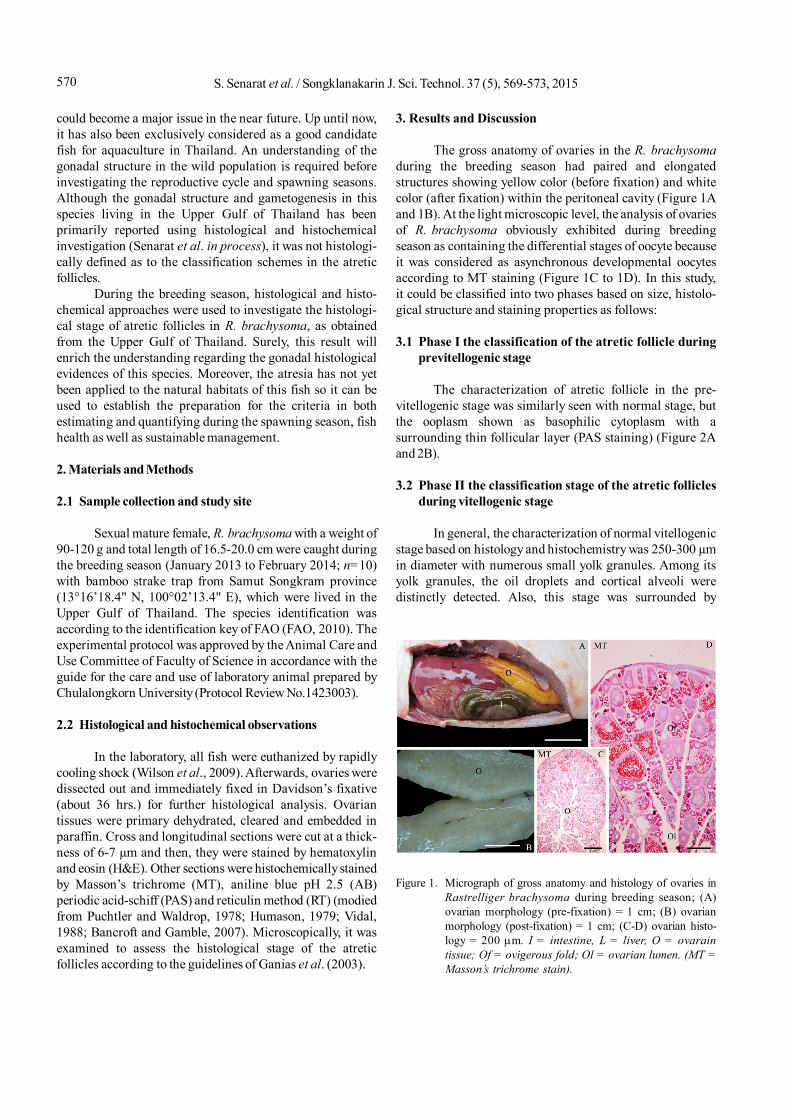
The gross anatomy of ovaries in the R. brachysomaduring the breeding season had paired and elongatedstructures showing yellow color (before fixation) and whitecolor (after fixation) within the peritoneal cavity (Figure 1Aand 1B). At the light microscopic level, the analysis of ovariesof R. brachysoma obviously exhibited during breedingseason as containing the differential stages of oocyte becauseit was considered as asynchronous developmental oocytesaccording to MT staining (Figure 1C to 1D). In this study,it could be classified into two phases based on size, histolo-gical structure and staining properties as follows:
3.1 Phase I the classification of the atretic follicle duringprevitellogenic stage
The characterization of atretic follicle in the pre-vitellogenic stage was similarly seen with normal stage, butthe ooplasm shown as basophilic cytoplasm with asurrounding thin follicular layer (PAS staining) (Figure 2Aand 2B).
3.2 Phase II the classification stage of the atretic folliclesduring vitellogenic stage
In general, the characterization of normal vitellogenicstage based on histology and histochemistry was 250-300 µmin diameter with numerous small yolk granules. Among itsyolk granules, the oil droplets and cortical alveoli weredistinctly detected. Also, this stage was surrounded by
Figure 1. Micrograph of gross anatomy and histology of ovaries inRastrelliger brachysoma during breeding season; (A)ovarian morphology (pre-fixation) = 1 cm; (B) ovarianmorphology (post-fixation) = 1 cm; (C-D) ovarian histo-logy = 200 m. I = intestine, L = liver, O = ovaraintissue; Of = ovigerous fold; Ol = ovarian lumen. (MT =Masson’s trichrome stain).
571S. Senarat et al. / Songklanakarin J. Sci. Technol. 37 (5), 569-573, 2015
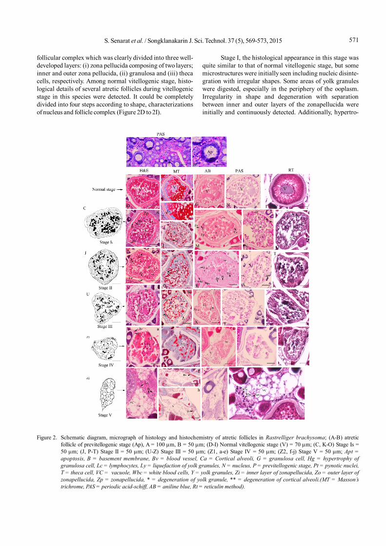
follicular complex which was clearly divided into three well-developed layers: (i) zona pellucida composing of two layers;inner and outer zona pellucida, (ii) granulosa and (iii) thecacells, respectively. Among normal vitellogenic stage, histo-logical details of several atretic follicles during vitellogenicstage in this species were detected. It could be completelydivided into four steps according to shape, characterizationsof nucleus and follicle complex (Figure 2D to 2I).
Stage I, the histological appearance in this stage wasquite similar to that of normal vitellogenic stage, but somemicrostructures were initially seen including nucleic disinte-gration with irregular shapes. Some areas of yolk granuleswere digested, especially in the periphery of the ooplasm.Irregularity in shape and degeneration with separationbetween inner and outer layers of the zonapellucida wereinitially and continuously detected. Additionally, hypertro-
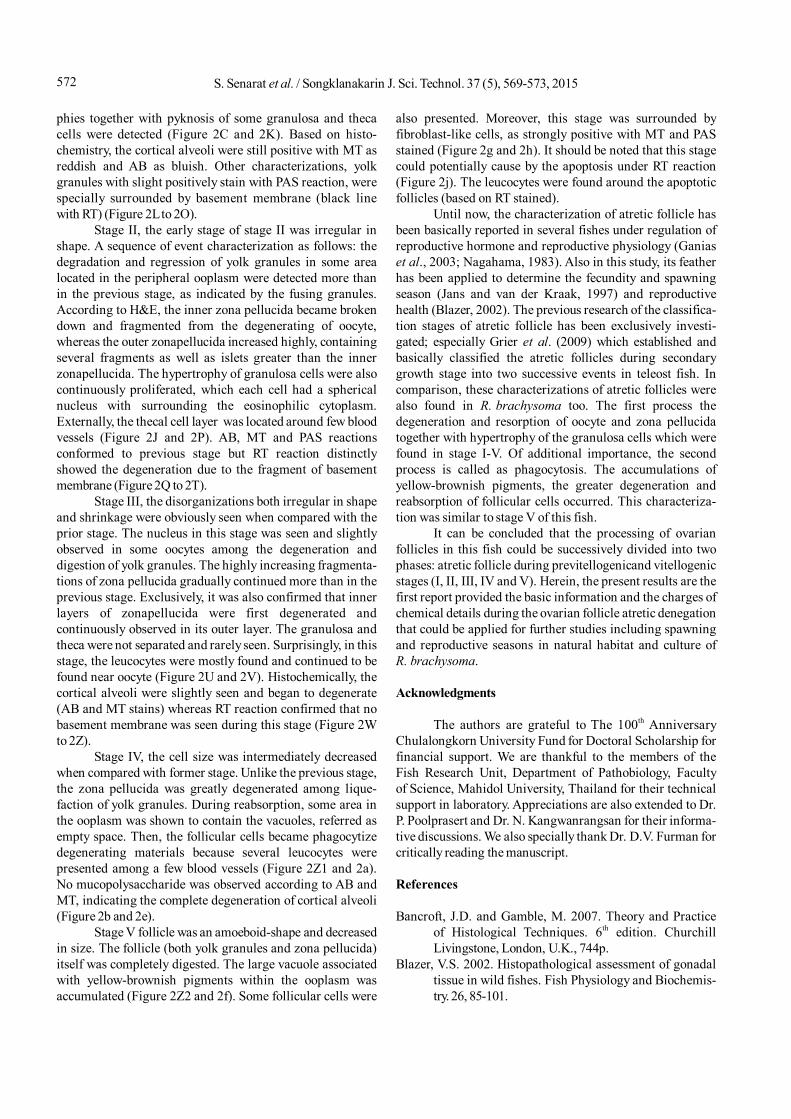
Figure 2. Schematic diagram, micrograph of histology and histochemistry of atretic follicles in Rastrelliger brachysoma; (A-B) atreticfollicle of previtellogenic stage (Ap), A = 100 m, B = 50 m; (D-I) Normal vitellogenic stage (V) = 70 m; (C, K-O) Stage Is =50 m; (J, P-T) Stage II = 50 m; (U-Z) Stage III = 50 m; (Z1, a-e) Stage IV = 50 m; (Z2, f-j) Stage V = 50 m; Apt =apoptosis, B = basement membrane, Bv = blood vessel, Ca = Cortical alveoli, G = granulosa cell, Hg = hypertrophy ofgranulosa cell, Lc = lymphocytes, Ly = liquefaction of yolk granules, N = nucleus, P = previtellogenic stage, Pt = pynotic nuclei,T = theca cell, VC = vacuole, Wbc = white blood cells, Y = yolk granules, Zi = inner layer of zonapellucida, Zo = outer layer ofzonapellucida, Zp = zonapellucida, * = degeneration of yolk granule, ** = degeneration of cortical alveoli.(MT = Masson’strichrome, PAS = periodic acid-schiff, AB = aniline blue, Rt = reticulin method).
S. Senarat et al. / Songklanakarin J. Sci. Technol. 37 (5), 569-573, 2015572
phies together with pyknosis of some granulosa and thecacells were detected (Figure 2C and 2K). Based on histo-chemistry, the cortical alveoli were still positive with MT asreddish and AB as bluish. Other characterizations, yolkgranules with slight positively stain with PAS reaction, werespecially surrounded by basement membrane (black linewith RT) (Figure 2L to 2O).
Stage II, the early stage of stage II was irregular inshape. A sequence of event characterization as follows: thedegradation and regression of yolk granules in some arealocated in the peripheral ooplasm were detected more thanin the previous stage, as indicated by the fusing granules.According to H&E, the inner zona pellucida became brokendown and fragmented from the degenerating of oocyte,whereas the outer zonapellucida increased highly, containingseveral fragments as well as islets greater than the innerzonapellucida. The hypertrophy of granulosa cells were alsocontinuously proliferated, which each cell had a sphericalnucleus with surrounding the eosinophilic cytoplasm.Externally, the thecal cell layer was located around few bloodvessels (Figure 2J and 2P). AB, MT and PAS reactionsconformed to previous stage but RT reaction distinctlyshowed the degeneration due to the fragment of basementmembrane (Figure 2Q to 2T).
Stage III, the disorganizations both irregular in shapeand shrinkage were obviously seen when compared with theprior stage. The nucleus in this stage was seen and slightlyobserved in some oocytes among the degeneration anddigestion of yolk granules. The highly increasing fragmenta-tions of zona pellucida gradually continued more than in theprevious stage. Exclusively, it was also confirmed that innerlayers of zonapellucida were first degenerated andcontinuously observed in its outer layer. The granulosa andtheca were not separated and rarely seen. Surprisingly, in thisstage, the leucocytes were mostly found and continued to befound near oocyte (Figure 2U and 2V). Histochemically, thecortical alveoli were slightly seen and began to degenerate(AB and MT stains) whereas RT reaction confirmed that nobasement membrane was seen during this stage (Figure 2Wto 2Z).
Stage IV, the cell size was intermediately decreasedwhen compared with former stage. Unlike the previous stage,the zona pellucida was greatly degenerated among lique-faction of yolk granules. During reabsorption, some area inthe ooplasm was shown to contain the vacuoles, referred asempty space. Then, the follicular cells became phagocytizedegenerating materials because several leucocytes werepresented among a few blood vessels (Figure 2Z1 and 2a).No mucopolysaccharide was observed according to AB andMT, indicating the complete degeneration of cortical alveoli(Figure 2b and 2e).
Stage V follicle was an amoeboid-shape and decreasedin size. The follicle (both yolk granules and zona pellucida)itself was completely digested. The large vacuole associatedwith yellow-brownish pigments within the ooplasm wasaccumulated (Figure 2Z2 and 2f). Some follicular cells were
also presented. Moreover, this stage was surrounded byfibroblast-like cells, as strongly positive with MT and PASstained (Figure 2g and 2h). It should be noted that this stagecould potentially cause by the apoptosis under RT reaction(Figure 2j). The leucocytes were found around the apoptoticfollicles (based on RT stained).
Until now, the characterization of atretic follicle hasbeen basically reported in several fishes under regulation ofreproductive hormone and reproductive physiology (Ganiaset al., 2003; Nagahama, 1983). Also in this study, its featherhas been applied to determine the fecundity and spawningseason (Jans and van der Kraak, 1997) and reproductivehealth (Blazer, 2002). The previous research of the classifica-tion stages of atretic follicle has been exclusively investi-gated; especially Grier et al. (2009) which established andbasically classified the atretic follicles during secondarygrowth stage into two successive events in teleost fish. Incomparison, these characterizations of atretic follicles werealso found in R. brachysoma too. The first process thedegeneration and resorption of oocyte and zona pellucidatogether with hypertrophy of the granulosa cells which werefound in stage I-V. Of additional importance, the secondprocess is called as phagocytosis. The accumulations ofyellow-brownish pigments, the greater degeneration andreabsorption of follicular cells occurred. This characteriza-tion was similar to stage V of this fish.
It can be concluded that the processing of ovarianfollicles in this fish could be successively divided into twophases: atretic follicle during previtellogenicand vitellogenicstages (I, II, III, IV and V). Herein, the present results are thefirst report provided the basic information and the charges ofchemical details during the ovarian follicle atretic denegationthat could be applied for further studies including spawningand reproductive seasons in natural habitat and culture ofR. brachysoma.
Acknowledgments
The authors are grateful to The 100th AnniversaryChulalongkorn University Fund for Doctoral Scholarship forfinancial support. We are thankful to the members of theFish Research Unit, Department of Pathobiology, Facultyof Science, Mahidol University, Thailand for their technicalsupport in laboratory. Appreciations are also extended to Dr.P. Poolprasert and Dr. N. Kangwanrangsan for their informa-tive discussions. We also specially thank Dr. D.V. Furman forcritically reading the manuscript.
References
Bancroft, J.D. and Gamble, M. 2007. Theory and Practiceof Histological Techniques. 6th edition. ChurchillLivingstone, London, U.K., 744p.
Blazer, V.S. 2002. Histopathological assessment of gonadaltissue in wild fishes. Fish Physiology and Biochemis-try. 26, 85-101.
573S. Senarat et al. / Songklanakarin J. Sci. Technol. 37 (5), 569-573, 2015
Department of Fisheries. 2009. Fisheries statistics of Thai-land. Available from: http://www.fisheries.go.th/itstat/yearbook/data_2552/Yearbook/Yearbook2009.pdf.[February 5, 2015].
FAO. 2010. Report of the First Workshop on the Assessmentof Fishery Stock Status in South and Southeast Asia.Bangkok, 16-19 June 2009. FAO Fisheries and Agri-cultural Report No. 913, Rome, FAO, 30p. Availablefrom: http://www.fao.org/docrep/012/i1555e/i1555e00.pdf [February 5, 2015].
Ganias, K., Somarakis, S., Koutsikopoulos, C., Machias, A.and Theodorou, A. 2003. Ovarian atresia in the Medi-terranean sardine, Sardinapilchrdussardina. Journalof the Marine Biological Association of the UnitedKingdom. 83, 1327-1332.
Genten, F., Terwinghe, E. and Danguy, A. 2009. Atlas of FishHistology, CRC Press, U.S.A., 223p.
Grier, J.H., Uribe, M.C. and Patiño, R. 2009. The ovary, folli-culogenesis and oogenesis in teleosts.In Reproduc-tive Biology and Phylogeny of Fishes (Agnathans andBony Fishes, B.G.M. Jamieson, editor. Enfield, NewHampshire, Science Publishers, U.S.A., pp. 25-84.
Humason, G.L. 1979. Animal Tissue Techniques.4th edition.San Francisco, Freeman, U.S.A.
Hunter, J.R. and Goldberg, S.R. 1980. Spawning incidenceand batch fecundity in northern anchovy, Engraulis-mordax. Fishery Bulletin. 77, 641-652.
Hunter, J.R. and Lo, N.C.H. 1997. The daily egg productionmethod of biomass estimation: some problems andpotential improvements. Ozeanografika. 2, 41-69.
Hunter, J.R. and Macewicz, B. 1985.Rates of atresia in theovary of captive and wild northern anchovy,Engraulismordax.Fishery Bulletin. 83, 119-136.
Hussein, M.R. 2005. Apoptosis in the ovary: molecularmechanisms. Human Reproduction Update. 11, 162-178.
Jans, D.M. and van der Kraak, G. 1997. Suppression ofapoptosis by gonadotropin, 17 -estradiol, and epi-dermal growth factor in rainbow trout preovulatoryovaries follicles. General and Comparative Endocri-nology. 105, 186-193.
Johnson, R., Wolf, J. and Braunbeck, T. 2009. OECD guidancedocument for the diagnosis of endocrine-related his-topathology of fish gonads. Available from: http://www.oecd.org/dataoecd/33/27/ 42140701.pdf [Feb-ruary 5, 2015]
Kennedy, A.M. 2002. Reproduction of striped bass Moro-nesaxatilis: a structural, biochemical and functionalcharacterization of atresia. MSc Thesis, North Caro-lina State University.Available from: http://www.lib.ncsu.edu/theses/available/etd-06162002-210408/[February 5, 2015]
Nagahama, Y. 1983. The function morphology of teleostgonads. In Fish Physiology: Volume IX: Reproduction,Part A: Endocrine Tissues and Hormones, W.S. Hoar,D.J. Randall, E.M. Donaldson, editors. No 9. AcademicPress, New York, U.S.A., pp 223-275.
Puchtler, H. and Waldrop, F.W. 1978. Silver impregnationmethods for reticulum bers and reticulin: A re-investi-gation of their origins and specicity. Histochemistry.57, 177-187.
Saidapur, S.K. 1978. Follicular atresia in the ovaries ofnonmammalian vertebrates. International Review ofCytology. 54, 225-244.
Santos, H.B., Sato, Y., Moro, L., Bazzoli, N. and Rizzo, E.2008. Relationship among follicular apoptosis, integrinbeta1 and collagen type IV during early ovarianregression in the teleost Prochilodusargenteus afterinduced spawning. Cell and Tissue Research. 332,159-170.
Vidal, B.C. 1988. Histochemical and anisotropical propertiescharacteristics of silver impregnation: the differentia-tion of reticulin bers from the other interstitialcollagens. ZoologischeJahrbücher. 117, 485–494.
Wilson, J.M, Bunte, R.M. and Carty, A.J. 2009. Evaluation ofrapid cooling and tricainemethanesulfonate (MS222)as methods of euthanasia in zebrafish (Daniorerio).American Association for Laboratory Animal Science.48, 785-789.
Wood, A.W. and van der Kraak, G. 2002. Inhibition ofapoptosis in vitellogenic ovarian follicles of rainbowtrout (Oncorhynchusmykiss) by salmon gonadotro-phin, epidermal growth factor and 17-estradiol.Molecular Reproduction and Development. 61, 511-518.
Wood, A.W. and van Der Kraak, G. 2003. Yolk proteolysis inrainbow trout oocytes after serum-free culture: Evi-dence for a novel biochemical mechanism of atresia inoviparous vertebrates. Molecular Reproduction andDevelopment. 65, 219-227.