Embed Size (px)
Citation preview
Clinical acceleration: from the consoleJames Hancock
Benson Radiology, North Adelaide, Australia
In Clinical MR the challenge to scan faster, in higher resolution and with consistent reproducibility always has been and always will be the challenge that drives technological development within the modality. Tradi- tional thinking in MR dictates that speed comes at the cost of compromised image quality and is achieved with a decrease in spatial resolution or at risk of increased artifacts often at the expense of signal-to-noise ratio (SNR).
The implementation of technology such as Compressed Sensing (CS), CAIPIRINHA and Simultaneous Multi-Slice (SMS) is changing our thinking on just what is possible and providing maximum flexibility in imaging. Imaging is now being better tailored to the patient and their capabilities rather than the patient having to bend to the requirements of the imaging. This is a fundamental principle of Siemens BioMatrix technology.
With a big focus on body imaging applications Benson Radiology installed a 3T MAGNETOM Vida system into a private imaging department in the central business district of Adelaide, South Australia in July 2017. Without access to government-based medical rebates the quality, speed and reproducibility of examinations performed on the MAGNETOM Vida system are key to a successful business outcome. The MAGNETOM Vida offers technology like Compressed Sensing (CS), CAIPIRINHA and Simultaneous Multi-Slice (SMS) providing maximum flexibility in imaging.
CAIPIRINHA CAIPIRINHA (Controlled Aliasing in Parallel Imaging Results in Higher Acceleration) is a unique k-space acquisition scheme for parallel imaging techniques (PAT) that improves the SNR of a sequence by up to 18% compared to conventional acceleration of the same sequence with SENSE or GRAPPA. With a particular focus on body imaging, Benson Radiology is leveraging the combination of very high coil density with CAIPIRINHA
VIBE to fit an imaging solution to the capabilities of the patient. In a competent breath-holder this allows traditional breath-hold examinations to be acquired at higher spatial resolutions than ever before. Here we use the SNR gain from the parallel imaging technique combined with high coil density to improve fine edge sharpness and better delineate the conspicuity of pathology in the abdomen.
In the subset of patients where the ability to hold ones breath is compromised acceleration factors can be increased, thereby driving down scan time to achieve robust reproducible outcomes without the traditional sacrifice in spatial resolution or the risk of imaging artifacts traditionally associated with high PAT factors. This is achievable at both 1.5T and 3T field strengths.
1.3 x 1.3 x 2 mm, 88 slices, complete liver coverage. Achieved using CAIPIRINHA total acceleration factor of 5. High resolution clearly defines liver lesion with features suggestive of heman- gioma in this delayed dataset acquired 5 minutes following administration of gadolinium contrast. Scan time of 16 seconds.
1
Figure 1: T1 VIBE CAIPIRINHA post contrast
MAGNETOM Flash (70) 1/2018
10 siemens.com/magnetom-world
Spotlight
Simultaneous Multi-Slice (SMS)With a focus on body imaging, diffusion-weighted imaging (DWI) is always going to be a key aspect of the imaging service offered at Benson Radiology. DWI offers well-known benefits in abdominal and pelvic imaging.
Conventional EPI-DWI techniques can be acquired in multiple ways, including a free-breathing (FB) approach with the use of navigators or with multiple breath-hold techniques. Breath-hold techniques are constrained by the breath-hold capacity of the patient as well as the reproducibility of these breath-holds. This further constrains the usefulness of the technique as b-values become limited, as does the spatial resolution that can be achieved. If using a free breathing technique the acquisition time itself is the tradeoff. Here navigating the approach can become inefficient depending on the patients respiratory cycle. Multiaverage free breathing, while the current gold standard at our institution, is still
a lengthy approach. This is despite making use of parallel imaging techniques like GRAPPA for acquisition acceleration. The conventional EPI-DWI approach is inefficient as every imaging slice has to be excited individ- ually, then the diffusion encoding gradients applied, and finally the image information acquired with EPI encoding. This process needs to be repeated multiple times, once for each imaging slice, until the entire volume of interest is covered.
The inefficiencies mentioned above can be overcome with Simultaneous Multi-Slice acquisition combined with a free breathing approach, which we have implemented at our institution. Instead of successive excitation of slices, slices are exited simultaneously with a multiband pulse and blipped-CAIPIRINHA SMS-EPI ensures preservation of high SNR and low artifact levels. Since multiple slices are excited simultaneously, the overall TR for a desired spatial coverage or number of slices is reduced, leading to a scan time reduction by the same factor. The big advantage of
2A
2D2C
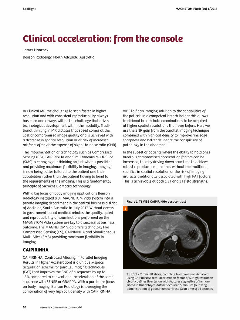
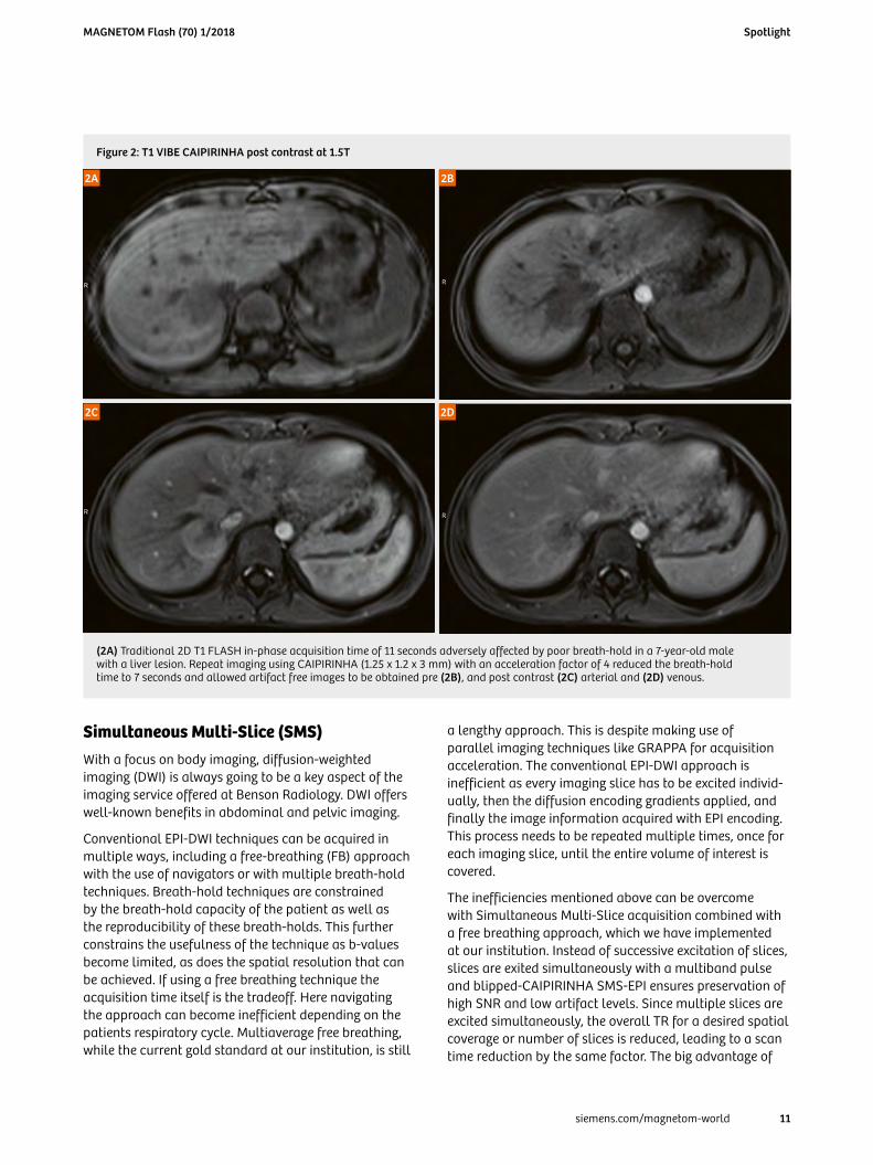
(2A) Traditional 2D T1 FLASH in-phase acquisition time of 11 seconds adversely affected by poor breath-hold in a 7-year-old male with a liver lesion. Repeat imaging using CAIPIRINHA (1.25 x 1.2 x 3 mm) with an acceleration factor of 4 reduced the breath-hold time to 7 seconds and allowed artifact free images to be obtained pre (2B), and post contrast (2C) arterial and (2D) venous.
Figure 2: T1 VIBE CAIPIRINHA post contrast at 1.5T
2B
MAGNETOM Flash (70) 1/2018
11siemens.com/magnetom-world
Spotlight
SMS over other acceleration techniques is that it does not suffer from the typical square-root of acceleration factor SNR penalty due to data under-sampling.
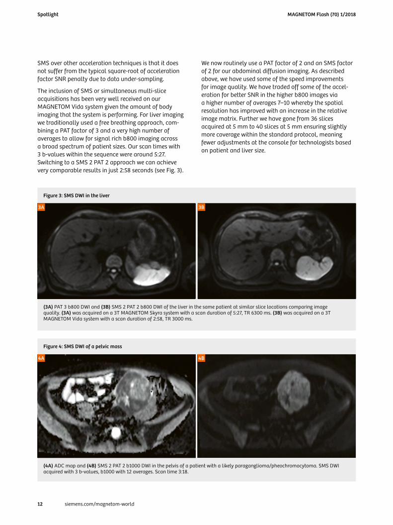
The inclusion of SMS or simultaneous multi-slice acquisitions has been very well received on our MAGNETOM Vida system given the amount of body imaging that the system is performing. For liver imaging we traditionally used a free breathing approach, com- bining a PAT factor of 3 and a very high number of averages to allow for signal rich b800 imaging across a broad spectrum of patient sizes. Our scan times with 3 b-values within the sequence were around 5:27. Switching to a SMS 2 PAT 2 approach we can achieve very comparable results in just 2:58 seconds (see Fig. 3).
We now routinely use a PAT factor of 2 and an SMS factor of 2 for our abdominal diffusion imaging. As described above, we have used some of the speed improvements for image quality. We have traded off some of the accel- eration for better SNR in the higher b800 images via a higher number of averages 7–10 whereby the spatial resolution has improved with an increase in the relative image matrix. Further we have gone from 36 slices acquired at 5 mm to 40 slices at 5 mm ensuring slightly more coverage within the standard protocol, meaning fewer adjustments at the console for technologists based on patient and liver size.
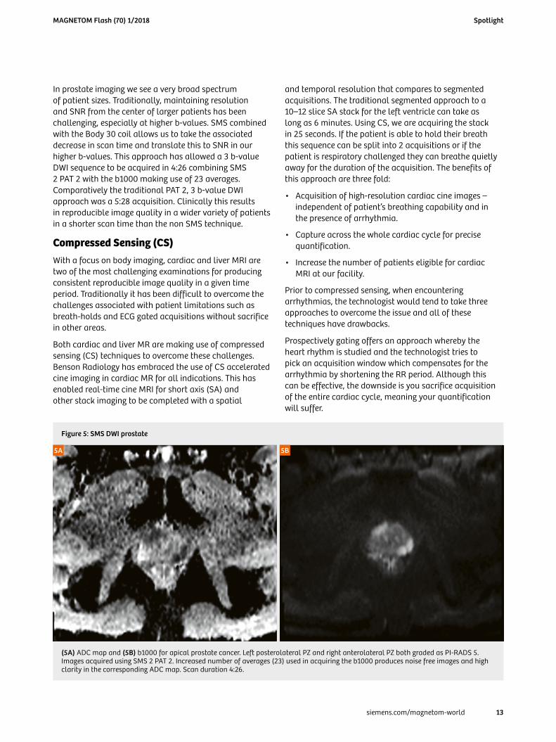
(4A) ADC map and (4B) SMS 2 PAT 2 b1000 DWI in the pelvis of a patient with a likely paraganglioma/pheochromocytoma. SMS DWI acquired with 3 b-values, b1000 with 12 averages. Scan time 3:18.
Figure 4: SMS DWI of a pelvic mass
4B4A
(3A) PAT 3 b800 DWI and (3B) SMS 2 PAT 2 b800 DWI of the liver in the same patient at similar slice locations comparing image quality. (3A) was acquired on a 3T MAGNETOM Skyra system with a scan duration of 5:27, TR 6300 ms. (3B) was acquired on a 3T MAGNETOM Vida system with a scan duration of 2:58, TR 3000 ms.
Figure 3: SMS DWI in the liver
3B3A
MAGNETOM Flash (70) 1/2018
12 siemens.com/magnetom-world
Spotlight
In prostate imaging we see a very broad spectrum of patient sizes. Traditionally, maintaining resolution and SNR from the center of larger patients has been challenging, especially at higher b-values. SMS combined with the Body 30 coil allows us to take the associated decrease in scan time and translate this to SNR in our higher b-values. This approach has allowed a 3 b-value DWI sequence to be acquired in 4:26 combining SMS 2 PAT 2 with the b1000 making use of 23 averages. Comparatively the traditional PAT 2, 3 b-value DWI approach was a 5:28 acquisition. Clinically this results in reproducible image quality in a wider variety of patients in a shorter scan time than the non SMS technique.
Compressed Sensing (CS)With a focus on body imaging, cardiac and liver MRI are two of the most challenging examinations for producing consistent reproducible image quality in a given time period. Traditionally it has been difficult to overcome the challenges associated with patient limitations such as breath-holds and ECG gated acquisitions without sacrifice in other areas.
Both cardiac and liver MR are making use of compressed sensing (CS) techniques to overcome these challenges. Benson Radiology has embraced the use of CS accelerated cine imaging in cardiac MR for all indications. This has enabled real-time cine MRI for short axis (SA) and other stack imaging to be completed with a spatial
and temporal resolution that compares to segmented acquisitions. The traditional segmented approach to a 10–12 slice SA stack for the left ventricle can take as long as 6 minutes. Using CS, we are acquiring the stack in 25 seconds. If the patient is able to hold their breath this sequence can be split into 2 acquisitions or if the patient is respiratory challenged they can breathe quietly away for the duration of the acquisition. The benefits of this approach are three fold:
• Acquisition of high-resolution cardiac cine images –independent of patient’s breathing capability and inthe presence of arrhythmia.
• Capture across the whole cardiac cycle for precisequantification.
• Increase the number of patients eligible for cardiacMRI at our facility.
Prior to compressed sensing, when encountering arrhythmias, the technologist would tend to take three approaches to overcome the issue and all of these techniques have drawbacks.
Prospectively gating offers an approach whereby the heart rhythm is studied and the technologist tries to pick an acquisition window which compensates for the arrhythmia by shortening the RR period. Although this can be effective, the downside is you sacrifice acquisition of the entire cardiac cycle, meaning your quantification will suffer.
(5A) ADC map and (5B) b1000 for apical prostate cancer. Left posterolateral PZ and right anterolateral PZ both graded as PI-RADS 5. Images acquired using SMS 2 PAT 2. Increased number of averages (23) used in acquiring the b1000 produces noise free images and high clarity in the corresponding ADC map. Scan duration 4:26.
Figure 5: SMS DWI prostate
5B5A
MAGNETOM Flash (70) 1/2018
13siemens.com/magnetom-world
Spotlight
6A6A
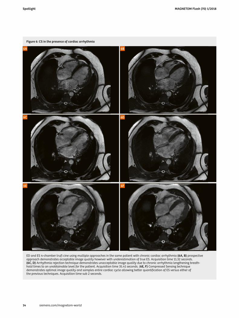
ED and ES 4-chamber trufi cine using multiple approaches in the same patient with chronic cardiac arrhythmia (6A, B) prospective approach demonstrates acceptable image quality however with underestimation of true ES. Acquisition time 11.32 seconds. (6C, D) Arrhythmia rejection technique demonstrates unacceptable image quality due to chronic arrhythmia lengthening breath- hold times to an unobtainable level for the patient. Acquisition time 35.41 seconds. (6E, F) Compressed Sensing technique demonstrates optimal image quality and samples entire cardiac cycle allowing better quantification of ES versus either of the previous techniques. Acquisition time sub 2 seconds.
Figure 6: CS in the presence of cardiac arrhythmia
6B
6D6C
6F6E
MAGNETOM Flash (70) 1/2018
14 siemens.com/magnetom-world
Spotlight

Another available tool is the arrhythmia rejection tool, whereby we set an acceptance window and the scanner rejects any RR periods which do not fall within this window. The downside of this is that if the patient has a chronic arrhythmia then the scan time can increase beyond the ability of the patient to hold their breath.
Prior to CS this leaves the technologist with nowhere to go other than to use real time imaging with poor spatial and temporal resolution. CS has changed this. Using compressed sensing the heart can be imaged with both high spatial and temporal resolution in a robust reproducible fashion independent of the patient’s breath-hold ability and in the presence of considerable arrhythmia.
In the liver or any abdominal/pelvic indication that requires a dynamic contrast injection, the quality of the final images is reliant on both a skilled technologist who can trigger the acquisition for an accurate arterial phase or phase(s) and a compliant patient who can follow breathing instructions at the right time and also maintain a breath-hold for the duration of the acquisition. In the liver in particular temporal resolution in lesion characterization is becoming important for diagnosis.
Compressed Sensing GRASP-VIBE offers a technique for patients with limited breath-hold capability or who are unable to follow breathing commands. The technique takes the pressure off technologists as the critical timing-dependant arterial phase of an acquisition is removed. The acquisition is a simple push-button exam.
The acquisition is performed in one continuous run, using a golden-angle stack-of-stars radial scheme that gives robustness towards motion and the flexibility to choose the temporal resolution. The sequence itself guides the user on the correct time point for contrast administration. Reconstruction is performed using a Compressed Sensing GPU accelerated algorithm.
This new approach gives us a reliable post contrast tech- nique on any patient. It is changing the way abdominal post contrast imaging is reviewed and making it a more dynamic cine experience at each slice location for the radiologist. Radiologists receive a temporal dataset which can be viewed akin to a cine series allowing contrast arrival, enhancement and wash out to be viewed on a slice by slice basis. The high temporal resolution is particularly useful for HCC detection and small thrombus within vessels. The insensitivity to breathing
7A
7F7E7D
7C7B
Compressed Sensing T1 Fatsat GRASP VIBE. 6 of 21 phases shown here. Technique is acquired in free breathing with a contrast timing independent acquisition running over approximately 5 minutes. (7A–C) are very early, mid and late arterial phases. (7D, E) are venous phases and (7F) being a delayed phase at nearly 5 minutes post contrast. Arterial enhancement, with contrast retention, central scar and minimal background T1/T2 change favours diagnosis of FNH.
Figure 7: CS GRASP VIBE liver
MAGNETOM Flash (70) 1/2018
15siemens.com/magnetom-world
Spotlight
Contact
James Hancock Modality Manager – MRI Benson Radiology 229 Melbourne Street North Adelaide, South Australia, 5006 Tel.: (08) 8239 0550 Mobile: 0434 279 466 [email protected]
8A
8F8E8D
8C8B
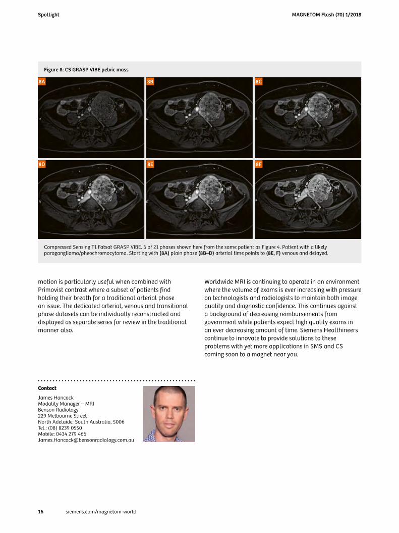
Compressed Sensing T1 Fatsat GRASP VIBE. 6 of 21 phases shown here from the same patient as Figure 4. Patient with a likely paraganglioma/pheochromocytoma. Starting with (8A) plain phase (8B–D) arterial time points to (8E, F) venous and delayed.
Figure 8: CS GRASP VIBE pelvic mass
motion is particularly useful when combined with Primovist contrast where a subset of patients find holding their breath for a traditional arterial phase an issue. The dedicated arterial, venous and transitional phase datasets can be individually reconstructed and displayed as separate series for review in the traditional manner also.
Worldwide MRI is continuing to operate in an environment where the volume of exams is ever increasing with pressure on technologists and radiologists to maintain both image quality and diagnostic confidence. This continues against a background of decreasing reimbursements from government while patients expect high quality exams in an ever decreasing amount of time. Siemens Healthineers continue to innovate to provide solutions to these problems with yet more applications in SMS and CS coming soon to a magnet near you.
MAGNETOM Flash (70) 1/2018
16 siemens.com/magnetom-world
Spotlight