Embed Size (px)
Citation preview
RESEARCH LETTER Open Access
Clinical analysis of sinus bradycardia inpatients with severe COVID-19 pneumoniaLijuan Hu1†, Linjing Gong1†, Zhilong Jiang1, Qibing Wang2, Yunzeng Zou2* and Lei Zhu1*
Keywords: COVID-19, Severe pneumonia, Sinus bradycardia, Clinical manifestation, ACE2
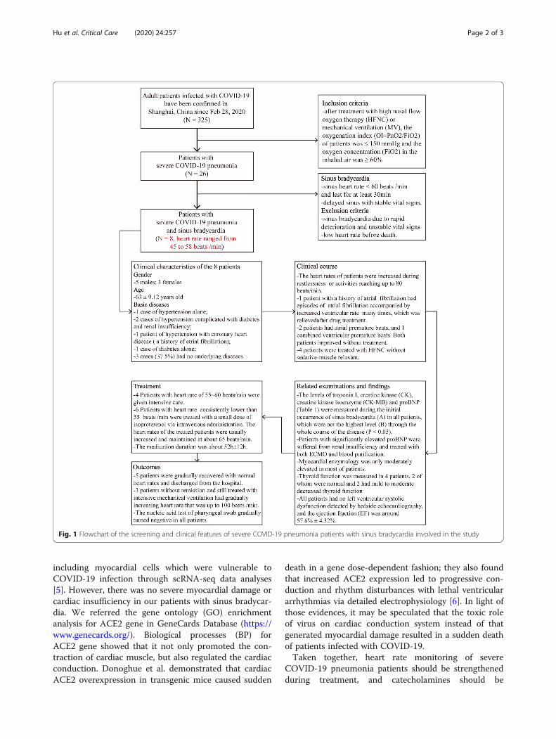
There were cases of sudden death in some patients in-fected with COVID-19 including a few of young physi-cians, which had a huge impact on medical communityand society [1]. The unexpected phenomenon lets us thinkabout the underlying problems that caused the suddendeath and some issues maybe ignored and that should beappropriately resolved. The initial manifestation of severeCOVID-19 pneumonia patients was hypoxemic respira-tory failure, accompanied by rapid increased reactive heartrate and susceptibility to supraventricular arrhythmia [2].It is notable that a proportion of these patients developedsinus bradycardia, which was significantly different fromother patients with multiple types of respiratory failure.In addition to lung injury, cardiac injury has often
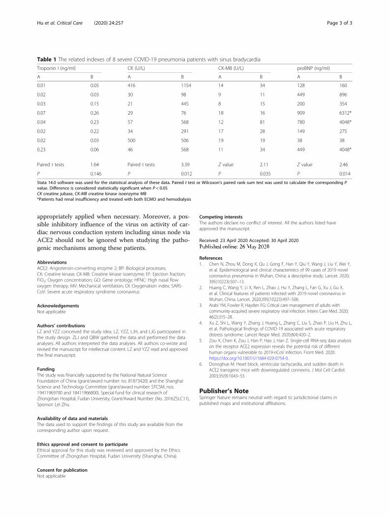
been reported in patients with COVID-19 [2]. Some ex-perts believed that the virus invasion into myocardiumled to severe myocarditis or the severe “cytokine storm”-induced acute myocardial injury may explain the suddendeath in some affected patients [3]. It is noteworthy thatabout 1/3 of the patients with severe illness in our studydeveloped sinus bradycardia (Fig. 1). The troponin andproBNP were basically normal among these patients ex-cept for those with renal failure (Table 1). The clinicalcharacteristics of explosive myocarditis and myocardialinfarction were not presented among these patients,
suggesting these are not the cause of sinus bradycardiain these patients. It was previously reported that nopathological evidence of myocarditis or myocardialmicroinfarction was observed in the heart of suffered pa-tients [4], consisting with our results. Therefore, wespeculated that sudden death among some severe pa-tients with improved symptoms post-treatment may becaused by severe arrhythmia such as ventricular fibrilla-tion induced by severe sinus delay.We found that sinus bradycardia often occurred dur-
ing sleep. So, deep sleep or sedation may be an import-ant risk factor for sinus bradycardia. A few patients hadmild to moderate decreased thyroid function, which wasconsistent with secondary pathological thyroid syndromeand may also be one of the causes of sinus bradycardia.When viral nucleic acid tests gradually turned negative,the heart rate returned to normal no matter whether thepatient’s condition improved or worsened and the usesof catecholamine were gradually discontinued. Accord-ing to the results, we speculated that the inhibitory effectof virus on sinus node activity was the main cause ofsinus bradycardia in these patients.Previous study indicated that COVID-19 invaded host
cells via the receptor angiotensin-converting enzyme 2(ACE2) [5]. Zou et al. identified specific cell types
© The Author(s). 2020 Open Access This article is licensed under a Creative Commons Attribution 4.0 International License,which permits use, sharing, adaptation, distribution and reproduction in any medium or format, as long as you giveappropriate credit to the original author(s) and the source, provide a link to the Creative Commons licence, and indicate ifchanges were made. The images or other third party material in this article are included in the article's Creative Commonslicence, unless indicated otherwise in a credit line to the material. If material is not included in the article's Creative Commonslicence and your intended use is not permitted by statutory regulation or exceeds the permitted use, you will need to obtainpermission directly from the copyright holder. To view a copy of this licence, visit http://creativecommons.org/licenses/by/4.0/.The Creative Commons Public Domain Dedication waiver (http://creativecommons.org/publicdomain/zero/1.0/) applies to thedata made available in this article, unless otherwise stated in a credit line to the data.
* Correspondence: [email protected]; [email protected]†Lijuan Hu and Linjing Gong contributed equally to this work.1Department of Pulmonary Medicine, Zhongshan Hospital, Fudan University,180 Feng Lin Rd., Shanghai 200032, China2Shanghai Institute of Cardiovascular Diseases, Zhongshan Hospital, FudanUniversity, 180 Feng Lin Rd., Shanghai 200032, China
Hu et al. Critical Care (2020) 24:257 https://doi.org/10.1186/s13054-020-02933-3
including myocardial cells which were vulnerable toCOVID-19 infection through scRNA-seq data analyses[5]. However, there was no severe myocardial damage orcardiac insufficiency in our patients with sinus bradycar-dia. We referred the gene ontology (GO) enrichmentanalysis for ACE2 gene in GeneCards Database (https://www.genecards.org/). Biological processes (BP) forACE2 gene showed that it not only promoted the con-traction of cardiac muscle, but also regulated the cardiacconduction. Donoghue et al. demonstrated that cardiacACE2 overexpression in transgenic mice caused sudden
death in a gene dose-dependent fashion; they also foundthat increased ACE2 expression led to progressive con-duction and rhythm disturbances with lethal ventriculararrhythmias via detailed electrophysiology [6]. In light ofthose evidences, it may be speculated that the toxic roleof virus on cardiac conduction system instead of thatgenerated myocardial damage resulted in a sudden deathof patients infected with COVID-19.Taken together, heart rate monitoring of severe
COVID-19 pneumonia patients should be strengthenedduring treatment, and catecholamines should be
Fig. 1 Flowchart of the screening and clinical features of severe COVID-19 pneumonia patients with sinus bradycardia involved in the study
Hu et al. Critical Care (2020) 24:257 Page 2 of 3
appropriately applied when necessary. Moreover, a pos-sible inhibitory influence of the virus on activity of car-diac nervous conduction system including sinus node viaACE2 should not be ignored when studying the patho-genic mechanisms among these patients.
AbbreviationsACE2: Angiotensin-converting enzyme 2; BP: Biological processes;CK: Creatine kinase; CK-MB: Creatine kinase isoenzyme; EF: Ejection fraction;FiO2: Oxygen concentration; GO: Gene ontology; HFNC: High nasal flowoxygen therapy; MV: Mechanical ventilation; OI: Oxygenation index; SARS-CoV: Severe acute respiratory syndrome coronavirus
AcknowledgementsNot applicable
Authors’ contributionsLZ and YZZ conceived the study idea. LZ, YZZ, LJH, and LJG participated inthe study design. ZLJ and QBW gathered the data and performed the dataanalyses. All authors interpreted the data analyses. All authors co-wrote andrevised the manuscript for intellectual content. LZ and YZZ read and approvedthe final manuscript.
FundingThe study was financially supported by the National Natural ScienceFoundation of China (grant/award number: no. 81873420) and the ShanghaiScience and Technology Committee (grant/award number: STCSM, nos.19411969700 and 18411966800). Special fund for clinical research ofZhongshan Hospital, Fudan University, Grant/Award Number: (No. 2016ZSLC11),Sponsor: Lei Zhu.
Availability of data and materialsThe data used to support the findings of this study are available from thecorresponding author upon request.
Ethics approval and consent to participateEthical approval for this study was reviewed and approved by the EthicsCommittee of Zhongshan Hospital, Fudan University (Shanghai, China).
Consent for publicationNot applicable
Competing interestsThe authors declare no conflict of interest. All the authors listed haveapproved the manuscript.
Received: 23 April 2020 Accepted: 30 April 2020
References1. Chen N, Zhou M, Dong X, Qu J, Gong F, Han Y, Qiu Y, Wang J, Liu Y, Wei Y,
et al. Epidemiological and clinical characteristics of 99 cases of 2019 novelcoronavirus pneumonia in Wuhan, China: a descriptive study. Lancet. 2020;395(10223):507–13.
2. Huang C, Wang Y, Li X, Ren L, Zhao J, Hu Y, Zhang L, Fan G, Xu J, Gu X,et al. Clinical features of patients infected with 2019 novel coronavirus inWuhan, China. Lancet. 2020;395(10223):497–506.
3. Arabi YM, Fowler R, Hayden FG: Critical care management of adults withcommunity-acquired severe respiratory viral infection. Intens Care Med. 2020;46(2):315–28.
4. Xu Z, Shi L, Wang Y, Zhang J, Huang L, Zhang C, Liu S, Zhao P, Liu H, Zhu L,et al. Pathological findings of COVID-19 associated with acute respiratorydistress syndrome. Lancet Respir Med. 2020;8(4):420–2.
5. Zou X, Chen K, Zou J, Han P, Hao J, Han Z. Single-cell RNA-seq data analysison the receptor ACE2 expression reveals the potential risk of differenthuman organs vulnerable to 2019-nCoV infection. Front Med. 2020.https://doi.org/10.1007/s11684-020-0754-0.
6. Donoghue M. Heart block, ventricular tachycardia, and sudden death inACE2 transgenic mice with downregulated connexins. J Mol Cell Cardiol.2003;35(9):1043–53.
Publisher’s NoteSpringer Nature remains neutral with regard to jurisdictional claims inpublished maps and institutional affiliations.
Table 1 The related indexes of 8 severe COVID-19 pneumonia patients with sinus bradycardia
Troponin I (ng/ml) CK (U/L) CK-MB (U/L) proBNP (ng/ml)
A B A B A B A B
0.01 0.05 416 1154 14 34 128 160
0.02 0.03 30 98 9 11 449 896
0.03 0.15 21 445 8 15 200 354
0.07 0.26 29 76 18 16 909 6312*
0.04 0.23 57 568 12 81 780 4048*
0.02 0.22 34 291 17 28 149 275
0.02 0.03 500 506 19 19 38 38
0.23 0.06 46 568 11 34 449 4048*
Paired t tests 1.64 Paired t tests 3.39 Z value 2.11 Z value 2.46
P 0.146 P 0.012 P 0.035 P 0.014
Stata 14.0 software was used for the statistical analysis of these data. Paired t test or Wilcoxon’s paired rank sum test was used to calculate the corresponding Pvalue. Difference is considered statistically significant when P < 0.05CK creatine jubase, CK-MB creatine kinase isoenzyme MB*Patients had renal insufficiency and treated with both ECMO and hemodialysis
Hu et al. Critical Care (2020) 24:257 Page 3 of 3