Embed Size (px)
Citation preview
FungiJournal of
Article
Clinical Characteristics and Relevance of Oral Candida Biofilmin Tongue Smears
Eunae Cho 1,2,† , YounJung Park 3,† , Ki-Yeol Kim 2,4, Dawool Han 1 , Hyun Sil Kim 1, Jeong-Seung Kwon 3,*and Hyung-Joon Ahn 3,*
�����������������
Citation: Cho, E.; Park, Y.; Kim, K.-Y.;
Han, D.; Kim, H.S.; Kwon, J.-S.; Ahn,
H.-J. Clinical Characteristics and
Relevance of Oral Candida Biofilm in
Tongue Smears. J. Fungi 2021, 7, 77.
https://doi.org/10.3390/jof7020077
Received: 22 December 2020
Accepted: 20 January 2021
Published: 22 January 2021
Publisher’s Note: MDPI stays neutral
with regard to jurisdictional claims in
published maps and institutional affil-
iations.
Copyright: © 2021 by the authors.
Licensee MDPI, Basel, Switzerland.
This article is an open access article
distributed under the terms and
conditions of the Creative Commons
Attribution (CC BY) license (https://
creativecommons.org/licenses/by/
4.0/).
1 Department of Oral Pathology, Oral Cancer Research Institute, Yonsei University College of Dentistry,Seoul 03722, Korea; [email protected] (E.C.); [email protected] (D.H.); [email protected] (H.S.K.)
2 BK21 FOUR Project, Yonsei University College of Dentistry, Seoul 03722, Korea; [email protected] Department of Orofacial Pain and Oral Medicine, Dental Hospital, Yonsei University College of Dentistry,
Seoul 03722, Korea; [email protected] Department of Dental Education, Yonsei University College of Dentistry, Seoul 03722, Korea* Correspondence: [email protected] (J.-S.K.); [email protected] (H.-J.A.);
Tel.: +82-2-2228-3111 (J.-S.K.); +82-2-2228-3112 (H.-J.A.)† Equally contributed to this article.
Abstract: Dimorphic Candida exist as commensal yeast carriages or infiltrate hyphae in the oral cavity.Here, we investigated the clinical relevance of Candida hyphae in non-pseudomembranous oralcandidiasis (OC) by smears of tongue biofilms. We conducted a retrospective study of 2829 patientswho had had tongue smears regardless of OC suspicion. Clinical characteristics were evaluatedusing a novel method of assessing hyphae. Clinical factors (moderate/severe stimulated pain, painaggravated by stimulation, tongue dorsum appearance and initial topical antifungal use) werehighly significant in the high-grade hyphae group but were statistically similar in the low-gradehyphae and non-observed hyphae group, suggesting low-grade hyphae infection as a subclinical OCstate. In addition to erythematous candidiasis (EC), a new subtype named “morphologically normalsymptomatic candidiasis” (MNSC) with specific pain patterns and normal tongue morphology wasidentified. MNSC had a significantly higher proportion of moderate and severe stimulated paincases than EC. Low unstimulated salivary flow rate (<0.1 mL/min) was found to be a commonrisk factor in MNSC and EC. In non-pseudomembranous OC, pain patterns were dependent onCandida hyphae degree regardless of tongue dorsum morphology. Morphologic differences seen inhigh-grade hyphae infection were not associated with systemic diseases or nutritional deficiencies.
Keywords: oral candidiasis; Candida; hyphae; biofilm; erythematous candidiasis; atrophic candidiasis;smears; tongue
1. Introduction
Candida are dimorphic fungus presented as commensal yeast carriages or invasivehyphae [1]. The hyphae phenotype is associated with virulence, epithelial infiltration,tissue damage, keratinization and biofilm formation [2–8]. Thus, hyphae identification isnecessary in detecting oral candidiasis (OC) transition from commensal Candida [9]. Re-cently, the biofilm formation is assumed to have a critical role in OC in addition to hyphaetransition [10–12]. Candida biofilm comprises a sessile-shaped heterogenous ecosystem,which includes Candida microorganisms, extracellular matrix and sometimes bacteria [13].Compared to planktonic Candida, biofilm formation induces persistent infection, recurrenceand antifungal resistance and is essential for Candida pathogenicity in mucosal candidia-sis [12–15].
OC is typically classified according to its clinical manifestations, representatively,pseudomembranous candidiasis (PC) and erythematous candidiasis (EC) [16,17]. In theoral cavity, the tongue is the primary reservoir for Candida colonization [18]. Candida
J. Fungi 2021, 7, 77. https://doi.org/10.3390/jof7020077 https://www.mdpi.com/journal/jof
J. Fungi 2021, 7, 77 2 of 13
biofilms are assumed to be causative for PC in the soft tissue, including the tongue [10,19].However, there is no known association between Candida biofilms and non-PC subtypes.Importantly, it is unclear what clinical manifestations, aside from whitish curd-like material(pseudomembrane), are relevant to Candida biofilms. In this study, we assessed the tonguebiofilm to evaluate the relevance of clinical characteristics and Candida hyphae in non-PC subtypes.
2. Materials and Methods2.1. Data Collection, Study Design and Variable Definition
This retrospective study, approved by the Institutional Review Board at Yonsei Uni-versity Dental Hospital (2-2017-0001), was based on medical records and smear slides ofpatients that had visited Yonsei University Dental Hospital from 2014 to 2019 and had re-ceived a tongue smear (n = 2829). Clinically confirmed cases of oral PC were later excludedduring the study.
Assessment factors were age, sex, pain characteristics (spontaneous pain [SpP], stimu-lated pain [StP] and pain difference by stimulation [PDSt]), tongue dorsum morphology,coexisting oral and systemic conditions, pathologic hyphae grade, salivary flow rate, com-plete blood counts and nutrition blood levels. The criteria of assessment factors are specifiedin Table 1. Pain characteristics had been evaluated by subjective pain response based on thenumerical rating scale (NRS). Ulcerative or erosive oral diseases that could mask tonguepain were excluded during pain record evaluation. Tongue dorsum morphology wasassessed by medical records and (if available) previous clinical photos.
Table 1. Criteria of clinical and pathologic factors.
Factors Criteria
Pain characteristics
Spontaneous pain (SpP) Intensity: 0–10 (NRS)
Stimulated pain (StP) Intensity: 0–10 (NRS)
Pain intensity Mild, <NRS4; moderate, ≤NRS4, <NRS7; severe, 7 ≤ NRS ≤ 10
Pain difference by stimulation(StP-SpP intensity)
Positive value: pain aggravation by stimulation (AggSt)
0: no pain difference
Negative value: pain alleviation by stimulation (AllSt)
Tongue dorsum morphologyAtrophy: any partial or full tongue papilla atrophy
Normal: a pink mucosa with normal tongue papilla or mild keratotictongue papilla/mucosa
Hyphae grade a
High-grade hyphae (HGH): hyphae value ≥10
Low-grade hyphae (LGH): hyphae value <10
No observed hyphae (NH): no hyphae
Insufficient material: smear slides with fewer than ten high-powerfields of valid oral material
Abbreviations: NRS, numeric rating scale. a Based on the total sum of Candida hyphae in ten selected high-powerfields in the order of highest hyphae aggregations on smear slides.
Tongue biofilms had been sampled from the dorsal tongue with a wooden tonguedepressor and smeared onto glass slides. The slides, which had been fixed with ethylalcohol and stained with Periodic acid-Schiff (PAS) stain, were put under a light microscopefor pathologic examination of Candida hyphae.
Candida hyphae were evaluated using a novel method established by our group.Hyphae were first divided into the existence of hyphae (H) group and the no observedhyphae (NH) group. The H group was further designated either low-grade hyphae (LGH)or high-grade hyphae (HGH) that was divided by a hyphae value cut-off point selectedbased on a preliminary study described in Figure S1 and Table S1. We analyzed clinical painand morphological characteristics with the total Candida hyphae value in 48 smeared slides
J. Fungi 2021, 7, 77 3 of 13
to identify specific correlation patterns. The clinical characteristics did not present a linearcorrelation pattern with the total hyphae count, but we noticed that certain characteristics,such as StP intensity and PDSt, revealed specific patterns in the range of hyphae valuesover a cut-off point (Figure S1). Next, we analyzed candidates of the hyphae value cut-offpoint within the range of 6 to 50, to determine an accurate cut-off point that had the mostsensitivity and specificity for clinical relevance—being 10 (Table S1). Based on the cut-offpoint 10, LGH and HGH were defined as detailed in Table 1. Since total hyphae countsequal and over 10 were uniformly classified as HGH, the total sum of Candida hyphae atten selected high-power fields in the order of highest hyphae aggregations were used forhyphae grade assessment in the final analysis.
Due to the severe imbalance between the NH group and the H (n = 135)/HGH(n = 69)/LGH (n = 66) groups, we randomly selected control cases (n = 205) from the NHgroup within 3 folds of the hyphae group of interest (HGH).
2.2. Statistics
The distribution and proportion of the data based on pathologic groups were analyzedby Mann–Whitney U test, Kruskal–Wallis H test and Chi-square test based on the charac-teristics of the dataset. Hyphae value cut-off points were analyzed in terms of sensitivity,specificity and accuracy. Risk factors were evaluated by simple and multiple logistic re-gression (odds ratio [OR] and 95% confidence interval [CI]), and the model was evaluatedusing receiver operating characteristic (ROC) curve analysis. p < 0.05 were consideredsignificant. Statistics were analyzed with IBM SPSS Statistics for Windows, Version 25.0.Armonk, NY: IBM Corp.
3. Results3.1. Basic Characteristics of the Study
Tongue smears from a total of 2829 patients were primarily included for evaluation.The smears had been routinely conducted in patients with a wide range of chief complaintsin the oral soft tissue, regardless of OC suspicion or tongue pain. Chief complaints includedbut were not limited to oral pain, painless benign or malignant soft tissue lesions, trauma,dry mouth, halitosis or neuropathic disorder in the overall oral cavity.
Among them, Candida hyphae were observed in the smears of 244 patients (8.6%).Twenty-three cases yielded insufficient smear material and were excluded. Eight of the 244patients were confirmed PC and, thus, excluded.
The final dataset (n = 340), after exclusion of inappropriate data and random selection,had a median age of 64 years (interquartile range [IQR], 55–73), 79.7% being female(Table 2).
Table 2. Clinical characteristics based on Candida hyphae grade.
Characteristics Entire StudyH
NH
p ValueH/NH
HGH LGH HGH/LGH/NH
Number of subjects/total (%), unless otherwise stated
Age
Median (IQR), years 64 (55–73)68 (60–77)
61 (53–70)<0.001
70 (61–76) 67 (58–77) <0.001
<49 years 55/340 (16.2)15/135 (11.1)
40/205 (19.5)<0.001
7/69 (10.1) 8/66 (12.1) <0.001
50–69 years 174/340 (51.2)58/135 (43.0)
116/205 (56.6)27/69 (39.1) 31/66 (47.0)
≤70 years 111/340 (32.6)62/135 (45.9)
49/205 (23.9)35/69 (50.7) 27/66 (40.9)
Sex
Female (%) 271/340 (79.7)112/135 (83.0)
159/205 (77.6)0.226
58/69 (84.1) 54/66 (81.8) 0.455
J. Fungi 2021, 7, 77 4 of 13
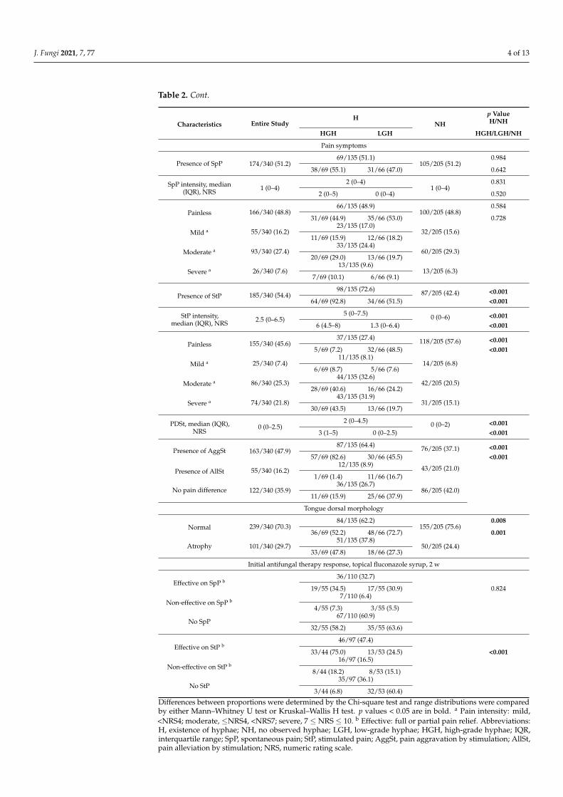
Table 2. Cont.
Characteristics Entire StudyH
NH
p ValueH/NH
HGH LGH HGH/LGH/NH
Pain symptoms
Presence of SpP 174/340 (51.2)69/135 (51.1)
105/205 (51.2)0.984
38/69 (55.1) 31/66 (47.0) 0.642
SpP intensity, median(IQR), NRS 1 (0–4)
2 (0–4)1 (0–4)
0.831
2 (0–5) 0 (0–4) 0.520
Painless 166/340 (48.8)66/135 (48.9)
100/205 (48.8)0.584
31/69 (44.9) 35/66 (53.0) 0.728
Mild a 55/340 (16.2)23/135 (17.0)
32/205 (15.6)11/69 (15.9) 12/66 (18.2)
Moderate a 93/340 (27.4)33/135 (24.4)
60/205 (29.3)20/69 (29.0) 13/66 (19.7)
Severe a 26/340 (7.6)13/135 (9.6)
13/205 (6.3)7/69 (10.1) 6/66 (9.1)
Presence of StP 185/340 (54.4)98/135 (72.6) 87/205 (42.4) <0.001
64/69 (92.8) 34/66 (51.5) <0.001
StP intensity,median (IQR), NRS 2.5 (0–6.5)
5 (0–7.5) 0 (0–6) <0.0016 (4.5–8) 1.3 (0–6.4) <0.001
Painless 155/340 (45.6)37/135 (27.4) 118/205 (57.6) <0.001
5/69 (7.2) 32/66 (48.5) <0.001
Mild a 25/340 (7.4)11/135 (8.1)
14/205 (6.8)6/69 (8.7) 5/66 (7.6)
Moderate a 86/340 (25.3)44/135 (32.6)
42/205 (20.5)28/69 (40.6) 16/66 (24.2)
Severe a 74/340 (21.8)43/135 (31.9)
31/205 (15.1)30/69 (43.5) 13/66 (19.7)
PDSt, median (IQR),NRS
0 (0–2.5)2 (0–4.5) 0 (0–2) <0.001
3 (1–5) 0 (0–2.5) <0.001
Presence of AggSt 163/340 (47.9)87/135 (64.4) 76/205 (37.1) <0.001
57/69 (82.6) 30/66 (45.5) <0.001
Presence of AllSt 55/340 (16.2)12/135 (8.9) 43/205 (21.0)
1/69 (1.4) 11/66 (16.7)
No pain difference 122/340 (35.9)36/135 (26.7)
86/205 (42.0)11/69 (15.9) 25/66 (37.9)
Tongue dorsal morphology
Normal 239/340 (70.3)84/135 (62.2)
155/205 (75.6)0.008
36/69 (52.2) 48/66 (72.7) 0.001
Atrophy 101/340 (29.7)51/135 (37.8)
50/205 (24.4)33/69 (47.8) 18/66 (27.3)
Initial antifungal therapy response, topical fluconazole syrup, 2 w
Effective on SpP b36/110 (32.7)
19/55 (34.5) 17/55 (30.9) 0.824
Non-effective on SpP b7/110 (6.4)
4/55 (7.3) 3/55 (5.5)
No SpP67/110 (60.9)
32/55 (58.2) 35/55 (63.6)
Effective on StP b46/97 (47.4)
33/44 (75.0) 13/53 (24.5) <0.001
Non-effective on StP b16/97 (16.5)
8/44 (18.2) 8/53 (15.1)
No StP35/97 (36.1)
3/44 (6.8) 32/53 (60.4)
Differences between proportions were determined by the Chi-square test and range distributions were comparedby either Mann–Whitney U test or Kruskal–Wallis H test. p values < 0.05 are in bold. a Pain intensity: mild,<NRS4; moderate, ≤NRS4, <NRS7; severe, 7 ≤ NRS ≤ 10. b Effective: full or partial pain relief. Abbreviations:H, existence of hyphae; NH, no observed hyphae; LGH, low-grade hyphae; HGH, high-grade hyphae; IQR,interquartile range; SpP, spontaneous pain; StP, stimulated pain; AggSt, pain aggravation by stimulation; AllSt,pain alleviation by stimulation; NRS, numeric rating scale.
J. Fungi 2021, 7, 77 5 of 13
3.2. Specific Characteristics of Candida Infection
Table 2 shows the clinical characteristics of the final dataset based on Candida hyphaegrade. The overall percentages of patients showing moderate and severe StP intensitywere significantly higher in the HGH group than in the LGH or NH group (40.6 and 43.5%,24.2 and 19.7%, 20.5 and 15.1%, respectively, p < 0.001). The rate of pain aggravation bystimulation (AggSt) was significantly higher in the HGH group compared to the LGH andNH groups (82.6, 45.5 and 37.1%, respectively, p < 0.001). Moreover, a higher proportion ofpain alleviation by stimulation (AllSt) was seen in the LGH and NH groups (16.7 and 21.0%,respectively), whereas there was only a single AllSt case (1.4%) in the HGH group. Clinicalcharacteristics based on hyphae grade revealed that moderate/severe StP intensity andAggSt, but not SpP, were significant in the HGH group; these were assumed to be specificcharacteristics of non-pseudomembranous subtypes of OC. This was supported by thedifferences in antifungal response between the hyphae groups. Initial topical fluconazoleuse for 2 weeks was more significantly effective in the HGH group than the LGH groupfor StP relief (p < 0.001), but not SpP relief (p = 0.502). Statistically, however, clinicalcharacteristics had a similar distribution in the LGH and NH groups, implying that thehyphae infection in the LHG group reflected a subclinical OC state.
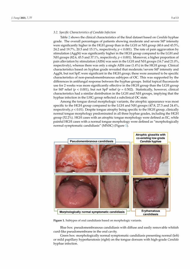
Among the tongue dorsal morphologic variants, the atrophic appearance was mostspecific to the HGH group compared to the LGH and NH groups (47.8, 27.3 and 24.4%,respectively, p < 0.01). Despite tongue atrophy being specific to the HGH group, clinicallynormal tongue morphology predominated in all three hyphae grades, including the HGHgroup (52.2%). HGH cases with an atrophic tongue morphology were defined as EC, whilepainful HGH cases with a normal tongue morphology were defined as “morphologicallynormal symptomatic candidiasis” (MNSC) (Figure 1).
J. Fungi 2021, 7, x FOR PEER REVIEW 5 of 13
bold. a Pain intensity: mild, <NRS4; moderate, NRS4≤, <NRS7; severe, 7 ≤ NRS ≤ 10. b Effective: full
or partial pain relief. Abbreviations: H, existence of hyphae; NH, no observed hyphae; LGH, low-
grade hyphae; HGH, high-grade hyphae; IQR, interquartile range; SpP, spontaneous pain; StP,
stimulated pain; AggSt, pain aggravation by stimulation; AllSt, pain alleviation by stimulation;
NRS, numeric rating scale.
3.2. Specific Characteristics of Candida Infection
Table 2 shows the clinical characteristics of the final dataset based on Candida hyphae
grade. The overall percentages of patients showing moderate and severe StP intensity
were significantly higher in the HGH group than in the LGH or NH group (40.6 and
43.5%, 24.2 and 19.7%, 20.5 and 15.1%, respectively, p < 0.001). The rate of pain aggravation
by stimulation (AggSt) was significantly higher in the HGH group compared to the LGH
and NH groups (82.6, 45.5 and 37.1%, respectively, p < 0.001). Moreover, a higher propor-
tion of pain alleviation by stimulation (AllSt) was seen in the LGH and NH groups (16.7
and 21.0%, respectively), whereas there was only a single AllSt case (1.4%) in the HGH
group. Clinical characteristics based on hyphae grade revealed that moderate/severe StP
intensity and AggSt, but not SpP, were significant in the HGH group; these were assumed
to be specific characteristics of non-pseudomembranous subtypes of OC. This was sup-
ported by the differences in antifungal response between the hyphae groups. Initial topi-
cal fluconazole use for 2 weeks was more significantly effective in the HGH group than
the LGH group for StP relief (p < 0.001), but not SpP relief (p = 0.502). Statistically, however,
clinical characteristics had a similar distribution in the LGH and NH groups, implying
that the hyphae infection in the LHG group reflected a subclinical OC state.
Among the tongue dorsal morphologic variants, the atrophic appearance was most
specific to the HGH group compared to the LGH and NH groups (47.8, 27.3 and 24.4%,
respectively, p < 0.01). Despite tongue atrophy being specific to the HGH group, clinically
normal tongue morphology predominated in all three hyphae grades, including the HGH
group (52.2%). HGH cases with an atrophic tongue morphology were defined as EC, while
painful HGH cases with a normal tongue morphology were defined as “morphologically
normal symptomatic candidiasis” (MNSC) (Figure 1).
Figure 1. Subtypes of oral candidiasis based on morphologic variants. Figure 1. Subtypes of oral candidiasis based on morphologic variants.
Blue box: pseudomembranous candidiasis with diffuse and easily removable whitishcurd-like pseudomembrane in the oral cavity.
Green box: morphologically normal symptomatic candidiasis presenting normal (left)or mild papillary hyperkeratosis (right) on the tongue dorsum with high-grade Candidahyphae infection.
J. Fungi 2021, 7, 77 6 of 13
Yellow box: Atrophic glossitis with diffuse severe tongue papilla atrophy, low-gradeCandida hyphae infection and accompanying systemic disorders (low red blood cell count,low hemoglobin level and vitamin B12 deficiency). The mucosal atrophy or moderateintensity of stimulated pain were not relieved by topical anti-fungal agent (fluconazole)use in this patient.
Orange box: erythematous candidiasis presenting partial tongue atrophy with high-grade Candida hyphae infection.
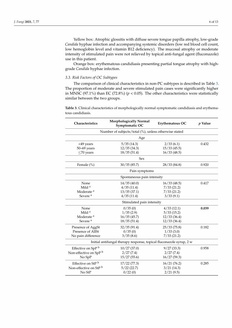
3.3. Risk Factors of OC Subtypes
The comparison of clinical characteristics in non-PC subtypes is described in Table 3.The proportion of moderate and severe stimulated pain cases were significantly higherin MNSC (97.1%) than EC (72.8%) (p < 0.05). The other characteristics were statisticallysimilar between the two groups.
Table 3. Clinical characteristics of morphologically normal symptomatic candidiasis and erythema-tous candidiasis.
Characteristics Morphologically NormalSymptomatic OC Erythematous OC p Value
Number of subjects/total (%), unless otherwise stated
Age
<49 years 5/35 (14.3) 2/33 (6.1) 0.43250–69 years 12/35 (34.3) 15/33 (45.5)≤70 years 18/35 (51.4) 16/33 (48.5)
Sex
Female (%) 30/35 (85.7) 28/33 (84.8) 0.920
Pain symptoms
Spontaneous pain intensity
None 14/35 (40.0) 16/33 (48.5) 0.417Mild a 4/35 (11.4) 7/33 (21.2)
Moderate a 13/35 (37.1) 7/33 (21.2)Severe a 4/35 (11.4) 3/33 (9.1)
Stimulated pain intensity
None 0/35 (0) 4/33 (12.1) 0.039Mild a 1/35 (2.9) 5/33 (15.2)
Moderate a 16/35 (45.7) 12/33 (36.4)Severe a 18/35 (51.4) 12/33 (36.4)
Presence of AggSt 32/35 (91.4) 25/33 (75.8) 0.182Presence of AllSt 0/35 (0) 1/33 (3.0)
No pain difference 3/35 (8.6) 7/33 (21.2)
Initial antifungal therapy response, topical fluconazole syrup, 2 w
Effective on SpP b 10/27 (37.0) 9/27 (33.3) 0.958Non-effective on SpP b 2/27 (7.4) 2/27 (7.4)
No SpP 15/27 (55.6) 16/27 (59.3)
Effective on StP b 17/22 (77.3) 16/21 (76.2) 0.285Non-effective on StP b 5/22 (22.7) 3/21 (14.3)
No StP 0/22 (0) 2/21 (9.5)
J. Fungi 2021, 7, 77 7 of 13
Table 3. Cont.
Characteristics Morphologically NormalSymptomatic OC Erythematous OC p Value
Accompanied conditions
Smoking c 1/32 (3.1) 1/32 (3.1) 0.982Alcohol c 1/32 (3.1) 0/32 (0) 0.313Denture c 12/32 (37.5) 10/29 (34.5) 0.806
Hypertension 15/35 (42.9) 9/33 (27.3) 0.179Diabetes mellitus 7/35 (20.0) 4/33 (12.1) 0.378
Hepatitis B 1/35 (2.9) 0/33 (0) 0.328
Salivary flow rate and presence of hyposecretion
Low USFR c 21/31 (67.7) 16/26 (61.5) 0.625Low SSFR c 25/31 (80.6) 17/26 (65.4) 0.193
Complete blood counts
Low RBC c 6/28 (21.4) 7/28 (25.0) 0.752Low hemoglobin c 5/28 (17.9) 8/28 (28.6) 0.342
Nutrition blood levels
Vitamin B12 def c 0/25 (0) 1/24 (4.2) 0.302Folate def c 0/25 (0) 0/24 (0) . . .Zinc def c 1/23 (4.3) 3/24 (12.5) 0.317
Albumin def c 1/26 (3.8) 1/28 (3.6) 0.957Serum iron def c 3/21 (14.3) 3/18 (16.7) 1.000
Differences between proportions were determined by the chi square test and range distributions were comparedby either Mann–Whitney U test or Kruskal–Wallis H test. p values <0.05 are in bold. a Pain intensity: mild,<NRS4; moderate, ≤NRS4, <NRS7; severe, 7 ≤ NRS ≤ 10. b Effective: full or partial pain relief. c Standard:denture, full or partial; smoking, daily; alcohol, consumption over 3 times/week; USFR, <0.1 mL/min; SSFR,<0.7 mL/min; RBC, F: <3.8 (106/µL), M: <4.4; (106/µL); hemoglobin, F: <11.7 (g/dL), M: <13.0 (g/dL); vitaminB12, <211 (pg/mL); folate, <3.1 (ng/mL); zinc, <66 (µg/dL); albumin, <3.3 (g/dL); serum iron, <40 (µg/dL).Abbreviations: SpP, spontaneous pain; StP, stimulated pain; AggSt, pain aggravation by stimulation; AllSt, painalleviation by stimulation; USFR, unstimulated salivary flow rate; SSFR, stimulated salivary flow rate; RBC, redblood cell count; F, female; M, male; def, deficiency; IQR, interquartile range.
Logistic regression of risk factors in MNSC and EC are detailed in Table 4. Multiplelogistic regression models for non-PC subtypes revealed that low unstimulated salivaryflow rate, but not stimulated salivary flow rate, was a significant risk factor in both MNSC(OR 5.3, 95% CI 1.8–15.4, p < 0.01) and EC (OR 8.2, 95% CI 2.3–28.9, p < 0.01) models.Denture was a significant risk factor only in the MNSC model (OR 4.9, 95% CI 1.5–15.6,p < 0.01). Other systemic factors, such as complete blood counts and nutrition blood levels,were not risk factors of either MNSC or EC.
Table 4. Logistic regression of risk factors in morphologically normal symptomatic candidiasis anderythematous candidiasis.
Variates aMorphologically Normal
Symptomatic OC Erythematous OC
Odds Ratio (95% CI) p Value Odds Ratio (95% CI) p Value
Simple logistic regression
Age 2.1 (1.2–3.8) 0.009 2.5 (1.4–4.7) 0.003
Sex 1.7 (0.6–4.7) 0.281 1.6 (0.6–4.4) 0.347
Smoking 0.4 (0.1–3.4) 0.418 0.4 (0.1–3.3) 0.401
Alcohol 0.5 (0.1–3.7) 0.464 . . .
Denture 5.3 (2.3–12.5) <0.001 4.7 (1.9–11.5) 0.001
Hypertension 1.9 (0.9–4.0) 0.087 1.0 (0.4–2.2) 0.904
J. Fungi 2021, 7, 77 8 of 13
Table 4. Cont.
Variates aMorphologically Normal
Symptomatic OC Erythematous OC
Odds Ratio (95% CI) p Value Odds Ratio (95% CI) p Value
Diabetes mellitus 1.7 (0.7–4.3) 0.250 1.0 (0.3–2.9) 0.928
Hepatitis B 2.0 (0.2–19.6) 0.559 . . .
Low USFR 6.6 (2.8–15.4) <0.001 5.0 (2.1–12.1) <0.001
Low SSFR 4.1 (1.6–10.6) 0.004 1.9 (0.8–4.5) 0.163
Low RBC 2.1 (0.7–5.6) 0.163 2.5 (1.0–6.6) 0.063
Low hemoglobin 2.6 (0.8–7.8) 0.097 4.7 (1.8–12.6) 0.002
Vit B12 def . . . 3.6 (0.3–41.1) 0.305
Folate def . . . . . .
Zinc def 0.2 (0.03–1.7) 0.150 0.7 (0.2–2.5) 0.582
Albumin def 7.1 (0.4–116.8) 0.171 6.6(0.4–107.9) 0.188
Iron def 8.4 (1.6–44.7) 0.013 8.4 (1.6–44.7) 0.013
Multiple logistic regression
Denture 4.9 (1.5–15.6) 0.007 3.3 (1.0–10.9) 0.054
Low USFR 5.3 (1.8–15.4) 0.002 8.2 (2.3–28.9) 0.001
Low SSFR 1.8 (0.5–6.0) 0.342 0.5 (0.1–1.8) 0.297
Low hemoglobin 2.7 (0.6–12.2) 0.205 2.5 (0.6–11.6) 0.231
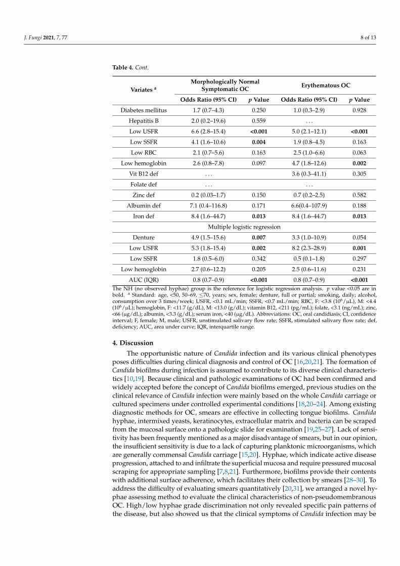
AUC (IQR) 0.8 (0.7–0.9) <0.001 0.8 (0.7–0.9) <0.001The NH (no observed hyphae) group is the reference for logistic regression analysis. p value <0.05 are inbold. a Standard: age, <50, 50–69, ≤70, years; sex, female; denture, full or partial; smoking, daily; alcohol,consumption over 3 times/week; USFR, <0.1 mL/min; SSFR, <0.7 mL/min; RBC, F: <3.8 (106/µL), M: <4.4(106/µL); hemoglobin, F: <11.7 (g/dL), M: <13.0 (g/dL); vitamin B12, <211 (pg/mL); folate, <3.1 (ng/mL); zinc,<66 (µg/dL); albumin, <3.3 (g/dL); serum iron, <40 (µg/dL). Abbreviations: OC, oral candidiasis; CI, confidenceinterval; F, female; M, male; USFR, unstimulated salivary flow rate; SSFR, stimulated salivary flow rate; def,deficiency; AUC, area under curve; IQR, interquartile range.
4. Discussion
The opportunistic nature of Candida infection and its various clinical phenotypesposes difficulties during clinical diagnosis and control of OC [16,20,21]. The formation ofCandida biofilms during infection is assumed to contribute to its diverse clinical characteris-tics [10,19]. Because clinical and pathologic examinations of OC had been confirmed andwidely accepted before the concept of Candida biofilms emerged, previous studies on theclinical relevance of Candida infection were mainly based on the whole Candida carriage orcultured specimens under controlled experimental conditions [18,20–24]. Among existingdiagnostic methods for OC, smears are effective in collecting tongue biofilms. Candidahyphae, intermixed yeasts, keratinocytes, extracellular matrix and bacteria can be scrapedfrom the mucosal surface onto a pathologic slide for examination [19,25–27]. Lack of sensi-tivity has been frequently mentioned as a major disadvantage of smears, but in our opinion,the insufficient sensitivity is due to a lack of capturing planktonic microorganisms, whichare generally commensal Candida carriage [15,20]. Hyphae, which indicate active diseaseprogression, attached to and infiltrate the superficial mucosa and require pressured mucosalscraping for appropriate sampling [7,8,21]. Furthermore, biofilms provide their contentswith additional surface adherence, which facilitates their collection by smears [28–30]. Toaddress the difficulty of evaluating smears quantitatively [20,31], we arranged a novel hy-phae assessing method to evaluate the clinical characteristics of non-pseudomembranousOC. High/low hyphae grade discrimination not only revealed specific pain patterns ofthe disease, but also showed us that the clinical symptoms of Candida infection may be
J. Fungi 2021, 7, 77 9 of 13
non-specific and indistinguishable from other painful or erythematous soft tissue lesions atthe low hyphae grade phase.
In addition to the classic PC and EC subtypes, we characterized a previously neglectedsubtype of OC, provisionally termed MNSC. Although a previous study has noted a fewcases of normal tongue appearance in healthy humans with observable Candida hyphae [18],this is the first study to suggest that normal tongue morphology in conjunction with painsymptoms is a definite clinical feature in OC. Since OC classification has been primarilyclinical-morphology-based, this subtype has gone unheeded in the clinic and may bemisdiagnosed as other painful oral diseases (e.g., burning mouth syndrome) with co-existing commensal Candida [16,17,32,33]. Contrary to conventional classification criteria,we believe that specific pain symptoms and hyphae grade are the major diagnostic factorsfor OC, rather than mucosal morphology. Although a previous study reported painfulOC without external abnormalities, the evaluation had been based on the whole Candidacarriage without discrimination of the disease phenotype, hyphae [34].
Candida-induced pain has been ambiguously described in the literature, such as burn-ing, sensitive or painful, mostly without clear distinction between SpP and StP [16,20,35].The association of StP and Candida carriage has been noticed in previous studies, but thecorrelation between StP and active Candida hyphae infection or soft tissue Candida biofilmshas not been examined before [33,34]. Our findings indicated moderate to severe StP as aspecific pain characteristic of Candida hyphae infection in the tongue biofilm. Moreover,SpP or AllSt, which are known features of burning mouth syndrome or atrophic glossitiscaused by nutritional deficiencies, did not show association with high-grade Candida hy-phae infection [36–39]. Consistent with our results, other studies have reported antifungaltreatment to be ineffective in patients with mainly SpP [33,34]. Altogether, these suggestthat concrete discrimination of pain features is informative for clinical differentiation ofOC. Interestingly, we observed that MNSC had a more advanced degree of StP intensitythan EC. Moreover, there were no painless patients in MNSC, unlike EC. This implied thatmucosal atrophy or erythema may not be the major cause of Candida-induced pain.
Factors associated with morphologic phenotype diversity are poorly understood inOC. As specific pain symptoms are universal in non-pseudomembranous OC, we furtherinvestigated factors associated with clinical morphology in Candida infection. Conspic-uous hyphae transition and proliferation have been observed in all PC, EC and MNSC,although the degree of direct hyphae/keratinocyte infiltration may be relatively indistinctin EC [21,37,40,41]. Candida hyphae are known to induce increased keratin differentiationand pseudomembrane formation [3,21,40], but whether they are responsible for contrastingclinical features of mucosal atrophy or normal differentiation is an intricate issue and rathera paradox. Despite that the presence of tongue atrophy and pain may suggest Candida in-fection, atrophic glossitis can arise from other conditions including salivary hyposecretion,anemia and nutritional deficiencies [17,35–39]. Possible local and systemic risk factors ofmorphological non-PC subtypes did not differ except for denture use. Although dentureuse was not statistically significant in EC, it showed a strong tendency as a risk factor aswell. These results indicated that co-existing anemia or nutritional deficiencies had minimalimpact on atrophic or erythematous morphology in definite OC with high-grade hyphae.
Other than clinical factors, the contents of Candida biofilm may contribute to OCmorphology phenotypes. Although our retrospective study focused mainly on the clinicalrelevance of the hyphae content within the smeared tongue material, tongue smears canprovide a comprehensive profile of non-hyphae contents in the biofilm. The compositionof extracellular matrix, immune cells and bacteria and their interactions with Candidaorganisms within the biofilm may influence the differentiation of underlying epithelialcells [11,27,29,42,43]. Further study of non-Candida biofilm contents may lead to systemicinsights into the clinical relevance of Candida infection.
Various local or systemic conditions have been suggested as causative or predispos-ing factors of OC [40–43]. These factors may directly affect fungal microorganisms ormay induce an immunosuppressive state or biofilm formation that can enhance Candida
J. Fungi 2021, 7, 77 10 of 13
pathogenesis [42,44–48]. In general, many patients have more than one possible risk factorfor OC, and the degree of the risk conditions vary among individuals. Furthermore, theassociation between Candida and possible risk factors can differ based on the method usedfor fungus measurement, as Candida has a dimorphic presence.
Candida infection related with denture use is called denture stomatitis. It typicallyarises on the palate mucosa in contact with the acrylic surface of maxillary dentures [48–51].Candida-induced denture stomatitis is presented as red, atrophic and sore mucosa at theinterface of the denture surface and soft tissue. Co-associated bacteria species have beensuggested to aid Candida biofilm formation in dentures [52–57]. Dentures were significantlyinvolved with MNSC showing normal tongue mucosa in this study. Although erythemaand atrophy are characteristic in denture stomatitis, they do not seem to be significantin the tongue mucosa of denture users. The tongue does not have a tight and intimatecontact with the denture surface as seen in the palate or gingiva, thus the associationbetween dentures and the tongue Candida biofilm is suspected to be indirect. Dentures maycontribute as a reservoir for increased oral Candida carriage that can lead to Candida hyphaeand biofilm formation at the tongue in appropriate conditions.
Hyposalivation is one of the most mentioned risk factors of OC. Candida carriageshave been commonly observed in xerostomia [52,56,57]. Saliva contains antifungal proteinsand antibodies that inhibit Candida adhesion and colonization and stimulates an innateimmune response against Candida microorganisms [52,57,58]. We identified that reducedunstimulated saliva, but not stimulated saliva, was a common risk factor in MNSC andEC multiple regression models. Other studies observed that Candida count had an inverserelation with both stimulated and unstimulated salivary flow, but the investigations werebased on planktonic fungal carriage collected from saliva [24,48,49,58]. Reduced stimulatedand unstimulated saliva have different molecule compositions [59], but their pathogeniceffect on Candida colonization and biofilm formation are not known.
Smoking, alcohol consumption, hypertension, diabetes mellitus and hepatitis B werenot risk factors for non-PC subtypes in this study. Chemicals in cigarette smoke has beenproposed to induce Candida infection and biofilm formation in vitro, but its affects inclinical settings are controversial [18,60–62]. Candida has been suspected to be related todiabetes in both adults and children [63–67]. In contrast to our results, Bartholomew et al.observed that invasive Candida was more frequently identified in diabetic patients thannon-diabetic subjects by oral cytologic smears [64]. Sato et al. reported that hypertensionand daily alcohol intake were associated with greater carriage of Candida albicans in theelderly resided at the post-disaster region, but this was not seen in our results based oninvasive Candida [68].
5. Conclusions
New insights on Candida biofilm formation during hyphae transition and infiltrationhave brought up the need for clinical assessment based on invasive hyphae within thebiofilm. In this study, we evaluated the clinical relevance of Candida infection in smearedtongue biofilms based on our novel hyphae grade method. We discovered that stimu-lated pain was a critical and specific characteristic, while tongue mucosal atrophy was asignificant but not a specific characteristic in non-PC subtypes. Importantly, we report amorphologically normal, yet symptomatic, OC subtype named “morphologically normalsymptomatic candidiasis” as a common non-PC subtype. The morphological differencesof MNSC and EC were thought to be minimally affected by clinical risk factors; instead,other factors such as non-fungal components of the Candida biofilm may contribute to thediversity of morphologic phenotypes in non-PC subtypes.
Supplementary Materials: The following are available online at https://www.mdpi.com/2309-608X/7/2/77/s1, Figure S1: Preliminary data of clinical characteristic distribution by total hyphae value,Table S1: Sensitivity, specificity and accuracy of hyphae value candidates on preliminary data.
J. Fungi 2021, 7, 77 11 of 13
Author Contributions: Conceptualization, E.C., J.-S.K., H.-J.A.; methodology, E.C., Y.P., K.-Y.K.;validation, H.S.K., K.-Y.K.; investigation, E.C., Y.P.; data curation, E.C., Y.P., D.H., H.S.K.; writing—original draft preparation, E.C., Y.P.; writing—review and editing, J.-S.K., H.-J.A.; supervision, J.-S.K.,H.-J.A.; funding acquisition, K.-Y.K., H.-J.A. All authors have read and agreed to the publishedversion of the manuscript.
Funding: This work was supported by the National Research Foundation of Korea (NRF) grantfunded by the Korea government (MSIP) (NRF-2016R1A5A2008630, NRF-2019R1A2C1003028) andthe BK21 FOUR Project, Yonsei University College of Dentistry.
Institutional Review Board Statement: The study was conducted according to the guidelines of theDeclaration of Helsinki and approved by the Institutional Review Board of Yonsei University DentalHospital (2-2017-0001).
Informed Consent Statement: Patient consent was waived due to its minimal risk to the subjectsand that all the retrospective information had been obtained during clinical procedures regardless ofresearch purpose.
Data Availability Statement: The data presented in this study are available on request from thecorresponding author. The data are not publicly available due to the privacy issues.
Acknowledgments: We thank Eph Tunkle for editing and proofreading of the manuscript.
Conflicts of Interest: The authors declare no conflict of interest. The funders had no role in the designof the study; in the collection, analyses, or interpretation of data; in the writing of the manuscript, orin the decision to publish the results.
References1. Thompson, D.S.; Carlisle, P.L.; Kadosh, D. Coevolution of morphology and virulence in Candida species. Eukaryot Cell 2011, 10,
1173–1182. [CrossRef] [PubMed]2. Sudbery, P.E. Growth of Candida albicans hyphae. Nat. Rev. Microbiol. 2011, 9, 737–748. [CrossRef] [PubMed]3. Rollenhagen, C.; Wöllert, T.; Langford, G.M.; Sundstrom, P. Stimulation of cell motility and expression of late markers of
differentiation in human oral keratinocytes by Candida albicans. Cell Microbiol. 2009, 11, 946–966. [CrossRef] [PubMed]4. Ene, I.V.; Bennett, R.J. Hwp1 and related adhesins contribute to both mating and biofilm formation in Candida albicans. Eukaryot
Cell 2009, 8, 1909–1913. [CrossRef]5. Nobile, C.J.; Mitchell, A.P. Genetics and genomics of Candida albicans biofilm formation. Cell Microbiol. 2006, 8, 1382–1391.
[CrossRef]6. Ramage, G.; VandeWalle, K.; López-Ribot, J.L.; Wickes, B.L. The filamentation pathway controlled by the Efg1 regulator protein is
required for normal biofilm formation and development in Candida albicans. FEMS Microbiol. Lett. 2002, 214, 95–100. [CrossRef]7. Yang, W.; Yan, L.; Wu, C.; Zhao, X.; Tang, J. Fungal invasion of epithelial cells. Microbiol. Res. 2014, 169, 803–810. [CrossRef]8. Richardson, J.P.; Ho, J.; Naglik, J.R. Candida-epithelial interactions. J. Fungi 2018, 4, 22. [CrossRef]9. Lo, H.-J.; Köhler, J.R.; DiDomenico, B.; Loebenberg, D.; Cacciapuoti, A.; Fink, G.R. Nonfilamentous C. albicans mutants are
avirulent. Cell 1997, 90, 939–949. [CrossRef]10. Dongari-Bagtzoglou, A.; Kashleva, H.; Dwivedi, P.; Diaz, P.; Vasilakos, J. Characterization of mucosal Candida albicans biofilms.
PLoS ONE 2009, 4, e7967. [CrossRef]11. O’Donnell, L.E.; Millhouse, E.; Sherry, L.; Kean, R.; Malcolm, J.; Nile, C.J.; Ramage, G. Polymicrobial Candida biofilms: Friends
and foe in the oral cavity. FEMS Yeast Res. 2015, 15. [CrossRef] [PubMed]12. Ganguly, S.; Mitchell, A.P. Mucosal biofilms of Candida albicans. Curr. Opin. Microbiol. 2011, 14, 380–385. [CrossRef]13. Cavalheiro, M.; Teixeira, M.C. Candida biofilms: Threats, challenges, and promising strategies. Front. Med. 2018, 5, 28. [CrossRef]
[PubMed]14. Wu, X.; Zhang, S.; Li, H.; Shen, L.; Dong, C.; Sun, Y.; Chen, H.; Xu, B.; Zhuang, W.; Deighton, M.; et al. Biofilm formation of
Candida albicans facilitates fungal infiltration and persister cell formation in vaginal candidiasis. Front. Microbiol. 2020, 11.[CrossRef] [PubMed]
15. Hawser, S.P.; Douglas, L.J. Biofilm formation by Candida species on the surface of catheter materials in vitro. Infect. Immun. 1994,62, 915–921. [CrossRef]
16. Lynch, D.P. Oral candidiasis. History, classification, and clinical presentation. Oral Surg. Oral Med. Oral Pathol. 1994, 78, 189–193.[CrossRef]
17. Holmstrup, P.; Axéll, T. Classification and clinical manifestations of oral yeast infections. Acta Odontol. Scand. 1990, 48, 57–59.[CrossRef]
18. Arendorf, T.M.; Walker, D.M. The prevalence and intra-oral distribution of Candida albicans in man. Arch. Oral Biol. 1980, 25,1–10. [CrossRef]
J. Fungi 2021, 7, 77 12 of 13
19. Harriott, M.M.; Noverr, M.C. Importance of Candida-bacterial polymicrobial biofilms in disease. Trends Microbiol. 2011, 19,557–563. [CrossRef]
20. Williams, D.W.; Lewis, M.A. Isolation and identification of Candida from the oral cavity. Oral Dis. 2000, 6, 3–11. [CrossRef]21. Tooyama, H.; Matsumoto, T.; Hayashi, K.; Kurashina, K.; Kurita, H.; Uchida, M.; Kasuga, E.; Honda, T. Candida concentrations
determined following concentrated oral rinse culture reflect clinical oral signs. BMC Oral Health 2015, 15, 150. [CrossRef]22. White, P.L.; Williams, D.W.; Kuriyama, T.; Samad, S.A.; Lewis, M.A.O.; Barnes, R.A. Detection of Candida in concentrated oral
rinse cultures by real-time PCR. J. Clin. Microbiol. 2004, 42, 2101–2107. [CrossRef]23. Epstein, J.B.; Pearsall, N.N.; Truelove, E.L. Quantitative relationships between Candida albicans in saliva and the clinical status of
human subjects. J. Clin. Microbiol. 1980, 12, 475–476. [CrossRef]24. Torres, S.R.; Peixoto, C.B.; Caldas, D.M.; Silva, E.B.; Magalhães, F.A.; Uzeda, M.; Nucci, M. Clinical aspects of Candida species
carriage in saliva of xerotomic subjects. Med. Mycol. 2003, 41, 411–415. [CrossRef]25. Rodrigues, M.E.; Gomes, F.; Rodrigues, C.F. Candida spp./bacteria mixed biofilms. J. Fungi 2019, 6, 5. [CrossRef]26. Chandra, J.; Kuhn, D.M.; Mukherjee, P.K.; Hoyer, L.L.; McCormick, T.; Ghannoum, M.A. Biofilm formation by the fungal pathogen
Candida albicans: Development, architecture, and drug resistance. J. Bacteriol. 2001, 183, 5385–5394. [CrossRef]27. Hawser, S.P.; Baillie, G.S.; Douglas, L.J. Production of extracellular matrix by Candida albicans biofilms. J. Med. Microbiol. 1998,
47, 253–256. [CrossRef]28. Nobile, C.J.; Schneider, H.A.; Nett, J.E.; Sheppard, D.C.; Filler, S.G.; Andes, D.R.; Mitchell, A.P. Complementary adhesin function
in C. albicans biofilm formation. Curr. Biol. 2008, 18, 1017–1024. [CrossRef]29. Soll, D.R. Candida biofilms: Is adhesion sexy? Curr. Biol. 2008, 18, R717–R720. [CrossRef]30. McCall, A.D.; Pathirana, R.U.; Prabhakar, A.; Cullen, P.J.; Edgerton, M. Candida albicans biofilm development is governed by
cooperative attachment and adhesion maintenance proteins. NPJ Biofilms Microbiomes 2019, 5, 21. [CrossRef]31. Byadarahally Raju, S.; Rajappa, S. Isolation and identification of Candida from the oral cavity. ISRN Dent. 2011, 2011, 487921.
[CrossRef] [PubMed]32. Osaki, T.; Yoneda, K.; Yamamoto, T.; Ueta, E.; Kimura, T. Candidiasis may induce glossodynia without objective manifestation.
Am. J. Med. Sci. 2000, 319, 100–105. [CrossRef]33. Terai, H.; Shimahara, M. Tongue pain: Burning mouth syndrome vs. Candida- associated lesion. Oral Dis. 2007, 13, 440–442.
[CrossRef]34. Terai, H.; Shimahara, M. Glossodynia from Candida-associated lesions, burning mouth syndrome, or mixed causes. Pain Med.
2010, 11, 856–860. [CrossRef] [PubMed]35. Sun, A.; Lin, H.P.; Wang, Y.P.; Chiang, C.P. Significant association of deficiency of hemoglobin, iron and vitamin B12, high
homocysteine level, and gastric parietal cell antibody positivity with atrophic glossitis. J. Oral Pathol. Med. 2012, 41, 500–504.[CrossRef] [PubMed]
36. Demir, N.; Dogan, M.; Koc, A.; Kaba, S.; Bulan, K.; Ozkol, H.U.; Dogan, S.Z. Dermatological findings of vitamin B12 deficiencyand resolving time of these symptoms. Cutan. Ocul. Toxicol. 2014, 33, 70–73. [CrossRef]
37. Bao, Z.X.; Yang, X.W.; Shi, J.; Liu, L.X. Serum zinc levels in 368 patients with oral mucosal diseases: A preliminary study. Med.Oral Patol. Oral Cir. Bucal 2016, 21, e335–e340. [CrossRef]
38. Lu, S.Y. Perception of iron deficiency from oral mucosa alterations that show a high prevalence of Candida infection. J. FormosMed. Assoc. 2016, 115, 619–627. [CrossRef]
39. Wu, Y.-C.; Wang, Y.-P.; Chang, J.Y.-F.; Cheng, S.-J.; Chen, H.-M.; Sun, A. Oral manifestations and blood profile in patients withiron deficiency anemia. J. Formos Med. Assoc. 2014, 113, 83–87. [CrossRef]
40. Singh, A.; Verma, R.; Murari, A.; Agrawal, A. Oral candidiasis: An overview. J. Oral Maxillofac. Pathol. 2014, 18, S81–S85.[CrossRef]
41. Martins, N.; Ferreira, I.C.; Barros, L.; Silva, S.; Henriques, M. Candidiasis: Predisposing factors, prevention, diagnosis andalternative treatment. Mycopathologia 2014, 177, 223–240. [CrossRef]
42. Cannon, R.D.; Holmes, A.R.; Mason, A.B.; Monk, B.C. Oral Candida: Clearance, colonization, or candidiasis? J. Dent. Res. 1995,74, 1152–1161. [CrossRef]
43. Vila, T.; Sultan, A.S.; Montelongo-Jauregui, D.; Jabra-Rizk, M.A. Oral candidiasis: A disease of opportunity. J. Fungi 2020, 6, 15.[CrossRef]
44. Netea, M.G.; Joosten, L.A.B.; van der Meer, J.W.M.; Kullberg, B.-J.; van de Veerdonk, F.L. Immune defence against Candida fungalinfections. Nat. Rev. Immunol. 2015, 15, 630–642. [CrossRef]
45. Fidel, P.L., Jr. Immunity to Candida. Oral Dis. 2002, 8 (Suppl. 2), 69–75. [CrossRef] [PubMed]46. Eix, E.F.; Nett, J.E. How biofilm growth affects Candida-host interactions. Front Microbiol. 2020, 11, 1437. [CrossRef] [PubMed]47. Heimdahl, A.; Nord, C.E. Oral yeast infections in immunocompromised and seriously diseased patients. Acta Odontol. Scand.
1990, 48, 77–84. [CrossRef]48. Nadig, S.D.; Ashwathappa, D.T.; Manjunath, M.; Krishna, S.; Annaji, A.G.; Shivaprakash, P.K. A relationship between salivary
flow rates and Candida counts in patients with xerostomia. J. Oral Maxillofac. Pathol. 2017, 21, 316. [CrossRef]49. Torres, S.R.; Peixoto, C.B.; Caldas, D.M.; Silva, E.B.; Akiti, T.; Nucci, M.; de Uzeda, M. Relationship between salivary flow
rates and Candida counts in subjects with xerostomia. Oral Surg. Oral Med. Oral Pathol. Oral Radiol. Endod. 2002, 93, 149–154.[CrossRef]
J. Fungi 2021, 7, 77 13 of 13
50. Karbach, J.; Walter, C.; Al-Nawas, B. Evaluation of saliva flow rates, Candida colonization and susceptibility of Candida strainsafter head and neck radiation. Clin. Oral Investig. 2012, 16, 1305–1312. [CrossRef]
51. Buranarom, N.; Komin, O.; Matangkasombut, O. Hyposalivation, oral health, and Candida colonization in independent dentateelders. PLoS ONE 2020, 15, e0242832. [CrossRef]
52. Kanaguchi, N.; Narisawa, N.; Ito, T.; Kinoshita, Y.; Kusumoto, Y.; Shinozuka, O.; Senpuku, H. Effects of salivary protein flowand indigenous microorganisms on initial colonization of Candida albicans in an in vivo model. BMC Oral Health 2012, 12, 36.[CrossRef] [PubMed]
53. Khan, S.A.; Fidel, P.L., Jr.; Thunayyan, A.A.; Varlotta, S.; Meiller, T.F.; Jabra-Rizk, M.A. Impaired Histatin-5 Levels and SalivaryAntimicrobial Activity against C. albicans in HIV Infected Individuals. J. AIDS Clin. Res. 2013, 4. [CrossRef] [PubMed]
54. Salvatori, O.; Puri, S.; Tati, S.; Edgerton, M. Innate immunity and saliva in Candida albicans-mediated oral diseases. J. Dent. Res.2016, 95, 365–371. [CrossRef]
55. Linden, S.K.; Sutton, P.; Karlsson, N.G.; Korolik, V.; McGuckin, M.A. Mucins in the mucosal barrier to infection. Mucosal Immunol.2008, 1, 183–197. [CrossRef] [PubMed]
56. Vila, T.; Rizk, A.M.; Sultan, A.S.; Jabra-Rizk, M.A. The power of saliva: Antimicrobial and beyond. PLoS Pathog. 2019, 15, e1008058.[CrossRef] [PubMed]
57. Lynge Pedersen, A.M.; Belstrøm, D. The role of natural salivary defences in maintaining a healthy oral microbiota. J. Dent. 2019,80, S3–S12. [CrossRef] [PubMed]
58. Navazesh, M.; Wood, G.J.; Brightman, V.J. Relationship between salivary flow rates and Candida albicans counts. Oral Surg. OralMed. Oral Pathol. Oral Radiol. Endod. 1995, 80, 284–288. [CrossRef]
59. Inui, T.; Palmer, R.J.; Shah, N.; Li, W.; Cisar, J.O.; Wu, C.D. Effect of mechanically stimulated saliva on initial human dental biofilmformation. Sci. Rep. 2019, 9, 11805. [CrossRef]
60. Soysa, N.S.; Ellepola, A.N. The impact of cigarette/tobacco smoking on oral candidosis: An overview. Oral Dis. 2005, 11, 268–273.[CrossRef]
61. Darwazeh, A.M.; Al-Dwairi, Z.N.; Al-Zwairi, A.A. The relationship between tobacco smoking and oral colonization with Candidaspecies. J. Contemp. Dent. Prac. 2010, 11, 017–024.
62. Muzurovic, S.; Hukic, M.; Babajic, E.; Smajic, R. The relationship between cigarette smoking and oral colonization with Candidaspecies in healthy adult subjects. Med. Glas. Off. Publ. Med. Assoc. Zenica Doboj Canton Bosnia Herzeg. 2013, 10, 397–399.
63. Olczak-Kowalczyk, D.; Pyrzak, B.; Dabkowska, M.; Panczyk-Tomaszewska, M.; Miszkurka, G.; Rogozinska, I.; Swoboda-Kopec,E.; Gozdowski, D.; Kalinska, A.; Piróg, A.; et al. Candida spp. and gingivitis in children with nephrotic syndrome or type 1diabetes. BMC Oral Health 2015, 15, 57. [CrossRef]
64. Bartholomew, G.A.; Rodu, B.; Bell, D.S. Oral candidiasis in patients with diabetes mellitus: A thorough analysis. Diabetes Care1987, 10, 607–612. [CrossRef]
65. Mohammadi, F.; Javaheri, M.R.; Nekoeian, S.; Dehghan, P. Identification of Candida species in the oral cavity of diabetic patients.Curr. Med. Mycol. 2016, 2, 1–7. [CrossRef]
66. Chouhan, S.; Kallianpur, S.; Prabhu, K.T.; Tijare, M.; Kasetty, S.; Gupta, S. Candidal prevalence in diabetics and its speciesidentification. Int. J. Appl. Basic Med. Res. 2019, 9, 49–54. [CrossRef]
67. Sampath, A.; Weerasekera, M.; Dilhari, A.; Gunasekara, C.; Bulugahapitiya, U.; Fernando, N.; Samaranayake, L. Type 2 diabetesmellitus and oral Candida colonization: Analysis of risk factors in a Sri Lankan cohort. Acta Odontol. Scand. 2019, 77, 508–516.[CrossRef]
68. Sato, T.; Kishi, M.; Suda, M.; Sakata, K.; Shimoda, H.; Miura, H.; Ogawa, A.; Kobayashi, S. Prevalence of Candida albicans andnon-albicans on the tongue dorsa of elderly people living in a post-disaster area: A cross-sectional survey. BMC Oral Health 2017,17, 51. [CrossRef]


![Prevalence and predictors of Haikael Martin oral thrush ... · Oral candidiasis (also known as “oral thrush”) is a fungal infection caused mainly by Candida albicans (c.alb) [1]](https://img.pdfslide.net/doc/110x75/5c96d57e09d3f26b0a8d0743/prevalence-and-predictors-of-haikael-martin-oral-thrush-oral-candidiasis.jpg)