Embed Size (px)
Citation preview

CLINICAL GUIDELINES
FOR THE MANAGEMENT
OF
CORONARY HEART DISEASE
Ministry of Health and Quality of Life/Mauritius Institute of Health/World Health Organisation 1

Table of Contents
1. INTRODUCTION 2. DEFINITION OF CORONARY ARTERY DISEASE 3. PATHOPHYSIOLOGY 4. GRADING OF ANGINA PECTORIS 5. CHRONIC STABLE ANGINA
5.1 Clinical Presentation 5.2 Examination and Investigation 5.3 Differential Diagnosis 5.4 Management 5.5 Follow-up 5.6 Indications for referral to cardiologist 5.7 Key points
6. ACUTE CORONARY SYNDROMES
6.1 Unstable Angina and non ST elevation acute myocardial infarction 6.1.1 Clinical Presentation 6.1.2 History, Examination and Investigations 6.1.3 Differential Diagnosis 6.1.4 Management 6.1.5 Follow-up and Second Prevention 6.1.6 Indications for Referral to Cardiologist 6.1.7 Key Points
6.2 Acute Coronary Syndromes – ST elevation acute myocardial infarction
6.2.1 Clinical Presentation 6.2.2 Examination and Investigations 6.2.3 Differential Diagnosis 6.2.4 Management 6.2.5 Follow-up & Rehabilitation 6.2.6 Post-infarction Evaluation & Secondary Prevention 6.2.7 Key Points
RECOMMENDATIONS
Ministry of Health and Quality of Life/Mauritius Institute of Health/World Health Organisation 2
1. INTRODUCTION
Of the cardiovascular diseases, acute myocardial infarction is the most common
cause of death. The incidence of acute myocardial infarction is estimated at
around 4,000/year in Mauritius, with a total mortality rate of 30% - 40% and a
25%-35% mortality rate before reaching the hospital. The underlying cause in
most cases is atherosclerotic coronary artery disease.
These clinical guidelines on the management of coronary artery disease have been
developed in this context in line with the decentralization and re-organization of
the Ministry of Health’s NCD programme.
They aim at improving the quality of health care through:
1. recommendations for active primary prevention of coronary artery disease
and its risk factors.
2. the improvement and standardization of the management of patients
suffering from coronary artery disease.
The recommendations made are, to a large extent, evidence-based.
The development of the guidelines is the combined effort of cardiologists, a
consultant in Internal Medicine and community physicians in association with the
Mauritius Institute of Health. It has sought consensus between the policy makers
at the Ministry of Health, cardiologists at hospital level and community
physicians at primary care level after having taken cognizance of local realities,
limitations and scope for further development.
Ministry of Health and Quality of Life/Mauritius Institute of Health/World Health Organisation 3

2. DEFINITION OF CORONARY ARTERY DISEASE
Coronary artery disease results from an imbalance between myocardial oxygen
supply and demand and is most commonly caused by the inability of
atherosclerotic coronary arteries to perfuse the heart due to partial or total
occlusion of the coronary arteries. Chronic angina pectoris is, by definition,
stable, i.e., the severity and/or frequency of chest pain is not increasing or
occurring at rest. Unstable angina, myocardial infarction and sudden ischaemic
death are also manifestations of chronic ischaemic heart disease, presenting as
acute coronary syndromes.
Ministry of Health and Quality of Life/Mauritius Institute of Health/World Health Organisation 4

3. PATHOPHYSIOLOGY
Myocardial ischaemia occurs when myocardial oxygen delivery cannot meet
metabolic myocardial demands. This discrepancy is termed supply-demand
mismatch. Although this term is overly simplistic, myocardial oxygen delivery is
mostly determined by the oxygen-carrying capacity of blood and coronary flow.
In normal coronary arteries, coronary blood flow can increase three to fivefold in
response to exercise. This increase, termed coronary flow reserve, occurs mostly
through decreased resistance in coronary microcirculation. Significant
atherosclerotic plaquing in epicardial coronary arteries (> 75% cross-sectional
area) results in a drop in blood pressure across the stenotic lesion. Coronary
arterioles dilate to compensate for the reduced distal perfusion pressure,
maintaining normal resting coronary blood flow. Consequently, at rest, most
patients with significant coronary artery stenosis obstructions have no ischaemia
and therefore no angina. During exercise, however, the capacity of coronary
arterioles to dilate further is limited, and the myocardial oxygen demand soon
outstrips the supply, resulting in ischaemia, followed usually by angina.
Acute coronary syndromes share a common pathophysiology: acute rupture
(fissuration or ulceration) of a lipid-rich intracoronary atheromatous plaque, with
subsequent mobilisation of a sequence of inflammatory and thrombotic cascades
culminating in the formation of thrombus.
The possible triggers of acute plaque rupture are: sheer stress, inflammation and
increased neurohormonal tone. Certain plaques are more susceptible to acute
rupture than others – for example those which are eccentric, with a lipid-rich
vulnerable core, a high content of active inflammatory cells, and a thin fibrous
cap. Rupture of the plaque (ulceration/fissuration), with exposure of the highly
thrombogenic contents to the bloodstream, initiates a cascade of cytokine release,
inflammatory cell activation and platelet activation, with resulting platelet
Ministry of Health and Quality of Life/Mauritius Institute of Health/World Health Organisation 5
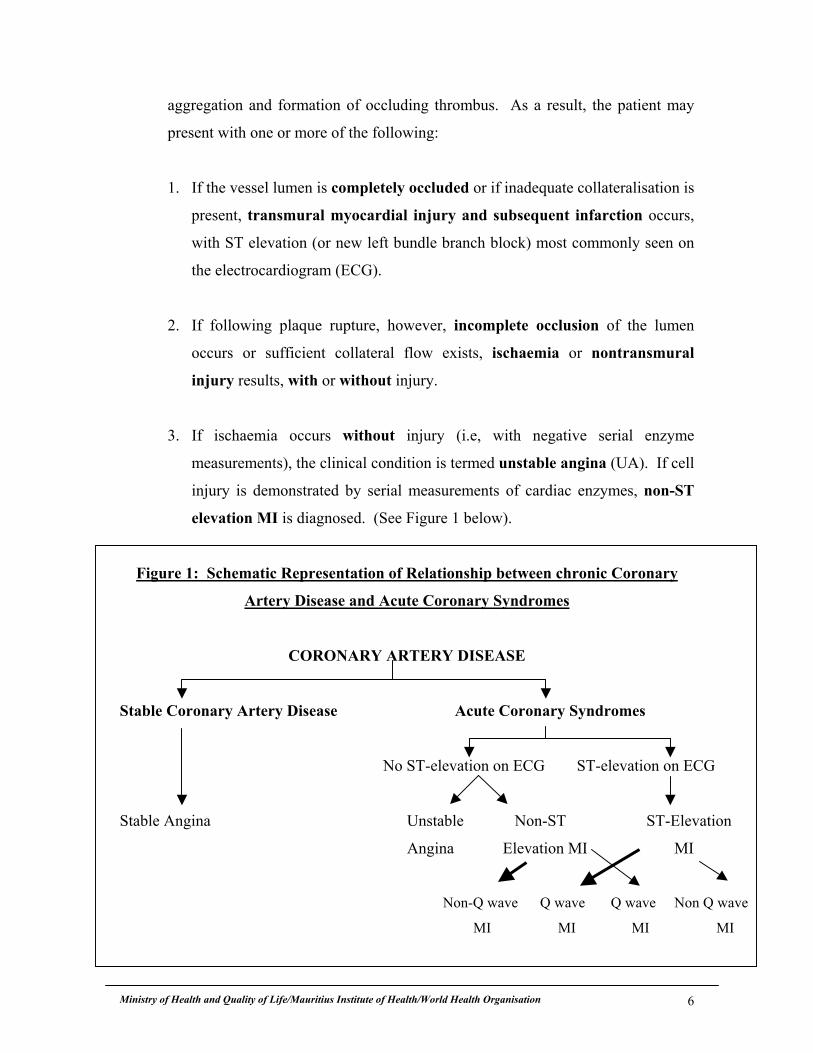
aggregation and formation of occluding thrombus. As a result, the patient may
present with one or more of the following:
1. If the vessel lumen is completely occluded or if inadequate collateralisation is
present, transmural myocardial injury and subsequent infarction occurs,
with ST elevation (or new left bundle branch block) most commonly seen on
the electrocardiogram (ECG).
2. If following plaque rupture, however, incomplete occlusion of the lumen
occurs or sufficient collateral flow exists, ischaemia or nontransmural
injury results, with or without injury.
3. If ischaemia occurs without injury (i.e, with negative serial enzyme
measurements), the clinical condition is termed unstable angina (UA). If cell
injury is demonstrated by serial measurements of cardiac enzymes, non-ST
elevation MI is diagnosed. (See Figure 1 below).
Figure 1: Schematic Representation of Relationship between chronic Coronary
Artery Disease and Acute Coronary Syndromes
CORONARY ARTERY DISEASE
Stable Coronary Artery Disease Acute Coronary Syndromes
No ST-elevation on ECG ST-elevation on ECG
Stable Angina Unstable Non-ST ST-Elevation
Angina Elevation MI MI
Non-Q wave Q wave Q wave Non Q wave
MI MI MI MI
Ministry of Health and Quality of Life/Mauritius Institute of Health/World Health Organisation 6

4. GRADING OF ANGINA PECTORIS
(According to the Canadian Cardiovascular Society
Classification System)
Class I: Ordinary physical activity (e.g. walking, climbing stairs) does not
cause angina.
Class II: Slight limitation of ordinary activity
Angina occurs on walking or climbing stairs rapidly, walking
uphill, walking or climbing stairs after meals or in cold, in wind,
under emotional stress, or during the few hours after awakening.
Angina occurs on walking more than two blocks on the level and
climbing more than one flight of ordinary stairs at a normal pace
and in normal condition.
Class III: Marked limitation of ordinary physical activity. Angina occurs on
walking one to two blocks on the level and climbing one flight of
stairs in normal conditions and at a normal pace.
Class IV: Inability to carry out any physical activity without discomfort –
anginal symptoms may be present at rest.
Ministry of Health and Quality of Life/Mauritius Institute of Health/World Health Organisation 7

5. CHRONIC STABLE ANGINA
5.1 CLINICAL PRESENTATION
Character: usually described more often as a discomfort, pressure, heaviness,
or squeezing sensation and less often as a pain. Sometimes
described as a burning or sharp sensation.
Location: substernal area, precordium or epigastrium with radiation to the
left arm, jaw or neck, less commonly felt only in radiation area and
not in the chest.
Precipitation: often provoked by exertion, cold weather, eating, smoking and
strong emotions. Relieved by rest, removal of provoking factors,
or sublingual nitrates.
Duration: few minutes usually, rarely more than 4-5 minutes.
History: description of the chest discomfort and its relationship to activity.
Past history of HBP, DM, hyperlipidaemia, or smoking or a family
history of premature IHD in lst degree relatives (age younger than
55 yrs).
Ministry of Health and Quality of Life/Mauritius Institute of Health/World Health Organisation 8

5.2 CLINICAL EXAMINATION AND INVESTIGATIONS
5.2.1 Clinical Examination
Often normal.
To look for confirmatory information, i.e., hypertension, peripheral artery disease,
xanthelasma, tendinous xanthomata, tobacco-stained fingers or teeth. Episodic
ischaemia alters left ventricular compliance, thus a transient S4, S3, may be heard
during an acute ischaemic episode.
5.2.2 Investigations
(i) Basic screening :
• Fasting blood glucose
• Serum lipids including high density lipoproteins (HDL) and
triglycerides
• Full blood count
• Blood urea and electrolytes
• Serum urates
(ii) ECG:
• Often normal in the absence of myocardial infarction or left ventricular
hypertrophy.
• An ECG during an episode of angina may show transient ST
depression, T wave inversion or ventricular arrythmias (VPB; isolated
or in runs, monomorph or polymorph). An ambulatory ECG (Holter)
may demonstrate ischaemic episodes with or without symptoms ‘Silent
Ischaemia’.
Ministry of Health and Quality of Life/Mauritius Institute of Health/World Health Organisation 9

(iii) Echocardiogram:
• May demonstrate wall motion abnormalities suggestive of ischaemia
or of infarction.
(iv) Exercise Stress Testing (EST):
• Is a sensitive and informative examination, particularly useful in the
detection and quantification of chronic ischaemic heart disease in
patients who are at increased risk.
Indications:
• Differential diagnosis of chest pain, i.e., evaluation of patients with
symptoms suggestive of IHD.
• Assessment of the threshold of angina in patient with known IHD.
• Evaluation of therapy for angina
• Evaluation of the asymptomatic patient over 40 yrs who has multiple
risk factors for IHD.
Contraindications:
• Recent acute MI (4-6 weeks), except for submaximal effort.
• Unstable angina
• Rapid ventricular or atrial arrhythmia
• Advanced atrio-ventricular (A-V) block
• Decompensated, uncontrolled congestive heart failure
• Acute noncardiac illness
• Severe aortic stenosis, hypertrophic obstructive cardiomyopathy
(HOCM)
• BP more than 170/100 before onset of exercise
Ministry of Health and Quality of Life/Mauritius Institute of Health/World Health Organisation 10

(v) Coronary angiography:
• Not necessarily needed in all patients with chronic stable angina to
confirm the diagnosis.
• It allows the localization and quantification of obstructive lesions,
evaluation of left ventricular (LV) function and assessment of valvular
or myocardial disease.
Indications:
• Angina refractory to medical management
• Angina or MI in patients less than 45 yrs of age
• Unstable angina (after medical stabilization)
• Patients with persistent angina and/or low level EST abnormalities
after MI.
• Marked ST changes at low-level exercise or persisting several minutes
after cessation of EST.
• Patients with life-treating arrhythmia associated with IHD.
• Suspected Prinzmetal’s (variant) angina (coronary vasospasm)
5.3 DIFFERENTIAL DIAGNOSIS
• Oesophageal reflux
• Chronic gastritis
• Musculoskeletal pain
• costochondritis
• Chronic pancreatitis
• Chronic dissecting aorta
• Pneumothorax
• Pericarditis
Ministry of Health and Quality of Life/Mauritius Institute of Health/World Health Organisation 11
5.4 MANAGEMENT
The treatment of chronic ischaemic heart disease has two major goals:
(i) to prevent myocardial infarction (MI) and death, thereby improving life
expectancy.
(ii) to reduce symptoms of angina and the occurrence of ischaemia, which
should improve quality of life.
Medical therapy with aggressive cardiovascular risk factor modification is the
cornerstone of therapy for chronic ischaemic heart disease. This holds true for
patients being treated either medically or with coronary revascularization. The
treatment strategies for chronic stable angina is separated into two important
divisions:
(1) anti-anginal therapy and
(2) education and risk factor modification.
5.4.1 Anti-anginal therapy
Conditions that exacerbate and provoke angina should be considered; these
include medications such as vasodilators, excessive thyroid replacement therapy,
and vasoconstrictors. Medical problems such as profound anaemia, uncontrolled
hypertension, hyperthyroidism, and hypoxemia should also be considered.
Primary cardiac disorders such as tachy-and brady-arrhythmias, valvular heart
disease (especially aortic stenosis), and hypertrophic cardiomyopathy may also
exacerbate angina pectoris and should be excluded. Careful attention to history,
thorough physical examination, and selection of appropriate laboratory and other
diagnostic studies can help identify these clinical conditions.
Ministry of Health and Quality of Life/Mauritius Institute of Health/World Health Organisation 12
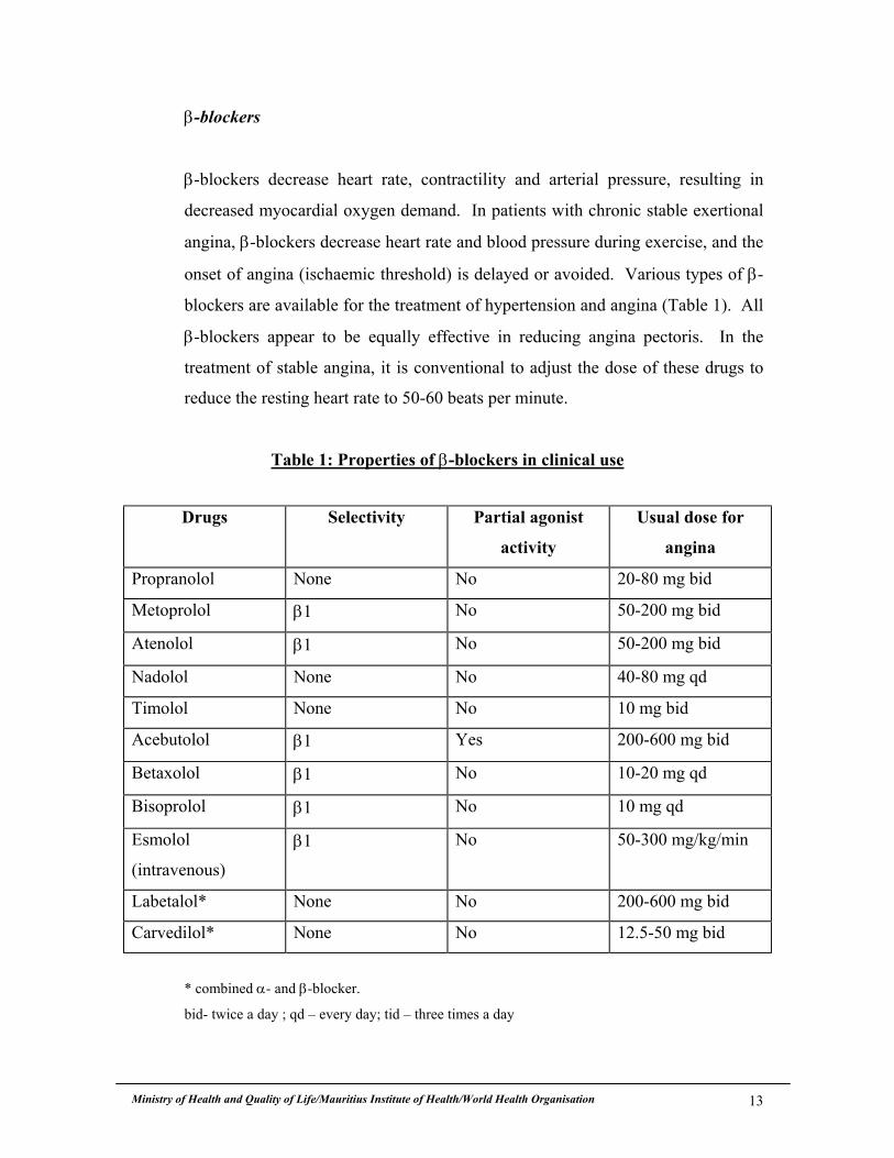
β-blockers
β-blockers decrease heart rate, contractility and arterial pressure, resulting in
decreased myocardial oxygen demand. In patients with chronic stable exertional
angina, β-blockers decrease heart rate and blood pressure during exercise, and the
onset of angina (ischaemic threshold) is delayed or avoided. Various types of β-
blockers are available for the treatment of hypertension and angina (Table 1). All
β-blockers appear to be equally effective in reducing angina pectoris. In the
treatment of stable angina, it is conventional to adjust the dose of these drugs to
reduce the resting heart rate to 50-60 beats per minute.
Table 1: Properties of β-blockers in clinical use
Drugs Selectivity Partial agonist
activity
Usual dose for
angina
Propranolol None No 20-80 mg bid
Metoprolol β1 No 50-200 mg bid
Atenolol β1 No 50-200 mg bid
Nadolol None No 40-80 mg qd
Timolol None No 10 mg bid
Acebutolol β1 Yes 200-600 mg bid
Betaxolol β1 No 10-20 mg qd
Bisoprolol β1 No 10 mg qd
Esmolol
(intravenous)
β1 No 50-300 mg/kg/min
Labetalol* None No 200-600 mg bid
Carvedilol* None No 12.5-50 mg bid
* combined α- and β-blocker.
bid- twice a day ; qd – every day; tid – three times a day
Ministry of Health and Quality of Life/Mauritius Institute of Health/World Health Organisation 13

The absolute cardiac contraindications for the use of β-blockers are severe
bradycardia, pre-existing high degree atrioventricular block, sick sinus syndrome,
and severe decompensated left ventricular failure (β-blockers have now been
shown to reduce total mortality in patients with compensated heart failure). β-
blockers should also be avoided in patients with pure vasospastic angina
(Prinzmetal angina) because these agents may induce coronary vasospasm from
unopposed α-receptor activity. Asthma and bronchospastic disease, severe
depression, and severe peripheral vascular disease are relative contraindications.
Most diabetic patients tolerate β-blockers, although these agents should be used
cautiously in patients who require insulin. In the absence of contraindications, β-
blockers are preferred as initial therapy. The evidence for this approach is
strongest in patients with a history of prior MI, for which this class of drugs has
been shown to reduce mortality.
Calcium antagonists
Calcium antagonists effectively treat hypertension and angina pectoris. These
agents are commonly divided into the dihydropyridine and nondihydropyridine
classes (Table 2). Calcium antagonists decrease coronary vascular resistance and
increase coronary blood flow. All of these agents cause dilatation of the
epicardial coronary vessels and the microcirculation arteriolar resistance vessels.
Dilatation of the epicardial coronary arteries is the principal mechanism that
allows calcium antagonists to relieve vasospastic angina. Calcium antagonists
also concurrently decrease myocardial oxygen demand, primarily by reduction of
systemic vascular resistance and reduction in arterial pressure. In addition,
certain calcium antagonists (verapamil and diltiazem) reduce myocardial oxygen
demand by decreasing heart rate and contractility.
Ministry of Health and Quality of Life/Mauritius Institute of Health/World Health Organisation 14

Table 2: Properties of calcium antagonists in clinical use
Drugs Usual dose Duration of action
Side effects
Dihydropyridines Nifedipine Immediate release: 30-90
mg daily orally Slow release: 30-180 mg orally
Short Hypotension, dizziness, flushing, nausea, constipation, edema
Amlodipine 5-10 mg qd Long Headache, edema Felodipine 5-10 mg qd Long Headache, edema Isradipine 2.5-10 mg bid Medium Headache, fatigue Nicardipine 20-40 mg tid Short Headache, dizziness,
flushing, edema Non-dihydropyridines Bepridil 200-400 mg qd Long Arrhythmias,
dizziness, nausea Diltiazem Immediate release: 30-80
mg 4 times daily Short Hypotension,
dizziness, flushing, bradycardia, edema
Slow release: 120-320 mg qd
Long
Verapamil Immediate release: 80-160 mg tid Slow release: 120-480 mg qd
Short Long
Hypotension, myocardial depression, heart failure, edema, bradycardia
bid – twice a day; qd – every day; tid - three times a day
Short-acting dihydropyridine calcium antagonists have the potential to enhance
the risk of adverse cardiac events and should be avoided. Long-acting calcium
antagonists of the dihydro-and nondihydropyridine class relieve angina and are
appropriate initial therapy in patients with contraindications to β-blockers. They
can also be substituted for β-blockers in patients who develop unacceptable side
effects or can be used in combination with β-blockers when initial β-blockers
therapy is unsuccessful.
Ministry of Health and Quality of Life/Mauritius Institute of Health/World Health Organisation 15
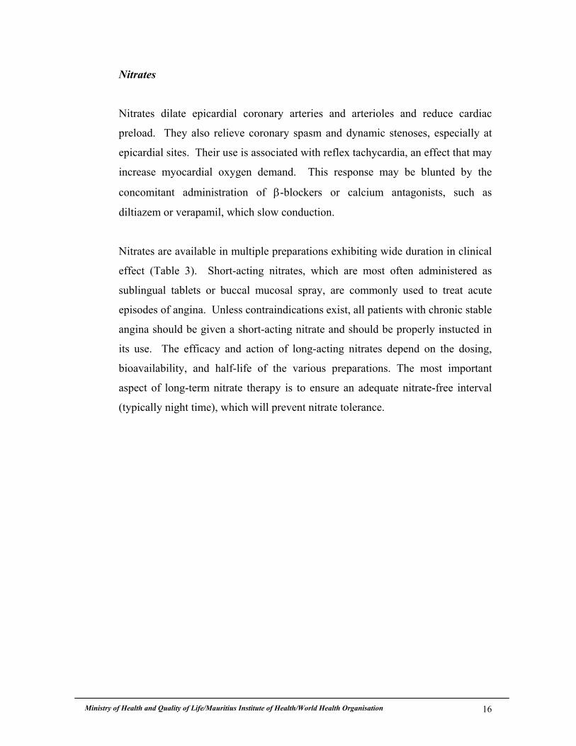
Nitrates
Nitrates dilate epicardial coronary arteries and arterioles and reduce cardiac
preload. They also relieve coronary spasm and dynamic stenoses, especially at
epicardial sites. Their use is associated with reflex tachycardia, an effect that may
increase myocardial oxygen demand. This response may be blunted by the
concomitant administration of β-blockers or calcium antagonists, such as
diltiazem or verapamil, which slow conduction.
Nitrates are available in multiple preparations exhibiting wide duration in clinical
effect (Table 3). Short-acting nitrates, which are most often administered as
sublingual tablets or buccal mucosal spray, are commonly used to treat acute
episodes of angina. Unless contraindications exist, all patients with chronic stable
angina should be given a short-acting nitrate and should be properly instucted in
its use. The efficacy and action of long-acting nitrates depend on the dosing,
bioavailability, and half-life of the various preparations. The most important
aspect of long-term nitrate therapy is to ensure an adequate nitrate-free interval
(typically night time), which will prevent nitrate tolerance.
Ministry of Health and Quality of Life/Mauritius Institute of Health/World Health Organisation 16

Table 3: Properties of nitrates in clinical use
Compound Route Dose Duration of effect Sublingual tablets 0.3-0.6 mg up to 1.5 mg Short 2-3 minutes Spray 0.4 mg as needed Short 2-3 minutes Ointment 2% 6 X 6 in., 15 X 15 cm
7.5-40 mg Effect up to 7 h
Transdermal 0.2—0.8 mg/h every 12 h 8-12 h during intermittent therapy
Oral sustained release
2.5-13 mg 4-8 h
Buccal 1-3 mg 3 times daily 3-5 h
Nitroglycerin
Intravenous 5-200 µg/min. Tolerance in 7-8 h Sublingual 2.5-15 mg Up to 60 min. Oral 5-80 mg, 2-3 times daily Up to 8h Spray 1.25 mg on tongue as
needed 2-3 min.
Chewable 5 mg 2-2.5 h Oral slow release 40 mg 1-2 times daily Up to 8 h Intravenous 1.25-5.0 mg/h Tolerance in 7-8 h
Isosorbide dinitrate
Ointment 100 mg/24 h Not effective 20 mg twice daily Isosorbide-5-
mononitrate Oral
60-240 mg once daily 12-24 h
In patients with exertional stable angina, nitrates improve exercise tolerance,
increase the time to onset of angina, and decrease ST-segment depression during
treadmill exercise testing. Combined with β-blockers or calcium antagonists,
nitrates produce greater anti-anginal and anti-ischaemic effects in patients with
stable angina.
Ministry of Health and Quality of Life/Mauritius Institute of Health/World Health Organisation 17
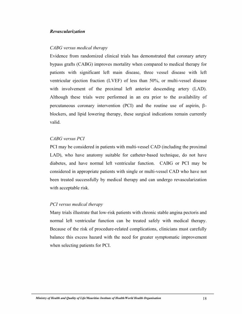
Revascularization
CABG versus medical therapy
Evidence from randomized clinical trials has demonstrated that coronary artery
bypass grafts (CABG) improves mortality when compared to medical therapy for
patients with significant left main disease, three vessel disease with left
ventricular ejection fraction (LVEF) of less than 50%, or multi-vessel disease
with involvement of the proximal left anterior descending artery (LAD).
Although these trials were performed in an era prior to the availability of
percutaneous coronary intervention (PCI) and the routine use of aspirin, β-
blockers, and lipid lowering therapy, these surgical indications remain currently
valid.
CABG versus PCI
PCI may be considered in patients with multi-vessel CAD (including the proximal
LAD), who have anatomy suitable for catheter-based technique, do not have
diabetes, and have normal left ventricular function. CABG or PCI may be
considered in appropriate patients with single or multi-vessel CAD who have not
been treated successfully by medical therapy and can undergo revascularization
with acceptable risk.
PCI versus medical therapy
Many trials illustrate that low-risk patients with chronic stable angina pectoris and
normal left ventricular function can be treated safely with medical therapy.
Because of the risk of procedure-related complications, clinicians must carefully
balance this excess hazard with the need for greater symptomatic improvement
when selecting patients for PCI.
Ministry of Health and Quality of Life/Mauritius Institute of Health/World Health Organisation 18
Antiplatelet agents
Aspirin (75-150 mg) exerts its antithrombotic effect by inhibiting cyclo-
oxygenase and synthesis of platelet thromboxane A2. It is effective in preventing
first heart attacks, improving mortality in acute coronary syndromes, and reducing
adverse cardiovascular events in patients with stable angina pectoris. Aspirin
should therefore be considered as first line therapy in all patients with chronic
ischaemic heart disease. In patients with absolute aspirin contraindications
alternative antiplatelet regimes such as dipyridamole (Persantine), ticlopidine
(Ticlid), glycoprotein 11b/111a receptor antagonists may be considered, although
the clinical data supporting their use in angina pectoris is less established.
Lipid lowering therapy
Of the classes of antihyperlipidemic drugs, the hydroxy-methylglutanyl coenzyme
A reductase inhibitors or statins are the most potent agents available in lowering
total and low density lipoprotein (LDL) and atherogenic cholesterol and are the
only lipid-lowering agents shown to improve overall mortality in clinical trials.
Furthermore, statins also exhibit anti-inflammatory properties, which may
positively modify the artherosclerotic process.
Therefore, statins should be considered in all patients with established CAD and
LDL cholesterol ≥ 130 mg/dL, targeting the on-therapy LDL cholesterol to less
than 100 mg/dL. In general, modification of diet and exercise are less effective
than statins in achieving the target levels of LDL cholesterol; thus, lipid lowering
pharmacotherapy is usually required in patients with angina and should be
considered as first-line therapy in disease modification.
A high density lipoprotein (HDL), antiatherogenic cholesterol level of less than
35 mg/dL is an independent risk factor for adverse cardiovascular events.
Hygienic measures such as weight loss, exercise, alcohol consumption, diabetes
Ministry of Health and Quality of Life/Mauritius Institute of Health/World Health Organisation 19

control, and smoking cessation may provide modest increases in HDL cholesterol.
Recently, a randomized trial comparing the fibrate gemfibrozil to placebo
demonstrated reduced cardiovascular mortality in patients with CAD and an
isolated HDL cholesterol of less than 35 mg/dL.
5.4.2 Education and Risk Factor Reduction
Identification and treatment of coronary disease risk factors in patients with
angina is necessary for optimal secondary prevention of chronic ischaemic heart
disease. Established and modifiable coronary disease risk factors, such as
cigarette smoking, hypertension, diabetes mellitus, and hyperlipidemia, are
common in patients with chronic ischaemic heart disease. These risk factors are
readily amenable to modification, and their treatment can affect clinical outcome
favourably.
Studies show that the risk of death from CAD is inversely proportional to
education. It is therefore important for the physician to assess the patient’s
baseline understanding of his or her condition and to appropriately educate the
patient and his or her family on the pathology and pathophysiology of the disease,
prognosis of condition, and treatment and risk factor reduction options.
Obesity
Obesity is a common condition associated with an increased risk for coronary
artery disease and mortality. Obesity is also associated with and contributes to
other coronary risk factors, including high blood pressure, glucose intolerance,
low levels of HDL cholesterol, and elevated triglycerides. Thus, much of the
increased CAD risk associated with obesity is mediated by these factors. Weight
reduction in obese patients with coronary disease can likely reduce the risk of
future events because weight reduction will reduce other modifiable risk factors
and reduce the increased myocardial oxygen demand imposed by obesity. Weight
Ministry of Health and Quality of Life/Mauritius Institute of Health/World Health Organisation 20

reduction is therefore recommended in all obese patients with ischaemic heart
disease. Strategies to improve weight reduction include:
a. lowering total caloric intake,
b. eating foods low in cholesterol and saturated fat and high in fibre and
unsaturated fat, and
c. exercise training.
Exercise
Exercise training improves blood pressure, glucose control, lipid profile, exercise
endurance, and objective measures of ischaemia. In addition, exercise training
may lead to changes in weight and a sense of well-being. For these reasons, a
regular exercise program should be part of a multifactorial intervention program
in patients with stable chronic ischaemic heart disease.
5.5 FOLLOW-UP
Patients who are successfully treated for chronic ischaemic heart disease should
have follow-up evaluation every 4 to 12 months. A more precise interval cannot
be recommended because many factors influence the length of follow-up period.
Five questions must be answered regularly during the follow-up evaluation:
(1) Has the patient decreased the level of physical activity?
(2) Have the patient’s anginal symptoms increased in frequency and become
more severe since the last visit?
(3) How well is the patient tolerating therapy?
(4) How successful has the patient been in reducing modifiable risk factors
and improving knowledge about ischaemic heart disease?
(5) Has the patient developed any new comorbid illnesses or has the severity
or treatment of known comorbid illnesses worsened the patient’s angina?
Ministry of Health and Quality of Life/Mauritius Institute of Health/World Health Organisation 21

Patients who cannot reliably identify and report changes in their status or who
need more support with their treatment or risk factor reduction should be seen
more frequently.
5.6 INDICATIONS FOR REFERRAL TO CARDIOLOGIST
In general, most patients with chronic stable angina initially present to their
primary care physician. The preliminary diagnosis, risk stratification, medical
management, risk factor modification, and patient education are performed under
direction of the primary care physician. Patients with worsening anginal
symptoms (Canadian Cardiovascular Society Angina Class III or IV) or
presenting with any acute coronary syndrome should be referred to a cardiologist.
Furthermore, patients with an increased risk for adverse cardiac events, left
ventricular ejection fraction (LVEF) ≤ 35%, high-risk exercise treadmill score,
multiple large ischaemic zones on stress imaging, successfully resuscitated from
sudden cardiac death, sustained ventricular tachycardia, significant angina
pectoris symptoms despite medical treatment should be considered for coronary
angiography and evaluated by a cardiology specialist. Those patients who are co-
managed by their primary care physician and cardiologist may alternate follow-up
evaluations, provided that communication among physicians is excellent and all
appropriate issues are addressed at each visit.
Ministry of Health and Quality of Life/Mauritius Institute of Health/World Health Organisation 22

5.7 KEY POINTS
• Angina pectoris is the most common initial manifestation of chronic ischaemic heart
disease.
• A mismatch between coronary blood flow and myocardial oxygen demand results in
myocardial ischaemia.
• Medical treatment of patients with chronic ischaemic heart disease should include:
aspirin and anti-anginals, β-blocker and blood pressure control, cholesterol lowering
and cigarette cessation, dietary modification and diabetes assessment, and exercise
and education.
• Coronary angiography should be considered in patients with chronic angina and any
of the following:
1. Left ventricular ejection fraction less than 35%
2. High risk exercise treadmill score
3. Multiple large ischaemic zones on stress imaging (especially if anterior
distribution),
4. Successful resuscitation from sudden cardiac death,
5. Sustained ventricular tachycardia, and
6. Significant angina pectoris despite medical treatment.
• Coronary artery bypass graft (CABG) should be considered in patients with chronic
angina and any of the following:
1. Significant left main disease,
2. Three vessel coronary artery disease (CAD) and depressed left ventricular
functions, and
3. Multi-vessel CAD that includes the proximal segment of the left anterior
descending (LAD).
• Percutaneous coronary intervention (PCI) may be considered in patients who have
multi-vessel CAD (including the proximal LAD), have anatomy suitable for catheter-
based technique, do not have diabetes, and have normal left ventricular function.
• Coronary revascularization with either PCI or CABG may be considered in
appropriate patients with single or multi-vessel CAD who have failed medical
management of chronic angina pectoris.
Ministry of Health and Quality of Life/Mauritius Institute of Health/World Health Organisation 23

6. ACUTE CORONARY SYNDROMES
Acute coronary syndromes share a common pathophysiology - rupture of a
vulnerable plaque with subsequent activation of dynamic inflammatory,
thrombotic and ischaemic cascades.
Acute coronary syndromes (ACS) may be divided into two broad categories:
1. non-ST elevation ACS: unstable angina (UA)
non-ST elevation MI and
2. ST elevation myocardial infarction (MI).
Acute non ST elevation coronary syndrome without injury (ie, with negative
serial enzyme measurements) is termed unstable angina (UA).
Acute non ST elevation coronary syndrome with cell injury, demonstrated by
serial measurements of cardiac enzymes is termed non-ST elevation MI.
6.2 UNSTABLE ANGINA AND NON ST ELEVATION ACUTE MYOCARDIAL
INFARCTION
6.1.1 Clinical presentation
Non ST elevation acute coronary syndrome (unstable angina and non-ST
elevation myocardial infarction) is defined as a change in the status of a patient’s
angina. It includes the following presentations: (a) new-onset angina, (b) angina
of increasing severity, frequency, duration and lesser or slower response to
rest and/or sublingual nitrates, (c) angina occuring at rest for the first time
and (d) post infarction angina.
Ministry of Health and Quality of Life/Mauritius Institute of Health/World Health Organisation 24

The term unstable angina implies a more serious clinical situation than chronic
stable angina since it may be an immediate precursor of myocardial infarction.
6.1.2 History, Examination and Investigations
History
A carefully obtained history is essential in establishing the diagnosis.
A non-ST elevation ACS should be suspected in any patient who presents with
new or worsening chest discomfort that may be consistent with angina. History
should focus on: risk factors for coronary artery disease, pre-existing diagnosis of
CAD and/or stress-testing, and the nature of the chest discomfort.
Important elements include location, quality, radiation, duration, associated
symptoms, and factors that trigger, worsen, or relieve the pain.
The diagnosis is easier in patients with preexisting stable effort angina, acquiring
new features during some recent time-usually 1 to 4 weeks.
In patients without prior (more than 4 weeks) history of angina (de novo angina)
history may be less easy though.
Some patients may not present with chest pain, but rather with arm or jaw pain,
epigastric discomfort, dyspnoea, or nausea.
Questions should also be directed toward establishing potential non-ischaemic
sources of pain which may mimic an acute coronary syndrome .
Ministry of Health and Quality of Life/Mauritius Institute of Health/World Health Organisation 25
Physical Examination
Physical examination during acute coronary syndromes may be relatively
unremarkable. The patient may appear anxious or diaphoretic (cold sweats).
Tachycardia or bradycardia, as well as extremes of blood pressure, may be
present, but vital signs are often normal.
Heart failure or valvular regurgitation are unusual acute consequences of non-ST
elevation ACS, but may be concurrently present in patients with underlying
cardiac disease.
Signs of peripheral vascular disease, such as carotid bruits or diminished
extremity pulses, should increase the suspicion of atherosclerotic coronary
disease.
Physical examination is perhaps most useful to diagnose non-ischaemic causes
of pain, such as costochondritis, pneumonia, obstructive pulmonary disease,
pericarditis, aortic dissection, upper gastrointestinal disease etc.
Laboratory Testing
Complete blood count, electrolytes, renal function tests, and chest x-ray are useful
to diagnose other causes of symptoms and to evaluate for concurrent medical
conditions, such as renal insufficiency. Clotting times should be obtained to
exclude bleeding diatheses prior to potential initiation of antithrombotic therapies.
.
The cornerstones of diagnostic testing, however, are ECG analysis and
measurements of serum cardiac markers.
Ministry of Health and Quality of Life/Mauritius Institute of Health/World Health Organisation 26

ECG analysis
A baseline ECG, preferably during the chest pain, is essential in any risk
stratification for the purpose of management.
Non ST elevation acute coronary syndromes are subdivided into
1. those without ECG changes, i.e a normal ECG (about 40% of patients)
2. those whose ECG shows ischemic changes (biphasic or inverted T waves, ST-
depression, and/or transient ST elevation – 60%)
The following ECG abnormalities are predictive of an incrementally higher risk:
transient T wave inversion, transient ST elevation, ST depression, and transient
both ST elevation and depression.
Transient or dynamic ECG changes are suggestive of ischaemia, and therefore
the need for serial ECGs in the first few hours after presentation especially when
pain persists, when pain recurs after initial improvement, or when pain subsides
and initial ECG abnormalities disappear..
Development of persistent ST elevation or left bundle branch block should
prompt urgent evaluation for thrombolytics or percutaneous intervention.
Serum cardiac markers
Various serum markers have demonstrated utility as markers of myocardial injury
(infarction), including serum creatine kinase (CK)-MB and troponin I or T.
One main advantage of using both markers is their different time course: while
either may detect early injury, CK-MB values typically return to normal by 1 to 2
days, while troponin levels usually remain elevated for 5 to 14 days.When both
Ministry of Health and Quality of Life/Mauritius Institute of Health/World Health Organisation 27

markers are measured in the first 24 hours after presentation, situations arise in
which one serum marker is normal while the other is mildly elevated.
Serum myoglobin is sometimes useful as an initial screen in doubtful cases
because of its higher early sensitivity (2 hours),
Initial Risk Stratification
One important goal of the initial evaluation is identification of high risk patients,
those with an increased risk of death or MI in the first 30 days.
Various clinical features are predictive of this higher risk.(Table 4).
Table 4: Features predictive of increased risk in non-ST elevation ACS
History
Older age
Diabetes mellitus
Known coronary artery disease
Postinfarction angina
Physical examination
Signs of depressed left ventricular function
Hypotension
ECG changes
ST depression (and/or transient elevation)
Laboratory: elevated CK-MB or troponin
Clinical course
Need for intravenous nitroglycerin
Recurrent or refractory ischaemia
Gallbladder, liver, gastric, or pancreatic disease
Ministry of Health and Quality of Life/Mauritius Institute of Health/World Health Organisation 28
Identification of patients with these features should lead to strong consideration
for institution of more intensive antithrombotic therapy
6.1.3 Differential Diagnosis
The diagnosis of non-ST elevation ACS should be suspected in any patient who
presents with new or worsening ischaemic chest pain consistent with angina,
with ECG abnormalities suggestive of ischaemia but without ST elevation.
Table 5: Non-ischaemic causes of chest discomfort
Costochondritis Muscle strain
Gastroesophageal reflux Oesophageal rupture
Aortic dissection Pericarditis
Congestive heart failure Arrhythmias
Pulmonary embolism Pneumothorax
Pneumonia Pleurisy
Obstructive always disease Anxiety
However, as noted previously, in many cases the initial ECG may be mildly
abnormal or even normal. In these cases, the diagnosis rests on the clinical
history of chest discomfort of a new or more severe nature, occurring more
frequently, at lower levels of activity, or at rest. It is this group of patients,
wherein typical ischaemic ECG changes are absent, that presents perhaps the
greatest diagnostic and management challenge to primary care providers,
emergency room personnel, and cardiologists alike.
In general the clinician should ask himself the following question: Given the
clinical history of new or changing chest discomfort, combined with the
Ministry of Health and Quality of Life/Mauritius Institute of Health/World Health Organisation 29

physical examination, ECG, and laboratory findings, do I believe that there
may have been acute plaque rupture with thrombus formation?
If the answer is no, then, although hospitalization and observation of the patient
might still be warranted, interventions such as treatment with heparin,
intravenous nitrates , glycoprotein receptor inhibitors, and cardiac catheterization
may not be indicated.
6.1.4 Management
Patients with low risk unstable angina may be managed as outpatients with early
follow-up evaluations.
Patients with high risk unstable angina (see table 4 above) require hospitalisation
with imposition of bed rest and sedation in an ECG-monitored environment.
Initial medical management of High Risk Unstable Angina and non Q-wave MI
Hospitalisation, preferably in a coronary care setting
Bed rest (24 – 48 hrs) Sedation as needed
Correction of precipitating conditions such as HBP, arrhythmias, anaemia, hypoxaemia Goals of treatment:
Relieve ischaemic symptoms with antianginal drugs at therapeutic dosage Inhibit thrombosis
Ministry of Health and Quality of Life/Mauritius Institute of Health/World Health Organisation 30

Drug treatment
A. Intravenous nitroglycerin (GTN, ISDN)- for 24 hrs. If asymptomatic after 24 hrs - oral long acting nitrates
B. Beta blockers (if not contraindicated) C. Calcium antagonists (verapamil, diltiazem) if beta-blockers contraindicated
N.B. Nifedipine only if associated with β-blockers D. Narcotic analgesics for pain refractory to antianginal drugs E. Heparin- bolus of 70-80 units/kg + infusion of 15-18 units/kg/hour.
KPTT should be checked every 6 hrs until therapeutic level of 1.5 – 2.0 (50-70 sec) Low molecular weight heparin (enoxaparin) – 1 mg/kg bd for 48 hours (or more if
angioplasty contemplated)
F. Aspirin 150-300 mg od Ticlopidine if aspirin contra-indicated - 250 mg bd Glycoprotein 11b- 111a receptor inhibitors (Reopro, Integrelin)
G. Lipid lowering therapy
Unless contraindicated, all patients with suspected ACS should be given aspirin
(150–300 mg orally).
If pain is present, nitroglycerin tablets should be given sublingually, with
subsequent further sublingual, transdermal, or intravenous doses as necessary.
If pain is not completely relieved with nitrates, intravenous opiates such as
morphine sulphate should be considered.
If oesophageal or gastric pain is suspected, an oral antacid can be given as well.
Heart rate, blood pressure, and rhythm should be monitored closely.
Ministry of Health and Quality of Life/Mauritius Institute of Health/World Health Organisation 31

If there is significant hypertension (e.g., blood pressure > 170/100 mmHg),
tachycardia, persistent chest pain, or ischaemic ECG changes, intravenous beta-
blockers should be administered in the absence of contraindications.
Oral beta-blockade (eg., atenolol 25–50 mg once daily) should be given to all
patients without contraindications, even when vital signs are normal.
If the clinical suspicion of ACS is quite low, then the above treatment combined
with observation in a monitored setting may be adequate.
For patients at higher risk, and for all patients with persistent pain, recurrent pain,
or ECG changes, further antithrombotic therapy is indicated, consisting of heparin
with or without a glycoprotein receptor inhibitor .
Antithrombotic and Anticoagulant Therapy
The mainstays of antithrombotic and anticoagulant therapy have been aspirin and
unfractionated heparin. Two newer classes of agents, however, show substantial
benefit in the treatment of non-ST elevation ACS. These are (a) low molecular
weight heparins and (b) glycoprotein IIb/IIIa receptor inhibitor
Low molecular weight heparins
Treatment with the low molecular weight enoxaparin (Lovenox) reduces
significantly the combined endpoint of death, myocardial infarction, and recurrent
angina or urgent revascularization, compared with unfractionated heparin.
Dosing is 1 mg/kg subcutaneously twice daily, started on admission and typically
continued until in-hospital risk stratification, revascularization, or hospital
discharge.
Ministry of Health and Quality of Life/Mauritius Institute of Health/World Health Organisation 32

Practical advantages of this therapy are simplified dosing and administration,
absence of coagulation profile monitoring, and potential cost savings.
Glycoprotein IIb/IIIa receptor antagonists
Platelet glycoprotein (GP) IIb/IIIa receptors represent the final common pathway
for platelet aggregation, via fibrin binding and cross-linking of the GP IIb/IIIa
receptor. Antibody fragment (abciximab) and peptide (eptifibatide, tirofiban)
antagonists to the GP IIb/IIIa receptor have been developed, which demonstrate
powerful in vitro and in vivo inhibition of platelet aggregation.
Use of GP IIb/IIIa antagonists, therefore, should be considered in addition to
aspirin and heparin in any individual suspected of having a non-ST elevation
ACS, especially if high-risk features are present, such as elevated serum enzymes,
persistent chest pain, or ST depression . In this setting, lower doses of intravenous
heparin are indicated, with goal of prothrombin time 1.5 to 2 times control.
Contraindications due to bleeding risk are similar to those for thrombolytics. The
cost of those drugs is still, however, very prohibitive.
In-hospital risk stratification
If chest pain persists or recurs on the medical and antithrombotic therapies
described, in association with high clinical suspicion or objective evidence of
ischemia such as positive serum markers or ECG changes, then cardiac
catheterization is typically indicated.
However, while this may occur in a significant proportion of patients, many
patients with non-ST elevation ACS can be made pain-free with nitrates, β-
blockers, opiates, and antithrombotic therapy.
Ministry of Health and Quality of Life/Mauritius Institute of Health/World Health Organisation 33

Additionally, others may have recurrent chest pain, but with low clinical
suspicion and no objective evidence of ischemia. In these groups of patients, in-
hospital risk stratification is indicated after an initial period of medical
stabilization (typically 18–48 hours).
Revascularization
If cardiac catheterization is performed, either due to refractory ischaemia or as
part of risk-stratification as described above, then revascularization should be
considered if suitable coronary anatomy is present.
The choice of percutaneous v/s surgical revascularization depends on both the
coronary anatomy and individual patient characteristics and preferences.
All patients should receive maximal preventive therapy.
6.1.5 Follow-up and Secondary Prevention
Many interventions have shown utility in the secondary prevention of ACS,
including control of cardiac risk factors such as smoking, hypertension,
hyperlipidemia, and diabetes, regular use of aspirin, use of beta-blockers and
angiotensin-converting-enzyme inhibitors in selected patients, and adherence to
cardioprotective diets containing n-3-omega fatty acids, monounsaturated fats,
and antioxidants .
All of these interventions act by reducing the inflammatory and thrombotic
milieu of the atherosclerotic vessel.
Ministry of Health and Quality of Life/Mauritius Institute of Health/World Health Organisation 34
6.1.6 Indications for referral to Cardiologist
Patients with worsening anginal symptoms or presenting with any acute coronary
syndrome should be referred to a cardiologist.
Those patients who are co-managed by their primary care physicians and
cardiologist may alternate follow-up evaluations, provided that communication
among physicians is excellent and all appropriate issues are addressed at each
visit.
Ministry of Health and Quality of Life/Mauritius Institute of Health/World Health Organisation 35
6.2 ST elevation acute myocardial infarction
6.2.1 Clinical presentation
ST elevation myocardial infarction occurs when myocardial oxygen supply is
abruptly stopped by prolonged and totally occlusive coronary thrombosis leading
to necrosis of the myocardial tissue.
Acute myocardial infarction is a medical emergency in which the mortality is
greatest within the first 2 hours after the onset of symptoms. Mortality from acute
myocardial infarction can be significantly reduced by rapid transport to a
coronary care setting, institution of prompt thrombolytic or mechanical
(angioplasty) reperfusion, treatment of ventricular arrhythmias and
haemodynamic complications.
History
Diagnosis
The diagnosis of acute myocardial infarction requires the presence of at least 2
of the following criteria (1) clinical symptoms suggestive of an acute MI
(2)ECG findings consistent with ischaemia or necrosis and (3) elevated
biochemical markers.
The decision to institute thrombolytic therapy usually is based on clinical
findings and ECG, because laboratory confirmation of the clinical diagnosis of
acute MI requires several hours.
Ministry of Health and Quality of Life/Mauritius Institute of Health/World Health Organisation 36

History
The cardinal characteristics of chest pain of acute myocardial infarction are:
a. localisation and to a lesser extent - radiation
b. duration of continuous ischaemic pain
c. response to oral nitrates
Prodromal symptoms of unstable angina such as de novo angina pectoris
during or after exercise or even at rest with increasing frequency are often
reported (unstable angina) if properly queried.
The pain of myocardial infarction is usually described as a pressure,
compression, constriction, squeezing, boring, or burning in the chest,
characteristically radiating to the left arm, but also sometimes involving the neck,
jaw, epigastrium, right arm, or back, lasting continuously for over 30 minutes, not
relieved by rest or nitroglycerin administration, and often accompanied by
anxiety, apprehension, restlessness, hypotension, nausea, paleness and sweating.
Almost 25% of patients with acute myocardial infarctions may be completely
asymptomatic at the onset of coronary occlusion (“silent infarcts”).
In the elderly or patients with diabetes, chest pain might be less severe and other
symptoms such as syncope, faintness, or dyspnea may be present.
The acute symptoms of MI are frequently associated with stress such as anger or
upsetting life events, or occasionally excessive physical exertion .
Ministry of Health and Quality of Life/Mauritius Institute of Health/World Health Organisation 37

6.2.2 Examination and Investigations
1. Physical Examination
The rapid, risk-stratification orientated physical examination should be directed
towards identification of:
1. haemodynamic instability, and
2. pulmonary congestion
New systolic murmurs are particularly important, suggesting the presence of
ischaemic mitral regurgitation (due to papillary muscle dysfunction or more
dramatically due to papillary muscle rupture) or an acquired ventricular septal
defect (due to rupture of the infarcted area of the septum)
There is no diagnostic physical sign of acute MI. The clinical appearance is
variable (from the healthy-looking athlete carrying his own suitcase to the
somnolent patient in cardiogenic shock). However, to detect complications of
acute MI, physical examination—in particular, auscultation of the lungs and the
heart—should be done repeatedly every day in all patients.
2. Electrocardiographic Findings
The electrocardiogram serves as the hallmark for diagnosis of acute MI because
characteristic ST, T, and Q-wave changes are detectable in most of the patients.
In the early stage of acute coronary occlusion (phase 1 or hyperacute phase) giant
positive T waves appear with taller than normal R waves .
In phase 2 (acute phase, more commonly seen at hospital admission), ST-segment
elevation is evident with a decrease in amplitude of the R wave, followed by
Ministry of Health and Quality of Life/Mauritius Institute of Health/World Health Organisation 38

pathologic Q-wave development that lasts 0.04 seconds or more and reaches 25%
of the amplitude of the R wave (pathologic Q- wave, phase 3, when cell necrosis
occurs over hours to days).
Over time, the ST segment returns to normal with T wave inversion (terminal
negative T wave); the R wave may be lost as a result of transmural necrosis and
may be replaced by a QS wave (phase 4, chronic phase). The Q wave or QS wave
persists, whereas the T wave may return to positive.
ST-wave elevation may persist in cases of left ventricular dyskinesis or aneurysm.
The electrocardiographic leads not representing the infarcted area may show
inverse, reciprocal ST-segment alterations.
A new left bundle branch block may be present and, if it persists, usually
indicates large anterior wall infarction.
The diagnosis of acute infarction in patients with acute chest pain who present
with left bundle branch block is highly probable when there is ST-segment
elevation of 1 mm or more concordant with the QRS complex, ST-segment
depression of 1 mm or more in lead V1, V2, or V3, and ST-segment elevation of
5 mm or more discordant to the QRS complex. When possible, the current ECG
tracing should be compared with previous records.
Determination of myocardial infarct localization according to ECG leads is listed
in Table 5.
Ministry of Health and Quality of Life/Mauritius Institute of Health/World Health Organisation 39

Table 5: Location of myocardial infarction according to electrocardiographic leads
and involved coronary artery
ST-segment elevation in Ventricular location Coronary artery involved V1 through V3 Anteroseptal Proximal LAD, septal
perforators V2 through V4 Anteroapical LAD, diagonal branches I, aVL, V6 Lateral LAD, diagonal branch, or
circumflex I, aVL High lateral 1st diagonal branch or
circumflex I, aVL, V3 through V6 Anterolateral Mid LAD or circumflex I, aVL, V1 through V6 Extensive anterior Proximal LAD II, III, aVF Inferior RCA or circumflex, distal LAD V1 through V2 Posterior Posterior descending of (ST depression) RCA, circumflex II, III, aVF, Inferolateral RCA or circumflex V5 through V6 V1, V3R, V4R Right ventricular RCA (LAD—left anterior descending RCA—right coronary artery.)
3. Cardiac enzymes
After myocardial injury and cell death, cellular enzymes are released into the
bloodstream. The typical time course of enzyme alterations that occur can help to
determine the phase of MI.
The most commonly used tests are creatinine kinase (CK), CK-MB, and the very
specific troponins (troponin I or T).
For clinical use, it is recommended that cardiac troponin T be measured to
distinguish between cardiac and other sources of chest pain. In case of elevated
troponin, follow-up measurements of CK are sufficient to determine the course of
Ministry of Health and Quality of Life/Mauritius Institute of Health/World Health Organisation 40

myocardial infarction. If chest pain is recurrent, intermittent analyses of CK-MB
or troponin T are recommended for detection of reinfarction.
4. Imaging Techniques
Global ventricular function and mechanical and hemodynamic complications such
as papillary muscle rupture, septal rupture, pericardial effusion, mitral
regurgitation, intraventricular thrombi, and their follow-up all can be assessed
accurately by echocardiography.
6.2.3 Differential diagnosis
Although in the majority of cases the diagnosis of ST elevation acute MI is
straightforward, a rapid but well thought differential diagnosis is essential. It is
mandatory that those diseases in which thrombolysis is not only of no use but may
even be contraindicated be excluded.
These are: pericarditis, early repolarisation on ECG, acute aortic dissection,
pneumothorax, ventricular aneurysms, Prinzmetal’s angina, left ventricular strain
pattern, left bundle branch block, hyperkalaemia, cerebro-vascular accident etc.
6.2.4 Management
Acute Management Strategies include:
1. Thrombolysis
2. Platelet antiaggregants
3. Anticoagulants
4. Maintenance of oxygen balance
Ministry of Health and Quality of Life/Mauritius Institute of Health/World Health Organisation 41

Because acute MI always represents a life-threatening event, the patient with
suspected acute myocardial infarction should be hospitalized immediately to
minimize delay in appropriate therapy.
Therapy for acute MI consists of the following six steps:1) relief of pain, 2) relief
of anxiety and restlessness, and sedation, 3) reperfusion, 4) anticoagulation,
5)therapy for complications, 6) secondary prevention.
For steps 1 and 2, repeated administration of intravenous morphine (2 to 10 mg or
more), is the treatment of choice, which can be combined with antiemetics to
reduce side effects.
Initial Management in ALL patients
(thrombolysis or not)
• General measures: IV line, blood samples for CE, electrolytes, FBC, lipid
profile
• Monitoring for cardiac rhythm, vital signs.
• If no ST elevation – repeat ECG in 1 hour (or earlier) if ongoing chest
discomfort
• Pain: adequate analgesia- Morphia IV 2-4 mg to be repeated in 10 min
interval until pain is under control (or side effects +)
• Nausea/vomiting – Primperan IV
• Hypotension – adequate volume expansion
• GTN s/l every 5 min (if no C/I), then infusion
• Oxygen 2-4 l/min
• Aspirin per os or Aspegic IV
• Heparin - IV bolus 5000 u/kg, then 1000u/h or enoxaparin 1 mg/kg bd sk
• Beta blockers- Atenolol 5-10 mg IV (if no C/I- bradycardia, hypotension,
LVF, HB, COPD, severe peripheral vascular diseases)
Ministry of Health and Quality of Life/Mauritius Institute of Health/World Health Organisation 42

Once the patient arrives at the hospital, initial evaluation of the patient’s history,
coronary risk factors, physical signs, 12-lead ECG tracing, possible
contraindications for reperfusion therapy, and steps for treatment should be
initiated within 20 to 30 minutes (“door-to-needle time”). The presence of a
special thrombolysis nurse might reduce the delay in instituting reperfusion
therapy for patients with acute MI.
In the emergency ward the patient should immediately receive aspirin in
chewable form, oxygen by nasal prongs or masks (blood gases may be checked,
especially in patients with chronic pulmonary diseases), and sublingual nitrates
(unless systolic arterial pressure is less than 90 mmHg or heart rate is less than 50
or greater than 100 beats per minute).
The patient should be monitored and have bed rest for 12 to 24 hours as life-
threatening arrhythmias, reinfarction, mechanical complications, and death occur
most frequently within the first 24 hours after acute MI.
Reperfusion Therapy
The main goal in the treatment of patients with acute MI is to reduce the extent of
irreversible myocardial tissue damage and necrosis.
Fibrinolytic therapy should be initiated as early as possible, preferably within the
first 3 hours after onset of persistent chest pain, but some benefit may still be
derived if administered up to even 12 hours (if pain persists with ST elevation).
Eligibility for thrombolytic therapy
• Chest pain of >30 min and < 12 hrs
• ST segment elevation of > 1.0 mV in two continuous ECG leads indicative of
AMI
Ministry of Health and Quality of Life/Mauritius Institute of Health/World Health Organisation 43
• New LBBB on ECG
Because fibrinolytic agents increase the risk of hemorrhage, including
intracerebral hemorrhage, several cautions and contraindications must be
considered before treatment can be initiated. These are:
A. Absolute contraindications:
• Active internal bleeding
• Suspected aortic dissection
• Known intracranial neoplasm
• History of any haemorragic CVA ever
• Or other cerebrovascular event during the past 1 year
B. Relative contraindications:
• Severe hypertension on presentation (BP > 180/110 mm.Hg.)
• History of chronic severe hypertension
• History of prior CVA or other intracranial pathology
• Recent trauma (< 2 weeks) or major surgery (< 3 weeks)
• Prolonged or traumatic CPR (> 10 min)
• Non-compressible vascular punctures
• Recent internal bleeding (< 2-45 weeks)
• Known bleeding diathesis or current INR > 2-3
• For streptokinase- prior exposure or known allergy
Primary Percutaneous Coronary Intervention
Primary percutaneous coronary intervention (PCI) can serve as an alternative to
thrombolysis only if performed in a timely fashion by individuals skilled in the
procedure and supported by experienced personnel in high-volume centers .
Indications include patients with contraindications to thrombolysis, patients at risk
for bleeding, and patients with cardiogenic shock
Ministry of Health and Quality of Life/Mauritius Institute of Health/World Health Organisation 44

Adjunctive Therapy
Platelet antiaggregants.
Early adjunctive therapy with thrombolysis consists of aspirin, which quickly
prevents thromboxane A2 production in platelets and prostacyclin in endothelial
cells. Aspirin should be administered at the time of admission to the emergency
ward at a dose of 150 to 300 mg, preferably in a chewable form because of its
faster absorption, followed by a daily administration indefinitely thereafter.
In case of true aspirin allergy, dipyridamole or ticlopidine may be used.
Heparin (which forms a complex with antithrombin III, thus inactivating
thrombin) is recommended in patients with large or anterior MI, known left
ventricular thrombus, or previous embolic events. In patients not treated with
thrombolytic therapy or with nonselected thrombolytics (streptokinase,
anistreplase, urokinase), and in patients without increased risk for systemic
emboli, subcutaneous administration of 7500 U (or 1 mg/kg of enoxaparin) twice
daily may be administered . Prolonged intravenous heparin therapy has not been
shown to decrease the rate of reocclusion.
β-Adrenergic blockers are known to reduce myocardial oxygen consumption, as
well as acute and long-term mortality and morbidity, and should be administered
intravenously as early as possible (on day 1 within 12 hours) followed by oral
therapy if contraindications are excluded. Furthermore, β-blockers may reduce
infarct size and the incidence of ventricular fibrillation and reinfarction.
ACE inhibitors should be initiated on day 1 within hours of hospitalization in
patients with evolving acute MI with ST-segment elevation or left bundle branch
block and continued indefinitely in case of impaired left ventricular function (the
lower the ejection fraction, the greater the benefit).
Ministry of Health and Quality of Life/Mauritius Institute of Health/World Health Organisation 45

Intravenous nitroglycerin is recommended during the first 12-24 hours and
beyond this time frame in patients with recurrent angina, large infarction, and
pulmonary congestion.
Calcium antagonists, predominantly short-acting nifedipine, may be harmful and
should be avoided in patients with acute MI. If β-blockers are ineffective,
verapamil or diltiazem might be used in patients with ischaemia or tachycardia in
the absence of left ventricular dysfunction or AV block .
Magnesium administration is recommended only to correct documented
magnesium deficits, especially in patients receiving diuretics, in the case of
torsades de pointes with a prolonged QT interval, and in high-risk patients not
receiving reperfusion therapy.
Intensive insulin-glucose administration (24h) during acute MI reduces long-
term mortality in patients with diabetes and might be considered in this cohort .
Complications of Acute Myocardial Infarction
Arrhythmias
The incidence of arrhythmias may be as high as 100% in patients with acute MI,
and manifests predominantly as premature ventricular beats.
The most serious and life-threatening arrhythmias are ventricular fibrillation and
sustained polymorphic ventricular tachycardia requiring early electrical
defibrillation (200 J), which is repeated if unsuccessful (up to 360 J).
Monomorphic ventricular tachycardia not associated with angina or acute heart
failure may be treated with the antiarrhythmic agents lidocaine (1 to 1.5 mg/kg as
a bolus, followed by 2 to 4 mg/min intravenously), procainamide, amiodarone, or
synchronized electrical cardioversion.
Ministry of Health and Quality of Life/Mauritius Institute of Health/World Health Organisation 46

For bradyarrhythmia and heart block, either intravenous atropine or temporary
transvenous (or, in some cases, transcutaneous) pacing is recommended.
Indications for pacing are symptomatic bradycardia (less than 50 beats per
minute) with symptoms of hypotension not responding to atropine; asystole;
bilateral bundle branch block; newly developed or indeterminate bifascicular
block with first-degree AV block; second-degree symptomatic or Mobitz type II
AV block, advanced (complete) AV-block; and incessant ventricular tachycardia,
which requires atrial or ventricular overdrive pacing.
Atrial fibrillation is often transient and may be associated with heart failure, atrial
ischemia, or pericarditis. It should be treated by either electric cardioversion in
patients with hemodynamic instability or ischemia; or by rapid digitalization, β-
blockade in patients without contraindications, diltiazem or verapamil, and
heparin to avoid embolic events.
Hemodynamic and Mechanical Complications
Congestive heart failure may result from systolic contractile dysfunction due to
large necrotic areas or to postischaemic contractile wall motion abnormalities
(stunned myocardium). Mechanical complications that worsen cardiac function
include myocardial infarct expansion and aneurysm formation, ruptured
ventricle, ventricular septal defect, papillary muscle dysfunction and rupture. The
primary symptom is dyspnea, caused by pulmonary congestion and tachycardia.
Therapy for congestive heart failure should be initiated with diuretics
(furosemide, 20 mg intravenously, which may be repeated), nitrates, and oxygen,
followed by positive inotropic agents (dobutamine, 2 to 20 µg/kg/min; dopamine
2 to 10 µg/kg/min intravenously).
In case of severe ventricular dysfunction or cardiogenic shock, a Swan-Ganz
pulmonary artery catheter for measurements of cardiac output, pulmonary artery
capillary wedge pressure pulmonary and systemic resistances may be placed.
Ministry of Health and Quality of Life/Mauritius Institute of Health/World Health Organisation 47

6.2.5 Follow-up and Rehabilitation
1. Follow-up
In all patients following myocardial infarction without contraindications, long-
term treatment with aspirin and β-adrenergic blockers is recommended .
Recurrent angina should be treated with intravenous nitroglycerin, aspirin,
heparin, analgesics, and β-blockers.
ACE inhibitors should be a part of secondary prevention in patients with left
ventricular dysfunction.
Calcium antagonists are not routinely recommended after infarction but may be
selectively prescribed in patients with specific indications such as hypertension or
angina in the presence of preserved left ventricular function.
Carvedilol has been shown to attenuate ventricular remodeling in patients with
left ventricular dysfunction after MI, but its long-term effects need to be
investigated further.
Ministry of Health and Quality of Life/Mauritius Institute of Health/World Health Organisation 48

Table 6: Long-term therapy and secondary prevention after myocardial infarction
Treatment
Drug therapy Comments
Aspirin In all patients without contraindications
β-Blockers In all patients without contraindications
ACE inhibitors In patients with left ventricular dysfunction
Dietary therapy Low saturated fats and cholesterol
Weight-reduction
and exercise in overweight patients
Lipid-lowering drugs If LDL cholesterol > 100 mg/dL,
HDL cholesterol < 35 mg/dL, triglycerides > 400
mg/dL
2. Rehabilitation after MI
Physical activity should be progressively advanced in stable patients. Before
discharge from the hospital patients should receive verbal and if possible written
instructions concerning physical activities, medications, diet, and secondary
preventive measures.
Rehabilitation should concern all facets of patient’s activities: physical,
psychological, emotional, socioeconomic, life-style changes, risk factors
management, smoking cessation, education, return to work (if feasible), self-
control, knowledge of limitations etc.
Ministry of Health and Quality of Life/Mauritius Institute of Health/World Health Organisation 49

6.2.6 Post-infarction Evaluation and Secondary Prevention
Coronary angiography and subsequent revascularization, either by PCI or bypass
grafting, ultimately should be performed in patients with recurrent angina or
evidence of large areas of reversible ischemia on stress testing, and might be
considered selectively in all survivors of acute MI.
The prognosis after acute myocardial infarction is related to four main factors:
• Extent of left ventricular dysfunction, including degree of left ventricular
dilation
• Presence of residual ischemia
• Degree of electrical instability of the myocardium
• Progression of coronary atherosclerosis
A risk stratification should be evaluated in each patient. Noninvasive evaluation
of low-risk patients includes submaximal or symptom-limited stress ECG at 4 to
6 days or 10 to 14 days, respectively. For prognostic assessment, after
mobilization, ie after 1 week, standard exercise testing should be performed to
assess potential exercise-induced angina or left ventricular dysfunction and
functional capacity. Stress ECG should be repeated early after hospital discharge
and after 4 to 6 weeks.
Patients should be educated about risk factor modification, ie, cessation of
smoking, necessity of physical exercise, and effective lifestyle intervention. Diet
recommendations consist of low saturated fat and cholesterol for all patients
following acute MI.
Ministry of Health and Quality of Life/Mauritius Institute of Health/World Health Organisation 50
6.2.7 Key Points
• Acute coronary syndromes share a common pathophysiology—rupture of a
vulnerable plaque with subsequent activation of dynamic inflammatory and
thrombotic cascades.
• Initial evaluation, using history, physical examination, electrocardiography,
and measurement of serum cardiac markers, should be directed toward risk-
stratification of the patient as low, intermediate, or high.
• Unless contraindicated, initial treatment should include aspirin, nitrates,
beta blockade, and unfractionated or low molecular weight heparin, with the
addition of more intensive antiplatelet therapies for high-risk patients.
• After medical stabilisation, strategies of either early cardiac catheterization or
initial stress testing are appropriate for further risk stratification.
• All patients should be discharged on preventive therapies, which are effective
by reducing the inflammatory and thrombotic milieu of the atherosclerotic
vessel.
• Myocardial infarction is defined as cardiomyocyte necrosis due to lack of
oxygen supply in relationship to oxygen demand. The underlying cause in
most cases is rupture of an intracoronary atherosclerotic plaque with
subsequent platelet aggregation and thrombotic occlusion.
• Q wave myocardial infarction is defined as myocardial cell necrosis with
development and maintenance of Q waves on the electrocardiogram.
• The key symptom occurring with acute coronary artery occlusion is severe
retrosternal chest pain, but symptoms may vary.
Ministry of Health and Quality of Life/Mauritius Institute of Health/World Health Organisation 51

• Early hospitalization and intensive monitoring in an intensive care or coronary
care unit is required.
• Worldwide mortality of myocardial infarction is 30% to 40%; one third die
within the first hours.
• Treatment of choice is early recanalization by either thrombolytic or
interventional therapy, including stent implantation, treatment of
complications, and secondary prevention.
• The main prospective goal is public education, early identification of
myocardial ischemia, and urgent reperfusion therapy.
• The future trend for acute management is the search for the optimal
reperfusion regimen with the smallest risk.
• For chronic management, the trend is toward inhibition of ventricular
remodeling and long-term improvement of ventricular function.
Ministry of Health and Quality of Life/Mauritius Institute of Health/World Health Organisation 52

GUIDELINES ON PRIMARY PREVENTION
OF
CORONARY ARTERY DISEASE IN MAURITIUS
AT
PRIMARY HEALTH CARE LEVEL
Ministry of Health and Quality of Life/Mauritius Institute of Health/World Health Organisation 53

Table of Contents
1.1 Introduction and Rationale
1.2 Definition of Primary Prevention
1.3 Objectives of Coronary Artery Disease Prevention
1.4 Principles of Screening for Coronary Artery Disease Risk Factors
1.5 Situational Analysis of Risk Factors for CAD
1.6 Definition of Risk Factors
1.7 Prioritisation for Coronary Heart Disease Prevention
1.8 Scientific Basis for Risk Factor Modification
Conclusion
References
Recommendations
Ministry of Health and Quality of Life/Mauritius Institute of Health/World Health Organisation 54

1. 1 INTRODUCTION AND RATIONALE
Worldwide, the mortality from cardiovascular disease is projected to rise from
13.2 million in 1985 to 24.5 million, an 85% increase in three decades. This
indication has led the World Health Organisation to term coronary heart disease
as the ‘World’s public health enemy number 1’. By 2015, the increase in
cardiovascular deaths will be more than double in developing nations, jumping
from 72 million to 167 million, or by 132 percent increase in thirty years, while
mortality in industrialized nations will rise by 28 percent. This means that
coronary artery disease will become the leading killer in both developing and
developed country.
Coronary artery disease has also been identified as the primary non-
communicable health problem in Mauritius, accounting for nearly one-third
deaths in Mauritius. It has a profound and adverse impact on household, families
and society and is a major contributor to morbidity, invalidity and mortality.
Pharmacological approaches, intensive care units, coronary angioplasty and
bypass surgery can reduce modality from coronary heart disease by one third.
However, two third of the decline is credited to measures such a healthy diets,
exercise and smoking cessation, optimum control of blood pressure and a normo-
glycemic level of glucose. Premature death of Mauritians can be avoided and
quality of life improved if a comprehensive risk factors intervention program for
CAD can be implemented at the primary health care level, widely perceived to be
the backbone of a rational health service system.
1.2 DEFINITION OF PRIMARY PREVENTION
Primary prevention refers to guidance given to persons with no known
cardiovascular disease. The first goal of prevention is to prevent the development
Ministry of Health and Quality of Life/Mauritius Institute of Health/World Health Organisation 55

of risk factors. The primary care physicians should regularly check for smoking,
physical inactivity, elevated lipid levels, elevated blood sugar, high blood
pressure and eating habits and other environmental hygiene factors.
1.3 OBJECTIVES OF CORONARY ARTERY DISEASE PREVENTION
The objectives of a coronary artery disease prevention program at the first level of
contact of a health system are to:
a. Remain free from coronary heart diseases and enjoy an improved quality of
life.
b. Reduce the risk of coronary heart disease events and thereby reduce premature
disability, mortality and extend overall survival.
c. Decrease the need for interventional procedures such as angioplasty, bypass
grafting and thereby decreasing hospital costs.
d. Reduce the incidence of subsequent myocardial infarctions
1.9 PRINCIPLES OF SCREENING FOR CORONARY ARTERY DISEASE
RISK FACTORS:
A coronary artery disease prevention program must meet a number of criteria
before it can be used in the general population. These criteria include:
1. Coronary heart disease is a serious public health problem.
2. Techniques for risk factor detection are easy, precise, cost-effective and
acceptable to the community.
3. There is a defined screening strategy and a defined intervention and
follow-up policy.
4. Trained staff and facilities for screening and intervention are available.
5. Screening and intervention results in a reduction in clinical events,
including morbidity and mortality.
6. Screening has no adverse effects.
7. Cost of screening and intervention is justified in relation to the outcome.
Ministry of Health and Quality of Life/Mauritius Institute of Health/World Health Organisation 56
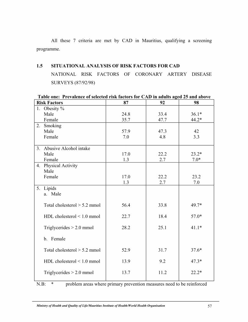
All these 7 criteria are met by CAD in Mauritius, qualifying a screening
programme.
1.5 SITUATIONAL ANALYSIS OF RISK FACTORS FOR CAD
NATIONAL RISK FACTORS OF CORONARY ARTERY DISEASE
SURVEYS (87/92/98)
Table one: Prevalence of selected risk factors for CAD in adults aged 25 and above Risk Factors 87 92 98 1. Obesity %
Male Female
24.8 35.7
33.4 47.7
36.1* 44.2*
2. Smoking Male
Female
57.9 7.0
47.3 4.8
42 3.3
3. Abusive Alcohol intake Male
Female
17.0 1.3
22.2 2.7
23.2* 7.0*
4. Physical Activity Male
Female
17.0 1.3
22.2 2.7
23.2 7.0
5. Lipids a. Male
Total cholesterol > 5.2 mmol HDL cholesterol < 1.0 mmol Triglycerides > 2.0 mmol
b. Female Total cholesterol > 5.2 mmol HDL cholesterol < 1.0 mmol Triglycerides > 2.0 mmol
56.4
22.7
28.2
52.9
13.9
13.7
33.8
18.4
25.1
31.7
9.2
11.2
49.7*
57.0*
41.1*
37.6*
47.3*
22.2*
N.B: * problem areas where primary prevention measures need to be reinforced
Ministry of Health and Quality of Life/Mauritius Institute of Health/World Health Organisation 57

DIABETES MELLITUS AND IMPAIRED GLUCOSE TOLERANCE
PREVALENCE NATIONAL SURVEYS 87/92/98 IN ADULTS AGED 25 AND
ABOVE
87 92 98 MEN DM Prevalence % IGT Prevalence %
14.2 15.6
16.3 14.9
20.8 11.8
WOMEN DM Prevalence % IGT Prevalence %
14.5 22.5
17.4 19.3
20.6 20.2
PREVALENCE OF HYPERTENSION IN ADULTS AGED 25 AND ABOVE
87 92 98 MEN
31.7 26.5 30.0
WOMEN
30.2 26.2 29.6
1.6 DEFINITION OF RISK FACTOR
The term risk factor describes those “characteristics found in healthy individuals
to be independently related to the subsequent occurrence of coronary heart disease
and, where modifiable, to be reversible.’ For example, cholesterol is an
independent risk factor for coronary heart disease. Epidemiological and
evidence-based studies have shown that cholesterol lowering by one percent is
associated with a decrease in the risk of coronary heart disease of about 2%.
There is overwhelming scientific evidence that lifestyle modification and risk
factor reduction can retard the development of coronary heart disease both before
and after the occurrence of a clinical event. Previously undue emphasis was
placed on individually high risk factors rather than the overall level of risk based
on a combination of risk factors. A multifactorial assessment of coronary heart
disease acknowledges 3 important facts:
Ministry of Health and Quality of Life/Mauritius Institute of Health/World Health Organisation 58

1. Coronary heart disease has a multifactorial aetiology
2. Risk factors have a multiplicative effect.
3. As physicians, we are dealing with the whole person (holistic view),
not isolated risk factors.
LIFESTYLE AND CHARACTERISTICS ASSOCIATED WITH INCREASED
RISK OF FUTURE CHD EVENTS
Lifestyles Modifiable characteristics Non-modificable
characteristics Diet high in saturated fat, cholesterol and calories Tobacco smoking Excess alcohol consumption Sedentary life Sustained psycho-emotional stress
Elevated blood pressure Elevated plasma Total cholesterol LDL cholesterol Low HDL cholesterol Elevated plasma TG Diabetes Obesity Thrombogenic factor ?
Age Sex Post menopause Family history of CHD at early age Male < 55 yrs Females < 65 yrs
1.7 PRIORITIZATION FOR CORONARY HEART DISEASE PREVENTION
Primary prevention activities for coronary heart disease should be downsized and
prioritized under the following targeted groups:
a. Patients with established coronary heart disease should be given first priority
for prevention as they are at high risk of fatal and non-fatal complications of
their disease.
b. Next priority should be given to individuals in the general population who are
at high risk of developing coronary heart disease.
Ministry of Health and Quality of Life/Mauritius Institute of Health/World Health Organisation 59
c. Third priority for prevention is close relatives of patients with very early onset
of coronary heart disease particularly the first-degree blood relatives of
patients.
d. Increasing age, ethnicity, socially economically deprived communities,
geographical regions are other variables to be considered for primary
prevention.
1.8 SCIENTIFIC BASIS FOR RISK FACTOR MODIFICATION
Unifactorial randomized control trials of diet; blood pressure and cholesterol
lowering have shown reductions in cardiovascular morbidity and mortality.
However, the WHO collaborating group study has confirmed that the multi
factorial intervention is more cost effective in modifying the risk factors of CAD.
Ministry of Health and Quality of Life/Mauritius Institute of Health/World Health Organisation 60

A. DIET
Diet is a determinant of CAD. Its effect on the development of atherosclerosis is
mediated through the influence of biological risk factors e.g. LDL, HDL, BP,
obesity, diabetes and hypertension.
A healthy diet is characterized as:
1. Low in saturated fatty acids
2. High in unsaturated fatty acids
3. Rich in complex carbohydrates
The traditional Mediterranean diet (low saturated fat + high unsaturated fat
especially monounsaturated) and Japanese diet (low saturated fat + high complex
carbohydrates) are associated with the best life expectancy in the world. The
physician’s training does not encompass aspects of dietary counseling. Therefore,
assistance from a dietician or a specially trained nurse for dietary interviews is of
considerable value.
B. OBESITY
Obesity can be assessed by three measures mainly:
a. Body mass index
b. Waist hip ratio
c. Waist circumference
a. Body mass index
Body weight is expressed in terms of BMl. It is calculated as weight in kg
/ height (in m2) squared and is recommended by the WHO expert
committee for assessing degree of obesity as shown in the table below.
Ministry of Health and Quality of Life/Mauritius Institute of Health/World Health Organisation 61

ASSESSMENT OF OBESITY USING BODY MASS INDEX
Body mass index (kg/m2) WHO classification Popular description
< 18.5 Underweight Thin
18.5 – 24.9 Normal weight Normal weight
25.0 – 29.9 Grade 1 overweight Overweight
30.0 – 39.9 Grade 2 overweight Obese
> 40.0 Grade 3 overweight Morbidity obese
b. Waist hip ratio
The ratio of hip to waist circumference is also widely used in
epidemiology as an index of central obesity. Central obesity with the
accumulation of fat to the trunk and abdominal cavity is associated with
high prevalence of lipid abnormalities, hypertension and abnormal glucose
tolerance and insulin resistance leading to increased coronary artery
disease. The normal value of waist hip ratio should not be greater than 1.
c. Waist circumference
There is also good evidence from population based studies that waist
circumference is also a useful index of obesity. Measurement of waist
circumference (one measurement and no calculation with the person
standing), midway between the lowest rib and the iliac crest, is also
recommended for clinical assessment of the degree of obesity and follow
up during weight reduction.
Ministry of Health and Quality of Life/Mauritius Institute of Health/World Health Organisation 62
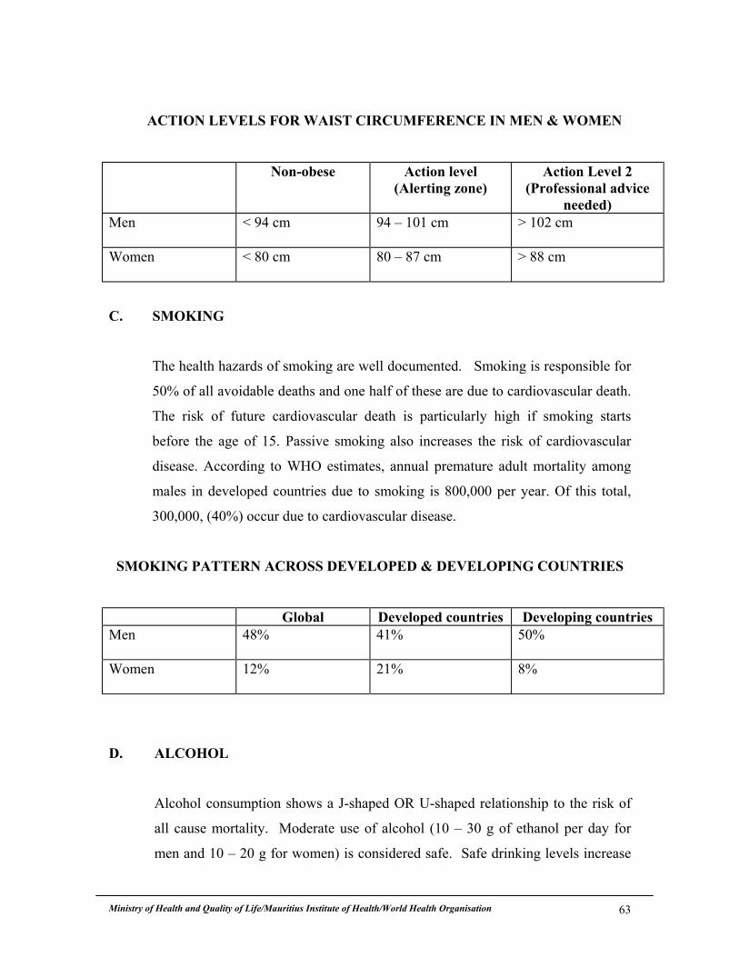
ACTION LEVELS FOR WAIST CIRCUMFERENCE IN MEN & WOMEN
Non-obese Action level (Alerting zone)
Action Level 2 (Professional advice
needed) Men
< 94 cm 94 – 101 cm > 102 cm
Women
< 80 cm 80 – 87 cm > 88 cm
C. SMOKING
The health hazards of smoking are well documented. Smoking is responsible for
50% of all avoidable deaths and one half of these are due to cardiovascular death.
The risk of future cardiovascular death is particularly high if smoking starts
before the age of 15. Passive smoking also increases the risk of cardiovascular
disease. According to WHO estimates, annual premature adult mortality among
males in developed countries due to smoking is 800,000 per year. Of this total,
300,000, (40%) occur due to cardiovascular disease.
SMOKING PATTERN ACROSS DEVELOPED & DEVELOPING COUNTRIES
Global Developed countries Developing countries Men
48% 41% 50%
Women
12% 21% 8%
D. ALCOHOL
Alcohol consumption shows a J-shaped OR U-shaped relationship to the risk of
all cause mortality. Moderate use of alcohol (10 – 30 g of ethanol per day for
men and 10 – 20 g for women) is considered safe. Safe drinking levels increase
Ministry of Health and Quality of Life/Mauritius Institute of Health/World Health Organisation 63

plasma HDL cholesterol and this may explain the protective effect of alcohol.
However, heavy drinking particularly binge drinking, increases the risk of sudden
arrhythmic death. According to the Mauritius non-communicable disease survey
1998 (preliminary report), alcohol consumption like smoking does not vary
significantly across age groups. However, the proportion of heavy drinkers is
highest in men in their forties. Hence, this group should be targeted to prevent
the harmful effects of alcohol related problems.
E. PLASMA LIPIDS
Plasma lipids can be measured by using:
1. Friedwald’s formula
2. Ratio of total cholesterol to HDL cholesterol
1. FRIEDWALD’S FORMULA
Total cholesterol, triglycerides and HDL cholesterol are measured in our
laboratory from a venous sample taken in the non-fasting state. With
these three measurements, the part of cholesterol carried in LDL can be
calculated according to Friedwald’s formula.
a. In mmol per litre (normal value LDL - C < 3.0 mmol per litre)
LDL cholesterol = total cholesterol – HDL cholesterol – (0.45 x
triglyceride level)
b. In mg per deciliter (normal LDL – C < 115 mg per deciliter)
LDL cholesterol = total cholesterol – HDL cholesterol – (0.2 x
triglyceride level)
Ministry of Health and Quality of Life/Mauritius Institute of Health/World Health Organisation 64

Friedwald’s calculation is a cheaper and more reliable estimation of LDL
cholesterol than commercially available direct measurement of LDL-
cholesterol based on immuno separation.
2. RATIO OF TOTAL CHOLESTEROL TO HDL-CHOLESTEROL
Measurement of the above ratio is also a good indicator of risk. A total
cholesterol/HDL ratio > 5, indicates coronary heart disease risk.
HDL cholesterol is cardio-protective whereas LDL cholesterol causes
atherosclerosis. High triglyceride levels (> 2.0 mmol) have also been
identified as a risk factor for coronary heart disease. A large body of
evidence has demonstrated that cholesterol lowering reduces the risk of
coronary heart disease events. In general, a reduction of LDL cholesterol
of 10% would lead to a reduction in the risk of coronary heart disease of
about 20% and this has turned out to be the case in clinical trials of both
primary and secondary prevention.
There are four classes of lipid lowering drugs in current use (statins,
fibrates, resins, niacin) and one or more drugs of each class have been
shown to reduce CHD morbidity and mortality. Evidence for efficacy and
safety in primary prevention is strongest for the statins.
F. PHYSICAL ACTIVITY
Regular aerobic physical activities have favourable effects on body weight, blood
pressure, plasma lipids, glucose tolerance and insulin sensitivity. Physical activity
recommendations have to define the intensity, duration and frequency of exercise.
The intensity of exercise for healthy individuals is best defined in terms of target
heart rate during peak exercise as shown below.
Ministry of Health and Quality of Life/Mauritius Institute of Health/World Health Organisation 65

SUITABLE TARGET HEART RATE DURING EXERCISE, ACCORDING TO
AGE [60-75% of maximum heart rate (220-age)]
Age (years) Heart rate per minute
20 – 29 115 – 145
30 – 39 110 – 140
40 – 49 105 – 130
50 – 59 100 –125
60 – 69 95 – 115
The target heart rate is easily achieved by exercises involving the use of large
muscle groups. Brisk walking, jogging, cycling, swimming, volleyball, aerobic
dancing and rope jumping are examples of such activities. Below is provided an
indication of the number of calories spent during various types of exercise:
Brisk Walking 3.6 calories/minute
Jogging 5.0 calories/minute
Running 5.0 calories/minute
Swimming 6.0 calories/minute
Cycling 4.5 calories/minute
Tennis 7.0 calories/minute
The duration of physical activity should preferably be 30 – 40 minutes, including
5 – 10 minutes warm up phase before the 20 – 30 minutes phase and 5 – 10
minutes cool down phase at its end and as frequent as 4-5 times weekly.
Ministry of Health and Quality of Life/Mauritius Institute of Health/World Health Organisation 66

G. BLOOD PRESSURE
Many epidemiological studies have demonstrated the importance of elevated
blood pressure as a risk for coronary heart disease in both men and women. The
decision to start drug therapy depends not only on the grading of blood pressure
level but also on the overall cardiovascular risk which calls for a proper history,
physical examination and laboratory examinations to identify the:
1. Presence of cardiovascular disease
2. Coexistence of risk factors
3. Incidence of target organ damage
Blood pressure should be lowered slowly and carefully because myocardial
necrosis, coronary atherosclerosis and cardiac hypertrophy may render coronary
autoregulation less effective in preserving organ perfusion when blood pressure is
reduced too rapidly.
(Refer to hypertension guidelines)
H. DIABETES
Type 1 and type 2 diabetes are associated with a marked increase of CHD. The
excess cardiovascular risk associated with type 1 diabetes becomes evident after
the age of 30. Type 2 diabetes is associated with more profound abnormalities in
cardiovascular risk factors than type 1 diabetes. Even IGT is associated with a
cardiovascular risk pattern characteristic of type 2 diabetes namely high
triglyceride level, low HDL value, increased value of hypertension and
hyperinsulinemia. The Diabetes Complication and Control Trial (DCCT) showed
that glucose control is important for the prevention of long term complications.
(Refer to Diabetes Guidelines)
Ministry of Health and Quality of Life/Mauritius Institute of Health/World Health Organisation 67

MODEL FOR PRIMARY AND SECONDARY PREVENTION PROGRAM FOR
NCD
Primary Prevention Secondary Prevention P Community I R Education Detection of Surveillance N I (Population) New Cases of old case T M E A R R V Y E N Early Improved T Treatment Treatment I P O R N E V C E Modified N Environment Family/ O T & Lifestyle Community Decreased Complications I (Population) Awareness N O N T Patient Education R O L NCD Complications
Ministry of Health and Quality of Life/Mauritius Institute of Health/World Health Organisation 68
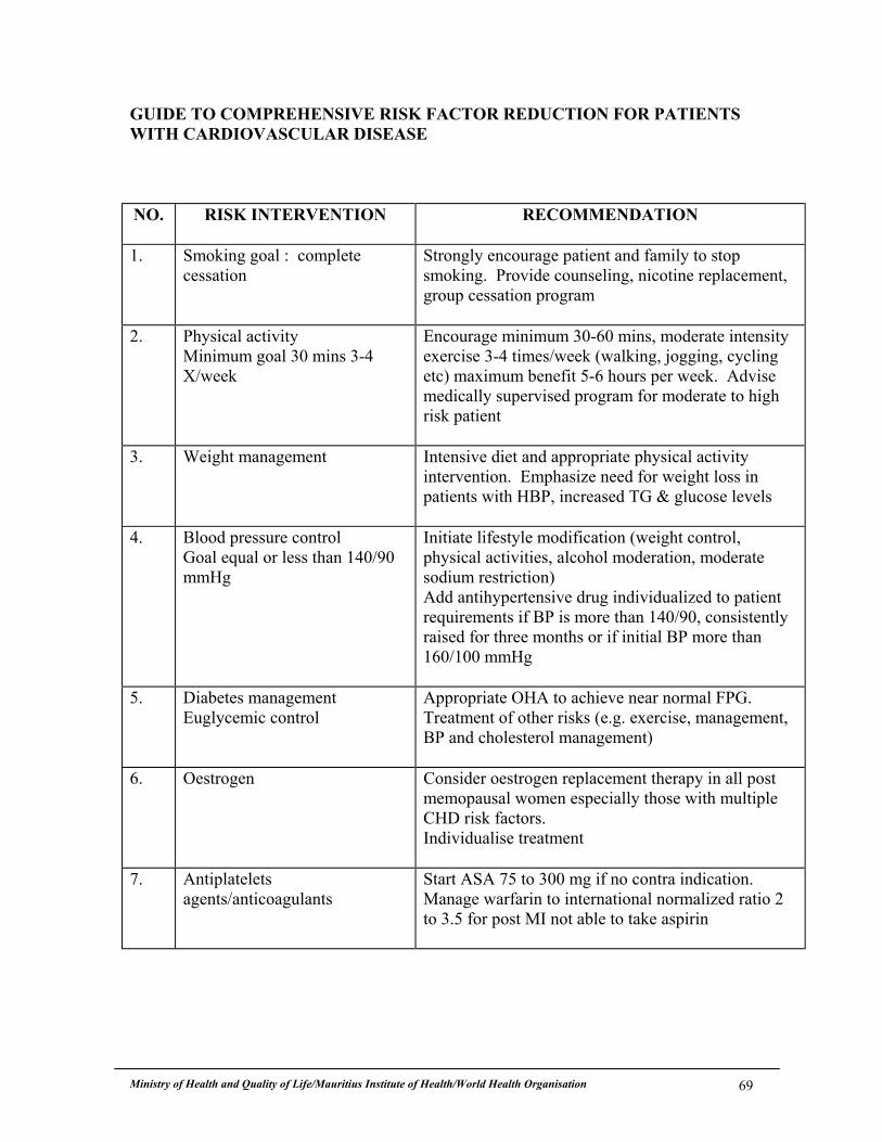
GUIDE TO COMPREHENSIVE RISK FACTOR REDUCTION FOR PATIENTS WITH CARDIOVASCULAR DISEASE
NO. RISK INTERVENTION RECOMMENDATION
1. Smoking goal : complete cessation
Strongly encourage patient and family to stop smoking. Provide counseling, nicotine replacement, group cessation program
2. Physical activity Minimum goal 30 mins 3-4 X/week
Encourage minimum 30-60 mins, moderate intensity exercise 3-4 times/week (walking, jogging, cycling etc) maximum benefit 5-6 hours per week. Advise medically supervised program for moderate to high risk patient
3. Weight management Intensive diet and appropriate physical activity intervention. Emphasize need for weight loss in patients with HBP, increased TG & glucose levels
4. Blood pressure control Goal equal or less than 140/90 mmHg
Initiate lifestyle modification (weight control, physical activities, alcohol moderation, moderate sodium restriction) Add antihypertensive drug individualized to patient requirements if BP is more than 140/90, consistently raised for three months or if initial BP more than 160/100 mmHg
5. Diabetes management Euglycemic control
Appropriate OHA to achieve near normal FPG. Treatment of other risks (e.g. exercise, management, BP and cholesterol management)
6. Oestrogen Consider oestrogen replacement therapy in all post memopausal women especially those with multiple CHD risk factors. Individualise treatment
7. Antiplatelets agents/anticoagulants
Start ASA 75 to 300 mg if no contra indication. Manage warfarin to international normalized ratio 2 to 3.5 for post MI not able to take aspirin
Ministry of Health and Quality of Life/Mauritius Institute of Health/World Health Organisation 69

NO. RISK INTERVENTION RECOMMENDATION
8. Lipid management
Primary goal LDL < 100 mg/ dl Secondary goal HDL > 35 mg/dl Tg < 200 mg/dl
Diet < 30 % fat, < 7% saturated fat, < 200 mg/day cholesterol Assess fasting lipid profile. Post MI patient – 4 to 6 weeks to stabilize. Add drug therapy according to following guide :
LDL < 100 mg/dl No drug therapy Follow up every 5 years
LDL 100-130 mg/dl Consider therapy to diet as follows
LDL > 130 mg/dl Add drug treatment to diet as follows
HDL < 35 mg/dl Emphasize weight management, physical activity, quit smoking, increased fruit consumption
Suggested drug therapy Tg < 200 mg/dl
Tg 200-400 mg/dl
Tg > 400 mg/dl
Statin
Statin
Niacin
Niacin
Combined treatment fibrate niacin + statin
If LDL goal not achieved, consider combination therapy
Ministry of Health and Quality of Life/Mauritius Institute of Health/World Health Organisation 70

CONCLUSION:
There is no ideal response or universal blue print to reverse the epidemic trends
of CAD. However, some common principles for a successful program on primary
prevention of CAD should comprise a combination of the following features:
1. Political will and leadership.
2. A strategic plan - stakeholders (rnulti-sectorial and multi-level).
• Situational analysis of past and present.
• Functional information support system.
• Goals, targets and implementation.
• Protocols and guidelines.
• Resources/downsizing and prioritization of services.
• Technical support from international agencies.
• Monitoring and evaluation.
• Re-programming.
3. Community involvement
4. Access to secondary and tertiary care
Thirty years ago, Eastern Finland was described as the world capital of
cardiovascular mortality. The North Karelia Health Promotion project based on
the adoption of healthy lifestyle had the following desirable outcomes:
1. Coronary mortality for males 35 - 54 dropped by 51% (720 reduced to 350
per 100,000).
2. 11% reduction in average blood cholesterol level.
Ministry of Health and Quality of Life/Mauritius Institute of Health/World Health Organisation 71

3. Male smoking prevalence decreased by half to one third.
4. Cancer mortality declined by 45 %.
5. Finland now ranks 6th for males and 14th for females in death from CAD.
Can Mauritius meet the challenge of reversing the trend in cardiovascular
disease? The answer is positive because many risk factors are associated with a
broad spectrum of non- communicable diseases which have been created by, and
can be controlled by Mauritians themselves.
Ministry of Health and Quality of Life/Mauritius Institute of Health/World Health Organisation 72

REFERENCES
1. Is primary care essential? Barbara Starfleld. The Lancet. Vol 344. October 22
1994.
2. Prevention of coronary heart disease in clinical practice. Task Force Report.
Prepared by David Wood et al. European Heart Journal (1998) 19, 1434 – 1503.
3. Guide to primary prevention of cardiovascular disease. A statement for Health
Care professional. From the Task Force on Risk Reduction. PREPARED BY Scot
M et al.
4. How to implement physical activity in primary and secondary prevention.
American Heart Association. Gerald F Fletcher. AHA Medical/ Scientific
Statement.
5. Mauritius Non Communicable Disease Surveys 87/92.
6. Mauritius Non Communicable Disease Surveys 98. A preliminary report.
7. National Health Statistic 1999.
8. 1999 World Health Organisation - International Society of hypertension. Journal
of hypertension 1999, 17: 151 – 183.
9. Epidemic of Cardiovascular Disease in post communist countries. Heart beat
number 3, September 1995. Journal of International society and federation of
cardiology.
10. Asian medical Journal. Vol 14 number 9, September 1997 Counter measures for
smoking cessation.
Ministry of Health and Quality of Life/Mauritius Institute of Health/World Health Organisation 73

11. Control of cardiovascular disease in developing countries – research, development
and institutional strengthening. Chrostopher P Housen et al. Bulletin of World
Health Federation. Number 1. March 1999.
12. Physical Activity for healthy adults as a primary prevention intervention in the
clinical setting. Sally S Haris et al. JAMA, June 23/30 - 1999 vol 261 number 24.
13. Physical exercise in the management of hypertension. Kikua Arakawa - Asian
Medical Journal. Vol 43. number 3- 107- 111, March 2000.
Ministry of Health and Quality of Life/Mauritius Institute of Health/World Health Organisation 74

Behavioural change
Procjaska and Diclemente have proposed a ‘stages of change’ model (387-388)
which argues that everyone is not equally ready to change their behaviour at a
given point in time even if they have all been invited to have screening and risk
factor modification. They maintain that it is important to assess the individual’s
behaviour thoughts, attitudes and beliefs concerning their perceived ability to
change, their behaviour over the past 6 months as well as the environmental
context in which they will attempt to change, and maintain the lifestyle change.
Five stages are proposed: pre-contemplation, contemplation, preparation action
and maintenance. In the pre-contemplation stage, individuals do not intend to
change their high-risk behaviour in the foreseeable future i.e over the next 6
months. Individuals can be in this stage because:
(a) they are unaware of the long-term consequences of their behaviour
(b) they are discouraged about their ability to change and do not want to think
about it
(c) they are defensive due to social pressure to change. Pre-contemplation is a
very stable stage. As a group they evaluate the pros of their risk behaviour as
greater than the cons.
Contemplation is the stage where people seriously intend to change their
behaviour within the next 6 months. Despite their intentions, however, they
typically stay in this stage for long periods of time, e.g. 2 years or more. They talk
about change, but keep putting it off. Those who substitute thinking for acting are
called chronic contemplators. Contemplators evaluate the pros and cons of their
behaviour as about equal; hence there is considerable ambivalence about
changing.
Ministry of Health and Quality of Life/Mauritius Institute of Health/World Health Organisation 75

People in these first two stages should not enter behaviour change programme, eg.
Smoking cessation, dietary or exercise programme. Most current smokers are in
these stages. Instead they should be provided with information, motivation and
advice that will help them move to the next stage, in which behavioural change is
seriously considered.
In the preparation stage, individuals intend to take action in the near future,
typically within the next month. They have a plan of action, and have usually
some modest behavioural change, such as reducing the number of cigarettes
smoked per day or slightly modifying diet. In this stage there are both behavioural
and intentional criteria; the cons are evaluated as greater than the pros. In this
stage, cues to action should be provided, such as the demonstration of association
between lifestyle and symptoms, illness in other family members, social pressures
and so on.
Overt behavioural change within the past six months characterises the action
stage, which is highly unstable and where the greatest risk for relapse may occur.
The criterion for achieving this stage is to have actually changed behaviour, eg.
Stopped smoking, as opposed to reducing smoking or switching brand to lower
tar, etc., During this period most of the processes of change come into play and
the intrapersonal (eg. Perceived self-efficacy), interpersonal (e.g. social support)
and environmental (eg. Unavoidable exposure to smoking environment) factors
associated with these processes are principal determinants of whether the newly
quit smoker proceeds to the next stage (maintenance) or regresses to the earliest
stage. Obviously this is the critical period where all the interventions strategies
must focus on the above mentioned processes. A wide variety of instructive
materials are usually available from national Heart Associations and Foundations
including tailored self-instruction programmes, support groups and counselling
strategies.
Ministry of Health and Quality of Life/Mauritius Institute of Health/World Health Organisation 76

Maintenance is considered from six months after an individual has changed
behaviour until about five years of continuous maintenance has elapsed. (N.B. :
Most smoking cessation programmes consider six months to one-year continuous
abstinence as “success”). For example, smokers who successfully move from
action to maintenance demonstrate a gradual decrease in overall temptation and
lower use of cognitive/experiential processes, and a heightened confidence and
greater use of behavioural techniques such as stimulus control, counter-
conditioning, helping relationship and contingency management. Mapping out
templates of change associated with successful recovery could allow comparisons
to be made across smoking and alcohol problems to highlight the general
principles underlying successful addictive behaviour change.
In summary, behaviour change programmes achieve much higher success rates
when they assess the state or readiness of the individual to commit to the change
process. The ‘stages of change’ model reviewed here is becoming increasing
employed by health care professionals who are concerned with treating and/or
referring patients for appropriate assistance, as it proves it’s value in providing
services to those who are ready to benefit from them.
Ministry of Health and Quality of Life/Mauritius Institute of Health/World Health Organisation 77

Coronary Risk Chart (not intended for clinically established CHD patients), has several
functions:
- absolute risk of developing CHD over next 10 years can be read off the chart without
any calculations.
Lifetime risk can be read by following the tables upwards (increased risk with increase
in age).
Relative risk can readily be estimated by comprising the risk in one cell with any other in
the same age group.
- One can predict the effect of changing from one risk category to another. For example,
the reduction in risk associated with cessation of smoking, reduction of BP or cholesterol
level can readily be shown to an individual.
- a Coronary Risk Chart developed by each country is recommended. May be constructed
from results of prospective cohort studies.
Ministry of Health and Quality of Life/Mauritius Institute of Health/World Health Organisation 78

Ministry of Health and Quality of Life/Mauritius Institute of Health/World Health Organisation 79

Ministry of Health and Quality of Life/Mauritius Institute of Health/World Health Organisation 80

Ministry of Health and Quality of Life/Mauritius Institute of Health/World Health Organisation 81

Ministry of Health and Quality of Life/Mauritius Institute of Health/World Health Organisation 82

Ministry of Health and Quality of Life/Mauritius Institute of Health/World Health Organisation 83

Ministry of Health and Quality of Life/Mauritius Institute of Health/World Health Organisation 84

RECOMMENDATIONS
1. All Area Health Centres and Community Health Centres
should have an ECG apparatus
2. All Nurses should be trained to do ECG
3. Doctors should be re-trained in interpreting ECG’s
4. Morphia should be made available at all Area Health Centres
and Community Health Centres
Ministry of Health and Quality of Life/Mauritius Institute of Health/World Health Organisation 85