Embed Size (px)
Citation preview
advanced
drug delivery reviews
ELSEVIER Advanced Drug Delivery Reviews 18 (1995) 53-71
Clinical investigation of gut immune responses
Anne Fergusona.*, Jamal Sallam”, Seamus O’Mahony”, Ian Poxtonh “University of Edinburgh Department of Medicine, Western General Hospital, Edinburgh EH4 2X(/. UK
hDepartment of Medical Microbiology, Western General Hospital, Edinburgh EH4 ZXCJ. UK
Accepted June 1995
Abstract
Mucosal and systemic immune systems are quite separate in their properties and in the factors which regulate them. In man, assessment of immune status at gut level, including the capacity to respond to oral vaccines, should follow the general lines which are used in research and clinical investigation of systemic immunity. All of the currently available methods for direct studies of gut immunity have limitations, but a new technique, per-oral whole gut lavage with non-absorbable cleansing fluid, provides a system of gut perfusion. The value of this technique is illustrated by data on intestinal antibodies to bacterial LPS core antigens, of gut immunity in healthy African children, on the isotype of antibodies elicited by oral killed whole cell/cholera toxin B vaccine in coeliac disease patients, and on the effects of smoking on intestinal antibodies elicited by the oral typhoid vacine Ty2la in healthy UK adults. Finally, results of a recent comprehensive study show that immunological tests using faeces are likely to be highly misleading.
Keywords: Intestinal antibody; Intestinal lymphocyte: Copro-antibody: Faecal immunology; Cholera toxin: Oral cholera vaccine; Oral typhoid vaccine
Contents
1. Introduction ......................................................
2. Mucosal immunity is distinct from systemic immunity ................ 2.1. Patterns of immune responses to fed antigen .................... 2.2. Induction and expression of immunity .......................... 2.3. Immunoregulation ............................................ 2.4. Spectrum of immune reactions to cholera toxin in mice ..........
3. Required information for full clinical description of mucosal immunity
3.1. Structured evaluation of systemic immune status ................ 3.1 .l. Clinical history ......................................... 3.1.2. Cells and cell-mediated immunity ........................ 3.1.3. Immunoglobulins and antibodies ......................... 3.1.4. Other immune parameters ...............................
3.2. Evaluation of gastrointestinal immune status .................... 3.2.1. Antigen handling ....................................... 3.2.2. Cells ...................................................
* Corresponding author: Fax 0131-537-1007.
................
................
................
................
................
................
................
................
................
................
................
........... .....
................
..........
..........
..........
..........
0169-409X/95/$29.00 @ 1995 Elsevier Science B.V. All rights reserved
SSDI 0169-409X(95)00050-X

3.23. Specilic cell-mediated immunity ....... ........ .... ............................................... 3.2.4. Immunoglobulins and antihodics ..................... ................ ........................... 3.2.5. Rcq!ulation of induction of active immunity and tolerance ..............................................
4. Source materl;lls for humoral immunity studies .................................................................. 1.1. Mucosalbiopsics ........................................................................................ 4.2. Directly aspirated intcstinnl secretions ................................................................. 4.3. Segmental perfusion ................................. ................. .......................... 4.4. Whole gut lavage ...................... ..... ............................................ 4.5. Critical importance of specimen handling and storage ........... ...........................................
5. Validation and applications of the whole gut lavapc technique .......................................... ........ 5.1. Clinical and laboratory protocols and methods .............................................................
5.1.1. Clinicalproccdure ..............................................................................
5.1.2. Processing of whole gut lavage Huid .................................................................. 5.1.3. Assaytechniqucs ..................................................................................
5.2. Validation as a whole gut perfusion ........................................................................
5.3. llse 01 gut Iavage for investigation of intestinal antibodies to bacterial endotoxin ................................ 53.1. Structure ol cndotoxin ........................................................................ -i.3.2. Serum and intestinal antibodies to LPS cow antigens ................................................. 53.3. Potential for active or passive immunisation ..........................................................
5.4. Studies of gut immunity m healthy African children ....... .............................................. 5.4.1. Rationale lor studying gut immunity in children ....................................................... 5.42. Asscsxmcnt of gut immune status in Atrican children ..................................................
6. Studiesoffaeccsareunhelpful ..................................................................................
6.1. Possible analytical and sampling problems associated with the use of faeces for immunological testing ............ 6.2. Comparison between faeces and whole gut lavage Huid .......................................................
62.1. Patientsandmethods ...............................................................................
6.2.2. Variable proportion of locally produced immunoglohulins and other proteins are detectable in faeccs ....... 7. Examples of the use of gut lavagc to study mucosal immunity to oral vaccines .......................................
7.1. Determinants of mucosal immune response to vaccination ..................................................
7.2. Immune responses to oral cholera vaccine in coeliac disease patients and controls ............................... 7.2.1. Subjects and methods ...........................................................................
7.22. Results and interpretation - intestinal antibodies ................ .................................... 7.23. Results and interprctatlon serum antibodies .......................................................
7.3. Effects of smoking on intestinal antibodies elicited hy the oral typhoid vacine Ty2la m healthy LJK adults ......... 73.1. Suhjectsandmethods ................................................................................
73.2. Results intestinal antibody responscy ............... .......................................... 8. Conclusions ...............................................................................................
References .................................................................................................
1. Introduction
The functions of the gastrointestinal tract
include the secretion of fluids and digestive enzymes; the absorption of nutrients; mixing and propulsion of foods, digestive secretions and the gut microflora; and the neuro-hormonal coordi- nation of these phenomena. These digestive processes have profound effects on the quantities and molecular forms of antigens within the gut lumen and tissues, and thus also on local immune reactions. Gastro-intestinal infections may direct- ly damage the tissues or cells, or may only affect functions such as secretion or motility. Local protective immunity will influence the severity of
58 5x
.5x
59 55,
5’)
SY
59
60
ho
ho
60
ho
hO
hl
61
61
61
62
62
62
62
62
63
64
64
64
65
65
66
66
66
67
67
68
68
6X
69
these pathological effects on the intestines in
addition to direct effects on the viability and pathogenicity of the organisms concerned. Per- haps because of the quite different properties of secretory IgA when compared with serum IgG
antibodies, it appears that a vigorous local im- mune response does not necessarily confer protection against re-infection, or even lead to complete elimination of an intestinal pathogen, although local immunity probably influences symptom severity.
The several types of immune responses in the gut are, normally, strictly regulated. Such active immune responses may be host-protective, e.g. conferring resistance to bacterial infection, neu-
A. Ferguson et al. I Advanced Drug Delivery Reviews I8 (199.5) 53-71 ss
tralising viruses or toxins; the immune responses may occur but may be completely irrelevant to any protective or other function of the gut (as in most reactions between antibodies and food antigens); and from time to time the immune responses cause tissue damage, for example by delayed type hypersensitivity (DTH) reactions, IgG- or IgM-mediated complement-fixing hyper- sensitivity, or IgE-mediated local anaphylaxis. Hypersensitivity may be entirely inappropriate, and thus a primary cause of disease (e.g. to gluten in coeliac disease), but also may be an unavoidable side effect of a protective immune response, as is well recognised in tuberculosis and leprosy, and in the elimination phase of helminth parasite expulsion from the gut.
Much research on gut immunology has been conducted in mice. It is likely that the general principles derived from the results of such ex- perimental work are also applicable in man, but this has been proven for only a few aspects of immunity. Good clinical studies of the physiology and regulation of the human GALT are urgently required.
2. Mucosal immunity is distinct from systemic immunity
The science of mucosal immunology has de- veloped rapidly during the last 20 years, and many of the principles and concepts are different from those of classical systemic immunity. A recently published handbook [l] gives a com- prehensive account of the biology of the gut epithelium, mucosal defence mechanisms, mucosal immunoglobulins, lymphoid cells, regu- lation of induction and expression of immunity, inflammation, immunisation and vaccines, and is highly recommended as a source of background information to this paper.
2.1. Patterns of immune responses to fed antigen
The immune system of the gut has a number of different roles. When the route of entry is through follicle-associated epithelium of Peyer’s patches of immunologically normal and mature mammals, the general trend is towards suppres-
sion of immunity-in other words, oral tolerance. However, active immunisation may also follow the feeding of antigen and this is typically in the form of harmless secretory ZgA antibody. In some circumstances there is, however, induction of potentially immunopathogenic immune reactions when antigen is fed, for example ZgE, ZgG antibody or T cell mediated immunity. Thus there may be either induction or suppression of a particular immune response, that is antibody- or T cell-mediated. Regulation of immunity is criti- cal to normal gut function, and, of course, vac- cine design and delivery must lead to active immunity without hypersensitivity, but not oral tolerance.
2.2. Induction and expression of immunity
Overall, mucosal immune functions can be separated into induction and effector phases.
The induction of a specific immune response is critically dependent on the situation of the in- dividual when antigen is first encountered. Fac- tors such as age, dose, route of encounter and physico-chemical form of antigen critically in- fluence the type of immune response thatwill predominate for the rest of the life of the individual. The state of activation of antigen- processing cells and T cells in the tissues where antigen is first encountered are also important factors.
In an immune individual, re-exposure to an- tigen will result in expression of an antibody- mediated or cell-mediated specific immune re- sponse, either at the site of exposure or, if the antigen is absorbed or otherwise transferred around the body, at other sites or systemically. As discussed below, there are circumstances when a local immune response in the gut causes disease such as malabsorption. A key factor in such immune-mediated tissue damage is the capacity of the tissue to respond to stimuli such as cytokines. The impaired capacity to mount non-specific inflammation, such as occurs with some nutrient deficiencies, may in fact protect the gut epithelium from some of the potentially adverse effects of a mucosal DTH reaction. We have observed failure of the usual crypt hy-
perplasia associated with intestinal DTH in pro-
tein malnourished mice [2].
2.3. Immunoregulation
There are many T lymphocytes dispersed in
the mucosa as well as in the organised lymphoid
tissues of the gut. T cells are critical to the induction of appropriate immune responses when antigen is encountered via the gut lumen,
and also as important effector cells for local protective and hypersensitivity reactions.
Handling of antigen by the gut is also irn-
portant. Experiments with protein antigens have
shown that there is subtle alteration - “process- ing” of antigen as it crosses the gut epithelium -
and such material is tolerogenic for cell-mediated
immunity, rather than immunogenic 13-51. In all normal mammals antigenic molecules gain access
from the gut lumen into the tissues of the body.
but immune responses to such materials do not normally cause disease.
2.4. Spectrum oj’ immune reactions to cholera toxin in mice
Experiments which we conducted in mice,
using cholera toxin (CT) as a fed antigen. illus- trate very well the distinct differences between
the mucosal and systemic compartments, humor-
al and cellular immunity, active immunisation and tolerance [6-81. Cholera toxin is a most
unusual antigen with considerable adjuvant ac- tivity when given orally and we found, as did several other investigators, that a single oral dose of CT induced not only intestinal but also a serum antibody response. However, the cellular limb of immunity (assessed by a footpad swelling
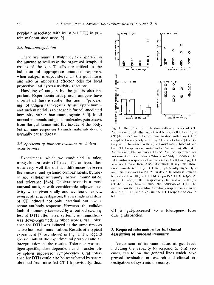
test of DTH after later, systemic immunisation) was down-regulated; in other words, oral toler- ance for DTH was induced at the same time as active humoral immunisation. Results of a typical experiment [7] are shown in Fig. 1. The legend gives details of the experimental protocol and an interpretation of the results. Tolerance was an- tigen-specific, dose-dependent and transferable by spleen suppressor lymphocytes. Oral toler- ance for DTH could also be transferred by serum obtained from mice fed CT 1 h previously: thus,
F;lp. I. The cf’f’ect of prefeeding different doses of C’T.
Ammals were Icd either ABS (inert buffer) or 0. I, I or 10 ~8
(“1. (day -7) 1 week before immunization with 1 gg CT in
complctc Freund’s adjuvant (day 0). 2 weeks later (day 14).
rhcv wcrc challenged with 5 fig toxoid into a footpad and
their DTH responses measured as footpad swelling after 24 h.
Animals wc’rc hled cm days 7. 13 and 22 of the experiment tar
;t\scssment of their strum antitoxin antibody responses. The
I$; antitoxin responses of animals fed either 0.1 or I pg CT
MCI-C’ no different f’l-om ABS-fed controls at any time. How-
c\cr. ;animals fed IO pccg C‘l‘ had significantly highcl lt$;
antitoxin responses ( p < 0.02) on day 7. In contrast. animals
led either I or IO pg (“I‘ had suppressed D’I‘H responses
( p .- O.Wl and ,I c’ 0.01. respectively) but a dose of 0.1 ,LLL~
(‘I’ did not significantly inhibit the induction of DTH. The
graphs show the IgG antitoxin antibody response in serum on
days 7 (a). 13 (b) and 22 (d) and the DTH response on day 15
(c)
CT is ‘gut-processed’ to a tolerogenic form
during absorption.
3. Required information for full clinical description of mucosal immunity
Assessment of immune status at gut level, including the capacity to respond to oral vac- cines, can follow the general lines which have proved invaluable in research and clinical in- vestigation of systemic immunity.
3.1. Structured evaluation of systemic immune status
Protocols for the clinical evaluation of systemic immunity which combine general clinical fea- tures, in vivo and in vitro laboratory tests, are agreed and widely applied by clinical and labora- tory immunologists, and are used in the the investigation and management of patients with primary, acquired and iatrogenic immuno- deficiency syndromes. Aberrant immunity can either be perceived and classified in relation to whether the problem lies in primary lymphoid organs, the peripheral lymphoid system, or in the non-antigen specific cellular or humoral mecha- nisms. A different approach, perhaps of greater relevance for day to day therapeutic decisions, classifies the patient by the type of effector mechanism involved, for example T cell me- diated immunity, immunoglobulin isotype, poly- morphonuclear or mononuclear cell function.
3.1.1. Clinical history Important features to be elicited include family
history, of immunodeficiency states, autoimmune disease and atopy. Evidence of previous normal recovery from bacterial or viral infections is important. Response to immunisation, particu- larly with live vaccines (smallpox, BCG) can give valuable evidence of normal cell mediated im- munity. The aberrant immune status of atopy can usually be implied from the history.
3.1.2. Cells and cell-mediated immunity There are now a range of techniques for
defining lymphocyte subsets in blood, extending into research orientated observations. Evidence of the existence of specific cell mediated immuni- ty (implying both normal afferent and efferent limbs) is obtained by in vivo tests of delayed type hypersensitivity using a range of recall antigens such as tuberculin, and many in vitro tests of antigen reactive T cell function are available, ranging from antigen driven blast transformation to the secretion of cytokines in culture with antigen. Usually, these tests are done with peripheral blood lymphocytes.
3.1.3. Immunoglobulins and antibodies Almost invariably, assays of total immuno-
globulins and of subclasses are done on serum, by a variety of techniques in which monoclonal or polyclonal anti-heavy chain antisera are used.
Apart from the use of immediate skin tests for in vivo detection of IgE class antibodies, tests for the presence, titres, avidity etc of antibody are also studied using serum. Precise information on induction and expression of humoral immunity is obtained by studying the primary and secondary immune responses to defined antigens not previ- ously encountered, for example, key-hole limpet haemocyanin.
3.1.4. Other immune parameters A wide range of other cellular and humoral
components may be aberrant and a full immune status evaluation should include an appraisal of eosinophils, mast cells, basophils, complement, and reticuloendothelial function.
3.2. Evaluation of gastrointestinal immune status (Table I)
3.2.1. Antigen handling The afferent limb, involving the follicle associ-
ated epithelium and organised lymphoid tissues such as Peyer’s patches, tonsils, appendix, is rarely investigated in man. Operative specimens and endoscopically targetted biopsies of ileal Peyer’s patches will provide tissues for such studies. Operative and post mortem specimens of mesenteric lymph nodes can also be examined.
3.2.2. Cells Most workers have concentrated on cells serv-
ing effector functions, and ideally, a range of properties should be assessed.
Mucosal biopsies can safely be taken from most levels of the gastrointestinal tract, and by using micro techniques and multiple biopsies, both histological and in vitro methodscan be applied. Immunofluorescence or immunoenzyme stains, using polyclonal or monoclonal antibodies targetted to either membrane or cytoplasmic antigens, have greatly expanded the information which can be accrued from mucosal biopsy histopathology.
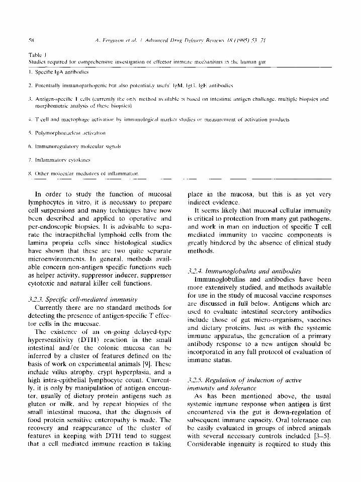
I.
2.
3.
4.
5,
h.
7.
X.
Specific IgA antibodies
Potentially immunopathogcnic but also potentially uaelul IgM. I@;. IpE antibodies
Antigen-specific ‘I cells (currently the onl! method ~1\xlahle 1s haxcd on intestinal antigen challcnye. multiple biopsies and
morphomctric analysis of thcsc biopsies)
I‘ cell and macrophage activatwn hy Immunological marker studies or mcasurcment of activation products
Polymorphonuclcar xtivatlon
Immunorcpulatory molecular sipndls
InfIammatory qtokines
Other molecular mediators 01 ~nllammat~on
In order to study the function of mucosal
lymphocytes in vitro, it is necessary to prepare cell suspensions and many techniques have now
been described and applied to operative and per-endoscopic biopsies. It is advisable to sepa- rate the intraepithelial lymphoid cells from the lamina propria cells since histological studies
have shown that these are two quite separate microenvironments. In general, methods avail-
able concern non-antigen specific functions such as helper activity, suppressor inducer, suppressor cytotoxic and natural killer cell functions.
3.2.3. Specific cell-mediated immunity Currently there are no standard methods for
detecting the presence of antigen-specific T effec- tor cells in the mucosae.
The existence of an on-going delayed-type hypersensitivity (DTH) reaction in the small intestinal and/or the colonic mucosa can be inferred by a cluster of features defined on the basis of work on experimental animals [9]. These
include villus atrophy, crypt hyperplasia, and a high intra-epithelial lymphocyte count. Current- ly. it is only by manipulation of antigen encoun- ter, usually of dietary protein antigens such as gluten or milk. and by repeat biopsies of the small intestinal mucosa, that the diagnosis of food protein sensitive enteropathy is made. The recovery and reappearance of the cluster of features in keeping with DTH tend to suggest that a cell mediated immune reaction is taking
place in the mucosa, but this is as yet very indirect evidence.
It seems likely that mucosal cellular immunity
is critical to protection from many gut pathogens, and work in man on induction of specific T cell mediated immunity to vaccine components is
greatly hindered by the absence of clinical study methods.
.1.2.4. Immunoglobulins und antibodies Immunoglobulins and antibodies have been
more extensively studied, and methods available for use in the study of mucosal vaccine responses are discussed in full below. Antigens which are
used to evaluate intestinal secretory antibodies
include those of gut micro-organisms, vaccines and dietary proteins. Just as with the systemic immune apparatus, the generation of a primary antibody response to a new antigen should be incorporated in any full protocol of evaluation of immune status.
.L?.S, Regulation of induction of active immunity and tolerance
As has been mentioned above, the usual systemic immune response when antigen is first encountered via the gut is down-regulation of subsequent immune capacity. Oral tolerance can be easily evaluated in groups of inbred animals with several necessary controls included [3-51. Considerable ingenuity is required to study this
A. Ferguson et al. I Advanced Drug Delivery Reviews 18 (199.5) S.?-71 59
phenomenon in man but it is possible, for exam- ple by using keyhole limpet haemocyanin [lo].
Experience with immunity to foods indicates that intestinal antigen-specific T cell mediated immunity does not normally develop to enterical- ly encountered antigens. Whether this is true tolerance, i.e. antigen-specific suppression, or whether it is merely the absence of this limb of the effector immune response at gut level, re- mains to be ascertained.
4. Source materials for humoral immunity studies
Animal and clinical studies show that with very few exceptions, tests on blood antibodies and circulating cells and cytokines (components of the systemic immune system), are virtually use- less as indices of mucosal immunity at gut level. Some general information on the function of the mucosa-associated lymphoid tissues can be ob- tained from studies of saliva or tears but these materials cannot provide organ-specific informa- tion relevant to the gut.
Unfortunately, for direct studies of gut im- munity, all of the currently available methods have limitations. Considerable information can be obtained by histological and other procedures applied to small bowel or colonic mucosal biop- sies, and fluid for immunological investigations may be aspirated from the jejunum or colon. How closely results obtained with these samples reflect events in other parts of the GI tract is uncertain. Furthermore, though there are many circumstances in which it is reasonable, and ethical, to intubate or biopsy symptomatic adults or children for research purposes, parallel data from clinically unaffected age-matched popula- tion controls are not easily available, and proper interpretation of the results obtained with the patients may thus be impossible.
4.1. Mucosal biopsies
It is possible to detect and count antibody- containing plasma cells in tissue sections by using isotope-labelled antigen and autoradiography, or by immunofluorescence or immunoenzyme tech-
niques. With double-labelling methods, the iso- type of antibody can also be determined. These approaches were extensively used in critical early research on mucosal immumity, using cholera toxin and tetanus toxin, in addition to soluble protein antigens such as hen egg ovalbumin.
Single cell suspensions of lamina propria cells, prepared by digestion and centrifugation of mac- erated biopsies, are suitable for in vitro-tests of antibody production such as the ELISPOT [13]. Antibody-producing cells can also be detected by combining culture and ELISA-based antibody detection [14].
4.2. Directly aspirated intestinal secretions
Direct jejunal intubation allows aspiration of upper small intestinal fluid [15], and similarly, duodenal or colonic fluid can be aspirated during upper GI endoscopy or colonoscopy [16], or through the tubing of a small bowel biopsy capsule [17]. When treated with protease in- hibitors and with appropriate storage conditions, these may yield much useful information on antibody production in these localised segments of the gut.
4.3. Segmental perfusion
Perfusion of a defined segment of the small intestine [18] or recta-sigmoid [19], with occlud- ing balloons above and below, allows measure- ments of locally-produced fluid, electrolytes, im- munoglobulins, cytokines, plasma-derived pro- teins and other materials relevant to gut immuni- ty. Basal and stimulated states can be assessed. This powerful approach has not, to our knowl- edge, been used to study vaccine responsiveness in man.
4.4. Whole gut lavage
Peroral gastrointestinal lavage with non-ab- sorbable fluid is now extensively used to cleanse the gastrointestinal tract prior to colonoscopy, barium enema examination or colonic surgery. This lavage fluid contains abundant quantities of IgA [20], and, in disease, also IgM and IgG [21]. After filtration and processing with protease
inhibitors, ELISA techniques can readily be used to study immunoglobulins and specific antibo- dies, as discussed below.
4.5. Critical importance of specimen handling and storage
Human intestinal secretions, including jejunal fluid and whole gut lavage fluid (WGLF) contain many and abundant proteases. If protease in- hibitors are not added to these materials immedi- ately after collection there is rapid degradation of proteins, including antibodies [20]. For exam- ple. we found in our early studies of WGLF that delays of 1 or 2 h in the addition of protease inhibitors resulted in loss of up to 92% of the IgA [21]. We also noted that early faecally-con- taminated specimens had much lower concen- trations of IgA than later clear specimens. Once WGLF specimens were clear, there was no significant difference in IgA content between serial specimens. In careful experiments with jejunal fluid, Forrest made the important ob- servation that heat-inactivation of proteases was an unsatisfactory method. leading to consider- able losses of IgA and antibodies 1221; he also suggested that data reported should take into account the total concentration of IgA in the fluid being examined.
5. Validation and applications of the whole gut lavage technique
Gut lavage with large volumes of saline has been used for some time to study intestinal antibodies in the clear fluid passed per rectum after ingestion of lo-20 1 of the fluid [23]. In 1988, Gaspari et al. [20] described a more accept- able technique. using 3-4 1 of a non-absorbable, commercially available polyethylene glycol (PEG)-based bowel cleansing fluid. We followed up this approach [21] and have since found that in patients with intestinal inflammatory diseases. this whole gut lavage fluid (WGLF) contains immunoglobulins. antibodies, plasma-derived proteins, inflammatory cells and cytokines [17,24-341. We have recently completed a study of fluid intake/output data, and concentrations of various substances in sequentially passed spec-
imens, confirming that WGLF is a gut perfusate
]351.
5.1. Clinical and laboratory protocols and methods
5 I. 1. Clinical procedure The lavage solution (available commercially as
Klean-prep (Norgine, Oxford, UK)) contains in one sachet: 59 g PEG with a molecular weight of 3350; 1.45 g NaCl; 1.63 g NaHCO,; 5.68 g Na,SO,; and 0.75 g KC1 (BP). One sachet is dissolved in 1 1 of tap water to give an osmolality of 260 mosm/l.
After an overnight fast, supervised and moni- tored by an experienced research nurse, patients or healthy volunteers drink the lavage solution, aiming for a rate of 200 ml every 12 min. After a period ranging from 30 min to 3.5 h, several formed or semi-liquid stools are passed, followed by faecal-stained fluid. These are discarded until clear fluid, resembling urine, is being passed per rectum. There is a very high success rate when the procedure is conducted as described-more than 95% in a series of hospital in-patients ranging in age from 14 to 89 years [36].
5.1.2. Processing of whole gut lavage fluid (261 20 ml of clear fluid are filtered through GF/A
(Whatman) glass fibre filters. To 10 ml of the filtered fluid the following reagents are added, with mixing after each addition (final concen- trations in parentheses): soya bean trypsin inhib- itor in phosphate-buffered saline (PBS) (80 pg/ ml); sodium ethylenediaminetetracetate in PBS (15 mM); phenylmethylsulphonyl fluoride in 95% ethanol (2 mM); sodium azide (1 mM); newborn calf serum (5% v/v).
Aliquots of processed WGLF are stored at -70°C for later analyses.
-5.1.3. Assay techniques Details are published elsewhere of the meth-
ods used for assays of immunoglobulin concen- trations [17], isotype-specific antibodies to diet- ary protein [17] and bacterial [31-331 antigens, haemoglobin [28], plasma-derived proteins [26], cytokines [29], and for cytological examination [30] of WGLF.
A. Ferguson et al. I Advanced Drug Delivery Reviews 18 (1995) 53-71 61
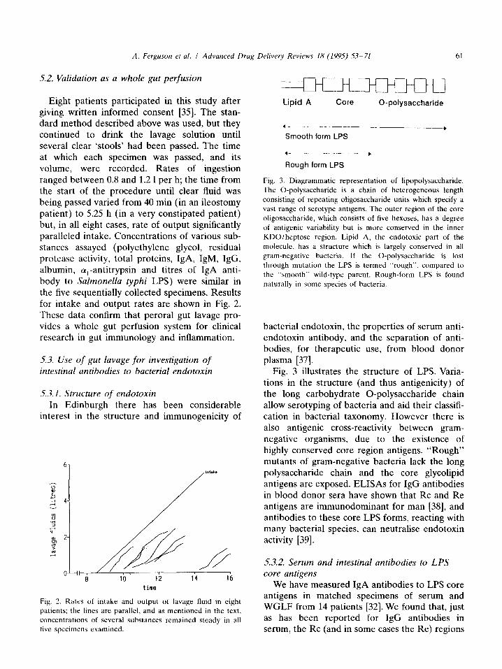
5.2. Validation as a whole gut perfusion
Eight patients participated in this study after giving written informed consent [35]. The stan- dard method described above was used, but they continued to drink the lavage solution until several clear ‘stools’ had been passed. The time at which each specimen was passed, and its volume, were recorded. Rates of ingestion ranged between 0.8 and 1.2 1 per h; the time from the start of the procedure until clear fluid was being passed varied from 40 min (in an ileostomy patient) to 5.25 h (in a very constipated patient) but, in all eight cases, rate of output significantly paralleled intake. Concentrations of various sub- stances assayed (polyethylene glycol, residual protease activity, total proteins, IgA, IgM, IgG, albumin, cu,-antitrypsin and titres of IgA anti- body to Salmonella typhi LPS) were similar in the five sequentially collected specimens. Results for intake and output rates are shown in Fig. 2. These data confirm that peroral gut lavage pro- vides a whole gut perfusion system for clinical research in gut immunology and inflammation.
5.3. Use of gut lavage for investigation of intestinal antibodies to bacterial endotoxin
5.3.1. Structure of endotoxin In Edinburgh there has been considerable
interest in the structure and immunogenicity of
“1 /
time
Fig. 2. Rates of intake and output of lavage fluid in eight
patients; the lines are parallel, and as mentioned in the text,
concentrations of several substances remained steady in all
five specimens examined
Lipid A Core 0-polysaccharide
,_-_-- _ Smooth form LPS
- -_.--+
Rough form LPS
Fig. 3. Diagrammatic representation of lipopolysaccharide.
The 0-polysaccharide is a chain of heterogeneous length
consisting of repeating oligosaccharide units which specify a
vast range of serotype antigens. The outer region of the core
oligosaccharide, which consists of five hexoses, has a degree
of antigenic variability but is more conserved in the inner
KDOlheptose region. Lipid A, the endotoxic part of the
molecule, has a structure which is largely conserved in all
gram-negative bacteria. If the 0-polysaccharide is lost
through mutation the LPS is termed “rough”, compared to
the “smooth” wild-type parent. Rough-form LPS is found
naturally in some species of bacteria.
bacterial endotoxin, the properties of serum anti- endotoxin antibody, and the separation of anti- bodies, for therapeutic use, from blood donor plasma [37].
Fig. 3 illustrates the structure of LPS. Varia- tions in the structure (and thus antigenicity) of the long carbohydrate 0-polysaccharide chain allow serotyping of bacteria and aid their classifi- cation in bacterial taxonomy. However there is also antigenic cross-reactivity between gram- negative organisms, due to the existence of highly conserved core region antigens. “Rough” mutants of gram-negative bacteria lack the long polysaccharide chain and the core glycolipid antigens are exposed. ELISAs for IgG antibodies in blood donor sera have shown that Rc and Re antigens are immunodominant for man [38], and antibodies to these core LPS forms. reacting with many bacterial species, can neutralise endotoxin activity [39].
5.3.2. Serum and intestinal antibodies to LPS core antigens
We have measured IgA antibodies to LPS core antigens in matched specimens of serum and WGLF from 14 patients [32]. We found that, just as has been reported for IgG antibodies in serum, the Rc (and in some cases the Re) regions
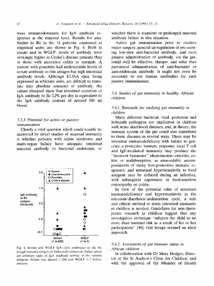
were immunodominant for IgA antibody re- sponses at the mucosal level. Results for anti- bodies to Rc in the 14 patients, expressed as empirical units, are shown in Fig. 4. Both in serum and in WGLF, levels of antibody were strikingly higher in Crohn’s disease patients than in those with ulcerative colitis or normals. A patient with pouchitis had undetectable levels of serum antibody to this antigen but high intestinal antibody levels. Although ELISA data, being expressed as arbitrary units, are difficult to trans- late into absolute amounts of antibody, the values obtained show that intestinal secretion of IgA antibody to Rc LPS per day is equivalent to the IgA antibody content of around 500 ml blood.
5.3.3. Potential for active or passive immunisation
Clearly a vital question which could readily be answered by direct studies of mucosal immunity is whether patients with sepsis syndrome and multi-organ failure have adequate intestinal mucosal antibody to bacterial endotoxin. or
2.0 0 Ulcerative colilis A
IgA antibody to Rc
q
antigen (Units/ml) 1.0.
f, A
t. 0.5 _ A
::
I o&B A 03
SO.1 OAU Serum WGLF
(1 In 200) (1 in 2)
Fig. 3. Serum and W(;LF IgA class antibodies to the Kc
(rough mutant) antigen of Sa/rmww/lu rninnesotu.Valucs given
are arbitrary units of IgA antibody activity to the various
antigens. Serum was diluted 1/2(X) and WGLF 112 before
analysis.
whether there is transient or prolonged mucosal antibody failure in this situation.
Active gut immunisation prior to elective major surgery, general up-regulation of pre-exist- ing low-titre anti-bacterial antibody, and even passive administration of antibody, via the gut, could well be effective, cheaper and safer than parenteral administration of anti-bacterial or anti-endotoxin antibody. It might not even be necessary to use human antibodies for such passive immunisation.
5.4. Studies of gut immunity in healthy African children
5.4.1. Rationale for studying gut immunity in children
Many different bacterial, viral, protozoa1 and helminth pathogens are implicated in children with acute diarrhoeal diseases, and, in theory, the immune system of the gut could also contribute to these diseases in several ways. There may be intestinal immunodeficiency with failure to gen- erate a protective immune response; local T cell and IgE-mediated immunity may produce the “innocent bystander” phenomenon-enteritis, co- litis or malabsorption, as unavoidable accom- paniments of many host-protective immune re- sponses; and intestinal hypersensitivity to food antigens may be induced during an infection. with subsequent expression as food-sensitive enteropathy or colitis.
In view of the potential roles of intestinal immunodeficiency and hypersensitivity in the infection/diarrhoea/malnutrition cycle, a safe and ethical method to study intestinal immunity in children is needed. Guidelines for non-thera- peutic research in children suggest that any investigative technique “subjects the child to no more than minimal risk as a result of his or her participation” [40]. Gut lavage seemed an ideal approach.
-5.4.2. Assessment of gut immune status in A,frican children
In collaboration with Dr Mary Hodges, Direc- tor of the St Andrew’s Clinic for Children, and with the approval of the Ministry of Health
A. Ferguson et al. I Advanced Drug Delivery Reviews 18 (1995) S%71 63
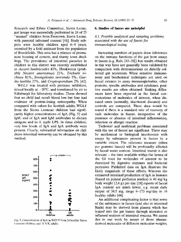
Research and Ethics Committee, Sierra Leone, gut lavage was successfully performed in 24 of 25 “normal” children from Freetown, Sierra Leone, with parental informed consent [41]. Study sub- jects were healthy children aged 6-9 years, recruited by a field assistant from the population of Brookfields. This area has a mixture of perma- nent housing of cement, and shanty town dwel- lings. The prevalence of intestinal parasites in children in this district was recently established as Ascaris Zumbricoides 43%, Hookworm (prob- ably Necator americanus) 21%) Trichuris tri- chiura 81%) Strongyloides stercoralis 5%) Giar- dia lamblia 27%, and Cryptosporidium 2% [42].
WGLF was treated with protease inhibitors, stored locally at -20°C and transferred by air to Edinburgh for laboratory studies. These showed that no child had occult blood loss but four had evidence of protein-losing enteropathy. When compared with values for Scottish adults, WGLF from the Sierra Leonean children had signifi- cantly higher concentrations of IgA (Fig. 5) and IgM, and of IgA and IgM antibodies to dietary antigens and to S. typhi LPS. In three children, very low levels of IgA and IgA antibody were present. Clearly, substantial information on chil- drens intestinal immunity can be obtained by this method.
.I -p < 0.005-_1
600 1 : 7004
WGLF
bA kxml)
600
/
l
500 f
400 2 0 *
300 am 0
l 200 : I- ?
% 100
0
0 SL UK
children adults
Fig. 5. Concentration of IgA in WGLF from 24 healthy Sierra
Leonean children and 16 UK adults.
6. Studies of faeces are unhelpful
6.1. Possible analytical and sampling problems associated with the use of faeces for immunological testing
Increasing numbers of papers draw inferences on the immune functions of the gut from assays in faeces (e.g. Refs. [43-501) but results obtained in this way have not generally been validated by comparison with determinations on directly col- lected gut secretions. When sensitive immuno- assay and biochemical techniques are used on faecal extracts to assay immunoglobulins, other proteins, specific antibodies and cytokines, posi- tive results are often obtained. Striking differ- ences have been reported in the faecal con- centrations of molecules of interest, when dis- eased cases (normally, diarrhoeal diseases) and controls are compared. These data would be sound if there is a standard rate of recovery of such molecules in faeces, irrespective of the presence or absence of intestinal inflammation, infection or allergic reaction.
Technical and analytical problems associated with the use of faeces are significant. There may be mechanical or biological interference with assays by substances present in faeces to a variable extent. The reference measure (often per gramme faeces) will be profoundly affected by faecal water content. Intestinal transit is also relevant - the time available within the lumen of the GI tract for molecules of interest to be destroyed by digestive enzymes and bacterial proteases. Published data on IgA illustrate the likely magnitude of these effects. Whereas the estimated intestinal production of IgA in humans (based on jejunal perfusion studies) is 40 mg/kg body weight (2.8 g) per day [18] reports of faecal IgA content are much lower, e.g. mean daily output of 58.5 mg, range 6-173 mg/day in 16 healthy adults [44].
An additional complicating factor is that some of the substances in faeces (and also in intestinal fluids) may be derived from plasma which has leaked into the gut lumen through ulcerated or inflamed sections of intestinal mucosa. We assess this in our work by assays of three plasma- derived molecules of different molecular weights,
IgG (some of which may be locally secreted), albumin and alpha-1-antitrypsin (AlAT) [26].
6.2. Comparison between ,faeces and whole gut lavage @id
We have described in the previous section some of the evidence that gut lavage with a polyethylene glycol based solution is a whole gut perfusion system. By using WGLF as a standard, it is thus possible to measure the extent to which detection rates and recoveries of intestinal im- munoglobulins, plasma-derived proteins and antibodies in faeces, truly reflect intestinal im- mune status. It should be borne in mind that the perfusion rate of gut lavage fluid is approximate- ly 20 ml/min, equivalent to 29 1 per day, whereas daily faecal output is normally in the range 50- 200 g daily.
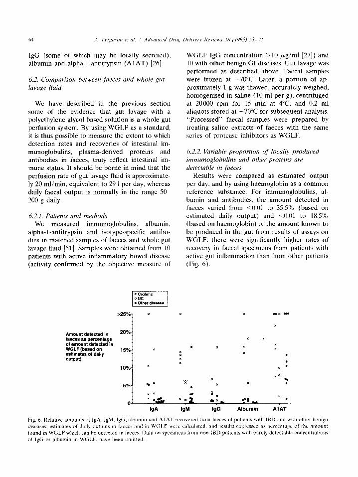
62.1. Patients and methods We measured immunoglobulins, albumin,
alpha-1-antitrypsin and isotype-specific antibo- dies in matched samples of faeces and whole gut lavage fluid [51]. Samples were obtained from 10 patients with active inflammatory bowel disease (activity confirmed by the objective measure of
J
Amount detected in 20%- faeces as percentage of amount detected in WGLF (based on 15%- 0 eatlmates of daily output)
lO%- x
5% 1 5c”
x 0
x
WGLF IgG concentration >lO pg/ml [27]) and 10 with other benign GI diseases. Gut lavage was performed as described above. Faecal samples were frozen at -70°C. Later, a portion of ap- proximately 1 g was thawed, accurately weighed, homogenised in saline (10 ml per g), centrifuged at 20000 rpm for 15 min at 4°C and 0.2 ml aliquots stored at -70°C for subsequent analysis. “Processed” faecal samples were prepared by treating saline extracts of faeces with the same series of protease inhibitors as WGLF.
6.2.2. Variable proportion of locally produced immunoglobulins and other proteins are detectable in ,faeces
Results were compared as estimated output per day, and by using haemoglobin as a common reference substance. For immunoglobulins, al- bumin and antibodies, the amount detected in faeces varied from co.01 to 35.5% (based on estimated daily output) and co.01 to 18.5% (based on haemoglobin) of the amount known to be produced in the gut from results of assays on WGLF: there were significantly higher rates of recovery in faecal specimens from patients with active gut inflammation than from other patients (Fig. 6).
. : x
x
x
a
:o
x
x
0 1
I
I
0
x x
.
.
0 l
x0 4 .
0
Oj x o& x 0 LO & ““8 _ .
I I km@ WI W Albumin AlAT
Fig. 6. Relative amounts of IgA. IgM. IgG. albumin and A I A’f ~reco\crcd Irom laeces ot patlcnts with IBD and with other benign
diseases; estimates of daily outputs in tacccs and in WG1.F wc~c calculated. and results cxprcsscd as percentage of the amount
found in WGLF which can hc detected in Paces. Data on spcclmens from non-IBD patients with barely detectable concentrations
of IgCi or albumin in WGLF, have been omitted.
A. Ferguson et al. I Advanced Drug Delivery Reviews 18 (199.5) X-71 65
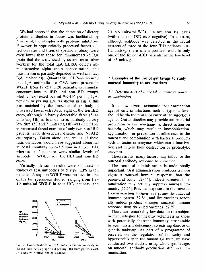
We had observed that the detection of dietary protein antibodies in faeces was facilitated by processing the samples with protease inhibitors. However, in appropriately processed faeces, de- tection rates and titres of specific antibody were even lower than those for immunoreactive IgA (note that the assay used by us and most other workers for the total IgA ELISA detects im- munoreactive alpha chain concentration, and thus measures partially degraded as well as intact IgA molecules). Quantitative ELISAs showed that IgA antibodies to OVA were present in WGLF from 19 of the 20 patients, with similar concentrations in IBD and non-IBD groups, whether expressed per ml WGLF, per mg IgA, per day or per mg Hb. As shown in Fig. 7, this was matched by the presence of antibody in processed faecal extracts in eight of the ten IBD cases, although in barely detectable titres (5-41 units/mg Hb) in four of these; antibody at very low titre (53 and 7 units/mg Hb) was detectable in processed faecal extracts of only two non-IBD patients, with diverticular disease and NSAID enteropathy. Taken alone, the results of these tests on faeces would have suggested abnormal mucosal immunity to ovalbumin in active IBD, whereas in fact there were similar levels of antibody in WGLF from the IBD and non-IBD cases.
Virtually identical results were obtained in studies of IgA antibodies to S. typhi LPS in ten patients. Assays on WGLF were positive in nine of the ten specimens studied, ranging from 1.2- 4.2 units/ml WGLF in four IBD patients, and
W antl-OVA antibody (units per nig lib)
Fig. 7. Concentrations of IgA anti-ovalbumin antibody in
WGLF and faeces (expressed per mg Hb) from patients with
IBD and with other benign diseases.
2.1-5.6 units/ml WGLF in five non-IBD cases (with one non-IBD case negative). In contrast, although antibody was detected in the faecal extracts of three of the four IBD patients, l.O- 1.2 units/g, there was a positive result in only one of the six non-IBD patients, at the low level of 0.6 units/g.
7. Examples of the use of gut lavage to study mucosal immunity to oral vaccines
7.1. Determinants of mucosal immune response to vaccination
It is now almost axiomatic that vaccination against enteric infections such as typhoid fever should be via the portal of entry of the infectious agents. Gut antibodies may provide antibacterial protection by two mechanisms: direct action on bacteria, which may result in immobilization, agglutination, or prevention of adherence to the mucosa; and combination with bacterial products such as toxins or enzymes which cause inactiva- tion and help in their destruction by proteolytic enzymes.
Theoretically, many factors may influence the mucosal antibody response to a vaccine.
The route of administration is undoubtedly important. Oral administration produces a more vigorous mucosal immune response than the parenteral route [52-541; indeed parenteral im- munization may actually suppress mucosal im- munity [55,56]. Previous exposure to the same or a cross-reacting antigen may prime the mucosal immune system [57,58], and live vaccines gener- ally induce produce stronger mucosal immune response than do killed vaccines [52,59].
There are remarkably few data on this subject in man, whether for healthy volunteers or those with potentially aberrant immunity attributable to age, nutrient deficiency, co-existing disease or genetic make-up. As part of a programme of research on the regulation of immunity and hypersensitivity in the human GI tract, we have conducted two studies, using whole gut lavage, on mucosal antibody production after oral im- munisation.
7.2. Immune responses to oral cholera vaccine in coeliac disease patients and controls
Cholera toxin (CT) is composed of one A subunit and five B subunits held together in a pentamer. The toxin B subunit binds to a mem- brane receptor (ganglioside GMl) of gut epi- thelial cells, and the enterotoxic effect is exerted through the A subunit.
Protective immunity against cholera is depen- dent on stimulation of the intestinal immune system, in particular secretory IgA antibodies [58] IgA anti-bacterial as well as antitoxic intesti- nal antibodies are associated with protection against cholera infection, and both are induced by natural infection or oral vaccination. Antitox- ic immunity as conferred by locally produced IgA antibodies probably exert their protective function by neutralising the toxin before it binds to the epithelial cell.
A new oral cholera vaccine (combined killed whole-cell/ B subunit), has been developed for use in endemic areas. This oral combined vaccine evokes high levels of specific intestinal antibodies along with antitoxic protection in endemic popu- lations as well as in human volunteers [58,60].
Given the profound disturbances in systemic and mucosal immunity in patients with coeliac disease, it might be expected that their immune response to vaccines may differ from those of healthy volunteers. To date, no study has ad- dressed this issue.
Z2.1. Subjects and methods Seven healthy volunteers and nine untreated
coeliac patients were studied, age range 24-46 years. All coeliac patients had had a recent jejunal biopsy showing subtotal or severe partial villus atrophy, and all were taking a normal (i.e., gluten-containing) diet.
The oral cholera vaccine used was a combined B subunit-whole cell killed vaccine, a gift of Professor Jan Holmgren (Gothenburg, Sweden). Each dose of vaccine contained 1 mg of B subunit and 10” killed vibrios. The vaccine was given mixed with 150 ml of a sodium bicarbonate solution.
Following baseline collections of serum and gut lavage fluid, three controls and four coeliacs
were given cholera vaccine on days 1 and 15; collection of serum and gut lavage fluid was repeated on day 25. A further three controls and five coeliacs were given vaccine on days 1, 15 and 29, with collection of serum and gut lavage fluid on day 39. In fact, results were similar for the 2 and 3 dose schedules and so results were com- bined.
Isotype-specific antibodies to cholera toxin were assayed in WGLF (IgA and IgM) and serum (IgA and IgG). The method used was a minor adaptation of a published “GM, ” method [20); precoating the ELISA plates with this protein ensures optimal binding of the cholera toxin B-subunit. Results were expressed semi- quantitatively as% of a reference standard which differed for each isotype.
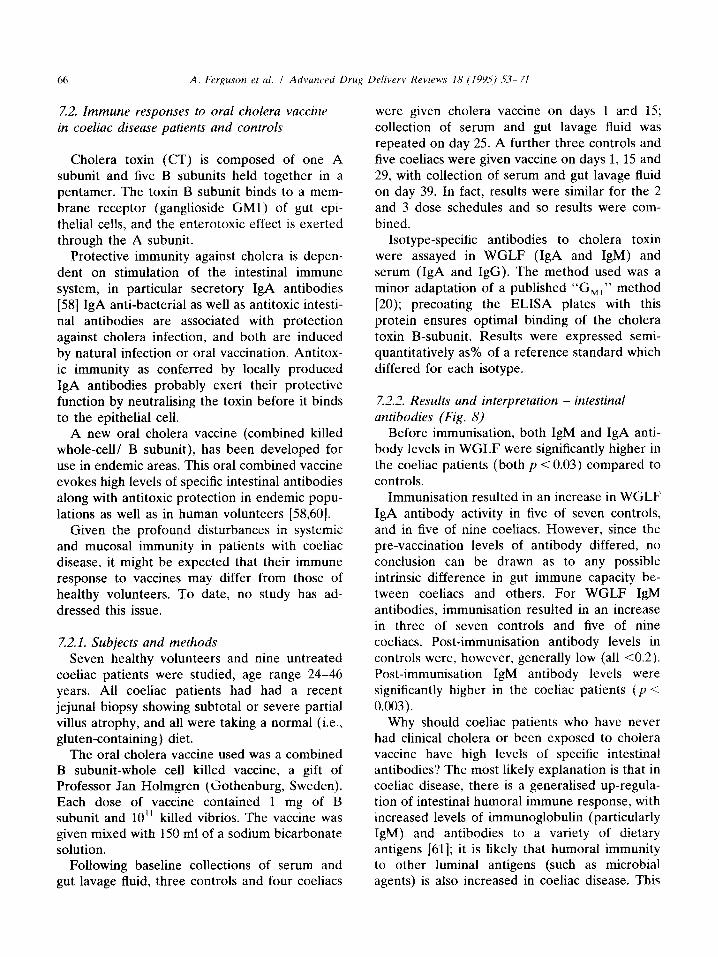
7.2.2. Results and interpretation - intestinal antibodies (Fig. 8)
Before immunisation, both IgM and IgA anti- body levels in WGLF were significantly higher in the coeliac patients (both p < 0.03) compared to controls.
Immunisation resulted in an increase in WGLF IgA antibody activity in five of seven controls, and in five of nine coeliacs. However, since the pre-vaccination levels of antibody differed, no conclusion can be drawn as to any possible intrinsic difference in gut immune capacity be- tween coeliacs and others. For WGLF IgM antibodies, immunisation resulted in an increase in three of seven controls and five of nine coeliacs. Post-immunisation antibody levels in controls were, however, generally low (all cO.2). Post-immunisation IgM antibody levels were significantly higher in the coeliac patients (p < 0.003).
Why should coeliac patients who have never had clinical cholera or been exposed to cholera vaccine have high levels of specific intestinal antibodies? The most likely explanation is that in coeliac disease, there is a generalised up-regula- tion of intestinal humoral immune response, with increased levels of immunoglobulin (particularly IgM) and antibodies to a variety of dietary antigens [61]; it is likely that humoral immunity to other luminal antigens (such as microbial agents) is also increased in coeliac disease. This
A. Ferguson et al. I Advanced Drug Delivery Reviews 18 (1995) 5.3-71
t1.2.
IgM 1 .o-
anti-CT6 in WOLF “‘-
(OD OS- reading) ’
0.4-
IM 1.
anti-CTB o In WGLF ’
P I’
,’ ,’
,’ I’
I’ I’ s* __
iii???
-s_ ,I --‘_o
I I , Pre post Pm post Pre Poet Pw Post
Control Coeliac Control Coeliac
Fig. 8. Gut lavage fluid IgA and IgM antibody responses to cholera toxin in coeliacs and controls.
being the case, coeliac patients would have high titres of intestinal antibodies to many microbial antigens including E. coli toxin which may have antigenic cross-reactivity with the cholera toxin B subunit.
7.2.3. Results and interpretation - serum antibodies
In serum, immunisation resulted in an increase in IgG antibody in six of seven controls, and seven of nine coeliacs, and of IgA antibody in six of seven controls, and seven of nine coeliacs. In general, serum responses tended to mirror in- testinal responses. However, an IgA serum re- sponse was seen in one control patient and two coeliac patients who did not have an intestinal IgA response. There were no significant differ- ences between controls and coeliacs in levels of either pre or post-immunisation serum IgG or IgA anti-toxin antibodies.
Serum anti-toxin responses seemed in general to reflect the intestinal immune responses; this is in accord with the study reported by Jertborn et al. [60] who found that serum antibody responses to oral cholera vaccine had a predictive accuracy of about 80% for reflecting the intestinal immune response. They pointed out that while measure- ment of serum antibody was a useful predictor of intestinal immune response in large field trials, it was of little use in the individual subject. It is also noteworthy, as discussed above, that CT is an unusually potent immunogen, and that feed- ing of many other antigens does not elicit serum antibodies, in contrast to CT.
7.3. Effects of smoking on intestinal antibodies elicited by the oral typhoid vacine Ty2la in healthy UK adults
The live attenuated oral typhoid vaccine Ty2la lacks the capsular antigen Vi which is the virul- ence factor in pathogenic strains of S. typhi [62]. When given as an oral typhoid vaccine (five to eight doses containing 3-10 X 10’” viable organ- isms) to healthy volunteers from non-endemic area, Ty2la provided 87% clinical protection against experimental challenge with 10” virulent S. typhi [63].
The vaccine has proved to be safe, with mild, if any, adverse reactions, and genetically stable. Large-scale field trials carried out in Egypt, Chile and Indonesia measuring its protective efficacy have shown significant although variable levels of protection against typhoid fever [64-671. The variation in the rates of protection was attributed to the differences in the formulations, vaccina- tion schedules and the incidence rates of in- fection in these countries.
The best published data on the capacity of the vaccine strain to induce local mucosal immunity are those of Forrest [15,22,68-701, who studied healthy Australian volunteers and specifically excluded individuals with a history of salmonella infection or food poisoning. However, most of his volunteers received doses of the organism one or two logs greater than those used in recent trials. He studied a variety of formulations, dosages and timing of administration using jejun- al aspirate as a material for the study, and

established that 3 weeks was the optimal timing for studying the intestinal immune responses to this vaccine. We have taken this work further by investigating the effect of smoking on gut im- munity by using the preparation of oral typhoid vaccine Ty2la, recently licenced for use in the UK, as an immunogen.
Z3.1. Subjects and methods We recruited 22 healthy adult volunteers, age
range 22-44 years. Subjects who drank more than 20 units of alcohol per week were excluded. Only subjects who were either heavy smokers (more than 20 cigarette per day for the last 3 years) or lifelong non-smokers, were selected. Subjects had gut lavage and baseline serum sample collected, and were then given three enteric-coated capsules of the oral typhoid vac- cine Ty2la, with each capsule containing 2 X 10” live S. typhi organims, to be taken on days 1, 3 and 5, not with food. 3 weeks WGLF and serum were again collected. IgA anti-S. typhi lipopolysaccharide (LPS) antibodies were mea- sured in WGLF and serum by a quantitative ELISA. Serum with a high titre (expressed as arbitrary units) was used as a reference standard and the results of the test samples were ex- pressed as units/ml and also, for WGLF, as units /mg IgA.
7.3.2. Results - intestinal antibody responses (Fig. 9)
The vaccine Ty2la was well-tolerated and there were no significant differences between pre- and post-vaccination concentrations of total IgA or IgM in WGLF (p = 0.63). Antibody results were considered to be significantly in- creased or decreased by vaccination if the changes were found to be more than 20% greater or less than the pre-immunisation values (the inter-assay coefficient of variation of the ELISA used was 15-20%). There was a significant increase in specific IgA antibody in WGLF specimens of 14 out of 22 volunteers (63.6%) at 3 weeks after vaccination with Ty2la. However, there was a significant decrease in specific IgA antibody in four volunteers (18.2%). A further four volunteers had no significant change in specific IgA antibody levels. Some of the vol-
: 2
1520 i
Pre Post
Fig. 9. IgA class antibodles to Salmonella typhr LPS in whole
gut lavage fluid from four healthy volunteers. before and at 3
weeks after vaccination with three doses of oral Ty2la
vaccine (given on days 1. 3 and 5)
unteers had a rise in serum anti-S. typhi LPS IgG and IgA antibodies, but there was no significant relationship between the changes in serum and those in WGLF.
Finally, there were no differences in any of the parameters measured when non-smokers and heavy smokers were compared.
8. Conclusions
Methods for safe and non-invasive investiga- tion of gut mucosal immunity, including immuni- ty to oral vaccines, are now available. We suggest that these techniques can be used to provide information critical to the understanding of the pathogenesis of diarrhoeal disease morbidity, and necessary for the design and delivery of effective oral immunisation. International and rural/urban comparisons will be of great interest, as will studies of the heterogeneity of intestinal immunity within members of a local community in the developing or developed world. Such investigations might address the pathogenesis and clinical significance of mucosal IgA de- ficiency; the characteristics of children or adults who, within the same highly contaminated en- vironment, remain pathogen-free in contrast to the majority who are frequently re-infected or are chronic carriers; nutrition, immunity, host- parasite interactions and malabsorption in giar- diasis; the relative importance of losses of pro-
A. Fergrcson et al. I Advanced Drug Delivery Rrviews I8 (19%) .5.?-71 69
tein and haemoglobin from the gut, as compared to other mechanisms, in children with anaemia and hypoproteinaemia; and factors that may influence intestinal antibody responses to peroral vaccines for cholera, typhoid and rotavirus.
References
III
PI
PI
[41
151
PI
[71
Strobe], S. and Ferguson, A. (1984) Immune responses
to fed protein antigens in mice. 3. Systemic tolerance or
priming is related to age at which antigen is first
encountered. Pediatr. Res. 18. 588-593.
Kay, R.A. and Ferguson. A. (1989) Systemic delayed-
type hypersensitivity to cholera toxin and a detoxified
derivative. Clin. Exp. Immunol. 76, 11 l-1 16.
Kay, R.A. and Ferguson, A. (1989) The immunological
consequences of feeding cholera toxin. I. Feeding cho-
era toxin suppresses the induction of systemic delayed-
type hypersensitivity but not humoral immunity. Immu-
nology 66. 410-415.
[8] Kay, R.A. and Ferguson. A. (1989) The immunological
conseyuenccs of feeding cholera toxin. II. Mechanisms
responsible for the induction of oral tolerance for DTH.
Immunology 66, 416-421.
Ogra. P.L., Mestecky, J., Lamm. M.E.. Strober. W.,
McGhec. J.R. and Bienenstock, J. (1994) Handbook of
Mucosal Immunology. Academic Press, New York.
Lamont, A.G.. Gordon. M. and Ferguson. A. (1988) T
lymphocyte function in protein deprived mice. Clin.
Exp. Immunol. 72, 113-I 17.
Troncone. R. and Ferguson. A. (1988) Gliadin pre-
sented via the gut induces oral tolerance in mice. Clin.
Exp. Immunol. 72, 284-287.
Strobe], S. and Ferguson, A. (1987) Persistence of oral
tolerance in mice fed ovalbumin is different for humoral
and cell-mediated immune responses. Immunology 60.
317-318.
[9] Mowat, A.McI. and Fcrguson. A. (1982) Intraepithelial
lymphocyte count and crypt hyperplasia measure the
mucosal component of the graft-versus-host reaction in
mouse small intestine. Gastroenterology 83. 417-423.
[IO] Husby. J., Elson, CO., Moldoveanu, Z. and Mestecky. J.
(1994) Oral tolerance in humans. T Cell but not B cell
tolerance to a soluble protein antigen. J. Immunol. 152.
4663-4670.
[II] Husband, A.J., Monie. H.J. and Gowans. J.L. (1977)
The natural history of the cells producing IgA in the gut.
Ciba. Found. Symp. 46, 29-54.
[12] Ghosh, S.. Ferguson A (1994) Mucosal immunology
research design should address not just cytokines and
cells. but also clinical data and controls. Editorial Re-
view. Clin. Exp. Immunol. 96, 377-378.
(131 Czcrkinsky. C.C.. Nilsson L.-A., Nygren, H., Ouchter-
lony. 0. and Tarkowski. A. (1983) A solid-phase en-
zyme-linked immunospot (ELISPOT) assay for enume-
(141
1151
1161
1171
1181
1191
w
1311
Gaspari. M.M., Brennan, P.T.. Soloman. S.M. and Elson.
C.O. (1988) A method of obtaining, processing. and
analysing human intestinal secretions for antibody con-
tent. J. Immunol. Methods I IO. 85-91.
O’Mahony, S.. Barton. J.R., Ct-ichton. S. ancl Ferguson.
A. (1990) Appraisal of gut lavagc in the study of
intestinal humoral immunity. Gut 31, l341- 1344.
1221 Forrest, B.D. (1992) Effects of sample processing on the
measurement of specific intestinal IgA immune rc-
sponses. Vaccine IO, 802-805.
12.31 Svcnnerholm A.-M., Sack, D.A.. Holmgrcn. J. and
Bardhan. P.K. (1982) Intestinal antibody responses after
immunization with cholera B subunit. Lancet i. 30.5~308.
1241 Arranz. E.. O’Mahony, S.. Barton, J.R. and Ferguson.
A. (1992) Immunosenescence and mucosal immunity:
significant effects of old age on serum and secretory IgA
concentrations and on intraepithelial lymphocyte counts.
Gut 33. X82-86.
12.51 O’Mahony, S.. Choudari, C.P.. Barton. J.R., Walker. S..
Ferguson A (1991) Gut lavagc fluid proteins as markers
of activity of inflammatory bowel disease. Stand. J.
Gastroenteroi. 26. 940-944.
[26] Brydon, W.G., Choudari, C.P. and Ferguson, A. (1993)
Relative specificity for active inflammatory bowel dis-
ease of plasma-derived proteins in gut lavage tluid. Eur.
J. Gastroenterol. Hepatol. S, 2699273.
[27] Choudari. C.P.. O’Mahony, S.. Brydon, (i.. Mwantembe.
0. and Ferguson. A. (1993) Concentrations of immuno-
globulin G. albumin and alpha-a-antitrypsin in whole gut
lavagc Huid: objective measures of disease activity in
ration of specific antibody-secreting cells. J. Immunol.
Methods 65. 1099121.
Forrest, B.D. (1988) Identification of an intestinal im-
mune response using peripheral blood lymphocytes.
Lancct i. 81-83.
Forrest, B.D.. LaBrooy. J.T.. Beyer, L.. Dearlove, C.E.
and Shearman. D.J.C (1991) The human humoral im-
mune response to Salmonella yphi Ty21a. J. Infect. Dis.
163, 336-345.
Kelly. C.P.. Pothoulakis, C.. Orellana, J. and Lamont.
J.T. (1992) Human colonic aspirates containing im-
munoglobulin A antibody to clostridiunt difficile toxin A
inhibit toxin A-receptor binding. Gastrocnterology 102.
35-40.
O’Mahony, S.. Arranz. E.. Barton. J.R. and Ferguson.
A. (1991) Dissociation between systemic and mucosal
humoral immune responses in cocliac disease. Gut 32,
29-15.
Conley, M.E. and Delacroix. D.L. (1987) Intravascular
and mucosal immunoglobulin A. Two separate but
related systems of immune defence? Ann. Intern. Med.
106. 8921899.
Cellier. C., Sahmoud. T.. Froguel. E:., Adenis, A..
Belaiche. J., Bretagne J.-F.. Florent, (‘., Bouvry. M..
Mary J.-Y. and Modigliani. R. (1994) r.‘orrelations be-
tween clinical activity. endoscopic severity. and bio-
logical parameters in colonic or ileocolonic Crohn’s
disease. A prospective multiccntrc study of 121 cases.
Gut 35, 231-s.
70
12x1
I291
I-701
1311
PI
[x3]
1341
[3S]
].761
I371
WI
inHammatory bowel discasc. Gastroenterology 104.
1064-1071.
Brydon, U I’;. and Fcrguson, A. (1992) Haemoglobin m gut lavage i .II~ as a measure of gastro-intestinal blood loss. Lancct 340. l381- 13x2.
Ferguson. A., Ghosh, S.. Handy. L.M.. C‘houdari. C‘..
Mwantembe. 0. and McIntyre. M.A. (1994) Analysis 01
disease distribution. activity and complications m the
patient with inllammatory bowel disease. Stand. J.
Gastroentcrol. 29 (Suppl. 203). 15-lY.
Handy. L.M.. Ghosh. S. and Fcrpuson A. (IYYS) In-
vestigation of neutrophil migration into the gut h\
cytology of whole gut Iavagc fluid. Eur. J. Gastrocntcrol.
Hepatol. 7. 53-58.
O’Mahony. S.. Anderson. N.. Nuki, (;. and Ferguson. A.
(19Y2) Systemic and mucosal antibodies to klebsiclla m
ankylosing spondylitis and Crohn’s disease. Ann.
Rheum. Dis. 51. 1296-1300.
Ferguson. A.. Sallam, J., McLintock. L.. (‘raft. N..
Poxton I (1994) The gut as an immune organ: Intestinal
anti-endotoxin antibodies. In: J.M. Kinney and H.N.
Tucker (Eds). Organ Metabolism and Nutrition: Idea\
for Future Critical Cart. Raven Press. New York, pp.
2.7 l-244.
Sallam. J. and Ferguson. A. (lYY2) Mucosal immunity to
oral vaccines. Lancet 3.19. 179.
Ferguson. A. and Mwantemhe, 0. (19%) Intestinal
lavage test of gut inflammation and immunity. In: S.
Auricchio. R. Tronconc and A. Ferguson (Eds). Mucos-
al Immunity In the Gut Epithelium. Karger. Basel. in
press.
Sallam, J. (lYY4) Intestinal humoral unmumty In man:
IgA and anti-salmonella antibodies. Thesis, llnivcrsity
of Edinburgh.
Ferguson. A.. Ghosh. S. and Brydon, W.C;. (1995) I!se 01
whole gut lavage Huid to mcasurc occult GI bleeding
and protein-losing enteropathy. 2. pastrocnterol. in
press.
Barclay. G.R. (1990) Antibodies to cndotoxm in health
and disease. Rev. Med. Microbial. I, 13.3~142.
Barclay. G.R. and Scott. B.B. ( 1987) Serological rela-
tionships between ~‘scheri&~ coli and Srrln~o&/rr
smooth- and rough-mutant lipoplysaccharides ‘1s rc-
vealed by enLyme-linked immunosorhent assay lor
human immunoglobulin (; antiendotoxin antibodies.
Infect. Immun. 55. 2706-2714.
l-191 Di Padova. F.. Brade. H.. Esarclay, G.R., Poxton. I.R..
Liehl, E.. Schuetze. E.. Kocher. H.P., Ramsay, G..
Schreier, M.H.. McClelland, D.B.L. and Rietschel. E.T.
(1993) A broadly cross-protective monoclonal antibody
binding to E.s&nc/C~ coli and Salmorzelln lipopolysac-
charide. Infect. Immun. 61. 3863-3872.
[40] Working Party on Research on Children. The ethical
conduct of research on children (1991) Medical Rc-
search Council.
1411 Hodges, M.. Kingstone. K.. Hrydon, W.G., Sallam. J. and
Ferguson, A. (1994) Use of gut lavage lluid to measure
intestinal immunity in healthy Sierra Leonean children.
J. Pediatr. Gastroentcrol. Nutr. 19. 65-70.
1421
1431
1441
(451
1461
I471
14x1
I491
1501
1511
1521
l5.3)
(541
[SS]
WI
Webster, J.. Hodges, M.H.. Crompton, D.W.T and Wal-
ters, D.E. (1990) Intestinal parasite infections in children
from Frcctown. Sierra Leone. J. Sierra Leone Med.
Dent. Assoc. 5. 144-1.55.
Haneberg. B. and Tonder, 0. (1973) Immunoglobulins
and other serum proteins in feces from infants and
children. Stand. J. Immunol. 2. 375-383.
Mcillct. D.. Raichvarg. D.. Tallet. F., Savel. J., Yongcr. J.
and Gohert. J.G. (1987) Measurcmcnt of total,
nomomcric and polymeric IgA in human faeces by
clcctro-immunodiffusion. Clin. Exp. Immunol. 69. 142-
147.
Koutras, A.K. and Vigorita, V.J. (1989) Fecal secretory
immunoglobulin A in breast milk versus formula feeding
in early infancy. J. Pediatr. Gastrocnterol. Nutr. 9, 5%
61.
Saha, K.. Sharma, S., Chopra. K. and Dua, N. (1990)
Raised stool and strum IgA levels in undernourished
infants with chronic diarrhoea and associated parasitic
infestations. J. Trop. Pediatr. 36. 69-74.
Sasai. K., Furukawa. S.. Sugawara. T.. Kaneko, K.. Baba,
M. and Yabuta, K. (1992) IgE levels in faecal extracts of
patients with food allergy. Allergy 47, 594-598.
Canccllicri. V. and Fara, G.M. (1985) Demonstration of
specilic IgA in human feces after immunization with Iivc
‘l‘y2la Salmotzella typhi vaccine. J. Infect. Dis. 151, 482-
4x4.
Stapleton. J.T.. Lange. D.K.. LeDuc, J.W.. Binn, L.N..
Janscn. R.W. and Lemon, S.M. (lY91) The role of
secretory immunity in hepatitis A virus infection. J.
Infect. Dis. 163. 7-l I.
Harendra de Silva, D.G., Mendis. L.N., Alexander,
G.J.M. Candy. D.C.A. Chart, H. and Rowe. B. (1993)
Concentrations of interleukin 6 and tumour necrosis
lactor in strum and stools of children with Sh&/lu
c/.v.serlr~ritrc, I infection. Gut 34. 194-198.
Ferguson. A.. Croft. N. and Humphrcys, K. (1995)
Technical Report: Results of immunological tests on
faecal extracts arc likely to be extremely misleading.
(‘iin. Exp. Immunol. 99. 70-75.
Ogra. P.. Karzon. D. and Righthand, F. (1968) Immuno-
globulin response in serum and secretions after immuni-
zation with live and inactivated polio vaccine and
natural infectnm. N. Engl. J. Med. 279, 89%YOO.
Lange, S. and Holmgrcn. J. (1978) Protective antitoxic
Cholera immunity in mice: influence of route and
number of immunizations and mode of action of protcc-
tive antibodies. Acta Pathol. Microbial. Stand. Sect. 86.
145-1.52.
Svennerholm A.-M., Hanson, L. and Holmgren, J.
( 1980) Different secretory immunoglobulin A antibody
responses to cholera vaccination in Swedish and Pakis-
tani women. Infect. Immun. 30. 427-430.
Pierce. N. (1984) Induction of optimal mucosal antibody
rcsponscs: effccls of age, immunization route(s) and dosing schedules in rats. Infect. Immun. 43. 341-346.
Pierce. N. and Koster. F. (1980) Priming and suppres-
sion of the intestinal immune response to cholera
A. Ferguson et al. I Advanced Drug Delivery Rcvirws 18 (199.~) 5-71 71
toxoid/toxin by parenteral toxoid in rats. J. Immunol.
124, 307-311.
[S7] Svennerholm A.-M., Holmgren. J. and Hanson. L.
(1977) Boosting of secretory 1gA antibody responses in
man by parcnteral Cholera vaccination. Stand. J. Im-
munol. 6, 1345.
[5X] Svennerholm A.-M., Gothefors, L. and Sack, D. (1984)
Local and systemic antibody responses and immunologi-
cal memory in humans after immunization with cholera
B subunit by different routes. Bull. WHO 62. 9099918.
1591 Levine. M.. Ferreccio, C. and Black, R. (1989) Progress
in vaccines against typhoid fever. Rev Infect. Dis. ii
(Suppl. 3). SSS2-SS67.
[60] Jertborn. M., Svennerholm A.-M. and Holmgren J.
(1984) Gut mucosal, salivary and serum antitoxic and
antibacterial antibody response in Swedes after oral
immunization with B subunit-whole cell Cholera vac-
cine. Int. Arch. Allergy Appl. Immunol. 75. X-43.
[hl] Arranz. E. and Ferguson, A. (1993) Intestinal antibody
pattern of coeliac disease: occurrence in patients with
normal jejunal biopsy histology. Gastroenterology 104.
1263-1272.
[62] Silva, B.. Gonzalez, C. and Mora. G. (1978) Genetic
characteristics of the Salmonella I_vphi strain Ty2la
vaccine. J. Infect. Dis. 155. 1077-1078.
[63] Gilman. R.. Hornick, R. and Woodward. W. (1977)
Evaluation of a UDP-glucose-4-epimeraseless mutant of
Salnronella ryphi as a live oral vaccine. J. Infect. Dis.
136. 717-723.
[64] Wahdan, M.. Seric, C. and Ccrisier, Y. (lY82) A con-
trolled held trial of live Salmonella fyphi strain Ty2la
oral vaccine against typhoid: Three-year results. J. In-
fect. Dis. 145. 292-295.
1651 Wahdan. M., Serie. C. and Germanier, R. (1980) A
controlled field trial of live oral typhoid vaccine Ty2la.
Bull. WHO 58. 469-474.
1661 Levine, M., Ferreccio, C. and Black, R. (1987) Large-
scale held trial of Ty2la live oral typhoid vaccine in
enteric-coated capsule formation. Lancet i, 1049%1052.
[67] Simanjuntak, C.. Paleologo. F. and Punjabi, N. (1991)
Oral immunization against typhoid fever in Indonesia
with Ty2la vaccine. Lancct 338. 10.55-1059.
[68] Forrest, B.. LaBrooy. J. and Attridgc. S. (1989) A
candidate live oral typhoid/cholera hybrid vaccine is
immunogenic in humans. J. Infect. 1.59. 145-146.
[69] Forrest. B. (1992) Impairment of immunogenicity of
Salmor~ella typhi Ty2la due to pre-existing cross-react-
ing intestinal antibodies. J. Infect. Dis. 166. 210-212.
1701 Forrest, B.. LaBrooy, J. and Dcarlove. C. (19Y2) Effect
of parenteral immuninzation on the intestmal immune
response to SahoneUa fyphi Ty2la. Infect. lmmun. 60.
365-47 1.