Embed Size (px)
Citation preview
Study Protocol
Clinical Investigation of the
TECNIS® Next-Generation Intraocular Lenses
NCT Number: NCT03111550
Version 1.0
Document date: February 2017
ABBOTT MEDICAL OPTICS INC. CONFIDENTIAL
Version 1.0 i PR/EDOF-121-NGPC
CONFIDENTIAL The following contains confidential, proprietary information
that is the property of Abbott Medical Optics Inc.
Clinical Investigation of the TECNIS® Next-Generation Intraocular Lenses
PROTOCOL NUMBER: EDOF-121-NGPC
SPONSOR: Abbott Medical Optics Inc.
1700 E. St. Andrew Place Santa Ana, CA 92705
714-247-8200 Investigator Agreement As an Investigator, I agree to:
• Implement and conduct this study diligently and in strict compliance with this agreement; the protocol; Good Clinical Practices; 21CFR812, ISO 14155 and all other applicable FDA regulations; conditions of approval imposed by the reviewing Institutional Review Board (IRB) or Independent Ethics Committee (IEC), FDA or other regulatory authorities; and all other applicable laws and regulations.
• Supervise all testing of the device where human subjects are involved.
• Ensure that the requirements for obtaining informed consent are met.
• Obtain authorization for use/disclosure of health information (e.g., HIPAA authorization or equivalent).
• Maintain all information supplied by Abbott Medical Optics in confidence and, when this information is submitted to an independent IRB or any other group, it will be submitted with a designation that the material is confidential.
I have read this protocol in its entirety and I agree to all aspects. Investigator Printed Name Signature Date Sub-Investigator Printed Name Signature Date Acknowledged By:
Signature of Sponsor’s Representative Date
Printed Name and Title
ABBOTT MEDICAL OPTICS INC. CONFIDENTIAL
Version 1.0 ii PR/EDOF-121-NGPC
TABLE OF CONTENTS
TITLE PAGE Table of Contents .......................................................................................................... ii Personnel and Facilities............................................................................................... iv Protocol Change History ............................................................................................... v 1. Synopsis ............................................................................................................. 1 2. Background/Introduction .................................................................................. 5 3. Clinical Hypothesis ............................................................................................ 6 4. Study Design ...................................................................................................... 6 5. Acronyms ........................................................................................................... 6 6. Study Objectives and Endpoints ...................................................................... 7
6.1 Primary Effectiveness Endpoints ....................................................................... 7 6.2 Primary Safety Endpoints .................................................................................. 7 6.3 Other Endpoints ................................................................................................. 7
7. Study Products .................................................................................................. 8 7.1 Intraocular Lenses ............................................................................................. 8 7.2 IOL Implantation Systems ................................................................................ 12
8. Study Population ............................................................................................. 12 8.1 Inclusion Criteria .............................................................................................. 12 8.2 Exclusion Criteria ............................................................................................. 13
9. Investigator Selection ...................................................................................... 14 9.1 Investigator Qualifications ................................................................................ 14 9.2 Investigator Obligations ................................................................................... 14 9.3 Investigator Approval ....................................................................................... 15
10. Experimental Plan ............................................................................................ 16 10.1 Overview ......................................................................................................... 16 10.2 Visit Schedule .................................................................................................. 17 10.3 Preoperative Procedures ................................................................................. 18 10.4 Randomization and Masking ............................................................................ 20 10.5 Study Lens Supply ........................................................................................... 21 10.6 Operative Procedures ...................................................................................... 21 10.7 Postoperative Procedures ............................................................................... 23 10.8 Exit of Subjects ................................................................................................ 29 10.9 Unscheduled Visits .......................................................................................... 30 10.10 Protocol Deviations .......................................................................................... 30
11. Adverse Events and Product Complaints ...................................................... 31 11.1 Adverse Event Definitions ................................................................................ 31 11.2 Product Complaint/Device Deficiency Definition .............................................. 33 11.3 Adverse Event and Complaint Reporting Requirements .................................. 33 11.4 Causal Relationship ......................................................................................... 34 11.5 Adverse Event Follow-up ................................................................................. 35
ABBOTT MEDICAL OPTICS INC. CONFIDENTIAL
Version 1.0 iii PR/EDOF-121-NGPC
12. Protocol Changes/Amendments ..................................................................... 36 13. Ethics Review and Patient Welfare ................................................................. 36
13.1 Institutional Review Board (IRB) ...................................................................... 36 13.2 Informed Consent ............................................................................................ 36
14. Documentation ................................................................................................. 37 14.1 Source Documents .......................................................................................... 37 14.2 Subject Confidentiality ..................................................................................... 37 14.3 Case Report Form Completion ........................................................................ 38 14.4 Study Summary ............................................................................................... 38
15. Monitoring ........................................................................................................ 38 15.1 Data Monitoring ............................................................................................... 38 15.2 Administrative Monitoring ................................................................................ 39 15.3 Safety Monitoring ............................................................................................. 40
16. Publications ..................................................................................................... 40 17. Risk Analysis ................................................................................................... 40 18. Records Retention ........................................................................................... 41 19. Termination of the Investigation ..................................................................... 42 20. Statistical Methods .......................................................................................... 42
20.1 Analysis Population ......................................................................................... 43 20.2 Primary Study Endpoints ................................................................................. 43 20.3 Additional Endpoints ........................................................................................ 45 20.4 Visual Acuity conventions and General Statistics ............................................ 46 20.5 Interim Reports ................................................................................................ 46 20.6 Sample Size Calculations ................................................................................ 46
Appendix A Summary of Procedures Required at Each Visit .................................. 48 Appendix B Equipment List ........................................................................................ 49 Appendix C Maximum Plus Manifest Refraction Technique with
Cylinder Refinement ............................................................................. 50 Appendix D Refraction Adjustments .......................................................................... 51 Appendix E Instructions for Using the M&S System ................................................ 52 Appendix F Instructions for Distance Visual Acuity Testing .................................... 54 Appendix G Instructions for Intermediate Visual Acuity testing .............................. 55 Appendix H Instructions For Near Visual Acuity Testing ......................................... 57 Appendix I Instructions for Depth of Focus Testing ................................................. 59 Appendix J Instructions for Manifest Cylinder Defocus Testing ............................. 60 Appendix K Instructions for Pupil Size Measurements ............................................ 61 Appendix L Instructions for Contrast sensitivity Testing ......................................... 62 Appendix M Slit-Lamp Exam Ratings ......................................................................... 64
ABBOTT MEDICAL OPTICS INC. CONFIDENTIAL
Version 1.0 v PR/EDOF-121-NGPC
PROTOCOL CHANGE HISTORY
Version Section(s) Page(s) Description of Change(s) Rationale for Change(s) 1.0 N/A N/A Original N/A
ABBOTT MEDICAL OPTICS INC. CONFIDENTIAL
Version 1.0 1 PR/EDOF-121-NGPC
1. SYNOPSIS
PROTOCOL: Clinical Investigation of the TECNIS® Next-Generation Intraocular Lenses Protocol Number: EDOF-121-NGPC
STUDY TREATMENTS:
Investigational Lens 1: • TECNIS® Next-Generation IOL, Model ZHR00
Investigational Lens 2: • TECNIS® Next-Generation IOL, Model ZQR00
Control Lens: • TECNIS Symfony® Extended Range of Vision IOL
(“Symfony”), Model ZXR00 (Abbott Medical Optics, Santa Ana, CA), commercially available
STUDY OBJECTIVE: The purpose of this clinical trial is to evaluate the safety and effectiveness of the next-generation TECNIS® IOLs.
CLINICAL HYPOTHESIS: The next generation of TECNIS IOLs, Models ZHR00 and ZQR00, will provide improved near (at 40 cm) visual acuity compared to the TECNIS Symfony control IOL. Complication and adverse event rates associated with the next-generation IOLs will be within the rates for posterior chamber IOLs given in ISO 11979-7:2006/ Amd 1:2012(E).
OVERALL STUDY DESIGN:
Structure: Prospective, multicenter, randomized, bilateral subject/evaluator-masked clinical trial
Number of sites: Up to 14 sites in the United States
Duration: 6 months
Administration: Surgeons will perform routine, small-incision, cataract surgery and implant the study lenses using a sponsor-recommended implantation system. The target for refractive outcomes will be emmetropia for both eyes.
ABBOTT MEDICAL OPTICS INC. CONFIDENTIAL
Version 1.0 2 PR/EDOF-121-NGPC
Visit Schedule: Subjects will be bilaterally implanted with the same lens type; the second eye is to be implanted within 1 month of the first-eye surgery.
All subjects will undergo a minimum of 9 visits: Preoperative for both eyes; Operative, 1-day and 1-week visits for each eye; and 1-month and 6-month visits for both eyes together.
STUDY POPULATION CHARACTERISTICS:
Condition: Bilateral cataracts with otherwise healthy eyes
Number of Subjects: Up to 260 subjects will be enrolled to achieve approximately 220 randomized and bilaterally-implanted subjects, resulting in approximately 195 evaluable subjects (65 in each test group and 65 in the control group) at 1 and 6 months.
Each site should enroll approximately 15 subjects, and no site may enroll more than 25% of the enrollment total.
Inclusion Criteria (all criteria apply to each study eye): • Minimum 22 years of age • Bilateral cataracts for which posterior chamber IOL implantation has been planned • Preoperative best corrected distance visual acuity (BCDVA) of 20/40 Snellen or
worse with or without a glare source • Potential for postoperative BCDVA of 20/30 Snellen or better • Corneal astigmatism:
o Normal corneal topography o Preoperative corneal astigmatism of 1.00 D or less in both eyes
• Clear intraocular media other than cataract in each eye • Availability, willingness and sufficient cognitive awareness to comply with
examination procedures • Signed informed consent and HIPAA authorization or equivalent documentation
necessary to comply with applicable privacy laws pertaining to medical treatment in the governing countries
• Ability to understand and respond to a questionnaire in English
Exclusion Criteria (all criteria apply to each study eye): • Requiring an intraocular lens power outside the available range of +16.0 D
to +28.0 D • Any clinically-significant pupil abnormalities (non-reactive, fixed pupils, or
abnormally-shaped pupils)
ABBOTT MEDICAL OPTICS INC. CONFIDENTIAL
Version 1.0 3 PR/EDOF-121-NGPC
• Irregular corneal astigmatism • Inability to focus or fixate for prolonged periods of time (e.g., due to strabismus,
nystagmus, etc.) • Prior corneal refractive (LASIK, LASEK, RK, PRK, etc.) or intraocular surgery.
Note: Prophylactic peripheral iridotomies and peripheral laser retinal repairs that, in the opinion of the investigator will not confound study outcome or increase risk to the subject, are acceptable.
• Corneal abnormalities such as stromal, epithelial or endothelial dystrophies that are predicted to cause visual acuity losses to a level worse than 20/30 Snellen during the study
• Inability to achieve keratometric stability for contact lens wearers (per procedure outlined in Section 10.3)
• Recent ocular trauma or ocular surgery that is not resolved/stable or may affect visual outcomes or increase risk to the subject
• Subjects with diagnosed degenerative visual disorders (e.g., macular degeneration or other retinal disorders) that are predicted to cause visual acuity losses to a level worse than 20/30 Snellen during the study
• Subjects with conditions associated with increased risk of zonular rupture, including capsular or zonular abnormalities that may lead to IOL decentration or tilt, such as pseudoexfoliation, trauma, or posterior capsule defects
• Use of systemic or ocular medications that may affect vision • Prior, current, or anticipated use during the course of the 6-month study of
tamsulosin or silodosin (e.g., Flomax, Flomaxtra, Rapaflo) that may, in the opinion of the investigator, confound the outcome or increase the risk to the subject (e.g., poor dilation or a lack of adequate iris structure to perform standard cataract surgery)
• Poorly-controlled diabetes • Acute, chronic, or uncontrolled systemic or ocular disease or illness that, in the
opinion of the investigator, would increase the operative risk or confound the outcome(s) of the study (e.g., immunocompromised, connective tissue disease, suspected glaucoma, glaucomatous changes in the fundus or visual field, ocular inflammation, etc.). Note: controlled ocular hypertension without glaucomatous changes (optic nerve cupping and visual field loss) is acceptable.
• Known ocular disease or pathology that, in the opinion of the investigator, o may affect visual acuity o may require surgical intervention during the course of the study (macular
degeneration, cystoid macular edema, diabetic retinopathy, uncontrolled glaucoma, etc.)
o may be expected to require retinal laser treatment or other surgical intervention during the course of the study (macular degeneration, cystoid macular edema, diabetic retinopathy, etc.)
• Patient is pregnant, plans to become pregnant, is lactating or has another condition associated with the fluctuation of hormones that could lead to refractive changes
• Concurrent participation or participation within 45 days prior to preoperative visit in any other clinical trial
• Desire for monovision correction
ABBOTT MEDICAL OPTICS INC. CONFIDENTIAL
Version 1.0 4 PR/EDOF-121-NGPC
EVALUATION CRITERIA: The purpose of this clinical study is to evaluate the safety and effectiveness of the next-generation TECNIS IOLs. The primary effectiveness endpoint is mean, monocular, distance corrected near visual acuity under photopic conditions at 40 cm. Other effectiveness endpoints include mean monocular diopters of defocus, monocular and binocular uncorrected distance, best corrected distance, uncorrected intermediate, distance corrected intermediate and uncorrected near visual acuities, monocular 10% low-contrast distance corrected and best corrected intermediate acuity and questionnaire responses.
The primary safety endpoint is adverse event rates versus ISO 11979-7:2006/ Amd.1:2012(E) Safety and Performance Endpoint (SPE) rates. Additional safety endpoints include the proportion of first-implanted eyes achieving 20/40 or better monocular photopic best corrected distance visual acuity vs. the ISO SPE rate, optical/visual symptoms, medical/lens findings, binocular best corrected distance contrast sensitivity and visual symptoms via PRO instrument.
DATA ANALYSIS: The investigational TECNIS Model ZHR00 and Model ZQR00 IOLs will each be compared separately to the TECNIS Symfony control IOL. For the primary effectiveness endpoints of monocular distance corrected near visual acuity, comparisons between the Model ZHR00 group vs the TECNIS Symfony control group, and comparisons between the Model ZQR00 group vs the TECNIS Symfony control group, will be performed using one-sided, two-sample t-tests with an alpha of 0.025. For the primary safety endpoints, adverse event rates, the adverse event rate of the Model ZHR00 IOL will be compared to the ISO grid value using one-sided exact tests with an alpha of 0.05 and the adverse event rate of the Model ZQR00 IOL will be compared to the ISO grid value using one-sided exact tests with an alpha of 0.05.
The safety population with available data will be used for all analyses. The key study timeframe for effectiveness endpoints will be 1 month and for safety endpoints it will be 6 months. Descriptive statistics including means, standard deviations, minimum and maximum values will be reported for visual acuity, refractive data and contrast sensitivity. The frequency and proportion will be reported for subjects with adverse events, medical findings, lens findings, ocular visual symptoms and questionnaires data.
STUDY VISITS AND PROCEDURES: Inclusion and exclusion qualifications will be assessed at the preoperative visit according to the inclusion/exclusion criteria. The Informed Consent Document and Authorization for Use/Disclosure of Health Information form (HIPAA authorization) must be signed by any patients who agree to participate in the study prior to undergoing any study-specific
ABBOTT MEDICAL OPTICS INC. CONFIDENTIAL
Version 1.0 5 PR/EDOF-121-NGPC
procedures. Those subjects who meet the inclusion/exclusion criteria and agree to participate will be randomized to receive lenses from the same lens group in both eyes, either Model ZHR00, Model ZQR00 or control, Model ZXR00. The eye implanted first will be considered the primary study eye. All subjects are intended to have bilateral cataract surgery with the second-eye surgery occurring after the 1-week exam for the first eye but no more than 30 days after the first-eye surgery. Subjects and study personnel performing the postoperative vision testing and refractions will be masked for the duration of the study.
Key preoperative data include ocular health and history, visual acuities, manifest refraction, keratometry, biomicroscopic slit-lamp findings, ocular symptoms and biometry. The operative visit will include standard procedures for cataract surgery and IOL implantation. Key postoperative data collection includes monocular and binocular uncorrected and distance corrected visual acuities, contrast sensitivity, defocus curve, slit-lamp findings, non-directed visual symptoms, questionnaires and adverse events.
2. BACKGROUND/INTRODUCTION
Presbyopia, defined as the age-related loss of accommodative amplitude, affects essentially all human beings beyond the age of 45 and impacts the ability of the eye to focus at near distances1,2. Current intraocular lens options for cataract patients who desire improved vision across a range of distances include a choice of monovision or multifocality. Patients implanted with standard monofocal lenses often need spectacles for reading or performing other near tasks, even if a monovision option is selected. Patients implanted with multifocal lenses, while being able to read and perform other near tasks without spectacles, sometimes experience dysphotopsias (e.g., halos), particularly at night, and may have limited intermediate ability (e.g., may need spectacles to work on a computer). Some accommodating lenses are also available on the market, although their effect depends upon fit within the capsular bag or capsular bag elasticity.
On July 15, 2016, another option was made available to cataract patients when the TECNIS Symfony Extended Range of Vision IOL, Model ZXR00, became commercially available. Utilizing a diffractive technology to elongate the depth of focus, the Symfony IOLs provide cataract patients with good distance vision, and improved intermediate and near vision compared to standard monofocal IOLs.
The investigational IOL devices in this protocol, Models ZHR00 and ZQR00, are enhanced versions of the commercially-available Symfony IOL. The diffractive technology on the posterior optic surface of the TECNIS Symfony IOL was modified slightly for both investigational IOLs, with the goal of further elongating the depth of focus compared to the Symfony IOL.
ABBOTT MEDICAL OPTICS INC. CONFIDENTIAL
Version 1.0 6 PR/EDOF-121-NGPC
3. CLINICAL HYPOTHESIS
This study will demonstrate that the TECNIS Model ZHR00 and Model ZQR00 IOLs will provide improved near (at 40 cm) visual acuity compared to the TECNIS Symfony control IOL. Complication and adverse event rates associated with the next generation IOLs will be within the rates for posterior chamber IOLs given in ISO 11979-7:2006/ Amd 1:2012(E).
4. STUDY DESIGN
This study is a 6-month, prospective, multicenter, subject/evaluator-masked, bilateral, randomized clinical investigation of the TECNIS Next-Generation Model ZHR00 and Model ZQR00 IOLs versus the TECNIS Symfony control IOL.
The study will be conducted at up to 14 sites in the U.S.A and will enroll up to 260 subjects to achieve approximately 220 randomized and bilaterally-implanted subjects, resulting in approximately 195 evaluable subjects (65 in each test group and 65 in the control group) at 1 and 6 months. Subjects are to be implanted with the same IOL in both eyes, the ZHR00 IOL, the ZQR00 IOL or the Symfony control IOL. The eye implanted first will be considered the primary study eye.
JUSTIFICATION OF STUDY DESIGN This study is being conducted to capture preliminary safety and effectiveness information on two design candidates, with the goal of selecting a final lens design. The prospective, multicenter, subject/evaluator-masked, bilateral, randomized study design was chosen to optimize comparison of visual outcomes between the Models ZHR00 and ZQR00 investigational IOLs and the Symfony control IOL, while minimizing the number of subjects by utilizing the same control group for statistical comparisons.
5. ACRONYMS
The following acronyms are used throughout the document:
• UCDVA: uncorrected distance visual acuity • BCDVA: best corrected distance visual acuity • UCIVA: uncorrected intermediate visual acuity • DCIVA: distance corrected intermediate visual acuity • BCIVA: best corrected intermediate visual acuity (DCIVA with add) • UCNVA: uncorrected near visual acuity • DCNVA: distance corrected near visual acuity • D: diopters
ABBOTT MEDICAL OPTICS INC. CONFIDENTIAL
Version 1.0 7 PR/EDOF-121-NGPC
6. STUDY OBJECTIVES AND ENDPOINTS
The purpose of this study is to evaluate the safety and effectiveness of the next-generation TECNIS Model ZHR00 and Model ZQR00 IOLs. The key study timeframe for effectiveness endpoints will be 1 month and for safety endpoints the key study timeframe is 6 months.
6.1 PRIMARY EFFECTIVENESS ENDPOINTS FOR MODEL ZHR00: MONOCULAR, PHOTOPIC DCNVA AT 40 CM
Success criteria: Statistically significant improvement in mean distance corrected near visual acuity for the investigational Model ZHR00 eyes vs. control eyes.
FOR MODEL ZQR00: MONOCULAR, PHOTOPIC DCNVA AT 40 CM
Success criteria: Statistically significant improvement in mean distance corrected near visual acuity for the investigational Model ZQR00 eyes vs. control eyes.
6.2 PRIMARY SAFETY ENDPOINTS For Model ZHR00: Rates of adverse events vs. ISO SPE rates For Model ZQR00: Rates of adverse events vs. ISO SPE rates
6.3 OTHER ENDPOINTS • Monocular and binocular, best corrected distance depth of focus • Monocular BCDVA percent 20/40 or better vs. ISO SPE rate • Binocular UCDVA, BCDVA, UCIVA, DCIVA, UCNVA and DCNVA • Monocular UCDVA, UCIVA, DCIVA and UCNVA • Monocular low-contrast DCIVA and BCIVA (10%) • Monocular and binocular best corrected distance contrast sensitivity vs
control (mesopic with and without glare at 1.5, 3 and 6 cpd, photopic with glare at 3, 6, 12, and 18 cpd)
• Binocular tolerance to cylinder • Visual symptoms via PRO instrument • Ocular/visual symptoms (non-directed responses as obtained from the open-
ended question “Are you having any difficulties with your eyes or vision?”) • Subject spectacle independence and satisfaction questionnaire responses • Medical findings/complications • Lens findings/complications
ABBOTT MEDICAL OPTICS INC. CONFIDENTIAL
Version 1.0 8 PR/EDOF-121-NGPC
7. STUDY PRODUCTS
7.1 INTRAOCULAR LENSES The three lens models used in this study include the investigational TECNIS Next-Generation IOLs, Model ZHR00 and ZQR00, and the TECNIS Symfony Extended Range of Vision IOL, Model ZXR00, control IOL. The investigational IOLs are modifications of the Symfony IOL, a single-piece, SENSAR acrylic IOL with a modified prolate (aspheric) design on the anterior optic surface to reduce spherical aberration and a diffractive posterior optic design to extend the depth of focus.
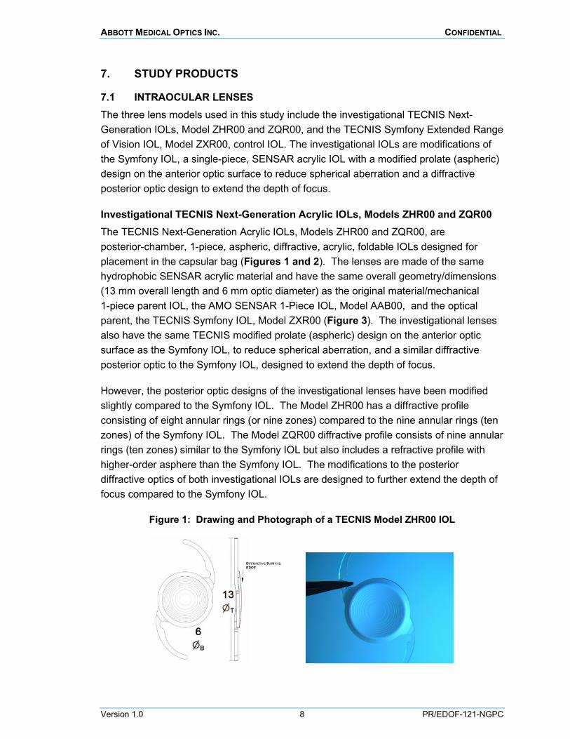
Investigational TECNIS Next-Generation Acrylic IOLs, Models ZHR00 and ZQR00 The TECNIS Next-Generation Acrylic IOLs, Models ZHR00 and ZQR00, are posterior-chamber, 1-piece, aspheric, diffractive, acrylic, foldable IOLs designed for placement in the capsular bag (Figures 1 and 2). The lenses are made of the same hydrophobic SENSAR acrylic material and have the same overall geometry/dimensions (13 mm overall length and 6 mm optic diameter) as the original material/mechanical 1-piece parent IOL, the AMO SENSAR 1-Piece IOL, Model AAB00, and the optical parent, the TECNIS Symfony IOL, Model ZXR00 (Figure 3). The investigational lenses also have the same TECNIS modified prolate (aspheric) design on the anterior optic surface as the Symfony IOL, to reduce spherical aberration, and a similar diffractive posterior optic to the Symfony IOL, designed to extend the depth of focus.
However, the posterior optic designs of the investigational lenses have been modified slightly compared to the Symfony IOL. The Model ZHR00 has a diffractive profile consisting of eight annular rings (or nine zones) compared to the nine annular rings (ten zones) of the Symfony IOL. The Model ZQR00 diffractive profile consists of nine annular rings (ten zones) similar to the Symfony IOL but also includes a refractive profile with higher-order asphere than the Symfony IOL. The modifications to the posterior diffractive optics of both investigational IOLs are designed to further extend the depth of focus compared to the Symfony IOL.
Figure 1: Drawing and Photograph of a TECNIS Model ZHR00 IOL
ABBOTT MEDICAL OPTICS INC. CONFIDENTIAL
Version 1.0 9 PR/EDOF-121-NGPC
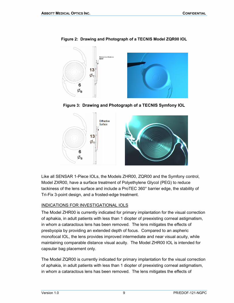
Figure 2: Drawing and Photograph of a TECNIS Model ZQR00 IOL
Figure 3: Drawing and Photograph of a TECNIS Symfony IOL
Like all SENSAR 1-Piece IOLs, the Models ZHR00, ZQR00 and the Symfony control, Model ZXR00, have a surface treatment of Polyethylene Glycol (PEG) to reduce tackiness of the lens surface and include a ProTEC 360° barrier edge, the stability of Tri-Fix 3-point design, and a frosted-edge treatment.
INDICATIONS FOR INVESTIGATIONAL IOLS
The Model ZHR00 is currently indicated for primary implantation for the visual correction of aphakia, in adult patients with less than 1 diopter of preexisting corneal astigmatism, in whom a cataractous lens has been removed. The lens mitigates the effects of presbyopia by providing an extended depth of focus. Compared to an aspheric monofocal IOL, the lens provides improved intermediate and near visual acuity, while maintaining comparable distance visual acuity. The Model ZHR00 IOL is intended for capsular bag placement only.
The Model ZQR00 is currently indicated for primary implantation for the visual correction of aphakia, in adult patients with less than 1 diopter of preexisting corneal astigmatism, in whom a cataractous lens has been removed. The lens mitigates the effects of
ABBOTT MEDICAL OPTICS INC. CONFIDENTIAL
Version 1.0 10 PR/EDOF-121-NGPC
presbyopia by providing an extended depth of focus. Compared to an aspheric monofocal IOL, the lens provides improved intermediate and near visual acuity, while maintaining comparable distance visual acuity. The Model ZQR00 IOL is intended for capsular bag placement only.
INDICATIONS FOR CONTROL IOL
The TECNIS Symfony® Extended range of Vision IOL, Model ZXR00, is indicated for primary implantation for the visual correction of aphakia, in adult patients with less than 1 diopter of pre-existing corneal astigmatism, in whom a cataractous lens has been removed. The lens mitigates the effects of presbyopia by providing an extended depth of focus. Compared to an aspheric monofocal IOL, the lens provides improved intermediate and near visual acuity, while maintaining comparable distance visual acuity. The Model ZXR00 IOL is intended for capsular bag placement only.
STORAGE AND DISTRIBUTION
Consignments of all three study lenses will be supplied to the sites. All study lenses should be stored in the original packaging and kept in a dry place. Lenses should not be stored in direct sunlight or at temperatures greater than 45° C (113°F). Each lens is packaged in a lens tray and sealed in a peel-pouch. The lens is sterile as long as the package has not been opened or damaged and the shelf-life expiration date has not been exceeded. The Principal Investigator is responsible for ensuring that the investigational lenses are only used for subjects enrolled in this study.
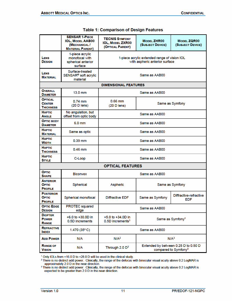
COMPARISON CHART Table 1 describes the dimensional and optical similarities between the study lenses and other associated AMO lenses.
ABBOTT MEDICAL OPTICS INC. CONFIDENTIAL
Version 1.0 12 PR/EDOF-121-NGPC
7.2 IOL IMPLANTATION SYSTEMS The investigational TECNIS Models ZHR00 and ZQR00 and the control TECNIS Model ZXR00 lenses are to be implanted using the UNFOLDER Platinum 1 Series Implantation System (DK7796 handpiece with the UNFOLDER Platinum 1 Series cartridge, Model 1MTEC30) or the ONE SERIES Ultra Implantation System (DK7786 or DK7791 handpiece with the One Series Ultra cartridge).
8. STUDY POPULATION
All study subjects will be enrolled from the normal surgical cataract population at up to 14 sites in the U.S.A. Approximately 260 subjects will be enrolled (signed informed consent document) to achieve approximately 220 randomized and bilaterally-implanted subjects, resulting in approximately 195 evaluable subjects (65 in each test group and 65 in the control group) at 1 and 6 months. This allows a screen failure rate of approximately 8% and a drop-out rate of approximately 5% for implanted subjects. Each site should implant a minimum of 15 subjects, and no site may implant more than 25% of the enrollment total.
This study will include only subjects undergoing bilateral primary cataract extraction and IOL implantation and who meet all of the study inclusion and exclusion criteria in both eyes. All subjects who meet the inclusion/exclusion criteria will be offered enrollment in the study. Eligibility criteria may not be waived by the investigator. Any questions regarding patient eligibility are to be discussed with AMO prior to subject enrollment. Those subjects who meet the inclusion/exclusion criteria and agree to participate will be randomized to receive lenses from the same lens group in both eyes, either Model ZHR00, Model ZQR00 or Symfony control. Subjects will be enrolled at each site sequentially until the recruitment goals are met or the site limit is reached.
8.1 INCLUSION CRITERIA Note: All criteria apply to each eye
• Minimum 22 years of age • Bilateral cataracts for which posterior chamber IOL implantation has been planned • Preoperative best corrected distance visual acuity (BCDVA) of 20/40 Snellen or
worse with or without a glare source • Potential for postoperative BCDVA of 20/30 Snellen or better • Corneal astigmatism:
o Normal corneal topography o Preoperative corneal astigmatism of 1.00 D or less in both eyes
• Clear intraocular media other than cataract in each eye • Availability, willingness and sufficient cognitive awareness to comply with
examination procedures
ABBOTT MEDICAL OPTICS INC. CONFIDENTIAL
Version 1.0 13 PR/EDOF-121-NGPC
• Signed informed consent and HIPAA authorization or equivalent documentation necessary to comply with applicable privacy laws pertaining to medical treatment in the governing countries
• Ability to understand and respond to a questionnaire in English
8.2 EXCLUSION CRITERIA Note: All criteria apply to each eye • Requiring an intraocular lens power outside the available range of +16.0 D
to +28.0 D • Any clinically-significant pupil abnormalities (non-reactive, fixed pupils, or
abnormally-shaped pupils) • Irregular corneal astigmatism • Inability to focus or fixate for prolonged periods of time (e.g., due to strabismus,
nystagmus, etc.) • Prior corneal refractive (LASIK, LASEK, RK, PRK, etc.) or intraocular surgery.
Note: Prophylactic peripheral iridotomies and peripheral laser retinal repairs that, in the opinion of the investigator will not confound study outcome or increase risk to the subject, are acceptable.
• Corneal abnormalities such as stromal, epithelial or endothelial dystrophies that are predicted to cause visual acuity losses to a level worse than 20/30 Snellen during the study
• Inability to achieve keratometric stability for contact lens wearers (per procedure outlined in Section 10.3)
• Recent ocular trauma or ocular surgery that is not resolved/stable or may affect visual outcomes or increase risk to the subject
• Subjects with diagnosed degenerative visual disorders (e.g., macular degeneration or other retinal disorders) that are predicted to cause visual acuity losses to a level worse than 20/30 Snellen during the study
• Subjects with conditions associated with increased risk of zonular rupture, including capsular or zonular abnormalities that may lead to IOL decentration or tilt, such as pseudoexfoliation, trauma, or posterior capsule defects
• Use of systemic or ocular medications that may affect vision • Prior, current, or anticipated use during the course of the 6-month study of
tamsulosin or silodosin (e.g., Flomax, Flomaxtra, Rapaflo) that may, in the opinion of the investigator, confound the outcome or increase the risk to the subject (e.g., poor dilation or a lack of adequate iris structure to perform standard cataract surgery)
• Poorly-controlled diabetes • Acute, chronic, or uncontrolled systemic or ocular disease or illness that, in the
opinion of the investigator, would increase the operative risk or confound the outcome(s) of the study (e.g., immunocompromised, connective tissue disease, suspected glaucoma, glaucomatous changes in the fundus or visual field, ocular inflammation, etc.). Note: controlled ocular hypertension without glaucomatous changes (optic nerve cupping and visual field loss) is acceptable.
• Known ocular disease or pathology that, in the opinion of the investigator, o may affect visual acuity o may require surgical intervention during the course of the study (macular
degeneration, cystoid macular edema, diabetic retinopathy, uncontrolled glaucoma, etc.)
ABBOTT MEDICAL OPTICS INC. CONFIDENTIAL
Version 1.0 14 PR/EDOF-121-NGPC
o may be expected to require retinal laser treatment or other surgical intervention during the course of the study (macular degeneration, cystoid macular edema, diabetic retinopathy, etc.)
• Patient is pregnant, plans to become pregnant, is lactating or has another condition associated with the fluctuation of hormones that could lead to refractive changes
• Concurrent participation or participation within 45 days prior to preoperative visit in any other clinical trial
• Desire for monovision correction
9. INVESTIGATOR SELECTION
9.1 INVESTIGATOR QUALIFICATIONS AMO will select ophthalmic surgeons who have completed a residency in ophthalmology (or its documented equivalent) and are licensed to practice medicine and perform surgery at his/her investigative site. Each site will have one designated principal investigator; some sites may have additional implanting sub-investigators/surgeons.
Investigators will be selected from surgeons who are experienced in small-incision, surgery and have implanted TECNIS Symfony IOLs in cataract patients. Investigators should have established their personalized A-constant for the TECNIS Symfony Model ZXR00 IOL. All sites are required to have adequate staff support for reporting and subject follow-up, as well as the necessary instrumentation to conduct study testing.
9.2 INVESTIGATOR OBLIGATIONS Investigators are required to fulfill the following obligations:
• Conduct the study in accordance with the relevant and current protocol. Investigator will only make changes to a protocol after notifying and obtaining approval from AMO, the FDA and the Investigational Review Board (IRB), except when necessary to protect the safety, rights or welfare of subjects
• Personally conduct and supervise the study
• Maintain a list of appropriately qualified persons to whom the investigator has delegated significant trial-related duties
• Be responsible for protecting the rights, safety and welfare of subjects under the investigator’s care and be responsible for the control and documentation of the devices under investigation
• Inform patients that the device(s) are being used for investigational purposes and that requirements relating to obtaining informed consent and IRB approval are met according to 21CFR50, 21CFR56, 21CFR812 and all other applicable laws and regulations
• Maintain confidentiality as required by HIPAA or similar laws and regulations
• Shall not obtain written informed consent from any subject to participate or allow any subject to participate before obtaining FDA and IRB approval
ABBOTT MEDICAL OPTICS INC. CONFIDENTIAL
Version 1.0 15 PR/EDOF-121-NGPC
• Document in each subject’s case history that informed consent was obtained prior to participation in the study as required by 21CFR812
• Report to AMO and the reviewing IRB any adverse experiences that occur during the course of the study in accordance with applicable laws and regulations
• Maintain adequate and accurate records in accordance with applicable laws and regulations and make available all study documents and subject medical records for inspection by either AMO, duly authorized regulatory agencies (e.g., FDA, PMDA, Health Canada, MOH, etc.) and/or the IRB
• Submit progress reports on the investigation to AMO and the reviewing IRB at regular intervals, but no less often than yearly as required by 21CFR812.150
• Ensure the IRB that is responsible for initial and continuing review of the study complies with applicable laws and regulations
• Report all changes in research activity and all unanticipated problems involving risks to patients to the IRB and AMO
• Supervise and permit investigational device use and disposition in accordance with applicable regulations and protocol requirements. Upon completion of enrollment or termination of the study or the investigator’s part of the study, or at AMO’s request, return to AMO any remaining supply of the investigational device
• Provide sufficient accurate financial information to AMO to allow AMO to submit complete and accurate certification or disclosure statements as required by 21CFR54. Promptly update this information if any relevant changes occur during the course of the investigation or for up to one year following completion of the study
• Comply with all other obligations of clinical investigators and requirements according to all applicable FDA regulations (e.g., 21CFR812), all other applicable laws and regulations, and all conditions of approval imposed by the reviewing IRB and the FDA
• Ensure that all associates, colleagues and employees assisting in the conduct of the study are adequately informed about the protocol, the investigational device, their study-related duties and functions and agree to fulfill their obligations in meeting the above commitments.
Investigators shall provide adequate time and resources to conduct and report on the study. The Investigator, or delegate, shall notify AMO of any change in the conduct of the study including changes in study personnel assigned to the study project, location of the investigational device(s), or maintenance of study records, etc.
9.3 INVESTIGATOR APPROVAL It is the responsibility of the investigator to obtain prospective approval of the study protocol, protocol amendments or changes, informed consent forms and other relevant documents (e.g., advertisements) from the IRB. All correspondence with the IRB should be retained in the Investigator Study Files/Notebook. Copies of IRB submissions and approvals should be forwarded to AMO. Study sites will obtain IRB approvals and fulfill any other site-specific regulatory requirements. The investigator is required to report to
ABBOTT MEDICAL OPTICS INC. CONFIDENTIAL
Version 1.0 16 PR/EDOF-121-NGPC
AMO within five working days any withdrawal of approval by the reviewing IRB for his/her participation in the investigation.
Prior to the start of subject enrollment, the following documents must be signed and returned to AMO:
• Confidentiality Agreement • Clinical Trial Agreement • Investigator Agreement/Protocol Signature page • Clinical Investigator Brochure Signature page • Financial Disclosure form • Signed and dated copy of investigator’s current curriculum vitae • Copy of the investigator’s current medical license • Hospital/Ambulatory Surgery Center Clinical Study Acknowledgement, if required By signing the study documents, the investigator agrees to conduct this study according to the obligations above and all other applicable regulatory and legal requirements.
10. EXPERIMENTAL PLAN
10.1 OVERVIEW This study will be conducted in accordance with U.S. Code of Federal Regulations, the Declaration of Helsinki, ISO 14155 and all other applicable laws and regulations. The study will not begin until regulatory and IRB approvals have been obtained.
This study will be a prospective, multicenter, bilateral, randomized, comparative subject/evaluator-masked clinical investigation conducted at up to 14 sites. Up to 260 subjects will be enrolled to achieve approximately 220 randomized and bilaterally implanted subjects, resulting in approximately 195 evaluable subjects (65 in each test group and 65 in the control group) at 1 and 6 months. After informed consent is obtained and confirmation that all inclusion/exclusion criteria are met, the eye(s) may be treated.
After signing the informed consent, subjects meeting all eligibility criteria will be randomized to receive lenses from the same lens group in both eyes, either the investigational Model ZHR00 or Model ZQR00 IOLs, or the Symfony control IOLs. For each subject, the investigator will choose which eye to operate on first at his/her discretion based on his/her standard clinical practice (e.g., the eye with the worse cataract, poorer best corrected distance vision and/or more severe optical/visual complaints). All subjects are intended to have bilateral cataract surgery with the second-eye surgery occurring after the 1-week postoperative exam for the first eye, but no more than 30 days after the first-eye surgery. All subjects will be examined through
ABBOTT MEDICAL OPTICS INC. CONFIDENTIAL
Version 1.0 17 PR/EDOF-121-NGPC
6 months postoperatively according to the visit schedule described in Section 10.2, Visit Schedule.
Although the investigators implanting the lenses cannot be masked, subjects and study evaluators responsible for conducting all vision testing will remain masked to which lenses were implanted through the 6-month study visit. Because differences between the investigative and control lenses may be discernible upon slit-lamp examination, special care must be taken to maintain masking of study technicians. As such, it is recommended that only the investigator, sub-investigator or other designated and trained clinician perform all biomicroscopic slit-lamp exams. To maintain consistency, as well as masking, it is recommended that a single individual (study technician or coordinator designated by the investigator) conduct all postoperative study-related vision testing, although a back-up person should also be designated and trained.
Key preoperative data include ocular health and history, visual acuities, manifest refraction, keratometry, biomicroscopic slit-lamp findings, ocular symptoms and biometry. The operative visit will include standard procedures for cataract surgery and IOL implantation. Key postoperative data collection includes monocular and binocular uncorrected and distance corrected visual acuities, contrast sensitivity, defocus curve, slit-lamp findings, non-directed visual symptoms, questionnaires and adverse events. A chart summary of all examination procedures required at each study visit is provided in Appendix A. If needed, specific equipment necessary to perform the required procedures will be supplied for the duration of the study (Appendix B).
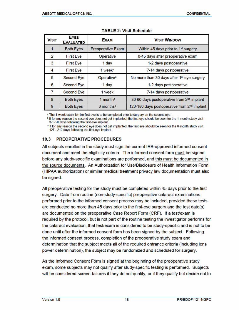
10.2 VISIT SCHEDULE The study visit schedule for all study subjects is outlined in Table 2.
All subjects are intended to have bilateral cataract surgery with the second-eye surgery occurring after the 1-week exam for the first eye but no more than 30 days after the first-eye surgery. After each surgery, each eye will be examined 1 day postoperatively (1-2 days) and again at 1 week (7-14 days). Following the second-eye surgery, both eyes will be evaluated at 1 month (30-60 days) and 6 months (120-180 days). Unscheduled visits may be conducted as necessary at the discretion of the investigator for medically-indicated follow-up.
ABBOTT MEDICAL OPTICS INC. CONFIDENTIAL
Version 1.0 19 PR/EDOF-121-NGPC
participate further in the study, or if they decide not to proceed with surgery. These subjects will be exited from the study.
Preoperative testing to be performed for each eye includes the following:
POTENTIAL DISTANCE VISUAL ACUITY
The subject must be capable of achieving Snellen 20/30 or better best corrected distance vision in each eye after cataract extraction and IOL implantation. The surgeon may use his/her judgment, the Potential Acuity Meter (PAM), or other methods (e.g., pinhole, laser interferometer, etc.) to estimate the subject’s potential postoperative acuity.
UNCORRECTED DISTANCE VISUAL ACUITY
Monocular uncorrected distance visual acuity (UCDVA) is to be measured using a standard Snellen chart or equivalent.
BEST CORRECTED DISTANCE VISUAL ACUITY AND MANIFEST REFRACTION
Preoperative manifest refraction is required. Monocular, best corrected distance visual acuity (BCDVA) is to be measured using a standard Snellen chart or equivalent and must be Snellen 20/40 or worse, with or without a glare source.
KERATOMETRY
Preoperative corneal astigmatism, as measured by keratometry or topography, should be 1.00 D or less. No irregular astigmatism should be present preoperatively, and topography (if performed) should be normal.
CONTACT LENS WEAR AND CORNEAL STABILITY For contact lens wearers, keratometric corneal stability following cessation of contact lens wear must be verified before surgery. PMMA contact lenses are not to be worn for at least 6 months; rigid gas-permeable contact lenses are not to be worn for at least 1 month; and extended-wear or daily-wear soft contact lenses are not to be worn for at least 1 week prior to the preoperative visit. Corneal stability must be verified for any subject who has worn PMMA lenses within 5 years or any other type of contact lenses within 6 months prior to the preoperative visit. To verify stability, repeat the keratometric measurements at least 1 week after the initial preoperative baseline keratometric measurement. Corneal curvature is considered to be stable if the difference in keratometric cylinder (vertical vs. horizontal keratometric readings) between the two time points does not exceed 0.50 D. Additionally, the difference between the two horizontal readings as well as the difference between the two vertical readings must also be no more than 0.50 D. Changes in keratometric axis must be no more than ±15○. If a change exceeding these criteria is noted, surgery is to be postponed until keratometric
ABBOTT MEDICAL OPTICS INC. CONFIDENTIAL
Version 1.0 20 PR/EDOF-121-NGPC
stability is demonstrated. Final biometry measurements and surgery should not take place until keratometric stability is achieved. Note: if this method of determining corneal stability is not a standard procedure in your practice, the subject must sign the informed consent form prior to starting the stability procedure.
IOL POWER AND TARGETED REFRACTION Axial length and anterior chamber depth (ACD) must be measured to determine the appropriate lens power to implant using an A-Constant. IOLMaster, Lenstar or immersion biometry methods are preferred; however, surgeons should use the biometry method with which they have the most experience and which was used in the determination of the personalized A-Constant for the TECNIS Symfony Model ZXR00 lens. The investigator’s personalized A-Constant for the TECNIS Symfony Model ZXR00 lens will also be used for the investigational TECNIS Models ZHR00 and ZQR00 lenses. The lens power should be calculated to achieve emmetropia at distance. Intentional over- or under-correction (outside ± 0.50 D) should NOT be planned for either eye; however, surgeons may adjust the targeted refraction as necessary to achieve emmetropia based on surgeon factors, study subject experience and/or subject first-eye outcomes.
ADDITIONAL PREOPERATIVE INFORMATION TO BE COLLECTED:
• Informed consent documentation • Subject demographic information • Planned surgery dates for each eye • Ocular history, including presence of ocular pathology for each eye • Intraocular pressure for each eye • Photopic (ambient) pupil size for each eye • Cataract type and density for each eye • Dilated fundus exam results for each eye • Medical findings from a biomicroscopic slit-lamp exam for each eye • Ocular symptoms for each eye • Ocular and systemic medications • Questionnaire to collect preoperative patient expectations
10.4 RANDOMIZATION AND MASKING A randomization list will be created by the AMO biostatistician for each investigative site and the randomization code will be uploaded into the electronic data capture system (EDC). Subjects will be randomized to the investigational Model ZHR00 IOL, the investigational Model ZQR00 IOL or the Symfony control IOL. Unmasked study personnel at the site will be trained to the randomization process through the EDC system and will randomize subjects after the subject has signed the informed consent form, has met all eligibility criteria and the investigator has documented which eye will be the first implanted.
ABBOTT MEDICAL OPTICS INC. CONFIDENTIAL
Version 1.0 21 PR/EDOF-121-NGPC
As part of the informed consent process, the investigator or delegate will explain to the subject the requirements of a randomized study and the differences expected between the three lens models: the Models ZHR00 and ZQR00 investigational lenses and the Symfony control lens. The surgeon and the operative staff will know which lens type is implanted. There may also be site coordinators and other site study staff, such as those performing slit-lamp exams, who will be unmasked. Unmasked study site staff will be instructed not to disclose the lens type the subject received or to talk about the lens to any masked evaluators or to the study subjects.
The subjects and the study technicians performing the postoperative vision tests are to be masked through study completion. To maintain subject/technician-masking through the 6-month study exams, a masking plan will be tailored for each site to detail how lens assignment information will be concealed from masked technicians. Recommended steps to maintain masking include ensuring that all items pertaining to lens group assignment and lens implantation records are kept separately from all other study documents and subject medical records until after completion of the final study visit. For example, lens stickers (indicating the lens model implanted) may be kept in the operating room study notebook until completion of the final study visit, at which time they may be placed in the subject medical charts. In the meantime, temporary lens stickers (without lens model designations) may be used in the subject’s medical chart.
To maintain subject masking, a temporary IOL implant identification card will be issued to the subject at the time of surgery. Following completion of the final study exam, each subject will be given the permanent IOL implant identification card.
10.5 STUDY LENS SUPPLY The investigational Model ZHR00 or ZQR00 lenses and the Symfony control lenses will be obtained from site consignments, supplied by AMO following IRB approval. Two lenses should be available for each case, a primary lens and a back-up lens. Unused back-up lenses are to be returned to the site consignment. At the completion of study enrollment, any remaining consignment lenses will be shipped back to AMO following reconciliation of investigational lens inventory by an AMO CRA. Any remaining control lenses will also be returned to AMO. At all times, the storage, access and use of all investigational lenses must be controlled and complete lens accountability maintained (See Section 15.2.1 Lens Accountability).
10.6 OPERATIVE PROCEDURES The investigator should use his or her standard, small-incision, cataract extraction surgical technique. Lenses should be folded for implantation and inserted into the capsular bag using one of the AMO-validated insertion systems described in Section 7.2.
ABBOTT MEDICAL OPTICS INC. CONFIDENTIAL
Version 1.0 22 PR/EDOF-121-NGPC
Investigators should manage surgical outcomes to ensure that the total postoperative refractive astigmatism is as minimal as possible. The total postoperative astigmatism, including surgically-induced astigmatism, should be targeted to be no greater than 1.0 D. Astigmatism may be managed by incision type and placement only. No additional refractive procedures are to be performed during the operative procedure or throughout the postoperative study period (e.g., LRI, OCCI, CRI, AK, PRK, LASIK or LASEK).
Operative case report forms will include the following information:
INCISION TYPE AND SIZE Lenses should be inserted through an incision ranging in size from approximately 2.2-3.0 mm, per the investigator’s standard technique when using the UNFOLDER Platinum 1 Series Implantation System or the ONE SERIES Ultra Implantation System. The incision may be clear corneal, limbal or scleral tunnel at the discretion of the investigator.
CAPSULORHEXIS SIZE AND METHOD
The anterior capsulotomy should be a continuous, curvilinear capsulorhexis approximately 5.0 to 5.5 mm in diameter to allow slight overlap of the lens optic edge. The anterior capsulotomy method may be manual (rhexis) or laser-assisted.
LENS REMOVAL Lens removal may occur using laser fragmentation combined with phacoemulsification/aspiration or using only phacoemulsification/aspiration.
VISCOELASTIC Viscoelastic materials should be used as is customary for each investigator and recorded on the case report form (CRF).
IMPLANT INSTRUMENTATION USED Lenses should be folded for implantation and inserted into the capsular bag using either the UNFOLDER Platinum 1 Series Implantation System (DK7796 handpiece with the Platinum 1 Series cartridge, Model 1MTEC30) or the ONE SERIES Ultra Implantation System (DK7786 [plunger] or the DK7791 [twist] handpieces with the ONE SERIES Ultra cartridge, Model 1VIPR30).
SURGICAL COMPLICATIONS Should a surgical complication occur, implantation of a study lens will be at the investigator’s discretion. In the event of capsular bag or zonular rupture, the lens should not be implanted if the complication may result in lens instability. Additionally, the lens is
ABBOTT MEDICAL OPTICS INC. CONFIDENTIAL
Version 1.0 23 PR/EDOF-121-NGPC
not to be implanted in the sulcus. In this case, the investigator may implant his/her choice of a back-up, non-investigational IOL. The subject should be exited from the study if a non-study lens is implanted as a result of a surgical complication during the first-eye implantation; however, the eye will be followed until resolution of the complication prior to exiting the subject. Should a surgical complication occur during the second-eye surgery and result in implantation of a non-study lens, the subject will not be exited from the study; the first eye will continue to be followed per-protocol, although data may be analyzed separately, and the second eye will be followed for safety until resolution of the complication.
MEDICATIONS Preoperative, operative and intraoperative medications should be used as is customary for each investigator and will be recorded on the CRF.
TYPE OF CLOSURE Wound closure is left to the surgeon’s discretion and will be recorded on the CRF.
ADDITIONAL OPERATIVE INFORMATION COLLECTED INCLUDES:
• Date of surgery • Operative eye • Lens power and serial number • Lens placement • Other surgical procedures • Surgical technique according to protocol • Product complaints • Serious and/or device-related adverse events
10.7 POSTOPERATIVE PROCEDURES Postoperatively, subjects will be examined according to the schedule in Section 10.2, Visit Schedule. Only the most recently operated eye will be evaluated at the 1-day and 1-week visits. Both eyes will be evaluated at the 1-month and 6-month visits.
Study technicians responsible for conducting all vision testing will be masked. Therefore, it is recommended that only the investigator/sub-investigator or other designated and trained clinician perform the biomicroscopic slit-lamp exams. To maintain consistency and masking throughout the study, it is recommended that a single individual (study technician or coordinator designated by the investigator) conduct all postoperative study-related vision testing, although a back-up person should also be designated and trained.
ABBOTT MEDICAL OPTICS INC. CONFIDENTIAL
Version 1.0 24 PR/EDOF-121-NGPC
Note: Subjects are not to wear contact lenses postoperatively until after completion of this study. Wearing contact lenses may potentially cause corneal edema or topography changes that may influence the visual acuity results. During the study, if correction is required, spectacles should be prescribed.
A postoperative CRF will collect the following information, although not all data are required at every visit (see Appendix A):
MANIFEST REFRACTION AND REFRACTION ADJUSTMENTS (MASKED PROCEDURE) Postoperative study manifest refractions are to be performed using the M&S System at a distance of 4.0 meters. Manifest refraction (MR) is to be performed using the Maximum Plus refraction method as detailed in Appendix C.
Because 4.0 meters is not optical infinity, refraction adjustments are necessary to ensure proper vision testing taking into account test distance and refraction distance. Appendix D lists the refraction adjustments required for the various vision tests using the BCDVA refraction.
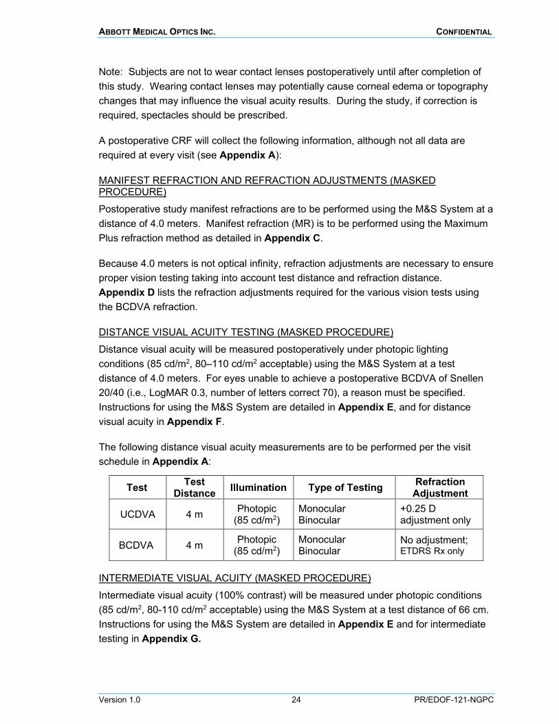
DISTANCE VISUAL ACUITY TESTING (MASKED PROCEDURE) Distance visual acuity will be measured postoperatively under photopic lighting conditions (85 cd/m2, 80–110 cd/m2 acceptable) using the M&S System at a test distance of 4.0 meters. For eyes unable to achieve a postoperative BCDVA of Snellen 20/40 (i.e., LogMAR 0.3, number of letters correct 70), a reason must be specified. Instructions for using the M&S System are detailed in Appendix E, and for distance visual acuity in Appendix F.
The following distance visual acuity measurements are to be performed per the visit schedule in Appendix A:
Test Test Distance Illumination Type of Testing Refraction
Adjustment
UCDVA 4 m Photopic (85 cd/m2)
Monocular Binocular
+0.25 D adjustment only
BCDVA 4 m Photopic (85 cd/m2)
Monocular Binocular
No adjustment; ETDRS Rx only
INTERMEDIATE VISUAL ACUITY (MASKED PROCEDURE) Intermediate visual acuity (100% contrast) will be measured under photopic conditions (85 cd/m2, 80-110 cd/m2 acceptable) using the M&S System at a test distance of 66 cm. Instructions for using the M&S System are detailed in Appendix E and for intermediate testing in Appendix G.
ABBOTT MEDICAL OPTICS INC. CONFIDENTIAL
Version 1.0 25 PR/EDOF-121-NGPC
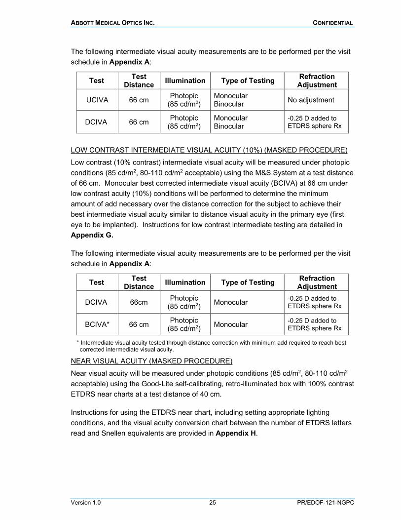
The following intermediate visual acuity measurements are to be performed per the visit schedule in Appendix A:
Test Test Distance Illumination Type of Testing Refraction
Adjustment
UCIVA 66 cm Photopic (85 cd/m2)
Monocular Binocular No adjustment
DCIVA 66 cm Photopic (85 cd/m2)
Monocular Binocular
-0.25 D added to ETDRS sphere Rx
LOW CONTRAST INTERMEDIATE VISUAL ACUITY (10%) (MASKED PROCEDURE) Low contrast (10% contrast) intermediate visual acuity will be measured under photopic conditions (85 cd/m2, 80-110 cd/m2 acceptable) using the M&S System at a test distance of 66 cm. Monocular best corrected intermediate visual acuity (BCIVA) at 66 cm under low contrast acuity (10%) conditions will be performed to determine the minimum amount of add necessary over the distance correction for the subject to achieve their best intermediate visual acuity similar to distance visual acuity in the primary eye (first eye to be implanted). Instructions for low contrast intermediate testing are detailed in Appendix G.
The following intermediate visual acuity measurements are to be performed per the visit schedule in Appendix A:
Test Test Distance Illumination Type of Testing Refraction
Adjustment
DCIVA 66cm Photopic (85 cd/m2) Monocular -0.25 D added to
ETDRS sphere Rx
BCIVA* 66 cm Photopic (85 cd/m2) Monocular -0.25 D added to
ETDRS sphere Rx
* Intermediate visual acuity tested through distance correction with minimum add required to reach best corrected intermediate visual acuity.
NEAR VISUAL ACUITY (MASKED PROCEDURE) Near visual acuity will be measured under photopic conditions (85 cd/m2, 80-110 cd/m2 acceptable) using the Good-Lite self-calibrating, retro-illuminated box with 100% contrast ETDRS near charts at a test distance of 40 cm.
Instructions for using the ETDRS near chart, including setting appropriate lighting conditions, and the visual acuity conversion chart between the number of ETDRS letters read and Snellen equivalents are provided in Appendix H.
ABBOTT MEDICAL OPTICS INC. CONFIDENTIAL
Version 1.0 26 PR/EDOF-121-NGPC
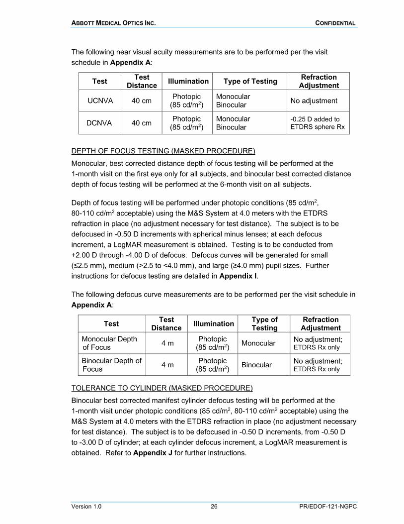
The following near visual acuity measurements are to be performed per the visit schedule in Appendix A:
Test Test Distance Illumination Type of Testing Refraction
Adjustment
UCNVA 40 cm Photopic (85 cd/m2)
Monocular Binocular No adjustment
DCNVA 40 cm Photopic (85 cd/m2)
Monocular Binocular
-0.25 D added to ETDRS sphere Rx
DEPTH OF FOCUS TESTING (MASKED PROCEDURE) Monocular, best corrected distance depth of focus testing will be performed at the 1-month visit on the first eye only for all subjects, and binocular best corrected distance depth of focus testing will be performed at the 6-month visit on all subjects.
Depth of focus testing will be performed under photopic conditions (85 cd/m2, 80-110 cd/m2 acceptable) using the M&S System at 4.0 meters with the ETDRS refraction in place (no adjustment necessary for test distance). The subject is to be defocused in -0.50 D increments with spherical minus lenses; at each defocus increment, a LogMAR measurement is obtained. Testing is to be conducted from +2.00 D through -4.00 D of defocus. Defocus curves will be generated for small (≤2.5 mm), medium (>2.5 to <4.0 mm), and large (≥4.0 mm) pupil sizes. Further instructions for defocus testing are detailed in Appendix I.
The following defocus curve measurements are to be performed per the visit schedule in Appendix A:
Test Test Distance Illumination Type of
Testing Refraction Adjustment
Monocular Depth of Focus 4 m Photopic
(85 cd/m2) Monocular No adjustment; ETDRS Rx only
Binocular Depth of Focus 4 m Photopic
(85 cd/m2) Binocular No adjustment; ETDRS Rx only
TOLERANCE TO CYLINDER (MASKED PROCEDURE) Binocular best corrected manifest cylinder defocus testing will be performed at the 1-month visit under photopic conditions (85 cd/m2, 80-110 cd/m2 acceptable) using the M&S System at 4.0 meters with the ETDRS refraction in place (no adjustment necessary for test distance). The subject is to be defocused in -0.50 D increments, from -0.50 D to -3.00 D of cylinder; at each cylinder defocus increment, a LogMAR measurement is obtained. Refer to Appendix J for further instructions.
ABBOTT MEDICAL OPTICS INC. CONFIDENTIAL
Version 1.0 27 PR/EDOF-121-NGPC
PUPIL SIZE Photopic, photopic with glare, mesopic and mesopic with glare pupil sizes will be measured postoperatively during the study. For consistency, the same method of measurement should be used throughout the study.
Photopic pupil size measurements are to be performed under the same lighting conditions at which photopic distance visual acuity is tested. Pupil sizes under the other lighting conditions will be measured during the contrast sensitivity testing procedures. Instructions for measuring pupil size are detailed in Appendix K.
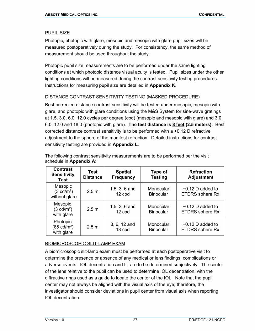
DISTANCE CONTRAST SENSITIVITY TESTING (MASKED PROCEDURE) Best corrected distance contrast sensitivity will be tested under mesopic, mesopic with glare, and photopic with glare conditions using the M&S System for sine-wave gratings at 1.5, 3.0, 6.0, 12.0 cycles per degree (cpd) (mesopic and mesopic with glare) and 3.0, 6.0, 12.0 and 18.0 (photopic with glare). The test distance is 8 feet (2.5 meters). Best corrected distance contrast sensitivity is to be performed with a +0.12 D refractive adjustment to the sphere of the manifest refraction. Detailed instructions for contrast sensitivity testing are provided in Appendix L.
The following contrast sensitivity measurements are to be performed per the visit schedule in Appendix A:
Contrast Sensitivity
Test Test
Distance Spatial
Frequency Type of Testing
Refraction Adjustment
Mesopic (3 cd/m2)
without glare 2.5 m 1.5, 3, 6 and
12 cpd Monocular Binocular
+0.12 D added to ETDRS sphere Rx
Mesopic (3 cd/m2) with glare
2.5 m 1.5, 3, 6 and 12 cpd
Monocular Binocular
+0.12 D added to ETDRS sphere Rx
Photopic (85 cd/m2) with glare
2.5 m 3, 6, 12 and 18 cpd
Monocular Binocular
+0.12 D added to ETDRS sphere Rx
BIOMICROSCOPIC SLIT-LAMP EXAM A biomicroscopic slit-lamp exam must be performed at each postoperative visit to determine the presence or absence of any medical or lens findings, complications or adverse events. IOL decentration and tilt are to be determined subjectively. The center of the lens relative to the pupil can be used to determine IOL decentration, with the diffractive rings used as a guide to locate the center of the IOL. Note that the pupil center may not always be aligned with the visual axis of the eye; therefore, the investigator should consider deviations in pupil center from visual axis when reporting IOL decentration.
ABBOTT MEDICAL OPTICS INC. CONFIDENTIAL
Version 1.0 28 PR/EDOF-121-NGPC
Findings of aqueous cells and flare, corneal edema, posterior capsule striae (wrinkles), posterior capsular opacification and IOL glistenings are to be rated using standardized grading scales of 0 to +4 (0 = none, +4 = severe) during the slit-lamp biomicroscopy. The specific grading scales are provided in Appendix M.
ND:YAG CAPSULOTOMY If an Nd:YAG capsulotomy is necessary, it is recommended that the procedure be performed at least 1 week prior to a study exam; this is particularly important for the 6-month study visit, as this is the key study exam for evaluation of safety and effectiveness.
DILATED FUNDUS EXAM
A dilated fundus exam is to be performed at the 6-month visit to evaluate retinal status and fundus visualization.
INTRAOCULAR PRESSURE AND KERATOMETRY
Intraocular pressure (IOP) and keratometry are to be measured using the investigator’s usual methods. It is recommended that the same methods be used for all study subjects at the site for the duration of the study.
OCULAR SYMPTOMS (NON-DIRECTED; SPONTANEOUS)
Subjective ocular symptoms are to be assessed at each postoperative visit by asking “Are you having any difficulties with your eyes/vision?” Subjects should not be prompted for specific responses; however, if a subject reports halos, night glare or starbursts, the level of severity should be determined (mild, moderate or severe).
MEDICATIONS Postoperative ocular medications should be used as is customary for each investigator and recorded in the source document for each subject. Medications will be recorded on a medication log CRF as applicable.
ADVERSE EVENTS
Subjects should be assessed at each visit for occurrence of and/or change in status of any adverse events, particularly serious and/or device-related adverse events. See Section 11.0, Adverse Events, for further information.
QUESTIONNAIRES Questionnaires will be administered at the 1-month and 6-month visits to collect information regarding spectacle usage, visual symptoms, visual quality and subject satisfaction. In order to minimize any effect the doctor-patient relationship may have on a subject’s responses on the questionnaire, the study questionnaires will be
ABBOTT MEDICAL OPTICS INC. CONFIDENTIAL
Version 1.0 29 PR/EDOF-121-NGPC
self-administered by the subjects. The questionnaires are to be administered at the start of the 1-month and 6-month study visits, prior to any visual acuity testing.
In addition, if a subject is seen at an Unscheduled visit due to an optical/visual symptom complaint, the PRO Visual Symptoms Questionnaire will be administered at that visit, as well as prior to any secondary surgical intervention for an optical/visual symptom complaint. If additional unscheduled visits and/or a secondary surgical intervention due to the same optical/visual symptom complaint occur within 2 weeks of each other, it is not necessary to complete the PRO Visual Symptoms Questionnaire a second time.
If the subject indicates in the PRO Visual Symptoms Questionnaire that they have visual symptoms or other problems with their vision that are bothersome enough to want the lenses removed and replaced, the investigator will document a determination for whether or not, in their opinion, the problem is related to the optical properties of the lens.
10.8 EXIT OF SUBJECTS An Exit CRF will be completed for all subjects, either when they complete the study or if they exit early.
It is the responsibility of the investigator to provide complete follow-up data to AMO for each subject, and every attempt should be made to gather that complete follow-up data for all subjects enrolled, as missing data can have a negative effect on the study results. Patients who would be traveling, relocating or otherwise unavailable for postoperative follow-up visits should not be enrolled in this clinical study.
A subject will be considered a "screen failure" if he/she does not meet the eligibility criteria or if consent is withdrawn prior to randomization.
A subject will be considered "discontinued prior to treatment" if the subject is randomized but does not undergo surgery or receive a study lens for various reasons including: the planned implant being aborted due to surgical complications, the subject withdrawing consent prior to treatment or the subject died prior to treatment.
Subjects will be "discontinued" from the study if one study lens (if implanted unilaterally) or both study lenses (if implanted bilaterally) are removed or if the subject dies.
If a subject receives at least one study lens, he/she is to be followed according to the schedule in Table 2 (Section 10.2) for visit windows.
Subjects will be considered “lost-to-follow-up” from the study only if irretrievably lost for unavoidable reasons such as: subject moved/unable to locate, subject ill/unable to travel, subject uncooperative/refuses further study participation. In the event of subject
ABBOTT MEDICAL OPTICS INC. CONFIDENTIAL
Version 1.0 30 PR/EDOF-121-NGPC
relocation, effort must be made by the investigator to secure follow-up information (i.e., slit-lamp findings and general visual acuity, etc.) from the subject’s new physician.
If a subject is exited early from the study, the investigator must indicate the reason for study exit on the CRF. In the event of a lens removal or other serious adverse event, the subject may be exited from the study; however, effort must be made by the investigator to follow the subject until resolution of the adverse event before exiting the subject from the study.
Following study completion or early exit, subjects will be informed about which lens model they received. Additionally, all study subjects are to be instructed to undergo regular eye examinations at least yearly and also to return to their doctor if any eye complications are experienced.
10.9 UNSCHEDULED VISITS During the study period, if a non-protocol-required visit is done for the purpose of medically-indicated follow-up for a study eye, data from this visit should be reported using the Unscheduled Visit CRF. The need for unscheduled visits is at the investigator’s discretion. Specific examinations to be performed at unscheduled visits are also at the discretion of the investigator (based on the reason for the unscheduled visit) and data are to be recorded in the appropriate section of the CRF.
Data to be collected may include: • Snellen manifest refraction • Uncorrected and best corrected distance visual acuity using a Snellen chart • Intraocular pressure • Slit-lamp examination for medical and/or lens findings • Dilated fundus exam • Ocular symptoms • Adverse events • Medications
In addition, if a subject is seen at an Unscheduled visit due to an optical/visual symptom complaint, the PRO Visual Symptoms Questionnaire will be administered at that visit, as well as prior to any secondary surgical intervention for an optical/visual symptom complaint. If additional unscheduled visits and/or a secondary surgical intervention due to the same optical/visual symptom complaint occur within 2 weeks of each other, it is not necessary to complete the PRO Visual Symptoms Questionnaire a second time.
10.10 PROTOCOL DEVIATIONS Any departure from the protocol procedures represents a protocol deviation. Protocol deviations may be subject-based (e.g., inclusion/exclusion criteria, informed consent deviation, etc.) or procedural-based (e.g., out-of-interval visits, non-compliance with
ABBOTT MEDICAL OPTICS INC. CONFIDENTIAL
Version 1.0 31 PR/EDOF-121-NGPC
testing procedures, etc.). All protocol deviations will be documented using the protocol deviation CRF. Any deviation made to protect the life or physical well-being of a subject in an emergency as well as any use of the investigational device without obtaining informed consent must be reported to AMO within 5 working days. Protocol deviations will be monitored by AMO, and if the non-compliance is persistent or egregious, AMO may take action, including but not limited to termination of the investigator’s participation in the study. The investigator is also responsible for informing the reviewing IRB of instances of protocol non-compliance in accordance with the IRB requirements.
11. ADVERSE EVENTS AND PRODUCT COMPLAINTS
11.1 ADVERSE EVENT DEFINITIONS
Adverse Event (AE) An adverse event is defined (following ISO 14155) as any untoward medical occurrence, unintended disease or injury, or untoward clinical signs (including abnormal laboratory findings) in subjects, users or other persons, whether or not related to the study device.
Serious Adverse Event (SAE) An adverse event is considered serious (following ISO 14155) if it is an untoward occurrence which may or may not be related to use of the study device that
• is sight- or life-threatening, • results in death, • requires inpatient hospitalization or prolongation of hospitalization (a planned
hospitalization for a pre-existing condition without a serious deterioration in health is not considered a serious adverse event),
• results in permanent impairment of a body structure or body function, • necessitates medical or surgical intervention to prevent permanent impairment to
a body structure or function, or • results in fetal distress, fetal death or a congenital abnormality or birth defect
Device-Related Adverse Event/Adverse Device Effect (ADE) A device-related adverse event is defined as any adverse event that is believed to be definitely, probably or possibly related to the study device (following the guidelines in Section 11.4, Causal Relationship). A device-related event is also considered an adverse device effect (ADE; following ISO 14155) resulting from the use of the study device that may result from user error, insufficiencies or inadequacies in the instructions for use, deployment, implantation, installation, operation of any malfunction of the device.
ABBOTT MEDICAL OPTICS INC. CONFIDENTIAL
Version 1.0 32 PR/EDOF-121-NGPC
Study-Specific Serious Anticipated Adverse Events The following is a list including, but not limited to, ocular serious adverse events (SAE) that are anticipated and must be reported to AMO for this study. Any events that are unlikely but anticipated (i.e., endophthalmitis) will be reported to the FDA and other appropriate regulatory agencies.
• Endophthalmitis/Intraocular infection • Hypopyon • Hyphema • IOL dislocation • Cystoid macular edema • Pupillary block • Retinal detachment/tear • Persistent corneal edema • Persistent iritis • Persistent raised IOP requiring treatment • Visual symptoms requiring secondary surgical intervention (e.g., lens removal) • Tilt and decentration requiring secondary surgical intervention (e.g.,
repositioning) • Residual refractive error resulting in a secondary surgical intervention • Retained lens material resulting in secondary surgical intervention
NOTE 1: Wound burps during the first week postoperatively, suture removal, planned blepharoplasty and Nd:YAG capsulotomy (for PCO) are not considered adverse events for this study.
NOTE 2: Raised IOP requiring treatment, cornea edema, and iritis will only be considered serious if persistent at the final study visit (120-180 days postoperative) or sight-threatening at the time of occurrence. Treatment merely to hasten the resolution of such conditions (and not intended to prevent permanent damage to the eye) will not be reported as serious adverse events.
Unanticipated Adverse Device Effect (UADE)/Unanticipated Serious Adverse Device Effect (USADE) Any UADE (USA 21CFR 812.3(s)) or USADE (ISO 14155) is defined as any serious adverse effect on health or safety or any life-threatening problem or death caused by, or associated with, a device, if that effect, problem, or death was not previously identified in nature, severity, or degree of incidence in the investigational plan (i.e., this protocol), application (including a supplementary plan or application), or risk assessment, or any other unanticipated serious problem associated with a device that relates to the rights, safety, or welfare of subjects.
ABBOTT MEDICAL OPTICS INC. CONFIDENTIAL
Version 1.0 33 PR/EDOF-121-NGPC
11.2 PRODUCT COMPLAINT/DEVICE DEFICIENCY DEFINITION A product complaint/device deficiency is defined (21 CFR 820.3(b) and ISO 14155) as any alleged deficiency related to the identity, quality, durability, reliability, safety, effectiveness, or performance of a device. This may include malfunctions, use error and inadequacies in labeling. Product complaints can pertain to any marketed AMO device being used in the study as well as the investigational device. The investigator is to assess whether the deficiency could have led to a serious adverse event without suitable action or intervention or under less fortunate circumstances.
11.3 ADVERSE EVENT AND COMPLAINT REPORTING REQUIREMENTS All adverse events and any complaint encountered using any AMO product, regardless of severity and whether or not attributed to the study device(s), are to be reported to AMO and recorded on the case report form corresponding to the visit during which awareness of the event occurred. Adverse events are also to be reported to the reviewing IRB as per the IRB’s reporting requirements. If required, adverse events will be reported to the appropriate regulatory agencies (e.g., FDA) according to all applicable laws and regulations.
Reporting of adverse events shall follow the USA Code of Federal Regulations (21CFR812) for sites in the USA. General guidelines are provided below:
Adverse Event Reporting An adverse event that is not serious or device-related is to be reported to AMO in a timely manner. Notification of non-serious and non-device related adverse events will occur by recording events on the CRF when noted. Such adverse events are also to be reported to the reviewing IRB per their reporting requirements.
Complaints/Device Deficiency Reporting A general product complaint or device deficiency is to be reported to AMO in a timely manner. Notification of complaints/device deficiencies will occur by recording complaints on the CRF at the visit the complaint occurs (e.g., operative visit) and/or by a phone call to AMO.
Any device deficiency that could have led to a serious adverse event without suitable action or intervention, or under less fortunate circumstances, must be reported to the sponsor immediately (no later than 48 hours after detection). Device deficiencies that could have led to a serious adverse event should also be reported to the investigator’s IRB per their reporting requirements.
ABBOTT MEDICAL OPTICS INC. CONFIDENTIAL
Version 1.0 34 PR/EDOF-121-NGPC
Serious and/or Device-Related Adverse Event Reporting SAEs and/or ADEs are to be documented using the SAE/ADE CRF. In the event of an SAE/ADE, AMO must be notified immediately (no later than 48 hours after detection). Any SAE/ADE is to be reported to AMO by phone, email and/or by submitting the completed SAE/ADE CRF. Any SAE or device-related AE should also be reported to the investigator’s IRB per their reporting requirements.
Unanticipated Adverse Device Effect (UADE)/Unanticipated Serious Adverse Device Effect (USADE) Reporting If during the study, a serious adverse event occurs that may reasonably be regarded as device-related and was not previously expected in nature, severity, or degree of incidence, the investigator is to report the UADE/USADE to AMO within 48 hours, and to the investigator’s IRB as soon as possible (and no later than 10 working days after learning of the event for sites in the USA as required by 21CFR812).
11.4 CAUSAL RELATIONSHIP The investigator should always be alert to adverse events that may be related to the study device or the use of the study device (i.e., the procedure specific to the initial application of the device). An attempt should be made in every case to determine the causality of the event. The following definitions are to be used as guidelines in determining the relationship between the event and the study device and/or use of the device.
ABBOTT MEDICAL OPTICS INC. CONFIDENTIAL
Version 1.0 35 PR/EDOF-121-NGPC
Definitely related: If the event is associated with the device and/or the use of the device beyond a reasonable doubt, a causal relationship exists between the adverse event and the device and/or the use of the study device.
Probably related: There is a reasonable possibility of a causal relationship between the adverse event and the device and/or the use of the study device and/or the adverse event cannot be reasonably explained by another cause.
Possibly related: The adverse event has not been determined to be related to the device or the use of the device, but no other cause has been identified and the device and/or the use of the study device cannot be ruled out as a possible cause.
Unlikely to be related: The possibility of a potential causal relationship between adverse event and the device and/or the use of the device could exist, but the adverse event can be reasonably explained by another cause.
Not related: There is no possibility of a causal relationship between the adverse event and the device and/or the use of the study device and/or the adverse event can be attributed to another cause.
If an adverse event is believed to be definitely, probably or possibly related to the study device and/or the use of the device, the event will be considered related to the study device and/or the use of the device.
11.5 ADVERSE EVENT FOLLOW-UP For every adverse event, appropriate measures should be undertaken to treat and/or monitor the subject until resolution occurs. The subject's files are to include all pertinent medical data relating to the event including the subject’s medical records, medical reports and/or judgments from colleagues or outside specialists who assisted in the treatment and follow-up of the subject. The investigator should keep AMO closely informed as to the outcome of serious and/or device-related adverse events, thereby allowing AMO to comply with the appropriate regulatory reporting requirements. A Detailed Adverse Event Update CRF should be completed each time the subject returns to the investigator or other specialist(s) for follow-up of serious and/or device-related adverse event until resolution of the event. Any subject who is to be exited from the study due to a serious and/or device-related adverse event should be followed until the outcome is determined prior to being exited from the study.
ABBOTT MEDICAL OPTICS INC. CONFIDENTIAL
Version 1.0 36 PR/EDOF-121-NGPC
12. PROTOCOL CHANGES/AMENDMENTS
If the investigator wishes to modify any procedure and/or the design of the study, he or she must contact and obtain consent from AMO regarding the proposed changes prior to implementation. Any modifications (including additional data collection) require approval by the FDA and all other appropriate regulatory agencies, as well as approval of the governing IRBs prior to implementation.
13. ETHICS REVIEW AND PATIENT WELFARE
13.1 INSTITUTIONAL REVIEW BOARD (IRB) It is the responsibility of the investigator to obtain prospective approval of the study protocol, protocol amendments or changes, informed consent forms and other relevant documents (e.g., advertisements) from the IRB. All correspondence with the IRB should be retained in the Investigator Notebook. Copies of IRB submissions and approvals should be forwarded to AMO.
The investigator is responsible for notifying the IRB of reportable adverse events as well as any other circumstance in which additional procedures outside the protocol were conducted to eliminate apparent hazards to subjects.
13.2 INFORMED CONSENT The current version of the IRB-approved study informed consent must be signed by each study subject prior to any study-specific examinations being performed. The IRB-approved informed consent is to be signed and dated by the subject as well as by the person who conducted the informed consent discussion. The signed informed consent will be maintained by the investigator as a permanent part of the subject’s medical records. A copy of the signed and dated form is to be provided to the subject. The investigator will provide AMO written acknowledgement on the preoperative case report form that a signed agreement of informed consent has been obtained and is in the investigator’s possession for each subject. As required by 21CFR812 Part G, the site shall document in the source documents that informed consent was obtained prior to participation in the study for each subject enrolled.
NOTE: The informed consent process also includes obtaining the subject’s signature on an Authorization for Use/Disclosure of Health Information for Research Form or equivalent documentation necessary to comply with applicable privacy laws pertaining to medical treatment in the governing countries.
NOTE: The sponsor will secure appropriate insurance for study subjects prior to study start.
ABBOTT MEDICAL OPTICS INC. CONFIDENTIAL
Version 1.0 37 PR/EDOF-121-NGPC
14. DOCUMENTATION
14.1 SOURCE DOCUMENTS Source documents must be kept for all study subjects. Source documents may include a subject’s medical records, hospital charts, clinic charts, the investigator’s subject study files, as well as results of any diagnostic tests or procedures such as topographies or laboratory tests with photographs or instrument printouts.
Each site is expected to adhere to the clinic’s own standard documentation requirements for medical charts/clinic notes. For the purposes of this clinical study, the medical charts/clinic notes must also include, at a minimum, the following data that will be considered source data and will be reviewed by AMO:
• Subject’s name and study identification number • Subject’s contact information • Study protocol number and the Sponsor name (AMO) • A statement that informed consent was obtained prior to participation in the study
(including the date) • Evidence of subject eligibility • Dates of all subject visits and surgeries throughout the duration of the study • Implant serial number identification (NOTE: This is masked information, and may
only be reviewed by unmasked study staff) • Concurrent medications • Corrected and uncorrected distance visual acuity (NOTE: M&S electronic data and
near visual acuity score sheets are considered source documentation and are to be retained by the site. A paper copy of the M&S results will be printed and validated by the site)
• Manifest refraction • Occurrence and status of any operative complications, postoperative medical or lens
findings and adverse events • Occurrence and status of any subject complaints, e.g., ocular/visual symptoms • The date the subject exited the study, and a notation as to whether the subject
completed the study or reason for early exit.
14.2 SUBJECT CONFIDENTIALITY Subjects will be assigned a site/subject number to maintain subject confidentiality. Subject names may possibly be disclosed to AMO or regulatory agencies during inspection of medical records related to the study, but reasonable precautions will be taken to maintain confidentiality of personal information to the extent permitted by applicable laws and regulations.
ABBOTT MEDICAL OPTICS INC. CONFIDENTIAL
Version 1.0 38 PR/EDOF-121-NGPC
14.3 CASE REPORT FORM COMPLETION This study will use an electronic data capture system. All study staff responsible for entering data into the system must complete certification prior to using the system. The investigator is responsible for ensuring that data are properly recorded on each subject’s case report forms and related documents. Prior to database lock, the investigator will verify completeness and accuracy of data submitted to AMO.
14.4 STUDY SUMMARY A final investigator’s summary (study close-out) will be provided to AMO and the reviewing IRB after termination or the completion of the study or the investigator’s part of the investigation, as directed by AMO.
15. MONITORING
AMO will perform three types of monitoring to ensure compliance with regulations: data monitoring, administrative monitoring, and safety monitoring.
15.1 DATA MONITORING In order to ensure a well-controlled clinical trial, AMO will follow specific data monitoring procedures, routinely generate reports and periodically review safety and effectiveness data. To avoid bias, any analyses generated prior to site closures will not be disseminated to any of the investigative sites.
An electronic data capture system (EDC) will be used to transmit case report forms from the investigative site to AMO. Requests for data clarification will be handled through this same system.
To minimize data omissions and inconsistencies on clinical reports and to ensure that data are accurately transcribed to computer data files, AMO will follow internal data processing procedures that include automated and manual quality control checks to identify any data discrepancies. Any such items will be resolved and documented as needed in EDC.
Prevention of Missing Data Methods used to safeguard against missing data that can have deleterious effects on the study integrity and reliability of its outcomes will include training study staff with WebEx, centralized and/or on-site programs. In addition, subjects will be encouraged at the time of informed consent to avoid missing study visits, as missing data may affect the study reliability and diminish the scientific value of their contribution to the study.
ABBOTT MEDICAL OPTICS INC. CONFIDENTIAL
Version 1.0 39 PR/EDOF-121-NGPC
15.2 ADMINISTRATIVE MONITORING Administrative monitoring procedures will ensure that study devices, subjects, and forms can be traced and will allow monitoring of investigator progress and compliance. Accountability and traceability of study devices will be monitored by trained AMO personnel.
Device Accountability Complete lens accountability will be maintained at the investigative site by maintaining records of all investigational lenses (Models ZHR00 and ZQR00 lenses) received from and returned to AMO. A site log will be used to track all lenses for date of receipt, eye implanted, serial number, lens power, use and disposition/return to AMO. This site log and any other investigational lens information will be maintained in the operative room study binder. During periodic investigative site monitoring visits, AMO personnel will review investigative site lens inventory records and logs to ensure IOL accountability compliance and complete investigational lens traceability.
Site Monitoring Plan Prior to performing any study implants, the requirements of the study and reporting mechanisms will be explained to each investigator either personally at the investigative site or at a formal study investigator meeting. When necessary, a pre-study site qualification visit may be performed to assess the adequacy of the site to perform the study for sites that have not previously worked with AMO or have undergone significant changes, or have not been visited in the past year. A study initiation visit will be conducted for all sites prior to the first implant.
Throughout the duration of the study, site visits to monitor compliance to this protocol will be made at each investigative site. During interim site monitoring visits, AMO will review informed consent documents and subject eligibility, and the data on study case report forms will be verified against subject charts and other source documents to ensure complete and accurate reporting. The subject files will also be reviewed to assure that all adverse events and any issues encountered with AMO products have been reported in a timely fashion.
AMO will also review source documents to verify that all required items have been documented in the subject medical charts. Refer to Section 14.1, Source Documents, for a list of items that are required for source documentation. In addition to subject files, study logs will be checked and conformance to lighting levels for visual acuity tests will be verified.
Training on study-specific procedures may also be conducted during monitoring visits. For this study in particular, a training/monitoring visit is likely to occur just prior to or
ABBOTT MEDICAL OPTICS INC. CONFIDENTIAL
Version 1.0 40 PR/EDOF-121-NGPC
during the first of the 1-month and 6-month visits, wherein the most extensive vision testing occurs.
Upon study completion, a final close-out site visit to each site will be made to monitor the last of the subject data records and finalize any outstanding study issues.
A separate Study Monitoring Plan will be established prior to study start that will define the type and frequency of monitoring visits and frequency of record monitoring.
15.3 SAFETY MONITORING The medical monitor will review results throughout the clinical trial as necessary to ensure the continued safety of the device and to ensure that no subjects are exposed to unreasonable risk. The medical monitor will be available to answer all questions from investigators. The medical monitor will review and assess any reports of serious and/or device-related adverse events as well as device deficiencies that could have led to a serious adverse event, and discuss these with the reporting investigator(s) as necessary. The medical monitor, as well as any other qualified personnel designated by AMO, shall also review any interim progress reports, as applicable.
16. PUBLICATIONS
Refer to the Clinical Trial Agreement for information regarding AMO publication policies.
17. RISK ANALYSIS
POTENTIAL RISKS AND RISK MANAGEMENT RISKS OF THE TECNIS NEXT-GENERATION IOLS, MODELS ZHR00 AND ZQR00
The TECNIS Models ZHR00 and ZQR00 IOLs are designed to provide far, intermediate and near vision; however, glasses may still be needed to improve distance vision and/or to have useful vision for intermediate or near tasks. Visual symptoms, particularly dysphotopsias such as halos, night glare, starbursts, etc., are expected to be less than with a standard multifocal IOL; however, such symptoms may still occur. Dysphotopsias may become less noticeable over time; however, the IOL may be removed if necessary. There may be a reduction in contrast sensitivity under certain conditions compared to a monofocal lens. Due to the diffractive optic design, the ability to perform some eye treatments (e.g., retinal photocoagulation) may be affected, and caution should be used when interpreting results of autorefractors or wavefront aberrometers that utilize infrared light, or when performing a duochrome test. These risks are not unlike other diffractive technology IOLs.
ABBOTT MEDICAL OPTICS INC. CONFIDENTIAL
Version 1.0 41 PR/EDOF-121-NGPC
GENERAL RISKS OF CATARACT SURGERY AND IOL IMPLANTATION
There are risks and complications associated with cataract surgery and IOL implantation in general. These can include worsening of vision, hemorrhage, loss of corneal clarity, inflammation, infections, retinal detachment, pupil changes, glaucoma, etc. Complications can result in poor vision, loss of vision or loss of the eye.
RISK MANAGEMENT
Subjects will be closely monitored thought the trial duration. The occurrence of adverse events and complaints will be assessed at each study visit and reported to AMO according to Section 11.0, Adverse Events and Product Complaints. Additionally, AMO will monitor incoming data following the procedures outlined in Section 15.0, Monitoring. The Medical Monitor will ensure subjects are not exposed to additional risks by monitoring serious adverse events, device-related adverse events, and device-deficiencies that could have led to serious adverse events (Section 15.3, Safety Monitoring).
POTENTIAL BENEFITS The general clinical performance of the TECNIS Next-Generation IOLs, Models ZHR00 and ZQR00, are expected to be similar to the TECNIS Symfony control IOL regarding distance and intermediate visual acuities and safety outcomes. An extended range of vision and improved near visual acuity and functionality may be achieved with the TECNIS Next-Generation IOLs, Models ZHR00 and ZQR00.
CONCLUSION
The hazards/risks associated with the TECNIS Next-Generation IOLs, Models ZHR00 and ZQR00, are acceptable and within those of AMO’s other advanced optic IOLs. The potential clinical benefits of the TECNIS Next-Generation IOLs, Models ZHR00 and ZQR00, outweigh the residual risks when the device is used as intended.
18. RECORDS RETENTION
All study-related correspondence, subject records, consent forms, Authorization for Use/Disclosure of Health Information Forms or similar medical treatment privacy law documentation, records of the distribution and use of all study products, and original case report forms should be maintained by the investigator.
The investigator must maintain and have access to the following essential documents until notified by the Sponsor. Note: This may be for a minimum of 15 years after completion of the study. AMO requires notification if the investigator wishes to relinquish ownership of the data so that mutually agreed-upon arrangements can be made for transfer of ownership to a suitably qualified, responsible person.
ABBOTT MEDICAL OPTICS INC. CONFIDENTIAL
Version 1.0 42 PR/EDOF-121-NGPC
• All case report forms • All adverse event information (i.e., medical records, medical reports and/or
judgments from colleagues or outside specialists who assisted in the treatment and follow-up of the subject)
• Investigational supply records/inventory • IRB approval documentation • Study correspondence • Study agreements • Site visit documentation • Protocol(s) • Subject log(s) • Clinical Investigator’s Brochure • Completed subject informed consent forms and medical privacy forms (e.g.,
Authorization for Use/Disclosure of Health information) • Subject medical chart/clinic notes (Not applicable for transfer of ownership to
AMO)
19. TERMINATION OF THE INVESTIGATION
The clinical investigation will be suspended in the event of high levels of complications and/or adverse events that are unexpected in nature and/or severity and evaluated as to causality relative to the study device. The clinical investigation may be suspended if the Medical Monitor or the IRB, upon review and evaluation of the clinical data, finds unacceptable clinical performance or the level of single or total complications and/or adverse events unacceptable for continuation of the investigation.
If causality is shown not to be related to the study device, the study may be resumed in accordance with the IRB and regulations of the FDA. The study will be terminated if causality is shown to be related to the study device.
Additionally, the investigator or AMO may stop a subject’s participation at any time. AMO may also stop the study at any time for reasons it determines appropriate. However, no suspension of the study would be made to disadvantage the study subjects. Following suspension of the study for any reason, all study subjects who have already received treatment would continue to be followed through completion of the study visit schedule.
20. STATISTICAL METHODS
This section highlights the analyses to be performed for key study endpoints. The key study timeframe for effectiveness endpoints will be 1 month and for safety endpoints the key study timeframe will be 6 months, although data will be reviewed at other time points
ABBOTT MEDICAL OPTICS INC. CONFIDENTIAL
Version 1.0 43 PR/EDOF-121-NGPC
as well. TECNIS Models ZHR00 and ZQR00 will both be compared to TECNIS Symfony control separately and independently for all analyses. All data will be reported by IOL group.
20.1 ANALYSIS POPULATION The safety population (SP) of all subjects implanted who have available data will be used for all analysis. No data imputation will be performed for missing data. For the monocular endpoints, the safety population will consist of first eyes implanted with a study IOL; for binocular endpoints, the safety population will consist of subjects implanted with the same IOL in both eyes. The primary analysis population will be the safety population (SP) for all endpoints.
20.2 PRIMARY STUDY ENDPOINTS
Primary Effectiveness Endpoints MODEL ZHR00: MONOCULAR DISTANCE CORRECTED NEAR VISUAL ACUITY (DCNVA) AT 40 CM The primary effectiveness endpoint for the Model ZHR00 IOL is mean (LogMAR), photopic, monocular, first-eye, distance corrected near visual acuity (40 cm) at 1 month postoperative. Note that a lower LogMAR value is a better acuity and a higher LogMAR value is a poorer acuity. The mean, SD, median, minimum, maximum and 95% confidence interval (CI) will be reported by IOL group.
o A statistical comparison between the Model ZHR00 and control lens groups will be performed using a 1-sided, 2-sample, t-test with an alpha set at 0.025. The null hypothesis is that the mean monocular DCNVA for the Model ZHR00 eyes is worse than or equal to that for control eyes. The alternate hypothesis is that the mean monocular DCNVA for Model ZHR00 eyes is better than that for control eyes.
Ho: μc - μt ≤ 0 (Model ZHR00 lens is worse than (higher LogMAR) or equal to control) H1: μc - μt > 0 (Model ZHR00 lens is better (lower LogMAR) than control) where μt = mean LogMAR DCNVA for the Model ZHR00 lens μc = mean LogMAR DCNVA for control lens Reject the null hypothesis if one-sided p-value ≤ 0.025.
The success criterion is a statistically significantly lower mean LogMAR value for the Model ZHR00 investigational lens compared to the control lens (p ≤ 0.025).
ABBOTT MEDICAL OPTICS INC. CONFIDENTIAL
Version 1.0 44 PR/EDOF-121-NGPC
MODEL ZQR00: MONOCULAR DISTANCE CORRECTED NEAR VISUAL ACUITY (DCNVA) AT 40 CM
The primary effectiveness endpoint for the Model ZQR00 IOL is mean (LogMAR), photopic, monocular, first-eye, distance corrected near visual acuity (40 cm) at 1 month postoperative. Note that a lower LogMAR value is a better acuity and a higher LogMAR value is a poorer acuity. The mean, SD, median, minimum, maximum and 95% CI will be reported by IOL group.
o Analysis for the Model ZQR00 will be similar to the Model ZHR00 analysis described above. A statistical comparison between the Model ZQR00 and control lens groups will be performed using a 1-sided, 2-sample, t-test with an alpha set at 0.025. The null hypothesis is that the mean monocular DCNVA for the Model ZQR00 lens eyes is worse than or equal to that for control eyes. The alternate hypothesis is that the mean monocular DCNVA for Model ZQR00 eyes is better than that for control eyes.
Ho: μc - μt ≤ 0 (Model ZQR00 lens is worse than (higher LogMAR) or equal to control) H1: μc - μt > 0 (Model ZQR00 lens is better (lower LogMAR) than control) where μt = mean LogMAR DCNVA for the Model ZQR00 lens μc = mean LogMAR DCNVA for control lens Reject the null hypothesis if one-sided p-value ≤ 0.025.
The success criterion is a statistically significantly lower mean LogMAR value for the Model ZQR00 compared to the control lens (p ≤ 0.025).
Primary Safety Endpoints MODEL ZHR00: RATES OF ADVERSE EVENTS VS. ISO SPE RATES
The primary Model ZHR00 safety endpoint for this study is the rate of adverse events vs. ISO SPE rates at 6 months postoperative. The frequency and proportion of first eyes, second eyes, and all subjects with these events will be reported over time by IOL group. Statistical comparisons to ISO SPE rates will be based on first-eye data; adverse event rates for the Model ZHR00 investigational lens first eyes will be compared to the ISO SPE rates using a one-sided, exact test based on the binomial distribution. The null hypothesis is that the AE rate for the Model ZHR00 investigational lens eyes is lower than or equal to the ISO SPE values, and the alternative hypothesis is that the AE rate for study eyes is higher than the ISO SPE values.
Ho: pt ≤ pi H1: pt > pi where pt = proportion of Model ZHR00 investigational lens eyes with the AE pi = proportion of eyes reported in ISO SPE rates with the AE
ABBOTT MEDICAL OPTICS INC. CONFIDENTIAL
Version 1.0 45 PR/EDOF-121-NGPC
Reject the null hypothesis if one-sided p-value < 0.05.
MODEL ZQR00: RATES OF ADVERSE EVENTS VS. ISO SPE RATES
The primary Model ZQR00 safety endpoint for this study is the rate of adverse events vs. ISO SPE rates at 6 months postoperative. The frequency and proportion of first eyes, second eyes, and all subjects with these events will be reported over time by IOL group. Statistical comparisons to ISO SPE rates will be based on first-eye data; adverse event rates for Model ZQR00 investigational lens first eyes will be compared to the ISO SPE rates using a one-sided, exact test based on the binomial distribution. The null hypothesis is that the AE rate for Model ZQR00 investigational lens eyes is lower than or equal to the ISO SPE values, and the alternative hypothesis is that the AE rate for study eyes is higher than the ISO SPE values.
Ho: pt ≤ pi H1: pt > pi where pt = proportion of Model ZQR00 investigational lens eyes with the AE pi = proportion of eyes reported in ISO SPE rates with the AE
Reject the null hypothesis if one-sided p-value < 0.05.
20.3 ADDITIONAL ENDPOINTS For BCDVA, the frequency and proportion of eyes achieving each acuity line will be reported over time by IOL group. The proportion of investigational lens eyes achieving 20/40 or better at 6 months will be compared to the ISO SPE rate for posterior chamber IOLs (all first eyes) using a one-sided, exact test based on binomial distribution. The null hypothesis (based on the ISO guidance document) is that the proportion of investigational lens eyes achieving 20/40 or better BCDVA is greater than or equal to the ISO SPE values, and the alternative hypothesis is that the proportion of investigational lens eyes achieving 20/40 or better BCDVA is less than the ISO SPE values.
In addition, the mean LogMAR BCDVA will be compared between lens groups for first eyes using a non-inferiority method. The null hypothesis is that the mean difference (control minus investigational lens) between the control and investigational IOLs is ≤-0.1 LogMAR (1 line) with the alternative hypothesis that the mean difference is >-0.1 LogMAR. A 90% confidence interval (CI) of a two-sample, two-sided, t-test will be used for evaluation.
For all other near endpoints, statistics similar to DCNVA described above will be used. In addition, the frequency and proportion of first eyes achieving each line will be reported over time by IOL group.
ABBOTT MEDICAL OPTICS INC. CONFIDENTIAL
Version 1.0 46 PR/EDOF-121-NGPC
For intermediate endpoints, the mean LogMAR values will be analyzed similar to BCDVA described above. The frequency and proportion of first eyes achieving each line will also be reported over time by IOL group.
For defocus results, the defocus curve will be produced by IOL group. The diopters of defocus where the mean visual acuity 20/32 or better is achieved will be derived by visual inspection of the defocus curve.
For questionnaire data, including visual symptoms, the frequency and proportion of subjects with a given response will be reported by IOL group. For comparisons between IOL groups, Wilcoxon Rank-Sum test will be used for ordinal data, and Fisher’s exact test will be used for categorical data. The null hypothesis is that there is no difference between responses, and the alternative hypothesis is that there is a difference between responses. Two-sided testing and alpha of 0.05 will be used to evaluate questionnaire data.
For contrast sensitivity data, descriptive analyses including mean, standard deviation, median, minimum, maximum and 90% confidence intervals will be presented by IOL group.
The frequency and percentage of medical findings, lens findings and non-directed ocular/visual symptoms will be reported over time by IOL group for both eyes.
20.4 VISUAL ACUITY CONVENTIONS AND GENERAL STATISTICS Visual acuity data will be converted to LogMAR values prior to analysis and adjusted for the test distance used if it is not the standard distance for the chart. Descriptive statistics will typically include sample size (N), mean, standard deviation (SD), median, minimum (Min) and maximum (Max) as appropriate for continuous variables. For continuous variables, statistical tests (e.g., t-test) assuming normality will generally be used. For categorical data, the frequency and proportion will be reported and Fisher’s exact test or Chi-square test will generally be applied. For ordinal categorical data, the frequency and proportion will be reported with the Wilcoxon Rank-Sum test generally used.
20.5 INTERIM REPORTS Although interim study progress reports will be conducted for this study, all masking of study subjects and masked study technicians will be maintained and interim reports will not be disseminated to investigators/site personnel.
20.6 SAMPLE SIZE CALCULATIONS For the primary endpoint of distance corrected near visual acuity, there is 90% power to detect a 1-line or greater difference in mean visual acuity between the investigational
ABBOTT MEDICAL OPTICS INC. CONFIDENTIAL
Version 1.0 47 PR/EDOF-121-NGPC
lens groups and the control group (assume one-sided testing with an alpha of 0.025 and standard deviation of 1.6 lines) with 65 subjects in each lens group.
For contrast sensitivity there is 80% power to detect a non-inferiority margin of 0.15 log units between the investigational lens groups and the control group (assume one-sided alpha=0.05 and standard deviation of 0.34) with 65 subjects in each lens group.
If the dropout rate is assumed at 10%, approximately 73 subjects will be implanted in each lens group to achieve a minimum of 65 subjects in each lens group at 1 and 6 months.
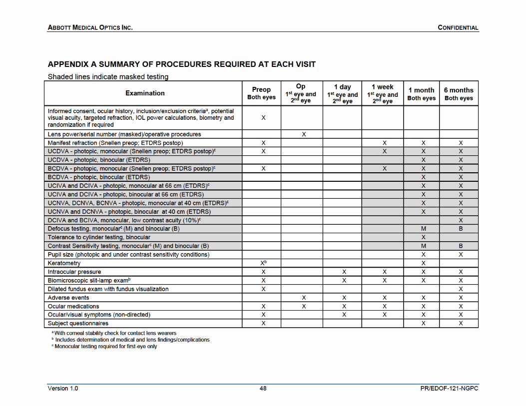
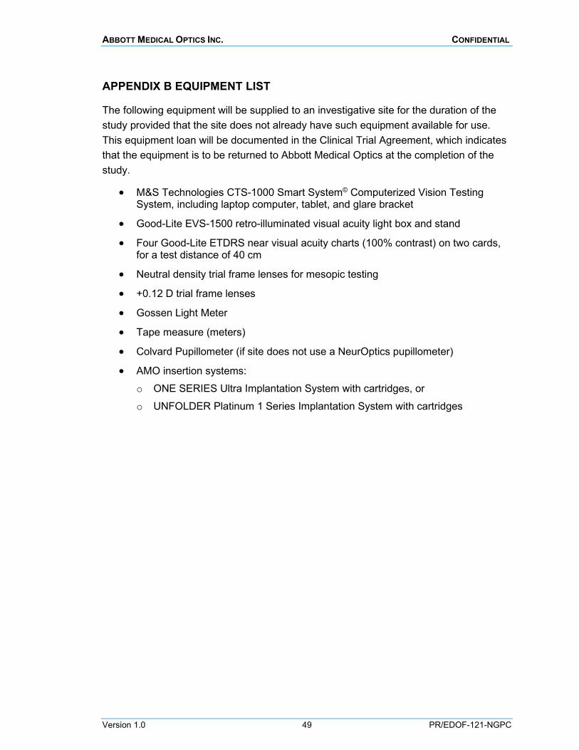
ABBOTT MEDICAL OPTICS INC. CONFIDENTIAL
Version 1.0 49 PR/EDOF-121-NGPC
APPENDIX B EQUIPMENT LIST
The following equipment will be supplied to an investigative site for the duration of the study provided that the site does not already have such equipment available for use. This equipment loan will be documented in the Clinical Trial Agreement, which indicates that the equipment is to be returned to Abbott Medical Optics at the completion of the study.
• M&S Technologies CTS-1000 Smart System© Computerized Vision Testing System, including laptop computer, tablet, and glare bracket
• Good-Lite EVS-1500 retro-illuminated visual acuity light box and stand
• Four Good-Lite ETDRS near visual acuity charts (100% contrast) on two cards, for a test distance of 40 cm
• Neutral density trial frame lenses for mesopic testing
• +0.12 D trial frame lenses
• Gossen Light Meter
• Tape measure (meters)
• Colvard Pupillometer (if site does not use a NeurOptics pupillometer)
• AMO insertion systems: o ONE SERIES Ultra Implantation System with cartridges, or o UNFOLDER Platinum 1 Series Implantation System with cartridges
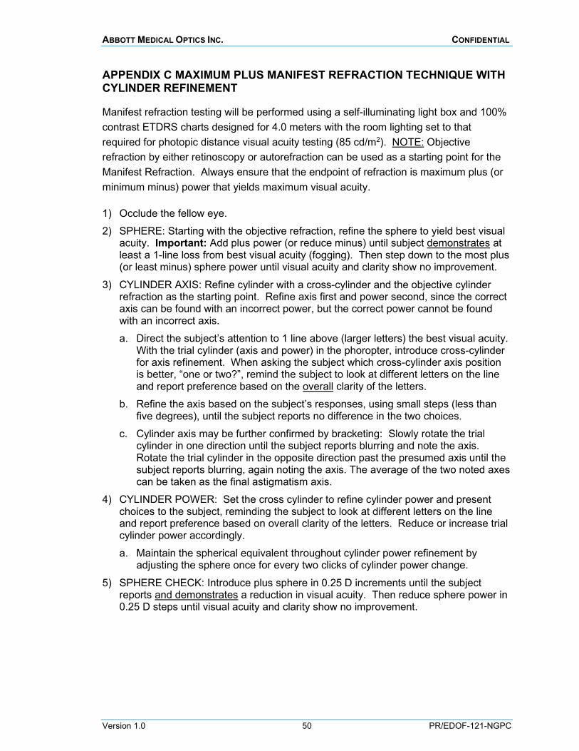
ABBOTT MEDICAL OPTICS INC. CONFIDENTIAL
Version 1.0 50 PR/EDOF-121-NGPC
APPENDIX C MAXIMUM PLUS MANIFEST REFRACTION TECHNIQUE WITH CYLINDER REFINEMENT
Manifest refraction testing will be performed using a self-illuminating light box and 100% contrast ETDRS charts designed for 4.0 meters with the room lighting set to that required for photopic distance visual acuity testing (85 cd/m2). NOTE: Objective refraction by either retinoscopy or autorefraction can be used as a starting point for the Manifest Refraction. Always ensure that the endpoint of refraction is maximum plus (or minimum minus) power that yields maximum visual acuity.
1) Occlude the fellow eye. 2) SPHERE: Starting with the objective refraction, refine the sphere to yield best visual
acuity. Important: Add plus power (or reduce minus) until subject demonstrates at least a 1-line loss from best visual acuity (fogging). Then step down to the most plus (or least minus) sphere power until visual acuity and clarity show no improvement.
3) CYLINDER AXIS: Refine cylinder with a cross-cylinder and the objective cylinder refraction as the starting point. Refine axis first and power second, since the correct axis can be found with an incorrect power, but the correct power cannot be found with an incorrect axis. a. Direct the subject’s attention to 1 line above (larger letters) the best visual acuity.
With the trial cylinder (axis and power) in the phoropter, introduce cross-cylinder for axis refinement. When asking the subject which cross-cylinder axis position is better, “one or two?”, remind the subject to look at different letters on the line and report preference based on the overall clarity of the letters.
b. Refine the axis based on the subject’s responses, using small steps (less than five degrees), until the subject reports no difference in the two choices.
c. Cylinder axis may be further confirmed by bracketing: Slowly rotate the trial cylinder in one direction until the subject reports blurring and note the axis. Rotate the trial cylinder in the opposite direction past the presumed axis until the subject reports blurring, again noting the axis. The average of the two noted axes can be taken as the final astigmatism axis.
4) CYLINDER POWER: Set the cross cylinder to refine cylinder power and present choices to the subject, reminding the subject to look at different letters on the line and report preference based on overall clarity of the letters. Reduce or increase trial cylinder power accordingly. a. Maintain the spherical equivalent throughout cylinder power refinement by
adjusting the sphere once for every two clicks of cylinder power change. 5) SPHERE CHECK: Introduce plus sphere in 0.25 D increments until the subject
reports and demonstrates a reduction in visual acuity. Then reduce sphere power in 0.25 D steps until visual acuity and clarity show no improvement.
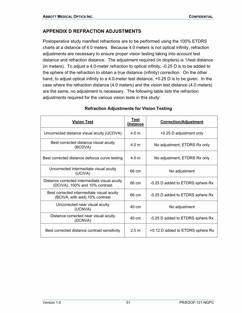
ABBOTT MEDICAL OPTICS INC. CONFIDENTIAL
Version 1.0 51 PR/EDOF-121-NGPC
APPENDIX D REFRACTION ADJUSTMENTS
Postoperative study manifest refractions are to be performed using the 100% ETDRS charts at a distance of 4.0 meters. Because 4.0 meters is not optical infinity, refraction adjustments are necessary to ensure proper vision testing taking into account test distance and refraction distance. The adjustment required (in diopters) is 1/test distance (in meters). To adjust a 4.0-meter refraction to optical infinity, -0.25 D is to be added to the sphere of the refraction to obtain a true distance (infinity) correction. On the other hand, to adjust optical infinity to a 4.0-meter test distance, +0.25 D is to be given. In the case where the refraction distance (4.0 meters) and the vision test distance (4.0 meters) are the same, no adjustment is necessary. The following table lists the refraction adjustments required for the various vision tests in this study:
Refraction Adjustments for Vision Testing
Vision Test Test Distance Correction/Adjustment
Uncorrected distance visual acuity (UCDVA) 4.0 m +0.25 D adjustment only
Best corrected distance visual acuity (BCDVA) 4.0 m No adjustment; ETDRS Rx only
Best corrected distance defocus curve testing 4.0 m No adjustment; ETDRS Rx only
Uncorrected intermediate visual acuity (UCIVA) 66 cm No adjustment
Distance corrected intermediate visual acuity (DCIVA), 100% and 10% contrast 66 cm -0.25 D added to ETDRS sphere Rx
Best corrected intermediate visual acuity (BCIVA, with add),10% contrast 66 cm -0.25 D added to ETDRS sphere Rx
Uncorrected near visual acuity (UCNVA) 40 cm No adjustment
Distance corrected near visual acuity (DCNVA) 40 cm -0.25 D added to ETDRS sphere Rx
Best corrected distance contrast sensitivity 2.5 m +0.12 D added to ETDRS sphere Rx
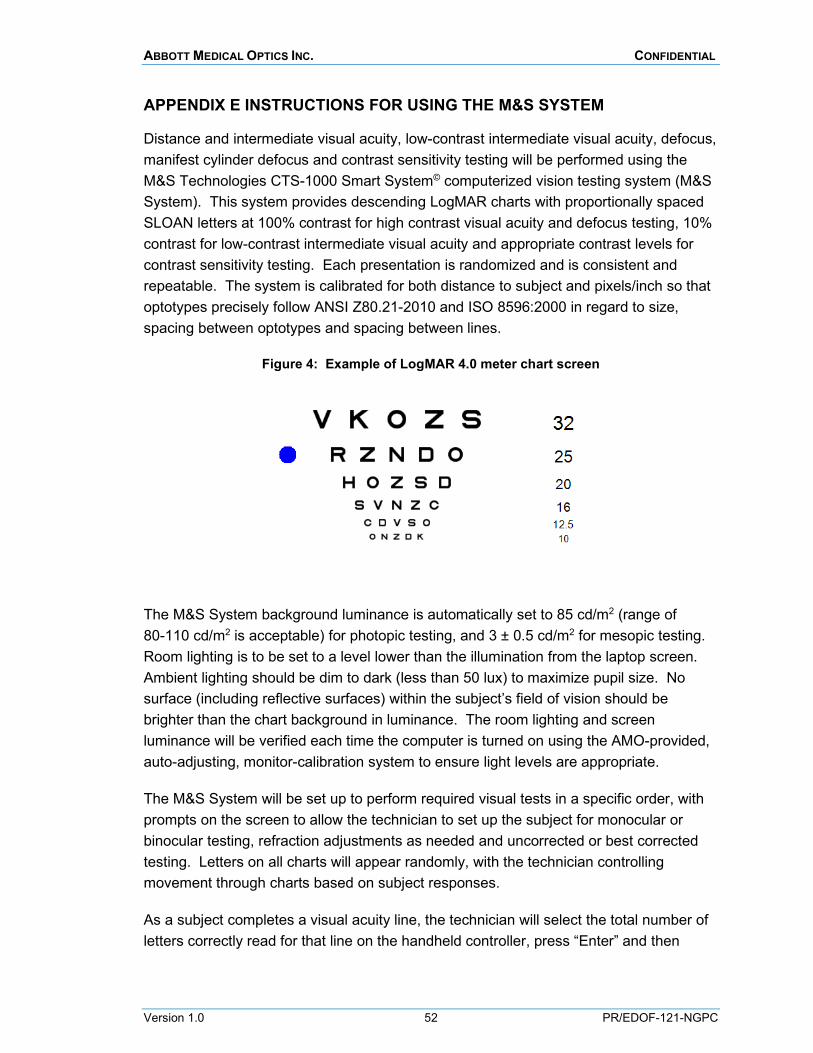
ABBOTT MEDICAL OPTICS INC. CONFIDENTIAL
Version 1.0 52 PR/EDOF-121-NGPC
APPENDIX E INSTRUCTIONS FOR USING THE M&S SYSTEM
Distance and intermediate visual acuity, low-contrast intermediate visual acuity, defocus, manifest cylinder defocus and contrast sensitivity testing will be performed using the M&S Technologies CTS-1000 Smart System© computerized vision testing system (M&S System). This system provides descending LogMAR charts with proportionally spaced SLOAN letters at 100% contrast for high contrast visual acuity and defocus testing, 10% contrast for low-contrast intermediate visual acuity and appropriate contrast levels for contrast sensitivity testing. Each presentation is randomized and is consistent and repeatable. The system is calibrated for both distance to subject and pixels/inch so that optotypes precisely follow ANSI Z80.21-2010 and ISO 8596:2000 in regard to size, spacing between optotypes and spacing between lines.
Figure 4: Example of LogMAR 4.0 meter chart screen
The M&S System background luminance is automatically set to 85 cd/m2 (range of 80-110 cd/m2 is acceptable) for photopic testing, and 3 ± 0.5 cd/m2 for mesopic testing. Room lighting is to be set to a level lower than the illumination from the laptop screen. Ambient lighting should be dim to dark (less than 50 lux) to maximize pupil size. No surface (including reflective surfaces) within the subject’s field of vision should be brighter than the chart background in luminance. The room lighting and screen luminance will be verified each time the computer is turned on using the AMO-provided, auto-adjusting, monitor-calibration system to ensure light levels are appropriate.
The M&S System will be set up to perform required visual tests in a specific order, with prompts on the screen to allow the technician to set up the subject for monocular or binocular testing, refraction adjustments as needed and uncorrected or best corrected testing. Letters on all charts will appear randomly, with the technician controlling movement through charts based on subject responses.
As a subject completes a visual acuity line, the technician will select the total number of letters correctly read for that line on the handheld controller, press “Enter” and then

ABBOTT MEDICAL OPTICS INC. CONFIDENTIAL
Version 1.0 53 PR/EDOF-121-NGPC
confirm the number of letters correct at the next prompt. The M&S System will then advance to the next line of testing and the process will repeat. The system will end the test when the subject no longer has any correct responses. The number of letters correctly read will be displayed on the laptop screen, along with the LogMAR value and Snellen acuity. Record the total number of letters read. A similar process will be done for contrast sensitivity grating responses. Once a test is completed, follow the prompts on the computer screen to start the next test.
Test results are stored in the M&S computer and a hard copy will be printed and validated as a back-up.
ABBOTT MEDICAL OPTICS INC. CONFIDENTIAL
Version 1.0 54 PR/EDOF-121-NGPC
APPENDIX F INSTRUCTIONS FOR DISTANCE VISUAL ACUITY TESTING
For distance visual acuities, the M&S System laptop should be placed at a test distance of 4.0 meters from the subject for testing distance visual acuities. A laptop setting may be used to reverse charts for rooms that require “folding” via a mirror to reach a distance of 4.0 meters. Whether standard or “folded”, measure and record the test distance accurately. If the room set-up does not allow the computer to be placed at precisely 4.0 meters, the M&S System can be adjusted to account for the actual test distance used.
The M&S System will be set up to perform the required distance visual acuity tests in a specific order, with prompts on the screen to allow the technician to set up the subject for monocular or binocular testing, refraction adjustment as needed and uncorrected or best corrected testing.
Subjects should be reminded prior to testing that squinting is not allowed. The technician is to observe the subject to ensure the subject is not squinting during visual acuity testing. If squinting is observed, the subject is to be reminded by the testing technician not to squint.
Distance visual acuity measurements are to be performed per the visit schedule in Appendix A. Monocular testing is required on the first eye implanted only; to test subjects monocularly, occlude the second eye in the phoropter or with an occluder if trial lenses are used.
Visual acuity measurements are based on the total number of correctly read letters. Subjects should be persuaded to read the smallest letters possible even if they have to guess. Follow the testing process listed in Appendix E. At the end of the test, the number of letters correctly read will be displayed on the laptop screen, along with the LogMAR value and Snellen acuity. Record the total number of letters read in the EDC system.
ABBOTT MEDICAL OPTICS INC. CONFIDENTIAL
Version 1.0 55 PR/EDOF-121-NGPC
APPENDIX G INSTRUCTIONS FOR INTERMEDIATE VISUAL ACUITY TESTING
Intermediate visual acuity will be measured using the M&S System at a test distance of 66 cm. Subjects should be reminded prior to testing that squinting is not allowed. Trial frames should be used for visual acuity testing with careful observation by the testing technician to ensure the subject is not squinting. If squinting is observed, the subject is to be reminded by the testing technician not to squint.
The M&S System will be set up to perform the required intermediate visual acuity tests in a specific order, with prompts on the screen to allow the technician to set up the subject for monocular or binocular testing, refraction adjustment as needed, and uncorrected or best corrected testing.
Subjects should be reminded prior to testing that squinting is not allowed. Trial frames should be used for visual acuity testing with careful observation by the testing technician to ensure the subject is not squinting. If squinting is observed, the subject is to be reminded by the testing technician not to squint.
Intermediate visual acuity measurements are to be performed per the visit schedule in Appendix A. Both 100% contrast and 10% contrast intermediate acuity tests will be done on the M&S System. Monocular testing is required on the first eye implanted only; to test subjects monocularly, occlude the second eye in the phoropter or with an occluder if trial lenses are used.
Visual acuity measurements are based on the total number of correctly read letters. Subjects should be persuaded to read the smallest letters possible even if they have to guess. Follow the testing process listed in Appendix E. At the end of the test, the number of letters correctly read will be displayed on the laptop screen, along with the LogMAR value and Snellen acuity. Record the total number of letters read in the EDC system.
BEST CORRECTED INTEMEDIATE VISUAL ACUITY TESTING WITH ADD (BCIVA): First eye monocular best corrected intermediate visual acuity (10% contrast) with minimum add will be performed to determine the minimum amount of add necessary to achieve an intermediate visual acuity similar to the best corrected distance visual acuity. BCIVA is to be tested at 66 cm under photopic conditions using the M&S System at 6 months. With distance correction in place, slowly build the amount of add necessary for the subject to achieve their best monocular low-contrast intermediate visual acuity in the primary eye. In order to ensure the appropriate amount of add, at least one of the criteria given below must be achieved:
ABBOTT MEDICAL OPTICS INC. CONFIDENTIAL
Version 1.0 56 PR/EDOF-121-NGPC
BCIVA similar to the subject’s BCDVA. Document the amount of add necessary to achieve a BCIVA that matches the subject’s BCDVA in the first eye. For example, if the subject’s BCDVA is 20/20, the minimum ADD power will be the amount of add necessary to achieve a BCIVA of 20/20 at 66 cm. When the subject reaches a BCIVA that matches the BCDVA, record the minimum add power and BCIVA (# of letters correct).
Increasing ADD power does not lead to improvement in near visual acuity. When an additional +0.50 add (in two +0.25 D increments) does not increase the subject’s visual acuity by 3 or more letters, stop the test and record the minimum add power and BCIVA (# of letters correct). For example, the subject shows progressive improvement in intermediate visual acuity and achieves 20/25 with +0.75 add over the distance correction. Increasing the add further by +0.50 (in two +0.25 D increments) only allows the subject to get 20/25+2 with a total of +1.25 add power. Stop the testing and record a minimum add of +0.75 with 75 letters correct.
ABBOTT MEDICAL OPTICS INC. CONFIDENTIAL
Version 1.0 57 PR/EDOF-121-NGPC
APPENDIX H INSTRUCTIONS FOR NEAR VISUAL ACUITY TESTING
Near visual acuity will be measured using a using Good-Lite 100% ETDRS near charts designed for 40 cm and the Good-Lite self-illuminated and self-calibrated illumination box. The Good-Lite illumination box is self-illuminated and self-calibrates to a light level of approximately 85 cd/m2 (range of 80–110 cd/m2 is acceptable). Room lighting is to be set at a level lower than the illumination from the light box, per manufacturer instructions. Ambient luminance should be from dim to dark to maximize pupil size. No surface (including reflective surfaces) within the subject’s field of vision should exceed the chart background in luminance.
The light box should be placed at a test distance of 40 cm from the subject for testing near visual acuities. Subjects should be reminded prior to testing that squinting is not allowed. Trial frames should be used for visual acuity testing with careful observation by the testing technician to ensure the subject is not squinting. If squinting is observed, the subject is to be reminded by the testing technician not to squint.
To avoid chart memorization, charts will be changed between every visual acuity measurement. Monocular testing will be required on the first eye implanted only, with the second eye occluded. Uncorrected and distance corrected monocular testing on the first eye will be completed before binocular testing is done.
Near visual acuity testing is to be performed per Appendix A. Visual acuity measurements are based on the total number of correctly read letters. Subjects should be persuaded to read the smallest letters possible even if they have to guess. Record the total number of letters read correctly for each near test on the case report form. Near score grids are to be used and must be retained as source documentation. A reference chart for the conversion of the number of correctly-read ETDRS letters to a Snellen line equivalent is provided later in this section.
PROCEDURE: Begin with monocular uncorrected near visual acuity testing for the first eye implanted, and instruct subjects to start from the top, reading each line from left to right until they cannot read any additional letters. Alternate the near chart per the score sheet and repeat the testing with distance correction in place. For binocular testing, change the near charts per the score sheet and test uncorrected and distance corrected binocular acuities. Record the total number of letters read for each test condition in the EDC system.
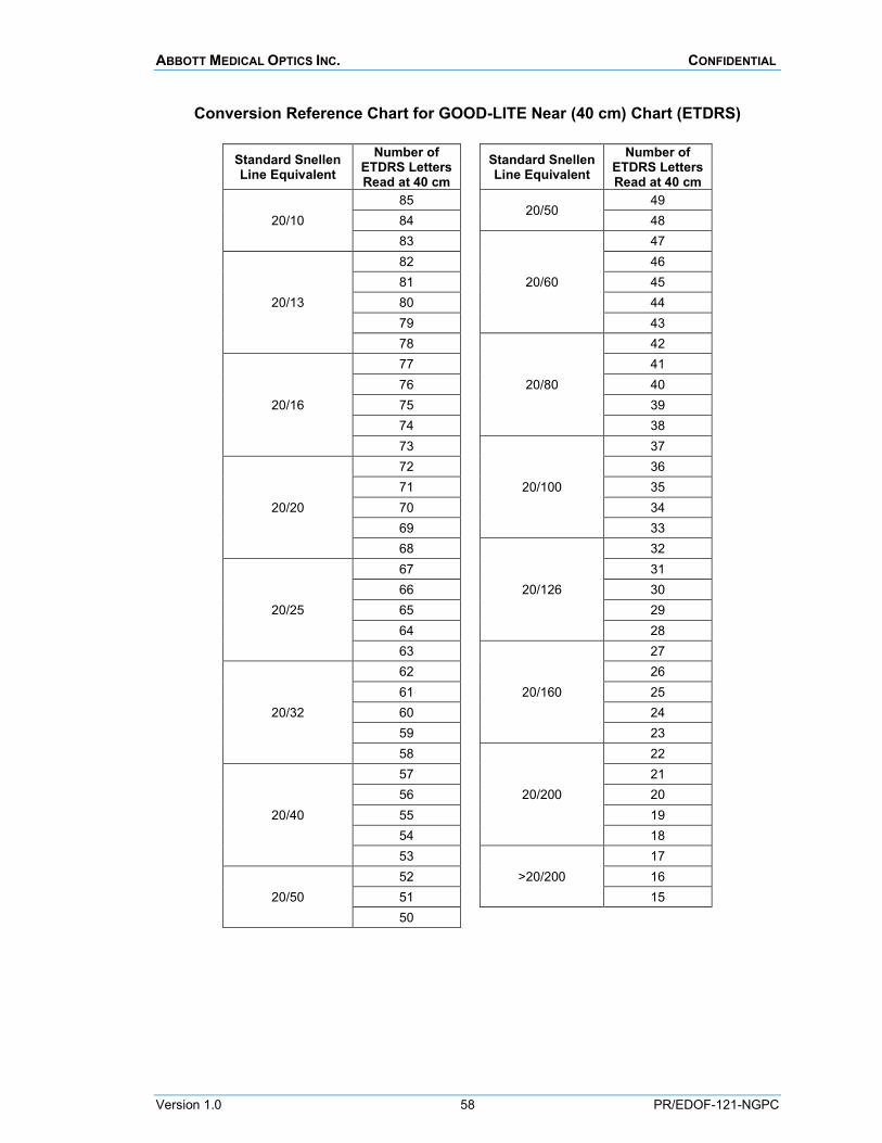
ABBOTT MEDICAL OPTICS INC. CONFIDENTIAL
Version 1.0 58 PR/EDOF-121-NGPC
Conversion Reference Chart for GOOD-LITE Near (40 cm) Chart (ETDRS)
Standard Snellen Line Equivalent
Number of ETDRS Letters Read at 40 cm
Standard Snellen Line Equivalent
Number of ETDRS Letters Read at 40 cm
20/10 85
20/50 49
84 48 83
20/60
47
20/13
82 46 81 45 80 44 79 43 78
20/80
42
20/16
77 41 76 40 75 39 74 38 73
20/100
37
20/20
72 36 71 35 70 34 69 33 68
20/126
32
20/25
67 31 66 30 65 29 64 28 63
20/160
27
20/32
62 26 61 25 60 24 59 23 58
20/200
22
20/40
57 21 56 20 55 19 54 18 53
>20/200 17
20/50 52 16 51 15 50
ABBOTT MEDICAL OPTICS INC. CONFIDENTIAL
Version 1.0 59 PR/EDOF-121-NGPC
APPENDIX I INSTRUCTIONS FOR DEPTH OF FOCUS TESTING
All depth of focus testing is to be performed under photopic conditions using the M&S System at a test distance of 4.0 meters. Subjects should be reminded prior to testing that squinting is not allowed. During defocus curve testing, trial frames will be used to allow observation by the testing technician to ensure the subject is not squinting. If squinting is observed, the subject is to be reminded by the testing technician not to squint.
This test is to be done monocularly at the 1-month visit for all subjects and binocularly at the 6-month visit for all subjects. Only the first eye implanted will be tested at the 1-month visit.
Monocular Depth of Focus Testing (1-month visit): With the subject’s distance correction in place for the first eye and the second eye occluded, defocus the image by +2.00 D over the manifest distance correction in the first eye. Start the test by instructing the subject to read as many letters as possible on the M&S system, even if they have to guess. The technician will control movement through charts based on subject responses and the system will end the testing when a threshold is reached. The number of letters correctly read and the LogMAR score will be displayed on the laptop screen; record the total number of letters correct in EDC. Continue to change the defocus in -0.50 D increments, repeating the test and recording the total number of letters correct at each level of defocus. Continue to change the defocus to -4.00 D over the subject’s distance correction.
Binocular Depth of Focus Testing (6-month visit): Begin binocular testing by defocusing the image by +2.00 D over the manifest distance correction in both eyes. Start the test by instructing the subject to read as many letters as they can on the M&S system, even if they have to guess. The technician will control movement through charts based on subject responses and the system will end the testing when a threshold is reached. The number of letters correctly read and the LogMAR score will be displayed on the laptop screen; record the total number of letters correct in EDC. Continue to change the defocus in -0.50 D increments in both eyes, repeating the test and recording the total number of letters correct at each level of defocus. Continue to change the defocus to -4.00 D over the subject’s distance correction.
ABBOTT MEDICAL OPTICS INC. CONFIDENTIAL
Version 1.0 60 PR/EDOF-121-NGPC
APPENDIX J INSTRUCTIONS FOR MANIFEST CYLINDER DEFOCUS TESTING
All manifest cylinder defocus testing is to be performed binocularly on all subjects at the 1-month study visit under photopic conditions using the M&S System at a test distance of 4.0 meters. Subjects should be reminded prior to testing that squinting is not allowed. During manifest cylinder defocus testing, trial frames will be used to allow observation by the testing technician to ensure the subject is not squinting. If squinting is observed, the subject is to be reminded by the testing technician not to squint.
Begin binocular testing by placing the manifest refraction (sphere and cylinder) in place for both eyes. Add cylindrical power -0.50 D over the distance correction in both eyes, maintaining the same axis as the manifest refraction (or place at 90 axis for subjects with no refractive cylinder). Start the test by instructing the subject to read as many letters as they can on the M&S system, even if they have to guess. The technician will control movement through charts based on subject responses and the system will end the testing when a threshold is reached. The number of letters correctly read and the LogMAR score will be displayed on the laptop screen; record the total number of letters correct in EDC. Continue to change the amount of in -0.50 D increments in both eyes, repeating the test and recording the total number of letters correct at each level of defocus. Continue to change the defocus to -3.00 D over the subject’s distance correction and record the number of letters correct for each cylinder lens.
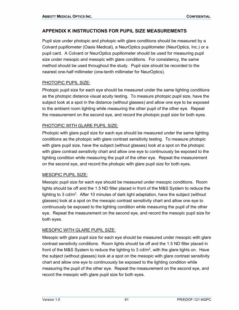
ABBOTT MEDICAL OPTICS INC. CONFIDENTIAL
Version 1.0 61 PR/EDOF-121-NGPC
APPENDIX K INSTRUCTIONS FOR PUPIL SIZE MEASUREMENTS
Pupil size under photopic and photopic with glare conditions should be measured by a Colvard pupillometer (Oasis Medical), a NeurOptics pupillometer (NeurOptics, Inc.) or a pupil card. A Colvard or NeurOptics pupillometer should be used for measuring pupil size under mesopic and mesopic with glare conditions. For consistency, the same method should be used throughout the study. Pupil size should be recorded to the nearest one-half millimeter (one-tenth millimeter for NeurOptics).
PHOTOPIC PUPIL SIZE: Photopic pupil size for each eye should be measured under the same lighting conditions as the photopic distance visual acuity testing. To measure photopic pupil size, have the subject look at a spot in the distance (without glasses) and allow one eye to be exposed to the ambient room lighting while measuring the other pupil of the other eye. Repeat the measurement on the second eye, and record the photopic pupil size for both eyes.
PHOTOPIC WITH GLARE PUPIL SIZE: Photopic with glare pupil size for each eye should be measured under the same lighting conditions as the photopic with glare contrast sensitivity testing. To measure photopic with glare pupil size, have the subject (without glasses) look at a spot on the photopic with glare contrast sensitivity chart and allow one eye to continuously be exposed to the lighting condition while measuring the pupil of the other eye. Repeat the measurement on the second eye, and record the photopic with glare pupil size for both eyes.
MESOPIC PUPIL SIZE: Mesopic pupil size for each eye should be measured under mesopic conditions. Room lights should be off and the 1.5 ND filter placed in front of the M&S System to reduce the lighting to 3 cd/m2. After 10 minutes of dark light adaptation, have the subject (without glasses) look at a spot on the mesopic contrast sensitivity chart and allow one eye to continuously be exposed to the lighting condition while measuring the pupil of the other eye. Repeat the measurement on the second eye, and record the mesopic pupil size for both eyes.
MESOPIC WITH GLARE PUPIL SIZE:
Mesopic with glare pupil size for each eye should be measured under mesopic with glare contrast sensitivity conditions. Room lights should be off and the 1.5 ND filter placed in front of the M&S System to reduce the lighting to 3 cd/m2, with the glare lights on. Have the subject (without glasses) look at a spot on the mesopic with glare contrast sensitivity chart and allow one eye to continuously be exposed to the lighting condition while measuring the pupil of the other eye. Repeat the measurement on the second eye, and record the mesopic with glare pupil size for both eyes.
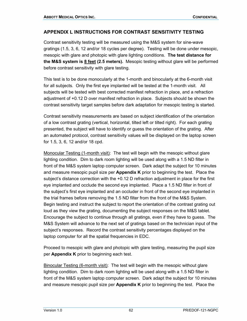
ABBOTT MEDICAL OPTICS INC. CONFIDENTIAL
Version 1.0 62 PR/EDOF-121-NGPC
APPENDIX L INSTRUCTIONS FOR CONTRAST SENSITIVITY TESTING
Contrast sensitivity testing will be measured using the M&S system for sine-wave gratings (1.5, 3, 6, 12 and/or 18 cycles per degree). Testing will be done under mesopic, mesopic with glare and photopic with glare lighting conditions. The test distance for the M&S system is 8 feet (2.5 meters). Mesopic testing without glare will be performed before contrast sensitivity with glare testing.
This test is to be done monocularly at the 1-month and binocularly at the 6-month visit for all subjects. Only the first eye implanted will be tested at the 1-month visit. All subjects will be tested with best corrected manifest refraction in place, and a refraction adjustment of +0.12 D over manifest refraction in place. Subjects should be shown the contrast sensitivity target samples before dark adaptation for mesopic testing is started.
Contrast sensitivity measurements are based on subject identification of the orientation of a low contrast grating (vertical, horizontal, tilted left or tilted right). For each grating presented, the subject will have to identify or guess the orientation of the grating. After an automated protocol, contrast sensitivity values will be displayed on the laptop screen for 1.5, 3, 6, 12 and/or 18 cpd.
Monocular Testing (1-month visit): The test will begin with the mesopic without glare lighting condition. Dim to dark room lighting will be used along with a 1.5 ND filter in front of the M&S system laptop computer screen. Dark adapt the subject for 10 minutes and measure mesopic pupil size per Appendix K prior to beginning the test. Place the subject’s distance correction with the +0.12 D refraction adjustment in place for the first eye implanted and occlude the second eye implanted. Place a 1.5 ND filter in front of the subject’s first eye implanted and an occluder in front of the second eye implanted in the trial frames before removing the 1.5 ND filter from the front of the M&S System. Begin testing and instruct the subject to report the orientation of the contrast grating out loud as they view the grating, documenting the subject responses on the M&S tablet. Encourage the subject to continue through all gratings, even if they have to guess. The M&S System will advance to the next set of gratings based on the technician input of the subject’s responses. Record the contrast sensitivity percentages displayed on the laptop computer for all the spatial frequencies in EDC.
Proceed to mesopic with glare and photopic with glare testing, measuring the pupil size per Appendix K prior to beginning each test.
Binocular Testing (6-month visit): The test will begin with the mesopic without glare lighting condition. Dim to dark room lighting will be used along with a 1.5 ND filter in front of the M&S system laptop computer screen. Dark adapt the subject for 10 minutes and measure mesopic pupil size per Appendix K prior to beginning the test. Place the
ABBOTT MEDICAL OPTICS INC. CONFIDENTIAL
Version 1.0 63 PR/EDOF-121-NGPC
subject’s distance correction with the +0.12 D refraction adjustment in place for both eyes. Place a 1.5 ND filter in front of the subject’s first eye implanted and an occluder in front of the second eye implanted in the trial frames before removing the 1.5 ND filter from the front of the M&S System. Begin testing and instruct the subjects to report the orientation of the contrast grating out loud as they view the grating, documenting the subject responses on the M&S tablet. Encourage the subject to continue through all gratings, even if they have to guess. The M&S System will advance to the next set of gratings based on the technician input of the subject’s responses. Record the contrast sensitivity percentages displayed on the laptop computer for all the spatial frequencies in EDC.
Proceed to mesopic with glare and photopic with glare testing, measuring the pupil size per Appendix K prior to beginning each test.
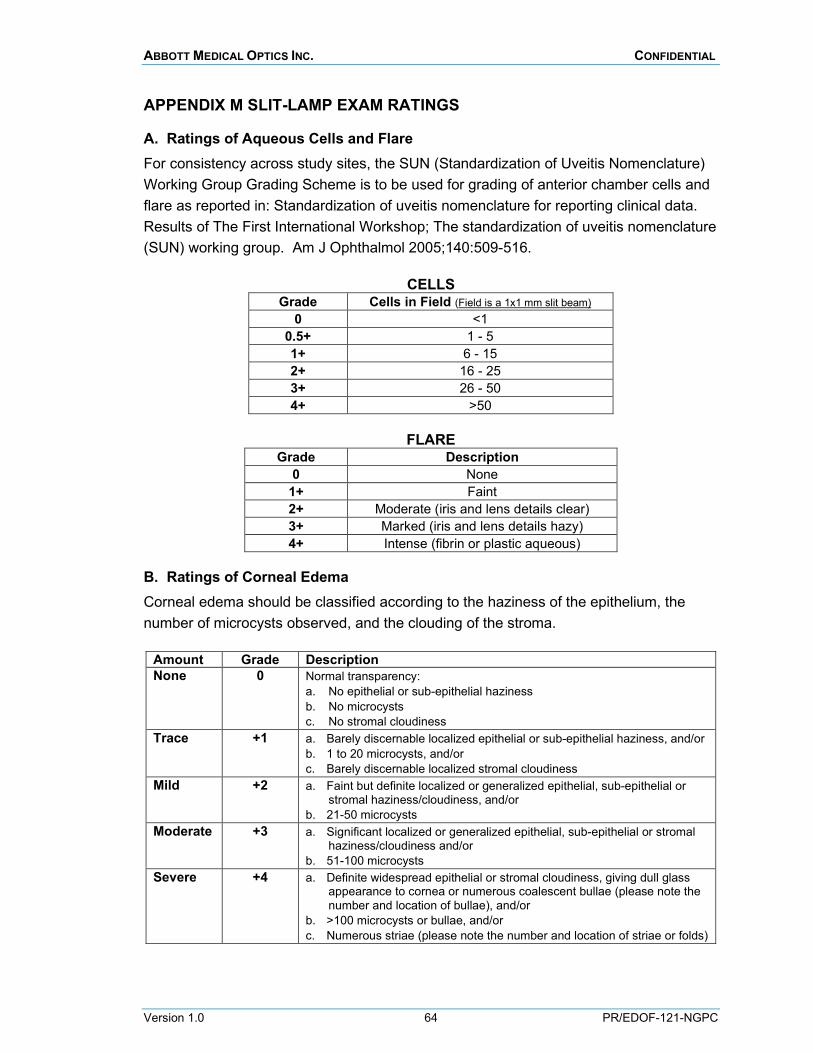
ABBOTT MEDICAL OPTICS INC. CONFIDENTIAL
Version 1.0 64 PR/EDOF-121-NGPC
APPENDIX M SLIT-LAMP EXAM RATINGS
A. Ratings of Aqueous Cells and Flare For consistency across study sites, the SUN (Standardization of Uveitis Nomenclature) Working Group Grading Scheme is to be used for grading of anterior chamber cells and flare as reported in: Standardization of uveitis nomenclature for reporting clinical data. Results of The First International Workshop; The standardization of uveitis nomenclature (SUN) working group. Am J Ophthalmol 2005;140:509-516.
CELLS Grade Cells in Field (Field is a 1x1 mm slit beam)
0 <1 0.5+ 1 - 5 1+ 6 - 15 2+ 16 - 25 3+ 26 - 50 4+ >50
FLARE
Grade Description 0 None
1+ Faint 2+ Moderate (iris and lens details clear) 3+ Marked (iris and lens details hazy) 4+ Intense (fibrin or plastic aqueous)
B. Ratings of Corneal Edema Corneal edema should be classified according to the haziness of the epithelium, the number of microcysts observed, and the clouding of the stroma.
Amount Grade Description None 0 Normal transparency:
a. No epithelial or sub-epithelial haziness b. No microcysts c. No stromal cloudiness
Trace +1 a. Barely discernable localized epithelial or sub-epithelial haziness, and/or b. 1 to 20 microcysts, and/or c. Barely discernable localized stromal cloudiness
Mild +2 a. Faint but definite localized or generalized epithelial, sub-epithelial or stromal haziness/cloudiness, and/or
b. 21-50 microcysts Moderate +3 a. Significant localized or generalized epithelial, sub-epithelial or stromal
haziness/cloudiness and/or b. 51-100 microcysts
Severe +4 a. Definite widespread epithelial or stromal cloudiness, giving dull glass appearance to cornea or numerous coalescent bullae (please note the number and location of bullae), and/or
b. >100 microcysts or bullae, and/or c. Numerous striae (please note the number and location of striae or folds)
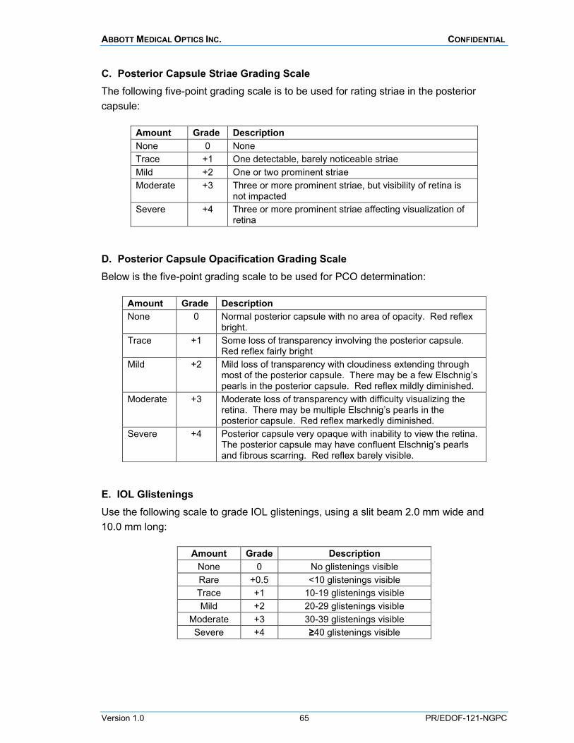
ABBOTT MEDICAL OPTICS INC. CONFIDENTIAL
Version 1.0 65 PR/EDOF-121-NGPC
C. Posterior Capsule Striae Grading Scale The following five-point grading scale is to be used for rating striae in the posterior capsule:
Amount Grade Description None 0 None Trace +1 One detectable, barely noticeable striae Mild +2 One or two prominent striae Moderate +3 Three or more prominent striae, but visibility of retina is
not impacted Severe +4 Three or more prominent striae affecting visualization of
retina
D. Posterior Capsule Opacification Grading Scale Below is the five-point grading scale to be used for PCO determination:
Amount Grade Description None 0 Normal posterior capsule with no area of opacity. Red reflex
bright. Trace +1 Some loss of transparency involving the posterior capsule.
Red reflex fairly bright Mild +2 Mild loss of transparency with cloudiness extending through
most of the posterior capsule. There may be a few Elschnig’s pearls in the posterior capsule. Red reflex mildly diminished.
Moderate +3 Moderate loss of transparency with difficulty visualizing the retina. There may be multiple Elschnig’s pearls in the posterior capsule. Red reflex markedly diminished.
Severe +4 Posterior capsule very opaque with inability to view the retina. The posterior capsule may have confluent Elschnig’s pearls and fibrous scarring. Red reflex barely visible.
E. IOL Glistenings Use the following scale to grade IOL glistenings, using a slit beam 2.0 mm wide and 10.0 mm long:
Amount Grade Description None 0 No glistenings visible Rare +0.5 <10 glistenings visible Trace +1 10-19 glistenings visible Mild +2 20-29 glistenings visible
Moderate +3 30-39 glistenings visible Severe +4 ≥40 glistenings visible