Embed Size (px)
Citation preview
RESEARCH Open Access
Clinical outcomes of previously untreatedpatients with unresectable intrahepaticcholangiocarcinoma following proton beamtherapyShosei Shimizu1*, Toshiyuki Okumura1, Yoshiko Oshiro1, Nobuyoshi Fukumitsu1, Kuniaki Fukuda2, Kazunori Ishige2,Naoyuki Hasegawa2, Haruko Numajiri1, Keiko Murofushi1, Kayoko Ohnishi1, Masashi Mizumoto1, Tetsuo Nonaka1,Hitoshi Ishikawa1 and Hideyuki Sakurai1
Abstract
Background: The effectiveness of proton beam therapy (PBT) as initial treatment for patients with unresectableintrahepatic cholangiocarcinoma (ICC) is unclear, particularly as related to ICC histological subtypes. We performedthis study to address this gap in knowledge.
Methods: Thirty-seven patients with unresectable ICC who underwent PBT as their initial treatment were evaluated.Twenty-seven patients had Child-Pugh class A liver function, 11 exhibited jaundice, and 10 had multiple tumors.Nineteen, 7, and 11 tumors were classified as mass forming (MF), periductal infiltrating (PI), and intraductal growth(IG) types, respectively, based on gross appearance in imaging studies. Patients were classified into the curativegroup (n = 25) and palliative group (n = 12) depending on whether the planning target volume covered all themacroscopic tumors.
Results: The 1- and 2-year overall survival rates were 60.3, and 41.4%, respectively; the median survival time (MST)was 15 months for all patients. The MSTs for curative and palliative groups were 25 and 7months, respectively.Curative treatment and adjuvant chemotherapy significantly improved overall survival, while the presence ofperiductal infiltrating type tumors was a negative prognostic factor. In the curative group, the 1- and 2-year localcontrol rates were 100 and 71.5%, respectively, while the 1-, and 2-year progression-free survival rates were 58.5,and 37.6%, respectively. No severe acute toxicities were observed. Three patients experienced grade 3 biliary tractinfection, although it was unclear whether this was radiotherapy-related.
Conclusion: PBT may yield to improve survival and local tumor control among patients with unresectable ICC.
Keywords: Intrahepatic cholangiocarcinoma, Proton beam therapy, Survival, Macroscopic type, Unresectable ICC
PrecisProton beam therapy as the initial treatment significantlyimproves survival in patients with intrahepatic cholan-giocarcinoma, and has a good safety profile. This is espe-cially true when administered as curative treatment incombination with chemotherapy.
IntroductionIntrahepatic cholangiocarcinoma (ICC) is the secondmost common primary liver cancer by hepatocellularcarcinoma and accounts for 10% of primary liver malig-nancies. Although surgery is considered the only cura-tive treatment for ICC, only 30% of such tumors areresectable at the time of diagnosis. The standard treat-ment for unresectable ICCs is chemotherapy; however,the median survival time (MST) for such patients is dis-mal [1]. Until recently, radiotherapy was indicated forintrahepatic malignancies only as a palliative treatment;
© The Author(s). 2019 Open Access This article is distributed under the terms of the Creative Commons Attribution 4.0International License (http://creativecommons.org/licenses/by/4.0/), which permits unrestricted use, distribution, andreproduction in any medium, provided you give appropriate credit to the original author(s) and the source, provide a link tothe Creative Commons license, and indicate if changes were made. The Creative Commons Public Domain Dedication waiver(http://creativecommons.org/publicdomain/zero/1.0/) applies to the data made available in this article, unless otherwise stated.
* Correspondence: [email protected] of Radiation Oncology and Proton Medical Research Center,Faculty of Medicine, University of Tsukuba, 1-1-1, Tennodai, Tsukuba, Ibaraki305-8575, JapanFull list of author information is available at the end of the article
Shimizu et al. Radiation Oncology (2019) 14:241 https://doi.org/10.1186/s13014-019-1451-5
however, recent technological advances have allowed forthe use of radiotherapy as curative-intent treatment,with favorable results even observed in patients withICC [2, 3]. In particular, proton beams can achieve ex-cellent dose localization, and we previously reported fa-vorable outcomes in patients who underwent protonbeam therapy (PBT) for hepatic tumors including hepa-tocellular carcinoma (HCC) [4–6], metastatic liver tu-mors [7, 8], and ICC (including in 20 patients treatedbetween 1995 and 2009) [2].The gross appearance of ICC is classified into 3 sub-
types according to the Liver Cancer Study Group ofJapan [9]; the mass forming (MF), periductal infiltrating(PI), and intraductal growth (IG) types. These 3 tumorsubtypes have different biological behaviors and are as-sociated with different prognoses after surgical resection.It is generally considered that lymph node metastasis isless frequent in IG-type ICCs [10–12], while perineuralinvasion is high in MF and PI types [10]. However, theinfluence of the macroscopic type on the results ofradiotherapy has not been previously described. In thisstudy, we analyzed the outcomes of patients who under-went PBT as an initial treatment for unresectable ICC.
MethodsPatientsForty-two patients with ICC were administered PBT astheir initial treatment at our current facility between 2001and 2017. Five of these patients had resectable disease butrefused to undergo surgery; of remaining 37 patients, 5,22, and 10 had unresectable tumors owing to their med-ical condition (old age or poor performance status [PS]),tumor progression, and both, respectively. The latter 37patients were investigated in our study. The patients’ char-acteristics are shown in Table 1; they included 22 menand 15 women with a median age of 68.4 years (range:32–87 years). The Eastern Cooperative Oncology GroupPS scores were 0, 1, and 2 for 12, 19, and 6 patients, re-spectively. In terms of liver function, 27 and 10 patientswere classified as Child-Pugh A and B, respectively. Elevenpatients were jaundiced at presentation; 4, 4, 19, and 10were diagnosed with stage I, II, IVA, and IVB disease, re-spectively, acording to the TNM classification (UICC ver-sion 7). Twenty patients had ICC confirmed via histologywhile the remainder were diagnosed based on imagingstudy including dynamic contrast-enhancement CT scansand MRI, positive tumor markers of carcinoembryonicantigen (CEA) or carbohydrate antigen 19–9 (CA19–9)with negative HCC-specific tumor markers such as alpha-fetoprotein (AFP) and protein induced by vitamin K ab-sence or antagonist-II (PIVKA-II). The tumor diametersranged from 15 to 140mm with a median of 57mm.Twenty-seven patients had a solitary tumor and 10 pa-tients had multiple tumors. The tumors in 19, 7, and 11
patients were classified as MF, PI, and IG types, respect-ively, based on CT findings. 25 and 12 patients receivedPBT with curative and palliative intent, respectively, de-pending on whether the planning target volume coveredall detected macroscopic tumors including positive lymphnodes (curative) or not (palliative). Five patients of the 10with multiple tumors received PBT in curative intent butother 5 patients had multiple tumor located at both lobeand lymph node or distant metastases, and they receivedPBT in palliative intent in order to avoid bile duct stenosisat hepatic portal region. In the curative group that com-prised 25 patients, the tumors of 13, 4, and 8 were classi-fied as MF, PI and IG types, respectively.
PBT administrationFor treatment planning, fiducial markers were implantedinto the normal liver tissue close to the tumor boundaryfor patient positioning. The patient’s body was immobi-lized using an individually shaped body cast (ESFORM;Engineering System Co., Matsumoto). CT using 5mm-thick slices was then performed in the treatment positionduring the end-expiratory phase with the support of arespiratory-gaiting system. The clinical target volume forthe primary lesion was delineated to encompass the grosstumor with 5–10mm margins in 3 dimensions, and cau-dal 5mm margins were added to compensate for any po-tential hepatic movement. The clinical target volume fornodal lesion was drawn to cover the clinically positivenodes with 5mm margins if required. Elective nodal ir-radiation was not performed. Next, 5–10mm marginswere added to define the planning target volume. Treat-ment beams were delivered via the double scatteringmode during the end of the expiratory phase using a re-spiratory gating system as described previously [13].The median prescribed dose was 72.6 GyE in 22
fractions; the doses ranged from 46.6 GyE in 12 frac-tions to 74.0 GyE in 37 fractions. Basically, the doseprescription for curative-intent therapy was dependenton tumor location, and was 72.6GyE in 22 fractions(biological effective dose (α/β = 10) [BED]: 96.6 Gy)for porta hepatis, 74GyE in 37 fractions (BED: 88.8Gy) for 2 cm from gastrointestinal tract, and 66 GyEin 10 fractions (BED:109.6 Gy) for tumor not adjacentto gastrointestinal tract and porta hepatis. The rela-tive biological effectiveness of the proton beam wasdesignated as 1.1 [14]. The tumor was covered by >95% of the prescribed dose at the isocenter, however,the target volume was usually modified according todose constraints for the gastrointestinal tract so asnot to exceed a maximum dose of 50 Gy. Also, thepercentage volumes of normal liver receiving at least0, 10, 20, and 30 GyE (V0, 10, 20, and 30) of 30, 20,26, and 18% were used as an adequate indication[15]. One, 21, and 5 patients received 66.0 Gy in 10
Shimizu et al. Radiation Oncology (2019) 14:241 Page 2 of 10
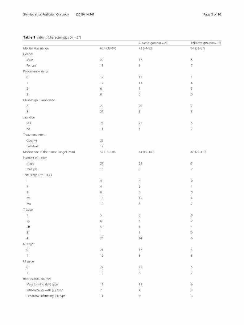
Table 1 Patient Characteristics (n = 37)
Curative group(n = 25) Palliative group(n = 12)
Median Age (range) 68.4 (32–87) 72 (44–82) 67 (32–87)
Gender
Male 22 17 5
Female 15 8 7
Performance status
0 12 11 1
1 19 13 6
2 6 1 5
3 0 0 0
Child-Pugh Classification
A 27 20 7
B 27 5 5
Jaundice
yes 26 21 5
no 11 4 7
Treatment intent
Curative 25
Palliative 12
Median size of the tumor (range) (mm) 57 (15–140) 44 (15–140) 60 (22–110)
Number of tumor
single 27 22 5
multiple 10 3 7
TNM stage (7th UICC)
I 4 4 0
II 4 3 1
III 0 0 0
IVa 19 15 4
IVb 10 3 7
T stage
1 5 5 0
2a 6 4 2
2b 5 1 4
3 1 1 0
4 20 14 6
N stage
0 21 17 4
1 16 8 8
M stage
0 27 22 5
1 10 3 7
macroscopic subtype
Mass forming (MF) type 19 13 6
Intraductal growth (IG) type 7 4 3
Periductal infiltrating (PI) type 11 8 3
Shimizu et al. Radiation Oncology (2019) 14:241 Page 3 of 10
fractions, 72.6 Gy in 22 fractions, and 74.0 Gy in 37fractions, respectively. 10 patients received less thanan BED of 88.8 GyE (equivalent to 74 Gy in 37 frac-tions): 70.0 Gy in 35fractions, 66.0 Gy in 20 fractions,66.0Gy in 30 fractions, 59.4 Gy in 18 fractions, 55.0Gy in 10 fractions, and 46.6 Gy in 12 fractions for 1,2, 1, 1, 2, and 1 patients, respectively (Table 1).
ChemotherapySixteen patients received concurrent chemotherapy toachieve a radiosensitizing effect, 15 of whom receivedoral chemotherapy with tegafur, gimeracil, and oteracil(TS-1), while the remaining patient received intravenousgemcitabine. Maintenance chemotherapy was adminis-tered after PBT to 19 patients, 10 of whom received TS-1 and 9 received intravenous chemotherapy (cisplatinplus gemcitabine: 5, gemcitabine alone: 4).
AnalysisThe overall survival rate was calculated from the date ofPBT commencement to that of death or March 2018.Local failure was defined as an increase of at least 20%in the sum of the target lesion diameters by diagnosticimaging such as CT and MRI. Progression was definedas local recurrence or the appearance of new legions.Toxicities were graded according to National Cancer In-stitution Common Terminology Criteria for AdverseEvents version 4.0 [16].Survival and local control rates were calculated using
the Kaplan-Meier method, and differences between 2groups were determined using the log-rank test. Factorsthat significantly influenced survival and local controlwere identified using the Cox proportional hazardsmodel. A P-value of < 0.05 was considered significant.
ResultsSurvivalAmong the 37 patients, 10 were alive at the lastfollow-up date, 25 had died from cancer progression,and 2 had died from reasons other than ICC. The me-dian follow-up time was 37.5 months, and the MSTwas 15.0 months with cumulative 1- and 2- years sur-vival rates of 60.3% (95% confidence interval [CI]:44.7–76.6%), and 41.4% (95% CI: 24.5–58.3%), respect-ively, for all patients (Fig. 1a). There was a significantdifference in survival between the curative and pallia-tive group (P = 0.001) (Fig. 1b). In the curative group,the MST was 25 months with 1- and 2-year overallsurvival rates of 66.3% (95% CI: 47.3–85.3%), and52.4% (95% CI: 31.8–73.0%), respectively. For patientsin the palliative group, the MST was 7 months with 1-,and 2-year overall survival rates of 27.5% (95% CI: 1–54%), and 18.3% (95% CI: 0–41.2%), respectively.Other factors that were associated with significantlyimproved survival were Child-Pugh A liver function(P = 0.004), absence of jaundice (P = 0.002), andundergoing maintenance chemotherapy (P = 0.000).The MST for patients treated with or without main-tenance chemotherapy were 49 months and 10months, respectively (Table 2).Although the macroscopic tumor type was not a sig-
nificant survival factor among all patients combined, itwas a significant factor specifically in the curativegroup, in which survival was significantly worse for pa-tients with PI-type tumors than for those with the other2 tumor types (PI vs. MF: P = 0.022; PI vs. IG: P =0.023; MF vs. IG: P = 0.784), and the median survivaltimes were 61 months, 31 months, and 9.0 months forpatients with IG-, MF-, and PI-type tumors (Fig. 1c). In
Table 1 Patient Characteristics (n = 37) (Continued)
Curative group(n = 25) Palliative group(n = 12)
Total dose
66.0 GyE in 10 Fraction (BED*:109.6 Gy) 1 1 0
72.6 GyE in 22 Fraction (BED:96.6 Gy) 21 19 2
74.0 GyE in 37 Fraction (BED:88.8 Gy) 5 2 3
Other (BED < 88.8Gy) 10 3 7
Concurrent chemotherapy
TS-1 15 11 4
Gemcitabine 1 1 0
None 21 13 8
Adjuvant Chemotherapy
TS-1 10 7 3
Cisplatine + Gemcitabine 5 4 1
Gemcitabine 4 2 2
None 18 11 7*BED: biological effective dose (α/β = 10)
Shimizu et al. Radiation Oncology (2019) 14:241 Page 4 of 10
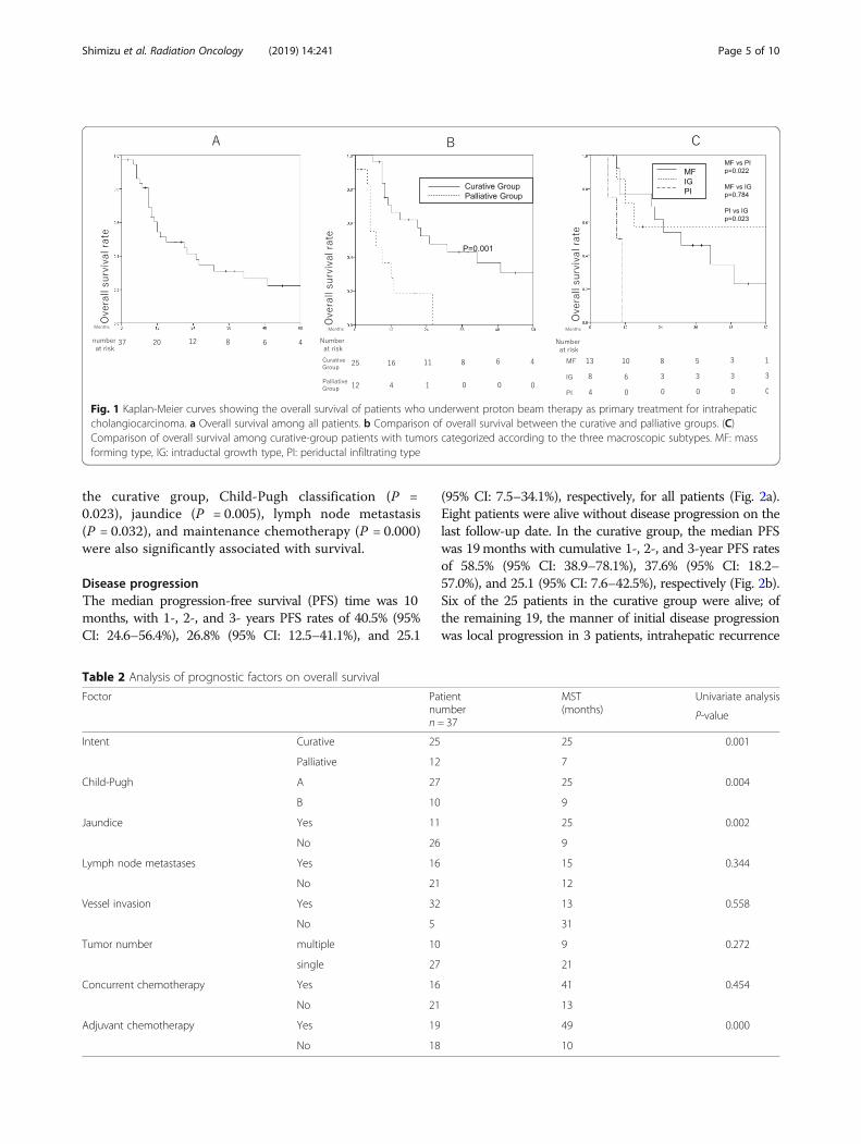
the curative group, Child-Pugh classification (P =0.023), jaundice (P = 0.005), lymph node metastasis(P = 0.032), and maintenance chemotherapy (P = 0.000)were also significantly associated with survival.
Disease progressionThe median progression-free survival (PFS) time was 10months, with 1-, 2-, and 3- years PFS rates of 40.5% (95%CI: 24.6–56.4%), 26.8% (95% CI: 12.5–41.1%), and 25.1
(95% CI: 7.5–34.1%), respectively, for all patients (Fig. 2a).Eight patients were alive without disease progression on thelast follow-up date. In the curative group, the median PFSwas 19months with cumulative 1-, 2-, and 3-year PFS ratesof 58.5% (95% CI: 38.9–78.1%), 37.6% (95% CI: 18.2–57.0%), and 25.1 (95% CI: 7.6–42.5%), respectively (Fig. 2b).Six of the 25 patients in the curative group were alive; ofthe remaining 19, the manner of initial disease progressionwas local progression in 3 patients, intrahepatic recurrence
Fig. 1 Kaplan-Meier curves showing the overall survival of patients who underwent proton beam therapy as primary treatment for intrahepaticcholangiocarcinoma. a Overall survival among all patients. b Comparison of overall survival between the curative and palliative groups. (C)Comparison of overall survival among curative-group patients with tumors categorized according to the three macroscopic subtypes. MF: massforming type, IG: intraductal growth type, PI: periductal infiltrating type
Table 2 Analysis of prognostic factors on overall survival
Foctor Patientnumbern = 37
MST(months)
Univariate analysis
P-value
Intent Curative 25 25 0.001
Palliative 12 7
Child-Pugh A 27 25 0.004
B 10 9
Jaundice Yes 11 25 0.002
No 26 9
Lymph node metastases Yes 16 15 0.344
No 21 12
Vessel invasion Yes 32 13 0.558
No 5 31
Tumor number multiple 10 9 0.272
single 27 21
Concurrent chemotherapy Yes 16 41 0.454
No 21 13
Adjuvant chemotherapy Yes 19 49 0.000
No 18 10
Shimizu et al. Radiation Oncology (2019) 14:241 Page 5 of 10
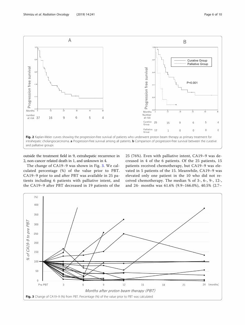
outside the treatment field in 9, extrahepatic recurrence in2, non-cancer related death in 1, and unknown in 4.The change of CA19–9 was shown in Fig. 3. We cal-
culated percentage (%) of the value prior to PBT.CA19–9 prior to and after PBT was available in 25 pa-tients including 6 patients with palliative intent, andthe CA19–9 after PBT decreased in 19 patients of the
25 (76%). Even with palliative intent, CA19–9 was de-creased in 4 of the 6 patients. Of the 25 patients, 15patients received chemotherapy, but CA19–9 was ele-vated in 5 patients of the 15. Meanwhile, CA19–9 waselevated only one patient in the 10 who did not re-ceived chemotherapy. The median % of 3-, 6-, 9-, 12-,and 24- months was 61.6% (9.9–166.0%), 40.5% (2.7–
Fig. 2 Kaplan-Meier curves showing the progression-free survival of patients who underwent proton beam therapy as primary treatment forintrahepatic cholangiocarcinoma. a Progression-free survival among all patients. b Comparison of progression-free survival between the curativeand palliative groups
Fig. 3 Change of CA19–9 (%) from PBT. Percentage (%) of the value prior to PBT was calculated
Shimizu et al. Radiation Oncology (2019) 14:241 Page 6 of 10
473.9%), 38.7% (4.4–250.0%), 41.5% (2.8–316.6%), and57.4% (1.8–373.4%), respectively.
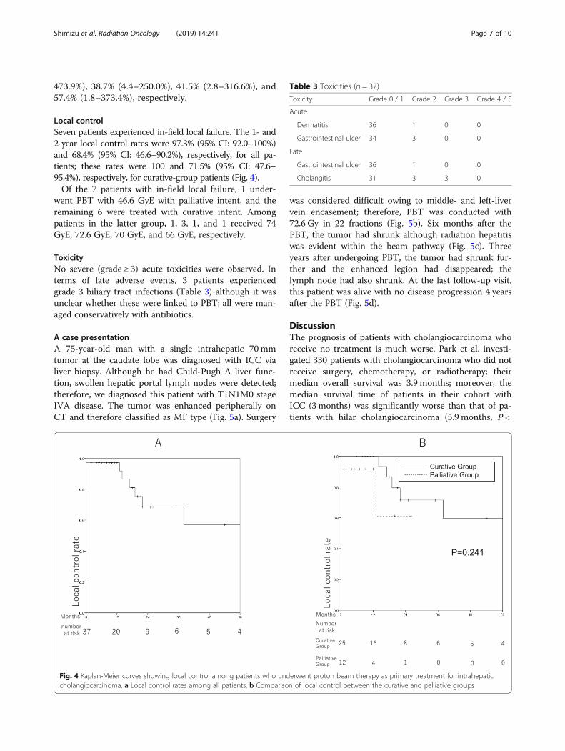
Local controlSeven patients experienced in-field local failure. The 1- and2-year local control rates were 97.3% (95% CI: 92.0–100%)and 68.4% (95% CI: 46.6–90.2%), respectively, for all pa-tients; these rates were 100 and 71.5% (95% CI: 47.6–95.4%), respectively, for curative-group patients (Fig. 4).Of the 7 patients with in-field local failure, 1 under-
went PBT with 46.6 GyE with palliative intent, and theremaining 6 were treated with curative intent. Amongpatients in the latter group, 1, 3, 1, and 1 received 74GyE, 72.6 GyE, 70 GyE, and 66 GyE, respectively.
ToxicityNo severe (grade ≥ 3) acute toxicities were observed. Interms of late adverse events, 3 patients experiencedgrade 3 biliary tract infections (Table 3) although it wasunclear whether these were linked to PBT; all were man-aged conservatively with antibiotics.
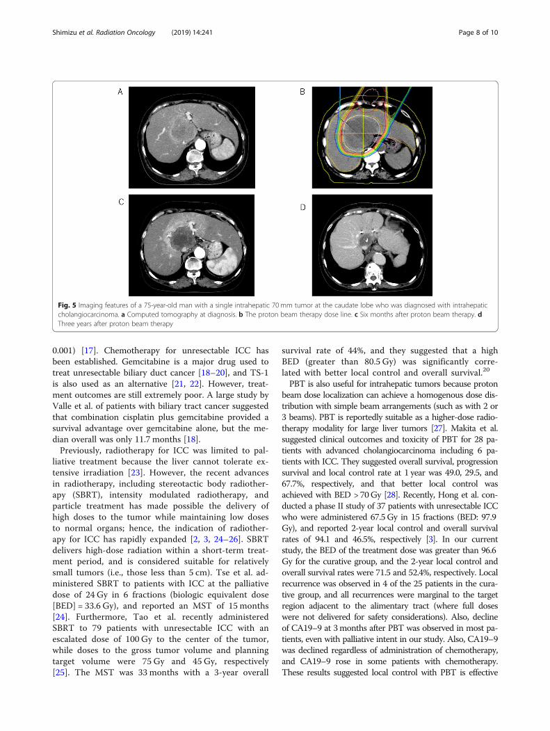
A case presentationA 75-year-old man with a single intrahepatic 70 mmtumor at the caudate lobe was diagnosed with ICC vialiver biopsy. Although he had Child-Pugh A liver func-tion, swollen hepatic portal lymph nodes were detected;therefore, we diagnosed this patient with T1N1M0 stageIVA disease. The tumor was enhanced peripherally onCT and therefore classified as MF type (Fig. 5a). Surgery
was considered difficult owing to middle- and left-livervein encasement; therefore, PBT was conducted with72.6 Gy in 22 fractions (Fig. 5b). Six months after thePBT, the tumor had shrunk although radiation hepatitiswas evident within the beam pathway (Fig. 5c). Threeyears after undergoing PBT, the tumor had shrunk fur-ther and the enhanced legion had disappeared; thelymph node had also shrunk. At the last follow-up visit,this patient was alive with no disease progression 4 yearsafter the PBT (Fig. 5d).
DiscussionThe prognosis of patients with cholangiocarcinoma whoreceive no treatment is much worse. Park et al. investi-gated 330 patients with cholangiocarcinoma who did notreceive surgery, chemotherapy, or radiotherapy; theirmedian overall survival was 3.9 months; moreover, themedian survival time of patients in their cohort withICC (3 months) was significantly worse than that of pa-tients with hilar cholangiocarcinoma (5.9 months, P <
Fig. 4 Kaplan-Meier curves showing local control among patients who underwent proton beam therapy as primary treatment for intrahepaticcholangiocarcinoma. a Local control rates among all patients. b Comparison of local control between the curative and palliative groups
Table 3 Toxicities (n = 37)
Toxicity Grade 0 / 1 Grade 2 Grade 3 Grade 4 / 5
Acute
Dermatitis 36 1 0 0
Gastrointestinal ulcer 34 3 0 0
Late
Gastrointestinal ulcer 36 1 0 0
Cholangitis 31 3 3 0
Shimizu et al. Radiation Oncology (2019) 14:241 Page 7 of 10
0.001) [17]. Chemotherapy for unresectable ICC hasbeen established. Gemcitabine is a major drug used totreat unresectable biliary duct cancer [18–20], and TS-1is also used as an alternative [21, 22]. However, treat-ment outcomes are still extremely poor. A large study byValle et al. of patients with biliary tract cancer suggestedthat combination cisplatin plus gemcitabine provided asurvival advantage over gemcitabine alone, but the me-dian overall was only 11.7 months [18].Previously, radiotherapy for ICC was limited to pal-
liative treatment because the liver cannot tolerate ex-tensive irradiation [23]. However, the recent advancesin radiotherapy, including stereotactic body radiother-apy (SBRT), intensity modulated radiotherapy, andparticle treatment has made possible the delivery ofhigh doses to the tumor while maintaining low dosesto normal organs; hence, the indication of radiother-apy for ICC has rapidly expanded [2, 3, 24–26]. SBRTdelivers high-dose radiation within a short-term treat-ment period, and is considered suitable for relativelysmall tumors (i.e., those less than 5 cm). Tse et al. ad-ministered SBRT to patients with ICC at the palliativedose of 24 Gy in 6 fractions (biologic equivalent dose[BED] = 33.6 Gy), and reported an MST of 15 months[24]. Furthermore, Tao et al. recently administeredSBRT to 79 patients with unresectable ICC with anescalated dose of 100 Gy to the center of the tumor,while doses to the gross tumor volume and planningtarget volume were 75 Gy and 45 Gy, respectively[25]. The MST was 33 months with a 3-year overall
survival rate of 44%, and they suggested that a highBED (greater than 80.5 Gy) was significantly corre-lated with better local control and overall survival.20
PBT is also useful for intrahepatic tumors because protonbeam dose localization can achieve a homogenous dose dis-tribution with simple beam arrangements (such as with 2 or3 beams). PBT is reportedly suitable as a higher-dose radio-therapy modality for large liver tumors [27]. Makita et al.suggested clinical outcomes and toxicity of PBT for 28 pa-tients with advanced cholangiocarcinoma including 6 pa-tients with ICC. They suggested overall survival, progressionsurvival and local control rate at 1 year was 49.0, 29.5, and67.7%, respectively, and that better local control wasachieved with BED > 70Gy [28]. Recently, Hong et al. con-ducted a phase II study of 37 patients with unresectable ICCwho were administered 67.5 Gy in 15 fractions (BED: 97.9Gy), and reported 2-year local control and overall survivalrates of 94.1 and 46.5%, respectively [3]. In our currentstudy, the BED of the treatment dose was greater than 96.6Gy for the curative group, and the 2-year local control andoverall survival rates were 71.5 and 52.4%, respectively. Localrecurrence was observed in 4 of the 25 patients in the cura-tive group, and all recurrences were marginal to the targetregion adjacent to the alimentary tract (where full doseswere not delivered for safety considerations). Also, declineof CA19–9 at 3months after PBT was observed in most pa-tients, even with palliative intent in our study. Also, CA19–9was declined regardless of administration of chemotherapy,and CA19–9 rose in some patients with chemotherapy.These results suggested local control with PBT is effective
Fig. 5 Imaging features of a 75-year-old man with a single intrahepatic 70 mm tumor at the caudate lobe who was diagnosed with intrahepaticcholangiocarcinoma. a Computed tomography at diagnosis. b The proton beam therapy dose line. c Six months after proton beam therapy. dThree years after proton beam therapy
Shimizu et al. Radiation Oncology (2019) 14:241 Page 8 of 10
for control of disease progression. Meanwhile, MST was sig-nificantly longer in the patients with maintenance chemo-therapy. Therefore, the role of chemotherapy also importantwith patients who received PBT.A tumor’s macroscopic subtype is important for deter-
mining the optimal surgical procedure because each ofthe 3 types of ICC has a distinct pathological extensionpattern. Intrahepatic recurrence is frequent in MF-typetumors, infiltration along the bile duct is frequent in PI-type tumors, and lymph node metastases are uncommonin patients with IG-type tumors. The postoperative prog-nosis of patients with these 3 subtypes are also dissimi-lar, the MSTs of patients with IG-, MF-, and PI-typetumors are reportedly 17–55months [29, 30], 18–50months [29–32], and 10–15months [29, 30], respect-ively. Although the patients in our cohort had unresect-able ICC, the MSTs for those with IG-, MF-, and PI-type tumors were 61, 31, and 9.0 months, respectively,which are comparable to those in patients who undergosurgery though our patient’s number with PI- and IG-type were small. Moreover, our contouring procedureduring dose calculation was the same among patientswith all 3 subtypes; however, it may be preferable tocontour the target regions while taking into accounttumor extension characteristics, which differ amongthese subtypes.
ConclusionOur study was limited in that it was retrospective and in-cluded a small number of patients; nevertheless, our datasuggested that PBT was safe and effective for patients withunresectable ICC, therefore, we consider that curativePBT for ICC is encouraged, though the definitive criteriahas not been established yet. Curative-intent treatmentand the administration of maintenance chemotherapywere significant predictors of improved survival; further-more, the macroscopic tumor type also appears to be animportant prognostic factor.
AcknowledgementsThis work was supported by Grant-in-Aid from the Ministry of Education, Sci-ence, Sports and Culture of Japan (15H04901, 19H03596), and also supportedby Japan Agency for Medical Research and Development, AMED (27251501).
Authors’ contributionsSS drafted initial manuscript, TO conceptualized and designed the study, YOdrafted manuscript and analyzed data, NF was doctor in proton therapy,KF,KI and NH were a doctor who specializes in liver of digestive organs andparticipated in general care, HN, KM and KO helped to make planning,MMparticipated in coordination and revised the article, TN and HI were doctorin proton therapy and participated in coordination and helped to draft themanuscript, HS conceptualized and designed the study, and criticallyreviewed the manuscript. All authors read and approved the finalmanuscript.
FundingThis work did not receive any specific funding.
Availability of data and materialsThe datasets supporting the conclusions of this article are included withinthe article.
Ethics approval and consent to participateAll study participants provided informed consent, and we obtained generalconsent to the research in written form from the participant.This study was conducted in accordance with the ethical standards definedin the Declaration of Helsinki and was approved by the ethics committee ofthe University of Tsukuba.(T-CReDO, H30–188).
Consent for publicationWe obtained consent for publication in written form from the participant.
Competing interestsWe have no competing interests.
Author details1Department of Radiation Oncology and Proton Medical Research Center,Faculty of Medicine, University of Tsukuba, 1-1-1, Tennodai, Tsukuba, Ibaraki305-8575, Japan. 2Department of Gastroenterology, Faculty of Medicine,University of Tsukuba, Tsukuba, Ibaraki, Japan.
Received: 18 September 2019 Accepted: 19 December 2019
References1. Ustundag Y, Bayraktar Y. Cholangiocarcinoma: a compact review of the
literature. World J Gastroenterol. 2008;14(42):6458–66.2. Ohkawa A, Mizumoto M, Ishikawa H, Abei M, Fukuda K, Hashimoto T, Sakae T,
Tsuboi K, Okumura T, Sakurai H. Proton beam therapy for unresectableintrahepatic cholangiocarcinoma. J Gastroenterol Hepatol. 2015;30(5):957–63.
3. Hong TS, Wo JY, Yeap BY, Ben-Josef E, McDonnell EI, Blaszkowsky LS, KwakEL, Allen JN, Clark JW, Goyal L, et al. Multi-institutional phase II study ofhigh-dose Hypofractionated proton beam therapy in patients with localized,Unresectable hepatocellular carcinoma and intrahepaticCholangiocarcinoma. J Clin Oncol. 2016;34(5):460–8.
4. Nakayama H, Sugahara S, Fukuda K, Abei M, Shoda J, Sakurai H, Tsuboi K,Matsuzaki Y, Tokuuye K. Proton beam therapy for hepatocellular carcinomalocated adjacent to the alimentary tract. Int J Radiat Oncol Biol Phys. 2011;80(4):992–5.
5. Hata M, Tokuuye K, Sugahara S, Kagei K, Igaki H, Hashimoto T, Ohara K,Matsuzaki Y, Tanaka N, Akine Y. Proton beam therapy for hepatocellularcarcinoma with portal vein tumor thrombus. Cancer. 2005;104(4):794–801.
6. Fukumitsu N, Sugahara S, Nakayama H, Fukuda K, Mizumoto M, Abei M,Shoda J, Thono E, Tsuboi K, Tokuuye K. A prospective study ofhypofractionated proton beam therapy for patients with hepatocellularcarcinoma. Int J Radiat Oncol Biol Phys. 2009;74(3):831–6.
7. Fukumitsu N, Okumura T, Takizawa D, Numajiri H, Ohnishi K, Mizumoto M,Aihara T, Ishikawa H, Tsuboi K, Sakurai H. Proton beam therapy for livermetastases from gastric cancer. J Radiat Res. 2017;58(3):357–62.
8. Fukumitsu N, Okumura T, Takizawa D, Makishima H, Numajiri H, Murofushi K,Ohnishi K, Mizumoto M, Aihara T, Ishikawa H, et al. Proton beam therapy formetastatic liver tumors. Radiother Oncol. 2015;117(2):322–7.
9. Liver Cancer Study Group of J. Primary liver cancer in Japan.Clinicopathologic features and results of surgical treatment. Ann Surg. 1990;211(3):277–87.
10. Hirohashi K, Uenishi T, Kubo S, Yamamoto T, Tanaka H, Shuto T,Yamasaki O, Horii K, Kinoshita H. Histologic bile duct invasion by amass-forming intrahepatic cholangiocarcinoma. J Hepato-Biliary-PancreatSurg. 2002;9(2):233–6.
11. Yamamoto M, Takasaki K, Yoshikawa T. Lymph node metastasis inintrahepatic cholangiocarcinoma. Jpn J Clin Oncol. 1999;29(3):147–50.
12. Tsuji T, Hiraoka T, Kanemitsu K, Takamori H, Tanabe D, Tashiro S.Lymphatic spreading pattern of intrahepatic cholangiocarcinoma.Surgery. 2001;129(4):401–7.
13. Tsunashima Y, Sakae T, Shioyama Y, Kagei K, Terunuma T, Nohtomi A, AkineY. Correlation between the respiratory waveform measured using arespiratory sensor and 3D tumor motion in gated radiotherapy. Int J RadiatOncol Biol Phys. 2004;60(3):951–8.
Shimizu et al. Radiation Oncology (2019) 14:241 Page 9 of 10
14. Paganetti H, Niemierko A, Ancukiewicz M, Gerweck LE, Goitein M, LoefflerJS, Suit HD. Relative biological effectiveness (RBE) values for proton beamtherapy. Int J Radiat Oncol Biol Phys. 2002;53(2):407–21.
15. Mizumoto M, Okumura T, Hashimoto T, Fukuda K, Oshiro Y, Fukumitsu N,Abei M, Kawaguchi A, Hayashi Y, Ohkawa A, et al. Evaluation of liverfunction after proton beam therapy for hepatocellular carcinoma. Int JRadiat Oncol Biol Phys. 2012;82(3):e529–35.
16. Common Terminology Criteria for Adverse Events (CTCAE) v4.0 [http://evs.nci.nih.gov/ftp1/CTCAE/CTCAE_4.03_2010-06-14_QuickReference_8.5x11.pdf].
17. Park J, Kim MH, Kim KP, Park Do H, Moon SH, Song TJ, Eum J, Lee SS, SeoDW, Lee SK. Natural history and prognostic factors of advancedCholangiocarcinoma without surgery, chemotherapy, or radiotherapy: alarge-scale observational study. Gut Liver. 2009;3(4):298–305.
18. Valle J, Wasan H, Palmer DH, Cunningham D, Anthoney A, Maraveyas A,Madhusudan S, Iveson T, Hughes S, Pereira SP, et al. Cisplatin plusgemcitabine versus gemcitabine for biliary tract cancer. N Engl J Med. 2010;362(14):1273–81.
19. Riechelmann RP, Townsley CA, Chin SN, Pond GR, Knox JJ. Expanded phaseII trial of gemcitabine and capecitabine for advanced biliary cancer. Cancer.2007;110(6):1307–12.
20. Andre T, Reyes-Vidal JM, Fartoux L, Ross P, Leslie M, Rosmorduc O, Clemens MR,Louvet C, Perez N, Mehmud F, et al. Gemcitabine and oxaliplatin in advancedbiliary tract carcinoma: a phase II study. Br J Cancer. 2008;99(6):862–7.
21. Sasaki T, Isayama H, Nakai Y, Ito Y, Kogure H, Togawa O, Toda N, Yasuda I,Hasebe O, Maetani I, et al. Multicenter, phase II study of gemcitabine and S-1 combination chemotherapy in patients with advanced biliary tract cancer.Cancer Chemother Pharmacol. 2010;65(6):1101–7.
22. Akahoshi K, Ban D, Kuboki R, Oba A, Ono H, Mitsunori Y, Kudo A, Tanaka S,Tanabe M. Orotate phosphoribosyltransferase as a predictor of benefit fromS-1 adjuvant chemotherapy for cholangiocarcinoma patients. JGastroenterol Hepatol. 2019;34(6):1108–15.
23. Foo ML, Gunderson LL, Bender CE, Buskirk SJ. External radiation therapy andtranscatheter iridium in the treatment of extrahepatic bile duct carcinoma.Int J Radiat Oncol Biol Phys. 1997;39(4):929–35.
24. Tse RV, Hawkins M, Lockwood G, Kim JJ, Cummings B, Knox J, Sherman M,Dawson LA. Phase I study of individualized stereotactic body radiotherapyfor hepatocellular carcinoma and intrahepatic cholangiocarcinoma. J ClinOncol. 2008;26(4):657–64.
25. Tao R, Krishnan S, Bhosale PR, Javle MM, Aloia TA, Shroff RT, Kaseb AO,Bishop AJ, Swanick CW, Koay EJ, et al. Ablative radiotherapy doses Lead to asubstantial prolongation of survival in patients with inoperable intrahepaticCholangiocarcinoma: a retrospective dose response analysis. J Clin Oncol.2016;34(3):219–26.
26. Klein J, Dawson LA. Hepatocellular carcinoma radiation therapy: review ofevidence and future opportunities. Int J Radiat Oncol Biol Phys. 2013;87(1):22–32.
27. Sugahara S, Oshiro Y, Nakayama H, Fukuda K, Mizumoto M, Abei M, Shoda J,Matsuzaki Y, Thono E, Tokita M, et al. Proton beam therapy for largehepatocellular carcinoma. Int J Radiat Oncol Biol Phys. 2010;76(2):460–6.
28. Makita C, Nakamura T, Takada A, Takayama K, Suzuki M, Ishikawa Y, Azami Y,Kato T, Tsukiyama I, Kikuchi Y, et al. Clinical outcomes and toxicity of protonbeam therapy for advanced cholangiocarcinoma. Radiat Oncol. 2014;9:26.
29. Guglielmi A, Ruzzenente A, Campagnaro T, Pachera S, Valdegamberi A,Nicoli P, Cappellani A, Malfermoni G, Iacono C. Intrahepaticcholangiocarcinoma: prognostic factors after surgical resection. World JSurg. 2009;33(6):1247–54.
30. Paik KY, Jung JC, Heo JS, Choi SH, Choi DW, Kim YI. What prognostic factorsare important for resected intrahepatic cholangiocarcinoma? J GastroenterolHepatol. 2008;23(5):766–70.
31. Okabayashi T, Yamamoto J, Kosuge T, Shimada K, Yamasaki S, Takayama T,Makuuchi M. A new staging system for mass-forming intrahepaticcholangiocarcinoma: analysis of preoperative and postoperative variables.Cancer. 2001;92(9):2374–83.
32. Suzuki S, Sakaguchi T, Yokoi Y, Okamoto K, Kurachi K, Tsuchiya Y, OkumuraT, Konno H, Baba S, Nakamura S. Clinicopathological prognostic factors andimpact of surgical treatment of mass-forming intrahepaticcholangiocarcinoma. World J Surg. 2002;26(6):687–93.
Publisher’s NoteSpringer Nature remains neutral with regard to jurisdictional claims inpublished maps and institutional affiliations.
Shimizu et al. Radiation Oncology (2019) 14:241 Page 10 of 10


![American Society of Clinical Oncology - P&T Community · A Phase 2 Trial [CALGB 50803] of Lenalidomide Plus Rituximab in Patients With Previously Untreated Follicular Lymphoma](https://img.pdfslide.net/doc/110x75/5b5bc99e7f8b9a885b8ebc4e/american-society-of-clinical-oncology-pt-community-a-phase-2-trial-calgb.jpg)